EP4537842A1 - Agent préventif ou agent thérapeutique pour la sclérose latérale amyotrophique, la maladie de parkinson, la maladie de huntington, l'ataxie spinocérébelleuse, une maladie dégénérative ou neurologique liée au vieillissement, le vieillissement cérébral ou des maladies associées au vieillissement cérébral - Google Patents
Agent préventif ou agent thérapeutique pour la sclérose latérale amyotrophique, la maladie de parkinson, la maladie de huntington, l'ataxie spinocérébelleuse, une maladie dégénérative ou neurologique liée au vieillissement, le vieillissement cérébral ou des maladies associées au vieillissement cérébral Download PDFInfo
- Publication number
- EP4537842A1 EP4537842A1 EP23819849.3A EP23819849A EP4537842A1 EP 4537842 A1 EP4537842 A1 EP 4537842A1 EP 23819849 A EP23819849 A EP 23819849A EP 4537842 A1 EP4537842 A1 EP 4537842A1
- Authority
- EP
- European Patent Office
- Prior art keywords
- mice
- amino acid
- monoclonal antibody
- human monoclonal
- acid sequence
- Prior art date
- Legal status (The legal status is an assumption and is not a legal conclusion. Google has not performed a legal analysis and makes no representation as to the accuracy of the status listed.)
- Pending
Links
Images


















Classifications
-
- C—CHEMISTRY; METALLURGY
- C07—ORGANIC CHEMISTRY
- C07K—PEPTIDES
- C07K16/00—Immunoglobulins [IG], e.g. monoclonal or polyclonal antibodies
- C07K16/18—Immunoglobulins [IG], e.g. monoclonal or polyclonal antibodies against material from animals or humans
- C07K16/24—Immunoglobulins [IG], e.g. monoclonal or polyclonal antibodies against material from animals or humans against cytokines, lymphokines or interferons
-
- A—HUMAN NECESSITIES
- A61—MEDICAL OR VETERINARY SCIENCE; HYGIENE
- A61P—SPECIFIC THERAPEUTIC ACTIVITY OF CHEMICAL COMPOUNDS OR MEDICINAL PREPARATIONS
- A61P21/00—Drugs for disorders of the muscular or neuromuscular system
-
- A—HUMAN NECESSITIES
- A61—MEDICAL OR VETERINARY SCIENCE; HYGIENE
- A61P—SPECIFIC THERAPEUTIC ACTIVITY OF CHEMICAL COMPOUNDS OR MEDICINAL PREPARATIONS
- A61P25/00—Drugs for disorders of the nervous system
-
- A—HUMAN NECESSITIES
- A61—MEDICAL OR VETERINARY SCIENCE; HYGIENE
- A61P—SPECIFIC THERAPEUTIC ACTIVITY OF CHEMICAL COMPOUNDS OR MEDICINAL PREPARATIONS
- A61P25/00—Drugs for disorders of the nervous system
- A61P25/14—Drugs for disorders of the nervous system for treating abnormal movements, e.g. chorea, dyskinesia
-
- A—HUMAN NECESSITIES
- A61—MEDICAL OR VETERINARY SCIENCE; HYGIENE
- A61P—SPECIFIC THERAPEUTIC ACTIVITY OF CHEMICAL COMPOUNDS OR MEDICINAL PREPARATIONS
- A61P25/00—Drugs for disorders of the nervous system
- A61P25/14—Drugs for disorders of the nervous system for treating abnormal movements, e.g. chorea, dyskinesia
- A61P25/16—Anti-Parkinson drugs
-
- A—HUMAN NECESSITIES
- A61—MEDICAL OR VETERINARY SCIENCE; HYGIENE
- A61P—SPECIFIC THERAPEUTIC ACTIVITY OF CHEMICAL COMPOUNDS OR MEDICINAL PREPARATIONS
- A61P25/00—Drugs for disorders of the nervous system
- A61P25/28—Drugs for disorders of the nervous system for treating neurodegenerative disorders of the central nervous system, e.g. nootropic agents, cognition enhancers, drugs for treating Alzheimer's disease or other forms of dementia
-
- C—CHEMISTRY; METALLURGY
- C07—ORGANIC CHEMISTRY
- C07K—PEPTIDES
- C07K16/00—Immunoglobulins [IG], e.g. monoclonal or polyclonal antibodies
- C07K16/18—Immunoglobulins [IG], e.g. monoclonal or polyclonal antibodies against material from animals or humans
-
- C—CHEMISTRY; METALLURGY
- C07—ORGANIC CHEMISTRY
- C07K—PEPTIDES
- C07K16/00—Immunoglobulins [IG], e.g. monoclonal or polyclonal antibodies
- C07K16/40—Immunoglobulins [IG], e.g. monoclonal or polyclonal antibodies against enzymes
-
- A—HUMAN NECESSITIES
- A61—MEDICAL OR VETERINARY SCIENCE; HYGIENE
- A61K—PREPARATIONS FOR MEDICAL, DENTAL OR TOILETRY PURPOSES
- A61K39/00—Medicinal preparations containing antigens or antibodies
- A61K2039/505—Medicinal preparations containing antigens or antibodies comprising antibodies
-
- A—HUMAN NECESSITIES
- A61—MEDICAL OR VETERINARY SCIENCE; HYGIENE
- A61K—PREPARATIONS FOR MEDICAL, DENTAL OR TOILETRY PURPOSES
- A61K39/00—Medicinal preparations containing antigens or antibodies
- A61K2039/54—Medicinal preparations containing antigens or antibodies characterised by the route of administration
-
- A—HUMAN NECESSITIES
- A61—MEDICAL OR VETERINARY SCIENCE; HYGIENE
- A61K—PREPARATIONS FOR MEDICAL, DENTAL OR TOILETRY PURPOSES
- A61K2239/00—Indexing codes associated with cellular immunotherapy of group A61K40/00
- A61K2239/38—Indexing codes associated with cellular immunotherapy of group A61K40/00 characterised by the dose, timing or administration schedule
-
- C—CHEMISTRY; METALLURGY
- C07—ORGANIC CHEMISTRY
- C07K—PEPTIDES
- C07K2317/00—Immunoglobulins specific features
- C07K2317/20—Immunoglobulins specific features characterized by taxonomic origin
- C07K2317/21—Immunoglobulins specific features characterized by taxonomic origin from primates, e.g. man
-
- C—CHEMISTRY; METALLURGY
- C07—ORGANIC CHEMISTRY
- C07K—PEPTIDES
- C07K2317/00—Immunoglobulins specific features
- C07K2317/20—Immunoglobulins specific features characterized by taxonomic origin
- C07K2317/24—Immunoglobulins specific features characterized by taxonomic origin containing regions, domains or residues from different species, e.g. chimeric, humanized or veneered
-
- C—CHEMISTRY; METALLURGY
- C07—ORGANIC CHEMISTRY
- C07K—PEPTIDES
- C07K2317/00—Immunoglobulins specific features
- C07K2317/40—Immunoglobulins specific features characterized by post-translational modification
- C07K2317/41—Glycosylation, sialylation, or fucosylation
-
- C—CHEMISTRY; METALLURGY
- C07—ORGANIC CHEMISTRY
- C07K—PEPTIDES
- C07K2317/00—Immunoglobulins specific features
- C07K2317/50—Immunoglobulins specific features characterized by immunoglobulin fragments
- C07K2317/56—Immunoglobulins specific features characterized by immunoglobulin fragments variable (Fv) region, i.e. VH and/or VL
- C07K2317/565—Complementarity determining region [CDR]
-
- C—CHEMISTRY; METALLURGY
- C07—ORGANIC CHEMISTRY
- C07K—PEPTIDES
- C07K2317/00—Immunoglobulins specific features
- C07K2317/70—Immunoglobulins specific features characterized by effect upon binding to a cell or to an antigen
- C07K2317/76—Antagonist effect on antigen, e.g. neutralization or inhibition of binding
-
- C—CHEMISTRY; METALLURGY
- C07—ORGANIC CHEMISTRY
- C07K—PEPTIDES
- C07K2317/00—Immunoglobulins specific features
- C07K2317/90—Immunoglobulins specific features characterized by (pharmaco)kinetic aspects or by stability of the immunoglobulin
- C07K2317/92—Affinity (KD), association rate (Ka), dissociation rate (Kd) or EC50 value
Definitions
- the present invention relates to a drug product for preventing or treating amyotrophic lateral sclerosis (hereinafter may be referred to as "ALS”), Parkinson's disease (hereinafter may be referred to as “PD”), Huntington's disease (hereinafter may be referred to as “HD”), spinocerebellar ataxia (hereinafter may be referred to as “SCA”), degenerative or neurological diseases related to aging (more specifically, cellular senescence), brain aging, or diseases associated with brain aging (hereinafter, these may be collectively referred to as "the diseases of the present invention and the like”); an anti-human HMGB1 antibody; or a drug product for preventing or treating Alzheimer's disease (hereinafter may be referred to as "AD”) or frontotemporal lobar degeneration (hereinafter may be referred to as "FTLD”), and the like.
- AD amyotrophic lateral sclerosis
- PD Parkinson's disease
- HD Huntington's disease
- FTLD is the second or third most common early-onset neurodegenerative dementia following AD and is considered closely related to motor neuron diseases.
- FTLD has symptoms of noticeable changes in behavior and personality which are often accompanied by language impairment, and these symptoms gradually progress to cognitive impairment and dementia.
- VCP valosin-containing protein
- PGRN progranulin
- CHMP2B charged multivesicular body protein 2B
- TDP-43 transactive response DNA-binding protein of 43 kD
- ALS and FTLD develop due to a combination of various genetic and environmental factors, and more than 20 genes are thought to be involved in these diseases.
- HMGB1 high mobility group box 1
- DAMP damage-associated molecule pattern
- HMGB1 leaking out of a cell owing to neuronal necrosis induces phosphorylation of serine at position 46 (Ser46) of MARCKS, which is a substrate of a phosphorylating enzyme, thereby inducing degeneration of neurites, prepared a mouse monoclonal antibody against HMGB1, and found that this mouse monoclonal antibody inhibits phosphorylation of Ser46 of MARCKS (Non-Patent Document 1). Further, the present inventors have reported that this mouse monoclonal antibody and a human monoclonal antibody resolve cognitive impairment in Alzheimer's disease model mice (Patent Documents 2 and 3).
- Non Patent Document 1 SCIENTIFIC REPORT, Aug 25, 2016; 6:31895
- An object of the present invention is to provide an agent for preventing or treating ALS, PD, HD, SCA, aging-related degenerative or neurological disease, brain aging, or diseases associated with brain aging, and an antibody that is more stable and exhibits an effect of preventing or treating AD or FTLD.
- a human monoclonal antibody #129 prepared in the examples of International Publication No. WO 2020/059847 was found to exhibit a treatment effect on four types of gene mutations (VCP T262A mutation, PGRN R504X mutation, CHMP2B Q165X mutation, and TDP43 N267S mutation) in the FTLD-ALS spectrum (hereinafter may be referred to as "FTLD-ALS"), and suppress transcriptional repression-induced atypical cell death (TRIAD) necrosis in nerve cells (may be referred to "neurons”) as well as further having an effect of inhibiting reciprocal amplification of TRIAD necrosis that is based on HMGB1-induced DNA damage.
- FTLD-ALS suppress transcriptional repression-induced atypical cell death
- the above-described human monoclonal antibody #129 was confirmed to reduce the number of senescent cells in the cerebral cortex of two types of AD model mice (5 ⁇ FAD mice and APP-KI mice), and also to suppress the cellular senescence of neurons, astrocytes and microglia.
- the human monoclonal antibody #129 and the anti-HMGB1 antibody having common CDRs thereto can be said to have an effect of treating aging-related degenerative or neurological diseases, brain aging itself, and diseases associated with brain aging.
- model mice HD model mice, PD model mice, ALS model mice, spinocerebellar ataxia [hereinafter may be referred to as "SCA”] model mice, and aging model mice
- SCA spinocerebellar ataxia
- aging model mice using, in addition to the human monoclonal antibody #129, a human monoclonal antibody #129-L2, which is a modified antibody of the light chain CDR3 of the human monoclonal antibody #129, the human monoclonal antibody #129 and the human monoclonal antibody #129-L2 were confirmed to exhibit treatment effects on HD, PD, ALS, SCA, and aging-associated cellular changes, cell death, cognitive function (memory), and motor function.
- the human monoclonal antibody #129-L2 has similar binding strength for human disulfide HMGB1 (ds-HMGB1) to the human monoclonal antibody #129 and treatment effects on FTLD-ALS model mice and AD model mice, and that the binding strength for human reduced HMGB1 is as weak as or weaker than that of the human monoclonal antibody #129. Also, it was confirmed that, unlike the human monoclonal antibody #129, the human monoclonal antibody #129-L2 does undergo N-glycan glycosylation, and that the stability of the antibody when stored is rather higher than that of the human monoclonal antibody #129.
- ds-HMGB1 human disulfide HMGB1
- the human monoclonal antibody #129-L2 does undergo N-glycan glycosylation, and that the stability of the antibody when stored is rather higher than that of the human monoclonal antibody #129.
- the present invention was accomplished based on these findings.
- the present invention provides the following.
- examples of other embodiments of the present invention include the following: a method for preventing or treating the diseases of the present invention and the like, comprising a step of administering a human monoclonal antibody (hereinafter may be referred to as "the human monoclonal antibody of the present invention") that specifically binds to human HMGB1 and comprises:
- examples of other embodiments of the present invention include the following: a method for preventing or treating Alzheimer's disease or frontotemporal lobar degeneration, and/or the diseases of the present invention and the like, comprising a step of administering a human monoclonal antibody (hereinafter may be referred to as "the human monoclonal antibody L1/L2 of the present invention") that specifically binds to human HMGB1 and comprises:
- the diseases of the present invention and the like can be prevented or treated without causing noticeable adverse reactions. Also, when the human monoclonal antibody L1/L2 of the present invention is used, Alzheimer's disease or frontotemporal lobar degeneration, and/or the diseases of the present invention and the like can be prevented or treated without causing noticeable adverse reactions.
- a first aspect of the preventing or treating agent of the present invention is an agent containing the human monoclonal antibody of the present invention (hereinafter may be referred to as "the agent 1 for preventing or treating of the present invention") used for a specific purpose of "preventing or treating one or more of amyotrophic lateral sclerosis; Parkinson's disease; Huntington's disease; aging-related degenerative or neurological disease; brain aging; and diseases associated with brain aging”.
- a second aspect of the preventing or treating agent of the present invention is an agent containing the human monoclonal antibody L1/L2 of the present invention (hereinafter may be referred to as “the agent 2 for preventing or treating of the present invention”) used for a specific purpose of "preventing or treating one or more of Alzheimer's disease; frontotemporal lobar degeneration; amyotrophic lateral sclerosis; Parkinson's disease; Huntington's disease; aging-related degenerative or neurological disease; brain aging; and diseases associated with brain aging” (hereinafter, "the agent 1 for preventing or treating of the present invention” and “the agent 2 for preventing or treating of the present invention” may be collectively referred to as “the agent for preventing or treating of the present invention”).
- the agent for preventing or treating of the present invention may be used solely as a medicine (drug product) or may be used in a form of a composition (pharmaceutical composition) by further mixing additives.
- a medicine drug product
- a composition pharmaceutical composition
- the "treatment of brain aging” may be referred to as “improvement in brain aging”.
- the human monoclonal antibody L1/L2 of the present invention is a human monoclonal antibody that specifically binds to human HMGB1, comprises an H chain CDR1 consisting of the amino acid sequence shown in SEQ ID No: 1, an H chain CDR2 consisting of the amino acid sequence shown in SEQ ID No: 2, and an H chain CDR3 consisting of the amino acid sequence shown in SEQ ID No: 3; and an L chain CDR1 consisting of the amino acid sequence shown in SEQ ID No: 4, an L chain CDR2 consisting of the amino acid sequence shown in SEQ ID No: 5, and an L chain CDR3 consisting of the amino acid sequence shown in SEQ ID No: 20 or 17, and falls within the scope of the human monoclonal antibody of the present invention.
- the expression "prevention of amyotrophic lateral sclerosis, Parkinson's disease, Huntington's disease, aging-related degenerative or neurological disease, brain aging, or diseases associated with brain aging” includes not only suppression of presence or development of amyotrophic lateral sclerosis, Parkinson's disease, Huntington's disease, aging-related degenerative or neurological disease, brain aging, or diseases associated with brain aging, but also delay of presence or onset time of amyotrophic lateral sclerosis, Parkinson's disease, Huntington's disease, aging-related degenerative or neurological disease, brain aging, or diseases associated with brain aging.
- treatment (improvement) of amyotrophic lateral sclerosis, Parkinson's disease, Huntington's disease, aging-related degenerative or neurological disease, brain aging, or diseases associated with brain aging includes not only resolution or improvement of lesions and symptoms of amyotrophic lateral sclerosis, Parkinson's disease, Huntington's disease, aging-related degenerative or neurological disease, brain aging, or diseases associated with brain aging, but also suppression of their progression.
- the human monoclonal antibody of the present invention contained in the agent 1 for preventing or treating of the present invention and the human monoclonal antibody L1/L2 of the present invention contained in the agent 2 for preventing or treating of the present invention have high affinity for human HMGB1 protein and have an effect of reducing or suppressing the phosphorylation level of serine at position 46 (Ser46) in MARCKS (pSer46-MARCKS); an effect of recovering the repair function of DNA damage in nerve cells; an effect of inhibiting TRIAD necrosis or its amplification (may be referred to as "propagation"); and an effect of reducing protein (for example, TDP43) aggregates in cerebral cortical neurons.
- those caused by one or more selected from HMGB1 and/or pSer46-MARCKS; decrease in the repair function of DNA damage in nerve cells; TRIAD necrosis or its amplification; and protein (for example, TDP43) aggregates in cerebral cortical neurons can be mentioned as a preferred example of the diseases of the present invention and the like to be prevented or treated with the agent 1 for preventing or treating of the present invention, and/or Alzheimer's disease or frontotemporal lobar degeneration, and/or the diseases of the present invention and the like to be prevented or treated with the agent 2 for preventing or treating of the present invention.
- TRIAD neurode
- RNA polymerase II a basic molecule in transcription
- YAP yes-associated protein
- TRIAD does not have morphological and biochemical features of apoptosis, and unlike autophagic cell death, it does not have autophagosome expansion and increase, but does have a morphological feature of swelling of endoplasmic reticulum.
- Target diseases of the agent for preventing or treating of the present invention may be a genetic disease (for example, diseases caused by genetic mutation, for example, valosin-containing protein (VCP) mutation such as VCP T262A mutation; progranulin (PGRN) mutation such as PGRN R504X mutation; charged multivesicular body protein 2B (CHMP2B) mutation such as CHMP2B Q165 X mutation; and transactive response DNA-binding protein of 43 kD (TDP43) mutation such as TDP43 N267S mutation), or may be a sporadic disease.
- VCP valosin-containing protein
- PGRN progranulin
- CHMP2B charged multivesicular body protein 2B
- TDP43 transactive response DNA-binding protein of 43 kD mutation
- the human monoclonal antibody of the present invention is preferably a human monoclonal antibody comprising an H chain variable region consisting of an amino acid sequence having at least 80% or more sequence identity to the amino acid sequence shown in SEQ ID No: 7 and an L chain variable region consisting of an amino acid sequence having at least 80% or more sequence identity to the amino acid sequence shown in SEQ ID No: 8, SEQ ID No: 21, or SEQ ID No: 18, more preferably a human monoclonal antibody comprising a heavy chain consisting of an amino acid sequence having at least 80% or more sequence identity to the amino acid sequence shown in SEQ ID No: 9 and a light chain consisting of an amino acid sequence having at least 80% or more sequence identity to the amino acid sequence shown in SEQ ID No: 10, SEQ ID No: 22, or SEQ ID No: 19.
- the human monoclonal antibody L1/L2 of the present invention is preferably a human monoclonal antibody comprising a heavy chain variable region consisting of an amino acid sequence having at least 80% or more sequence identity to the amino acid sequence shown in SEQ ID No: 7 and a light chain variable region consisting of an amino acid sequence having at least 80% or more sequence identity to the amino acid sequence shown in SEQ ID No: 21 or 18, and more preferably a human monoclonal antibody comprising a heavy chain consisting of an amino acid sequence having at least 80% or more sequence identity to the amino acid sequence shown in SEQ ID No: 9 and a light chain consisting of an amino acid sequence having at least 80% or more sequence identity to the amino acid sequence shown in SEQ ID No: 22 or 19.
- HMGB1 high mobility group box 1
- HMG1, HMG3, SBP-1, or HMG-1 Human-derived HMGB1 is typically a protein consisting of an amino acid sequence identified as NCBI reference sequence: NP_002119.1 (a protein encoded by the nucleotide sequence identified as NCBI reference sequence: NM_002128.5).
- NP_002119.1 a protein encoded by the nucleotide sequence identified as NCBI reference sequence: NM_002128.5.
- the DNA sequence of the gene is mutated in nature (that is, non-artificially) by a mutation thereof or the like, and the amino acid sequence of the protein encoded thereby is also modified along with the mutation.
- variants of this protein existing in nature also fall within the scope of HMGB1, to which the human monoclonal antibody of the present invention or the human monoclonal antibody L1/L2 of the present invention binds.
- the term "antibody that specifically binds to human HMGB1” means an antibody that recognizes and binds to human HMGB1 through a highly specific antigen-antibody recognition mechanism.
- the human monoclonal antibody of the present invention or the human monoclonal antibody L1/L2 of the present invention is preferably in an isolated condition.
- isolated used herein means that an antibody exists in a condition different from the condition in which it originally exists, achieved by extracting the antibody by artificial manipulation from the environment where it originally exists, expressing the antibody in an environment that is not the environment where it originally exists antibody, or the like.
- the term "isolated antibody” does not include an antibody derived from a certain individual and contained in the body of the individual or in a tissue or body fluid (for example, blood, plasma, or serum) derived from the body without undergoing an external operation (artificial manipulation).
- the human monoclonal antibody of the present invention or the human monoclonal antibody L1/L2 of the present invention is preferably an antibody produced from an organism or a cell prepared by artificial manipulation (for example, an antibody produced from hybridoma).
- the "antibody produced from an organism or a cell prepared by artificial manipulation” does not include an antibody produced from an organism or a B cell that exists naturally (not undergoing artificial manipulation).
- the term "monoclonal antibody” means an antibody (including a functional fragment of an antibody) obtained from a group of substantially uniform antibodies. A monoclonal antibody recognizes a single determinant on an antigen.
- the human monoclonal antibody of the present invention or the human monoclonal antibody L1/L2 of the present invention includes human immunoglobulin classes and subclasses, as well as forms of functional fragments of these antibodies.
- the classes and subclasses of the human monoclonal antibody of the present invention or the human monoclonal antibody L1/L2 of the present invention include IgG such as IgG1, IgG2, IgG3, and IgG4, IgA such as IGA1 and IGA2, IgD, IgE, and IgM.
- the human monoclonal antibody of the present invention and the human monoclonal antibody L1/L2 of the present invention typically comprise heavy chain CDR1, heavy chain CDR2, heavy chain CDR3, light chain CDR1, light chain CDR2, and light chain CDR3, and have a framework region (FR) linked to the amino (N) terminus and the carboxyl (C) terminus of each region of these CDR1 to CDR3, and comprise a heavy chain variable region and light chain variable region.
- FR framework region linked to the amino (N) terminus and the carboxyl (C) terminus of each region of these CDR1 to CDR3, and comprise a heavy chain variable region and light chain variable region.
- examples of a heavy chain FR include heavy chain FR1 linked to the N terminus of heavy chain CDR1, heavy chain FR2 linked between the C terminus of heavy chain CDR1 and the N terminus of heavy chain CDR2, heavy chain FR3 linked between the C terminus of heavy chain CDR2 and the N terminus of heavy chain CDR3, and heavy chain FR4 linked to the C terminus of heavy chain CDR3.
- examples of a light chain FR include light chain FR1 linked to the N terminus of the light chain CDR1, light chain FR2 linked between the C terminus of light chain CDR1 and the N terminus of light chain CDR2, light chain FR3 linked between the C terminus of light chain CDR2 and the N terminus of light chain CDR3, and light chain FR4 linked to the C terminus of light chain CDR3.
- heavy chain FR1 examples include (HF1) a polypeptide consisting of amino acid residues 1 to 30 of the amino acid sequence shown in SEQ ID No: 7 and (HF1') a polypeptide consisting of an amino acid sequence having at least 80% or more sequence identity to this polypeptide;
- LF1 a polypeptide consisting of amino acid residues 1 to 23 of the amino acid sequence shown in SEQ ID No: 8 and (LF1') a polypeptide consisting of an amino acid sequence having at least 80% or more sequence identity to this polypeptide;
- the human monoclonal antibody of the present invention and the human monoclonal antibody L1/L2 of the present invention are human antibodies.
- Examples of the "human antibodies” include a human chimeric antibody, a humanized antibody, and a complete human antibody, and preferred examples thereof are a humanized antibody and a complete human antibody.
- the "human chimeric antibody” has the variable region of an antibody derived from a non-human animal (for example, a non-human mammal such as a chicken, a mouse, a rat, or a bovine) and the constant region of a human-derived antibody linked to each other.
- a non-human animal for example, a non-human mammal such as a chicken, a mouse, a rat, or a bovine
- a human-derived antibody for example, a non-human mammal such as a chicken, a mouse, a rat, or a bovine
- the human chimeric antibody can be obtained by, for example, immunizing a non-human animal (preferably a non-human mammal) with an antigen, excising an antibody variable region that binds to the antigen from the gene of the mouse monoclonal antibody, binding it to the gene of the constant region of an antibody derived from the human bone marrow, and incorporating the gene into an expression vector, so that the expression vector is introduced into a host to produce the human chimeric antibody (for example, Japanese unexamined Patent Application Publication No. 8-280387 ; U.S. Patent No. 4816397 ; U.S. Patent No. 4816567 ; and U.S. Patent No. 5807715 ).
- the "humanized antibody” is an antibody obtained by transplanting the gene sequence of the antigen-binding site (a CDR) of an antibody (that is, CDR grafting) derived from a non-human animal (for example, a non-human mammal such as a chicken, a mouse, a rat, or a bovine) into an antibody gene derived from a human, and the methods for preparing it, such as overlap extension PCR, are known (see, for example, European Patent Application No. 239400 , European Patent Application No. 125023 , International Publication No. WO 90/07861 , and International Publication No. WO 96/02576 ).
- a CDR antigen-binding site
- variable region of an antibody is usually composed of three CDRs sandwiched by four framework regions (FRs).
- a CDR is a region that substantially determines the binding specificity of an antibody. While the amino acid sequence of a CDR is rich in diversity, amino acid sequences constituting an FR often show high homology among antibodies having different binding specificity. Thus, it is generally said that the binding specificity of an antibody can be transplanted to other antibodies by transplanting a CDR. Further, in transplantation of a non-human-derived CDR to a human FR, a human FR with high homology to the non-human animal-derived FR is selected from viewpoints of maintaining the function of the CDR.
- amino acids in a CDR not only recognize an antigen, but also coordinate with amino acids of an FR in the vicinity of the CDR, and are involved in maintenance of the loop structure of the CDR. Therefore, a human FR consisting of amino acid sequences with high homology to the amino acid sequences of an FR adjacent to a CDR to be transplanted is preferably used.
- a known human FR with high homology to a non-human animal-derived FR can be searched by using, for example, a search system which is available on the Internet and specialized in antibodies ( ⁇ http://www.bioinf.org.uk/abysis/>).
- a mutation can be introduced into the sequence of a non-human-derived antibody other than CDR so that it is consistent with the sequence of a thus-obtained human FR.
- a gene (cDNA) encoding the amino acid sequence of a human FR obtained by search is available, a non-human-derived CDR may be introduced into the sequence.
- Introduction of a mutation and the like can be performed by using techniques known to the field, such as nucleic acid synthesis and site specific mutation induction.
- an FR of a human-derived antibody in which the FR is linked via a CDR, and the CDR forms a favorable antigen-binding site can be suitably selected.
- an amino acid residue in the FR can also be substituted in accordance with the method described in " Cancer Res., 1993, 53, 851-856 " or the like, so that a CDR of a humanized antibody forms an appropriate antigen-binding site.
- a variant FR sequence having an intended property can be selected by measuring and assessing the antigen affinity of a variant antibody in which an amino acid has been substituted.
- the human monoclonal antibody of the present invention or the human monoclonal antibody L1/L2 of the present invention also includes one portion (a partial fragment) of an antibody which is a functional fragment specifically recognizing HMGB1 protein, in addition to an antibody consisting of a whole antibody.
- a functional fragment include Fab, Fab', F(ab') 2 , a variable region fragment (Fv), a disulfide-bond Fv, a single-chain Fv (scFv), sc(Fv) 2 , a diabody, a polyspecific antibody, and polymers thereof.
- Fab used herein means a monovalent antigen-binding fragment of an immunoglobulin consisting of one light chain and a portion of a heavy chain.
- a “Fab” can be obtained by papain digestion of an antibody or a genetic recombination method.
- the term “Fab'” is made different from Fab by adding a small number of residues to the carboxyl terminus of the heavy chain CH1 domain, including one or more cysteines in the hinge region of an antibody.
- F(ab') 2 means a bivalent antigen-binding fragment of an immunoglobulin consisting of portions of two light chains and two heavy chains.
- a "variable region fragment (Fv)” is the smallest antibody fragment that has a complete antigen recognizing and binding site.
- the Fv is a dimer in which a heavy chain variable region and a light chain variable region are strongly linked to each other by a noncovalent bond.
- the "single-chain Fv (scFv)” comprises a heavy chain variable region and a light chain variable region of an antibody, and these regions exist in a single polypeptide chain.
- the “sc(Fv) 2 " is obtained as a single chain by linking two heavy chain variable regions and two light chain variable regions with linkers or the like.
- the “diabody” is a small antibody fragment having two antigen-binding sites, and this fragment comprises a heavy chain variable region bound to a light chain variable region in the same polypeptide chain, and each region is paired with a complementary region in another chain.
- the "polyspecific antibody” is a monoclonal antibody having binding specificity to at least two different antigens.
- a polyspecific antibody can be prepared by simultaneously expressing two pairs of an immunoglobulin heavy chain and a light chain in which two heavy chains have different specificity.
- the H chain CDR1 to CDR3 and the L chain CDR1 to CDR3 in the human monoclonal antibody of the present invention include modified CDRs in which one or more amino acids have been substituted, deleted, added, and/or inserted in the amino acid sequences shown in SEQ ID Nos: 1 to 6 without reducing a desirable activity (that is, affinity for HMGB1).
- the human monoclonal antibody of the present invention comprising the modified CDRs can be prepared by, for example, introducing a mutation into a DNA encoding the antibody chain of the human monoclonal antibody #129 or synthesizing a peptide.
- Modification of amino acids in regions other than CDRs in an antibody is considered to have a relatively small effect on the affinity for an antigen.
- techniques for screening for an antibody with antigen affinity that has been enhanced by modifying an amino acid of a CDR are known (for example, " PNAS, 102:8466-8471 (2005 ),” “ Protein Engineering, Design & Selection, 21:485-493 (2008 ),” International Publication No. WO 2002/051870 , “ J. Biol. Chem., 280:24880-24887 (2005 ),” “ Protein Engineering, Design & Selection, 21:345-351 (2008 ),” or " MAbs.
- the expression "(one or) more amino acids” in "one or more amino acids have been substituted, deleted, added, and/or inserted” means preferably 10 or less amino acids, more preferably five or less amino acids, yet more preferably three or less amino acids (for example, two or less amino acids, one amino acid).
- a preferred example of the substitution, deletion, addition, and insertion of amino acids is conservative substitution.
- the "conservative substitution” means substitution with other amino acid residues having a chemically similar side chain. Groups of amino acid residues having a chemically similar amino acid side chain are well known in this technical field.
- acidic amino acids (aspartic acid and glutamic acid), basic amino acids (lysine, arginine, and histidine), and neutral amino acids can be classified into amino acids having a hydrocarbon chain (glycine, alanine, valine, leucine, isoleucine, and proline), amino acids having a hydroxy group (serine and threonine), amino acids containing sulfur (cysteine and methionine), amino acids having an amide group (asparagine and glutamine), an amino acid having an imino group (proline), and amino acids having an aromatic group (phenylalanine, tyrosine, and tryptophan).
- amino acids having a hydrocarbon chain glycine, alanine, valine, leucine, isoleucine, and proline
- amino acids having a hydroxy group serine and threonine
- amino acids containing sulfur (cysteine and methionine)
- amino acids having an amide group asparagine and
- the H chain CDR1 in the human monoclonal antibody of the present invention is preferably an H chain CDR1 consisting of the amino acid sequence shown in SEQ ID No: 1 as its effect is demonstrated in the example described later
- the H chain CDR2 in the human monoclonal antibody of the present invention is preferably an H chain CDR2 consisting of the amino acid sequence shown in SEQ ID No: 2 as its effect is demonstrated in the example described later
- the H chain CDR3 in the human monoclonal antibody of the present invention is preferably an H chain CDR3 consisting of the amino acid sequence shown in SEQ ID No: 3 as its effect is demonstrated in the example described later
- the L chain CDR1 in the human monoclonal antibody of the present invention is preferably an L chain CDR1 consisting of the amino acid sequence shown in SEQ ID No: 4 as its effect is demonstrated in the example described later
- the L chain CDR2 in the human monoclonal antibody of the present invention is preferably an L chain CDR2 consisting of the amino acid sequence shown in SEQ
- the expression "at least 80% or more sequence identity" to a predetermined amino acid sequence means preferably at least 85% or more sequence identity, more preferably at least 90% or more sequence identity, yet more preferably at least 95% or more (for example, at least 96% or more, at least 97% or more, at least 98% or more, at least 99% or more, 100%).
- Homology of amino acid sequences is determined by using a BLASTP (amino acid level) program (for example, " J. Mol. Biol., 215:403-410, 1990 ").
- the program is based on a BLAST algorithm (for example, " Proc. Natl. Acad. Sci. USA, 87:2264-2268, 1990 " and " Proc. Natl.
- the human monoclonal antibody of the present invention or the human monoclonal antibody L1/L2 of the present invention also includes human monoclonal antibodies obtained by modifying a process following translation of an antibody, for example, changing the number or position of glycosylation sites.
- modified human monoclonal antibodies can improve the antibody-dependent cellular cytotoxicity (ADCC) activity of an antibody.
- the glycosylation of an antibody is typically a N-linked or O-linked glycosylation. Glycosylation of an antibody is largely dependent on a host cell used to express the antibody.
- the glycosylation pattern can be modified by a known method such as introduction or deletion of a specific enzyme involved in sugar production ( Japanese unexamined Patent Application Publication No. 2008-113663 , U.S. Patent No.
- the human monoclonal antibody of the present invention or the human monoclonal antibody L1/L2 of the present invention can be prepared by a known hybridoma method or a known DNA recombination method.
- Representative examples of the hybridoma method include the method of Kohler and Milstein (see “ Nature, 256:495 (1975 )").
- Examples of the antibody-producing cell used in the cell fusion process in this method include a spleen cell, a lymph node cell, and a peripheral white blood cell of an animal (for example, a mammal such as a mouse, a rat, a hamster, a rabbit, a monkey, or a goat) immunized with an antigen (for example, HMGB1 protein, a partial peptide thereof, a protein obtained by fusing these proteins with Fc protein or the like, a cell expressing these proteins).
- an antigen for example, HMGB1 protein, a partial peptide thereof, a protein obtained by fusing these proteins with Fc protein or the like, a cell expressing these proteins.
- An antibody-producing cell obtained by allowing an antigen to act on the above-mentioned cell or a lymphocyte isolated beforehand from an unimmunized animal in a medium can also be used.
- a myeloma cell various known cell lines can be used.
- An antibody-producing cell and a myeloma cell may be derived from different animal species as long as they can be fused, but are preferably derived from an identical animal species as a source.
- a hybridoma is produced by cell fusion of, for example, a spleen cell obtained from a mouse immunized with an antigen and a mouse myeloma cell, and a hybridoma producing a monoclonal antibody specific to HMGB1 protein can be obtained by subsequent screening.
- a human monoclonal antibody that specifically binds to HMGB1 protein can be obtained by culturing a hybridoma or from the ascites of a mammal to which a hybridoma has been administered.
- the DNA recombination method is a technique in which an antibody gene encoding the human monoclonal antibody of the present invention or the human monoclonal antibody L1/L2 of the present invention is cloned from a hybridoma, a B cell, or the like and incorporated into a suitable vector, and this vector is introduced into a host cell (for example, a mammal cell line such as HEK cell, Escherichia coli, a yeast cell, an insect cell, a plant cell) to produce the human monoclonal antibody of the present invention or the human monoclonal antibody L1/L2 of the present invention as a recombinant antibody (for example, " Eur. J. Biochem. 192:767-775 (1990 )").
- a host cell for example, a mammal cell line such as HEK cell, Escherichia coli, a yeast cell, an insect cell, a plant cell
- a host cell When an antibody gene encoding the human monoclonal antibody of the present invention or the human monoclonal antibody L1/L2 of the present invention is expressed, a host cell may be transformed by incorporating antibody genes encoding a heavy chain and a light chain into separate expression vectors or by incorporating the antibody genes encoding a heavy chain and a light chain into a single expression vector (International Publication No. WO 94/11523 ).
- the human monoclonal antibody of the present invention or the human monoclonal antibody L1/L2 of the present invention can be obtained in a substantially pure and uniform form by culturing the above-mentioned host cell and isolating the antibody out of the host cell or a culture broth and purifying it.
- Isolation and purification of an antibody can be performed by using a method usually used in purification of a polypeptide.
- a transgenic animal for example, a bovine, a goat, a sheep, a swine
- a large amount of a human monoclonal antibody derived from the human monoclonal antibody of the present invention gene or the human monoclonal antibody L1/L2 of the present invention gene can be obtained from milk of the transgenic animal.
- Examples of the additive as used herein include pharmaceutically acceptable formulation ingredients such as a carrier, a binder, a stabilizer, an excipient, a diluent, a pH buffer, an isotonic agent, a dissolving aid, and a nutrient.
- Examples of the pharmaceutically acceptable carrier include mannitol, lactose, sucrose, and human albumin when the agent for preventing or treating of the present invention is a powder; and physiological saline, water for injection, a phosphate buffer solution, and aluminum hydroxide when the agent for preventing or treating of the present invention is a liquid.
- a subject for administration of the agent 1 for preventing or treating of the present invention is sufficient as long as the subject (human) requires prevention or treatment of the diseases of the present invention and the like, and examples thereof include a subject diagnosed as having a high risk of onset or development of the diseases of the present invention and the like, and a patient who has the onset of or is developing the diseases of the present invention and the like.
- a subject for administration of the agent 2 for preventing or treating of the present invention is sufficient as long as the subject (human) requires prevention or treatment of Alzheimer's disease or frontotemporal lobar degeneration, and/or the diseases of the present invention and the like, and examples thereof include a subject diagnosed as having a high risk of onset or development of Alzheimer's disease or frontotemporal lobar degeneration, and/or the diseases of the present invention and the like, and a patient who has the onset of or is developing Alzheimer's disease or frontotemporal lobar degeneration, and/or the diseases of the present invention and the like.
- Examples of a method for administering the agent for preventing or treating of the present invention include administration by injection (for example, subcutaneous administration, intramuscular administration, intravenous administration, intraarterial administration), administration through an oral cavity mucous membrane, intrarectal administration, local administration into the brain (intracerebroventricular administration), percutaneous administration, and oral administration, and a preferred example is intravenous administration.
- the dose of the human monoclonal antibody of the present invention and the human monoclonal antibody L1/L2 of the present invention can vary depending on the age, body weight, sex, health condition, and severity of progression of symptoms of a subject for administration, but the dose for adults is usually 0.1 to 1000 mg, preferably 1 to 100 mg per kg of body weight daily.
- the agent 1 for preventing or treating of the present invention may be used in combination with a known medicine used in the prevention or treatment of the diseases of the present invention and the like. Further, the agent 2 for preventing or treating of the present invention may be used in combination with a known medicine used in the prevention or treatment of Alzheimer's disease or frontotemporal lobar degeneration, and/or the diseases of the present invention and the like.
- a human monoclonal antibody (humanized IgG antibody) #129 prepared in the examples in an international patent application ( PCT-JP2019-36926 [International Publication No. WO 2020/059847 ]) was used as an anti-human HMGB1 antibody.
- the amino acid sequences of the H chain and the L chain of the human monoclonal antibody #129 are shown in Tables 1 and 2.
- Tables 1 and 2 amino acid sequences of the H chain and the L chain of the human monoclonal antibody #129 were shown in Tables 1 and 2.
- the human monoclonal antibody #129 was subcutaneously administered to a mouse, and the antibody concentrations in the brain and the serum were measured by the ELISA method. The results demonstrated that 2% to 8% of the administered human monoclonal antibody #129 had passed the blood brain barrier (BBB) and had been transferred into the brain.
- BBB blood brain barrier
- the underlined region in the table represents the H chain V region (SEQ ID No: 7).
- the three regions surrounded in a square in the table represent the H chain CDR1 (SEQ ID No: 1), the H chain CDR2 (SEQ ID No: 2), and the H chain CDR3 (SEQ ID No: 3) in order from the amino terminus side.
- H chain CDR1 SEQ ID No: 1
- H chain CDR2 SEQ ID No: 2
- H chain CDR3 SEQ ID No: 3
- FTLD-ALS model mice VCP T262A -KI mice, PGRN R504X -KI mice, CHMP2B Q165X -KI mice, and TDP-43 N267S -KI mice
- the PGRN R504X -KI mouse was prepared in accordance with the method described in " Nat. Commun., 2018 9, 433 .”
- a targeting vector for preparing the VCP T262A -KI mouse was constructed. Specifically, a PCR product of a 5.4-kb Not I -Xho I fragment amplified from a Bac clone (ID: RP23-111G9 or RP23-124L1) was subcloned at the PspOM I -Xho I site in pBS-DTA. Similarly, a 2.9-kb BamHI-NotI fragment amplified from the same Bac clone was subcloned in pBS-LNL(-) having a Neo cassette (loxP-Neo-loxP).
- a 4.5-kb fragment (the 1.6-kb Neo cassette + the 2.9-kb fragment) was excised by treatment with NotI, and a 0.8-kb Xho I -Sma I fragment having a T262A mutation was amplified from the same Bac clone by PCR and treated with Xho I and Sma I . These 0.8-kb and 4.5-kb fragments were subcloned at the XhoI-NotI site in the targeting vector. After the targeting vector was linearized by treatment with NotI, these fragments were electroporated into ES cells having a C57BL/6J background.
- VCP T262A -KI mice were selected from non-transgenic littermates by PCR using a primer set (Primer 1 [5'-ATATGCTCTCACTGTATGGTATTGC-3'; SEQ ID No: 11] and Primer 2 [5'-TCCAGATGAGCTTAGAAGATTAGAA-3'; SEQ ID No: 12]) to amplify a DNA fragment containing a LoxP sequence.
- a targeting vector for preparing a CHMP2B Q165X -KI mouse was constructed. Specifically, a PCR product of a 5.7-kb Cla I -Sal I fragment amplified from a Bac clone (ID: RP23-13H5 or RP23-273E18) was subcloned at the Clal I -Sal I site in pBS-DTA (manufactured by Unitech Inc.). A 3.0-kb SacII-NotI fragment was amplified from the Bac clone and was subcloned in pBS-LNL(+) having a Neo cassette (loxP-Neo-loxP).
- a 4.6-kb SacII-NotI fragment (1.6-kb Neo cassette + 3.0-kb fragment) was subcloned at the Sac II -Cla I site in the targeting vector.
- CHMP2B Q165X -KI mice were selected from non-transgenic littermates by PCR using a primer set (Primer 3 [5'-TGGATTTTATTTATGTCTGAATGTG-3'; SEQ ID No: 13] and Primer 4 [5'-ATAAGCAACTTCACAAGGCATCTTA-3'; SEQ ID No: 14]) to amplify a DNA fragment containing a LoxP sequence.
- a targeting vector for preparing a TDP-43 N2675 -KI mouse was constructed. Specifically, a 5.9-kb Cla I -Sal I fragment was amplified from a Bac clone (ID: RP23-364M1 or RP23-331P21) and subcloned at the Cla I -Sal I site in pBS-TK (manufactured by Unitech Inc.). A 2.7-kb SacII-NotI fragment was amplified from the Bac clone and subcloned in pA-LNL(+) having a Neo cassette (loxP-Neo-loxP).
- TDP-43 N267S -KI mice were selected from non-transgenic littermates by PCR using a primer set (Primer 5 [5'-ACAGTTGGGTGTGATAGCAGGTACT-3'; SEQ ID No: 15] and Primer 6 [5'-TCGAGAATTACAGGAATGTATCATC-3'; SEQ ID No: 16]) to amplify a DNA fragment containing a LoxP sequence.
- the brain was enucleated from four types of FTLD-ALS model mice or C57BL/6J mice of various months of age and immobilized in PBS containing 4% paraformaldehyde for 12 hours, and a paraffin-embedded brain tissue section with a thickness of 5 ⁇ m was prepared using a microtome (manufactured by Yamato Kohki Industrial Co., Ltd.).
- the prepared paraffin-embedded brain tissue section was deparaffinized using xylene, immersed in a 0.01 M citric acid buffer solution (pH 6.0) for re-hydration, and then heated at 120°C for 15 minutes.
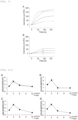
- the number of pSer46-MARCKS-positive cells per region of 143 ⁇ m ⁇ 143 ⁇ m was calculated on the basis of the DAPI fluorescence image and the pSer46-MARCKS fluorescence image ( Figures 2-1 and 9 ). Further, a KDEL signaling region in MAP2-positive cells, wherein the MAP2 is a mature neuron marker, was measured on the basis of the DAPI fluorescence image, the KDEL fluorescence image, and the MAP2 fluorescence image ( Figure 10 ).
- the ⁇ H2AX signaling region in MAP2-positive cells was measured on the basis of the DAPI fluorescence image, the ⁇ H2AX fluorescence image, and the MAP2 fluorescence image ( Figure 11 ).
- the 53BP1 signaling region in MAP2-positive cells was measured on the basis of the DAPI fluorescence image, the 53BP1 fluorescence image, and the MAP2 fluorescence image ( Figure 12 ).
- the pSer409/410-TDP-43 signaling region in a ubiquitin-positive inclusion was measured on the basis of the DAPI fluorescence image, the pSer409/410-TDP-43 fluorescence image, and the ubiquitin fluorescence image ( Figure 13 ).
- the p62 signaling region in an inclusion was measured on the basis of the DAPI fluorescence image and the p62 fluorescence image ( Figure 14 ). Further, the proportion of cells with a VAMP2 signal and a PSD95 signal colocalized therein was measured on the basis of the DAPI fluorescence image, the VAMP2 fluorescence image, and the PSD95 fluorescence image ( Figure 15 ). Further, the proportion of cells with a pSer203-tau signal and a PSD95 signal colocalized therein was measured on the basis of the DAPI fluorescence image, the pSer203-tau fluorescence image, and the PSD95 fluorescence image ( Figure 16 ).
- VCP T262A -KI PGRN R504X -KI CHMP2B Q165X -KI TDP43 N267S -KI Duration of treatment with antibody 6 to 10 months of age 3 to 7 months of age 1 to 6 months of age 6 to 10 months of age Duration of Morris water maze test 1 to 5 days from 10 months of age 1 to 5 days from 7 months of age 1 to 5 days from 6 months of age 1 to 5 days from 10 months of age Duration of Y-maze test 7 to 10 months of age 5 to 7 months of age 4 to 6 months of age 7 to 10 months of age Date of fear conditioning test 10 months of age 7 months of age 6 months of age 10 months of age
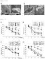
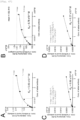
- mice underwent a Morris water maze test four times (60 seconds) once daily for five days, and the mean time to reach the platform (latency) (the vertical axis in Figure 3 ) was measured.
- a Y-maze consisting of three identical arms whose angles therebetween are equal (manufactured by O'Hara & Co., Ltd.) was used in the Y-maze test.
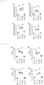
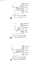
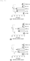
- a mouse was placed at the end of one arm and allowed to behave freely in the maze for eight minutes, and the percentage of spontaneous alternation (expressed as "Alternation rate" on the vertical axis in Figures 4 to 7 ) was calculated by dividing the number of entries into a new arm different from the previous arm by the total number of movements from one arm to another arm.
- the cerebral cortex tissue from four types of FTLD-ALS model mice or C57BL/6J mice at various months of age was dissolved in the presence of a dissolution buffer (100 mM Tris-HCl [pH 7.5], 2% SDS) containing a protease inhibitor cocktail (#539134; manufactured by Calbiochem; dilution factor, 1:100) at 4°C for one hour.
- a dissolution buffer 100 mM Tris-HCl [pH 7.5], 2% SDS
- protease inhibitor cocktail #1539134; manufactured by Calbiochem; dilution factor, 1:100
- PVDF polyvinylidene difluoride
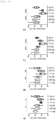
- the membrane was washed three times with a TBST solution, and antibody reactions were allowed to occur at room temperature for one hour using secondary antibodies against various primary antibodies (Table 5). Then, the above-mentioned 13 types of proteins were detected using an ECL Prime Western blot detection reagent (RPN2232; manufactured by GE Healthcare) and a luminescence image analyzer (ImageQuant LAS 500; manufactured by GE Healthcare) ( Figure 17 ).
- ECL Prime Western blot detection reagent RPN2232; manufactured by GE Healthcare
- a luminescence image analyzer ImageQuant LAS 500; manufactured by GE Healthcare
- the KD value of the mouse monoclonal antibody 2C8C was 1.95 ⁇ 10 -9 M
- the KD value of the human monoclonal antibody #129 of the present invention was 3.79 ⁇ 10 -11 M.
- the human monoclonal antibody #129 of the present invention is shown to be an antibody with very high affinity for human HMGB1.
- pSer46-MARCKS occurs from an early stage before onset of Alzheimer's disease, and HMGB1 leaking out of a cell owing to necrosis of a neuron induces phosphorylation of MARCKS, inducing degeneration of a neurite (" SCIENTIFIC REPORT, 2016 Aug 25; 6:31895 "). Accordingly, whether phosphorylation of MARCKS and necrosis of neurons also occurred at an early stage of development of FTLD was analyzed using four types of FTLD-ALS model mice one to 12 months of age.
- FTLD-ALS symptoms improved when the human monoclonal antibody #129 of the present invention was administered to four types of FTLD-ALS model mice was analyzed using three types of cognitive function tests (a Morris water maze test, a Y-maze test, and a fear-conditioning test).
- a Morris water maze test demonstrated that the time to reach the platform was prolonged as the four types of FTLD-ALS model mice aged by month ("KI n.t.” in Figure 3 ), as compared with C57BL/6J mice ("B6 n.t.” in Figure 3 ), which are normal mice ( Figure 3 ).
- the protein agglutination marker (pSer409/410-TDP-43 and p62)-derived signaling region expanded in the cerebral cortex.
- the human monoclonal antibody #129 of the present invention was administered, the expansion of the pSer409/410-TDP-43 and p62-derived signaling region was significantly suppressed ( Figures 13 and 14 ).
- the present inventors investigated synapse damage by analysis of a phosphorylated proteome using colocalization of VAMP2 and PSD95 and colocalization of pSer203-tau and PSD95 as indicators.
- the results of the analysis of synapse damage using these colocalizations as indicators showed that colocalization of VAMP2 and PSD95 in the cerebral cortex had decreased in the four types of FTLD-ALS model mice, as compared with the C57BL/6J mice, but the decrease was significantly suppressed when the human monoclonal antibody #129 of the present invention was administered ( Figure 15 ).
- the phosphorylation level of the synapse instability-associated protein (pSer203-tau) increased in the four types of FTLD-ALS model mice as compared with the C57BL/6J mice.
- the increase in the pSer203-tau level was suppressed (Lane (5) in Figure 17 ).
- the expression levels of synapse-associated proteins (PSD95 and VAMP2) decreased in the four types of FTLD-ALS model mice as compared with the C57BL/6J mice.
- the phosphorylation level of the cell death-associated protein increased in the four types of FTLD-ALS model mice as compared with the C57BL/6J mice.
- the human monoclonal antibody #129 of the present invention was administered to the four types of FTLD-ALS model mice, the increase in the pSer46-MARCKS level was suppressed (Lane (10) in Figure 17 ).
- the phosphorylation level of the DNA repair-associated protein (pSer77/78-Ku70) increased in the four types of FTLD-ALS model mice as compared with the C57BL/6J mice.
- the human monoclonal antibody #129 of the present invention was administered to the four types of FTLD-ALS model mice, the increase in the pSer77/78-Ku70 level was suppressed (Lane (11) in Figure 17 ).
- the protein agglutination marker level (pSer409/410-TDP-43) increased in the four types of FTLD-ALS model mice as compared with the C57BL/6J mice.
- the human monoclonal antibody #129 of the present invention was administered to the four types of FTLD-ALS model mice, the increase in the pSer409/410-TDP-43 level was suppressed (Lane (12) in Figure 17 ).
- the human monoclonal antibody #129 of the present invention was administered to the four types of FTLD-ALS model mice and the C57BL/6J mice, then the lung, liver, kidney, intestine, spleen, skeletal muscle, heart, and skin were removed therefrom by incision, and a pathological examination was performed.
- the results showed no abnormal findings (for example, obvious inflammation, cell death, tumor) due to administration of the human monoclonal antibody #129 of the present invention in any tissue.
- the results indicate that the human monoclonal antibody #129 of the present invention and the anti-HMGB1 antibody having the CDRs common thereto can prevent and improve symptoms in FTLD-ALS model mice without causing noticeable adverse reactions.
- 5 ⁇ FAD mice that is, transgenic mice that have overexpressed mutant human APP (770) having Swedish (KM670/671NL) mutation, Florida (I716V) mutation, and London (V717I) familial Alzheimer's disease (FAD) mutation, and human PS1 having two FAD mutations (M146L and L285V) were purchased from The Jackson Laboratory. Genes that encode the above-mentioned APP and PS1 in 5 ⁇ FAD mice are under the control of a mouse Thy1 promoter (" J Neurosci 26, 10129-10140 (2006 )"). The background of 5 ⁇ FAD mice was C57BL/SJL, which had been created by mating C57BL/6J females and SJL/J males. Comprehensive proteomic analysis was performed as mentioned earlier, by using brain tissue samples from male 5 ⁇ FAD mice at 1, 3, 6, and 12 months of age (" Hum Mol Genet 24, 540-558 (2015 )”).
- APP-KI mice (“App NL-G-F/NL-G-F ) mice (" Nat. Neurosci. 17, 661-663 (2014 )"), that is, mice having Swedish (KM670/671NL) mutation, Beyreuther/Iberian (I716F) mutation, and Arctic (E693G) mutation of humanized APP mouse genes were obtained from Riken BioResource Research Center.
- a control IgG human IgG isotype control, #12000C; manufactured by Thermo Fisher Scientific, Inc.
- the human monoclonal antibody #129 of the present invention was subcutaneously administered once a week from 6 months to 8 months of age (hereinafter simply referred to as "subcutaneously administered") (9 times in total).
- the human monoclonal antibody #129 of the present invention When the human monoclonal antibody #129 of the present invention was intravenously injected into each mouse, the human monoclonal antibody #129 of the present invention was diluted with physiological saline to a final volume of 50 ⁇ l and administered once a month into the caudal vein from 6 months to 8 months of age after birth (3 times in total).
- mouse and human brain tissue samples were immobilized in 4% paraformaldehyde and paraffin-embedded. Sagittal sections or coronal sections with a thickness of 5 ⁇ m were obtained using a microtome (manufactured by Yamato Kohki Industrial Co., Ltd.).
- the immunohistochemical staining was performed using the following primary antibodies: rabbit anti-phosphorylated Ser46-MARCKS antibody (1:2000 [manufactured by GL Biochem (Shanghai) Ltd.]); mouse anti-A ⁇ antibody (1:1000; clone 82E1; #10323; manufactured by IBL Co., Ltd.); mouse anti-MAP2 antibody (1:200; sc-32791; manufactured by Santa Cruz Biotechnology, Inc.); rabbit anti-MAP2 antibody (1:2000; ab32454; manufactured by Abcam Limited); mouse anti-phosphorylated H2X ( ⁇ H2X) antibody (1:300; JBW301; manufactured by Millipore); rabbit anti-53BP1 antibody (1:5000; NB100-304; manufactured by Novus Biologicals, LLC); rabbit anti-YAP antibody (1:50; GTX129151; manufactured by GeneTex, Inc.); rabbit anti-NF ⁇ B antibody (1:100; #8242; manufactured by Cell Signaling Technology, Inc.); mouse anti-IL6 antibody (1:50; ab208113;
- the entire cerebral cortex in various mice was subjected to Western blotting to detect ten types of proteins (yH2AX, 53BP1, ⁇ H2AX, 53BP1, pSer77/78-Ku70, ⁇ -actin, pThr638-PKC ⁇ , pThr641-PKC ⁇ I, pSer660-PKC ⁇ II/ ⁇ , and pan-PKC) using the antibodies in Tables 5 and 6, according to the method described in the above-described section of [Western blot method] in Example 1.
- ds-HMGB1 (6 ng/mL, 0.24 nM) and/or the human monoclonal antibody #129 of the present invention (6 ng/mL, 0.04 nM) were added to the medium.
- U2OS cells were treated with 2 ⁇ m Hoechst 33258 (DOJINDO LABORATORIES, Kumamoto, Japan) for 20 minutes and sensitized to DSB.
- Ku70 is known to assemble at DSB sites through nonhomologous end-joining (NHEJ) pathway and to be involved in DNA damage repair.
- NHEJ nonhomologous end-joining
- time-lapse imaging analysis of EGFP-Ku70 expressing cells after laser micro-irradiation was performed to examine how trigger signals from extracellular HMGB1 affect Ku70 assembling at DSB sites, in human U2OS cells that endogenously express TLR4 and RAGE receptors.
- CSF cerebrospinal fluid
- Senescent glial cells and nerve cells have been identified in neurodegenerative and aging brains (" Aging Cell 17, (2018 )”; “ J. Clin. Invest. 128, 1208-1216 (2018 )”; “ Nat. Neurosci. 22, 719-728 (2019 )”; “ Nat. Neurosci. 22, 683-684 (2019 )”; “ Front. Cell. Neurosci. 14, 1-14 (2020 )”; and “ Nat. Rev. Neurosci. 21, 433-444 (2020 ) ”) .
- a nuclear membrane protein Lamin B1 as a senescence marker, was localized in a ring shape in nerve cells, astrocytes, and microglia of normal mice (C57BL/6J mice) ( Figure 19a , arrows), whereas the localization of Lamin B1 in a ring shape became unclear or disappeared in a part of nerve cells and astrocytes of two types of AD model mice (5 ⁇ FAD mice and APP-KI mice) ( Figure 19a , arrows). Further, although abnormal morphology of Lamin B1 ring was observed in the microglia of the two types of AD model mice, the Lamin B1 ring by itself mostly remained ( Figure 19a , arrows).
- the intravenous administration of the human monoclonal antibody #129 of the present invention to two types of AD model mice significantly reduced, in all mice, the number of senescent cells in the cerebral cortex ( Figure 20b ), as well as the number of senescent cells in the neurons, astrocytes and microglia ( Figure 19b ) as compared with the case of intravenous administration of the control IgG to the above-mentioned two types of AD model mice.
- NF ⁇ B transcription factor
- MCP1 and IL6 inflammatory cytokines
- a protein kinase of Ku70, PBS (control) or a PKC ⁇ inhibitor (Go6976) was subcutaneously administered to B6/SJL mice or 5 ⁇ FAD mice at 6 weeks (1.5 months) of age (left diagram of Figure 22a ), and the expression levels of ⁇ H2AX and 53BP1 in the cerebral cortex were analyzed by immunohistochemical staining and Western blotting, as well as a cognitive function test (Y-maze test) was used to analyze whether the AD symptoms were improved.
- the control IgG or the human monoclonal antibody #129 of the present invention was subcutaneously administered to B6/SJL mice or 5 ⁇ FAD mice at 6 months of age (center diagram of Figure 22a ) to analyze using a cognitive function test (Y-maze test).
- dsHMGB1 activates a PKC-ERK system through binding with TLR4 and RAGE and phosphorylates MARCKS ("2016 Aug 25;6:31895. doi:10.1038/srep31895 "), and also revealed this time that the activated PKC phosphorylated Ku70 to decrease the repair function of DNA damage ( Figure 18 of Example 2). Further, it was revealed this time that the human monoclonal antibody #129 of the present invention suppresses TRIAD necrosis in nerve cells and also inhibits reciprocal amplification of TRIAD necrosis that is based on HMGB1-induced DNA damage ( Figures 18 and 22 of Example 2).
- the present Example 1 indicated that the human monoclonal antibody #129 of the present invention and the anti-HMGB1 antibody having the CDRs common thereto exhibit a treatment effect on FTLD-ALS model mice.
- the VCP T262A mutation, PGRN R504X mutation, CHMP2B Q165 X mutation, and TDP43 N267S mutation that the FTLD-ALS model mice carry are known to cause not only FTLD but also ALS.
- FTLD and ALS are understood to be the same disease spectrum in this field (for example, Patent Documents 1 to 13 below).
- HMGB1 neurodegenerative disease
- FTLD and ALS sporadic case
- ALS sporadic case
- the human monoclonal antibody on HMGB1 in the present invention shows equal effects on pathological conditions that may arise due to various causes as shown in the present Example, and hence, it can be developed as a therapeutic agent for more patients with not only familial neurodegenerative disease but also sporadic neurodegenerative disease.
- necrosis associated with decreased YAP has been confirmed in Huntington's disease (HD) mice (" Mao et al, Hum Mol Genet 2016 ”) and human HD patients (" Yamanishi et al, Acta Neuropathol Commun 2017 ”) in addition to AD and FTLD, and in mutant SOD1 transgenic mice, which are model mice for amyotrophic lateral sclerosis (ALS), decreased motoneurons have been confirmed (“ Morimoto et al, J Neurosci Res 2009 ").
- the human monoclonal antibody #129 of the present invention has an effect of reducing TDP43 aggregates of cerebral cortical neurons in four types of FTLD-ALS model mice, as shown in Example 1. Therefore, the human monoclonal antibody #129 of the present invention is fully expected to have a suppressing effect on degenerative diseases such as ALS associated with TDP43 aggregation.
- the human monoclonal antibody #129 of the present invention and the anti-HMGB1 antibody having the CDRs common thereto are fully expected to exhibit a treatment effect on general degenerative diseases, including ALS, PD, and HD.
- the human monoclonal antibody #129 of the present invention reduces the number of senescent cells in the cerebral cortex of two types of AD model mice (5 ⁇ FAD mice and APP-KI mice), and also suppresses the cellular senescence of neurons, astrocytes and microglia ( Figures 19 and 20 of Example 2), the human monoclonal antibody #129 of the present invention and the anti-HMGB1 antibody having the CDRs common thereto have been confirmed to have effects of treating aging-related degenerative or neurological disease, brain aging, and diseases associated with brain aging.
- mice As Huntington's disease model mice, R6/2 mice (Document 1) were used. As Parkinson's disease model mice, ⁇ -synuclein A53T mice (Document 2) were used. As amyotrophic lateral sclerosis model mice, SOD1-Tg mice (Document 3) were used. As spinocerebellar ataxia model mice, mutant ataxin-1 knock-in mice (Document 4) were used. As aging model mice, C57BL/6 mice at 12 months of age were used.
- the human monoclonal antibody #129 adjusted with physiological saline to 6 ⁇ g per 30 g of body weight (equivalent to 0.2 mg/kg BW) or the same amount of physiological saline without antibody was administered into the caudal vein of model mice of various diseases. Also, as a normal control group, the same amount of physiological saline was administered into the caudal vein of normal genotype sibling mice.
- the amount to be administered was increased to 60 ⁇ g per 30 g of body weight (equivalent to 2 mg/kg BW) from the third administration.
- mice 6 ⁇ g per 30 g of body weight (equivalent to 0.2 mg/kg BW) was intravenously administered once per 4 weeks.
- [Table 7] R6/2 mouse ⁇ -synuclein A53T mouse SOD1-Tg mouse Mutant ataxin-1 knock-in mouse Aged mouse Duration of treatment with antibody 6 to 12 weeks of age 8 to 18 weeks of age 6 to 20 weeks of age 8 to 18 weeks of age 12 to 19 months of age Duration of rotarod test 6 to 14 weeks of age 8 to 20 weeks of age 6 to 22 weeks of age 8 to 20 weeks of age - Duration of open field test 6 to 14 weeks of age 8 to 20 weeks of age 6 to 22 weeks of age - - Duration of footprint test 6 to 14 weeks of age 8 to 20 weeks of age 6 to 22 weeks of age 8 to 20 weeks of age - Y-maze test - - - - 12 to 20 months of age
- a human monoclonal antibody #129-L2 that is an amino acid sequence variant of the light chain CDR3 used in Example 4, which is described later, was administered in place of the human monoclonal antibody #129, in antibody administration experiments on the amyotrophic lateral sclerosis model mice (SOD1-Tg mice) and aging model mice.
- the alignment of the light chain variable region amino acid sequence of the human monoclonal antibody #129 and the human monoclonal antibody #129-L2 is as shown in Figure 25 .
- the human monoclonal antibody #129 antibody and the human monoclonal antibody #129-L2 antibody were used to study the binding to human dsHMGB1 by surface plasmon resonance (SPR), and as a result, the dissociation constants (K D ) of the human monoclonal antibody #129 antibody and the human monoclonal antibody #129-L2 antibody for the human dsHMGB1 were 1.47 ⁇ 10 -11 and 1.44 ⁇ 10 -11 , respectively, confirming that the two have almost equivalent binding strength.
- the average time taken to fall from the rotarod was calculated and used for subsequent analysis.
- the mice were placed in the center of an open field space (50 cm ⁇ 50 cm ⁇ 40 cm[height]) and allowed to explore for 10 minutes.
- the light intensity was 70 lux at the center of the field.
- the traveling distance (cm) was used as an index.
- footprints during walking were recorded to compare walking patterns.
- the hind paws were covered with blue ink, the forepaws were covered with red ink, and the mice were placed on a passage of a length of 40 cm and a width of 7.5 cm, which served as a runway to a sealed box.
- White paper on the passage floor was replaced after each run. Footprint patterns were measured using the footprint pattern parameter: stride described in Document 8 below. Further, the Y-maze test was performed according to the method described in Example 1.
- the brain was enucleated from disease model mice or normal genotype sibling mice at various months of age and immobilized in PBS containing 4% paraformaldehyde for 12 hours, and a paraffin-embedded brain tissue section with a thickness of 5 ⁇ m was prepared using a microtome (manufactured by Yamato Kohki Industrial Co., Ltd.).
- the prepared paraffin-embedded brain tissue section was deparaffinized using xylene, immersed in a 0.01 M citric acid buffer solution (pH 6.0) for re-hydration, and then heated at 120°C for 15 minutes.
- the number of the cells corresponding to the conditions described in the description of respective figures, per region of 143 ⁇ m ⁇ 143 ⁇ m for the cerebral cortex was calculated on the basis of the DAPI fluorescence image. Further, signal values of the regions of interest were measured on the basis of the pSer46-MARCKS fluorescence image, the ⁇ H2AX fluorescence image, the 53BP1 fluorescence image, and the KDEL fluorescence image.
- the cerebral cortex tissues from disease model mice and normal genotype sibling mice, and cerebellar tissues from spinocerebellar ataxia model mice and their background mice at various months of age were dissolved in the presence of a dissolution buffer (100 mM Tris-HCl[pH 7.5], 2% SDS) containing a protease inhibitor cocktail (#539134; manufactured by Calbiochem; dilution factor, 1:100) at 4°C for one hour.
- a dissolution buffer 100 mM Tris-HCl[pH 7.5], 2% SDS
- protease inhibitor cocktail #1539134; manufactured by Calbiochem; dilution factor, 1:100
- sample buffer 125 mM Tris-HCl, pH 6.8, 4% [w/v] SDS, 12% [v/v] 2-mercaptoethanol, 20% [v/v] glycerol, and 0.005% [w/v] bromophenol blue
- PVDF polyvinylidene difluoride
- the membrane was washed three times with a TBST solution, and antibody reactions were allowed to occur at room temperature for one hour using secondary antibodies against various primary antibodies (Table 9). Then, the above-mentioned 9 types of proteins were detected using an ECL Prime Western blot detection reagent (RPN2235; manufactured by Cytiva) and a luminescence image analyzer (ImageQuant LAS 500; manufactured by GE Healthcare).
- ECL Prime Western blot detection reagent RPN2235; manufactured by Cytiva
- a luminescence image analyzer imageQuant LAS 500; manufactured by GE Healthcare
- Huntington's disease model mice underwent experiments using post-symptomatic R6/2 mice (Document 1) as shown in "1. Materials and methods” described above, and intravenous administration was performed once per 2 weeks ( Figure 23A ). At 6 weeks of age, which was the timing of initiating administration, the rotarod test indicated decreased motor function in R6/2 mice. As shown in “1. Materials and methods” described above, in the rotarod test after administering 6 ⁇ g of the human monoclonal antibody #129 per 30 g of body weight (equivalent to 0.2 mg/kg BW) twice, improved motor function had not been clarified, and therefore, the amount to be administered was increased to 60 ⁇ g per 30 g of body weight (equivalent to 2 mg/kg BW) from the third administration.
- mice of three groups of physiological saline-administered normal mice, physiological saline-administered R6/2 mice, and human monoclonal antibody #129-administered R6/2 mice that is, "three groups for analyzing Huntington's disease model mice"
- triple staining of Htt, Ub and MAP2 was performed, and in the physiological saline-administered R6/2 mice, Htt intranuclear inclusions positive for Ub were observed in the MAP2-positive cerebral nerve cells ( Figure 24A ).
- TRIAD neuronal necrosis that is referred to as TRIAD releases HMGB1 (Documents 9 and 10), and that the released HMGB1 induces TRIAD necrosis in surrounding nerve cells (Document 11).
- a TRIAD necrosis marker positivity of cell body pSer46-MARCKS (Documents 9 to 11) and decreased nuclear YAP (Documents 10 to 13) are known. Accordingly, triple staining of YAP, pSer46-MARCKS and DAPI was performed, and in the physiological saline-administered R6/2 mice, TRIAD necrosis that shows pSer46-MARCKS positivity and decreased nuclear YAP was increased ( Figure 26A ).
- DNA damage in nerve cells is known to increase in the pathological conditions of neurodegenerative diseases including Huntington's disease (Documents 14 and 15). Therefore, DNA damage in nerve cells in the cerebral cortex was measured by quadruple staining with MAP2 and DAPI, using DNA damage markers 53BP1 and ⁇ H2AX. The results indicated that DNA damage in nerve cells increased in R6/2 mice compared with normal mice, and that the number of nerve cells having DNA damage was suppressed by administration of the human monoclonal antibody #129 ( Figure 26C ).
- the human monoclonal antibody #129 has an effect of suppressing pathological change and cell death in the pathological conditions of Huntington's disease (HD), and suppressing the decrease in motor function.
- Parkinson's disease model mice underwent experiments using post-symptomatic ⁇ -synuclein A53T mice (Document 2) as shown in "1. Materials and methods" described above, and intravenous administration was performed once per 2 weeks ( Figure 29A ). Once per 2 weeks from 8 weeks to 18 weeks of age, 6 ⁇ g of the human monoclonal antibody #129 per 30 g of body weight (equivalent to 0.2 mg/kg BW) was administered six times in total ( Figure 29A ). At 8 weeks of age, which was the timing of initiating administration, the rotarod test indicated that ⁇ -synuclein A53T mice were hyperactive ( Figure 29B ), indicating that Parkinson's disease had already been developed.
- mice of the three groups were sampled from mice of the three groups for analyzing Parkinson's disease model mice after motor function analysis to pathologically analyze.
- quadruple staining of pSer129-Syn, Ub, MAP2, and DAPI was performed, and in the physiological saline-administered ⁇ -synuclein A53T mice, increase in Ub-positive proteins and increase in pSer129-Syn in cell nuclei were observed in the cytoplasm of MAP2-positive cerebral nerve cells ( Figure 30A ).
- the findings of pSer129-Syn positive and Ub-positive were normalized by administration of the human monoclonal antibody #129.
- TRIAD neuronal necrosis that is referred to as TRIAD was examined (Document 12). As described above, it is known that TRIAD releases HMGB1 (Documents 9 and 10), and that the released HMGB1 induces TRIAD necrosis in surrounding nerve cells (Document 11). Further, in another immunohistochemical staining for Parkinson's disease model mice, findings have been reported that pSer46-MARCKS is stained in nerve cells (Document 16).
- DNA damage in nerve cells was measured in the cerebral cortex by quadruple staining with MAP2 and DAPI, using DNA damage markers 53BP1 and ⁇ H2AX.
- the human monoclonal antibody #129 has an effect of suppressing pathological change and cell death in the pathological conditions of Parkinson's disease (PD), and suppressing the decrease in motor function.
- PD Parkinson's disease
- Amyotrophic lateral sclerosis model mice underwent experiments using post-symptomatic SOD1-Tg mice (Document 3) as shown in "1. Materials and methods" described above, and intravenous administration was performed once per 2 weeks ( Figure 33A ). Once per 2 weeks from 6 weeks to 9 weeks of age, 6 ⁇ g of the human monoclonal antibody #129-L2 per 30 g of body weight (equivalent to 0.2 mg/kg BW) was administered twice in total ( Figure 33A ). The rotarod test immediately before initiating administration indicated decreased motor function in SOD1-Tg mice at 6 weeks of age ( Figure 33B ), indicating that SOD1-Tg mice had already developed amyotrophic lateral sclerosis.
- the amount to be administered was increased to 60 ⁇ g per 30 g of body weight (equivalent to 2 mg/kg BW) from the third administration at 10 weeks of age to the eighth administration at 20 weeks of age.
- mice of three groups of physiological saline-administered normal mice, physiological saline-administered SOD1-Tg mice, and human monoclonal antibody #129-L2-administered SOD1-Tg mice that is, "three groups for analyzing amyotrophic lateral sclerosis model mice"
- human monoclonal antibody #129-L2-administered SOD1-Tg mice that is, "three groups for analyzing amyotrophic lateral sclerosis model mice”
- immunohistochemical staining when quadruple staining of Ub, SOD1, MAP2, and DAPI was performed, there was an increase in the staining signals of SOD1 and Ub in large neurons in spinal cord anterior horn of SOD1-Tg mice, and some of them had inclusion-like morphology.
- TRIAD neuronal necrosis that is referred to as TRIAD was examined (Document 12).
- TRIAD neuronal necrosis that is referred to as TRIAD was examined (Document 12).
- HMGB1 Documents 9 and 10
- HMGB1 induces TRIAD necrosis in surrounding nerve cells
- Document 18 YAP fluctuations have been reported (Document 18).
- a TRIAD necrosis marker positivity of cell body pSer46-MARCKS (Document 9) and decreased nuclear YAP (Document 10) are known.
- the human monoclonal antibody #129-L2 has an effect of suppressing pathological change and cell death in the pathological conditions of amyotrophic lateral sclerosis (ALS), and suppressing the decrease in motor function.
- ALS amyotrophic lateral sclerosis
- mice of three groups of physiological saline-administered normal mice, physiological saline-administered mutant ataxin-1 knock-in mice, and human monoclonal antibody #129-administered mutant ataxin-1 knock-in mice that is, "three groups for analyzing spinocerebellar ataxia model mice"
- TRIAD which is cell death targeted by the human monoclonal antibody #129
- TRIAD neuronal necrosis that is referred to as TRIAD (Document 12) releases HMGB1 as described above (Documents 9 and 10), and that the released HMGB1 induces TRIAD necrosis (Document 11) or neurite degeneration (Document 9) in surrounding nerve cells.
- a TRIAD necrosis marker positivity of cell body or neurite pSer46-MARCKS (Documents 9 to 11) and decreased nuclear YAP (Documents 10 to 13) are known.
- the expression level of YAP affects the prognosis of mutant ataxin-1 knock-in mice even in the pathological conditions of spinocerebellar ataxia type 1 (SCA1) (Document 19).
- DNA damage in nerve cells is known to increase in the pathological conditions of neurodegenerative diseases including SCA1 (Documents 14 and 15). Therefore, DNA damage in Purkinje cells was measured by quadruple staining with MAP2 and DAPI, using DNA damage markers 53BP1 and ⁇ H2AX. The results indicated that DNA damage in Purkinje cells increased in mutant ataxin-1 knock-in mice compared with normal mice, and that DNA damage in Purkinje cells was suppressed by administration of the human monoclonal antibody #129 ( Figure 39C ).
- KDEL staining performed to detect abnormal endoplasmic reticulum which is a characteristic of TRIAD
- KDEL signal values of the Purkinje cell layer and granule cell layer were increased in mutant ataxin-1 knock-in mice compared with normal mice, confirming endoplasmic reticulum instability, which is observed in TRIAD ( Figure 40A ). Whether such findings eventually affect the number of Purkinje cells or the number of microglia and astrocytes in cerebellar cortex was confirmed by MAP2, Iba1, and GFAP staining ( Figure 40B ).
- the human monoclonal antibody #129 has an effect of suppressing pathological change and cell death in spinocerebellar ataxia, and suppressing the decrease in motor function.
- Lamin B1 is an inner nuclear membrane protein and shows a ring-shaped nuclear stain pattern in immunohistochemical staining; however, it is said that the ring-shaped nuclear localization disappears in cellular senescence (Document 20).
- DNA damage in nerve cells in the cerebral cortex was measured by quadruple staining with MAP2 and DAPI, using DNA damage markers 53BP1 and ⁇ H2AX.
- the above results indicate that the human monoclonal antibody #129-L2 has an effect of suppressing aging-associated cellular changes and cell death, and suppressing the decrease in cognitive function.
- amino acid sequences shown by underlines in the table indicate the modified sites of the amino acid sequences of the light chain CDR3 of the human monoclonal antibody #129.
- the antibody affinity of the human monoclonal antibody #129 and the human monoclonal antibody #129-L2 for human dsHMGB1 was analyzed by SPR described in Example 1.
- the dissociation constants (K D ) of the human monoclonal antibody #129 and the human monoclonal antibody #129-L2 for the human reduced HMGB1 were 2.07 ⁇ 10 -7 and 1.51 ⁇ 10 -5 , respectively, confirming that the human monoclonal antibody #129-L2 has weaker binding strength than the human monoclonal antibody #129 ( Figures 46D and C ).
- the same amount of physiological saline was similarly administered to various FTLD-ALS model mice or normal mice (sibling mice that do not have mutant genes of various FTLD-ALS model mice).
- the administration was initiated after development of the diseases was confirmed by a Y-maze test.
- the human monoclonal antibody #129 was purified from a plurality of clones of a CHO cell line that stably expresses human monoclonal antibody #129 according to a conventional method, and analyzed by non-reducing capillary electrophoresis according to a conventional method, and as a result, gentle peaks (arrow indicated site in the figure) were observed on the high-molecular weight side from the peak of IgG in all of the clones, as shown in Figure 54 .
- the human monoclonal antibody #129-L2 was purified from a plurality of clones of a CHO cell line that stably expresses human monoclonal antibody #129-L2, and analyzed by non-reducing capillary electrophoresis, and as a result, no gentle peaks were observed on the high-molecular weight side in any of the clones, as shown in Figure 55 .
- K D of the human monoclonal antibody #129-L2 was 1.44 ⁇ 10 -11 before the storage for 6 months at 4°C ( Figure 57A ), whereas it was 3.51 ⁇ 10 -11 after the storage for 6 months at 4°C ( Figure 57B ), indicating an increase of only about 2.5-fold.
- the human monoclonal antibody #129 and the human monoclonal antibody #129-L2
- treatment effects on Alzheimer's disease model mice 5 ⁇ FAD mice
- physiological saline containing 0.2 mg/kg body weight (6 ⁇ g/animal) of the human monoclonal antibody #129 or human monoclonal antibody #129-L2 was administered to one mouse of the Alzheimer's disease model mice by intravenous injection via caudal vein, once per month for a total of three times starting from 6 months of age after development of the disease ( Figure 58A ).
- the present invention contributes to prevention and/or treatment of amyotrophic lateral sclerosis, Parkinson's disease, Huntington's disease, spinocerebellar ataxia, Alzheimer's disease, frontotemporal lobar degeneration, aging-related degenerative or neurological disease, brain aging, or diseases associated with brain aging.
Landscapes
- Health & Medical Sciences (AREA)
- Chemical & Material Sciences (AREA)
- Organic Chemistry (AREA)
- Engineering & Computer Science (AREA)
- Bioinformatics & Cheminformatics (AREA)
- Neurology (AREA)
- Life Sciences & Earth Sciences (AREA)
- Medicinal Chemistry (AREA)
- General Health & Medical Sciences (AREA)
- Neurosurgery (AREA)
- Biomedical Technology (AREA)
- Veterinary Medicine (AREA)
- Chemical Kinetics & Catalysis (AREA)
- General Chemical & Material Sciences (AREA)
- Nuclear Medicine, Radiotherapy & Molecular Imaging (AREA)
- Pharmacology & Pharmacy (AREA)
- Animal Behavior & Ethology (AREA)
- Public Health (AREA)
- Molecular Biology (AREA)
- Immunology (AREA)
- Biochemistry (AREA)
- Biophysics (AREA)
- Genetics & Genomics (AREA)
- Proteomics, Peptides & Aminoacids (AREA)
- Psychology (AREA)
- Hospice & Palliative Care (AREA)
- Psychiatry (AREA)
- Orthopedic Medicine & Surgery (AREA)
- Physical Education & Sports Medicine (AREA)
- Peptides Or Proteins (AREA)
- Medicines Containing Antibodies Or Antigens For Use As Internal Diagnostic Agents (AREA)
Applications Claiming Priority (2)
| Application Number | Priority Date | Filing Date | Title |
|---|---|---|---|
| JP2022092342 | 2022-06-07 | ||
| PCT/JP2023/021039 WO2023238869A1 (fr) | 2022-06-07 | 2023-06-06 | Agent préventif ou agent thérapeutique pour la sclérose latérale amyotrophique, la maladie de parkinson, la maladie de huntington, l'ataxie spinocérébelleuse, une maladie dégénérative ou neurologique liée au vieillissement, le vieillissement cérébral ou des maladies associées au vieillissement cérébral |
Publications (1)
| Publication Number | Publication Date |
|---|---|
| EP4537842A1 true EP4537842A1 (fr) | 2025-04-16 |
Family
ID=89118335
Family Applications (1)
| Application Number | Title | Priority Date | Filing Date |
|---|---|---|---|
| EP23819849.3A Pending EP4537842A1 (fr) | 2022-06-07 | 2023-06-06 | Agent préventif ou agent thérapeutique pour la sclérose latérale amyotrophique, la maladie de parkinson, la maladie de huntington, l'ataxie spinocérébelleuse, une maladie dégénérative ou neurologique liée au vieillissement, le vieillissement cérébral ou des maladies associées au vieillissement cérébral |
Country Status (5)
| Country | Link |
|---|---|
| US (1) | US20250353904A1 (fr) |
| EP (1) | EP4537842A1 (fr) |
| JP (1) | JPWO2023238869A1 (fr) |
| CN (1) | CN119730880A (fr) |
| WO (1) | WO2023238869A1 (fr) |
Family Cites Families (24)
| Publication number | Priority date | Publication date | Assignee | Title |
|---|---|---|---|---|
| GB8308235D0 (en) | 1983-03-25 | 1983-05-05 | Celltech Ltd | Polypeptides |
| US4816567A (en) | 1983-04-08 | 1989-03-28 | Genentech, Inc. | Recombinant immunoglobin preparations |
| US5807715A (en) | 1984-08-27 | 1998-09-15 | The Board Of Trustees Of The Leland Stanford Junior University | Methods and transformed mammalian lymphocyte cells for producing functional antigen-binding protein including chimeric immunoglobulin |
| GB8607679D0 (en) | 1986-03-27 | 1986-04-30 | Winter G P | Recombinant dna product |
| US5047335A (en) | 1988-12-21 | 1991-09-10 | The Regents Of The University Of Calif. | Process for controlling intracellular glycosylation of proteins |
| IL162181A (en) | 1988-12-28 | 2006-04-10 | Pdl Biopharma Inc | A method of producing humanized immunoglubulin, and polynucleotides encoding the same |
| US6657103B1 (en) | 1990-01-12 | 2003-12-02 | Abgenix, Inc. | Human antibodies derived from immunized xenomice |
| ATE300615T1 (de) | 1990-08-29 | 2005-08-15 | Genpharm Int | Transgene mäuse fähig zur produktion heterologer antikörper |
| US5661016A (en) | 1990-08-29 | 1997-08-26 | Genpharm International Inc. | Transgenic non-human animals capable of producing heterologous antibodies of various isotypes |
| US5625126A (en) | 1990-08-29 | 1997-04-29 | Genpharm International, Inc. | Transgenic non-human animals for producing heterologous antibodies |
| US5545806A (en) | 1990-08-29 | 1996-08-13 | Genpharm International, Inc. | Ransgenic non-human animals for producing heterologous antibodies |
| US5633425A (en) | 1990-08-29 | 1997-05-27 | Genpharm International, Inc. | Transgenic non-human animals capable of producing heterologous antibodies |
| US5278299A (en) | 1991-03-18 | 1994-01-11 | Scripps Clinic And Research Foundation | Method and composition for synthesizing sialylated glycosyl compounds |
| WO1993010260A1 (fr) | 1991-11-21 | 1993-05-27 | The Board Of Trustees Of The Leland Stanford Junior University | Utilisation de la neuraminidase extracellulaire pour lutter contre la degradation d'oligosaccharides de la glycoproteine |
| FR2688138B1 (fr) | 1992-03-06 | 1995-05-05 | Rhone Poulenc Rorer Sa | Application de l'amino-2 trifluoromethoxy-6 benzothiazole pour obtenir un medicament destine au traitement de la sclerose laterale amyotrophique. |
| US5648267A (en) | 1992-11-13 | 1997-07-15 | Idec Pharmaceuticals Corporation | Impaired dominant selectable marker sequence and intronic insertion strategies for enhancement of expression of gene product and expression vector systems comprising same |
| CU22615A1 (es) | 1994-06-30 | 2000-02-10 | Centro Inmunologia Molecular | Procedimiento de obtención de anticuerpos monoclonales murinos menos inmunogénicos. anticuerpos monoclonales obtenidos |
| CA2194907A1 (fr) | 1994-07-13 | 1996-02-01 | Kouji Matsushima | Anticorps humain reconstitue contre l'interleukine-8 humaine |
| ES2434961T5 (es) | 1998-04-20 | 2018-01-18 | Roche Glycart Ag | Ingeniería de glicosilación de anticuerpos para mejorar la citotoxicidad celular dependiente del anticuerpo |
| ES2639222T5 (es) | 2000-10-06 | 2023-11-24 | Kyowa Kirin Co Ltd | Células que producen unas composiciones de anticuerpo |
| CN1487996B (zh) | 2000-11-30 | 2010-06-16 | 米德列斯公司 | 用于生产人类抗体的转基因转染色体啮齿动物 |
| EP1345967B1 (fr) | 2000-12-22 | 2011-08-10 | Grad, Carole, Legal representative of Kaplan, Howard | Bibliotheques d'affichage de phages de fragments v h humains |
| EP3502248B1 (fr) | 2016-08-09 | 2024-05-29 | National University Corporation Tokyo Medical and Dental University | Anticorps dirigés contre hmgb1 et composition les comprenant pour le traitement ou la prévention de la maladie d'alzheimer |
| JP7475687B2 (ja) | 2018-09-21 | 2024-04-30 | 国立大学法人 東京医科歯科大学 | ヒトhmgb1に特異的に結合するヒトモノクローナル抗体、及びそれを含有するアルツハイマー病を治療又は予防するための医薬組成物 |
-
2023
- 2023-06-06 US US18/871,213 patent/US20250353904A1/en active Pending
- 2023-06-06 EP EP23819849.3A patent/EP4537842A1/fr active Pending
- 2023-06-06 CN CN202380044547.9A patent/CN119730880A/zh active Pending
- 2023-06-06 JP JP2024526987A patent/JPWO2023238869A1/ja active Pending
- 2023-06-06 WO PCT/JP2023/021039 patent/WO2023238869A1/fr not_active Ceased
Also Published As
| Publication number | Publication date |
|---|---|
| WO2023238869A1 (fr) | 2023-12-14 |
| JPWO2023238869A1 (fr) | 2023-12-14 |
| US20250353904A1 (en) | 2025-11-20 |
| CN119730880A (zh) | 2025-03-28 |
Similar Documents
| Publication | Publication Date | Title |
|---|---|---|
| JP7028923B2 (ja) | 抗トランスサイレチン抗体 | |
| JP7101927B2 (ja) | 抗トランスサイレチン抗体 | |
| CN106574258B (zh) | 抗-运甲状腺素蛋白人源化抗体 | |
| JP6875280B2 (ja) | 抗トランスサイレチン抗体 | |
| CN114174330B (zh) | Apoe抗体、融合蛋白及其用途 | |
| JP7016470B2 (ja) | 抗トランスサイレチン抗体 | |
| JP7017013B2 (ja) | 抗トランスサイレチン抗体 | |
| CN106459956B (zh) | 抗-运甲状腺素蛋白人抗体 | |
| JP7475687B2 (ja) | ヒトhmgb1に特異的に結合するヒトモノクローナル抗体、及びそれを含有するアルツハイマー病を治療又は予防するための医薬組成物 | |
| EP3502248B1 (fr) | Anticorps dirigés contre hmgb1 et composition les comprenant pour le traitement ou la prévention de la maladie d'alzheimer | |
| CN110072888B (zh) | 药剂、用途和方法 | |
| JP2022033757A (ja) | タウオパチーを治療するための組成物及び方法 | |
| US20210079075A1 (en) | Novel means and methods for treating neurodegenerative diseases | |
| EP4537842A1 (fr) | Agent préventif ou agent thérapeutique pour la sclérose latérale amyotrophique, la maladie de parkinson, la maladie de huntington, l'ataxie spinocérébelleuse, une maladie dégénérative ou neurologique liée au vieillissement, le vieillissement cérébral ou des maladies associées au vieillissement cérébral | |
| US20230416375A1 (en) | Antibody variants against wnt receptor ryk | |
| AU2016235685B2 (en) | Method of diagnosis or treatment of neurological disorders with p75ECD and/or p75 | |
| HK40125870A (zh) | 肌萎缩侧索硬化症、帕金森病、亨廷顿病、脊髓小脑性共济失调、与老化有关的变性疾病或神经疾病、脑老化、或者伴有脑老化的疾病的预防剂或治疗剂 | |
| US20240025985A1 (en) | Agent for Preventing or Treating Frontotemporal Lobar Degeneration | |
| US20240352102A1 (en) | Compositions for treating tauopathies and methods of use thereof | |
| KR20250116724A (ko) | 항-헌팅틴 항체 | |
| WO2024101345A1 (fr) | Agent pour prévenir ou traiter une maladie associée à l'accumulation d'agrégats de protéines anormales |
Legal Events
| Date | Code | Title | Description |
|---|---|---|---|
| STAA | Information on the status of an ep patent application or granted ep patent |
Free format text: STATUS: THE INTERNATIONAL PUBLICATION HAS BEEN MADE |
|
| PUAI | Public reference made under article 153(3) epc to a published international application that has entered the european phase |
Free format text: ORIGINAL CODE: 0009012 |
|
| STAA | Information on the status of an ep patent application or granted ep patent |
Free format text: STATUS: REQUEST FOR EXAMINATION WAS MADE |
|
| 17P | Request for examination filed |
Effective date: 20241129 |
|
| AK | Designated contracting states |
Kind code of ref document: A1 Designated state(s): AL AT BE BG CH CY CZ DE DK EE ES FI FR GB GR HR HU IE IS IT LI LT LU LV MC ME MK MT NL NO PL PT RO RS SE SI SK SM TR |
|
| DAV | Request for validation of the european patent (deleted) | ||
| DAX | Request for extension of the european patent (deleted) |