EP4552699A2 - Protéines de fusion à domaine de liaison à l'albumine - Google Patents
Protéines de fusion à domaine de liaison à l'albumine Download PDFInfo
- Publication number
- EP4552699A2 EP4552699A2 EP25153140.6A EP25153140A EP4552699A2 EP 4552699 A2 EP4552699 A2 EP 4552699A2 EP 25153140 A EP25153140 A EP 25153140A EP 4552699 A2 EP4552699 A2 EP 4552699A2
- Authority
- EP
- European Patent Office
- Prior art keywords
- abd
- fusion protein
- cytokine
- cells
- scfv
- Prior art date
- Legal status (The legal status is an assumption and is not a legal conclusion. Google has not performed a legal analysis and makes no representation as to the accuracy of the status listed.)
- Pending
Links
Images







































Classifications
-
- C—CHEMISTRY; METALLURGY
- C07—ORGANIC CHEMISTRY
- C07K—PEPTIDES
- C07K16/00—Immunoglobulins [IG], e.g. monoclonal or polyclonal antibodies
- C07K16/18—Immunoglobulins [IG], e.g. monoclonal or polyclonal antibodies against material from animals or humans
- C07K16/24—Immunoglobulins [IG], e.g. monoclonal or polyclonal antibodies against material from animals or humans against cytokines, lymphokines or interferons
- C07K16/244—Interleukins [IL]
-
- A—HUMAN NECESSITIES
- A61—MEDICAL OR VETERINARY SCIENCE; HYGIENE
- A61K—PREPARATIONS FOR MEDICAL, DENTAL OR TOILETRY PURPOSES
- A61K47/00—Medicinal preparations characterised by the non-active ingredients used, e.g. carriers or inert additives; Targeting or modifying agents chemically bound to the active ingredient
- A61K47/50—Medicinal preparations characterised by the non-active ingredients used, e.g. carriers or inert additives; Targeting or modifying agents chemically bound to the active ingredient the non-active ingredient being chemically bound to the active ingredient, e.g. polymer-drug conjugates
- A61K47/51—Medicinal preparations characterised by the non-active ingredients used, e.g. carriers or inert additives; Targeting or modifying agents chemically bound to the active ingredient the non-active ingredient being chemically bound to the active ingredient, e.g. polymer-drug conjugates the non-active ingredient being a modifying agent
- A61K47/62—Medicinal preparations characterised by the non-active ingredients used, e.g. carriers or inert additives; Targeting or modifying agents chemically bound to the active ingredient the non-active ingredient being chemically bound to the active ingredient, e.g. polymer-drug conjugates the non-active ingredient being a modifying agent the modifying agent being a protein, peptide or polyamino acid
- A61K47/64—Drug-peptide, drug-protein or drug-polyamino acid conjugates, i.e. the modifying agent being a peptide, protein or polyamino acid which is covalently bonded or complexed to a therapeutically active agent
- A61K47/643—Albumins, e.g. HSA, BSA, ovalbumin or a Keyhole Limpet Hemocyanin [KHL]
-
- A—HUMAN NECESSITIES
- A61—MEDICAL OR VETERINARY SCIENCE; HYGIENE
- A61K—PREPARATIONS FOR MEDICAL, DENTAL OR TOILETRY PURPOSES
- A61K38/00—Medicinal preparations containing peptides
- A61K38/16—Peptides having more than 20 amino acids; Gastrins; Somatostatins; Melanotropins; Derivatives thereof
- A61K38/17—Peptides having more than 20 amino acids; Gastrins; Somatostatins; Melanotropins; Derivatives thereof from animals; from humans
- A61K38/19—Cytokines; Lymphokines; Interferons
- A61K38/20—Interleukins [IL]
- A61K38/2086—IL-13 to IL-16
-
- A—HUMAN NECESSITIES
- A61—MEDICAL OR VETERINARY SCIENCE; HYGIENE
- A61K—PREPARATIONS FOR MEDICAL, DENTAL OR TOILETRY PURPOSES
- A61K39/00—Medicinal preparations containing antigens or antibodies
- A61K39/0005—Vertebrate antigens
- A61K39/0011—Cancer antigens
- A61K39/001136—Cytokines
- A61K39/001138—Tumor necrosis factors [TNF] or CD70
-
- A—HUMAN NECESSITIES
- A61—MEDICAL OR VETERINARY SCIENCE; HYGIENE
- A61K—PREPARATIONS FOR MEDICAL, DENTAL OR TOILETRY PURPOSES
- A61K39/00—Medicinal preparations containing antigens or antibodies
- A61K39/0005—Vertebrate antigens
- A61K39/0011—Cancer antigens
- A61K39/001136—Cytokines
- A61K39/00114—Interleukins [IL]
-
- A—HUMAN NECESSITIES
- A61—MEDICAL OR VETERINARY SCIENCE; HYGIENE
- A61K—PREPARATIONS FOR MEDICAL, DENTAL OR TOILETRY PURPOSES
- A61K39/00—Medicinal preparations containing antigens or antibodies
- A61K39/395—Antibodies; Immunoglobulins; Immune serum, e.g. antilymphocytic serum
- A61K39/39533—Antibodies; Immunoglobulins; Immune serum, e.g. antilymphocytic serum against materials from animals
- A61K39/3955—Antibodies; Immunoglobulins; Immune serum, e.g. antilymphocytic serum against materials from animals against proteinaceous materials, e.g. enzymes, hormones, lymphokines
-
- A—HUMAN NECESSITIES
- A61—MEDICAL OR VETERINARY SCIENCE; HYGIENE
- A61K—PREPARATIONS FOR MEDICAL, DENTAL OR TOILETRY PURPOSES
- A61K47/00—Medicinal preparations characterised by the non-active ingredients used, e.g. carriers or inert additives; Targeting or modifying agents chemically bound to the active ingredient
- A61K47/50—Medicinal preparations characterised by the non-active ingredients used, e.g. carriers or inert additives; Targeting or modifying agents chemically bound to the active ingredient the non-active ingredient being chemically bound to the active ingredient, e.g. polymer-drug conjugates
- A61K47/51—Medicinal preparations characterised by the non-active ingredients used, e.g. carriers or inert additives; Targeting or modifying agents chemically bound to the active ingredient the non-active ingredient being chemically bound to the active ingredient, e.g. polymer-drug conjugates the non-active ingredient being a modifying agent
- A61K47/62—Medicinal preparations characterised by the non-active ingredients used, e.g. carriers or inert additives; Targeting or modifying agents chemically bound to the active ingredient the non-active ingredient being chemically bound to the active ingredient, e.g. polymer-drug conjugates the non-active ingredient being a modifying agent the modifying agent being a protein, peptide or polyamino acid
- A61K47/65—Peptidic linkers, binders or spacers, e.g. peptidic enzyme-labile linkers
-
- A—HUMAN NECESSITIES
- A61—MEDICAL OR VETERINARY SCIENCE; HYGIENE
- A61P—SPECIFIC THERAPEUTIC ACTIVITY OF CHEMICAL COMPOUNDS OR MEDICINAL PREPARATIONS
- A61P35/00—Antineoplastic agents
-
- C—CHEMISTRY; METALLURGY
- C07—ORGANIC CHEMISTRY
- C07K—PEPTIDES
- C07K14/00—Peptides having more than 20 amino acids; Gastrins; Somatostatins; Melanotropins; Derivatives thereof
- C07K14/435—Peptides having more than 20 amino acids; Gastrins; Somatostatins; Melanotropins; Derivatives thereof from animals; from humans
- C07K14/52—Cytokines; Lymphokines; Interferons
- C07K14/54—Interleukins [IL]
- C07K14/5443—IL-15
-
- C—CHEMISTRY; METALLURGY
- C07—ORGANIC CHEMISTRY
- C07K—PEPTIDES
- C07K14/00—Peptides having more than 20 amino acids; Gastrins; Somatostatins; Melanotropins; Derivatives thereof
- C07K14/435—Peptides having more than 20 amino acids; Gastrins; Somatostatins; Melanotropins; Derivatives thereof from animals; from humans
- C07K14/705—Receptors; Cell surface antigens; Cell surface determinants
- C07K14/715—Receptors; Cell surface antigens; Cell surface determinants for cytokines; for lymphokines; for interferons
- C07K14/7155—Receptors; Cell surface antigens; Cell surface determinants for cytokines; for lymphokines; for interferons for interleukins [IL]
-
- C—CHEMISTRY; METALLURGY
- C07—ORGANIC CHEMISTRY
- C07K—PEPTIDES
- C07K16/00—Immunoglobulins [IG], e.g. monoclonal or polyclonal antibodies
- C07K16/18—Immunoglobulins [IG], e.g. monoclonal or polyclonal antibodies against material from animals or humans
-
- C—CHEMISTRY; METALLURGY
- C07—ORGANIC CHEMISTRY
- C07K—PEPTIDES
- C07K16/00—Immunoglobulins [IG], e.g. monoclonal or polyclonal antibodies
- C07K16/18—Immunoglobulins [IG], e.g. monoclonal or polyclonal antibodies against material from animals or humans
- C07K16/22—Immunoglobulins [IG], e.g. monoclonal or polyclonal antibodies against material from animals or humans against growth factors ; against growth regulators
-
- C—CHEMISTRY; METALLURGY
- C07—ORGANIC CHEMISTRY
- C07K—PEPTIDES
- C07K16/00—Immunoglobulins [IG], e.g. monoclonal or polyclonal antibodies
- C07K16/18—Immunoglobulins [IG], e.g. monoclonal or polyclonal antibodies against material from animals or humans
- C07K16/28—Immunoglobulins [IG], e.g. monoclonal or polyclonal antibodies against material from animals or humans against receptors, cell surface antigens or cell surface determinants
- C07K16/2803—Immunoglobulins [IG], e.g. monoclonal or polyclonal antibodies against material from animals or humans against receptors, cell surface antigens or cell surface determinants against the immunoglobulin superfamily
- C07K16/2818—Immunoglobulins [IG], e.g. monoclonal or polyclonal antibodies against material from animals or humans against receptors, cell surface antigens or cell surface determinants against the immunoglobulin superfamily against CD28 or CD152
-
- C—CHEMISTRY; METALLURGY
- C07—ORGANIC CHEMISTRY
- C07K—PEPTIDES
- C07K16/00—Immunoglobulins [IG], e.g. monoclonal or polyclonal antibodies
- C07K16/46—Hybrid immunoglobulins
- C07K16/468—Immunoglobulins having two or more different antigen binding sites, e.g. multifunctional antibodies
-
- A—HUMAN NECESSITIES
- A61—MEDICAL OR VETERINARY SCIENCE; HYGIENE
- A61K—PREPARATIONS FOR MEDICAL, DENTAL OR TOILETRY PURPOSES
- A61K39/00—Medicinal preparations containing antigens or antibodies
- A61K2039/60—Medicinal preparations containing antigens or antibodies characteristics by the carrier linked to the antigen
- A61K2039/6031—Proteins
- A61K2039/6081—Albumin; Keyhole limpet haemocyanin [KLH]
-
- A—HUMAN NECESSITIES
- A61—MEDICAL OR VETERINARY SCIENCE; HYGIENE
- A61K—PREPARATIONS FOR MEDICAL, DENTAL OR TOILETRY PURPOSES
- A61K38/00—Medicinal preparations containing peptides
-
- C—CHEMISTRY; METALLURGY
- C07—ORGANIC CHEMISTRY
- C07K—PEPTIDES
- C07K14/00—Peptides having more than 20 amino acids; Gastrins; Somatostatins; Melanotropins; Derivatives thereof
- C07K14/435—Peptides having more than 20 amino acids; Gastrins; Somatostatins; Melanotropins; Derivatives thereof from animals; from humans
- C07K14/76—Albumins
-
- C—CHEMISTRY; METALLURGY
- C07—ORGANIC CHEMISTRY
- C07K—PEPTIDES
- C07K2317/00—Immunoglobulins specific features
- C07K2317/30—Immunoglobulins specific features characterized by aspects of specificity or valency
- C07K2317/31—Immunoglobulins specific features characterized by aspects of specificity or valency multispecific
-
- C—CHEMISTRY; METALLURGY
- C07—ORGANIC CHEMISTRY
- C07K—PEPTIDES
- C07K2317/00—Immunoglobulins specific features
- C07K2317/30—Immunoglobulins specific features characterized by aspects of specificity or valency
- C07K2317/33—Crossreactivity, e.g. for species or epitope, or lack of said crossreactivity
-
- C—CHEMISTRY; METALLURGY
- C07—ORGANIC CHEMISTRY
- C07K—PEPTIDES
- C07K2317/00—Immunoglobulins specific features
- C07K2317/60—Immunoglobulins specific features characterized by non-natural combinations of immunoglobulin fragments
- C07K2317/62—Immunoglobulins specific features characterized by non-natural combinations of immunoglobulin fragments comprising only variable region components
- C07K2317/622—Single chain antibody (scFv)
-
- C—CHEMISTRY; METALLURGY
- C07—ORGANIC CHEMISTRY
- C07K—PEPTIDES
- C07K2317/00—Immunoglobulins specific features
- C07K2317/70—Immunoglobulins specific features characterized by effect upon binding to a cell or to an antigen
- C07K2317/76—Antagonist effect on antigen, e.g. neutralization or inhibition of binding
-
- C—CHEMISTRY; METALLURGY
- C07—ORGANIC CHEMISTRY
- C07K—PEPTIDES
- C07K2319/00—Fusion polypeptide
-
- C—CHEMISTRY; METALLURGY
- C12—BIOCHEMISTRY; BEER; SPIRITS; WINE; VINEGAR; MICROBIOLOGY; ENZYMOLOGY; MUTATION OR GENETIC ENGINEERING
- C12N—MICROORGANISMS OR ENZYMES; COMPOSITIONS THEREOF; PROPAGATING, PRESERVING, OR MAINTAINING MICROORGANISMS; MUTATION OR GENETIC ENGINEERING; CULTURE MEDIA
- C12N15/00—Mutation or genetic engineering; DNA or RNA concerning genetic engineering, vectors, e.g. plasmids, or their isolation, preparation or purification; Use of hosts therefor
- C12N15/09—Recombinant DNA-technology
- C12N15/11—DNA or RNA fragments; Modified forms thereof; Non-coding nucleic acids having a biological activity
- C12N15/62—DNA sequences coding for fusion proteins
-
- C—CHEMISTRY; METALLURGY
- C12—BIOCHEMISTRY; BEER; SPIRITS; WINE; VINEGAR; MICROBIOLOGY; ENZYMOLOGY; MUTATION OR GENETIC ENGINEERING
- C12N—MICROORGANISMS OR ENZYMES; COMPOSITIONS THEREOF; PROPAGATING, PRESERVING, OR MAINTAINING MICROORGANISMS; MUTATION OR GENETIC ENGINEERING; CULTURE MEDIA
- C12N15/00—Mutation or genetic engineering; DNA or RNA concerning genetic engineering, vectors, e.g. plasmids, or their isolation, preparation or purification; Use of hosts therefor
- C12N15/09—Recombinant DNA-technology
- C12N15/63—Introduction of foreign genetic material using vectors; Vectors; Use of hosts therefor; Regulation of expression
-
- C—CHEMISTRY; METALLURGY
- C12—BIOCHEMISTRY; BEER; SPIRITS; WINE; VINEGAR; MICROBIOLOGY; ENZYMOLOGY; MUTATION OR GENETIC ENGINEERING
- C12N—MICROORGANISMS OR ENZYMES; COMPOSITIONS THEREOF; PROPAGATING, PRESERVING, OR MAINTAINING MICROORGANISMS; MUTATION OR GENETIC ENGINEERING; CULTURE MEDIA
- C12N2510/00—Genetically modified cells
-
- C—CHEMISTRY; METALLURGY
- C12—BIOCHEMISTRY; BEER; SPIRITS; WINE; VINEGAR; MICROBIOLOGY; ENZYMOLOGY; MUTATION OR GENETIC ENGINEERING
- C12N—MICROORGANISMS OR ENZYMES; COMPOSITIONS THEREOF; PROPAGATING, PRESERVING, OR MAINTAINING MICROORGANISMS; MUTATION OR GENETIC ENGINEERING; CULTURE MEDIA
- C12N2800/00—Nucleic acids vectors
Definitions
- Biologics are useful for the treatment of cancers in a variety of ways.
- Cytokine-based therapies can work directly on cancer cells by interfering with how such cells grow and multiply. Cytokines may also stimulate the immune system, by encouraging the growth of killer T cells and other cells that attack cancer cells. Further, cytokines can promote cancer cells to send out chemicals that attract immune system cells. See, e.g., Dranoff, Nature Reviews Cancer 4: 11-22 (2004 ); and Zhang et al., Proc Natl Acad Sci U.S.A. 106(18): 7513-7518 (2009 ). Antibodies are desirable as therapeutics due to their ability to recognize targets with both specificity and high affinity.
- Monoclonal antibody based therapies including those that target tumor surface antigens and inhibitory signals that limit T-cell activation, have been a standard component of cancer therapeutics for over 20 years. See, e.g., Weiner, Nat Rev Cancer 15(6): 361-370 (2015 ).
- Short circulatory half-life represents a major obstacle for many biologics. See, e.g., Perdreau et al., European Cytokine Network 21: 297-307 (2010 ). Such short-acting therapeutics require frequent dosing profiles that can reduce applicability to the clinic, particular for chronic conditions. Long serum half-life is desirable as it would decrease the need for repetitive injections of the molecule to achieve a therapeutically relevant serum concentration.
- Methods of extending the half-life of therapeutic proteins include PEGylation, fusion to human serum albumin (HSA), fusion to the constant fragment (Fc) of a human immunoglobulin IgG, and fusion to non-structured polypeptides such as XTEN.
- Half-life extension technologies enable new and improved biologic therapies that reduce the cost and burden of frequent dosing.
- novel reagents and methods useful for extending the half-lives of protein and peptide based therapeutics See, e.g., Stohl, BioDrugs 29(4): 215-239 (2015 ).
- compositions that include albumin binding domain are provided herein.
- albumin binding domain i.e., albumin binding domain fusion proteins
- Short circulatory half-life represents a major obstacle for many biologics. See, e.g., Perdreau et al., European Cytokine Network 21: 297-307 (2010 ). Such short-acting therapeutics require frequent dosing profiles that can reduce applicability to the clinic, particular for chronic conditions. Long serum half-life is desirable as it would decrease the need for repetitive injections of the molecule to achieve a therapeutically relevant serum concentration.
- Methods of extending the half-life of therapeutic proteins include PEGylation, fusion to human serum albumin (HSA), fusion to the constant fragment (Fc) of a human immunoglobulin IgG, and fusion to non-structured polypeptides such as XTEN.
- Half-life extension technologies enable new and improved biologic therapies that reduce the cost and burden of frequent dosing.
- novel reagents and methods useful for extending the half-lives of protein and peptide based therapeutics See, e.g., Stohl, BioDrugs 29(4): 215-239 (2015 ).
- composition that includes an albumin binding domain (ABD).
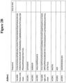
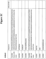
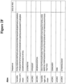
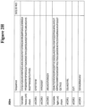
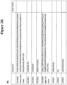
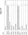
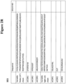
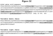
- ABD includes: a) a variable heavy chain that includes a vhCDR1, a vhCDR2 and a vhCDR3 of any one of the variable heavy chains depicted in Figure 2 ; and b) a variable light chain that includes a vlCDR1, a vlCDR2 and a vlCDR3 of any one of the variable light chains depicted in Figure 2 .
- the vhCDR1 includes a vhCDR1 sequence
- the vhCDR2 comprises a vhCDR2 sequence
- the vhCDR3 includes a vhCDR3 sequence according to any of the vhCDR1, vhCDR2, and vhCDR3 sequences depicted in Figure 2 .
- the variable heavy chain includes the sequence of any one of the variable heavy chains depicted in Figure 2 .
- the vlCDR1 includes a vlCDR1 sequence
- the vlCDR2 includes a vlCDR2 sequence
- the vlCDR3 includes a vlCDR3 sequence according to any of the vlCDR1, vlCDR2, and vhCDR3 sequences depicted in Figure 2 .
- the variable light chain includes the sequence of any one of the variable light chains depicted in Figure 2 .
- the albumin binding domain comprises the variable heavy chain and variable light chain of A10m3 ( Figure 2D ).
- composition that includes a variant IL-15.
- the variant IL-15 includes one or more amino acid substitutions selected from the group consisting of K86A, K86R, N112A, N112S, N112Q, K86A/N112A, K86R/N112A, K86A/N112S, K86R/N112S, K86A/N112Q, K86R/N112Q, K86A/N112A/N79A, K86R/N112A/N79A, K86A/N112A/N79D, K86R/N112A/N79D, K86A/N112A/N79Q, K86R/N112A/N79Q, K86A/N112A/N71D, K86R/N112A/N71D, K86A/N112A/N71Q, K86R/N112A/N71Q, K86A/N112A/N112A/N
- the variant IL-15 includes an amino acid sequence of any one of the variant IL-15s depicted in Figure 3 .
- the variant IL-15 further includes an IL-15 receptor alpha (IL-15R ⁇ ) attached to said IL-15.
- IL-15R ⁇ IL-15 receptor alpha
- an albumin binding domain (ABD) fusion protein that includes an ABD attached to a fusion partner.
- the ABD includes a variable heavy chain that includes a vhCDR1, a vhCDR2 and a vhCDR3 of any one of the variable heavy chains depicted in Figure 2 , and a variable light chain that includes a vlCDR1, a vlCDR2 and a vlCDR3 of any one of the variable light chains depicted in Figure 2 .
- the vhCDR1 includes a vhCDR1 sequence
- the vhCDR2 includes a vhCDR2 sequence
- the vhCDR3 includes a vhCDR3 sequence according to any of the vhCDR1, vhCDR2, and vhCDR3 sequences depicted in Figure 2
- the variable heavy chain includes the sequence of any one of the variable heavy chain sequences depicted in Figure 2 .
- the vlCDR1 includes a vlCDR1 sequence
- the vlCDR2 includes a vlCDR2 sequence
- the vlCDR3 includes a vlCDR3 sequence according to any of the vlCDR1, vlCDR2, and vhCDR3 sequences depicted in Figure 2
- the variable light chain includes the sequence of any one of the variable light chain sequences depicted in Figure 2 .
- variable heavy chain and the variable light chain include the variable heavy chain and variable light chain of A10m3, respectively ( Figure 2D ).
- the fusion partner is a cytokine.
- the cytokine is selected from: IL-2, IL-7, IL-12, IL-15, IL-18, IL-21, GM-CSF and IFN- ⁇ .
- the fusion partner is a binding moiety.
- the binding moiety is an scFv that includes an scFv variable heavy chain and an scFv variable light chain.
- the scFv is selected from: an anti-TGF ⁇ scFv, an anti-PD-L1 scFv, and an anti-TNF scFv.
- the scFv is an anti-interleukin scFv.
- the scFv is an anti-IL-1, IL-6, IL-8, IL-17(A-F) or IL-23 scFv.
- the ABD is attached to said fusion partner by a linker.
- the linker is (GGGGS) x , wherein x is an integer from 1-10
- an IL15-albumin binding domain (ABD) fusion protein according to the formula (IL-15)-L-(ABD).
- the ABD includes a variable heavy chain that includes a vhCDR1, a vhCDR2 and a vhCDR3 of any one of the variable heavy chains depicted in Figure 2 , and a variable light chain that includes a viCDR1, a vlCDR2 and a vlCDR3 of any one of the variable light chains depicted in Figure 2 , and L is a linker.
- the vhCDR1 includes a vhCDR1 sequence
- the vhCDR2 includes a vhCDR2 sequence
- the vhCDR3 includes a vhCDR3 sequence according to any of the vhCDR1, vhCDR2, and vhCDR3 sequences depicted in Figure 2
- the variable heavy chain includes the sequence of any one of the variable heavy chain sequences depicted in Figure 2 .
- the vlCDR1 includes a vlCDR1 sequence
- the vlCDR2 includes a vlCDR2 sequence
- the vlCDR3 includes a vlCDR3 sequence according to any of the vlCDR1, vlCDR2, and vhCDR3 sequences depicted in Figure 2
- the variable light chain includes the sequence of any one of the variable light chain sequences depicted in Figure 2 .
- variable heavy chain and the variable light chain include the variable heavy chain and variable light chain of A10m3, respectively ( Figure 2D ).
- IL15-ABD fusion protein according to any one of claims 22 to 27, wherein said IL-15 is a variant IL-15 comprising one or more amino acid substitutions selected from the group consisting of K86A, K86R, N112A, N112S, N112Q, K86A/N112A, K86R/N112A, K86A/N112S, K86R/N112S, K86A/N112Q, K86R/N112Q, K86A/N112A/N79A, K86R/N112A/N79A, K86A/N112A/N79D, K86R/N112A/N79D, K86A/N112A/N79Q, K86R/N112A/N79Q, K86A/N112A/N71D, K86R/N112A/N71D, K86A/N112A/N71Q, K86R/N112A/N71, K
- the IL-15 is a wild-type IL-15. In some embodiments, the IL-15 comprises a wild-type IL-15 attached to a IL-15 receptor alpha (IL-15R ⁇ ).
- IL-15R ⁇ IL-15 receptor alpha
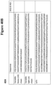
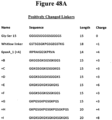
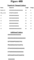
- the linker is selected from any of the linkers depicted in Figure 48 .
- the linker is (GGGGS) x , wherein x is an integer from 1-10.
- the IL15-ABD fusion protein has an amino acid sequence according to any one of the amino acid sequences depicted in Figure 4 .
- an IL12-albumin binding domain (ABD) fusion protein according to the formula (IL-12)-L-(ABD).
- the ABD includes a variable heavy chain that includes a vhCDR1, a vhCDR2 and a vhCDR3 of any one of the variable heavy chains depicted in Figure 2 , and a variable light chain that includes a vlCDR1, a vlCDR2 and a vlCDR3 of any one of the variable light chains depicted in Figure 2 , and L is a linker.
- the vhCDR1 includes a vhCDR1 sequence
- the vhCDR2 includes a vhCDR2 sequence
- the vhCDR3 includes a vhCDR3 sequence according to any of the vhCDR1, vhCDR2, and vhCDR3 sequences depicted in Figure 2
- the variable heavy chain includes the sequence of any one of the variable heavy chain sequences depicted in Figure 2 .
- the vlCDR1 includes a vlCDR1 sequence
- the vlCDR2 includes a vlCDR2 sequence
- the vlCDR3 includes a vlCDR3 sequence according to any of the vlCDR1, vlCDR2, and vhCDR3 sequences depicted in Figure 2
- the variable light chain includes the sequence of any one of the variable light chain sequences depicted in Figure 2 .
- variable heavy chain and the variable light chain include the variable heavy chain and variable light chain of A10m3, respectively ( Figure 2D ).
- the IL-12 is a single chain IL-12 that includes a p35 subunit, a p40 subunit and an IL-12 linker, and the IL-12 linker covalently attaches the p35 subunit to the p40 subunit.
- the linker is selected from any of the linkers depicted in Figure 48 .
- the linker is (GGGGS) x , where x is an integer from 1-10.
- the IL12-ABD fusion protein includes an amino acid sequence according to any one of the amino acid sequences of Figure 20 .
- n albumin binding domain (ABD) fusion protein having a formula, from N-terminus to C-terminus, selected from: a) (IL-12)-L1-(ABD)-L2-(IL-15); and b) (IL-15)-L1-(ABD)-L2-(IL-12).
- the ABD includes a variable heavy chain that includes a vhCDR1, a vhCDR2 and a vhCDR3 of any one of the variable heavy chains depicted in Figure 2 , and a variable light chain that includes a vlCDR1, a vlCDR2 and a vlCDR3 of any one of the variable light chains depicted in Figure 2 , and L1 and L2 are a first and second linker, respectively.
- the vhCDR1 includes a vhCDR1 sequence
- the vhCDR2 includes a vhCDR2 sequence
- the vhCDR3 includes a vhCDR3 sequence according to any of the vhCDR1, vhCDR2, and vhCDR3 sequences depicted in Figure 2
- the variable heavy chain includes the sequence of any one of the variable heavy chain sequences depicted in Figure 2 .
- the vlCDR1 includes a vlCDR1 sequence
- the vlCDR2 includes a vlCDR2 sequence
- the vlCDR3 includes a vlCDR3 sequence according to any of the vlCDR1, vlCDR2, and vhCDR3 sequences depicted in Figure 2
- the variable light chain includes the sequence of any one of the variable light chain sequences depicted in Figure 2 .
- variable heavy chain and the variable light chain include the variable heavy chain and variable light chain of A10m3, respectively ( Figure 2D ).
- the IL-15 includes a wild-type IL-15 polypeptide.
- the wild-type IL-15 is attached to an IL-15 receptor alpha (IL-15R ⁇ ).
- the IL-15 is a variant IL-15 that includes one or more amino acid substitutions selected from: K86A, K86R, N112A, N112S, N112Q, K86A/N112A, K86R/N112A, K86A/N112S, K86R/N112S, K86A/N112Q, K86R/N112Q, K86A/N112A/N79A, K86R/N112A/N79A, K86A/N112A/N79D, K86R/N112A/N79D, K86A/N112A/N79Q, K86R/N112A/N79Q, K86A/N112A/N71D, K86R/N112A/N71D, K86A/N112A/N71Q, K86R/N112A/N71Q, K86A/N112A/N71Q, K86A/N112A/N71
- the IL-12 is a single chain IL-12 that includes a p35 subunit, a p40 subunit and an IL-12 linker, and where the IL-12 linker attaches the p35 subunit to the p40 subunit.
- the first linker and second linker are each independently selected from any of the linkers depicted in Figure 48 .
- the linker is (GGGGS) x , where x is an integer from 1-10.
- an ABD fusion protein that includes an albumin binding domain (ABD), a cytokine, and a linker (L), according to the formula (cytokine)-L-(ABD).
- the ABD includes a variable heavy chain that includes a vhCDR1, a vhCDR2 and a vhCDR3 of any one of the variable heavy chains depicted in Figure 2 , and a variable light chain that includes a vlCDR1, a vlCDR2 and a vlCDR3 of any one of the variable light chains depicted in Figure 2 , and L is a linker.
- the cytokine is selected from: IL-2, IL-7, IL-12, IL-15, IL-18, IL-21, GM-CSF and IFN- ⁇ .
- the vhCDR1 includes a vhCDR1 sequence
- the vhCDR2 includes a vhCDR2 sequence
- the vhCDR3 includes a vhCDR3 sequence according to any of the vhCDR1, vhCDR2, and vhCDR3 sequences depicted in Figure 2
- the variable heavy chain includes the sequence of any one of the variable heavy chain sequences depicted in Figure 2 .
- the vlCDR1 includes a vlCDR1 sequence
- the vlCDR2 includes a vlCDR2 sequence
- the vlCDR3 includes a vlCDR3 sequence according to any of the vlCDR1, vlCDR2, and vhCDR3 sequences depicted in Figure 2
- the variable light chain includes the sequence of any one of the variable light chain sequences depicted in Figure 2 .
- variable heavy chain and the variable light chain include the variable heavy chain and variable light chain of A10m3, respectively ( Figure 2D ).
- the linker is selected from any of the linkers depicted in Figure 48 .
- the linker is (GGGGS) x , where x is an integer from 1-10.
- an ABD fusion protein according to the formula: (FP1)-L1-(ABD)-L2-(FP2), where ABD is an albumin binding domain that includes a variable heavy chain and a variable light chain; where FP1 and FP2 are a first fusion protein and a second fusion protein, respectively; and where L1 and L2 are a first and second linker, respectively.
- the ABD includes a variable heavy chain that includes a vhCDR1, a vhCDR2 and a vhCDR3 of any one of the variable heavy chains depicted in Figure 2 , and a variable light chain that includes a vlCDR1, a vlCDR2 and a vlCDR3 of any one of the variable light chains depicted in Figure 2 .
- the vhCDR1 includes a vhCDR1 sequence
- the vhCDR2 includes a vhCDR2 sequence
- the vhCDR3 includes a vhCDR3 sequence according to any of the vhCDR1, vhCDR2, and vhCDR3 sequences depicted in Figure 2
- the variable heavy chain includes the sequence of any one of the variable heavy chain sequences depicted in Figure 2 .
- the vlCDR1 includes a vlCDR1 sequence
- the vlCDR2 includes a vlCDR2 sequence
- the vlCDR3 includes a vlCDR3 sequence according to any of the vlCDR1, vlCDR2, and vhCDR3 sequences depicted in Figure 2
- the variable light chain includes the sequence of any one of the variable light chain sequences depicted in Figure 2 .
- variable heavy chain and the variable light chain include the variable heavy chain and variable light chain of A10m3, respectively ( Figure 2D ).
- FP1 and FP2 are a first cytokine and a second cytokine respectively.
- the first cytokine and said second cytokine are selected from IL-2 and IL-12; IL-7 and IL-15; IL-15 and IL-12; IL-18 and GM-CSF; IL-21 and IL-15; GM-CSF and IL-12; GM-CSF and IL-21; and IFN- ⁇ and IL-15.
- the first and second fusion partner are selected from: an anti-PD-L1 scFv and an IL-12; an anti-PD-L1 scFv and an IL-15; an anti-PD-L1 scFv and a anti-TGF ⁇ scFv; a first anti-PD-L1 scFv and a second PD-L1 scFv; an anti-TGF ⁇ scFv and an IL-12; an anti-TGF ⁇ scFv and an IL-15; a anti-TGF ⁇ scFv and a PD-L1 scFv; and a first anti-TGF ⁇ scFv and a second anti-TGF ⁇ scFv.
- An ABD fusion protein according to claim 68 wherein FP1 and FP2 are a first binding moiety and a second binding moiety, respectively.
- the first binding moiety and the second binding moiety are each an scFv.
- the first binding moiety and the second binding moiety are selected from: a TNF scFv and an IL-1 scFv; a TNF scFv and an IL-6 scFv; a TNF scFv and an IL-8 scFv; a TNF scFv and an IL-17 (isoforms A-F) scFv; TNF scFv and an IL-23 scFv;and a first TNF scFv and a second TNF scFv.
- first linker and second linker are each independently selected from any of the linkers depicted in Figure 48 .
- first linker and second linker are each independently (GGGGS) x , where x is an integer from 1-10.
- an albumin binding domain (ABD) fusion protein that includes a TGF ⁇ binding domain and an albumin binding domain.
- the albumin binding domain includes a ABD variable heavy chain and a ABD variable light chain having the amino acid sequences of any of the variable heavy chains and variable light chains in Figure 2 .
- the ABD variable heavy chain and said ABD variable light chain comprises the amino acid sequence of the variable heavy chain and variable light chain of A10m3.
- the TGF ⁇ binding domain is an scFv that includes a variable heavy chain and a variable light chain of 4D9 ( Figure 40B ).
- the ABD further includes an IL-12, an IL-15, a PD-L1 binding domain, or a second TGF ⁇ binding domain.
- an albumin binding domain (ABD) fusion protein that includes a PD-L1 binding domain and an albumin binding domain.
- the albumin binding domain includes a ABD variable heavy chain and a ABD variable light chain having the amino acid sequences of any of the variable heavy chains and variable light chains in Figure 2 .
- the ABD variable heavy chain and the ABD variable light chain comprises the amino acid sequence of the variable heavy chain and variable light chain of A10m3 ( Figure 2D ).
- the PD-L1 binding domain is an scFv comprising a variable heavy chain and a variable light chain of 10D12 ( Figure 50 ).
- the ABD further includes an IL-12, an IL-15, a TGF ⁇ binding domain, or a second PD-L1 binding domain.
- nucleic acid that encodes any of the albumin binding domains, variant IL-15s or ABD fusion proteins described herein, host cells that include any such nucleic acids, and methods of making such ABDs, variant IL-15s or ABD fusion proteins.
- a method of inhibiting or reducing a tumor in a subject in need thereof includes administering to the subject a ABD fusion protein.
- Biologics including cytokine and antibody based therapeutics are useful for the treatment of cancers.
- cytokine based therapeutics include those utilizing IL-2, IL-7, IL-12, IL-15, IL-18, IL-21 GM-CSF and IFN- ⁇ .
- IL-12 is capable of mediating immune effector functions in a manner compatible to enhancing pro-inflammatory, endogenous anti-tumor immune response.
- Boggio et al. J Exp Med 188:589-96 (1998 ); Cavallo et al., Cancer Res 59:414-21 (1999 ); Yu et al. Int Immunol 8:855-65 (1996 ); Nastala et al., J Immunal 153:1697:706 (1994 ); Brunda et al., J Exp Med 178. 1223-30 (1993 ).
- IL-12 is known to induce an inflammatory Th1 CD4+ T cell response as well as enhance CD8+ T cell cytotoxicity.
- T cell secretion of IFN ⁇ mediated by IL-12 may reverse T cell anergy and confer effector T cell resistance to immune suppressive regulatory T cells.
- the ability of IL-12 to not only activate the adaptive and the innate immune systems, but also further modulate the otherwise immune-hostile tumor microenvironment makes IL-12 an ideal candidate for tumor immunotherapy.
- IL-15 is capable of stimulating T cell proliferation inside tumors (see , e.g. , Miecnik et al., Sci Transl Med 6(228):228ra37 (2014 ) as well as extend the survivability of effector memory CD8+ T cells and is critical for the development of NK cells. Thus, it is believed that IL-15 can boost the potency of checkpoint inhibitors and other immunotherapies that harness T cells to attack cancer cells.
- IL-15 monomer has a short half-life of less than 40 minutes in vivo. Modifications to IL-15 monomer can improve its in vivo pharmacokinetics in the treatment of cancers.
- modifications have generally centered on improving the trans -presentation of IL-15 with the alpha subunit of IL-15 receptor, IL-15R ⁇ .
- modifications include: 1) pre-association of IL-15 and its soluble receptor a-subunit-Fc fusion to form IL-15: IL-15R ⁇ -Fe complex (see, e.g., Rubinstein et al., Proc Natl Acad Sci U.S.A.
- Monoclonal antibody based therapies including those that target tumor surface antigens and inhibitory signals that limit T-cell activation, have been a standard component of cancer therapeutics for over 20 years. See, e.g., Weiner, Nat Rev Cancer 15(6): 361-370 (2015 ).
- Short circulatory half-life represents a major obstacle for many biologics, including cytokine and antibody based therapeutics. See, e.g., Herrington-Symes et al., Advances in Bioscience and Biotechnology 4: 689-698 (2013 ) and Perdreau et al., European Cytokine Network 21: 297-307 (2010 ). Such short-acting therapeutics require frequent dosing profiles that can reduce applicability to the clinic, particular for chronic conditions. Long serum half-life is desirable as it would decrease the need for repetitive injections of the molecule to achieve a therapeutically relevant serum concentration.
- Methods of extending the half-life of therapeutic proteins include PEGylation, fusion to human serum albumin (HSA), fusion to the constant fragment (Fc) of a human immunoglobulin IgG, and fusion to non-structured fusion proteins such as XTEN. See, e.g., Stohl, BioDrugs 29(4): 215-239 (2015 ).
- Half-life extension technologies enable improved or new biologic therapies that reduce the cost and burden of frequent dosing.
- novel reagents and methods that can extend the half-lives of protein and peptide based therapeutics.
- albumin binding domain (ABD) fusion proteins useful for extending the half-lives of biologics (e.g ., interleukins and antibodies).
- Serum albumin possess a long half-life in the range of 2-4 weeks due to recycling through the neonatal Fc receptor (FcRn).
- Albumin is taken up by endothelial cells through macropinocytosis and binds to the FcRn in a pH-dependent manner in the acidic environment of the early endosome.
- Albumin-FcRn binding diverts albumin molecules from degradation in the lysosomal compartment and redirects the albumin molecules to the plasma membrane, where they are released back into the blood plasma due to the neutral pH.
- Albumin binding domains (ABDs) described herein do not compete with FcRN for albumin binding and bind albumin at a pH range that allows for the ABD to also undergo FcRn-driven endosomal albumin recycling when bound to albumin.
- biologics that include the subject albumin binding domain (ABD) are capable of evading lysosomal degradation using the albumin-FcRn pathway and, consequently, exhibit longer serum half-lives than counterparts lacking ABDs.
- Such ABD containing therapeutics advantageously localize to tumors, which are known to contain high levels of serum albumin.
- tumors which are known to contain high levels of serum albumin.
- ABD containing therapeutics are particularly useful for the treatment of cancers.
- compositions that include an albumin binding domain.
- serum albumin refers to a member of a family of globular proteins produced by the liver that functions primarily as a carrier protein of steroids, fatty acids, and thyroid hormones in the blood. Serum albumin also plays a major role in stabilizing extracellular fluid volume by contributing to oncotic pressure of plasma, and includes, but is not limited to, human serum albumin (HSA, Genbank Accession numbers: NM_000477 and NP_000468) and mouse serum albumin (MSA, Genbank accession numbers: NM_009654 and NP_0033784). The structure of albumin is characterized by several long ⁇ helices and contains eleven distinct binding domains for hydrophobic compounds. In humans, serum albumin is encoded by the ALB gene.
- Fusion proteins that include the subject albumin binding domain can bind serum albumin (SA), which allows the fusion protein to be taken in by cells by macropinocytosis.
- SA serum albumin
- the ABDs described herein bind at a pH range of about pH 5.5 to about pH 7.2.
- SA bound ABD fusion proteins bind to FcRn via SA at an acidic pH (e.g., pH 5.5), which in turn diverts the SA bound ABD fusion protein from the lysosome compartment of the cell and back to the plasma membrane.
- the SA dissociates from FcRn due to the neutral pH (e.g., pH 7.1-7.5) and the SA and ABD fusion protein are released back into the bloodstream.
- the neutral pH e.g., pH 7.1-7.5
- therapeutics that include the subject ABD are capable of binding to albumin at a pH range of about pH 5.5 to about pH 7.2, such therapeutics advantageously also undergo FcRn-driven endosomal albumin recycling and, thus, evade lysosomal degradation.
- therapeutics that include such ABDs advantageously exhibit longer serum half-lives than counterparts lacking ABDs.
- Such therapeutics are particular useful for the treatment of cancers, which are known to contain high levels of serum albumin.
- the albumin binding domain binds albumin at a site that does not interfere with SA binding to a neonatal Fc receptor (FcRn).
- FcRn neonatal Fc receptor
- FcRn neonatal Fc receptor
- a neonatal Fc receptor FcRn
- FcRn neonatal Fc receptor
- the FcRn may be from any organism, including but not limited to humans, mice, rats, rabbits, and monkeys.
- the functional FcRn protein comprises two fusion proteins, often referred to as the heavy chain and light chain. The light chain is beta-2-microglobulin and the heavy chain is encoded by the FcRn gene.
- FcRn or an FcRn protein refers to the complex of FcRn heavy chain with beta-2-microglobulin.
- the albumin binding domain described or exemplified herein preferably specifically binds to serum albumin (e.g ., HSA) at an epitope on the serum albumin molecule that does not participate in the interaction of the serum albumin molecule with the FcRn. Binding of the SA binding moiety to the serum albumin molecule thus preferably does not substantially interfere with, inhibit, prevent, or otherwise reduce binding of the serum albumin molecule ( e.g ., IISA) with the FcRn.
- the albumin binding domain does not compete with the FcRn for binding to the serum albumin molecule.
- the albumin binding domain does not sterically inhibit binding of serum albumin to the FcRn.
- the SA binding moiety does not change the conformation of the serum albumin molecule such that the albumin cannot interact with the FcRn.
- the albumin binding domain binds SA (e.g ., HSA) at a pH of 5.0 ⁇ 0.1, 0.2, 0.3, 0.4, 0.5, 0.6, 0.7, 0.8, 0.9, 1.0, 1.1, 1.2, 1.3, 1.4, 1.5, 1.6, 1.7, 1.8, 1.9, 2.0. 2.1, 2.2, .2.3, .2.4, .2.5, .2.6, 2.7, 2.8, 2.9, or 3.0.
- the albumin binding domain binds SA at a range of pH of about pH 5.5- about pH 7.2.
- the SA binding moiety binds SA at a pH of 5.5.
- the albumin binding domain is a human serum albumin (HSA) binding domain.
- HSA binding domains include, but are not limited to, albumin binding domains that can bind to an HSA molecule such as a whole HSA molecule or a fragment of an HSA.
- the HSA binding domain also binds mouse serum albumin.
- the HSA binding domain also binds cyno monkey albumin.
- the HSA binding domain does not bind to bovine serum albumin (BSA).
- BSA bovine serum albumin
- Albumin binding domains provided herein can include a variable heavy chain alone or a variable heavy chain in association with a variable light chain.
- the albumin binding domain includes a variable heavy chain.
- the variable heavy chain includes a vhCDR1, vhCDR2, and vhCDR3 (variable heavy chain Complementary Determining Regions 1-3).
- the antigen binding domain also includes a variable light chain.
- the variable light chain includes a vlCDR1, vlCDR2 and vlCDR3 (variable light chain Complementary Determining Regions 1-3).
- the albumin binding domain includes a variable heavy chain that includes the vhCDR1, vhCDR2, and vhCDR3 of any of the variable heavy chains depicted in Figure 2
- the albumin binding domain includes the vhCDR1, vhCDR2, and vhCDR3 of an A10m3 variable heavy chain, as shown in Figure 2D .
- the albumin binding domain includes the vhCDR1, vhCDR2, and vhCDR3 of A10m3 as shown in Figure 2D .
- the albumin binding domain also includes a variable light chain.
- the albumin binding domain includes a variable light chain that includes the vlCDR1, vlCDR2, and vlCDR3 of any of the variable light chains depicted in Figure 2 .
- the albumin binding domain includes the vlCDR1, vlCDR2, and vlCDR3 of an A10m3 variable light chain, as shown in Figure 2D .
- the albumin binding domain includes the vlCDR1, vlCDR2, and vlCDR3 of A10m3 as shown in Figure 2D .
- the albumin binding domain (e.g ., HSA binding domain) is an antibody or an antibody fragment. In some embodiments, the albumin binding domain (e.g ., HSA binding domain) is an scFv.
- the variable heavy chain and the variable light chain are attached to each other by a linker (e.g ., an scFv linker).
- the linker is attached to the variable heavy chain at its C-terminus and the variable light chain at its N-terminus. Suitable linkers are described herein and in Figure 48 .
- the linker is a (Gly4Ser) x linker, where x is 1, 2, 3, 4, 5, 6, 7, or 8.
- the linker is a (Gly4Ser) 5 linker.
- the albumin binding domain also includes a variable heavy chain that includes the vhCDR1, vhCDR2 and vhCDR3 of A10m3 and a variable light chain that includes the vlCDR1, vlCDR2 and vlCDR3 of A10m3 ( Figure 2D ).
- the albumin binding domain includes the variable heavy sequence and variable light sequence of the A10m3 ABD depicted in Figure 2D .
- compositions that include variant IL-15s with improved in vivo stability and/or biological activity as compared to wildtype IL-15.
- interleukin 15 refers to an interleukin that binds to and signals through a complex composed of an IL-15 specific receptor alpha chain, an IL-2/IL-15 receptor beta chain (CD122) and the common gamma chain (gamma-C, CD132) (Genbank Accession numbers: NM_00000585 and NP_000576 (human); and NM_001254747 and NP_001241676 (mouse)).
- IL-15 has been shown to stimulate T cell proliferation inside tumors (see, e.g., Miecnik et al., Sci Transl Med 6(228):228ra37 (2014 ). IL-15 also is able to extend the survivability of effector memory CD8+ T cells and is critical for the development of NK cells. It is believed that IL-15 can boost the potency of checkpoint inhibitors and other immunotherapies that harness T cells to attack cancer cells. Therefore, without being bound by any particular theory of operation, it is believed that the IL-15s described herein are useful for the treatment of cancers.
- IL-15 monomer however, has a short half-life of less than 40 minutes in vivo.
- Modifications to IL-15 monomer can improve its in vivo pharmacokinetics in the treatment of cancers. These modifications have generally centered on improving the trans -presentation of IL-15 with the alpha subunit of IL-15 receptor, IL-15R ⁇ .
- Such modifications include: 1) pre-association of IL-15 and its soluble receptor a-subunit-Fc fusion to form IL-15: IL-15R ⁇ -Fe complex ( see, e.g., Rubinstein et al., Proc Natl Acad Sci U.S.A.
- the IL-15 is a variant of a parental IL-15 with increased stability as compared to wildtype IL-15.
- the variant IL-15 is a variant of a wildtype human IL-15.
- the variant IL-15 includes an amino acid substitution at position K86 of the parental IL-15 shown in Figure 3 .
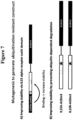
- K86 is a putative site for ubiquitin-dependent degradation ( See Example 2) when made using particular cell types ( e.g ., HEK293 T cells). Therefore, without being bound by any particular theory of operation, it is believed that removal of the K86 ubiquitination site by amino acid substitution improves the stability of IL-15 ( See Examples 2 and 3).
- the IL-15 is a variant IL-15 having an amino acid substitution at position N112.
- Amino acid position N112 is a key site for IL-15 bioactivity, as it is critical for a proper IL-15/IL-15 receptor gamma interaction, particularly when IL-15 is attached to an ABD. Therefore, without being bound by any particular theory of operation, it is believed that mutations at position N112 can enhance one or more functions of IL-15 including, but not limited to, promoting T cell proliferation in tumor environments, enhancing survivability of CD8+ T cells and promoting NK cell development.
- Particular amino acid substitutions that can improve IL-15 in vivo stability and/or biological activity include, but are not limited to: K86A, K86R, N112A, N112S, N112Q, K86A/N112A, K86R/N112A, K86A/N112S, K86R/N112S, K86A/N112Q, K86R/N112Q, K86A/N112A/N79A, K86R/N112A/N79A, K86A/N112A/N79D, K86R/N112A/N79D, K86A/N112A/N79Q, K86R/N112A/N79Q, K86A/N112A/N71D, K86R/N112A/N71D, K86A/N112A/N71Q, K86R/N112A/N71Q, K86A/N112A/N71Q, K86A/N
- the IL-15 described herein is attached to IL-15R alpha.
- Such IL-15 presented in trans with its receptor has been shown to have a prolonged half-life and higher potency as compared to native Il-15 alone. See, e.g., Wu, J Mol Genet Med 7, 85 (2013 ).
- compositions that include IL-12.
- interleukin 12 “interleukin 12”, “IL-12” and “IL12” all refer to an interleukin that is a heterodimeric cytokine encoded by the IL-12A and IL-12B genes (Genbank Accession numbers: NM_000882 (IL-12A) and NM_002187 (IL-12B)).
- IL-12 is composed of a bundle of four alpha helices and is involved in the differentiation of native T cells into TH1 cells.
- IL-12 binds to the IL-12 receptor, which is a heterodimeric receptor formed by IL-12R- ⁇ 1 and IL-12R- ⁇ 2.
- IL-12 is known as a T cell-stimulating factor that can stimulate the growth and function of T cells.
- IL-12 can stimulate the production of interferon gamma (IFN- ⁇ ), and tumor necrosis factor-alpha (TNF- ⁇ ) from T cells and natural killer (NK) cells and reduce IL-4 mediated suppression of IFN- ⁇ .
- IFN- ⁇ interferon gamma
- TNF- ⁇ tumor necrosis factor-alpha
- IL-12 can further mediate enhancement of the cytotoxic activity of NK cells and CD8+ cytotoxic T lymphocytes.
- IL-12 can also have anti-angiogenic activity by increasing production of interferon gamma, which in turn increases the production of the chemokine inducible protein-10 (IP-10 or CXCL10). IP-10 then mediates this anti-angiogenic effect.
- IP-10 chemokine inducible protein-10
- the IL-12 is a mouse IL-12. In other embodiments, the IL-12 is a human 1L-12.
- the IL-12 is a single chain IL-12 polypeptide comprising an IL-12 p35 subunit attached to an IL-12 p40 subunit.
- Such IL-12 single chain polypeptides advantageously retain one or more of the biological activities of wildtype IL-12.
- the single chain IL-12 polypeptide described herein is according to the formula, from N-terminus to C-terminus, (p40)-(L)-(p35), wherein "p40" is an IL-12 p40 subunit, "p35” is IL-12 p35 subunit and L is a linker.
- the single chain IL-12 is according to the formula from N-terminus to C-terminus, (p35)-(L)-(p40).
- Any suitable linker can be used in the single chain IL-12 polypeptide including those described herein and disclosed in Figure 49C .
- Suitable linkers can include, for example, linkers having the amino acid sequence (GGGGS) x wherein x is an integer from 1-10.
- Other suitable linkers include, for example, the amino acid sequence GGGGGGS.
- Exemplary single chain IL-12 linkers than can be used with the subject single chain IL-12 polypeptides are also described in Lieschke et al., Nature Biotechnology 15: 35-40 (1997 ), which is incorporated herein in its entirety by reference and particularly for its teaching of IL-12 polypeptide linkers.
- the single chain IL-12 polypeptide is a single chain human IL-12 polypeptide (i.e., it includes a human p35 and p40 IL-12 subunit).
- the single chain IL-12 polypeptide is a single chain mouse IL-12 polypeptide. Exemplary single chain human and mouse IL-12s are depicted in Figures 20 (shown as fusion peptide with ABD) and 49C.
- ABD compositions that include an albumin binding domain attached to one or more fusion partners (e.g ., a first fusion partner, a second fusion partner, etc.) via a linker.
- fusion partners e.g ., a first fusion partner, a second fusion partner, etc.
- ABDs useful for such ABD fusion proteins include, but are not limited to, those described herein.
- the amino acids sequences of such ABDs including vhCDR1-3, vlCDR1-3, variable heavy chain and variable light chain sequences, are disclosed, for example, in Figure 2 .
- the ABD fusion protein includes a variable heavy chain that includes the vhCDR1, vhCDR2 and vhCDR3 of any of the ABD variable heavy chains in Figure 2 , and a variable light chain that includes the vlCDR1, vlCDR2 and vCDR3 of any of the ABD variable light chains in Figure 2 .
- the ABD includes a variable heavy chain having a vhCDR1, a vhCDR2 and a vhCDR3 of an ABD disclosed in Figure 2 and a variable light chain having a vhCDR1, a vhCDR2 and a vhCDR3 of an ABD disclosed in Figure 2 .
- the ABD fusion protein includes the variable heavy chain and variable light chain of an ABD disclosed in Figure 2 .
- the ABD fusion protein includes a variable heavy chain that includes the vhCDR1, vhCDR2 and vhCDR3 of the A10m3 variable heavy chain, and a variable light chain that includes the vlCDR1, vlCDR2 and vCDR3 of the A10m3 variable light chain ( Figure 2D ).
- the ABD includes a variable heavy chain having a vhCDR1, a vhCDR2 and a vhCDR3 of A10m3 and a variable light chain having a vhCDR1, a vhCDR2 and a vhCDR3 of A10m3 ( Figure 2D ).
- the ABD fusion protein includes the variable heavy chain and variable light chain of A10m3 ( Figure 2D ).
- ABD fusion proteins described herein include a fusion partner.
- the fusion partner include two fusion partners (a first fusion partner (FP1) and a second fusion partner (FP2).
- the fusion partners can be attached to the ABD in several orientations.
- the ABD fusion protein is according to the formula, from N-terminus to C-terminus: FP1-ABD-FP2, FP1-PF2-ABD or ABD-FP1-FP2, wherein FP1 is a first fusion partner and FP2 is a second fusion partner.
- Fusion partners may include, for example, cytokines (e.g ., interferons and interleukins), growth factors, polypeptides, proteins, and hormones (e.g ., growth hormones, parathyroid hormone).
- cytokines e.g ., interferons and interleukins
- growth factors e.g ., growth factor, polypeptides, proteins, and hormones (e.g ., growth hormones, parathyroid hormone).
- the fusion partner is an antibody-based binding moiety that includes a variable heavy chain and a variable light chain. Such binding moieties can bind to any target of interest including, for example, tumor specific targets or cytokines.
- the fusion partner is a single chain variable fragment (scFv).
- Antibody-based fusion partners also include, but are not limited to, ds-scFv, single domain antibodies (sdAb), diabodies, dsFvs, ds-scFvs, Fabs, and full length antibodies.
- Antibody-based fusion partners also include multispecific (e.g ., bispecific) antibodies and fragments.
- the subject ABD fusion proteins described herein utilize linkers between components (ABD and fusion partners) and within components.
- scFv fusion partners utilize standard peptide linkers, generally based on glycine and serine, to attach the variant heavy and light chains to form the scFv.
- standard peptide linkers are utilize to attached ABDs to fusion partners (e.g ., cytokine fusion partners).
- linkers are used to attach components of particular moieties, for example, the p35 and p40 subunit of IL-12 and IL-15 with IL-15R ⁇ .
- the linker is a (Gly4Ser) x linker, where x is 1, 2, 3 ,4 ,6, 7 or 8.
- the ABD is connected to fusion partners by (Gly4Ser) 5 linkers.
- linker peptide may predominantly include the following amino acid residues: Gly, Ser, Ala, or Thr.
- the linker peptide should have a length that is adequate to link two molecules in such a way that they assume the correct conformation relative to one another so that they retain the desired activity.
- the linker is from about 1 to 50 amino acids in length, preferably about 1 to 30 amino acids in length.
- linkers of 1 to 20 amino acids in length may be used, with from about 5 to about 10 amino acids finding use in some embodiments.
- Useful linkers include glycine-serine polymers, including for example (GS)n, (GSGGS)n, (GGGGS)n, and (GGGS)n, where n is an integer of at least one (and generally from 3 to 4), glycine-alanine polymers, alanine-serine polymers, and other flexible linkers.
- glycine-serine polymers including for example (GS)n, (GSGGS)n, (GGGGS)n, and (GGGS)n, where n is an integer of at least one (and generally from 3 to 4), glycine-alanine polymers, alanine-serine polymers, and other flexible linkers.
- nonproteinaceous polymers including but not limited to polyethylene glycol (PEG), polypropylene glycol, polyoxyalkylenes, or copolymers of polyethylene glycol and polypropylene glycol, may find use as linkers, that is may find use as linkers.
- linker sequences may include any sequence of any length of CL/CH1 domain but not all residues of CL/CH1 domain; for example the first 5-12 amino acid residues of the CL/CH1 domains.
- Linkers can be derived from immunoglobulin light chain, for example C ⁇ or C ⁇ .
- Linkers can be derived from immunoglobulin heavy chains of any isotype, including for example C ⁇ 1 Cy2, Cy3, C ⁇ 4, C ⁇ 1, C ⁇ 2, C ⁇ , C ⁇ , and C ⁇ .
- Linker sequences may also be derived from other proteins such as Ig-like proteins (e.g., TCR, FcR, KIR), hinge region-derived sequences, and other natural sequences from other proteins.
- linker is not substantially sensitive to the extracellular environment.
- "not substantially sensitive to the extracellular environment” in the context of a linker means that no more than about 20%, 15%, 10%, 5%, 3%, or no more than about 1% of the linkers, in a sample of antibody-drug conjugate compound, are cleaved when the antibody-drug conjugate compound presents in an extracellular environment (for example, in plasma).
- Whether a linker is not substantially sensitive to the extracellular environment can be determined, for example, by incubating with plasma the antibody-drug conjugate compound for a predetermined time period (for example, 2, 4, 8, 16, or 24 hours) and then quantitating the amount of free drug present in the plasma.
- a predetermined time period for example, 2, 4, 8, 16, or 24 hours
- the linker promotes cellular internalization. In certain embodiments, the linker promotes cellular internalization when conjugated to the therapeutic agent (that is, in the milieu of the linker-therapeutic agent moiety of the antibody-drug conjugate compound as described herein). In yet other embodiments, the linker promotes cellular internalization when conjugated to both the auristatin compound and the ABD fusion proteins of the invention.
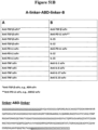
- Exemplary linkers that can be utilized with the subject ABDs as domain linkers, scFv linkers, as well as attach components of particular fusion partners are further depicted in Figure 48 .
- the linker is a "domain linker", used to link any two domains as outlined herein together (e.g., a cytokine fusion partner (e.g., an interleukin) and an ABD). While any suitable linker can be used, many embodiments utilize a glycine-serine polymer, including for example (GS)n, (GSGGS)n, (GGGGS)n, and (GGGS)n, where n is an integer of at least one (and generally from 3 to 4 to 5) as well as any peptide sequence that allows for recombinant attachment of the two domains with sufficient length and flexibility to allow each domain to retain its biological function.
- a cytokine fusion partner e.g., an interleukin
- ABD a cytokine fusion partner
- the fusion partner is an scFv that includes a variable heavy chain and a variable light chain.
- the ABD variable heavy chain is attached to the variable light chain with an scFv linker.
- ABD fusion proteins are further discussed below.
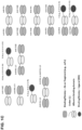
- the ABD fusion protein includes a cytokine fusion partner, i.e., a cytokine-albumin binding domain (cytokine-ABD) fusion proteins ( Figure 1 ).
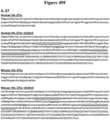
- the cytokine-ABD fusion protein includes an IL-2, IL-7, IL-12, IL-15, IL-18, IL-21, IL-27, GM-CSF or IFN- ⁇ .
- Subject cytokine-ABD fusion proteins are useful for immunomodulatory effects in a subject where such immunomodulatory effects are needed (e.g ., treatment of a cancer or an autoimmune disease).
- subject cytokine-ABDs exhibit a longer half-life and improved pharmacokinetic properties as compared to interleukin therapeutics alone.
- the ABD includes a ABD variable heavy chain. In certain embodiments the ABD includes a ABD variable light chain. In exemplary embodiments, the ABD is an scFv that includes a variable heavy chain attached to a variable light chain by a linker ( e.g ., any one of the linkers disclosed herein and in Figure 48 ).
- a linker e.g ., any one of the linkers disclosed herein and in Figure 48 .
- variable heavy chain includes the vhCDR1-3 of any of the ABD variable heavy chains described herein, including those ABD variable heavy chains depicted in Figure 2 .
- the ABD variable heavy chain includes the vhCDR1-3 of the A10m3 variable heavy chain ( Figure 2D ).
- the ABD variable heavy chain includes the A10m3 vhCDR1-3 as depicted in Figure 2D .
- the ABD variable heavy chain has the amino acid sequence of A10m3 variable heavy chain.
- the ABD includes a ABD variable light chain.
- the ABD variable light chain includes the vlCDR1-3 of any of the ABD variable light chains described herein, including those ABD variable light chains depicted in Figure 2 .
- the ABD variable light chain includes the vlCDR1-3 of the A10m3 variable light chain ( Figure 2D ).
- the ABD variable light chain includes the A10m3 vlCDR1-3 as depicted in Figure 2D .
- the ABD variable light chain has the sequence of A10m3 variable light chain.
- the IL-ABD is according to the formula, from N-terminus to C-terminus, cytokine-L-ABD or ABD-L-cytokine, where L is a linker that attaches the cytokine to the ABD (e.g., a peptide linker).
- the cytokine -ABD includes a variable heavy chain and the cytokine is attached to the N-terminal of the ABD variable heavy chain. In some embodiments, the cytokine is attached to the C-terminal of the ABD variable heavy chain.
- the interleukin-ABD includes an ABD that also includes a variable light chain (e.g ., an ABD scFv).
- the cytokine is attached to the C-terminal of the variable light chain.
- the N-terminal of the cytokine is attached to the ABD.
- the C-terminal of the cytokine is attached to the ABD.
- the cytokine is cytokine selected from IL-2, IL-7, IL-12, IL-15, IL-18, IL-21, IL-27, GM-CSF and IFN- ⁇ .
- Cytokine molecules include, for example, full length cytokine and cytokine fragments, for example, a part that is important for the particular cytokine's function (e.g ., a part of the interleukin that binds to its receptor).
- the cytokine-ABD includes an IL-2 molecule or fragment thereof.
- IL-2 interleukin 2
- IL2 refers to a member of the cytokine having four alpha helix bundles and signals through the IL-2 receptor (Genbank Accession numbers: NM_000586 and NP_000577 (human) and NM_008366 and NP_032392 (mouse)).
- IL-2 plays key roles in immune system function, tolerance and immunity, primarily via its direct effects on T cells.
- IL-2 prevents autoimmune diseases by promoting the differentiation of certain immature T cells into regulatory T cells, which kill off other T cells that are primed to attack normal healthy cells in the body.
- IL-2 has been used for the treatment of cancers (malignant melanoma, renal cell cancer) in large intermittent doses and has been extensively used in continuous doses.
- Exemplary IL-2-ABDs are shown in Figure 49A .
- the cytokine-ABD fusion protein includes an IL-7 molecule or fragment thereof.
- IL-7 interleukin 7
- IL-7 Interleukin 7
- IL7 Interleukin 7
- IL-7 is a non-hematopoietic cell-derived cytokine with a central role in the adaptive immune system.
- IL-7 is an ideal solution for the immune reconstitution of immunosuppressed cancer patients by promoting peripheral T cell expansion. In animal models, IL-7 has been proven to prolong the survival of tumor-bearing hosts. Exemplary IL-7-ABDs are shown in Figure 49B .
- cytokine-ABD includes a an IL-12 molecule or fragment thereof.
- the IL-12 is a single chain IL-12 as described herein and as depicted in Figures 20 (shown as a fusion peptide with ABD) and 49C.
- the cytokine-ABD includes an IL-15 molecule or fragment thereof.
- the IL-15-ABD includes a variant IL-15 as described herein and at Figures 3 and 4 .
- the IL-15 is a wildtype IL-15, as shown in Figure 3 ( e.g ., "parental IL-15").
- the wild-type IL-15 is attached to an IL-15 receptor alpha (IL-15R ⁇ ).
- the cytokine-ABD includes an IL-21 molecule or fragment thereof.
- interleukin 21 “IL-21”, and “IL21” (Genbank Accession numbers: NM_001207006 and NP_001193935 (human); and NM_0001291041 and NP_001277970 (mouse)) all refer to a member of a cytokine that binds to IL-21 receptor and has potent regulatory effects on cells of the immune system, including natural killer (NK) cells and cytotoxic cells and binds to IL-21 receptor that can destroy virally infected or cancerous cells. Therefore, without being bound by any particular theory of operation, it is believed that the subject cytokine-ABD having an IL-21 are useful for the treatment of various cancers. Exemplary IL-21-ABDs are shown in Figure 49E .
- cytokine-ABDs include, not are not limited to IL-27, IFN- ⁇ and GM-CSF.
- the interleukin-ABD polypeptide also includes an interferon-alpha, interferon-beta or GM-CSF molecule. Such molecules are useful for delivery into tumors and reduction of toxicity.
- the cytokine-ABD includes two cytokines attached to an ABD selected from the following: IL-2 and IL-12; IL-7 and IL-15;IL-15 and IL-12; IL-18 and GMC SF; IL-21 and IL-15; GMC-SF and IL-12; GMC-SF and IL-21; and IFN- ⁇ and IL-15.
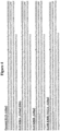
- Figure 1 depicts several exemplary orientations in which two cytokines can be attached to the ABD. Any ABD can be used in such fusion protein, including those that include any of the ABD variable heavy domains and variable light domains shown in Figure 2 .
- the ABD is A10m3.
- Any linker can be used to attached each cytokine to the ABD, including any linker depicted in Figure 48 .
- An exemplary backbone linker-A10m3-linker sequence for use in such fusion proteins is depicted in Figure 50 .
- Exemplary linkers than can be used include (GGGGS) x linkers wherein X is 1-10.
- a (GGGGS) 5 is used to attach the cytokine to the ABD.
- the cytokine-ABD is according to a formula selected from the following: (from N-terminus to C-terminus): Cytokine 1-L1-ABD-L2-Cytokine 2; Cytokine 1-L1-Cytokine 2-L2-ABD; and ABD-L1-Cytokine 1-L2- Cytokine 2, where L1 and L2 are linkers that connect the cytokines and ABD components and Cytokine 1 and Cytokine 2 are selected from the following cytokine pairs: IL-2 and IL-12; IL-7 and IL-15;IL-15 and IL-12; IL-18 and GM-CSF; IL-21 and IL-15; GM-CSF and IL-12; GM-CSF and IL-21; and IFN- ⁇ and IL-15.
- the ABD is A10m3 ( Figure 2D ).
- either cytokine can be "Cytokine 1" and the other cytokine can be "Cytokine 2".
- cytokine 1 the cytokine 1
- IL-12 the cytokine 2
- IL-12 is "Cytokine 1"
- IL-2 is "Cytokine 2”.
- the cytokine-ABD include two of the same interleukins.
- the interleukin-ABD polypeptide includes two different interleukins (e.g ., an IL-12 and an IL-15), as described above.
- ABD fusion proteins that include particular cytokine combinations are disclosed in Figure 51 .
- Exemplary cytokine-ABD fusion proteins that include a combination of different cytokines are further discussed in detail below.
- the cytokine-ABD fusion protein includes an IL-12 and an IL-15.
- Exemplary IL-12 and IL-15 sequences that can be included in such embodiments are depicted in Figures 3 and 49C . It is believed that cytokine-ABDs that include both an IL-12 and IL-15 fusion partner exhibit the anti-tumor effects of both interleukins. The combination of IL-12 and IL-15 was shown to induce enhanced anti-tumor activity as compared to either cytokine alone. Such enhanced anti-tumor activity was correlated with the reciprocal upregulation of each cytokine's receptors through the synergistic induction of IFN- ⁇ .
- IL-12 in combination with IL-15 was further shown to promote anti-tumor activity in peritoneal macrophages through the synthesis of nitric oxide. Without being bound by any particular theory of operation, it is believed that polypeptides having both an IL-12 and an IL-15 fusion partner are capable of rapidly activate the innate response (IL-12) as well as potently stimulate the proliferation of T cells and maintain memory CD8+ T cells (IL-15). Animal studies show that sequential delivery of IL-12 and IL-15 expressing cells also cured mice in a therapeutic setting of established tumor. Depleting CD8+ cells eliminated the protection of this therapy, suggesting clonal expansion of tumor CTL. See, e.g., Croce et al., Clin Cancer Res 11(2 Pt 1):735-742 (2005 ).
- the cytokine-ABD is according to the formula, from N-terminus to C-terminus:
- any suitable ABD can be used including, for example, an ABD having a ABD variable heavy chain that includes the vhCDR1-3 of any of the ABD variable heavy chains listed in Figure 2 .
- the variable heavy chain includes the vhCDR1-3 of a A10m3 variable heavy chain ( Figure 2D ).
- the ABD includes a ABD variable light chain that includes the vlCDR1-3 of a A10m3 variable light chain.
- the ABD is an A10m3 scFv.
- the IL-15 is a variant IL-15 as described herein ( see, e.g., Figure 3 ).
- the IL-15 can also be a wild-type IL-15 or a wild-type IL-15 that is attached to an IL-15R ⁇ .
- the IL-15 is a variant IL-15 selected from those depicted in Figure 3 .
- the Il-15 is a variant IL-15 having amino acid substitutions K86R and N112A.
- IL-12s that can be used include those IL-12s that have a p35 and p40 domain.
- the IL-12 is a single chain IL-12 as described herein ( see, e.g., Figure 20 , as shown as part of an IL-12-ABD fusion polypeptide and Figure 49C ).
- L1 and L2 are a first and second linker, respectively, L1 and L2 can be any linker that is suitable for attaching the IL-15 and IL-12 domains to the ABD domain ( e.g ., the linkers listed in Figure 48 ).
- Exemplary linkers than can be used include (GGGGS) x linkers wherein X is 1-10.
- L1 and L2 are each (GGGGS) 5 .
- the cytokine-ABD includes an IL-2 and an IL-12.
- IL-2 and IL-12 reciprocally upregulate each other's receptors and use separate signaling pathways to induce different but complementary biological effects.
- Both IL-2 and IL-12 can stimulate mitogen or CD3-activated T cells to proliferate & produce IFN- ⁇ .
- studies show that the delivery of both IL-2 and IL-12 genes into mice bearing B16 melanoma elicited a significant reduction in tumor burden and enhancement in overall survival ( see, e.g., Dietrich et al., Arch Surg 387(34):177-182 (2002 )).
- cytokine-ABD fusion proteins having both an IL-2 and an IL- are useful for the reduction of tumors and treatment of cancers.
- the cytokine-ABD fusion protein includes an IL-2 and an IL-15. Both IL-2 and IL-15 are capable of stimulating the proliferation of NK cells and activated T cells, as well as supporting the expansion of effector T cells. It is believed that cytokine-ABD fusion proteins that include both an IL-2 and IL-15 are useful for the reduction of tumors and treatment of cancers.
- the cytokine-ABD fusion protein includes an IL-7 and an IL-12 fusion partner.
- Interleukin-7 is a non-hematopoietic cell-derived cytokine with a central role in the adaptive immune system. It promotes lymphocyte development in the thymus maintains survival of naive and memory T cell homeostasis in the periphery. Moreover, it is important for the organogenesis of lymph nodes (LN) and for the maintenance of activated T cells recruited into the secondary lymphoid organs (SLOs).
- LN lymph nodes
- SLOs secondary lymphoid organs
- the immune capacity of cancer patients is suppressed and is characterized by lower T cell counts, less effector immune cells infiltration, higher levels of exhausted effector cells and higher levels of immunosuppressive cytokines, such as transforming growth factor ⁇ (TGF- ⁇ ).
- TGF- ⁇ transforming growth factor ⁇
- IL-7 is an ideal solution for the immune reconstitution of immunosuppressed cancer patients by promoting peripheral T cell expansion. In animal models, IL-7 has been proven to prolong the survival of tumor-bearing hosts. See Gao et al., Int. J. Mol. Sci. 16: 10267-10280 (2015 ).
- IL-12 acts directly on CD8+ T cells to enhance their IL-7 mediated proliferation. It is believed that cytokine-ABD fusion proteins that include an IL-7 and IL-12 binding domain advantageously promote the proliferation of CD8+ T cells and enhance cytolytic activity against tumors.
- the cytokine-ABD fusion protein includes an IL-7 and an IL-15.
- interleukin 7 and 15 are considered powerful pro-inflammatory cytokines that have the ability to reduce tumorgenesis. It is believed that cytokine ABD fusion proteins that include both an IL-7 and IL-15 are useful for the reduction of tumors and treatment of cancers.
- the cytokine-ABD includes an IL-12 and an IL-21.
- IL-12 is capable of stimulating the proliferation of NK cells and activated T cells and supporting the expansion of effector T cells.
- IL-21 is a regulator of NK and T cell function that bridges innate and adaptive immune systems. IL-21 promotes the maturation of NK cells from bone marrow progenitors, activates human peripheral NK cells, promotes NK expansion and maturation, and enhances CD8+ T cell-mediated effector functions.
- cytokine-ABD fusion proteins that include both an IL-12 and IL-21 are useful for the reduction of tumors and treatment of cancers.
- the cytokine-ABD includes an IL-12 and an IL-18.
- IL-18 is known to induce IFN- ⁇ production, promote Th1 cell development and NK activation.
- IL-12 is known to induce the upregulation of the IL-18 receptor in an IFN- ⁇ -dependent manner.
- Administration of SCK murine mammary carcinoma cells co-expressing IL-18 and IL-12 to mice reduced tumor burden and inhibited angiogenesis (see, e.g., Coughlin et al., J Clin Invest 101 (6):1441-1452 (1998 )).
- IL-18 in combination with IL-12 to tumor bearing mice synergistically induced a prolonged serum level of IFN-y, while tumor bearing mice treated with IL-18 or IL-12 alone induced minimal serum IFN- ⁇ that was rapidly attenuated ( See, e.g. , Subleski et al., Cancer Res 66(22):11005-11012 (2006 )).
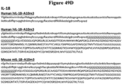
- Exemplary IL-18-ABDs are shown in Figure 37 .
- cytokine-ABD fusion proteins that include both an IL-12 and IL-18 fusion partner are useful for the reduction of tumors and treatment of cancers.
- the cytokine-ABD includes a GM-CSF and an IL-18.
- GM-CSF regulates hematopoietic progenitor cell differentiation and proliferation.
- GM-CSF also enhances the capacity of APC to process and present antigen, which in turn leads to the activation of cytotoxic T cells, increased IFN- ⁇ production and, ultimately, tumor regression.
- Both GM-CSF and IL-12 are able to elicit significant anti-tumor responses in several different preclinical tumor models, including a liver tumor model and a lung tumor model. ( See, e.g. , Kilinc et al., J Immunol 177(10):6962-6973 (2006 )).
- cytokine-ABD fusion proteins that include both a GM-CSFand IL-12 fusion partner are useful for the reduction of tumors and treatment of cancers.
- IL-12 which is well known to induce IFN- ⁇ production, can lead to the production of additional soluble factors that enhance IFN- ⁇ signaling.
- Interferons including, interferon alpha, are known to induce apoptosis in malignant cells. See, e.g., Thyrell, L. et al., Oncogene 21, 1251-1262 (2002 ).
- cytokine-ABD fusion proteins that include both an IFN alpha and IL-12 are useful for the reduction of tumors and treatment of cancers.
- cytokine-cytokine combinations that can be included in the subject cytokine-ABD fusion proteins are depicted in Figure 51A .
- the ABD fusion proteins includes a binding moiety (e.g., an scFv) fusion partner, i.e., a BM-ABD fusion protein.
- the BM-ABD includes one binding moiety.
- the BM-ABD includes two binding moieties (e.g ., scFvs).
- the BM-ABD includes a cytokine and a binding moiety ( Figure 1B ).
- Figure 1 depicts several exemplary orientations in which the binding moiety or binding moiety-cytokine/binding moiety-binding moiety combinations can be attached to the ABD.
- binding moieties that are useful for practice with the subject BM-ABDs binding moieties that are based on antibody variable heavy domain and variable light domain.
- the binding moiety includes a variable heavy domain and a variable light domain.
- the binding moiety is a single chain variable fragment (scFv).
- any ABD can be used in such fusion protein, including those that include any of the ABD variable heavy domains and variable light domains shown in Figure 2 .
- the ABD includes the variable heavy and light domains of A10m3.
- the ABD is A10m3 scFv.
- any linker can be used to attached the cytokine and/or binding moiety to the ABD, including any linker depicted in Figure 48 .
- the linker is (GGGGS) 5 .
- An exemplary backbone linker-A10m3-linker sequence for use in such fusion proteins is depicted in Figure 48 .
- binding moieties e.g ., scFvs
- cytokine e.g., IL-4 kine
- binding moiety that can be such ABD fusion proteins
- the BM-ABD includes an anti-TGF ⁇ binding domain (e.g., an anti-TGF ⁇ scFv). Exemplary anti-TGF ⁇ scFv sequences are depicted in Figure 45 .
- the anti-TGF ⁇ scFv includes the variable heavy and variable light domain of 4D9 anti-TGF ⁇ scFv.
- the ABD fusion protein includes an anti-TGF ⁇ scFv and a cytokine or additional binding moiety, where the cytokine or additional binding moiety is a second anti-TGF ⁇ scFv, IL-15, IL-12, or an anti-PD-L1 binding domain (10D12). See Figure 51B .
- the BM-ABD includes an anti-PD-L1 binding domain (e.g., an anti-T PD-L1 scFv).
- the anti- PD-L1 scFv includes the variable heavy and variable light domain of 4D9 anti- PD-L1 10D12 scFv.
- the ABD fusion protein includes an anti- PD-L1 scFv and a cytokine or additional binding moiety, where the cytokine or additional binding moiety is a second anti-anti- PD-L1 scFv, IL-15, IL-12, or an anti- TGF ⁇ binding domain (4D9). See Figure 51B .
- the BM-ABD provided herein includes a TGF- ⁇ binding moiety.
- TGF- ⁇ TGF- ⁇
- TGF ⁇ TGF ⁇
- TGFb transforming growth factor beta
- TGF ⁇ 1 Genbank Accession numbers: NM_000660 and NP_000651 (human); and NM_011577 and NP_035707 (mouse)
- TGF ⁇ 2 Genbank Accession numbers NM_001135599 and NP_001129071 (human)
- NM_009367 and NP_33393 mimouse
- TGF ⁇ family members have an N-terminal signal peptide of 20-30 amino acids that are required for secretion from cells, a pro-region, and a 112-113 amino acid C-terminal region that becomes the mature TGF ⁇ molecule following its release form the pro-region by proteolytic cleavage.
- the mature TGF ⁇ protein can dimerize to produce a 25kDa active molecule with many conserved structural motifs, include nine cysteine residues, eight of which form disulfide bonds within the TGF ⁇ molecule to create a cysteine knot structure. The ninth conserved cysteine forms a bond with the ninth cysteine of another TGF ⁇ to produce the dimer.
- subject BM-ABD that bind TGF ⁇ can be used to treat subjects having cancers (e.g ., a late stage cancer).
- the BM-ABD includes a TGF ⁇ 1 binding moiety.
- the BM-ABD includes a TGF ⁇ 2 binding moiety.
- the BM-ABD includes a TGF ⁇ 3 binding moiety.
- the multivalent binding polypeptide includes a binding moiety that can bind TGF ⁇ 1, TGF ⁇ 2, and/or TGF ⁇ 3 or any combination thereof (e.g., binds TGF ⁇ 1 and TGF ⁇ 2; binds TGF ⁇ 2 and TGF ⁇ 3; binds TGF ⁇ 1 and TGF ⁇ 3; or binds TGF ⁇ 1, TGF ⁇ 2 and TGF ⁇ 3).
- the TGF ⁇ binding moiety binds TGF ⁇ 1, TGF ⁇ 2 and TGF ⁇ 3.
- the TGF ⁇ binding moiety that includes the variable heavy domain and variable light domain of a TGF ⁇ binding moiety in Figure 40 .
- the TGF ⁇ binding moiety includes the variable heavy domain and variable light domain of TGF ⁇ binding moiety 4D9 ( Figure 40B ).
- the TGF ⁇ binding moiety is the 4D9 scFv. 4D9 has been shown to prevent the expansion of CD4 + FoxP3 + regulatory T cell, prevent Smad activation (e.g ., Smad2 phosphorylation), as well as prevent cellular epithelial-to-mesenchymal transition and/or cancer cell migration.
- the TGF ⁇ binding moiety-ABD fusion protein is further attached to another binding moiety or cytokine.
- the other binding moiety is a PD-L1 binding moiety or another TGF ⁇ binding moiety.
- the cytokine is IL-15 or IL-12 ( See Figure 51B ).
- the BM-ABD provided herein includes a Programmed Cell Death 1 Ligand 1 (PD-L1) binding moiety.
- P-L1 Programmed Cell Death 1 Ligand 1
- “Programmed Cell Death 1 Ligand 1” “Programmed Death Ligand 1,” “PDL1,” and “PD-L1” (Genbank Accession numbers NM_001267706 and NP_001254635 (human) and NM_021893 and NP_068693 (mouse)) all refer to a member of a 40kDa type 1 transmembrane protein that has binds to PD1 receptor, found on activated T cells, B cells, and myeloid cells, to modulate activation or inhibition.
- PD-L1 upregulation allows cancers to evade the immune system.
- BM-ABDs that include a PD-L1 binding moiety may be useful in the treatment of cancers.
- the PD-L1 binding moiety includes the variable heavy domain and variable light domain of PD-L1 binding moiety 10D12 ( Figure 50 ).
- the PD-L1 binding moiety is the 10D12 scFv ( Figure 50 ).
- 10D12 binds to hPD-L1 at low pH and is crossreactive with mPD-L1.
- 10D12 does not bind to hPD-L2 or mPD-L2. Further, 10D12 blocks PD-1/PD-L1 interaction, as well as B71/PD-L1 interaction.
- the PD-L1 binding moiety-ABD fusion protein is further attached to another binding moiety or cytokine.
- the other binding moiety is a TGF ⁇ binding moiety or another PD-L1 binding moiety.
- the cytokine is IL-15 or IL-12 ( See Figure 51B ).
- the ABD fusion protein provided herein includes a tumor nercosis factor (TNF) binding moiety.
- TNF tumor nercosis factor
- the TNF binding moiety is an scFv.
- the TNF binding moiety-ABD fusion protein is further attached to another fusion partner that is a binding moiety or an inhibitor peptide.
- the second binding moiety is a second TNF binding moiety, an IL-1 binding moiety, an IL-6 binding moiety, an IL-8 binding moiety, an IL-17 (isoforms A-F) binding moiety, or an IL-23 binding moiety.
- the ABD fusion protein includes a binding moiety selected from an IL-1, IL-6, IL-8, IL-17(A-F) and IL-23 binding moiety.
- TNF and interleukin binding moieties-ABDs are useful for the treatment of diseases such as rheumatoid arthritis, Crohn's disease, psoriatic arthritis, ankylosing spondylitis, ulcerative colitis, chronic plaque psoriasis and TNF based diseases.
- kits for imaging and/or detecting tumors comprise contacting a tumor cell, tumor cell culture, tumor vasculature cell, tumor vasculature cell culture, tumor tissue, and other tissues and cells with a subject ABD fusion protein of the invention that is labeled.
- the ABD fusion protein also find use in the in vitro or in vivo imaging of tumors or autoimmune disease states associated with the antigen binding partners of the ABD fusion protein herein.
- the fusion protein described herein are used for both diagnosis and treatment, or for diagnosis alone.
- a subject ABD fusion protein is labeled.
- Diagnosis can be done either in vivo, by administration of a diagnostic protein that allows whole body imaging as described below, or in vitro, on samples removed from a patient.
- sample in this context includes any number of things, including, but not limited to, bodily fluids (including, but not limited to, blood, urine, serum, lymph, saliva, anal and vaginal secretions, perspiration and semen), as well as tissue samples such as result from biopsies of relevant tissues.
- labels fall into several classes: a) immune labels, which may be an epitope incorporated as a fusion partner that is recognized by an antibody, b) isotopic labels, which may be radioactive or heavy isotopes, c) small molecule labels, which may include fluorescent and colorimetric dyes, or molecules such as biotin that enable other labeling methods, and d) labels such as particles (including bubbles for ultrasound labeling) or paramagnetic labels that allow body imagining.
- immune labels which may be an epitope incorporated as a fusion partner that is recognized by an antibody
- isotopic labels which may be radioactive or heavy isotopes
- small molecule labels which may include fluorescent and colorimetric dyes, or molecules such as biotin that enable other labeling methods
- labels such as particles (including bubbles for ultrasound labeling) or paramagnetic labels that allow body imagining.
- Labels may be incorporated into the proteins at any position (for example, through one or more of the linkers described herein) and may be incorporated in vitro or in vivo during protein expression, as is known in the art.
- Specific labels include optical dyes, including, but not limited to, chromophores, phosphors and fluorophores, with the latter being specific in many instances.
- Fluorophores can be either "small molecule" fluores, or proteinaceous fluores.
- fluorescent label any molecule that may be detected via its inherent fluorescent properties. Suitable fluorescent labels include, but are not limited to, fluorescein, rhodamine, tetramethylrhodamine, eosin, erythrosin, coumarin, methyl-coumarins, pyrene, Malacite green, stilbene, Lucifer Yellow, Cascade BlueJ, Texas Red, IAEDANS, EDANS, BODIPY FL, LC Red 640, Cy 5, Cy 5.5, LC Red 705, Oregon green, the Alexa-Fluor dyes (Alexa Fluor 350, Alexa Fluor 430, Alexa Fluor 488, Alexa Fluor 546, Alexa Fluor 568, Alexa Fluor 594, Alexa Fluor 633, Alexa Fluor 660, Alexa Fluor 680), Cascade Blue, Cascade Yellow and R-phycoerythrin (PE) (Molecular Probes, Eugene, Oreg.), FITC, Rhod
- Suitable proteinaceous fluorescent labels also include, but are not limited to, green fluorescent protein, including a Renilla, Ptilosarcus, or Aequorea species of GFP ( Chalfie et al., Science 263:802-805 (1994 )), EGFP (Clontech Laboratories, Inc., Genbank Accession Number U55762), blue fluorescent protein (BFP, Quantum Biotechnologies, Inc. 1801 de Maisonneuve Blvd. West, 8th Floor, Montreal, Quebec, Canada H3H 1J9; Stauber, 1998, Biotechniques 24:462-471 ; Heim et al., 1996, Curr. Biol.
- green fluorescent protein including a Renilla, Ptilosarcus, or Aequorea species of GFP ( Chalfie et al., Science 263:802-805 (1994 )), EGFP (Clontech Laboratories, Inc., Genbank Accession Number U55762), blue fluorescent protein (BFP, Quantum Biotechnologies, Inc. 1801 de
- EYFP enhanced yellow fluorescent protein
- luciferase Ichiki et al., J. Immunol. 150:5408-5417 (1993 )
- .beta galactosidase Nolan et al., Proc. Natl. Acad. Sci. U.S.A. 85:2603-2607 (1998 )
- Renilla WO92/15673 , WO95/07463 , WO98/14605 , WO98/26277 , WO99/49019 , U.S. Pat. Nos.
- nucleic acids are created that encode the ABD fusion protein, and that may then be cloned into host cells, expressed and assayed, if desired.
- nucleic acids, and particularly DNA may be made that encode each protein sequence.
- These practices are carried out using well-known procedures. For example, a variety of methods that may find use in generating ABD fusion proteins, similar to the production of antibodies, are disclosed herein are described in Molecular Cloning - A Laboratory Manual, 3rd Ed. (Maniatis, Cold Spring Harbor Laboratory Press, New York, 2001 ), and Current Protocols in Molecular Biology (John Wiley & Sons ), both incorporated entirely by reference.
- Such methods include, but are not limited to, gene assembly methods, PCR-based method and methods which use variations of PCR, ligase chain reaction-based methods, pooled oligo methods such as those used in synthetic shuffling, error-prone amplification methods and methods which use oligos with random mutations, classical site-directed mutagenesis methods, cassette mutagenesis, and other amplification and gene synthesis methods.
- gene assembly methods PCR-based method and methods which use variations of PCR
- ligase chain reaction-based methods pooled oligo methods such as those used in synthetic shuffling
- error-prone amplification methods and methods which use oligos with random mutations
- classical site-directed mutagenesis methods cassette mutagenesis
- cassette mutagenesis cassette mutagenesis
- other amplification and gene synthesis methods there are a variety of commercially available kits and methods for gene assembly, mutagenesis, vector subcloning, and the like, and such commercial products find use in for generating nucleic acids that encode
- the ABDs disclosed herein may be produced by culturing a host cell transformed with nucleic acid, e.g ., an expression vector, containing nucleic acid encoding the ABD fusion proteins, under the appropriate conditions to induce or cause expression of the protein.
- the conditions appropriate for expression will vary with the choice of the expression vector and the host cell, and will be easily ascertained by one skilled in the art through routine experimentation.
- a wide variety of appropriate host cells may be used, including but not limited to mammalian cells, bacteria, insect cells, yeast, and plant cells.
- a variety of cell lines that may find use in generating ABD fusion proteins disclosed herein are described in the ATCC ® cell line catalog, available from the American Type Culture Collection.
- the ABDs are expressed in mammalian expression systems, including systems in which the expression constructs are introduced into the mammalian cells using virus such as retrovirus or adenovirus.
- Any mammalian cells may be used, e.g ., human, mouse, rat, hamster, and primate cells. Suitable cells also include known research cells, including but not limited to Jurkat T cells, NIH3T3, CHO, BHK, COS, HEK293, PER C.6, HeLa, Sp2/0, NS0 cells and variants thereof.
- library proteins are expressed in bacterial cells.
- Bacterial expression systems are well known in the art, and include Escherichia coli (E.
- the ABD fusion proteins are produced in insect cells (e.g ., Sf21/Sf9, Trichoplusia ni Bti-Tn5b1-4) or yeast cells ( e.g., S. cerevisiae, Pichia, etc).
- insect cells e.g ., Sf21/Sf9, Trichoplusia ni Bti-Tn5b1-4
- yeast cells e.g., S. cerevisiae, Pichia, etc.
- the ABD polypetides are expressed in vitro using cell free translation systems. In vitro translation systems derived from both prokaryotic (e.g., E.
- coli coli
- eukaryotic e.g., wheat germ, rabbit reticulocytes
- in vitro translation is required for some display technologies, for example ribosome display.
- the ABD fusion proteins may be produced by chemical synthesis methods.
- transgenic expression systems both animal (e.g ., cow, sheep or goat milk, embryonated hen's eggs, whole insect larvae, etc.) and plant (e.g ., corn, tobacco, duckweed, etc.).
- the nucleic acids that encode ABD fusion proteins disclosed herein may be incorporated into an expression vector in order to express the protein.
- a variety of expression vectors may be utilized for protein expression.
- Expression vectors may comprise self-replicating extra-chromosomal vectors or vectors which integrate into a host genome. Expression vectors are constructed to be compatible with the host cell type.
- expression vectors which find use in generating antibodies disclosed herein include, but are not limited to, those which enable protein expression in mammalian cells, bacteria, insect cells, yeast, and in in vitro systems.
- a variety of expression vectors are available, commercially or otherwise, that may find use for expressing antibodies disclosed herein.
- the disclosed ABD fusion proteins can be encoded by multiple nucleic acid molecules.
- the variable heavy and light chains can be introduced into a host cell independently. Though present on separate nucleic acids, their expression yields a single polypeptide.
- Expression vectors typically comprise a protein operably linked with control or regulatory sequences, selectable markers, any fusion partners, and/or additional elements.
- operably linked herein is meant that the nucleic acid is placed into a functional relationship with another nucleic acid sequence.
- these expression vectors include transcriptional and translational regulatory nucleic acid operably linked to the nucleic acid encoding the multivalent ABD fusion proteins, and are typically appropriate to the host cell used to express the protein.
- the transcriptional and translational regulatory sequences may include promoter sequences, ribosomal binding sites, transcriptional start and stop sequences, translational start and stop sequences, and enhancer or activator sequences.
- expression vectors typically contain a selection gene or marker to allow the selection of transformed host cells containing the expression vector. Selection genes are well known in the art and will vary with the host cell used.
- ABDs are purified or isolated after expression.
- ABDs and ABD fusion proteins may be isolated or purified in a variety of ways known to those skilled in the art. Purification may be particularly useful for separating heterodimeric heavy chain species from homodimeric heavy chain species, as described herein. Standard purification methods include chromatographic techniques, including ion exchange, hydrophobic interaction, affinity, sizing or gel filtration, and reversed-phase, carried out at atmospheric pressure or at high pressure using systems such as FPLC and HPLC. Purification methods also include electrophoretic, isoelectric focusing, immunological, precipitation, dialysis, and chromatofocusing techniques. Ultrafiltration and diafiltration techniques, in conjunction with protein concentration, are also useful.
- ABDs and ABD fusion proteins find use in a variety of therapeutic uses as described herein.
- ABD fusion proteins include, but are not limited to, those disclosed in Figures 4 , 20 , 34 , 36 , 40 , 45 , 50-51 .
- the ABD fusion protein includes an IL-12 or IL-15 (e.g., an IL-12-ABD or IL-15-ABD fusion protein).
- IL-15 ABD fusion proteins are capable of inhibiting tumor growth in a dose dependent manner. Such IL-15 mediated tumor growth inhibition is accompanied by an increase in tumor infiltrating lymphocytes, including cytotoxic T lymphocytes (CTLs) and activated natural killer (NK) cells.
- the ABD fusion protein includes an IL-12 molecule.
- the ABD fusion protein includes an IL-15.
- the cytokine-ABD includes an IL-12 and an IL-15.
- the IL-15-ABD includes a variant IL-15 as described herein ( see, e.g., Figure 3 ).
- the ABD fusion protein includes an IL-12 or IL-15 (e.g., an IL-12-ABD or IL-15-ABD fusion protein).
- IL-12 and IL-15 are cytokines for immunomodulation of the tumor microenvironment owing to its ability to proliferate and extend survivability of CD8+ T cells.
- Other useful ABD fusion proteins include, but are not limited to, those disclosed in Figures 4 , 20 , 34 , 36 , 40 , 45 , 50-51 .
- cancers to be treated herein include, but are not limited to, carcinoma, blastoma, sarcoma, certain leukemia or lymphoid malignancies. More particular examples of such cancers include squamous cell cancer (e.g ., epithelial squamous cell cancer), lung cancer including small-cell lung cancer, non-small cell lung cancer, adenocarcinoma of the lung and squamous carcinoma of the lung, cancer of the peritoneum, hepatocellular cancer, gastric or stomach cancer including gastrointestinal cancer, pancreatic cancer, glioblastoma, cervical cancer, ovarian cancer, liver cancer, bladder cancer, hepatoma, breast cancer, colon cancer, rectal cancer, colorectal cancer, endometrial or uterine carcinoma, salivary gland carcinoma, kidney or renal cancer, prostate cancer, vulval cancer, thyroid cancer, hepatic carcinoma, anal carcinoma, penile carcinoma, skin cancer/melanoma, as well as head and neck cancer and metasteses associated with
- the method comprises contacting the cell with an albumin binding domain fusion protein that includes in IL-12 and/or IL-15 (e.g., an IL-12-ABD or IL-15-ABD fusion protein).
- an albumin binding domain fusion protein that includes in IL-12 and/or IL-15 (e.g., an IL-12-ABD or IL-15-ABD fusion protein).
- the ABD fusion protein includes an IL-12 molecule.
- the ABD fusion protein includes an IL-15 molecule.
- the fusion protein the ABD fusion protein includes an IL-12 and an IL-15.
- a therapeutic composition comprising any subject albumin binding domain (ABD) polypeptide and a carrier.
- subject therapeutic compositions used in the practice of the foregoing methods can be formulated into pharmaceutical compositions comprising a carrier suitable for the desired delivery method.
- Suitable carriers include any material that when combined with the therapeutic composition retains the anti-tumor function of the therapeutic composition and is generally non-reactive with the patient's immune system. Examples include, but are not limited to, any of a number of standard pharmaceutical carriers such as sterile phosphate buffered saline solutions, bacteriostatic water, and the like (see, generally, Remington's Pharmaceutical Sciences 16th Edition, A. Osal., Ed., 1980 ).
- Formulations of the albumin binding domain (ABD) fusion protein used in accordance with the present invention are prepared for storage by mixing an ABD fusion protein having the desired degree of purity with optional pharmaceutically acceptable carriers, excipients or stabilizers ( Remington's Pharmaceutical Sciences 16th edition, Osol, A. Ed. [1980 ]), in the form of lyophilized formulations or aqueous solutions.
- Acceptable carriers, excipients, or stabilizers are nontoxic to recipients at the dosages and concentrations employed, and include buffers such as phosphate, citrate, and other organic acids; antioxidants including ascorbic acid and methionine; preservatives (such as octadecyldimethylbenzyl ammonium chloride; hexamethonium chloride; benzalkonium chloride, benzethonium chloride; phenol, butyl or benzyl alcohol; alkyl parabens such as methyl or propyl paraben; catechol; resorcinol; cyclohexanol; 3-pentanol; and m-cresol); low molecular weight (less than about 10 residues) fusion proteins; proteins, such as serum albumin, gelatin, or immunoglobulins; hydrophilic polymers such as polyvinylpyrrolidone; amino acids such as glycine, glutamine, asparagine, histidine, arg
- the formulation herein may also contain more than one active compound as necessary for the particular indication being treated, preferably those with complementary activities that do not adversely affect each other.
- the composition may comprise a cytotoxic agent, cytokine, growth inhibitory agent and/or small molecule antagonist.
- cytotoxic agent cytokine, growth inhibitory agent and/or small molecule antagonist.
- the active ingredients may also be entrapped in microcapsules prepared, for example, by coacervation techniques or by interfacial polymerization, for example, hydroxymethylcellulose or gelatin-microcapsules and poly-(methylmethacylate) microcapsules, respectively, in colloidal drug delivery systems (for example, liposomes, albumin microspheres, microemulsions, nano-particles and nanocapsules) or in macroemulsions.
- colloidal drug delivery systems for example, liposomes, albumin microspheres, microemulsions, nano-particles and nanocapsules
- the formulations to be used for in vivo administration should be sterile, or nearly so. This is readily accomplished by filtration through sterile filtration membranes.
- Sustained-release preparations may be prepared. Suitable examples of sustained-release preparations include semipermeable matrices of solid hydrophobic polymers containing the antibody, which matrices are in the form of shaped articles, e.g ., films, or microcapsules. Examples of sustained-release matrices include polyesters, hydrogels (for example, poly(2-hydroxyethyl-methacrylate), or poly(vinylalcohol)), polylactides ( U.S. Pat. No.
- copolymers of L-glutamic acid and gamma ethyl-L-glutamate non-degradable ethylene-vinyl acetate
- degradable lactic acid-glycolic acid copolymers such as the LUPRON DEPOT TM (injectable microspheres composed of lactic acid-glycolic acid copolymer and leuprolide acetate)
- poly-D-(-)-3-hydroxybutyric acid While polymers such as ethylene-vinyl acetate and lactic acid-glycolic acid enable release of molecules for over 100 days, certain hydrogels release proteins for shorter time periods.
- encapsulated albumin binding domain fusion proteins When encapsulated albumin binding domain fusion proteins remain in the body for a long time, they may denature or aggregate as a result of exposure to moisture at 37°C, resulting in a loss of biological activity and possible changes in immunogenicity. Rational strategies can be devised for stabilization depending on the mechanism involved. For example, if the aggregation mechanism is discovered to be intermolecular S--S bond formation through thio-disulfide interchange, stabilization may be achieved by modifying sulfhydryl residues, lyophilizing from acidic solutions, controlling moisture content, using appropriate additives, and developing specific polymer matrix compositions.
- the subject albumin binding domain fusion proteins and therapeutic agents are administered to a subject, in accord with known methods, such as intravenous administration as a bolus or by continuous infusion over a period of time, by intramuscular, intraperitoneal, intracerobrospinal, subcutaneous, intra-articular, intrasynovial, intrathecal, oral, topical, or inhalation routes. Intravenous or subcutaneous administration of the antibody is preferred.
- therapy is used to provide a positive therapeutic response with respect to a disease or condition.
- positive therapeutic response is intended an improvement in the disease or condition, and/or an improvement in the symptoms associated with the disease or condition.
- a positive therapeutic response would refer to one or more of the following improvements in the disease: (1) a reduction in the number of tumor cells; (2) an increase in tumor cell death; (3) inhibition of tumor cell survival; (5) inhibition (i.e., slowing to some extent, preferably halting) of tumor growth; (6) an increased patient survival rate; and (7) some relief from one or more symptoms associated with the disease or condition.
- Positive therapeutic responses in any given disease or condition can be determined by standardized response criteria specific to that disease or condition.
- Tumor response can be assessed for changes in tumor morphology (i.e., overall tumor burden, tumor size, and the like) using screening techniques such as magnetic resonance imaging (MRI) scan, x-radiographic imaging, computed tomographic (CT) scan, bone scan imaging, endoscopy, and tumor biopsy sampling including bone marrow aspiration (BMA) and counting of tumor cells in the circulation.
- MRI magnetic resonance imaging
- CT computed tomographic
- BMA bone marrow aspiration
- the subject undergoing therapy may experience the beneficial effect of an improvement in the symptoms associated with the disease.
- B cell tumors for example, the subject may experience a decrease in the so-called B symptoms, i.e., night sweats, fever, weight loss, and/or urticaria.
- therapy with an multivalent therapeutic agent may block and/or prolong the time before development of a related malignant condition, for example, development of multiple myeloma in subjects suffering from monoclonal gammopathy of undetermined significance (MGUS).
- MGUS monoclonal gammopathy of undetermined significance
- An improvement in the disease may be characterized as a complete response.
- complete response is intended an absence of clinically detectable disease with normalization of any previously abnormal radiographic studies, bone marrow, and cerebrospinal fluid (CSF) or abnormal monoclonal protein in the case of myeloma.
- CSF cerebrospinal fluid
- Such a response may persist for at least 4 to 8 weeks, or sometimes 6 to 8 weeks, following treatment according to the subject methods.
- an improvement in the disease may be categorized as being a partial response.
- partial response is intended at least about a 50% decrease in all measurable tumor burden (i.e., the number of malignant cells present in the subject, or the measured bulk of tumor masses or the quantity of abnormal monoclonal protein) in the absence of new lesions, which may persist for 4 to 8 weeks, or 6 to 8 weeks.
- Treatment includes a "therapeutically effective amount” of the medicaments used.
- a “therapeutically effective amount” refers to an amount effective, at dosages and for periods of time necessary, to achieve a desired therapeutic result.
- a therapeutically effective amount may vary according to factors such as the disease state, age, sex, and weight of the individual, and the ability of the medicaments to elicit a desired response in the individual.
- a therapeutically effective amount is also one in which any toxic or detrimental effects of the antibody or antibody portion are outweighed by the therapeutically beneficial effects.
- a “therapeutically effective amount” for tumor therapy may also be measured by its ability to stabilize the progression of disease.
- the ability of a compound to inhibit cancer may be evaluated in an animal model system predictive of efficacy in human tumors.
- this property of a composition may be evaluated by examining the ability of the compound to inhibit cell growth or to induce apoptosis by in vitro assays known to the skilled practitioner.
- a therapeutically effective amount of a therapeutic compound may decrease tumor size, or otherwise ameliorate symptoms in a subject.
- One of ordinary skill in the art would be able to determine such amounts based on such factors as the subject's size, the severity of the subject's symptoms, and the particular composition or route of administration selected.
- candidate ABDs were chosen for their ability to bind human serum albumin (kD ⁇ 20-60 nM), mouse serum albumin (kD ⁇ 10-30 nM) and cyno serum albumin (kD ⁇ 20-60 nM) at low pH (pH5.5) and neutral pH (pH 7.7).
- Candidate ABDs were assayed to ensure that they did not compete with FcRn binding to serum albumin.
- ABDs that bind at such pH and did not compete with FcRn binding are capable of undergoing FcRn mediated endosomal recycling.
- biologics e.g ., cytokines and antibody based biologics
- ABDs biologics that include such ABDs are also capable of undergoing such FcRn mediated recycling and, thus, exhibit a longer half-life compared to counterparts that do not include such ABDs.
- albumin binding clones Five albumin binding clones were selected based on these criteria: A9, A10, A6, 2B4, 2H10. Of these five clones, A10 was selected based on high expression level and best activity profile. A10 was then mutated to eliminate regions that could putatively cause immunogenicity. From these A10 variants, A10m3 was selected as the lead based on its high affinity for serum albumin.
- Example 2 Variant IL-15 and IL-15-ABDs
- IL15-A10m3 protein is extremely poor by HEK293T cells, which cannot be accounted by transcription
- IL-15-ABD (IL-15-A10m3) constructs were produced in HEK293 cells in three independently transfected cell cultures and assessed by Western blotting using either anti-His tag antibody ( Figure 5A , left) or by functional ELISA binding to mouse serum albumin ( Figure 5A , right). As shown in Figure 5A , expression of IL-15-A10m3 could not be assessed by either of these methods.
- mRNA was prepared from four independently IL-15-A10m3 transfected cells and RT-PCR was performed to quantify the mRNA level of IL15-A10m3 mRNA ( Figure 5B , lanes 2-5) in comparison with that of a house-keeping gene, GAPDH ( Figure 5B , lane 6).
- IL15-A10m3 mRNA was detected in significant quantities from the transfected cells relative to that control GAPDH, suggesting that the low expression levels of IL-15-A10m3 produced in HEK293 cells is not due to transcription, but most likely due associated with the translational or post-translational process.
- IL15 is a very potent proinflammatory cytokine, whose expression is tightly controlled by cells, it is possible that cells utilize ubiquitination to actively control IL15 protein levels.
- Potential ubiquitination sites on IL-15 that are putatively protected upon binding to IL-15R ⁇ were identified ( Figure 6 ).
- amino acid K86 is a putative ubiquitination site that is next to the IL-15/ IL-15R ⁇ binding site ( Figure 6A ), suggesting the possibility that the binding of IL15R ⁇ to IL15 blocks the accessibility of ubiquitin ligase (e.g., E3) to K86 on the IL15 protein.
- K86 was further confirmed as a ubiquitination site using UbPred, an online ubiquitination site database (www.ubpred.org) Figure 6B .
- IL-15 variants containing amino acid substitutions at K86, including K86A and K86R, were made.
- the sequences of several of these IL-15 variants are shown in Figure 3 . Without being bound by any particular theory of operation, it is believed that amino acid substitutions at these particular ubiquitination sites lead to ubiquitination resistant IL-15 that exhibit increased stability over wildtype IL-15.
- Such IL-15 variants were attached to an ABD (A10m3) for further half-life extension ( Figure 4 ).
- Figure 7 provides schematic illustrations of such IL-15-ABD fusion proteins including an IL-15-ABD with a IL-15R ⁇ /IL15 "sushi domain" (A) and IL-15-ABD fusion proteins that include IL-15 variants having amino acid substitutions at ubiquitination site K86 (B).
- HEK293 cell-produced IL-15-ABD K86R and K86A variants and IL-15R ⁇ /IL15-ABD were assessed for their ability to bind mouse serum albumin (MSA) and IL-15R ⁇ .
- MSA mouse serum albumin
- HEK293-produced IL-15-ABD K86R and K86A variants (12 clones) and IL-15R ⁇ /IL15-ABD (12 clones) exhibited higher expression as compared to HEK-293-produced wildtype IL-15-ABD (12 clones).
- Such constructs also were capable of binding to MSA.
- IL-15 K86R-A10m3 A scaled up production of one particular variant HEK293 cell-produced IL-15-ABD containing amino acid substitution K86R (IL-15 K86R-A10m3) was carried out and the IL-15 K86R-A10m3 construct was assessed for serum albumin binding in vitro.
- IL-15-ABD having a K86 mutation IL-15 K86R-A10m3
- IL-15R ⁇ /IL15-ABD IL-15R ⁇ /IL15-A10m3
- K86R-A10m3 exhibited binding to mouse serum albumin (MSA).
- the bioactivity of IL15 K86R -A10m3 produced from HEK293Tcells is impaired, which can be rescued by de-glycosylation.
- IL-15-ABDs tested include wildtype IL-15-A10m3 and three different IL-15 K86R-A10m3s produced in HEK293T cells.
- IL-15 K86R-A10m3 produced from HEK293T showed a significantly reduced ability to promote CTLL2 proliferation compared to controls produced in E. coli, including commercial wildtype IL-15 (R&D) and IL-15-A10m3 produced in-house.
- IL-15 K86R-A10m3 was deglycosylated using PNGase and CTLL2 proliferation assays were carried out to assess bioactivity of the deglycosylated IL15 K86R-A10m3.
- glycan was completely removed and visualized by SDS-PAGE followed by glycan staining ( Figure 12A , left) and coomassie blue staining ( Figure 12A , right).
- Amino acid position N112 of IL-15 K86R-A10m3 is a key site for IL-15 bioactivity, particularly in the context of IL-15-ABD, as it is critical for a proper establishment of the interaction between IL-15 and IL15 receptor gamma.
- Variant IL-15 K86R was further mutated at N112A to determine whether mutations at this site could restore IL-15 bioactivity, similar to deglycosylated IL-15.
- IL15 K86R-A10m3 were mutated to further include a N112Q, N112A or N112S IL-15 amino acid substitution and CTTLL2 proliferation assays were carried out to test bioactivity.
- N112Q big, red
- N112S medium, green
- N112A small, blue
- the suggested hydrogen bond established by N112 of 1L-15 and Y103 of IL15 receptor gamma does not seem to be important for this activity, as N112A cannot form such a bond.
- WT IL-15 from R&D systems black and in-house E.coli produced IL-15-A10m3 (purple) were used as the positive control; parental IL15 K86R-A10m3 without deglycosylation (yellow) was used as the negative control.
- Example 3 In vivo Activity of Variant IL-15 and IL-15-ABDs
- mice were treated with IL-15, PBS placebo or various doses of IL-15-ABD by IV injection at four different time points, spaced 48 hours apart.
- IL-15-ABD inhibits tumor growth in a dose dependent manner.
- IL-15-ABD exhibited a greater PK as compared to IL-15 WT.
- IL-15 T 1 ⁇ 2 ⁇ 0.6 hrs, similar to those reported in the public domain ( ⁇ 0.5 hrs).
- Study results show ABD extends IL-15 T 1 ⁇ 2 ⁇ to ⁇ 7.0 hours, which is a ⁇ 10X fold increase.
- IL-15-ABD was also assayed for stability in human serum using a cell based assay.
- IL-15-ABD was more stable in human serum compared to commercial IL-15 control, without ABD.
- IL-12 single chain-ABD constructs were made in HEK293T cells and purified by size exclusion chromatograph.
- IL-12-A10m3 produced from HEK293T cells are fully active in both in vitro assays and cell based assays.
- IL12-A10m3 is capable of binding to mouse serum albumin, with an equilibrium dissociation constant (KD) of 2.1nM.
- KD equilibrium dissociation constant
- IL12-A10m3 produced from HEK293T were also capable of stimulating human PBMC proliferation, comparable to that of in-house produced mouse IL12 and commercial-available mouse IL-12 (R&D)( Figure 21B ).
- IL12-A10m3 produced from HEK293T stimulated secretion of interferon gamma from human PBMC, comparable to that of in-house produced mouse IL-12 and commercial-available mouse IL-12 (R&D) ( Figure 22 ).
- Treatment with IL-12-ABD reduces tumor volume in vivo.
- mice were treated with either IL-12-ABD or IL-12 at three similar doses by IV injection on day 7 after tumor inoculation (day 0), when tumor volume reached 100 mm 3 . Tumor growth was monitored every 2 days post treatment for 10 days. PBS placebo served as a control. As shown in Figures 24-26 and 28 , both IL-12 and IL-12-ABD were able to reduce tumor growh in a dose dependent manner.
- IL-12-ABD was able to reduce tumor volume more effectively compared to IL-12 alone at similarly concentrations (See, e.g., Figure 26 at 10 days and Figure 28 , median days for 50% tumors to reach 2000 mm 3 ). Longitudinal body mass measurements of mice from these studies show minimal changes in weight across all IL-12-ABD treatment groups ( Figure 27 ). Lack of observed significant changes in mass, suggest a lack of IL-12-ABD toxicity in treatment groups over the 12 day post treatment course.
- IL-12-ABD 4.5 ⁇ g IL-12-ABD, same molar dose as 3 ⁇ g IL-12 control
- IL-12-ABD treated mice also exhibited a corresponding increase in immune activation as shown by an increase in spleen weight and IFN- ⁇ , without an effect on mouse bodyweight, as compared to control ( Figure 29 ).
- Figure 30 further shows the result of a study comparing tumor volume of B16-F10 tumor-bearing mice at 10 days, injected either with IL-12-ABD (1.3 ⁇ g), IL-12 (30 ⁇ g), or placebo.
- IL-12 (1 ⁇ g) and IL-12-ABD (1.3 ⁇ g) are molar equivalent and have the same bioactivity in vitro
- IL-12-ABD is ⁇ 30+ fold more potent than IL-12 in vivo (compare results at day 10 in Figure 30 , 1.3 ⁇ g IL-12-ABD ⁇ 30 ⁇ g IL-12).
- Figure 31 further depicts the hematopoietic effects of IL-12-ABD and IL-12 in mice from the study depicted in Figure 30 at 3 and 7 days.
- mice treated with IL-12-ABD exhibited a transient lowering of WBC, neutrophils and lymphocytes on day 3 as compared to IL-12 treated mice and placebo control. Such cell populations, however, returned to normal by day 7. Further, IFN- ⁇ levels in mice treated with IL-12-ABD were higher at days 3 and 7, as compared to mice treated with IL-12 and control.
- PBS Placebo
- IL12-ABD 1.5 ug, 5 ug, 15 ug
- IL15-ABD-IL12 1.7 ug, 6 ug, 17 ug.
- IL-12-ABD was more effective than treatment with either anti-PD-1 or the molar equivalent dose of recombinant IL-12. Moreover, IL-12-ABD was as effective as the combination of recombinant IL-12 and anti-PD-1 treatment. Interestingly, the addition of anti-PD-1 Ab to recombinant IL-12 improved the efficacy of either treatment alone, whereas, anti-PD-1 treatment provided no further benefit to IL-12-ABD
- IL-12-ABD IL-12-ABD
- serum concentrations of IL-12-ABD and IL-12 were assessed.
- IL-12-ABD exhibited a higher PK than IL-12 WT.
- IL-12 T 1 ⁇ 2 ⁇ 2.5 hrs, similar to those reported in the public domain ( ⁇ 3.5 hrs).
- Study results show ABD extends IL-12 T 1 ⁇ 2 ⁇ to 9.5 hours, a ⁇ 4X fold increase.
- IL-15-ABD-IL-12, hIL15 (K86R/N112A)-A10m3-mIL-12sc and mIL-12sc - A10m3- hIL15 (K86R/N112A) constructs were made in HEK293T cells and purified by size exclusion chromatograph . The sequences of these constructs are depicted in Figure 34 . The ability of hIL15 (K86R/N112A)-A10m3-mIL-12sc and mIL-12sc -A10m3- hIL15 (K86R/N112A) constructs to bind MSA, IL12 receptor beta 2 and IL-15 receptor alpha were assessed by ELISA.
- both IL-15-ABD-IL-12 constructs were able to bind to MSA in a dose dependent manner in cell culture media. Further, both bispecific constructs were able to bind to IL12 receptor beta2 and IL15 receptor alpha in a dose dependent manner in cell culture media. As shown in Figure 35 , IL-12/IL-15-ABDs having the orientation, for N-terminus to C-terminus, IL-15-ABD-IL-12, exhibited better antigen binding as compared to IL-12-ABD-IL-15. Additional bispecifics that include IL-15 and IL-12 are disclosed in Figure 36 .
- IL-15-ABD-IL-12 was further assessed for IL-12 and IL-15 activity ( Figures 37 and 38 ).
- lymphocytes from PBMCs were triggered undergo blast formation by treatment with PHA-P for four days and rhIL-2 on the third day. Lymphoblasts were then treated with either IL-15-ABD-IL-12 or IL-12 control for two days and IL-12 activity was assessed, based on lymphoblast proliferation and IFN- ⁇ secretion ( Figure 37A ). IL-15 activity was assessed using a CTLL-2 cytotoxic T lymphocyte proliferation assay ( Figure 38A ).
- IL-15-ABD-IL-12 exhibited IL-12 activity as assess by lymphoblast proliferation ( Figure 37B ) and IFN- ⁇ secretion ( Figure 37C ). Moreover, IL-15-ABD-IL-12 exhibited IL-15 in the CTLL-2 proliferation assay ( Figure 38B ). As such, subject IL-15-ABD-IL-12 exhibited both IL-12 and IL-15 bioactivity.
- IL-15/IL-12 ABDs provide synergistic biological activity.
- IL-12 increases IL-15 alpha receptor, IFN- ⁇ .
- IL-15 increases IL-12 beta 1 receptor, and NK cells , while reducing CD8 cell memory loss.
- PBS Prolacebo
- molar does equivalents of IL12-ABD (1.5 ug, 5 ug, 15 ug) or IL15-ABD-IL12 (1.7 ug, 6 ug, 17 ug).
- IL-15-ABD-IL-12 was superior in anti-tumor activity as compared to IL-12-ABD in the B16-F10 mouse model at equal molar concentration.
- Other similar in-vivo studies show that free IL-12 (5ug) in combination with I-15 (1ug) is less than 50% as potent as IL-15-ABD-IL-12 (6ug) (data not shown).
- anti-hTGF ⁇ 1 binding domains were identified: 1A10, 1F11, 2H6, 4B9, 4C10, 4D9, 4G3, 4G6, 4H4, 4H7, and 6H11. These clones exhibited cross reactivity to hTGF ⁇ 2, 3 and mTGF ⁇ 1 and with potential inhibition of the binding of the hTGF ⁇ 1 to its receptor II. The clones were subsequently selected for purification as scFvs and further characterization
- the scFvs were screened for cross-reactivity to mouse and human TGF ⁇ -1 using standard ELISA techniques. Binding ELISAs show that 2H6, 4G3, 4H7, 4B9, 4D9 & 6H11 have good cross-reactivity against both mTGF ⁇ -1 and hTGF ⁇ -1.
- Binding & blocking ELISA was performed to determine whether the anti-TGF ⁇ -1 scFvs were able to bind TGF ⁇ -1 and block its interaction with TGF ⁇ R-II. 2H6, 4G3, 4H7, 4B9 & 4D9 all show good blocking efficacy and inhibition of mTGF ⁇ & mTGF ⁇ R-II interaction.
- anti-TGF ⁇ -1 scFvs 4H7 and 4D9 were tested for ability to interfere with TGF ⁇ -1 bioactivity.
- the sequences of anti-TGF ⁇ -1 scFvs 4H7 and 4D9 are shown in Figures 40A and B .
- Tregs Regulatory T cells
- cytokines that affect immune system homeostasis.
- tumor cells may secrete TGF ⁇ , which then can affect the number of circulating Tregs. It has been previously demonstrated that exposure to TGFB1 leads to an expansion of CD4 + Foxp3 + Treg subsets from CD4 + Foxp3 - T cells. These induced Tregs are then able to contribute to the induction of a T cell anergic response by inhibiting the activation of tumor antigen specific cytotoxic CD8 + T cells.
- recombinant TGF ⁇ 1 is capable of stimulating the expansion of CD4 + Foxp3 + Tregs from a mixed T cell population isolated from healthy human donor PBMCs.
- TGF ⁇ blockade with anti- TGF ⁇ 1D11 antibody or the TGF- ⁇ 1 scFv 2H6, 4H7, and 4D9 all significantly inhibit the TGF ⁇ induced expansion of CD4 + Foxp3 + Tregs also in a dose dependent manner.
- TGF ⁇ 1scFvs are useful for reducing TReg expansion in cancers.
- E-cadherin is a commonly used marker of epithelial cells, and is localized to the adherens junctions between epithelial cells. Loss of E-cadherin is a strong marker of EMT that is indicative of the transdifferentiation process. ( Figure 42A ). Additionally, vimentin is associated with highly motile cells. Thus, the induction of vimentin expression in cells is also indicative of increased motility and increased localized invasion in vivo.
- TGF- ⁇ blockade by anti- TGF ⁇ 1D11 antibody or the TGF- ⁇ 1 scFv reverses TGF- ⁇ 1-induced epithelial-to-mesenchymal transition ( Figure 42 ) and migration ( Figure 43 ).
- Mouse 4T1 cells were cultured in growth media supplemented with TGF- ⁇ 1 (panel 2); TGF- ⁇ 1 and 1D11 (panel 3); or TGF- ⁇ 1 and anti-TGF- ⁇ 1 scFv (panel 4), then fixed and stained with E-cadherin antibody (green) and vimentin antibody (purple). Nuclei were counterstained with DAPI (blue).
- TGF- ⁇ 1 Treatment with TGF- ⁇ 1 induced loss of E-cadherin from cell-cell junctions and increased expression of vimentin. This effect is reversed by the addition of 1D11 or subject anti-TGF- ⁇ 1 scFv described herein ( Figure 42 panels 3 and 4). Moreover, the anti-TGF- ⁇ 1 scFv described herein is capable of blocking TGF- ⁇ 1-mediated carcinoma cell migration ( Figure 43 ).
- TGF ⁇ superfamily consists of pleiotropic cytokines that regulate various biological processes including cell proliferation, differentiation, migration, cell survival, angiogenesis, wound healing and immune surveillance.
- TGF ⁇ 1 which is expressed in various tissue types.
- TGF ⁇ inhibits the proliferation of most normal epithelial cells.
- TGF ⁇ functions as a cell growth inhibitor.
- TGF ⁇ acts as a tumor suppressor.
- tumor cells become resistant to the growth inhibitory effects of TGF ⁇ , and TGF ⁇ takes on a tumor promoter role. Indeed, TGF ⁇ 1 has been shown to be overexpressed in various tumors.
- TGF ⁇ pathway Activation of the TGF ⁇ pathway occurs through the binding of TGF ⁇ ligand to the type II TGF ⁇ receptor (T ⁇ RII), which then induces an association and oligomerization between T ⁇ RII and T ⁇ RI.
- T ⁇ RII type II TGF ⁇ receptor
- Smad2 and Smad3 are recruited and phosphorylated by T ⁇ RI.
- Phosphorylated Smad2 or Smad3 then bind to Smad4 in the cytoplasm, and this complex translocates to the nucleus where it interacts with the promoter region and activate transcription of target genes.
- the activation of the TGF ⁇ pathway is measurable by the rapid phosphorylation of Smad2 after addition of TGF ⁇ to serum starved cells.
- TGF- ⁇ 1 scFv-ABD constructs 4D9M-A6m and 4H7-A6m
- anti-TGF ⁇ -1-ABD extended the anti-TGF ⁇ -1 scFv T 1 ⁇ 2 ⁇ from 106 minutes to 10.6 hours.
- Anti-TGF- ⁇ 1 scFv-ABDs (bivalent for TGF- ⁇ 1) produced in E. coli and HEK cells were assessed for binding to mouse serum albumin ( Figure 47 ).
- Three different orientations of the constructs were assessed: two anti-TGF- ⁇ 1 scFvs attached to the N terminal of the ABD ("Bi N Term”); two anti-TGF- ⁇ 1 scFvs attached to the C terminal of the ABD ("Bi C Term); or one anti-TGF- ⁇ 1 scFv attached to each of the N terminal and C terminal of the ABD ("Bi Mid”).
- All constructs exhibited binding to mouse serum albumin.
- Bi Mid exhibited better binding to MSA than the N Term orientation.
- the Bi N Term orientation exhibited better binding to MSA than either the Bi Mid or Bi C term orientation.
- TGF ⁇ -1 mediated inhibition of T cell proliferation was reversed (i.e., increase T cell proliferation) by such constructs (Figure 46B).
- 4D9M-ABD has been shown to block human TGF ⁇ -1 and human TGF ⁇ -3 binding with cognate receptors (data not shown).
Landscapes
- Health & Medical Sciences (AREA)
- Chemical & Material Sciences (AREA)
- Life Sciences & Earth Sciences (AREA)
- Organic Chemistry (AREA)
- Medicinal Chemistry (AREA)
- General Health & Medical Sciences (AREA)
- Immunology (AREA)
- Proteomics, Peptides & Aminoacids (AREA)
- Molecular Biology (AREA)
- Biochemistry (AREA)
- Biophysics (AREA)
- Genetics & Genomics (AREA)
- Public Health (AREA)
- Animal Behavior & Ethology (AREA)
- Veterinary Medicine (AREA)
- Pharmacology & Pharmacy (AREA)
- Epidemiology (AREA)
- Mycology (AREA)
- Microbiology (AREA)
- Bioinformatics & Cheminformatics (AREA)
- Engineering & Computer Science (AREA)
- Zoology (AREA)
- Gastroenterology & Hepatology (AREA)
- Oncology (AREA)
- Toxicology (AREA)
- Nuclear Medicine, Radiotherapy & Molecular Imaging (AREA)
- General Chemical & Material Sciences (AREA)
- Chemical Kinetics & Catalysis (AREA)
- Endocrinology (AREA)
- Cell Biology (AREA)
- Peptides Or Proteins (AREA)
- Micro-Organisms Or Cultivation Processes Thereof (AREA)
- Medicinal Preparation (AREA)
- Preparation Of Compounds By Using Micro-Organisms (AREA)
- Medicines That Contain Protein Lipid Enzymes And Other Medicines (AREA)
- Medicines Containing Antibodies Or Antigens For Use As Internal Diagnostic Agents (AREA)
Applications Claiming Priority (4)
| Application Number | Priority Date | Filing Date | Title |
|---|---|---|---|
| US201762459981P | 2017-02-16 | 2017-02-16 | |
| US201762459975P | 2017-02-16 | 2017-02-16 | |
| EP18710585.3A EP3583125B1 (fr) | 2017-02-16 | 2018-02-20 | Protéines de fusion à domaine de liaison à l'albumine |
| PCT/US2018/000085 WO2018151868A2 (fr) | 2017-02-16 | 2018-02-20 | Protéines de fusion à domaine de liaison à l'albumine |
Related Parent Applications (1)
| Application Number | Title | Priority Date | Filing Date |
|---|---|---|---|
| EP18710585.3A Division EP3583125B1 (fr) | 2017-02-16 | 2018-02-20 | Protéines de fusion à domaine de liaison à l'albumine |
Publications (2)
| Publication Number | Publication Date |
|---|---|
| EP4552699A2 true EP4552699A2 (fr) | 2025-05-14 |
| EP4552699A3 EP4552699A3 (fr) | 2025-07-16 |
Family
ID=61622675
Family Applications (2)
| Application Number | Title | Priority Date | Filing Date |
|---|---|---|---|
| EP18710585.3A Active EP3583125B1 (fr) | 2017-02-16 | 2018-02-20 | Protéines de fusion à domaine de liaison à l'albumine |
| EP25153140.6A Pending EP4552699A3 (fr) | 2017-02-16 | 2018-02-20 | Protéines de fusion à domaine de liaison à l'albumine |
Family Applications Before (1)
| Application Number | Title | Priority Date | Filing Date |
|---|---|---|---|
| EP18710585.3A Active EP3583125B1 (fr) | 2017-02-16 | 2018-02-20 | Protéines de fusion à domaine de liaison à l'albumine |
Country Status (17)
| Country | Link |
|---|---|
| US (3) | US11028166B2 (fr) |
| EP (2) | EP3583125B1 (fr) |
| JP (3) | JP7200138B2 (fr) |
| CN (2) | CN110461871B (fr) |
| AU (2) | AU2018220516B2 (fr) |
| BR (1) | BR112019017001A2 (fr) |
| CA (1) | CA3053906A1 (fr) |
| DK (1) | DK3583125T3 (fr) |
| ES (1) | ES3018262T3 (fr) |
| HR (1) | HRP20250319T1 (fr) |
| HU (1) | HUE070957T2 (fr) |
| LT (1) | LT3583125T (fr) |
| NZ (1) | NZ756674A (fr) |
| PL (1) | PL3583125T3 (fr) |
| PT (1) | PT3583125T (fr) |
| SI (1) | SI3583125T1 (fr) |
| WO (1) | WO2018151868A2 (fr) |
Families Citing this family (45)
| Publication number | Priority date | Publication date | Assignee | Title |
|---|---|---|---|---|
| PL3583125T3 (pl) | 2017-02-16 | 2025-07-28 | Sonnet BioTherapeutics, Inc. | Białka fuzyjne domeny wiążącej albuminę |
| JP7165717B2 (ja) | 2017-03-15 | 2022-11-04 | パンディオン・オペレーションズ・インコーポレイテッド | 標的免疫寛容 |
| AU2018273914A1 (en) | 2017-05-24 | 2019-11-14 | Pandion Operations, Inc. | Targeted immunotolerance |
| US10946068B2 (en) | 2017-12-06 | 2021-03-16 | Pandion Operations, Inc. | IL-2 muteins and uses thereof |
| USRE50550E1 (en) | 2017-12-06 | 2025-08-26 | Pandion Operations, Inc. | IL-2 muteins and uses thereof |
| US10174091B1 (en) | 2017-12-06 | 2019-01-08 | Pandion Therapeutics, Inc. | IL-2 muteins |
| KR20210003813A (ko) | 2018-04-18 | 2021-01-12 | 젠코어 인코포레이티드 | IL-15/IL-15Rα Fc-융합 단백질 및 LAG-3 항원 결합 도메인을 함유하는 LAG-3 표적화 이종이량체 융합 단백질 |
| US20210130430A1 (en) * | 2018-05-14 | 2021-05-06 | Werewolf Therapeutics, Inc. | Activatable cytokine polypeptides and methods of use thereof |
| EP3807321A4 (fr) | 2018-06-18 | 2022-03-02 | Anwita Biosciences, Inc. | Constructions anti-mésothéline et utilisations associées |
| CN113438961A (zh) | 2018-12-20 | 2021-09-24 | Xencor股份有限公司 | 含有IL-15/IL-15Rα和NKG2D抗原结合结构域的靶向异二聚体Fc融合蛋白 |
| CN121971599A (zh) | 2019-02-25 | 2026-05-05 | 芝加哥大学 | 用与抗炎剂连接的ecm亲和肽治疗炎症性和自身免疫性病症的方法和组合物 |
| CA3141327A1 (fr) | 2019-05-20 | 2020-11-26 | Pandion Operations, Inc. | Immunotolerance ciblee madcam |
| JP7762570B2 (ja) | 2019-05-20 | 2025-10-30 | サイチューン ファーマ | 癌又は感染性疾患を処置するためのIL-2/IL-15Rβγアゴニスト投薬レジメン |
| WO2020249757A1 (fr) * | 2019-06-14 | 2020-12-17 | Philogen S.P.A | Immunoconjugués comprenant un dianticorps à chaîne unique et de l'interleukine-15 ou l'interleukine-15 et un domaine sushi du récepteur alpha de l'interleukine-15 |
| TW202120560A (zh) * | 2019-08-16 | 2021-06-01 | 國立陽明大學 | 重組多肽及其用途 |
| TW202128757A (zh) * | 2019-10-11 | 2021-08-01 | 美商建南德克公司 | 具有改善之特性的 PD-1 標靶 IL-15/IL-15Rα FC 融合蛋白 |
| WO2021102063A1 (fr) * | 2019-11-20 | 2021-05-27 | Anwita Biosciences, Inc. | Protéines de fusion de cytokine, leurs compositions pharmaceutiques et leurs applications thérapeutiques |
| WO2021168079A1 (fr) | 2020-02-21 | 2021-08-26 | Pandion Operations, Inc. | Immunotolérance ciblée sur un tissu avec un effecteur cd39 |
| PH12022552630A1 (en) * | 2020-04-01 | 2023-01-04 | Xilio Dev Inc | Masked il-15 cytokines and their cleavage products |
| WO2021222150A2 (fr) | 2020-04-28 | 2021-11-04 | Anwita Biosciences, Inc. | Polypeptides d'interleukine-2 et protéines de fusion de ceux-ci, ainsi que leurs compositions pharmaceutiques et leurs applications thérapeutiques |
| WO2021239999A1 (fr) * | 2020-05-28 | 2021-12-02 | Universität Zürich | Protéine de fusion de ligand pd-l1 il-12 |
| EP4171611A4 (fr) * | 2020-07-30 | 2024-10-02 | Anwita Biosciences, Inc. | Protéines de fusion de l'interleukine-22, leurs compositions pharmaceutiques et leurs applications thérapeutiques |
| KR20230096047A (ko) | 2020-10-26 | 2023-06-29 | 싸이튠 파마 | 비흑색종 피부암 치료용 IL-2/IL-15Rβγ 작용제 |
| JP2023550685A (ja) | 2020-10-26 | 2023-12-05 | サイチューン ファーマ | 扁平上皮癌を治療するためのIL-2/IL-15Rβγアゴニスト |
| EP4237438A4 (fr) * | 2020-10-27 | 2024-11-27 | Beijing QL Biopharmaceutical Co., Ltd. | Protéines de fusion de gdf15 et leur utilisation |
| JP2023553323A (ja) | 2020-11-25 | 2023-12-21 | エクシリオ デベロップメント, インコーポレイテッド | 腫瘍特異的に切断可能なリンカー |
| EP4262840A4 (fr) * | 2020-12-18 | 2024-11-20 | Cullinan Amber Corp. | Molécules immunomodulatrices localisées dans le collagène à fusion linéaire bi-fonctionnelle et leurs méthodes |
| CN114478800B (zh) * | 2021-02-05 | 2022-10-11 | 华南理工大学 | 基于血清白蛋白的融合蛋白、纳米组装体及其制备方法和应用 |
| JP7642857B2 (ja) * | 2021-04-22 | 2025-03-10 | 広東菲鵬制▲薬▼股▲ふん▼有限公司 | 二重特異性多機能性融合ポリペプチド |
| IL309522B2 (en) | 2021-06-23 | 2025-12-01 | Cytune Pharma | Interleukin 15 variants |
| CA3225815A1 (fr) | 2021-08-13 | 2023-02-16 | Cytune Pharma | Combinaison d'agonistes de l'il-2/il-15rbetagamma avec des conjugues anticorps-medicament pour le traitement du cancer |
| TW202321315A (zh) * | 2021-09-24 | 2023-06-01 | 大陸商廣東菲鵬製藥股份有限公司 | 白介素15及其受體的多肽複合物 |
| KR20250004698A (ko) * | 2022-04-08 | 2025-01-08 | 파트너 테라퓨틱스 인코포레이티드 | 장기-작용 과립구 대식세포-콜로니 자극 인자 |
| JP2025523362A (ja) * | 2022-05-27 | 2025-07-23 | ソネット バイオセラピューティクス,インコーポレイテッド | Il-12-アルブミン結合ドメイン融合タンパク質製剤及びその使用方法 |
| US20260048099A1 (en) * | 2022-08-16 | 2026-02-19 | The United States Of America, As Represented By The Secretary, Deparment Of Health And Human Service | Synergistic interactions for improved cancer treatment |
| WO2024039238A1 (fr) * | 2022-08-19 | 2024-02-22 | 주식회사 프로앱텍 | Protéine modifiée d'un fragment variable à chaîne unique |
| AU2023373880A1 (en) * | 2022-11-02 | 2025-04-03 | Sonnet BioTherapeutics, Inc. | Recombinant il-12 albumin binding domain fusion proteins for use in methods for the treatment of cancer |
| CN115838748B (zh) * | 2022-11-07 | 2023-09-12 | 吉林大学 | shIL-27毕赤酵母高效表达基因及表达生产方法 |
| EP4680761A1 (fr) * | 2023-03-14 | 2026-01-21 | Sonnet BioTherapeutics, Inc. | Procédés de fabrication de protéines de fusion recombinées à domaine de liaison à l'albumine de l'il-12 |
| EP4694916A1 (fr) | 2023-04-14 | 2026-02-18 | SOTIO Biotech Inc. | Cellules immunitaires modifiées pour le traitement d'un cancer en combinaison avec des agonistes ?? du récepteur il-2/il-15 |
| CN117964755B (zh) * | 2023-12-26 | 2025-01-21 | 华润生物医药有限公司 | 抗tslp抗体及其用途 |
| US12134635B1 (en) * | 2023-12-29 | 2024-11-05 | Sonnet BioTherapeutics, Inc. | Interleukin 18 (IL-18) variants and fusion proteins comprising same |
| WO2025170982A2 (fr) | 2024-02-06 | 2025-08-14 | Paragon Therapeutics, Inc. | Compositions d'anticorps anti-il-23 et méthodes d'utilisation |
| WO2025217446A1 (fr) * | 2024-04-12 | 2025-10-16 | Sonnet BioTherapeutics, Inc. | Procédés de fabrication de protéines de fusion du domaine de liaison à l'albumine il-12/il-15 recombinantes |
| WO2026089759A1 (fr) | 2024-10-21 | 2026-04-30 | Sonnet BioTherapeutics, Inc. | Peptides d'amarrage et conjugués de médicament à base d'anticorps associés |
Citations (18)
| Publication number | Priority date | Publication date | Assignee | Title |
|---|---|---|---|---|
| US3773919A (en) | 1969-10-23 | 1973-11-20 | Du Pont | Polylactide-drug mixtures |
| WO1992015673A1 (fr) | 1991-03-11 | 1992-09-17 | The University Of Georgia Research Foundation, Inc. | Clonage et expression de renilla luciferase |
| US5292658A (en) | 1989-12-29 | 1994-03-08 | University Of Georgia Research Foundation, Inc. Boyd Graduate Studies Research Center | Cloning and expressions of Renilla luciferase |
| WO1995007463A1 (fr) | 1993-09-10 | 1995-03-16 | The Trustees Of Columbia University In The City Of New York | Methodes d'utilisation de proteines fluorescentes vertes |
| US5683888A (en) | 1989-07-22 | 1997-11-04 | University Of Wales College Of Medicine | Modified bioluminescent proteins and their use |
| WO1998014605A1 (fr) | 1996-10-04 | 1998-04-09 | Loma Linda University | Luciferase renilla et genes de fusion de proteines fluorescentes vertes |
| US5741668A (en) | 1994-02-04 | 1998-04-21 | Rutgers, The State University Of New Jersey | Expression of a gene for a modified green-fluorescent protein |
| WO1998026277A2 (fr) | 1996-12-12 | 1998-06-18 | Prolume, Ltd. | Appareil permettant de detecter et d'identifier des agents infectieux et procede correspondant |
| US5777079A (en) | 1994-11-10 | 1998-07-07 | The Regents Of The University Of California | Modified green fluorescent proteins |
| US5804387A (en) | 1996-02-01 | 1998-09-08 | The Board Of Trustees Of The Leland Stanford Junior University | FACS-optimized mutants of the green fluorescent protein (GFP) |
| US5874304A (en) | 1996-01-18 | 1999-02-23 | University Of Florida Research Foundation, Inc. | Humanized green fluorescent protein genes and methods |
| US5876995A (en) | 1996-02-06 | 1999-03-02 | Bryan; Bruce | Bioluminescent novelty items |
| US5925558A (en) | 1996-07-16 | 1999-07-20 | The Regents Of The University Of California | Assays for protein kinases using fluorescent protein substrates |
| WO1999049019A2 (fr) | 1998-03-27 | 1999-09-30 | Prolume, Ltd. | Luciferases, proteines fluorescentes, acides nucleiques codant pour les luciferases et les proteines fluorescentes, et leur utilisation dans des diagnostics, des criblages a haut rendement et des articles nouveaux |
| WO2004010957A2 (fr) | 2002-07-31 | 2004-02-05 | Seattle Genetics, Inc. | Conjugues de medicaments et leur utilisation dans le traitement du cancer, d'une maladie auto-immune ou d'une maladie infectieuse |
| US20050238649A1 (en) | 2003-11-06 | 2005-10-27 | Seattle Genetics, Inc. | Monomethylvaline compounds capable of conjugation to ligands |
| US20060024317A1 (en) | 2004-05-19 | 2006-02-02 | Medarex, Inc | Chemical linkers and conjugates thereof |
| WO2018151868A2 (fr) | 2017-02-16 | 2018-08-23 | Sonnet Bio Therapeutics | Protéines de fusion à domaine de liaison à l'albumine |
Family Cites Families (20)
| Publication number | Priority date | Publication date | Assignee | Title |
|---|---|---|---|---|
| US9321832B2 (en) * | 2002-06-28 | 2016-04-26 | Domantis Limited | Ligand |
| US8921528B2 (en) | 2004-06-01 | 2014-12-30 | Domantis Limited | Bispecific fusion antibodies with enhanced serum half-life |
| GB2416122A (en) | 2004-07-12 | 2006-01-18 | Ipsen Ltd | Botulinum neurotoxin composition |
| BRPI0518761A2 (pt) * | 2004-12-02 | 2008-12-09 | Domantis Ltd | fusço de droga, conjugado de droga, Ácido nucleico recombinante, construÇço de Ácido nucleico, cÉlula hospedeira, mÉtodo para produzir uma fusço de droga, composiÇço farmacÊutica, droga, mÉtodo de tratamento e/ou prevenÇço de uma condiÇço em um paciente, mÉtodo de retardo ou prevenÇço de progressço de doenÇa, e, mÉtodo para diminuir a absorÇço de alimentos por um paciente |
| US20090111745A1 (en) * | 2004-12-02 | 2009-04-30 | Domantis Limited | Plad Domain Peptides With Increased Serum Half Life Due To Conjugation To Domain Antibodies |
| AU2006249144B2 (en) | 2005-05-18 | 2011-11-17 | Ablynx Nv | Improved NanobodiesTM against Tumor Necrosis Factor-alpha |
| MX2011008752A (es) | 2009-02-19 | 2011-09-06 | Glaxo Group Ltd | Variantes de union a anti-albumina de suero mejoradas. |
| EP2454285A2 (fr) | 2009-07-16 | 2012-05-23 | Glaxo Group Limited | Domaines variables uniques de liaison anti-albumine sérique améliorés |
| US9012609B2 (en) | 2010-08-13 | 2015-04-21 | Glaxosmithkline Intellectual Property Development Limited | Anti-serum albumin binding variants |
| WO2013014149A1 (fr) * | 2011-07-27 | 2013-01-31 | Philogen S.P.A. | Immunoconjugué d'il-12 |
| US9803004B2 (en) * | 2011-11-11 | 2017-10-31 | Ucb Biopharma Sprl | Albumin binding antibodies and binding fragments thereof |
| EP2844269A4 (fr) * | 2012-03-28 | 2016-01-06 | Amylin Pharmaceuticals Llc | Administration transmuqueuse de polypeptides génétiquement modifiés |
| CN102875675B (zh) * | 2012-04-26 | 2015-05-27 | 拜明(苏州)生物技术有限公司 | 抗人血清白蛋白单链抗体及其氮端连接多肽药物的方法 |
| WO2013167883A1 (fr) * | 2012-05-07 | 2013-11-14 | The University Court Of The University Of Aberdeen | Molécule de liaison à domaine unique |
| EP2752426A1 (fr) | 2013-01-03 | 2014-07-09 | Covagen AG | Composés de liaison d'albumine de sérum humain et leurs protéines de fusion |
| CA2898100C (fr) | 2013-01-14 | 2023-10-10 | Xencor, Inc. | Nouvelles proteines heterodimeres |
| CN103115445B (zh) | 2013-02-05 | 2014-09-24 | 中盈长江国际新能源投资有限公司 | 太阳能自动均热聚热管、槽式组件、热发电系统和工艺 |
| JP6775422B2 (ja) * | 2014-03-28 | 2020-10-28 | ゼンコー・インコーポレイテッドXencor、 Inc. | Cd38及びcd3に結合する二重特異性抗体 |
| JP2017536830A (ja) * | 2014-11-26 | 2017-12-14 | ゼンコー・インコーポレイテッドXencor、 Inc. | Cd3及びcd38に結合するヘテロ二量体抗体 |
| US10100106B2 (en) | 2016-05-20 | 2018-10-16 | Harpoon Therapeutics, Inc. | Single domain serum albumin binding protein |
-
2018
- 2018-02-20 PL PL18710585.3T patent/PL3583125T3/pl unknown
- 2018-02-20 CN CN201880016019.1A patent/CN110461871B/zh active Active
- 2018-02-20 US US15/932,387 patent/US11028166B2/en active Active
- 2018-02-20 EP EP18710585.3A patent/EP3583125B1/fr active Active
- 2018-02-20 NZ NZ756674A patent/NZ756674A/en unknown
- 2018-02-20 AU AU2018220516A patent/AU2018220516B2/en active Active
- 2018-02-20 CA CA3053906A patent/CA3053906A1/fr active Pending
- 2018-02-20 CN CN202410411002.3A patent/CN118403180A/zh active Pending
- 2018-02-20 HR HRP20250319TT patent/HRP20250319T1/hr unknown
- 2018-02-20 JP JP2019566563A patent/JP7200138B2/ja active Active
- 2018-02-20 LT LTEPPCT/US2018/000085T patent/LT3583125T/lt unknown
- 2018-02-20 HU HUE18710585A patent/HUE070957T2/hu unknown
- 2018-02-20 PT PT187105853T patent/PT3583125T/pt unknown
- 2018-02-20 ES ES18710585T patent/ES3018262T3/es active Active
- 2018-02-20 EP EP25153140.6A patent/EP4552699A3/fr active Pending
- 2018-02-20 BR BR112019017001A patent/BR112019017001A2/pt unknown
- 2018-02-20 SI SI201831226T patent/SI3583125T1/sl unknown
- 2018-02-20 DK DK18710585.3T patent/DK3583125T3/da active
- 2018-02-20 WO PCT/US2018/000085 patent/WO2018151868A2/fr not_active Ceased
-
2021
- 2021-05-04 US US17/307,884 patent/US12006361B2/en active Active
-
2022
- 2022-12-21 JP JP2022203856A patent/JP7447232B2/ja active Active
-
2024
- 2024-02-28 JP JP2024029068A patent/JP7714710B2/ja active Active
- 2024-06-07 US US18/737,713 patent/US20240309083A1/en active Pending
-
2025
- 2025-02-25 AU AU2025201337A patent/AU2025201337A1/en active Pending
Patent Citations (20)
| Publication number | Priority date | Publication date | Assignee | Title |
|---|---|---|---|---|
| US3773919A (en) | 1969-10-23 | 1973-11-20 | Du Pont | Polylactide-drug mixtures |
| US5683888A (en) | 1989-07-22 | 1997-11-04 | University Of Wales College Of Medicine | Modified bioluminescent proteins and their use |
| US5292658A (en) | 1989-12-29 | 1994-03-08 | University Of Georgia Research Foundation, Inc. Boyd Graduate Studies Research Center | Cloning and expressions of Renilla luciferase |
| US5418155A (en) | 1989-12-29 | 1995-05-23 | University Of Georgia Research Foundation, Inc. | Isolated Renilla luciferase and method of use thereof |
| WO1992015673A1 (fr) | 1991-03-11 | 1992-09-17 | The University Of Georgia Research Foundation, Inc. | Clonage et expression de renilla luciferase |
| WO1995007463A1 (fr) | 1993-09-10 | 1995-03-16 | The Trustees Of Columbia University In The City Of New York | Methodes d'utilisation de proteines fluorescentes vertes |
| US5741668A (en) | 1994-02-04 | 1998-04-21 | Rutgers, The State University Of New Jersey | Expression of a gene for a modified green-fluorescent protein |
| US5777079A (en) | 1994-11-10 | 1998-07-07 | The Regents Of The University Of California | Modified green fluorescent proteins |
| US5874304A (en) | 1996-01-18 | 1999-02-23 | University Of Florida Research Foundation, Inc. | Humanized green fluorescent protein genes and methods |
| US5804387A (en) | 1996-02-01 | 1998-09-08 | The Board Of Trustees Of The Leland Stanford Junior University | FACS-optimized mutants of the green fluorescent protein (GFP) |
| US5876995A (en) | 1996-02-06 | 1999-03-02 | Bryan; Bruce | Bioluminescent novelty items |
| US5925558A (en) | 1996-07-16 | 1999-07-20 | The Regents Of The University Of California | Assays for protein kinases using fluorescent protein substrates |
| WO1998014605A1 (fr) | 1996-10-04 | 1998-04-09 | Loma Linda University | Luciferase renilla et genes de fusion de proteines fluorescentes vertes |
| WO1998026277A2 (fr) | 1996-12-12 | 1998-06-18 | Prolume, Ltd. | Appareil permettant de detecter et d'identifier des agents infectieux et procede correspondant |
| WO1999049019A2 (fr) | 1998-03-27 | 1999-09-30 | Prolume, Ltd. | Luciferases, proteines fluorescentes, acides nucleiques codant pour les luciferases et les proteines fluorescentes, et leur utilisation dans des diagnostics, des criblages a haut rendement et des articles nouveaux |
| WO2004010957A2 (fr) | 2002-07-31 | 2004-02-05 | Seattle Genetics, Inc. | Conjugues de medicaments et leur utilisation dans le traitement du cancer, d'une maladie auto-immune ou d'une maladie infectieuse |
| US20060074008A1 (en) | 2002-07-31 | 2006-04-06 | Senter Peter D | Drug conjugates and their use for treating cancer, an autoimmune disease or an infectious disease |
| US20050238649A1 (en) | 2003-11-06 | 2005-10-27 | Seattle Genetics, Inc. | Monomethylvaline compounds capable of conjugation to ligands |
| US20060024317A1 (en) | 2004-05-19 | 2006-02-02 | Medarex, Inc | Chemical linkers and conjugates thereof |
| WO2018151868A2 (fr) | 2017-02-16 | 2018-08-23 | Sonnet Bio Therapeutics | Protéines de fusion à domaine de liaison à l'albumine |
Non-Patent Citations (36)
| Title |
|---|
| "Genbank Accession", Database accession no. U55762 |
| "Genbank", Database accession no. NM_0001291041 |
| "Protein Purification: Principles and Practice", 1994, JOHN WILEY & SONS |
| "Remington's Pharmaceutical Sciences", 1980 |
| BESSARD ET AL., MOLECULAR CANCER THERAPEUTICS, vol. 8, 2009, pages 2736 - 45 |
| BOGGIO ET AL., J EXP MED, vol. 188, 1998, pages 589 - 96 |
| BRUNDA ET AL., J EXP MED, vol. 178, 1993, pages 1223 - 30 |
| CAVALLO ET AL., CANCER RES, vol. 59, 1999, pages 414 - 21 |
| CHALFIE ET AL., SCIENCE, vol. 263, 1994, pages 802 - 805 |
| COUGHLIN ET AL., J CLIN INVEST, vol. 101, no. 6, 1998, pages 1441 - 1452 |
| CROCE ET AL., CLIN CANCER RES, vol. 11, 2005, pages 735 - 742 |
| DIETRICH ET AL., ARCH SURG, vol. 387, no. 34, 2002, pages 177 - 182 |
| DRANOFF, NATURE REVIEWS CANCER, vol. 4, 2004, pages 11 - 22 |
| GAO ET AL., INT. J. MOL. SCI., vol. 16, 2015, pages 10267 - 10280 |
| HAMANISHI ET AL., PROC NATL ACAD SCI USA, vol. 104, no. 9, 2007, pages 3360 - 5 |
| HAYHURSTGEORGIOU: "Molecular Cloning - A Laboratory Manual", vol. 5, 2001, COLD SPRING HARBOR LABORATORY PRESS, pages: 683 - 689 |
| HEIM ET AL., CURR. BIOL., vol. 6, 1996, pages 178 - 182 |
| HERRINGTON-SYMES ET AL., ADVANCES IN BIOSCIENCE AND BIOTECHNOLOGY, vol. 4, 2013, pages 689 - 698 |
| ICHIKI ET AL., J. IMMUNOL., vol. 150, 1993, pages 5408 - 5417 |
| KILINC ET AL., J IMMUNOL, vol. 177, no. 10, 2006, pages 6962 - 6973 |
| LIESCHKE ET AL., NATURE BIOTECHNOLOGY, vol. 15, 1997, pages 35 - 40 |
| MAYNARDGEORGIOU, ANNU REV BIOMED ENG, vol. 2, 2000, pages 339 - 76 |
| MIECNIK ET AL., SCI TRANSLMED, vol. 6, no. 228, 2014, pages 228 - 37 |
| NASTALA ET AL., J IMMUNAL, vol. 153, no. 1697, 1994, pages 706 |
| NOLAN ET AL., PROC. NATL. ACAD. SCI. U.S.A., vol. 85, 1998, pages 2603 - 2607 |
| PERDREAU ET AL., EUROPEAN CYTOKINE NETWORK, vol. 21, 2010, pages 297 - 307 |
| RUBINSTEIN ET AL., PROC NATL ACAD SCI U.S.A., vol. 103, 2006, pages 9166 - 71 |
| STAUBER, BIOTECHNIQUES, vol. 24, 1998, pages 462 - 471 |
| STOHL, BIODRUGS, vol. 29, no. 4, 2015, pages 215 - 239 |
| SUBLESKI ET AL., CANCER RES, vol. 66, no. 22, 2006, pages 11005 - 11012 |
| THYRELL, L. ET AL., ONCOGENE, vol. 21, 2002, pages 1251 - 1262 |
| WEINER, NAT REV CANCER, vol. 15, no. 6, 2015, pages 361 - 370 |
| WU, J MOL GENET MED, vol. 7, 2013, pages 85 |
| YU ET AL., INT IMMUNOL, vol. 8, 1996, pages 855 - 65 |
| ZHANG ET AL., PROC NATL ACAD SCI U.S.A., vol. 106, no. 18, 2009, pages 7513 - 7518 |
| ZHU ET AL., JOURNAL OF IMMUNOLOGY, vol. 183, 2009, pages 3598 - 6007 |
Also Published As
Similar Documents
| Publication | Publication Date | Title |
|---|---|---|
| US12006361B2 (en) | Albumin binding domain fusion proteins | |
| RU2711979C2 (ru) | Белковый комплекс интерлейкина 15 и его применение | |
| US11866481B2 (en) | TGF-β-receptor ectodomain fusion molecules and uses thereof | |
| DK1210425T4 (en) | BAFF receptor (BCMA), an immunoregulatory agent | |
| KR20090008467A (ko) | 치료용의 항체표적 사이토카인 | |
| CN116322785A (zh) | 包含il-10的双细胞因子融合蛋白 | |
| JP7404343B2 (ja) | 融合タンパク質、並びに腫瘍及び/又はウイルス感染の治療薬を製造するためのその使用 | |
| HK40126456A (en) | Albumin binding domain fusion proteins | |
| RU2786444C2 (ru) | Слитые белки с альбумин-связывающими доменами | |
| TW202409088A (zh) | 抗lilrb2抗體及其用途 | |
| HK40015886B (en) | Albumin binding domain fusion proteins | |
| HK40015886A (en) | Albumin binding domain fusion proteins | |
| TW202502807A (zh) | 融合il10多肽 | |
| KR20260034079A (ko) | Il10 역전된 단량체 | |
| HK40114315A (zh) | 包含抗cd73抗体和il-2的融合蛋白及其用途 | |
| WO2025179282A1 (fr) | Anticorps ciblant des protéines du virus d'epstein-barr et méthodes d'utilisation | |
| WO2025256479A1 (fr) | Protéine de fusion et son utilisation | |
| CN118974093A (zh) | 包含抗cd73抗体和il-2的融合蛋白及其用途 | |
| EA046818B1 (ru) | Интерферон-ассоциированные антигенсвязывающие белки для лечения инфекции, вызванной вирусом гепатита в |
Legal Events
| Date | Code | Title | Description |
|---|---|---|---|
| PUAI | Public reference made under article 153(3) epc to a published international application that has entered the european phase |
Free format text: ORIGINAL CODE: 0009012 |
|
| STAA | Information on the status of an ep patent application or granted ep patent |
Free format text: STATUS: THE APPLICATION HAS BEEN PUBLISHED |
|
| AC | Divisional application: reference to earlier application |
Ref document number: 3583125 Country of ref document: EP Kind code of ref document: P |
|
| AK | Designated contracting states |
Kind code of ref document: A2 Designated state(s): AL AT BE BG CH CY CZ DE DK EE ES FI FR GB GR HR HU IE IS IT LI LT LU LV MC MK MT NL NO PL PT RO RS SE SI SK SM TR |
|
| REG | Reference to a national code |
Ref country code: DE Ref legal event code: R079 Free format text: PREVIOUS MAIN CLASS: A61P0035000000 Ipc: C07K0016180000 |
|
| PUAL | Search report despatched |
Free format text: ORIGINAL CODE: 0009013 |
|
| AK | Designated contracting states |
Kind code of ref document: A3 Designated state(s): AL AT BE BG CH CY CZ DE DK EE ES FI FR GB GR HR HU IE IS IT LI LT LU LV MC MK MT NL NO PL PT RO RS SE SI SK SM TR |
|
| RIC1 | Information provided on ipc code assigned before grant |
Ipc: C07K 16/18 20060101AFI20250606BHEP Ipc: C07K 14/715 20060101ALI20250606BHEP Ipc: A61K 38/20 20060101ALI20250606BHEP Ipc: A61K 39/395 20060101ALI20250606BHEP Ipc: C07K 16/22 20060101ALI20250606BHEP Ipc: C07K 16/28 20060101ALI20250606BHEP Ipc: A61P 35/00 20060101ALI20250606BHEP |
|
| REG | Reference to a national code |
Ref country code: HK Ref legal event code: DE Ref document number: 40126456 Country of ref document: HK |
|
| STAA | Information on the status of an ep patent application or granted ep patent |
Free format text: STATUS: REQUEST FOR EXAMINATION WAS MADE |
|
| 17P | Request for examination filed |
Effective date: 20260115 |
|
| GRAP | Despatch of communication of intention to grant a patent |
Free format text: ORIGINAL CODE: EPIDOSNIGR1 |
|
| STAA | Information on the status of an ep patent application or granted ep patent |
Free format text: STATUS: GRANT OF PATENT IS INTENDED |
|
| GRAJ | Information related to disapproval of communication of intention to grant by the applicant or resumption of examination proceedings by the epo deleted |
Free format text: ORIGINAL CODE: EPIDOSDIGR1 |
|
| STAA | Information on the status of an ep patent application or granted ep patent |
Free format text: STATUS: REQUEST FOR EXAMINATION WAS MADE |