EP4571650A1 - Génération d'images synthétiques - Google Patents
Génération d'images synthétiques Download PDFInfo
- Publication number
- EP4571650A1 EP4571650A1 EP23216049.9A EP23216049A EP4571650A1 EP 4571650 A1 EP4571650 A1 EP 4571650A1 EP 23216049 A EP23216049 A EP 23216049A EP 4571650 A1 EP4571650 A1 EP 4571650A1
- Authority
- EP
- European Patent Office
- Prior art keywords
- image
- examination
- machine learning
- learning model
- synthetic
- Prior art date
- Legal status (The legal status is an assumption and is not a legal conclusion. Google has not performed a legal analysis and makes no representation as to the accuracy of the status listed.)
- Pending
Links
Images

Classifications
-
- G—PHYSICS
- G16—INFORMATION AND COMMUNICATION TECHNOLOGY [ICT] SPECIALLY ADAPTED FOR SPECIFIC APPLICATION FIELDS
- G16H—HEALTHCARE INFORMATICS, i.e. INFORMATION AND COMMUNICATION TECHNOLOGY [ICT] SPECIALLY ADAPTED FOR THE HANDLING OR PROCESSING OF MEDICAL OR HEALTHCARE DATA
- G16H30/00—ICT specially adapted for the handling or processing of medical images
-
- G—PHYSICS
- G06—COMPUTING OR CALCULATING; COUNTING
- G06N—COMPUTING ARRANGEMENTS BASED ON SPECIFIC COMPUTATIONAL MODELS
- G06N3/00—Computing arrangements based on biological models
- G06N3/02—Neural networks
- G06N3/04—Architecture, e.g. interconnection topology
- G06N3/045—Combinations of networks
-
- G—PHYSICS
- G06—COMPUTING OR CALCULATING; COUNTING
- G06N—COMPUTING ARRANGEMENTS BASED ON SPECIFIC COMPUTATIONAL MODELS
- G06N3/00—Computing arrangements based on biological models
- G06N3/02—Neural networks
- G06N3/04—Architecture, e.g. interconnection topology
- G06N3/0475—Generative networks
-
- G—PHYSICS
- G06—COMPUTING OR CALCULATING; COUNTING
- G06N—COMPUTING ARRANGEMENTS BASED ON SPECIFIC COMPUTATIONAL MODELS
- G06N3/00—Computing arrangements based on biological models
- G06N3/02—Neural networks
- G06N3/08—Learning methods
-
- G—PHYSICS
- G06—COMPUTING OR CALCULATING; COUNTING
- G06T—IMAGE DATA PROCESSING OR GENERATION, IN GENERAL
- G06T11/00—Two-dimensional [2D] image generation
-
- G—PHYSICS
- G06—COMPUTING OR CALCULATING; COUNTING
- G06V—IMAGE OR VIDEO RECOGNITION OR UNDERSTANDING
- G06V10/00—Arrangements for image or video recognition or understanding
- G06V10/70—Arrangements for image or video recognition or understanding using pattern recognition or machine learning
- G06V10/77—Processing image or video features in feature spaces; using data integration or data reduction, e.g. principal component analysis [PCA] or independent component analysis [ICA] or self-organising maps [SOM]; Blind source separation
- G06V10/774—Generating sets of training patterns; Bootstrap methods, e.g. bagging or boosting
-
- G—PHYSICS
- G16—INFORMATION AND COMMUNICATION TECHNOLOGY [ICT] SPECIALLY ADAPTED FOR SPECIFIC APPLICATION FIELDS
- G16H—HEALTHCARE INFORMATICS, i.e. INFORMATION AND COMMUNICATION TECHNOLOGY [ICT] SPECIALLY ADAPTED FOR THE HANDLING OR PROCESSING OF MEDICAL OR HEALTHCARE DATA
- G16H30/00—ICT specially adapted for the handling or processing of medical images
- G16H30/40—ICT specially adapted for the handling or processing of medical images for processing medical images, e.g. editing
-
- G—PHYSICS
- G06—COMPUTING OR CALCULATING; COUNTING
- G06T—IMAGE DATA PROCESSING OR GENERATION, IN GENERAL
- G06T2210/00—Indexing scheme for image generation or computer graphics
- G06T2210/41—Medical
-
- G—PHYSICS
- G06—COMPUTING OR CALCULATING; COUNTING
- G06V—IMAGE OR VIDEO RECOGNITION OR UNDERSTANDING
- G06V2201/00—Indexing scheme relating to image or video recognition or understanding
- G06V2201/03—Recognition of patterns in medical or anatomical images
Definitions
- Systems, methods, and computer programs disclosed herein relate to training a machine learning model and using the trained machine learning model to generate synthetic images, preferably synthetic medical images.
- Machine learning models are being used not only to recognize signs of disease in medical images of the human or animal body (see, for example, WO2018202541A1 , WO2020229152A1 ), but also increasingly to generate synthetic (artificial) medical images.
- WO2019/074938A1 and WO2022184297A1 describe methods for generating an artificial radiological image showing an examination region of an examination object after application of a standard amount of a contrast agent, although only a smaller amount of contrast agent than the standard amount was applied.
- the standard amount is the amount recommended by the manufacturer and/or distributor of the contrast agent and/or the amount approved by a regulatory authority and/or the amount listed in a package insert for the contrast agent.
- the methods described in WO2019/074938A1 and WO2022184297A can therefore be used to reduce the amount of contrast agent.
- WO2021052896A1 and WO2021069338A1 describe methods for generating an artificial medical image showing an examination region of an examination object in a first time period.
- the artificial medical image is generated using a trained machine learning model based on medical images showing the examination region in a second time period.
- the method can be used, for example, to speed up radiological examinations. Instead of measuring radiological images over a longer period of time, radiological images are measured only within a part of the time period and one or more radiological images are predicted for the remaining part of the time period using the trained model.
- US11170543B2B2 and US11181598B2 describe methods for MRI image reconstruction from undersampled data using machine learning models.
- WO2016/175755A1 and WO2014/036473A1 describe methods for generating a high radiation dose CT image based on a low radiation dose CT image using machine learning models.
- Synthetic medical images which are generated with the help of trained machine learning models, are often blurred, i.e., structures in the synthetic medical images are often less recognizable than in the measured medical images on which the synthetic medical images are based.
- the present disclosure relates to a computer-implemented method of training a machine learning model to generate synthetic images, the method comprising:
- the present disclosure relates to a computer-implemented method of generating a synthetic image, the method comprising:
- the present disclosure provides a computer system comprising:
- the present disclosure provides a computer system comprising:
- the present disclosure provides a non-transitory computer readable storage medium having stored thereon software instructions that, when executed by a processor of a computer system, cause the computer system to execute the following steps:
- the present disclosure provides a non-transitory computer readable storage medium having stored thereon software instructions that, when executed by a processor of a computer system, cause the computer system to execute the following steps:
- the articles “a” and “an” are intended to include one or more items and may be used interchangeably with “one or more” and “at least one.”
- the singular form of “a”, “an”, and “the” include plural referents, unless the context clearly dictates otherwise. Where only one item is intended, the term “one” or similar language is used.
- the terms “has”, “have”, “having”, or the like are intended to be open-ended terms. Further, the phrase “based on” is intended to mean “based at least partially on” unless explicitly stated otherwise.
- the present disclosure provides means for generating a synthetic image.
- synthetic means that the image is not the result of a physical measurement on a real object under examination, but that the image has been generated by a machine learning model.
- a synonym for the term “synthetic” is the term “artificial”.
- a synthetic image may however be based on one or more measured images, i.e., the machine learning model may be able to generate the synthetic image based on one or more measured images (and/or other/further data).
- the synthetic image represents an examination region of an examination object.
- the "examination object” is preferably a living being, more preferably a mammal, most preferably a human.
- the "examination region” is a part of the examination object, for example an organ or part of an organ or a plurality of organs or another part of the examination object.
- the examination region may be a liver, kidney, heart, lung, brain, stomach, bladder, prostate, intestine, thyroid, eye, breast or a part of said parts or another part of the body of a mammal (for example a human).
- a mammal for example a human
- the examination region includes a liver or part of a liver or the examination region is a liver or part of a liver of a mammal, preferably a human.
- the examination region includes a brain or part of a brain or the examination region is a brain or part of a brain of a mammal, preferably a human.
- the examination region includes a heart or part of a heart or the examination region is a heart or part of a heart of a mammal, preferably a human.
- the examination region includes a thorax or part of a thorax or the examination region is a thorax or part of a thorax of a mammal, preferably a human.
- the examination region includes a stomach or part of a stomach or the examination region is a stomach or part of a stomach of a mammal, preferably a human.
- the examination region includes a pancreas or part of a pancreas or the examination region is a pancreas or part of a pancreas of a mammal, preferably a human.
- the examination region includes a kidney or part of a kidney or the examination region is a kidney or part of a kidney of a mammal, preferably a human.
- the examination region includes one or both lungs or part of a lung of a mammal, preferably a human.
- the examination region includes a thyroid or part of a thyroid of a mammal, preferably a human.
- the examination region includes an eye or part of an eye of a mammal, preferably a human.
- the examination region includes a breast or part of a breast or the examination region is a breast or part of a breast of a female mammal, preferably a female human.
- the examination region includes a prostate or part of a prostate or the examination region is a prostate or part of a prostate of a male mammal, preferably a male human.
- image means preferably a data structure that represents a spatial distribution of a physical signal.
- the spatial distribution may be of any dimension, for example 2D, 3D, 4D or any higher dimension.
- the spatial distribution may be of any shape, for example forming a grid and thereby defining pixels or voxels, the grid being possibly irregular or regular.
- the physical signal may be any signal, for example proton density, tissue echogenicity, tissue radiolucency, measurements related to the blood flow, information of rotating hydrogen nuclei in a magnetic field, color, level of gray, depth, surface or volume occupancy, such that the image may be a 2D or 3D RGB/grayscale/depth image, or a 3D surface/volume occupancy model.
- An image is usually composed of discrete image elements (e.g., pixels for 2D images, voxels for 3D images, doxels for 4D images).
- the present invention is described in this disclosure predominantly using the example of images representing an examination region of an examination object in real space.
- the present invention can also be applied to representations of an examination region in other spaces, for example, representations of the examination region in frequency space or projection space.
- image is to be interpreted broadly.
- the term “representation” is used instead of the term “image”.
- the terms “representation” and “image” are used interchangeably in the present disclosure.
- the "real space” is the three-dimensional Euclidean space, which corresponds to the space that we humans experience with our senses and in which we move. A representation in real space is therefore the more familiar representation for humans.
- the examination region is usually represented by a plurality of image elements (for example pixels or voxels or doxels), which may for example be in a raster arrangement in which each image element represents a part of the examination region, wherein each image element may be assigned a colour value or grey value.
- the colour value or grey value represents a signal intensity, for example the attenuation of X-rays.
- the examination region is represented by a superposition of fundamental vibrations.
- the examination region may be represented by a sum of sine and cosine functions having different amplitudes, frequencies, and phases.
- the amplitudes and phases may be plotted as a function of the frequencies, for example, in a two- or three-dimensional plot.
- the lowest frequency (origin) is placed in the centre. The further away from this centre, the higher the frequencies.
- Each frequency can be assigned an amplitude representing the frequency in the frequency-space representation and a phase indicating the extent to which the respective vibration is shifted towards a sine or cosine vibration.
- the k-space data produced by MRI is an example of a representation in frequency space.
- a representation in real space can for example be converted (transformed) by a Fourier transform into a representation in frequency space.
- a representation in frequency space can for example be converted (transformed) by an inverse Fourier transform into a representation in real space.
- a representation of an examination region in the projection space is usually the result of a computed tomography examination prior to image reconstruction.
- the raw data obtained in the computed tomography examination can be understood as a projection-space representation.
- computed tomography the intensity or attenuation of X-radiation as it passes through the examination object is measured. From this, projection values can be calculated.
- the object information encoded by the projection is transformed into an image (e.g., a real-space representation) through a computer-aided reconstruction.
- the reconstruction can be effected with the Radon transform.
- the Radon transform describes the link between the unknown examination object and its associated projections.
- the synthetic image is a synthetic medical image.
- a "medical image” is a preferably visual representation of the human body or a part thereof or a visual representation of the body of an animal or a part thereof. Medical images can be used, e.g., for diagnostic and/or treatment purposes.
- Techniques for generating medical images include X-ray radiography, computerized tomography, fluoroscopy, magnetic resonance imaging, ultrasonography, endoscopy, elastography, tactile imaging, thermography, microscopy, positron emission tomography, optical coherence tomography, fundus photography, and others.
- Examples of medical images include CT (computer tomography) scans, X-ray images, MRI (magnetic resonance imaging) scans, PET (positron emission tomography) scans, fluorescein angiography images, OCT (optical coherence tomography) scans, histological images, ultrasound images, fundus images and/or others.
- CT computer tomography
- X-ray images MRI (magnetic resonance imaging) scans
- PET positron emission tomography
- fluorescein angiography images fluorescein angiography
- OCT optical coherence tomography
- the medical image is a radiologic image.
- Radiology is the branch of medicine concerned with the application of electromagnetic radiation and mechanical waves (including, for example, ultrasound diagnostics) for diagnostic, therapeutic and/or scientific purposes.
- X-rays other ionizing radiation such as gamma rays or electrons are also used.
- imaging other imaging procedures such as sonography and magnetic resonance imaging (MRI) are also included in radiology, although no ionizing radiation is used in these procedures.
- MRI magnetic resonance imaging
- the term "radiology” as used in the present disclosure includes, in particular, the following examination procedures: computed tomography, magnetic resonance imaging, sonography, positron emission tomography.
- the radiologic image can be, e.g., a 2D or 3D CT scan or MRI scan.
- the radiologic image may be a representation of an examination region generated with or without a contrast agent.
- Contrast agents are substances or mixtures of substances that improve the depiction of structures and functions of the body in radiological examinations.
- contrast agents In computed tomography, iodine-containing solutions are usually used as contrast agents.
- MRI magnetic resonance imaging
- superparamagnetic substances for example iron oxide nanoparticles, superparamagnetic iron-platinum particles (SIPPs)
- paramagnetic substances for example gadolinium chelates, manganese chelates, hafnium chelates
- sonography liquids containing gas-filled microbubbles are usually administered intravenously. Examples of contrast agents can be found in the literature (see for example A.S.L. Jascinth et al.: Contrast Agents in computed tomography: A Review, Journal of Applied Dental and Medical Sciences, 2016, vol.
- the synthetic image is generated with the help of a trained machine learning model.
- Such a “machine learning model”, as used herein, may be understood as a computer implemented data processing architecture.
- the machine learning model can receive input data and provide output data based on that input data and on parameters of the machine learning model (model parameters).
- the machine learning model can learn a relation between input data and output data through training. In training, parameters of the machine learning model may be adjusted in order to provide a desired output for a given input.
- the process of training a machine learning model involves providing a machine learning algorithm (that is the learning algorithm) with training data to learn from.
- the term "trained machine learning model” refers to the model artifact that is created by the training process.
- the training data must contain the correct answer, which is referred to as the target.
- the learning algorithm finds patterns in the training data that map input data to the target, and it outputs a trained machine learning model that captures these patterns.
- input data are inputted into the machine learning model and the machine learning model generates an output.
- the output is compared with the (known) target.
- Parameters of the machine learning model are modified in order to reduce the deviations between the output and the (known) target to a (defined) minimum.
- a loss function can be used for training, where the loss function can quantify the deviations between the output and the target.
- the aim of the training process can be to modify (adjust) parameters of the machine learning model in order to reduce the loss to a (defined) minimum.
- a machine learning model is trained to generate synthetic images.
- the machine learning model is provided.
- the machine learning model is configured to generate a synthetic image based on input data and parameters of the machine learning model.
- the machine learning model can be or include an artificial neural network.
- An "artificial neural network” is a biologically inspired computational model.
- An ANN usually comprises at least three layers of processing elements: a first layer with input neurons (nodes), a k th layer with output neurons, and k-2 inner layers, where k is a natural number greater than 2.
- the input neurons serve to receive the input data.
- the input data constitute or comprise an n-dimensional vector (e.g., a feature vector), with n being an integer equal to or greater than 1, there is usually one input neuron for each component of the vector.
- the input data constitute or comprise an image, there is usually one input neuron for each colour/greyscale channel of each image element.
- the output neurons serve to output a synthetic image.
- the processing elements of the layers are interconnected in a predetermined pattern with predetermined connection weights therebetween.
- Each network node represents a pre-defined calculation of the weighted sum of inputs from prior nodes and a non-linear output function. The combined calculation of the network nodes relates the inputs to the outputs.
- an ANN can also comprise connection biases.
- the machine learning model can be or comprise a convolutional neural network (CNN).
- CNN convolutional neural network
- a "CNN” is a class of deep neural networks, most commonly applied to processing images.
- a CNN comprises an input layer with input neurons, an output layer with output neurons, as well as multiple hidden layers between the input layer and the output layer.
- the nodes in the CNN input layer are usually organized into a set of "filters" (feature detectors), and the output of each set of filters is propagated to nodes in successive layers of the network.
- the computations for a CNN include applying the convolution mathematical operation to each filter to produce the output of that filter.
- Convolution is a specialized kind of mathematical operation performed by two functions to produce a third function that is a modified version of one of the two original functions.
- the first function to the convolution can be referred to as the input, while the second function can be referred to as the convolution kernel.
- the output may be referred to as the feature map.
- the input to a convolution layer can be a multidimensional array of data that defines the various colour/greyscale components of an input image.
- the convolution kernel can be a multidimensional array of parameters, where the parameters are adapted by the training process for the neural network.
- the objective of the convolution operation is to extract features (such as, e.g., edges from an input image).
- the first convolutional layer is responsible for capturing the low-level features such as edges, colour, gradient orientation, etc.
- the architecture adapts to the high-level features as well, giving a network which has the wholesome understanding of images in the dataset.
- the pooling layer is responsible for reducing the spatial size of the feature maps. It is useful for extracting dominant features with some degree of rotational and positional invariance, thus maintaining the process of effectively training of the model.
- Adding a fully-connected layer is a way of learning non-linear combinations of the high-level features as represented by the output of the convolutional part.
- the machine learning model can have an autoencoder architecture.
- An “autoencoder” is a type of neural network that is often used for unsupervised learning and dimensionality reduction.
- An autoencoder is designed to learn a compressed representation (encoding) of the input data and subsequently reconstruct (decode) the original input as accurately as possible or generate an output derived from the input data.
- An autoencoder usually consists of two main components: the encoder and the decoder.
- the encoder maps the input data to a lower-dimensional space, typically referred to as the latent space or bottleneck.
- the dimensionality of this latent space is usually smaller than the dimensionality of the input data, effectively compressing the information.
- the decoder then reconstructs the original input from the encoded representation using another neural network, mirroring the encoder's structure. However, the decoder can also be used to generate a different output from the encoded representation, e.g., a synthetic image.
- the machine learning model can have a U-Net architecture.
- U-Net refers to a convolutional neural network (CNN) architecture that was introduced by Ronneberger et al. in 2015 (see, e.g., O. Ronneberger et al.: U-Net: Convolutional Networks for Biomedical Image Segmentation, arXiv:1505.04597v1 ).
- CNN convolutional neural network
- the U-Net architecture is notable for its symmetric and U-shaped structure, which inspired its name. It is designed to capture both local and global information while maintaining fine-grained spatial resolution.
- the U-Net architecture typically consists of two main parts: the contracting path (encoder) and the expansive path (decoder).
- the contracting path in the U-Net architecture resembles a traditional CNN, where each layer consists of convolutional and pooling operations. As the name suggests, this part of the network is responsible for capturing the low-level and high-level features, gradually reducing the spatial resolution and increasing the number of feature channels.
- the expansive path in the U-Net architecture which is the decoder part, performs a series of upscaling operations to gradually restore the spatial resolution of the encoded features.
- the upsampling is typically achieved using transpose convolutions or other upsampling techniques.
- skip connections are used to concatenate feature maps from the corresponding layer in the contracting path. These skip connections help in preserving low-level and fine-grained spatial details, aiding in accurate segmentation.
- the machine learning model can be or comprise a transformer model.
- transformer architecture which relies heavily on self-attention mechanisms to process sequential data efficiently.
- Transformer models use self-attention mechanisms to capture contextual relationships between portions of the input data in a sequence. This enables transformer models to model long-range dependencies effectively, allowing them to produce more accurate predictions.
- the transformer architecture consists of two main components: the encoder and the decoder.
- the encoder processes the input sequence, modeling its contextual relationships, while the decoder generates the output sequence based on the encoded information.
- Both the encoder and decoder are composed of multiple layers of self-attention mechanisms and feed-forward neural networks. Details about transformers may be found in scientific publication (see, e.g., S. Khan et al.: Transformers in Vision: A Survey, arXiv:2101.01169v5 ).
- the machine learning model of the present disclosure is configured to generate a synthetic image based on input data and parameters of the machine learning model.
- the input data may comprise one or more images, in particular medical images.
- the machine learning model can be configured and trained to generate a synthetic image based on one or more images (input images). Each image may represent the examination region of the examination object.
- the one or more images of the input data can be measured images, i.e., images that are the result of a measurement on an examination object.
- the one or more images of the input data can be one or more medical images.
- the synthetic image may be generated on other or additional input data.
- Such additional input data can comprise additional images, such as one or more images adjacent to one or more input images.
- Such other/additional input data can be information about the examination object, the examination region and/or the (measurement) method used to generate the input data and/or the target data.
- the other/additional data can be patient data, for example.
- Patient data may include, e.g.: demographic data (age, sex, body size (height), body weight, body mass index, ethnicity), resting heart rate, heart rate variability and other data derived from heart rate, glucose concentration in blood and urine, body temperature, impedance (e.g., thoracic impedance), blood pressure (e.g., systolic and/or diastolic arterial peripheral blood pressure), (estimated) glomerular filtration rate, urine albumin-to-creatinine ratio (uACR), blood measurement values (e.g., blood sugar, oxygen saturation, erythrocyte count, hemoglobin content, leukocyte count, platelet count, inflammation values, blood lipids including low-density lipoprotein cholesterol (LDL) and high-density lipoprotein cholesterol (HDL), ions including Na+ and corrected calcium), pre-existing disease(s), genetic background, lifestyle information about the life of the patient, such as consumption of alcohol, smoking, and/or exercise and/or the patient's
- Patient data may include information from an electronic medical record (EMR, also referred to as electronic health record (EHR)).
- EMR electronic medical record
- EHR electronic health record
- the EMR may contain information about a hospital's or physician's practice where certain treatments were performed and/or certain tests were performed, as well as various other (meta-)information about the patient's treatments, medications, tests, and physical and/or mental health records.
- Patient data may include information about a person's condition obtained from the person himself/herself (self-assessment data, (electronic) patient reported outcome data (e)PRO)).
- Patient data can be provided by the patient and/or any other person such as a physician and/or a physician assistant.
- Patient data may be entered into one or more computer systems by said person or persons via input means (such as a keyboard, a touch-sensitive surface, a mouse, a microphone, and/or the like).
- Patient data can be captured (e.g., automatically) by one or more sensors, e.g., blood pressure sensor, motion sensor, activity tracker, blood glucose meter, heart rate meter, thermometer, impedance sensor, microphone (e.g., for voice analysis) and/or others.
- sensors e.g., blood pressure sensor, motion sensor, activity tracker, blood glucose meter, heart rate meter, thermometer, impedance sensor, microphone (e.g., for voice analysis) and/or others.
- Patient data can be measured by a laboratory and stored in a data storage by laboratory personnel.
- Patient data can be read from one or more data storages.
- the machine learning model is configured and trained to generate a synthetic radiologic image representing an examination region of an examination object after application of a second amount of a contrast agent based on one or more radiologic images (and optionally additional input data) representing the examination region of the examination object before and/or after application of a first amount of the contrast agent.
- the first amount is different from the second amount.
- the first amount may be less than the second amount, or the first amount may be greater than the second amount. If the first amount is less than the second amount, the method of the present disclosure may be used to reduce the amount of contrast agent in a radiologic examination (see, e.g., WO2019/074938A1 , WO2022184297A1 ).
- a first radiologic image of the examination region of an examination object may be generated after application of an amount less than the standard amount, and based on this measured radiologic image, a synthetic image of the examination region of the examination object after application of the standard amount may be generated using the trained machine learning model.
- a first radiologic image of the examination region of an examination object may be generated without contrast agent
- a second radiologic image of the examination region of the examination object may be generated after application of an amount less than the standard amount
- a synthetic image of the examination region of the examination object after application of the standard amount may be generated using the trained machine learning model.
- the standard amount is the amount recommended by the manufacturer and/or distributor of the contrast agent and/or the amount approved by a regulatory authority and/or the amount listed in a package insert for the contrast agent.
- the machine learning model is configured and trained to generate a synthetic radiologic image representing an examination region of an examination object after application of a second contrast agent based on one or more radiologic images (and optionally additional input data) representing the examination region of the examination object before and/or after application of a first contrast agent, wherein the first contrast agent and the second contrast agent are different contrast agents (see, e.g., WO2021197996A1 ).
- the machine learning model is configured and trained to generate a synthetic high-dose CT image of an examination region of an examination object based on one or more low-dose CT image(s) of the examination region of the examination object.
- high-dose and low-dose refer to the radiation dose and have the definitions commonly used in the state of the art (see, e.g., WO2016/175755A1 ).
- the machine learning model is configured and trained to generate a synthetic medical image of an examination region of an examination object at a first time point based on one or more medical image(s) of the examination region of the examination object at one or more other time points.
- the machine learning model may be configured and trained to generate a synthetic radiologic image representing an examination region of an examination object at a point in time in a dynamic radiologic examination (e.g., a functional magnetic resonance imaging examination).
- the synthetic radiologic image may be generated based on one or more measured radiologic images (and optionally additional input data) representing the examination region of the examination object at one or more other points in time in the dynamic radiologic examination.
- fMRI Functional magnetic resonance imaging
- fMRI is a technique used in radiology and neuroscience to measure and map brain activity by detecting changes in blood flow and oxygenation levels.
- a strong magnetic field and radio waves are used to create images of the brain.
- fMRI can indirectly measure neural activity in different regions of the brain. This is based on the principle that increased neural activity in a specific brain area leads to an increased demand for oxygenated blood supply to that region (see, e.g.: Z. Huang et al.: Timescales of Intrinsic BOLD Signal Dynamics and Functional Connectivity in Pharmacologic and Neuropathologic States of Unconsciousness, The Journal of Neuroscience, 2018, 38(9):2304-2317 ).
- the machine learning model can be configured and trained to generate a synthetic radiologic image of an examination region of an examination object at a first time point before or after application of a contrast agent based on one or more radiologic image(s) (and optionally further input data) of the examination region of the examination object at one or more other time points before or after the application of the contrast agent.
- the invention can be used to shorten the time an examination object has to spend in an MRI scanner, for example. Instead of measuring MRI images over a longer period of time, MRI images are measured only within a part of the time period and one or more MRI images are predicted for the remaining part of the time period using the trained machine learning model (see, e.g. WO2021052896A1 and WO2021069338A1 ).
- the machine learning model is configured and trained to generate a synthetic medical image of a first modality, based on one or more medical image(s) (and optionally additional input data) of one or more other modalities.
- the machine learning model can be configured and trained to generate a synthetic CT image of an examination region of an examination object based on one or more MRI images of the examination region of the examination object.
- the machine learning model can be configured and trained to generate a synthetic MRI image of an examination region of an examination object based on one or more CT images of the examination region of the examination object.
- the machine learning model is configured and trained to generate a synthetic medical image according to a first measurement protocol, based on one or more medical image(s) (and optionally additional input data) according to one or more other measurement protocols.
- the measurement protocols may be different MRI imaging sequences, for example.
- the machine learning model can be configured and trained to generate a synthetic T1-weighted MRI image of an examination region of an examination object based on a T2-weighted MRI image of the examination region of the examination object or vice versa. Transformations between other measurement protocols such as diffusion weighted imaging (DWI), fluid attenuated inversion recovery (Flair), proton density (PD), and/or others are also possible.
- DWI diffusion weighted imaging
- Flair fluid attenuated inversion recovery
- PD proton density
- the machine learning model is configured and trained to generate a synthetic medical image resulting from applying first measurement parameters based on one or more measured medical images (and optionally additional input data) resulting from applying second measurement parameters.
- the second measurement parameters differ from the first measurement parameters.
- Such a trained machine learning model can be used, for example, to harmonize medical images generated at different locations (sites) and/or under different conditions in order to (better) compare them.
- harmonization of scan protocols can be achieved by image-to-image translations between medical images created with different parameters (e.g., repetition time (TR), echo time (TE) in case of MRI images) in multi-site datasets.
- TR repetition time
- TE echo time
- training data comprises, for each examination object of a plurality of examination objects, (i) input data and (ii) target data, wherein the target data comprises an image of an examination region of the examination object.
- plurality means an integer greater than 1, usually greater than 10, preferably greater than 100.
- the input data has already been described above.
- the target data comprises an image of an examination region of the examination object (target image).
- the image of the target data specifies what the synthetic image should look like.
- the machine learning model is trained to generate a synthetic image that comes as close as possible to the target image, i.e., has as few deviations as possible from the target image.
- the target image is usually (but not necessarily) a measured image.
- the machine learning model can be configured and trained to not only generate a synthetic image based on input data, but also to perform another task that requires additional (further) target data.
- image features are determined from the target image. These image features of the target image are also referred to as “target image features" in this disclosure.
- Image features refer to characteristics and/or patterns extracted from images that are used to represent and analyze visual data. These features help in capturing relevant information about the content, style and/or structure of an image.
- Image features can be roughly divided into low-level image features and high-level image features (see, e.g., T. G. Nisia and S. Rajesh: Extraction of High-level and Low-level feature for classification of Image using Ridgelet and CNN based Image Classification, Journal of Physics: Conference Series, 2021, 1911. 012019 ). Both can be used to characterize an image.
- Low-level image features also known as primitive features, are basic and local characteristics that can be extracted directly from the pixel/voxel values of an image. They typically capture low-level visual properties such as color/greyscales, texture, edges, and gradients. Examples of low-level image features include color histograms, edge detectors, and texture descriptors. These features provide fundamental information about the image's visual content but may lack semantic meaning or higher-level context.
- high-level image features are more abstract and capture complex patterns and semantics within an image. They are derived from lower-level features and often involve more sophisticated processing techniques. High-level features can represent objects, scenes, or concepts in an image. They encode higher-level information such as shapes, structures, or relationships between objects. Examples of high-level image features include object detectors, semantic segmentation maps, or features extracted from deep convolutional neural networks (CNNs). These features are learned through training on large datasets and can capture rich semantic information, enabling tasks like object recognition, scene understanding, and image captioning.
- CNNs deep convolutional neural networks
- Image features can also be roughly divided into content features and style features (see, e.g. J. B. Tenenbaum, W. T. Freeman: Separating style and content with bilinear models, Neural Computation, 2020, 12, pp. 1247-1283 ; A. Elgammal, C.-S. Lee: Separating style and content on a nonlinear manifold, Proceedings of the 2004 IEEE Computer Society Conference on Computer Vision and Pattern Recognition, 2004, pp. I-I ). Both can be used to characterize an image.
- Content features capture the underlying objects, shapes, and structures within an image. They represent the high-level information that defines what is present in the image (e.g., objects and their arrangements). These features are typically derived by extracting feature map activations from deeper layers of deep convolutional neural networks (CNNs) trained on large datasets. By extracting content features, a model can understand and recognize objects, scenes, and other semantic elements in an image.
- CNNs deep convolutional neural networks
- Style features capture the artistic or visual style of an image. They represent the texture, colors, patterns, and other aesthetic properties that define the unique style or artistic characteristics of an image. These features are typically extracted from feature map activations of earlier layers of the CNN, and computed via a Gram matrix. The Gram matrix doesn't hold onto spatial information (where objects are located in the image) but captures how often different features appear together and the prevalence of certain features. This defines the "style" of the image and captures patterns, textures, and the general essence of the image's style but not the specific arrangement of objects.
- models can disentangle the underlying content of an image from its artistic expression. This separation enables various applications, such as transferring the style of one image onto the content of another, generating new images with a specific style, or analyzing and classifying images based on their artistic characteristics.
- the image features are or comprise one or more feature maps.
- such feature maps can be generated with the help of a pre-trained convolutional neural network.
- a feature map refers to the output produced by a layer in the network. It represents the activation or response of a group of neurons, also called a feature detector, to a specific pattern or feature within the input data.
- feature maps are usually generated through the application of convolutional filters to the input data.
- Each filter scans the input data and performs a convolution operation by taking the dot product between the filter weights and a portion of the input data. This operation produces a scalar value, also known as an activation or feature response, which indicates the presence or strength of the detected feature at that particular spatial location in the input data.
- a feature map usually consists of multiple activation values, one for each location in the input data, usually forming a spatial grid. Each individual activation value represents the response of the corresponding feature detector to a specific pattern or feature within the input data. As the network architecture becomes deeper, multiple layers of feature maps are generated by applying different sets of filters, each specializing in detecting specific patterns or features.
- Feature maps serve as intermediate representations within the neural network, which subsequently undergo additional operations such as pooling, activation functions, and dense layers to extract and refine more complex representations.
- the machine learning model is trained using the training data.
- the input data for each examination object of the plurality of examination objects is inputted into the machine learning model.
- the machine learning model generates a synthetic image based on the input data and model parameters.
- image features are determined.
- the image features of the synthetic image are generated in the same way as the target image features of the target image, so that there is an image feature of the synthetic image for each target image feature.
- Deviations (i) between the synthetic image and the target image and (ii) between the image features of the synthetic image and the target image features are determined.
- the deviations can be quantified using one or more loss functions.
- the deviations between the synthetic image and the target image can be quantified by determining the absolute differences in color value/grey values element by element and adding them up across all image elements.
- the deviations between the image features of the synthetic image and the target image features can be quantified in an analogous way. If single values are involved, the absolute difference between the single values can be calculated. In the case of vectors, matrices or tensors, the absolute differences can be calculated element by element and added across all elements.
- the deviations (i) between synthetic image and the target image and (ii) between the image features of the synthetic images and the target image features can be combined in a joint loss function, e.g., by addition.
- the aim of the training can be to reduce the deviations by minimizing the loss function.
- the loss function is usually minimized in an optimization process (e.g., a gradient descent process) by modifying the model parameters.
- Training can be carried out until a stop criterion is met.
- Such a stop criterion can be for example: a predefined maximum number of training steps/epochs has been performed, deviations between output data and target data can no longer be reduced by modifying the model parameters, a predefined minimum of the loss function is reached, and/or an extreme value (e.g., maximum or minimum) of another performance value is reached.
- the trained machine learning model and/or the modified parameters of the trained machine learning model may be output, stored and/or transmitted to a separate computer system.
- the trained machine learning model can be used for inference.
- “Inference” is understood as the generation of a synthetic image of the examination region of a new examination object.
- new means that data from the new examination object has usually not already been used when training the machine learning model.
- New input data is received to generate a synthetic representation of the examination region of the new examination object.
- the new input data of the new examination object usually corresponds to the input data of the plurality of examination objects that were used to train the machine learning model.
- the input data is inputted into the trained machine learning model.
- a synthetic image is received as output from the trained machine learning model.
- the synthetic image represents the examination region of the new examination object.
- the synthetic image can be outputted, stored and/or transmitted to a separate computer system.
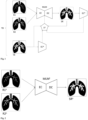
- Fig. 1 shows an exemplary and schematic embodiment of the computer-implemented method for training a machine learning model.
- the training method generates a model that can be used to generate synthetic images of an examination region, which is why the method can also be referred to as a method for generating a model for generating synthetic images of an examination region.
- the machine learning model MLM shown in Fig. 1 is trained on training data.
- the training data comprises, for each examination object of a plurality of examination objects, (i) input data and (ii) target data.
- This data set TD comprises a first representation R1 of a human lung and a second representation R2 of the human lung as input data, as well as a third representation TR of the human lung as target data.
- the third representation TR is also referred to here as the target representation.
- the first representation R1 may be a radiologic image, such as a CT image or an MRI image.
- the first representation R1 may represent the human lung without a contrast agent.
- the second representation R2 may also be a radiologic image, such as a CT image or an MRI image.
- the second representation R2 may represent the human lung after application of a first amount of a contrast agent.
- the target representation TR may also be a radiologic image, such as a CT image or an MRI image.
- the target representation TR may represent the human lung after application of a second amount of a contrast agent.
- the second amount differs from the first amount.
- the second amount may be greater than the first amount.
- the first representation R1 and the second representation R2 are inputted into the machine learning model MLM.
- the machine learning model MLM comprises an encoder EC and a decoder DC.
- the encoder EC is configured to extract features from the first representation R1 and the second representation R2 and to generate a feature vector based on the first representation R1 and the second representation R2.
- the decoder DC is configured to generate a synthetic representation SR based on the feature vector.
- the feature vector is not explicitly shown in Fig. 1 .
- the synthetic representation SR may be a synthetic radiologic image, such as a synthetic CT image or a synthetic MRI image.
- the synthetic representation SR may represent the human lung after application of the second amount of a contrast agent.
- the machine learning model MLM may be configured and trained to generate a synthetic representation SR that looks like the target representation TR based on the first representation R1 and the second representation R2 and parameters of the machine learning model MLM.
- the synthetic representation SR is fed to an additional encoder EC*.
- the additional encoder EC* is configured to extract image features from the synthetic representation SR.
- the additional encoder EC* can, for example, be an encoder of a pre-trained autoencoder that contains a CNN.
- the features can, for example, be feature maps that are generated by the CNN.
- the additional encoder EC* can also be the encoder EC of the machine learning model MLM or be based on it or replaced by it in the course of training.
- the target representation TR is also fed to the additional encoder EC*.
- the additional encoder EC* is shown twice in Fig. 1 , but it is the same additional encoder EC*.
- the additional encoder EC* therefore also extracts image features from the target representation TR.
- a loss function LF is used to quantify (i) deviations between the synthetic representation SR and the target representation TR and (ii) deviations between the features extracted from the synthetic representation SR and the features extracted from the target representation TR.
- the deviations can be reduced.
- the above steps are repeated for a plurality of input data and target data of a plurality of human lungs until a stop criterion is met.
- additional input data can be used to train the machine learning model MLM.
- Such additional input data can comprise additional representations, such as one or more representations of the examination object which represent areas adjacent to the examination region represented by the representations R1 and/or R2.
- Such other/additional input data can be information about the examination object, the examination region and/or the (measurement) method used to generate the input data and/or the target data.
- the machine learning model can be used to predict, i.e., generate a synthetic representation of a new human lung.
- new means that no representation of the human lung was usually used when training the machine learning model.
- Fig. 2 shows an exemplary and schematic embodiment of the computer-implemented method for generating a synthetic representation using a trained machine learning model.
- the trained machine learning model MLM t shown in Fig. 2 may have been trained in a method as described with respect to Fig. 1 .
- a first representation R1 n and a second representation R2 n are received. It is possible that additional input data is received.
- the first representation R1 n may be a radiologic image, such as a CT image or an MRI image.
- the first representation R1 n may represent a human lung without a contrast agent.
- the second representation R2 n may also be a radiologic image, such as a CT image or an MRI image.
- the second representation R2 n may represent the human lung after application of a first amount of a contrast agent.
- the first representation R1 n and the second representation R2 n are inputted into the trained machine learning model MLM t .
- the trained machine learning model MLM t is configured and was trained to generate a synthetic representation SR n based on the first representation R1 n and the second representation R2 n .
- the synthetic representation SR n may be a synthetic radiologic image, such as a synthetic CT image or a synthetic MRI image.
- the synthetic representation SR represents the human lung after application of a second amount of a contrast agent.
- the second amount differs from the first amount.
- the second amount may be greater than the first amount (this depends on what the machine learning model MLM t was trained for).
- the synthetic representation SR n can be outputted (e.g., displayed on a screen and/or printed using a printer) and/or stored in a data memory and/or transmitted to a separate computer system.
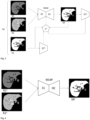
- Fig. 3 shows another exemplary and schematic embodiment of the computer-implemented method for training a machine learning model.
- the training method generates a model that can be used to generate synthetic images of an examination region, which is why the method can also be referred to as a method for generating a model for generating synthetic images of an examination region.
- the machine learning model MLM shown in Fig. 3 is trained on training data.
- the training data comprises, for each examination object of a plurality of examination objects, (i) input data and (ii) target data.
- This data set TD comprises a first representation R1 of a human liver and a second representation R2 of the human liver as input data, as well as a third representation TR of the human liver as target data.
- the third representation TR is also referred to here as the target representation.
- the first representation R1 may be a radiologic image, such as a CT image or an MRI image.
- the first representation R1 may represent the human liver at a first time point before or after application of a contrast agent.
- the second representation R2 may also be a radiologic image, such as a CT image or an MRI image.
- the second representation R2 may represent the human liver at a second time point before or after application of the contrast agent.
- the target representation TR may also be a radiologic image, such as a CT image or an MRI image.
- the target representation TR may represent the human liver at a third time point before or after application of the contrast agent.
- the first point in time, the second point in time and the third point in time are different points in time.
- the second point in time can follow the first point in time and the third point in time can follow the second point in time.
- the second time point follows the third time point and the first time point follows the second time point.
- the contrast agent can be a hepatobiliary contrast agent.
- a hepatobiliary contrast agent has the characteristic features of being specifically taken up by liver cells (hepatocytes), accumulating in the functional tissue (parenchyma) and enhancing contrast in healthy liver tissue.
- An example of a hepatobiliary MRI contrast agent is the disodium salt of gadoxetic acid (Gd-EOB-DTPA disodium), which is described in US Patent No. 6 039 931A and is commercially available under the trade names Primovist ® and Eovist ® . Further hepatobiliary contrast agents are described inter alia in WO2022/194777 .
- such a hepatobiliary MRI contrast agent is not limited to use in an MRI examination. It is possible that such a hepatobiliary MRI contrast agent is also used in a CT examination. In a CT examination, such a hepatobiliary MRI contrast agent will of course produce less contrast than a CT contrast agent. However, no hepatobiliary CT contrast agents are currently approved for CT examinations. Therefore, the present invention can also be used to increase the contrast of an MRI contrast agent in a CT examination and to utilize the hepatobiliary properties of a hepatobiliary MRI contrast agent in CT examinations.
- the first representation R1 can represent a human liver in the native phase, i.e. without contrast agent.
- the second representation R2 may represent the human liver at a first time point after administration of a (e.g., hepatobiliary) contrast agent.
- the second representation R2 may represent the human liver, e.g., in the arterial phase, the portal venous phase or the transitional phase after administration of a hepatobiliary contrast agent.
- the target representation TR may represent the human liver, e.g., in the hepatobiliary phase after administration of the hepatobiliary contrast agent.
- first representation R1 and the second representation R2 there are one or more further representations that represent the human liver at further points in time before and/or after the application of a (e.g., hepatobiliary) contrast agent.
- a (e.g., hepatobiliary) contrast agent e.g., hepatobiliary
- the input data may comprise only one representation, e.g., only the first representation R1 or only the second representation R2.
- the machine learning model MLM comprises an encoder EC and a decoder DC.
- the encoder EC is configured to extract features from the first representation R1 and the second representation R2 and to generate a feature vector based on the first representation R1 and the second representation R2.
- the decoder DC is configured to generate a synthetic representation SR based on the feature vector.
- the feature vector is not explicitly shown in Fig. 3 .
- the machine learning model MLM is configured and trained to generate a synthetic representation SR that looks like the target representation TR based on the first representation R1 and the second representation R2 and parameters of the machine learning model MLM.
- the synthetic representation SR may represent the human lung at a third time point before or after application of the contrast agent, e.g., in the hepatobiliary phase after administration of a hepatobiliary contrast agent.
- the synthetic representation SR is fed to an additional encoder EC*.
- the additional encoder EC* is configured to extract image features from the synthetic representation SR.
- the additional encoder EC* can, for example, be an encoder of a pre-trained autoencoder that contains a CNN.
- the features can, for example, be feature maps that are generated by the CNN.
- the additional encoder EC* can also be the encoder EC of the machine learning model MLM or be based on it or replaced by it in the course of training.
- the target representation TR is also fed to the additional encoder EC*.
- the additional encoder EC* is shown twice in Fig. 3 , but it is the same additional encoder EC*.
- the additional encoder EC* therefore also extracts image features from the target representation TR.
- a loss function LF is used to quantify (i) deviations between the synthetic representation SR and the target representation TR and (ii) deviations between the features extracted from the synthetic representation SR and the features extracted from the target representation TR.
- the deviations can be reduced.
- the above steps are repeated for a plurality of input data and target data of a plurality of human livers until a stop criterion is met.
- additional input data can be used to train the machine learning model MLM.
- Such additional input data can comprise additional representations, such as one or more representations of the examination object which represent areas adjacent to the examination region represented by the representations R1 and/or R2.
- Such other/additional input data can be information about the examination object, the examination region and/or the (measurement) method used to generate the input data and/or the target data.
- the machine learning model can be used to predict, i.e., generate a synthetic representation of a new human liver.
- new means that no representation of the human liver was usually used when training the machine learning model.
- Fig. 4 shows another exemplary and schematic embodiment of the computer-implemented method for generating a synthetic representation using a trained machine learning model.
- the trained machine learning model MLM t shown in Fig. 4 may have been trained in a method as described with respect to Fig. 3 .
- a first representation R1 n and a second representation R2 n are received. It is possible that additional input data is received.
- the first representation R1 n may be a radiologic image, such as a CT image or an MRI image.
- the first representation R1 n may represent a human liver at a first time point before or after application of a contrast agent.
- the second representation R2 n may also be a radiologic image, such as a CT image or an MRI image.
- the second representation R2 n may represent the human liver at a second time point before or after application of the contrast agent.
- the first representation R1 n and the second representation R2 n are inputted into the trained machine learning model MLM'.
- the trained machine learning model MLM t is configured and was trained to generate a synthetic representation SR n based on the first representation R1 n and the second representation R2 n .
- the synthetic representation SR n may be a synthetic radiologic image, such as a synthetic CT image or a synthetic MRI image.
- the synthetic representation SR n represents the human liver at a third time point before or after application of the contrast agent.
- the machine learning model MLM t may be configured and trained to generate, based on a first representation R1 n representing a human liver without contrast agent and a second representation R2 n representing the liver at a time point after the application of a hepatobiliary contrast agent (e.g., in the arterial phase or portal venous phase or transitional phase), a synthetic representation SR n of the human liver in the hepatobiliary phase after the application of the hepatobiliary contrast agent.
- a hepatobiliary contrast agent e.g., in the arterial phase or portal venous phase or transitional phase
- the synthetic representation SR n can be output (e.g. displayed on a screen or printed using a printer) and/or stored in a data memory and/or transmitted to a separate computer system.
- a “computer system” is an electronic data processing system that processes data by means of programmable calculation rules. Such a system typically comprises a “computer”, which is the unit that includes a processor for carrying out logic operations, and peripherals.
- peripherals refers to all devices that are connected to the computer and are used for control of the computer and/or as input and output devices. Examples thereof are monitor (screen), printer, scanner, mouse, keyboard, drives, camera, microphone, speakers, etc. Internal ports and expansion cards are also regarded as peripherals in computer technology.
- Fig. 5 shows by way of example and in schematic form a computer system according to the present disclosure.
- the computer system (1) shown in Fig. 5 comprises a receiving unit (11), a control and calculation unit (12) and an output unit (13).
- the control and calculation unit (12) serves for control of the computer system (1), coordination of the data flows between the units of the computer system (1), and for the performance of calculations.
- the control and calculation unit (12) is configured:
- Fig. 6 shows by way of example and in schematic form a further embodiment of the computer system.
- the computer system (1) comprises a processing unit (21) connected to a storage medium (22).
- the processing unit (21) and the storage medium (22) form a control and calculation unit, as shown in Fig. 5 .
- the processing unit (21) may comprise one or more processors alone or in combination with one or more storage media.
- the processing unit (21) may be customary computer hardware that is able to process information such as digital images, computer programs and/or other digital information.
- the processing unit (21) usually consists of an arrangement of electronic circuits, some of which can be designed as an integrated circuit or as a plurality of integrated circuits connected to one another (an integrated circuit is sometimes also referred to as a "chip").
- the processing unit (21) may be configured to execute computer programs that can be stored in a working memory of the processing unit (21) or in the storage medium (22) of the same or of a different computer system.
- the storage medium (22) may be customary computer hardware that is able to store information such as digital images (for example representations of the examination region), data, computer programs and/or other digital information either temporarily and/or permanently.
- the storage medium (22) may comprise a volatile and/or non-volatile storage medium and may be fixed in place or removable. Examples of suitable storage media are RAM (random access memory), ROM (read-only memory), a hard disk, a flash memory, an exchangeable computer floppy disk, an optical disc, a magnetic tape or a combination of the aforementioned.
- Optical discs can include compact discs with read-only memory (CD-ROM), compact discs with read/write function (CD-R/W), DVDs, Blu-ray discs and the like.
- the processing unit (21) may be connected not just to the storage medium (22), but also to one or more interfaces (11, 12, 31, 32, 33) in order to display, transmit and/or receive information.
- the interfaces may comprise one or more communication interfaces (11, 32, 33) and/or one or more user interfaces (12, 31).
- the one or more communication interfaces may be configured to send and/or receive information, for example to and/or from an MRI scanner, a CT scanner, an ultrasound camera, other computer systems, networks, data storage media or the like.
- the one or more communication interfaces may be configured to transmit and/or receive information via physical (wired) and/or wireless communication connections.
- the one or more communication interfaces may comprise one or more interfaces for connection to a network, for example using technologies such as mobile telephone, wifi, satellite, cable, DSL, optical fibre and/or the like.
- the one or more communication interfaces may comprise one or more close-range communication interfaces configured to connect devices having close-range communication technologies such as NFC, RFID, Bluetooth, Bluetooth LE, ZigBee, infrared (e.g. IrDA) or the like.
- the user interfaces may include a display (31).
- a display (31) may be configured to display information to a user. Suitable examples thereof are a liquid crystal display (LCD), a light-emitting diode display (LED), a plasma display panel (PDP) or the like.
- the user input interface(s) (11, 12) may be wired or wireless and may be configured to receive information from a user in the computer system (1), for example for processing, storage and/or display. Suitable examples of user input interfaces are a microphone, an image- or video-recording device (for example a camera), a keyboard or a keypad, a j oystick, a touch-sensitive surface (separate from a touchscreen or integrated therein) or the like.
- the user interfaces may contain an automatic identification and data capture technology (AIDC) for machine-readable information.
- AIDC automatic identification and data capture technology
- This can include barcodes, radiofrequency identification (RFID), magnetic strips, optical character recognition (OCR), integrated circuit cards (ICC) and the like.
- RFID radiofrequency identification
- OCR optical character recognition
- ICC integrated circuit cards
- the user interfaces may in addition comprise one or more interfaces for communication with peripherals such as printers and the like.
- One or more computer programs (40) may be stored in the storage medium (22) and executed by the processing unit (21), which is thereby programmed to fulfil the functions described in this description.
- the retrieving, loading and execution of instructions of the computer program (40) may take place sequentially, such that an instruction is respectively retrieved, loaded and executed. However, the retrieving, loading and/or execution may also take place in parallel.
- the computer system of the present disclosure may be designed as a laptop, notebook, netbook and/or tablet PC; it may also be a component of an MRI scanner, a CT scanner or an ultrasound diagnostic device.
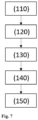
- Fig. 7 shows schematically in the form of a flow chart a preferred embodiment of the computer-implemented method for generating a synthetic representation.
- the method (100) comprises the steps:
Landscapes
- Engineering & Computer Science (AREA)
- Theoretical Computer Science (AREA)
- Physics & Mathematics (AREA)
- Health & Medical Sciences (AREA)
- General Physics & Mathematics (AREA)
- General Health & Medical Sciences (AREA)
- Evolutionary Computation (AREA)
- Artificial Intelligence (AREA)
- Software Systems (AREA)
- Computing Systems (AREA)
- Medical Informatics (AREA)
- Biomedical Technology (AREA)
- Life Sciences & Earth Sciences (AREA)
- Molecular Biology (AREA)
- Computational Linguistics (AREA)
- General Engineering & Computer Science (AREA)
- Mathematical Physics (AREA)
- Biophysics (AREA)
- Data Mining & Analysis (AREA)
- Nuclear Medicine, Radiotherapy & Molecular Imaging (AREA)
- Radiology & Medical Imaging (AREA)
- Epidemiology (AREA)
- Primary Health Care (AREA)
- Public Health (AREA)
- Databases & Information Systems (AREA)
- Computer Vision & Pattern Recognition (AREA)
- Multimedia (AREA)
- Image Analysis (AREA)
Priority Applications (2)
| Application Number | Priority Date | Filing Date | Title |
|---|---|---|---|
| EP23216049.9A EP4571650A1 (fr) | 2023-12-12 | 2023-12-12 | Génération d'images synthétiques |
| US18/978,116 US20250191734A1 (en) | 2023-12-12 | 2024-12-12 | Generating synthetic images |
Applications Claiming Priority (1)
| Application Number | Priority Date | Filing Date | Title |
|---|---|---|---|
| EP23216049.9A EP4571650A1 (fr) | 2023-12-12 | 2023-12-12 | Génération d'images synthétiques |
Publications (1)
| Publication Number | Publication Date |
|---|---|
| EP4571650A1 true EP4571650A1 (fr) | 2025-06-18 |
Family
ID=89222074
Family Applications (1)
| Application Number | Title | Priority Date | Filing Date |
|---|---|---|---|
| EP23216049.9A Pending EP4571650A1 (fr) | 2023-12-12 | 2023-12-12 | Génération d'images synthétiques |
Country Status (2)
| Country | Link |
|---|---|
| US (1) | US20250191734A1 (fr) |
| EP (1) | EP4571650A1 (fr) |
Families Citing this family (1)
| Publication number | Priority date | Publication date | Assignee | Title |
|---|---|---|---|---|
| CN120355929B (zh) * | 2025-06-24 | 2025-09-05 | 贵州大学 | 一种医学图像领域泛化分割方法及系统 |
Citations (14)
| Publication number | Priority date | Publication date | Assignee | Title |
|---|---|---|---|---|
| US6039931A (en) | 1989-06-30 | 2000-03-21 | Schering Aktiengesellschaft | Derivatized DTPA complexes, pharmaceutical agents containing these compounds, their use, and processes for their production |
| WO2014036473A1 (fr) | 2012-08-31 | 2014-03-06 | Kenji Suzuki | Technique d'apprentissage supervisée par machine servant à la réduction de la dose de rayonnement en imagerie tomographique par ordinateur |
| WO2016175755A1 (fr) | 2015-04-28 | 2016-11-03 | Siemens Healthcare Gmbh | Procédé et système pour synthétiser des images de tomographie calculée à dose élevée ou kv élevé virtuelles à partir d'images de tomographie calculée à faible dose ou faible kv |
| WO2018202541A1 (fr) | 2017-05-02 | 2018-11-08 | Bayer Aktiengesellschaft | Améliorations dans la détection radiologique de l'hypertension pulmonaire thromboembolique chronique |
| WO2019074938A1 (fr) | 2017-10-09 | 2019-04-18 | The Board Of Trustees Of The Leland Stanford Junior University | Réduction de dose de contraste pour imagerie médicale à l'aide d'un apprentissage profond |
| WO2020229152A1 (fr) | 2019-05-10 | 2020-11-19 | Bayer Consumer Care Ag | Identification de signes candidats indiquant une fusion oncogénique ntrk |
| WO2021052896A1 (fr) | 2019-09-18 | 2021-03-25 | Bayer Aktiengesellschaft | Prévision d'images irm au moyen d'un modèle de prévision entraîné par apprentissage supervisé |
| WO2021069338A1 (fr) | 2019-10-08 | 2021-04-15 | Bayer Aktiengesellschaft | Génération d'images irm du foie sans amélioration du contraste |
| WO2021197996A1 (fr) | 2020-04-03 | 2021-10-07 | Bayer Aktiengesellschaft | Génération d'images radiologiques |
| US11170543B2 (en) | 2020-01-13 | 2021-11-09 | The Board Of Trustees Of The Leland Stanford Junior University | MRI image reconstruction from undersampled data using adversarially trained generative neural network |
| US11181598B2 (en) | 2019-04-25 | 2021-11-23 | Siemens Healthcare Gmbh | Multi-contrast MRI image reconstruction using machine learning |
| WO2022184297A1 (fr) | 2021-03-02 | 2022-09-09 | Bayer Aktiengesellschaft | Apprentissage automatique dans le domaine de la radiologie avec injecton d'agent de contraste |
| WO2022194777A1 (fr) | 2021-03-15 | 2022-09-22 | Bayer Aktiengesellschaft | Nouvel agent de contraste destiné à être utilisé dans l'imagerie par résonance magnétique |
| EP4210069A1 (fr) * | 2022-01-11 | 2023-07-12 | Bayer Aktiengesellschaft | Images tomodensitométriques synthétiques à contraste amélioré |
-
2023
- 2023-12-12 EP EP23216049.9A patent/EP4571650A1/fr active Pending
-
2024
- 2024-12-12 US US18/978,116 patent/US20250191734A1/en active Pending
Patent Citations (15)
| Publication number | Priority date | Publication date | Assignee | Title |
|---|---|---|---|---|
| US6039931A (en) | 1989-06-30 | 2000-03-21 | Schering Aktiengesellschaft | Derivatized DTPA complexes, pharmaceutical agents containing these compounds, their use, and processes for their production |
| WO2014036473A1 (fr) | 2012-08-31 | 2014-03-06 | Kenji Suzuki | Technique d'apprentissage supervisée par machine servant à la réduction de la dose de rayonnement en imagerie tomographique par ordinateur |
| US20150201895A1 (en) * | 2012-08-31 | 2015-07-23 | The University Of Chicago | Supervised machine learning technique for reduction of radiation dose in computed tomography imaging |
| WO2016175755A1 (fr) | 2015-04-28 | 2016-11-03 | Siemens Healthcare Gmbh | Procédé et système pour synthétiser des images de tomographie calculée à dose élevée ou kv élevé virtuelles à partir d'images de tomographie calculée à faible dose ou faible kv |
| WO2018202541A1 (fr) | 2017-05-02 | 2018-11-08 | Bayer Aktiengesellschaft | Améliorations dans la détection radiologique de l'hypertension pulmonaire thromboembolique chronique |
| WO2019074938A1 (fr) | 2017-10-09 | 2019-04-18 | The Board Of Trustees Of The Leland Stanford Junior University | Réduction de dose de contraste pour imagerie médicale à l'aide d'un apprentissage profond |
| US11181598B2 (en) | 2019-04-25 | 2021-11-23 | Siemens Healthcare Gmbh | Multi-contrast MRI image reconstruction using machine learning |
| WO2020229152A1 (fr) | 2019-05-10 | 2020-11-19 | Bayer Consumer Care Ag | Identification de signes candidats indiquant une fusion oncogénique ntrk |
| WO2021052896A1 (fr) | 2019-09-18 | 2021-03-25 | Bayer Aktiengesellschaft | Prévision d'images irm au moyen d'un modèle de prévision entraîné par apprentissage supervisé |
| WO2021069338A1 (fr) | 2019-10-08 | 2021-04-15 | Bayer Aktiengesellschaft | Génération d'images irm du foie sans amélioration du contraste |
| US11170543B2 (en) | 2020-01-13 | 2021-11-09 | The Board Of Trustees Of The Leland Stanford Junior University | MRI image reconstruction from undersampled data using adversarially trained generative neural network |
| WO2021197996A1 (fr) | 2020-04-03 | 2021-10-07 | Bayer Aktiengesellschaft | Génération d'images radiologiques |
| WO2022184297A1 (fr) | 2021-03-02 | 2022-09-09 | Bayer Aktiengesellschaft | Apprentissage automatique dans le domaine de la radiologie avec injecton d'agent de contraste |
| WO2022194777A1 (fr) | 2021-03-15 | 2022-09-22 | Bayer Aktiengesellschaft | Nouvel agent de contraste destiné à être utilisé dans l'imagerie par résonance magnétique |
| EP4210069A1 (fr) * | 2022-01-11 | 2023-07-12 | Bayer Aktiengesellschaft | Images tomodensitométriques synthétiques à contraste amélioré |
Non-Patent Citations (21)
| Title |
|---|
| A. ELGAMMALC.-S. LEE: "Separating style and content on a nonlinear manifold", PROCEEDINGS OF THE 2004 IEEE COMPUTER SOCIETY CONFERENCE ON COMPUTER VISION AND PATTERN RECOGNITION, 2004, pages I - I |
| A. IGNEE ET AL.: "Ultrasound contrast agents", ENDOSC ULTRASOUND., vol. 5, no. 6, November 2016 (2016-11-01), pages 355 - 362 |
| A.S.L. JASCINTH ET AL.: "Contrast Agents in computed tomography: A Review", JOURNAL OF APPLIED DENTAL AND MEDICAL SCIENCES, vol. 2, 2016, pages 143 - 149 |
| C. L. HUI ET AL.: "Patterns of enhancement in the hepatobiliary phase of gadoxetic acid-enhanced MRI", BR J RADIOL., vol. 93, 2020, pages 20190989 |
| CAO KAIBIN ET AL: "A Multi-constraint Hybrid Network for Ultrasound Image Synthesis", 2023 IEEE 7TH INFORMATION TECHNOLOGY AND MECHATRONICS ENGINEERING CONFERENCE (ITOEC), IEEE, vol. 7, 15 September 2023 (2023-09-15), pages 2147 - 2151, XP034457158, DOI: 10.1109/ITOEC57671.2023.10292010 * |
| D. G. LOWE: "Distinctive Image Features from Scale Invariant Keypoints", INTERNATIONAL JOURNAL OF COMPUTER VISION, vol. 60, 2004, pages 91 - 110, XP019216426, DOI: 10.1023/B:VISI.0000029664.99615.94 |
| H. LUSIC ET AL.: "X-ray-Computed Tomography Contrast Agents", CHEM. REV., vol. 113, no. 3, 2013, pages 1641 - 1666, XP055559399, Retrieved from the Internet <URL:https://www.radiology.wisc.edu/wp-content/uploads/2017/10/contrast-agents-tutorial.pdf> DOI: 10.1021/cr200358s |
| J. B. TENENBAUMW. T. FREEMAN, SEPARATING STYLE AND CONTENT WITH BILINEAR MODELS, NEURAL COMPUTATION, vol. 12, 2020, pages 1247 - 1283 |
| K. CATCH: "The Radon Transformation and Its Application in Tomography", JOURNAL OF PHYSICS CONFERENCE SERIES, no. 1, 1903, pages 012066 |
| L- ARMI ET AL.: "Texture Image Analysis and Texture Classification Methods - A Review", INTERNATIONAL ONLINE JOURNAL OF IMAGE PROCESSING AND PATTERN RECOGNITION, vol. 2, no. 1, 2019, pages 1 - 29 |
| L.C. ABONYI ET AL.: "Intravascular Contrast Media in Radiography: Historical Development & Review of Risk Factors for Adverse Reactions", SOUTH AMERICAN JOURNAL OF CLINICAL RESEARCH, vol. 3, 2016, pages 1 - 10 |
| LECTURE NOTES IN COMPUTER SCIENCE, pages 827 - 836 |
| M. BLACHNIK ET AL.: "Image classification by histogram features created with Learning Vector Quantization", PROCEEDINGS OF INTERNATIONAL CONFERENCE ON ARTIFICIAL NEURAL NETWORKS, ICANN, vol. 5163, 2008, XP019106104 |
| M. R. WANKHADE ET AL., INTERNATIONAL JOURNAL OF COMPUTER SCIENCE AND MOBILE COMPUTING, vol. 6, 2017, pages 336 - 345 |
| M.R. NOUH ET AL.: "Radiographic and magnetic resonances contrast agents: Essentials and tips for safe practices", WORLD J RADIOL., vol. 9, no. 9, 28 September 2017 (2017-09-28), pages 339 - 349 |
| O. RONNEBERGER ET AL.: "t7 Net: Convolutional Networks for Biomedical Image Segmentation", ARXIV:1505.04597V1, 2015 |
| S. KHAN ET AL.: "Transformers in Vision: A Survey", ARXIV:2101.01169V5 |
| T. G. NISIAS. RAJESH: "Extraction of High-level and Low-levelfeature for classification of Image using Ridgelet and CAW based Image Classification", JOURNAL OF PHYSICS: CONFERENCE SERIES, vol. 1911, 2021, pages 012019 |
| W. ZHOU ET AL.: "Histogram of Oriented Gradients Feature Extraction From Raw Bayer Pattern Images", IEEE TRANSACTIONS ON CIRCUITS AND SYSTEMS II: EXPRESS BRIEFS, 2020, pages 1 - 5 |
| Y. H. LIU ET AL.: "Feature Extraction and Image Recognition with Convolutional Neural Networks", JOURNAL OF PHYSICS CONFERENCE SERIES, vol. 1087, no. 6, 2018, pages 062032, XP055980588 |
| Z. HUANG ET AL.: "Timescales of Intrinsic BOLD Signal Dynamics and Functional Connectivity in Pharmacologic and Neuropathologic States of Unconsciousness", THE JOURNAL OF NEUROSCIENCE, vol. 38, no. 9, 2018, pages 2304 - 2317 |
Also Published As
| Publication number | Publication date |
|---|---|
| US20250191734A1 (en) | 2025-06-12 |
Similar Documents
| Publication | Publication Date | Title |
|---|---|---|
| US12008759B2 (en) | Method and system for identifying pathological changes in follow-up medical images | |
| Gibson et al. | NiftyNet: a deep-learning platform for medical imaging | |
| US12611152B2 (en) | Machine learning in the field of contrast-enhanced radiology | |
| US20240303973A1 (en) | Actor-critic approach for generating synthetic images | |
| US12198343B2 (en) | Multi-modal computer-aided diagnosis systems and methods for prostate cancer | |
| US20240193738A1 (en) | Implicit registration for improving synthesized full-contrast image prediction tool | |
| JP2021140769A (ja) | 医用情報処理装置、医用情報処理方法及び医用情報処理プログラム | |
| Kunapuli et al. | A review of deep learning models for medical diagnosis | |
| US20240185577A1 (en) | Reinforced attention | |
| Zhang et al. | The optimal tetralogy of Fallot repair using generative adversarial networks | |
| US20250191734A1 (en) | Generating synthetic images | |
| US20250201408A1 (en) | Prediction of representations of an examination area of an examination object after applications of different amounts of a contrast agent | |
| US20250201407A1 (en) | Prediction of a representation of an examination area of an examination object in a state of a sequence of states | |
| US20250078474A1 (en) | Synthetic contrast-enhanced ct images | |
| US20250292447A1 (en) | Generation of a synthetic medical image | |
| US20250349101A1 (en) | Segmentation of medical images | |
| EP4575997A1 (fr) | Génération d'images synthétiques | |
| EP4618009A1 (fr) | Génération d'une image médicale synthétique | |
| Emami et al. | Elements of Medical Image Processing 20 | |
| EP4621717A1 (fr) | Génération d'une image médicale synthétique | |
| Tabatabaei et al. | Generative AI in medical imaging | |
| US20240404255A1 (en) | Generation of artificial contrast-enhanced radiological images | |
| WO2025260252A1 (fr) | Segmentation d'images médicales | |
| Hachaj et al. | Nowadays and future computer application in medicine | |
| Lamrani et al. | U-Net-based Artificial Intelligence for Accurate and Robust Brain Tumor Diagnosis using Magnetic Resonance Imaging |
Legal Events
| Date | Code | Title | Description |
|---|---|---|---|
| PUAI | Public reference made under article 153(3) epc to a published international application that has entered the european phase |
Free format text: ORIGINAL CODE: 0009012 |
|
| STAA | Information on the status of an ep patent application or granted ep patent |
Free format text: STATUS: THE APPLICATION HAS BEEN PUBLISHED |
|
| AK | Designated contracting states |
Kind code of ref document: A1 Designated state(s): AL AT BE BG CH CY CZ DE DK EE ES FI FR GB GR HR HU IE IS IT LI LT LU LV MC ME MK MT NL NO PL PT RO RS SE SI SK SM TR |
|
| RAP3 | Party data changed (applicant data changed or rights of an application transferred) |
Owner name: BAYER AKTIENGESELLSCHAFT |
|
| STAA | Information on the status of an ep patent application or granted ep patent |
Free format text: STATUS: REQUEST FOR EXAMINATION WAS MADE |
|
| 17P | Request for examination filed |
Effective date: 20251218 |