EP4574055A1 - Ultraschallabbildungsverfahren zur genauen lokalisierung oder dimensionierung eines objekts - Google Patents
Ultraschallabbildungsverfahren zur genauen lokalisierung oder dimensionierung eines objekts Download PDFInfo
- Publication number
- EP4574055A1 EP4574055A1 EP24218568.4A EP24218568A EP4574055A1 EP 4574055 A1 EP4574055 A1 EP 4574055A1 EP 24218568 A EP24218568 A EP 24218568A EP 4574055 A1 EP4574055 A1 EP 4574055A1
- Authority
- EP
- European Patent Office
- Prior art keywords
- interest
- point
- ultrasonic
- size
- flight
- Prior art date
- Legal status (The legal status is an assumption and is not a legal conclusion. Google has not performed a legal analysis and makes no representation as to the accuracy of the status listed.)
- Pending
Links
Images



Classifications
-
- A—HUMAN NECESSITIES
- A61—MEDICAL OR VETERINARY SCIENCE; HYGIENE
- A61B—DIAGNOSIS; SURGERY; IDENTIFICATION
- A61B8/00—Diagnosis using ultrasonic, sonic or infrasonic waves
- A61B8/08—Clinical applications
- A61B8/0833—Clinical applications involving detecting or locating foreign bodies or organic structures
- A61B8/085—Clinical applications involving detecting or locating foreign bodies or organic structures for locating body or organic structures, e.g. tumours, calculi, blood vessels, nodules
-
- A—HUMAN NECESSITIES
- A61—MEDICAL OR VETERINARY SCIENCE; HYGIENE
- A61B—DIAGNOSIS; SURGERY; IDENTIFICATION
- A61B8/00—Diagnosis using ultrasonic, sonic or infrasonic waves
- A61B8/08—Clinical applications
- A61B8/0858—Clinical applications involving measuring tissue layers, e.g. skin, interfaces
-
- A—HUMAN NECESSITIES
- A61—MEDICAL OR VETERINARY SCIENCE; HYGIENE
- A61B—DIAGNOSIS; SURGERY; IDENTIFICATION
- A61B8/00—Diagnosis using ultrasonic, sonic or infrasonic waves
- A61B8/08—Clinical applications
- A61B8/0891—Clinical applications for diagnosis of blood vessels
-
- A—HUMAN NECESSITIES
- A61—MEDICAL OR VETERINARY SCIENCE; HYGIENE
- A61B—DIAGNOSIS; SURGERY; IDENTIFICATION
- A61B8/00—Diagnosis using ultrasonic, sonic or infrasonic waves
- A61B8/44—Constructional features of the ultrasonic, sonic or infrasonic diagnostic device
- A61B8/4483—Constructional features of the ultrasonic, sonic or infrasonic diagnostic device characterised by features of the ultrasound transducer
-
- A—HUMAN NECESSITIES
- A61—MEDICAL OR VETERINARY SCIENCE; HYGIENE
- A61B—DIAGNOSIS; SURGERY; IDENTIFICATION
- A61B8/00—Diagnosis using ultrasonic, sonic or infrasonic waves
- A61B8/52—Devices using data or image processing specially adapted for diagnosis using ultrasonic, sonic or infrasonic waves
- A61B8/5215—Devices using data or image processing specially adapted for diagnosis using ultrasonic, sonic or infrasonic waves involving processing of medical diagnostic data
- A61B8/5223—Devices using data or image processing specially adapted for diagnosis using ultrasonic, sonic or infrasonic waves involving processing of medical diagnostic data for extracting a diagnostic or physiological parameter from medical diagnostic data
-
- A—HUMAN NECESSITIES
- A61—MEDICAL OR VETERINARY SCIENCE; HYGIENE
- A61B—DIAGNOSIS; SURGERY; IDENTIFICATION
- A61B8/00—Diagnosis using ultrasonic, sonic or infrasonic waves
- A61B8/54—Control of the diagnostic device
Definitions
- the invention relates to the field of ultrasound imaging, in particular medical imaging, and relates to a method for improving the accuracy of such measurements when they are carried out using an ultrasound probe having an opening field of the emitted ultrasound field which has a given size.
- the invention applies in particular to the imaging of blood vessels, for example arteries, with a view to precisely determining the diameter of such vessels, but it is not limited to this application and aims more generally at improving the measurement precision for the ultrasound imaging of all types of object in the medical field or the field of non-destructive testing by ultrasound.
- ultrasound imaging is a tool that allows for the precise monitoring, sometimes in real time, of certain anatomical objects such as blood vessels. In particular, it allows the location and measurement of the dimensions of these vessels.
- the accuracy of ultrasound measurements varies depending on the type of equipment used.
- ultrasonic probes are generally based on multi-element transducers that use several active elements to synthesize an ultrasonic beam in order to focus the beam by coherent summation of the amplitudes of the different elements.
- the invention is based on an algorithmic solution that allows the resolution and accuracy of the measurement to be improved without modifying the acquisition equipment.
- the method is based on carrying out several successive measurements by varying the number of active elements or the aperture size of the generated ultrasonic field and then extrapolating the measurement that would have been obtained with an aperture close to zero from the different measurement points carried out.
- the ultrasonic probe comprises a plurality of elements and the variation of the size of the opening of the ultrasonic field is obtained by varying the number of active receiving elements.
- the number of active receiving elements is a multiple of two.
- the number of active elements in transmission is equal to the number of active elements in reception or to the maximum number of elements that the probe comprises.
- the object to be imaged is a blood vessel and the point of interest is a point on a wall of the blood vessel.
- the method is applied for two points of interest corresponding to a first point of the distal wall of the blood vessel and a second point of the proximal wall of the blood vessel, diametrically opposite the first point and the method further comprises an estimation of the diameter of the blood vessel from the extrapolated flight times of the two points.
- the subject of the invention is an ultrasound imaging device comprising an ultrasound probe and a processing unit configured to carry out the steps of the method according to the invention.
- the invention thus makes it possible to optimize the precision of the measurements without the need to modify the ultrasonic probe.
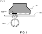
- the probe 101 is positioned in contact with the skin 103 so that the row of elements 102 is located substantially in a transverse plane of an artery 104 to be imaged.
- the probe 101 is not necessarily centered on the artery 104, it is sufficient that the artery is covered in emission and reception by the ultrasound beam generated by the probe so as to obtain a transverse imprint of the artery.
- the probe is positioned at a given point and a group of transmitting elements is activated to generate an ultrasonic field in a given direction.
- the echoes of the field over the covered area are then measured by the same group of elements or a small number of elements.
- the ultrasonic field is emitted in a direction perpendicular to the axis of the alignment of the elements 102 which also corresponds locally to the plane of the skin 103 on which the probe 101 is positioned.
- FIG. 3b shows a column of the 2D image of the Figure 3a which corresponds to a signal measured for an acquisition corresponding to a position of the probe located directly above the center of the vessel to be imaged.
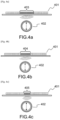
- FIGS. 4a, 4b and 4c show three different acquisition configurations for the same ultrasound probe 401 placed directly above a radial artery 402
- the number of active elements 403 in transmission and reception is equal to 16.
- the ultrasonic field is generated from the 16 central elements of the probe and also measured by these 16 elements. This acquisition makes it possible to obtain a first measurement of the signal represented in the Figure 3b .
- Figures 4a, 4b and 4c show that it is possible to perform the same measurement from a variable active window size.
- a minimum active window size is required to obtain a measurement with a usable signal-to-noise ratio.
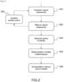
- the measurement method begins with steps 201, 202, 203 which consist of carrying out several successive acquisitions of the same ultrasonic signal (for the same position of the probe relative to the object to be imaged) by varying the size of the active window at each iteration.
- the number of active receiving elements can be reduced at each iteration relative to the maximum number of elements used to generate the ultrasonic beam.
- the number of active transmitting elements can be left constant or the group of active transmitting elements can be taken equal to the group of active receiving elements.
- the number of active receiving elements can be reduced by a factor of 2.
- the ultrasound probe is manufactured using a technology which allows the opening of the ultrasound field to be varied with a single element, then the variation in the number of active receiving elements is replaced by a variation in the opening field at each iteration.
- One or more points of interest are then identified on the acquired ultrasound signal. For example, in the case of the signal from the Figure 5 , two points of interest 501,502 are identified which correspond to echoes of the signal on the distal and proximal internal walls of the blood vessel.
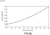
- step 205 a numerical model of the evolution of the flight time variation curves as a function of the number of channels for each point of interest is then determined by mathematical optimization.
- This mathematical model is, for example, determined by a model in the form of a second-degree polynomial function or any other suitable numerical model. It can be defined by means of an interpolation of the measured points of the curve. This interpolation can be carried out by means of a mixture of Gaussian functions or learned by a trained model of neural networks or any other suitable numerical optimization algorithm.
- the mathematical model can be determined for each curve associated with each point of interest or for the final quantity that one wishes to determine from several points of interest, in this case the difference between the two flight times.
- step 206 the mathematical model obtained is extrapolated to determine an asymptotic value for a number of channels tending towards 0. Indeed, this value corresponds to the most precise measurement in terms of resolution and its determination via an extrapolation of a model makes it possible not to be subject to the problem of low signal-to-noise ratio inherent in a measurement carried out with a very small active window.
Landscapes
- Health & Medical Sciences (AREA)
- Life Sciences & Earth Sciences (AREA)
- Engineering & Computer Science (AREA)
- Medical Informatics (AREA)
- Surgery (AREA)
- Pathology (AREA)
- Radiology & Medical Imaging (AREA)
- Biophysics (AREA)
- Biomedical Technology (AREA)
- Heart & Thoracic Surgery (AREA)
- Physics & Mathematics (AREA)
- Molecular Biology (AREA)
- Nuclear Medicine, Radiotherapy & Molecular Imaging (AREA)
- Animal Behavior & Ethology (AREA)
- General Health & Medical Sciences (AREA)
- Public Health (AREA)
- Veterinary Medicine (AREA)
- Vascular Medicine (AREA)
- Gynecology & Obstetrics (AREA)
- Physiology (AREA)
- Computer Vision & Pattern Recognition (AREA)
- Investigating Or Analyzing Materials By The Use Of Ultrasonic Waves (AREA)
- Ultra Sonic Daignosis Equipment (AREA)
Applications Claiming Priority (1)
| Application Number | Priority Date | Filing Date | Title |
|---|---|---|---|
| FR2314348A FR3156647B1 (fr) | 2023-12-18 | 2023-12-18 | Méthode d’imagerie ultrasonore pour la localisation ou le dimensionnement précis d’un objet |
Publications (1)
| Publication Number | Publication Date |
|---|---|
| EP4574055A1 true EP4574055A1 (de) | 2025-06-25 |
Family
ID=90721168
Family Applications (1)
| Application Number | Title | Priority Date | Filing Date |
|---|---|---|---|
| EP24218568.4A Pending EP4574055A1 (de) | 2023-12-18 | 2024-12-10 | Ultraschallabbildungsverfahren zur genauen lokalisierung oder dimensionierung eines objekts |
Country Status (3)
| Country | Link |
|---|---|
| US (1) | US12402856B2 (de) |
| EP (1) | EP4574055A1 (de) |
| FR (1) | FR3156647B1 (de) |
Citations (3)
| Publication number | Priority date | Publication date | Assignee | Title |
|---|---|---|---|---|
| JP5854072B2 (ja) * | 2014-03-19 | 2016-02-09 | セイコーエプソン株式会社 | 血管径測定装置、血管径の測定方法 |
| WO2016057233A1 (en) | 2014-10-06 | 2016-04-14 | Sperion Medical Devices, LLC | System and method for non-invasive blood pressure measurement |
| US20210100523A1 (en) * | 2019-10-04 | 2021-04-08 | Invensense, Inc. | Determination of blood vessel characteristic change using an ultrasonic sensor |
-
2023
- 2023-12-18 FR FR2314348A patent/FR3156647B1/fr active Active
-
2024
- 2024-12-10 EP EP24218568.4A patent/EP4574055A1/de active Pending
- 2024-12-11 US US18/977,422 patent/US12402856B2/en active Active
Patent Citations (3)
| Publication number | Priority date | Publication date | Assignee | Title |
|---|---|---|---|---|
| JP5854072B2 (ja) * | 2014-03-19 | 2016-02-09 | セイコーエプソン株式会社 | 血管径測定装置、血管径の測定方法 |
| WO2016057233A1 (en) | 2014-10-06 | 2016-04-14 | Sperion Medical Devices, LLC | System and method for non-invasive blood pressure measurement |
| US20210100523A1 (en) * | 2019-10-04 | 2021-04-08 | Invensense, Inc. | Determination of blood vessel characteristic change using an ultrasonic sensor |
Non-Patent Citations (2)
| Title |
|---|
| "System A feasibility study of a PMUT-based wearable sensor for the automatic monitoring of carotid artery parameters", IEEE INTERNATIONAL ULTRASONICS SYMPOSIUM, 2021 |
| J. H. GAGAN ET AL.: "Automated Segmentation of Common Carotid Artery in Ultrasound Images", IEEE ACCESS, vol. 10, 2022, pages 58419 - 58430 |
Also Published As
| Publication number | Publication date |
|---|---|
| FR3156647B1 (fr) | 2025-12-05 |
| US20250195030A1 (en) | 2025-06-19 |
| US12402856B2 (en) | 2025-09-02 |
| FR3156647A1 (fr) | 2025-06-20 |
Similar Documents
| Publication | Publication Date | Title |
|---|---|---|
| EP0409054B1 (de) | Verfahren zur Verarbeitung eines elektrischen Distanzsignals | |
| EP0335759A1 (de) | Ultraschallwindmesser | |
| EP0077585B1 (de) | Vorrichtung zur Untersuchung von Medien mittels Ultraschallechographie | |
| EP3847450B1 (de) | Verfahren zur ultraschalldetektion und charakterisierung von defekten in einem heterogenen material | |
| EP0751743A1 (de) | Verfahren und gerät zur auswertung und charakterisierung von knocheneigenschaften | |
| CA2765203A1 (fr) | Procede et dispositif ultrasonores pour caracteriser un milieu | |
| EP0338618A1 (de) | Gerät zur Messung der Geschwindigkeit von Organen und der Blutströmung durch Korrelationsverfahren | |
| EP3899523B1 (de) | Verfahren zur ultraschallbildgebung mittels zweidimensionaler fourier-transformation, zugehörige ultraschallsondenvorrichtung und computerprogramm | |
| EP0196713B1 (de) | Verfahren und Vorrichtung zur Untersuchung von Medien mittels Ultraschall-Echographie | |
| WO2021023933A1 (fr) | Procédé et système de caractérisation ultrasonore non invasive d'un milieu hétérogène | |
| EP4083659B1 (de) | Vorrichtung und verfahren zur verarbeitung von signalen aus einer gesamtheit von ultraschallwandlern | |
| WO2024074252A1 (fr) | Procede d'imagerie ultrasonore par transformee de fourier multidimensionnelle a l'aide de deux transducteurs multielements distincts | |
| EP0197582B1 (de) | Verfahren und Vorrichtung zur Untersuchung von Medien mittels Ultraschall-Echographie | |
| EP0458384A1 (de) | Mess- und Anzeigevorrichtung für die Geschwindigkeit der Blutströmung mittels Ultraschallechographie | |
| EP4574055A1 (de) | Ultraschallabbildungsverfahren zur genauen lokalisierung oder dimensionierung eines objekts | |
| EP0402968B1 (de) | Vorrichtung zum Messen der Fliessgeschwindigkeit von Blut | |
| EP0430374A1 (de) | Einrichtung zum Messen und Darstellen physiologischer Grössen der Blutströmung mittels Ultraschall-Echographie | |
| FR3075373A1 (fr) | Dispositif, installation et procede d'imagerie de defauts d'une structure par emission et reception d'ondes mecaniques dans cette structure | |
| EP4574056A1 (de) | Verfahren zur charakterisierung eines zielobjekts in einem medium | |
| EP2825876A1 (de) | Multisensor-ultraschalltestvorrichtung und verfahren zur herstellung einer solchen vorrichtung, verfahren zur steuerung einer derartigen vorrichtung und entsprechendes computerprogramm | |
| FR3045165A1 (fr) | Procede et systeme sur puce d'imagerie par ultrasons | |
| FR3156911A1 (fr) | Procédé et système de caracterisation ultrasonore d’un milieu | |
| EP2929343B1 (de) | Vorrichtung und verfahren zur ultraschalllbildgebeung mit filterung von artefakten aufgrund von interferenzen zwischen rekonstruktionsmoden | |
| FR3098302A1 (fr) | Dispositif d’imagerie par ondes de surface | |
| FR3153517A1 (fr) | Procédé et système de caracterisation ulstrasonore d’un milieu pour analyse medicale |
Legal Events
| Date | Code | Title | Description |
|---|---|---|---|
| PUAI | Public reference made under article 153(3) epc to a published international application that has entered the european phase |
Free format text: ORIGINAL CODE: 0009012 |
|
| STAA | Information on the status of an ep patent application or granted ep patent |
Free format text: STATUS: REQUEST FOR EXAMINATION WAS MADE |
|
| 17P | Request for examination filed |
Effective date: 20241210 |
|
| AK | Designated contracting states |
Kind code of ref document: A1 Designated state(s): AL AT BE BG CH CY CZ DE DK EE ES FI FR GB GR HR HU IE IS IT LI LT LU LV MC ME MK MT NL NO PL PT RO RS SE SI SK SM TR |
|
| GRAP | Despatch of communication of intention to grant a patent |
Free format text: ORIGINAL CODE: EPIDOSNIGR1 |
|
| STAA | Information on the status of an ep patent application or granted ep patent |
Free format text: STATUS: GRANT OF PATENT IS INTENDED |
|
| RIC1 | Information provided on ipc code assigned before grant |
Ipc: A61B 8/08 20060101AFI20260311BHEP Ipc: A61B 8/00 20060101ALI20260311BHEP |
|
| INTG | Intention to grant announced |
Effective date: 20260330 |