EP4574971A2 - Criblage protéomique de maladies lysosomales - Google Patents
Criblage protéomique de maladies lysosomales Download PDFInfo
- Publication number
- EP4574971A2 EP4574971A2 EP25169255.4A EP25169255A EP4574971A2 EP 4574971 A2 EP4574971 A2 EP 4574971A2 EP 25169255 A EP25169255 A EP 25169255A EP 4574971 A2 EP4574971 A2 EP 4574971A2
- Authority
- EP
- European Patent Office
- Prior art keywords
- seq
- gaa
- pmol
- pompe disease
- signature peptide
- Prior art date
- Legal status (The legal status is an assumption and is not a legal conclusion. Google has not performed a legal analysis and makes no representation as to the accuracy of the status listed.)
- Pending
Links
Images

















Classifications
-
- G—PHYSICS
- G01—MEASURING; TESTING
- G01N—INVESTIGATING OR ANALYSING MATERIALS BY DETERMINING THEIR CHEMICAL OR PHYSICAL PROPERTIES
- G01N33/00—Investigating or analysing materials by specific methods not covered by groups G01N1/00 - G01N31/00
- G01N33/48—Biological material, e.g. blood, urine; Haemocytometers
- G01N33/50—Chemical analysis of biological material, e.g. blood, urine; Testing involving biospecific ligand binding methods; Immunological testing
- G01N33/68—Chemical analysis of biological material, e.g. blood, urine; Testing involving biospecific ligand binding methods; Immunological testing involving proteins, peptides or amino acids
- G01N33/6803—General methods of protein analysis not limited to specific proteins or families of proteins
- G01N33/6848—Methods of protein analysis involving mass spectrometry
-
- G—PHYSICS
- G01—MEASURING; TESTING
- G01N—INVESTIGATING OR ANALYSING MATERIALS BY DETERMINING THEIR CHEMICAL OR PHYSICAL PROPERTIES
- G01N33/00—Investigating or analysing materials by specific methods not covered by groups G01N1/00 - G01N31/00
- G01N33/48—Biological material, e.g. blood, urine; Haemocytometers
- G01N33/50—Chemical analysis of biological material, e.g. blood, urine; Testing involving biospecific ligand binding methods; Immunological testing
- G01N33/68—Chemical analysis of biological material, e.g. blood, urine; Testing involving biospecific ligand binding methods; Immunological testing involving proteins, peptides or amino acids
- G01N33/6854—Immunoglobulins
-
- G—PHYSICS
- G01—MEASURING; TESTING
- G01N—INVESTIGATING OR ANALYSING MATERIALS BY DETERMINING THEIR CHEMICAL OR PHYSICAL PROPERTIES
- G01N2800/00—Detection or diagnosis of diseases
- G01N2800/04—Endocrine or metabolic disorders
-
- G—PHYSICS
- G01—MEASURING; TESTING
- G01N—INVESTIGATING OR ANALYSING MATERIALS BY DETERMINING THEIR CHEMICAL OR PHYSICAL PROPERTIES
- G01N2800/00—Detection or diagnosis of diseases
- G01N2800/38—Pediatrics
-
- G—PHYSICS
- G01—MEASURING; TESTING
- G01N—INVESTIGATING OR ANALYSING MATERIALS BY DETERMINING THEIR CHEMICAL OR PHYSICAL PROPERTIES
- G01N2800/00—Detection or diagnosis of diseases
- G01N2800/38—Pediatrics
- G01N2800/385—Congenital anomalies
Definitions
- the current disclosure provides clinical diagnosis and newborn screening for lysosomal storage diseases (LSDs) including Mucopolysaccharidosis Type I (MPS I or Hurler Syndrome) and Pompe Disease.
- LSDs lysosomal storage diseases
- MPS I or Hurler Syndrome Mucopolysaccharidosis Type I
- ERT enzyme replacement therapy
- the disclosed methods and assays can further reduce the number of false positives caused by pseudo deficiency cases of LSD.
- LSDs lysosomal storage diseases
- LSDs include a group of more than 50 rare inherited metabolic disorders that result from defects in lysosome function. Lysosomes are intracellular compartments filled with enzymes responsible for the breakdown of large molecules and the relay of the breakdown fragments to other parts of the cell for recycling. This process requires several critical enzymes, and defects in one or more of these enzymes can cause the large molecules to accumulate within the cell, eventually killing the cell. Patients having an LSD can have damage to skeletal muscle, bones, and the nervous system.
- Treatment for LSDs include providing functioning exogenous enzymes (e.g., in a form of a drug) in enzyme replacement therapy (ERT).
- ERT enzyme replacement therapy
- some patients will develop immune-mediated inhibitory reactions, including neutralizing antibodies, to ERT. Immunomodulation can be undertaken to combat this immune response in a patient but is most effective when initiated prior to ERT. Therefore, knowing whether a patient suffering from an LSD will develop an immune reaction to ERT before starting the treatment can be critical.
- molecular analyses to predict which patients will develop such immune reactions are slow and labor-intensive, taking months to complete. During this time, patients may have developed ERT-neutralizing antibodies. Nonetheless, currently there are no standard clinical tests with fast turn-around times that can reliably help predict immune reactions to ERT.
- Newborn screening is a standard public preventive mandatory screening test carried out routinely for the 4 million babies born every year in the U.S.
- NBS usually involves a blood test performed 24 to 48 hours after birth.
- the screening usually uses a few drops of blood from a newborn's heel deposited on filter paper.
- the paper containing dried blood spots (DBS) can be stored until the tests are conducted.
- NBS assessments punches of dried blood are taken from the DBS and laboratory tests are performed to detect the presence or absence of specific substances within the blood (called markers or biomarkers) that are indicative of disorders not apparent at birth but that cause serious health problems later in life. Though the disorders screened vary from state to state, most states screen for phenylketonuria, primary congenital hypothyroidism, cystic fibrosis, and sickle cell disease. NBS has proven to be highly effective at improving patient outcomes and avoiding long-term disability in affected individuals, while at the same time reducing healthcare costs.
- NBS for several LSDs has been approved in many states.
- the screening involves measurement of lysosomal enzymatic activities in DBSs, typically by tandem mass spectrometry or digital microfluidics fluorimetry. Newborns having an assay value for enzyme activity below a predetermined cut-off value are considered positive for an LSD. However, lack of analytical precision can warrant additional second-tier tests to confirm screen-positive results.
- the enzymatic assay relies on synthetic substrates, which are not identical to the natural substrates; thus, the enzymes will behave differently towards those artificial substrates and potentially cause misdiagnosis. Furthermore, the enzymatic assay requires that the functions and structures of the relevant enzymes remain intact, which will be difficult to control during transportation and storage of the NBS samples from various parts of the country or states.
- the current disclosure describes development of multiplexed assays that can be used to screen subjects for LSDs including Mucopolysaccharidosis Type I (MPS I; Hurler Syndrome) and Pompe Disease.
- the assays can significantly improve outcomes for affected individuals by reliably diagnosing these disorders before devastating and often fatal clinical symptoms emerge.
- the assays can detect the presence or absence of markers associated with these disorders using DBS, among other sources of biological samples.
- the subjects are newborns and the DBS are already routinely collected as part of existing NBS procedures.
- the samples can include buccal swabs, peripheral blood mononuclear cells (PBMCs), or white blood cells (WBCs) collected in the clinic for follow up confirmation after presumptive positive result from NBS.
- the assays can predict whether a subject will develop an immune reaction to ERT and distinguish cases of enzyme pseudo deficiency from confirmed LSD patients.
- the current disclosure describes peptides associated with each of the disorders that can be reliably detected and quantified using peptide immunoaffinity enrichment coupled to selected reaction monitoring mass spectrometry (immuno-SRM).
- immuno-SRM selected reaction monitoring mass spectrometry
- the current disclosure also provides high affinity antibodies that can be used to enrich for the indicated peptides.
- an antibody or antigen binding fragment thereof of the disclosure includes: a heavy chain variable (VH) domain including CDR1 of SEQ ID NO: 5, CDR2 of SEQ ID NO: 6, and CDR3 of SEQ ID NO: 7, and a light chain variable (VL) domain including CDR1 of SEQ ID NO: 8, CDR2 of SEQ ID NO: 9, and CDR3 of SEQ ID NO: 10.
- VH heavy chain variable
- VL light chain variable
- an antibody or antigen binding fragment thereof of the disclosure includes a VH domain as set forth in SEQ ID NO: 13 and a VL domain as set forth in SEQ ID NO: 16.
- an antibody or antigen binding fragment thereof of the disclosure includes: a VH domain including CDR1 of SEQ ID NO: 17, CDR2 of SEQ ID NO: 18, and CDR3 of SEQ ID NO: 19, and a VL domain including CDR1 of SEQ ID NO: 20, CDR2 of SEQ ID NO: 21, and CDR3 of SEQ ID NO: 22.
- an antibody or antigen binding fragment thereof of the disclosure includes a VH domain as set forth in SEQ ID NO: 25 and a VL domain as set forth in SEQ ID NO: 28.
- the disclosure provides assays and kits including an antibody or antigen binding fragment thereof described herein.
- the antibody or antigen binding fragment thereof is a recombinant antibody or antigen binding fragment thereof.
- Particular embodiments include using the antibodies or antigen binding fragments thereof of the disclosure to screen for MPS I and/or Pompe Disease in newborns and also high-risk subjects.
- the antibodies or antigen-binding fragments thereof can be used to determine true positive cases, to eliminate pseudo deficiency, and to determine efficacy of one or more treatments in a subject being treated for MPS I and/or Pompe Disease.
- Particular embodiments include using the antibodies or antigen binding fragments thereof of the disclosure to detect one or more signature peptides of MPS I and/or Pompe Disease in one or more biological samples.
- the disclosure also provides a method for predicting whether a subject will develop an immune response to enzyme replacement therapy (ERT) for MPS I and/or Pompe Disease.
- ERT enzyme replacement therapy
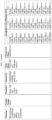
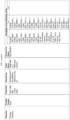
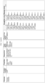
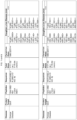
- FIG. 1 Table listing protein targets and peptide sequences used for peptide immunoaffinity enrichment coupled to selected reaction monitoring mass spectrometry (immuno-SRM-MS) for Mucopolysaccharidosis Type I (MPS I; Hurler Syndrome) and Pompe Disease. Total mass, parent ion mass, daughter y-ion masses, and daughter b-ion masses are also shown.
- FIG. 2 Schematic illustrating the process of immuno-SRM-MS.
- FIGs. 3A-3D Schematic illustrating the process of immuno-SRM-MS.
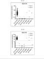
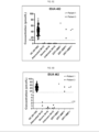
- FIG. 3A IDUA 218 peptide biomarker level in 100 normal controls and 11 MPS I patients (dotted line represents the cutoff for IDUA 218).
- FIG. 3B enlarged version of FIG. 3A with the focus from 0 to 10 pmol/L (dashed line represents the LOD of IDUA 218).
- FIG. 3C IDUA 462 peptide biomarker level in 100 normal controls and 11 MPS I patients (dotted line represents the cutoff for IDUA 462).
- FIG. 3D enlarged version of FIG. 3C with the focus from 0 to 5 pmol/L
- FIGs. 4A, 4B Linear response of IDUA peptides when an internal standard was spiked into the dried blood spot (DBS) matrix (the dotted line represents the lowest level of peptide found in normal cohort).
- FIGs. 5A, 5B IDUA peptide concentrations in DBS and peripheral blood mononuclear cell (PBMC) samples. (DBS: five 3mm punches; PBMC: 250 ⁇ g of protein).
- FIGs 6A, 6B IDUA peptide concentrations in DBS and buccal swab samples.
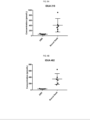
- FIGs. 7A, 7B Comparison of IDUA concentrations among 100 normal controls (NC), 9 MPS I patients (MPS I pt), and 4 MPS I pseudo deficient cases (MPS I Pseudo) for IDUA 218 peptide biomarker ( FIG. 7A ) and IDUA 462 peptide biomarker ( FIG. 7B ).
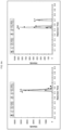
- FIGs. 8A , 8B Multiple reaction monitoring (MRM) traces for GAA peptides from purified peptides (left) and DBS samples (right) after peptide capture by sera antibodies from immunized rabbits.
- MRM Multiple reaction monitoring
- FIG. 8B GAA 855 MRM traces.
- FIG. 9 Endogenous multiple reaction monitoring (MRM) traces for GAA 855 from PBMC sample after peptide capture by supernatant antibodies from isolated plasma cells.
- FIGs. 10A , 10B Multiple reaction monitoring (MRM) traces for GAA peptides from purified peptides (I), DBS samples (II), and buccal swab samples (III) after peptide capture by sera antibodies from immunized rabbits.
- FIG. 10A GAA 155 MRM traces, TTPTFFPK (SEQ ID NO: 3), parent ion mass 469.7527++;
- FIG. 10A GAA 155 MRM traces, TTPTFFPK (SEQ ID NO: 3), parent ion mass 469.7527++;
- FIG. 10B GAA 376 MRM traces, WGYSSTAITR (SEQ ID NO: 5), parent ion mass 571.2855++.
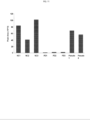
- FIG 11 Comparison of GAA concentrations in DBS among three normal controls (NC1, NC2, NC3), three true positive Pompe patients (PD1, PD2, PD3), and two pseudo-deficient cases (Pseudo 1 and Pseudo 2) for GAA 376 peptide biomarker (WGYSSTAITR (SEQ ID NO: 5), parent ion mass 571.2855++).
- FIG. 12 Comparison of GAA concentrations in DBS among three normal controls (NC1, NC2, NC3), three true positive Pompe patients (PD1, PD2, PD3), and two pseudo-deficient cases for GAA 376 peptide biomarker (WGYSSTAITR (SEQ ID NO: 5), parent ion mass 571.2855++).
- sequences of the disclosure including: SEQ ID NO: Description 16 anti-IDUA 218 variable heavy domain coding sequence with leader sequence 17 anti-IDUA 218 variable heavy domain amino acid sequence with leader sequence 18 anti-IDUA 218 variable heavy domain amino acid sequence without leader sequence 19 anti-IDUA 218 variable light domain coding sequence with leader sequence 20 anti-IDUA 218 variable light domain amino acid sequence with leader sequence 21 anti-IDUA 218 variable light domain amino acid sequence without leader sequence 28 anti-IDUA 462 variable heavy domain coding sequence with leader sequence 29 anti-IDUA 462 variable heavy domain amino acid sequence with leader sequence 30 anti-IDUA 462 variable heavy domain amino acid sequence without leader sequence 31 anti-IDUA 462 variable light domain coding sequence with leader sequence 32 anti-IDUA 462 variable light domain amino acid sequence with leader sequence 33 anti-IDUA 462 variable light domain amino acid sequence without leader sequence 40 anti-IDUA 218 variable heavy domain coding sequence without leader sequence 41 anti-IDUA 218 variable light domain coding sequence without leader sequence 42 anti-IDUA 462 variable heavy domain coding sequence without leader sequence
- LSDs lysosomal storage diseases
- MPS I Mucopolysaccharidosis Type I
- Mucopolysaccharidosis Type I (MPS I; Hurler Syndrome) is a rare LSD belonging to the group of mucopolysaccharidoses.
- MPS I can be further divided into severe and attenuated types.
- the severe form of MPS I is also known as Hurler Syndrome.
- Severe and attenuated forms of MPS I have similar symptoms and complications; however, attenuated forms of MPS I show slower disease progression and/or later age of onset of symptoms.
- subjects with attenuated MPS I do not show early developmental delay and/or do not experience progressive decline in mental capabilities.
- onset of symptoms and complications occur between ages three and ten years for individuals with attenuated MPS I.
- the spectrum of disease severity is broad for individuals with attenuated MPS I, including life-threatening complications leading to death in their twenties to thirties, to a normal life span with significant joint problems and cardiorespiratory disease.
- linear growth decreases by age three years in individuals with severe MPS I.
- mental disability is progressive and profound in individuals with severe MPS I.
- death occurs within the first ten years of life for individuals with severe MPS I.
- diagnosis of individuals with severe versus attenuated MPS I is made with clinical and laboratory findings.
- MPS I may have macrocephaly, a buildup of fluid in the brain, heart valve abnormalities, distinctive-looking facial features, short stature, joint deformities, an enlarged liver and spleen, and a large tongue. Upper respiratory infections and sleep apnea can occur due to a narrowed airway. People with MPS I often develop clouding of the cornea, have hearing loss, and have recurrent ear infections. Children with severe MPS I usually have a shortened lifespan, sometimes living only into late childhood. Heart disease and airway obstruction are major causes of death in people with both severe and attenuated MPS I.
- MPS I is caused by a variation in the IDUA gene, which encodes an alpha-L-iduronidase enzyme that is needed to break down complex carbohydrates produced in the body called glycosaminoglycans (or mucopolysaccharides).
- a deficiency of the IDUA enzyme leads to build up of glycosaminoglycans in the lysosomes of all cells and progressive damage of tissues.
- Therapies for MPS I include: providing the deficient or missing alpha-L-iduronidase enzyme in enzyme replacement therapy (ERT); hematopoietic stem cell transplantation (HSCT); bone marrow transplantation; and alleviating symptoms of the disease.
- Pompe Disease is a rare inherited lysosomal storage disorder that disables the heart and skeletal muscles.
- PD can include infantile onset and late onset.
- the infantile onset PD can be further characterized as classic or non-classic.
- symptoms can emerge within a few months of birth for infants with the classic form of infantile onset PD.
- infants with the classic form of infantile onset PD can have the following symptoms: muscle weakness; poor muscle tone; an enlarged liver; heart defects; failure to gain weight and grow at the expected rate; and/or breathing problems.
- infants with the classic form of infantile onset PD die from heart failure in the first year of life.
- alglucosidase alfa Myozyme ⁇
- Another alglucosidase alfa drug, Lumizyme ⁇ has been approved for late-onset (non-infantile) Pompe Disease.
- compositions and methods can further be used to reduce the number of false positives caused by pseudo deficiency cases in NBS for MPS I and Pompe disease.
- a pseudo deficient allele of a gene associated with an LSD includes one or more mutations (as compared to a corresponding wild type gene) in one copy of the gene that encodes an altered protein or changes the expression of the gene but does not cause disease.
- Individuals with a pseudo deficient allele show greatly reduced enzyme activity, but they are healthy.
- clinically healthy subjects with a pseudo deficient allele of a gene associated with an LSD show reduced activity of the enzyme encoded by the gene in vitro.
- clinically healthy subjects with a pseudo deficient allele of a gene associated with an LSD show reduced activity of the enzyme encoded by the gene in vitro but functional activity of the same enzyme in vivo.
- clinically healthy subjects can have one or two copies of a pseudo deficient allele.
- a false positive result occurs in an enzyme assay test when test results are positive, but disease or morbidity is not present in the tested subject, or disease is present at a subclinical level in the tested subject.
- healthy subjects do not exhibit or exhibit very few symptoms of a disease.
- healthy subjects do not need treatment for a disease.
- healthy subjects with a pseudo deficient allele associated with an LSD do not exhibit or exhibit very few symptoms of the LSD.
- healthy subjects with a pseudo deficient allele associated with an LSD do not need treatment for the LSD.
- carriers have one copy of a pathogenic variant of a gene that causes an LSD and one pseudo deficient allele of the corresponding gene.
- carriers of a pathogenic variant of a gene that causes LSD do not exhibit or exhibit very few symptoms of the LSD.
- carriers of a pathogenic variant of a gene that causes LSD do not need treatment for the LSD.
- Tandem mass spectrometry was first applied to NBS in the 1990s, paving the way for rapid screening of multiple metabolites and thus several diseases from DBS samples collected at birth ( Chace, J Mass Spectrom. Wiley-Blackwell; 2009;44: 163-170 ; Millington et al., J. Inherit. Metab. Dis. 1990;13: 321-324 ; Sweetman et al. Pediatrics. 2006;117: S308-S314 ; Almannai et al., Curr. Opin. Pediatr. 2016;28: 694-699 ; Watson et al., Genet. Med. Nature Publishing Group; 2006. pp. 1S-252S ; Chace et al., Clin.
- SRM-MS Selected reaction monitoring mass spectrometry
- Each patient in the blinded study was deficient in the signature peptide specific for their respective disease (i.e., XLA patient lacking Bruton's Tyrosine Kinase (BTK) and WAS patient missing WAS protein (WASP), etc.).
- BTK Bruton's Tyrosine Kinase
- WASP WAS patient missing WAS protein
- SRM-MS utilizes proteolytically-generated signature peptides as stoichiometric surrogates of a protein of interest. This may, in turn, be used to estimate the number of a particular cell-type expressing that protein in a sample (i.e. quantification of CD3 ⁇ for an indication of the amount of CD3+ T-cells in blood).
- the high specificity of MS for each signature peptide is conferred by three physiochemical properties-its mass, retention times upon high-performance liquid chromatography (HPLC) separation, and resultant target-specific fragmentation patterns ( Kennedy et al. Nat. Methods. 2014;11: 149-155 ).
- Immuno-SRM Peptide immunoaffinity enrichment coupled to SRM-MS is a method that enables precise quantification of low abundance markers.
- Immuno-SRM generally involves the following steps: (i) selection of target proteins that are indicative of the presence or absence of a disorder; (ii) treatment of a biological sample that would include the target protein, if present, with enzymes to digest all proteins in the biological sample into smaller fragments called peptides; (iii) enrichment for selected peptide markers derived from the target protein; and (iv) analysis and quantification of the enriched peptides of interest in a mass spectrometer.
- Immuno-SRM also referred to as Stable Isotope Standards and Capture by Anti-Peptide Antibodies (SISCAPA)
- SISCAPA Stable Isotope Standards and Capture by Anti-Peptide Antibodies
- proteins may be precipitated with acetone, the protein pellet may be resuspended in an appropriate buffer (e.g., guanidine hydrochloride in 95% ethanol supplemented with 2.5% glycerol), the pellet may be dispersed by sonication, the pellet may be centrifuged and washed, the pellet may be dried, and the pellet may be solubilized in an appropriate buffer (e.g., PBS and sodium dodecyl sulfate). In particular embodiments, the solubilized pellet may be heated at 100°C and then centrifuged to obtain a supernatant for use.
- an appropriate buffer e.g., guanidine hydrochloride in 95% ethanol supplemented with 2.5% glycerol
- the solubilized pellet may be heated at 100°C and then centrifuged to obtain a supernatant for use.
- signature proteotypic peptides that are unique to the protein of interest and that are consistently observed in MS experiments are selected to stoichiometrically represent the protein of interest ( Mallick et al. Nat Biotechnol 2007;25: 125-131 ).
- Signature peptides can be selected by detection in previous MS experiments, use of computational tools to predict the peptides most likely observable by MS, or a combination of both.
- tryptic peptides 5-22 amino acids in length with moderate hydrophobicity can be selected.
- Very hydrophilic and very hydrophobic peptides can be less stable due to retention time variation in HPLC and loss to surfaces.
- methionine residues can be undesirable (Whiteaker and Paulovich, Clin Lab Med. 2011;31(3): 385-396 ).
- Shorter peptides and those containing proline residues can be better targets for SRM ( Lange et al., Molecular Systems Biology 2008; 4: 222 ).
- the peptides include portions of IDUA and/or GAA.
- the peptides include SEQ ID NOs: 1-9.
- peptides of the present disclosure include those described in Table 1A-1C and FIG. 1 .
- exemplary CDR sequences of antibodies of the present disclosure are shown in Table 1A.
- exemplary variable heavy (VH) and variable light (VL) domain sequences of antibodies of the present disclosure are shown in Table 1B.
- SEQ ID NOs of exemplary peptides and antibodies of the present disclosure are shown in Table 1C. Table 1A.
- CDR1 RYWMH (SEQ ID NO: 10)
- CDR1 KSSQSLLHSDGKTYLN (SEQ ID NO: 13)
- CDR2 EINPSNGGTNYNEKFKN (SEQ ID NO: 11)
- CDR2 LVSKLDS (SEQ ID NO: 14)
- CDR3 AMDY (SEQ ID NO: 12)
- CDR3 WQGLHFPWT (SEQ ID NO: 15)
- Anti-IDUA 462/ MPS I CDR1: DTYMH (SEQ ID NO: 22)
- variable heavy (VH) and variable light (VL) domain sequences of antibodies of the present disclosure Variable domain SEQ ID NO: Amino acid sequence Anti-IDUA 218 VH 18 Anti-IDUA 218 VL 21 Anti-IDUA 462 VH 30 Anti-IDUA 462 VL 33 Anti-GAA 155 VH 57 Anti-GAA 155 VL 65 Anti-GAA 376 VH 79 Anti-GAA 376 VL 87 Table 1C .
- SEQ ID NOs for exemplary peptides and antibodies of the present disclosure SEQ ID NOs Disease or Target Cell Target Protein Peptides SEQ ID NO VH CDRs VL CDRs VH domain VL domain Mucopolysaccharidosis Type 1 (MPS I) IDUA IDUA 218-230 SEQ ID NO: 1 10-12 13-15 16 -18, 40 19 -21, 41 MPS I IDUA IDUA 462-474 SEQ ID NO: 2 22-24 25-27 28 -30, 42 31 -33, 43 Pompe Disease GAA GAA 155-162 SEQ ID NO: 3 44-46 47-49 52 , 53 , 56, 57 60 , 61 , 64, 65 [50 , 51 , 54, 55] ⁇ [58 , 59 , 62, 63] ⁇ Pompe Disease GAA GAA 332-348 SEQ ID NO: 4 Pompe Disease GAA GAA 376-385 SEQ ID NO: 5 66-68 69-71 74 , 75 , 78, 79 82 , 83
- a biological sample includes DBS, cells from a buccal swab sample, PBMCs, or WBCs.
- proteins in a biological sample can be digested into peptides with trypsin.
- Trypsin cleaves exclusively C-terminal to arginine and lysine residues and can be a preferred choice to generate peptides because the masses of generated peptides are compatible with the detection ability of most mass spectrometers (up to 3000 m/z) and because there are efficient algorithms available for the generation of databases of theoretical trypsin-generated peptides. High cleavage specificity, availability, and low cost are other advantages of trypsin.
- Peptides formed by the treatment of a protein with trypsin are known as tryptic peptides.
- An antibody includes a polypeptide ligand substantially encoded by an immunoglobulin gene or immunoglobulin genes, or functional fragments thereof, whether natural, or partially or wholly synthetically produced.
- An antibody specifically (or selectively) binds and recognizes an epitope (e.g., an antigen).
- An antibody can include any protein having a binding domain that is homologous or largely homologous to an immunoglobulin binding domain.
- An antibody preparation may be monoclonal or polyclonal.
- the antibody may be a member of any immunoglobulin class, including any of the human classes: IgG, IgM, IgA, IgD, and IgE, etc.
- the recognized immunoglobulin genes include the kappa and lambda light chain constant region genes, the alpha, gamma, delta, epsilon and mu heavy chain constant region genes, and the myriad immunoglobulin variable region genes.
- "Fc" portion of an antibody refers to that portion of an immunoglobulin heavy chain that includes one or more heavy chain constant region domains, CH1, CH2 and CH3, but does not include the heavy chain variable region.
- An intact antibody can include at least two heavy (H) chains and two light (L) chains interconnected by disulfide bonds.
- Each heavy chain is composed of a heavy chain variable region (abbreviated herein as VH or V H ) and a heavy chain constant region.
- the heavy chain constant region includes three domains, CH1, CH2 and CH3.
- Each light chain is composed of a light chain variable region (abbreviated herein as VL or V L ) and a light chain constant region.
- the light chain constant region includes one domain, CL.
- the VH and VL regions can be further subdivided into regions of hypervariability, termed complementarity determining regions (CDR), interspersed with regions that are more conserved, termed framework regions (FR).
- CDR complementarity determining regions
- Each VH and VL is composed of three CDRs and four FRs, arranged from amino-terminus to carboxy-terminus in the following order: FR1, CDR1, FR2, CDR2, FR3, CDR3, FR4.
- the variable regions of the heavy and light chains contain a binding domain that interacts with an antigen.
- the constant regions of the antibodies can mediate the binding of the immunoglobulin to host tissues or factors, including various cells of the immune system (e.g., effector cells) and the first component (Clq) of the classical complement system.
- an exemplary antibody includes a heavy chain or light chain coding sequence with a leader sequence. In particular embodiments, an exemplary antibody includes a variable heavy domain or variable light domain coding sequence with a leader sequence. In particular embodiments, an exemplary antibody includes a heavy chain or light chain amino acid sequence with a leader peptide. In particular embodiments, an exemplary antibody includes a heavy chain or light chain amino acid sequence without a leader peptide. In particular embodiments, an exemplary antibody includes a variable heavy domain or variable light domain amino acid sequence with a leader peptide. In particular embodiments, an exemplary antibody includes a variable heavy domain or variable light domain amino acid sequence without a leader peptide.
- (V) Variants Variants of the sequences disclosed and referenced herein are also included.
- Functional variants include one or more residue additions or substitutions that do not substantially impact the physiological effects of the protein.
- Functional fragments include one or more deletions or truncations that do not substantially impact the physiological effects of the protein. A lack of substantial impact can be confirmed by observing experimentally comparable results in a binding study.
- Functional variants and functional fragments of binding domains bind their cognate antigen or ligand at a level comparable to a wild-type reference.
- amino acid changes in the protein variants disclosed herein are conservative amino acid changes, i.e., substitutions of similarly charged or uncharged amino acids.
- a conservative amino acid change involves substitution of one of a family of amino acids which are related in their side chains.
- Suitable conservative substitutions of amino acids are known to those of skill in this art and generally can be made without altering a biological activity of a resulting molecule.
- Those of skill in this art recognize that, in general, single amino acid substitutions in non-essential regions of a polypeptide do not substantially alter biological activity (see, e.g., Watson et al. Molecular Biology of the Gene, 4th Edition, 1987, The Benjamin/Cummings Pub. Co., p. 224 ).
- Naturally occurring amino acids are generally divided into conservative substitution families as follows: Group 1: Alanine (Ala), Glycine (Gly), Serine (Ser), and Threonine (Thr); Group 2: (acidic): Aspartic acid (Asp), and Glutamic acid (Glu); Group 3: (acidic; also classified as polar, negatively charged residues and their amides): Asparagine (Asn), Glutamine (Gln), Asp, and Glu; Group 4: Gln and Asn; Group 5: (basic; also classified as polar, positively charged residues): Arginine (Arg), Lysine (Lys), and Histidine (His); Group 6 (large aliphatic, nonpolar residues): Isoleucine (Ile), Leucine (Leu), Methionine (Met), Valine (Val) and Cysteine (Cys); Group 7 (uncharged polar): Tyrosine (Tyr), Gly, Asn, Gln, Cys, Ser,
- An insertion, deletion or substitution may be anywhere in the VH region, including at the amino- or carboxy-terminus or both ends of this region, provided that each CDR includes zero changes or at most one, two, or three changes and provided a binding domain containing the modified VH region can still specifically bind its target with an affinity similar to the wild type binding domain.
- a VL region in a binding domain is derived from or based on a VL of a known antibody or an antibody disclosed herein and optionally contains one or more (e.g., 2, 3, 4, 5, 6, 7, 8, 9, 10) insertions, one or more (e.g., 2, 3, 4, 5, 6, 7, 8, 9, 10) deletions, one or more (e.g., 2, 3, 4, 5, 6, 7, 8, 9, 10) amino acid substitutions (e.g., conservative amino acid substitutions), or a combination of the above-noted changes, when compared with the VL of the known antibody or antibody disclosed herein.
- one or more e.g., 2, 3, 4, 5, 6, 7, 8, 9, 10 insertions, one or more (e.g., 2, 3, 4, 5, 6, 7, 8, 9, 10) deletions, one or more (e.g., 2, 3, 4, 5, 6, 7, 8, 9, 10) amino acid substitutions (e.g., conservative amino acid substitutions), or a combination of the above-noted changes, when compared with the VL of the known antibody or antibody disclosed here
- An insertion, deletion or substitution may be anywhere in the VL region, including at the amino- or carboxy-terminus or both ends of this region, provided that each CDR includes zero changes or at most one, two, or three changes and provided a binding domain containing the modified VL region can still specifically bind its target with an affinity similar to the wild type binding domain.
- % sequence identity refers to a relationship between two or more sequences, as determined by comparing the sequences.
- identity also means the degree of sequence relatedness between protein, nucleic acid, or gene sequences as determined by the match between strings of such sequences.
- Identity (often referred to as “similarity") can be readily calculated by known methods, including (but not limited to) those described in: Computational Molecular Biology (Lesk, A. M., ed.) Oxford University Press, NY (1988 ); Biocomputing: Informatics and Genome Projects (Smith, D. W., ed.) Academic Press, NY (1994 ); Computer Analysis of Sequence Data, Part I (Griffin, A. M., and Griffin, H.
- Relevant programs also include the GCG suite of programs (Wisconsin Package Version 9.0, Genetics Computer Group (GCG), Madison, Wisconsin); BLASTP, BLASTN, BLASTX ( Altschul, et al., J. Mol. Biol. 215:403-410 (1990 ); DNASTAR (DNASTAR, Inc., Madison, Wisconsin); and the FASTA program incorporating the Smith-Waterman algorithm ( Pearson, Comput. Methods Genome Res., [Proc. Int.
- Magnetic particles are available in a wide array of chemistries allowing for coupling to antibodies. Enrichment with antibodies attached to particles can allow parallel processing of samples. Magnetic particle processing has been automated in 96 well plates for the SISCAPA enrichment step with elution in the plates for analysis by mass spectrometry. Other particular embodiments use a novel bead trap device developed to perform the bead handling steps in line with a nanoflow chromatography system ( Anderson et al. Mol Cell Proteomics 2009;8(5): 995-1005 ). This minimizes losses of peptides to containers between elution and analysis steps.
- Peptide enrichment can also be implemented by immobilizing anti-peptide antibodies in pipet tips ( Nelson et al., Anal Chem. 1995;67(7): 1153-1158 ). After separation of the antibody bound peptide from free peptides, the bound peptide can be eluted. Any elution means can be used. One elution means which has been found to be efficient is 5% acetic acid/3% acetonitrile. Other elution means, including other acids, and other concentrations of acetic acid can be used, as is efficient for a particular peptide.
- VI Liquid Chromatography
- one or more LC purification steps are performed prior to SRM-MS.
- a mixture of enriched peptides (the mobile phase) can be passed through a column packed with material (stationary phase) to separate the peptides based on their weight and affinity for the mobile and stationary phases of the column.
- Traditional LC analysis relies on the chemical interactions between sample components and column packing materials, where laminar flow of the sample through the column is the basis for separation of the analyte of interest from the test sample. The skilled artisan will understand that separation in such columns is a diffusional process.
- column packing materials are available for chromatographic separation of samples, and selection of an appropriate separation protocol is an empirical process that depends on the sample characteristics, the analyte of interest, the interfering substances present and their characteristics, etc.
- Various packing chemistries can be used depending on the needs (e.g., structure, polarity, and solubility of compounds being purified).
- the columns are polar, ion exchange (both cation and anion), hydrophobic interaction, phenyl, C-2, C-8, C-18 columns, polar coating on porous polymer, or others that are commercially available.
- an analyte may be purified by applying a sample to a column under conditions where the analyte of interest is reversibly retained by the column packing material, while one or more other materials are not retained.
- a first mobile phase condition can be employed where the analyte of interest is retained by the column, and a second mobile phase condition can subsequently be employed to remove retained material from the column, once the non-retained materials are washed through.
- a mass spectrometer includes a gas phase ion spectrometer that measures a parameter that can be translated into mass-to-charge (m/z) ratios of gas phase ions. Mass spectrometry refers to the use of a mass spectrometer to detect gas phase ions. Mass spectrometers generally include an ion source and a mass analyzer. Examples of mass spectrometers are time-of-flight (TOF), magnetic sector, quadrupole filter, ion trap, ion cyclotron resonance, electrostatic sector analyzer and hybrids of these.
- TOF time-of-flight
- magnetic sector magnetic sector
- quadrupole filter quadrupole filter
- ion trap ion trap
- ion cyclotron resonance electrostatic sector analyzer and hybrids of these.
- the fragment ion(s) is generated through collisional induced dissociation in the second quadrupole.
- the two levels of mass selection allow high selectivity, as co-eluting background ions are filtered out very effectively.
- SRM analysis selectively targets (filters) particular analytes, which translates into an increased sensitivity by one or two orders of magnitude compared with conventional 'full scan' techniques.
- SRM provides a linear response over a wide dynamic range up to five orders of magnitude. This enables the detection of low-abundance proteins in highly complex mixtures. Therefore, SRM is a highly specific detection/monitoring method with low background interference.
- MRM multiple reaction monitoring
- SRM/MRM-MS Selected reaction monitoring/multiple reaction monitoring mass spectrometry
- the following parameters can be used to specify an LC-SRM-MS assay of a protein under a particular LC-SRM-MS system: (1) an enriched tryptic peptide of a given protein; (2) the retention time (RT) of the peptide on an LC column; (3) the m/z value of the peptide precursor ion; (4) the declustering potential used to ionize the precursor ion; (5) the m/z value of a fragment ion generated from the peptide precursor ion; and (6) the collision energy (CE) used to fragment the peptide precursor ion that is optimized for the particular peptide.
- RT includes the elapsed time between injection and elution of an analyte.
- Declustering potential includes a voltage potential to dissolvate and dissociate ion clusters. It is also known as “fragmentor voltage” or “ion transfer capillary offset voltage” depending on the manufacturer.
- Collision energy includes the amount of energy precursor ions receive as they are accelerated into the collision cell.
- a set of isotopically labeled synthetic versions of the peptides of interest may be added in known amounts to the sample for use as internal standards. Since the isotopically labeled peptides have physical and chemical properties identical to the corresponding surrogate peptide, they co-elute from the chromatographic column and are easily identifiable on the resultant mass spectrum ( Gerber et al. Proc. Natl. Asso. Sci. 2003;100: 6940-6945 ; Kirkpatrick et al. Methods 2005; 35: 265-273 ).
- the isotopes with which amino acids in a given peptide can be labeled include 13 C, 2 H, 15 N, 17 O, 18 O, and 34 S.
- a peptide is labeled with 13 C and/or 15 N heavy isotopes.
- the addition of the labeled standards may occur before or after proteolytic digestion.
- the labeled internal standard peptides are added after proteolytic digestion. Methods of synthesizing isotopically labeled peptides will be known to those of skill in the art.
- the experimental samples contain internal standard peptides.
- internal standard peptides include reference signature peptides.
- a signature peptide concentration can be determined by combining: (i) a ratio calculated from comparing the peak area of the signature peptide to the peak area of its corresponding reference signature peptide obtained from an LC-MRM-MS assay, and (ii) the known concentration of the reference signature peptide.
- Peptides selected as reference standards and suitable for quantification are sometimes referred to as quantotypic peptides (Q-peptides).
- Q-peptides include all of the characteristics of proteotypic peptides but also place restrictions on the residues that can constitute the reference peptide to eradicate artefactual modification and/or incomplete cleavage ( Holman et al. Bioanalysis 2012;4(14): 1763-1786 ).
- Absolute quantitative levels of a given protein, or proteins can be determined by the SRM/MRM methodology whereby the SRM/MRM signature peak area of an individual peptide from a given protein in one biological sample is compared to the SRM/MRM signature peak area of a known amount of a "spiked" internal standard.
- the internal standard is a synthetic version of the same exact peptide that contains one or more amino acid residues labeled with one or more heavy isotopes.
- isotope labeled internal standards are synthesized so that mass spectrometry analysis generates a predictable and consistent SRM/MRM signature peak that is different and distinct from the native peptide signature peak, and which can be used as a comparator peak.
- the signature peak area of the native peptide is compared to the signature peak area of the internal standard peptide, and this numerical comparison indicates either the absolute molarity and/or absolute weight of the native peptide present in the original protein preparation from the biological sample.
- Absolute quantitative data for fragment peptides are displayed according to the amount of protein analyzed per sample. Absolute quantitation can be performed across many peptides, and thus proteins, simultaneously in a single sample and/or across many samples to gain insight into absolute protein amounts in individual biological samples and in entire cohorts of individual samples.
- Another strategy for absolute quantitation of peptides is equimolarity through equalizer peptide.
- This methodology involves chemically synthesizing the isotopically labeled Q-peptides of interest as dipeptides.
- a common amino acid sequence is positioned N-terminal to the Q-peptide and is referred to as the equalizer peptide.
- the amount of Q-peptide can be accurately determined through reference to a single light-labeled peptide.
- Appropriate amounts of each standard peptide can then be added to a sample of interest (either predigested or prior to proteolysis) to facilitate absolute quantification ( Holzmann et al. Anal. Chem. 2009;81: 10254-10261 ).
- Absolute quantification can also employ quantification concatemer (QconCAT) proteins ( Beynon et al. Nat. Methods 2005;2: 587-589 ; Johnson et al. J. Am. Soc. Mass Spectrom. 2009;20: 2211-2220 ; Ding et al. J. Proteome Res. 2011;10: 3652-3659 ; Carroll et al. Molecular & Cellular Proteomics 2011;Sep 19: mcp-M111 ).
- QconCAT quantification concatemer
- the QconCAT protein is then affinity purified and co-digested with the sample, generating a stoichiometric mixture of all the 'heavy' Q-peptides of which it is composed, and the proteolytic peptides from the native proteins and internal standard are subsequently analyzed.
- a variant of the QconCAT approach termed peptide-concatenated standards (PCS)
- PCS peptide-concatenated standards
- PCS peptide-concatenated standards
- PSAQ protein standards for absolute quantification
- PSAQ uses recombinant proteins but rather than being a concatenation of peptides from several proteins, the entire protein to be quantified is expressed in stable isotope-labeled form. One or several PSAQs can then be added to the sample predigestion to facilitate quantification.
- Particular embodiments use label-free strategies for protein quantification such as intensity-based measurements ( America and Cordewener, Proteomics 2008;8: 731-749 ) or spectral counting ( Lundgren et al. Expert Rev. Proteomics 2010;7: 39-53 ).
- relative quantitation can be determined for a given peptide, or peptides, from a single protein within a single sample by comparing the signature peak area for that peptide for that given protein by SRM/MRM methodology to the signature peak area for another different peptide, or peptides, from a different protein within the same protein preparation from the biological sample. In this way, the amount of a particular peptide from a given protein, and therefore the amount of the given protein, is determined relative to another protein within the same sample.
- Signature peptide levels can be expressed in concentration units (e.g., pmol/L).
- the mean concentration of a signature peptide in a test sample derived from a subject being screened for MPS I and/or Pompe Disease can be compared to the mean concentration of the corresponding peptide in a normal control sample.
- a normal control sample can be derived from one or more normal control subjects or from a population of normal control subjects.
- a normal control subject includes a subject who does not have or is not known to have MPS I and/or Pompe Disease.
- a normal control subject includes a subject who does not have genetic mutations associated with MPS I or Pompe Disease.
- the mean concentration of an IDUA 218 signature peptide in DBS from a population of normal control subjects includes a concentration in a range of 10 pmol/L to 350 pmol/L, in a range of 15 pmol/L to 300 pmol/L, and in a range of 20 pmol/L to 250 pmol/L.
- the mean concentration of an IDUA 218 signature peptide in DBS from a population of normal control subjects includes a concentration of 10 pmol/L, 15 pmol/L, 20 pmol/L, 25 pmol/L, 30 pmol/L, 35 pmol/L, 40 pmol/L, 45 pmol/L, 50 pmol/L, 55 pmol/L, 60 pmol/L, 65 pmol/L, 70 pmol/L, 75 pmol/L, 80 pmol/L, 85 pmol/L, 90 pmol/L, 95 pmol/L, 100 pmol/L, 110 pmol/L, 120 pmol/L, 130 pmol/L, 140 pmol/L, 150 pmol/L, 160 pmol/L, 170 pmol/L, 180 pmol/L, 190 pmol/L, 200 pmol/L, 210 pmol/L, 220 pmol/L,
- the mean concentration of an IDUA 218 signature peptide in PBMCs from a population of normal control subjects includes a concentration in a range of 300 pmol/L to 1000 pmol/L, in a range of 350 pmol/L to 800 pmol/L, and in a range of 400 pmol/L to 700 pmol/L.
- the mean concentration of an IDUA 218 signature peptide in PBMCs from a population of normal control subjects includes a concentration of 300 pmol/L, 310 pmol/L, 320 pmol/L, 330 pmol/L, 340 pmol/L, 350 pmol/L, 360 pmol/L, 370 pmol/L, 380 pmol/L, 390 pmol/L, 400 pmol/L, 410 pmol/L, 420 pmol/L, 430 pmol/L, 440 pmol/L, 450 pmol/L, 460 pmol/L, 470 pmol/L, 480 pmol/L, 490 pmol/L, 500 pmol/L, 510 pmol/L, 520 pmol/L, 530 pmol/L, 540 pmol/L, 550 pmol/L, 560 pmol/L, 570 pmol/L,
- the mean concentration of an IDUA 218 signature peptide in buccal swab samples from a population of normal control subjects includes a concentration in a range of 100 pmol/L to 1000 pmol/L, in a range of 100 pmol/L to 900 pmol/L, and in a range of 100 pmol/L to 800 pmol/L.
- the mean concentration of an IDUA 218 signature peptide in buccal swab samples from a population of normal control subjects includes a concentration of 100 pmol/L, 125 pmol/L, 150 pmol/L, 175 pmol/L, 200 pmol/L, 225 pmol/L, 250 pmol/L, 275 pmol/L, 300 pmol/L, 325 pmol/L, 350 pmol/L, 375 pmol/L, 400 pmol/L, 425 pmol/L, 450 pmol/L, 475 pmol/L, 500 pmol/L, 525 pmol/L, 550 pmol/L, 575 pmol/L, 600 pmol/L, 625 pmol/L, 650 pmol/L, 675 pmol/L, 700 pmol/L, 725 pmol/L, 750 pmol/L, 775 pmol/L, 800
- the mean concentration of an IDUA 462 signature peptide in PBMCs from a population of normal control subjects includes a concentration in a range of 350 pmol/L to 1000 pmol/L, in a range of 400 pmol/L to 900 pmol/L, and in a range of 450 pmol/L to 850 pmol/L.
- the mean concentration of an IDUA 462 signature peptide in buccal swab samples from a population of normal control subjects includes a concentration in a range of 30 pmol/g of protein to 80 pmol/g of protein, in a range of 30 pmol/g of protein to 75 pmol/g of protein, and in a range of 30 pmol/g of protein to 70 pmol/g of protein.
- the mean concentration of an IDUA 462 signature peptide in buccal swab samples from a population of normal control subjects includes a concentration of 30 pmol/g of protein, 35 pmol/g of protein, 40 pmol/g of protein, 45 pmol/g of protein, 50 pmol/g of protein, 55 pmol/g of protein, 60 pmol/g of protein, 65 pmol/g of protein, 70 pmol/g of protein, 75 pmol/g of protein, 80 pmol/g of protein, or more.
- the mean concentration of a GAA 376 signature peptide in DBS from a population of normal control subjects includes a concentration in a range of 25 pmol/L to 200 pmol/L, in a range of 30 pmol/L to 180 pmol/L, and in a range of 35 pmol/L to 160 pmol/L.
- the mean concentration of a GAA 376 signature peptide in DBS from a population of normal control subjects includes a concentration of 25 pmol/L, 30 pmol/L, 35 pmol/L, 40 pmol/L, 45 pmol/L, 50 pmol/L, 55 pmol/L, 60 pmol/L, 65 pmol/L, 70 pmol/L, 75 pmol/L, 80 pmol/L, 85 pmol/L, 90 pmol/L, 95 pmol/L, 100 pmol/L, 110 pmol/L, 120 pmol/L, 130 pmol/L, 140 pmol/L, 150 pmol/L, 160 pmol/L, 170 pmol/L, 180 pmol/L, 190 pmol/L, 200 pmol/L, or more.
- One or more standard peptides may be synthesized with any method known in the pertinent art. Such synthetic peptides may further include amino acids with one or more natural modifications. Such natural modifications may include deamination of glutamine and asparagine, amination, oxidation, and hydroxylation.
- the methods of the present disclosure include identifying individuals with MPS I and/or Pompe Disease. In particular embodiments, diagnosing individuals with MPS I and/or Pompe Disease, is performed early, for example, as part of NBS, or before symptoms of a disorder are evident in the individual. In particular embodiments, the methods of the present disclosure include identifying individuals with a severe form of MPS I. In particular embodiments, the methods of the present disclosure include identifying individuals with an attenuated form of MPS I. In particular embodiments, the methods of the present disclosure include differentiating individuals with a severe form of MPS I from individuals with an attenuated form of MPS I. In particular embodiments, the methods of the present disclosure include identifying individuals with infantile onset Pompe Disease. In particular embodiments, the methods of the present disclosure include identifying individuals with late onset Pompe Disease. In particular embodiments, the methods of the present disclosure include differentiating individuals with a infantile onset Pompe Disease from individuals with late onset Pompe Disease.
- the methods of the present disclosure include obtaining DBS, buccal swab, PBMC, or WBC samples.
- DBS, buccal swab, PBMC, or WBC samples are obtained according to a method described herein.
- DBS, buccal swab, PBMC, or WBC samples are obtained from a DBS, a buccal swab, a PBMC, or a WBC repository or lab that stores DBS, buccal swab, PBMC, or WBC samples for future testing.
- a biological sample includes DBS, cells from buccal swabs, PBMCs, or WBCs.
- one or more punches of the DBS, whole DBS, cells from buccal swabs, PBMCs, or WBCs can be solubilized in an appropriate buffer, and an appropriate digestion enzyme described herein can be added to digest proteins present in DBS, cells from buccal swabs, PBMCs, or WBCs into peptide fragments.
- DBS, cells from buccal swabs, PBMCs, or WBCs can be solubilized with 0.1% Triton X-100 in 50 mM ammonium bicarbonate and digested with trypsin.
- the methods of the present disclosure include enriching for signature peptides that are used in screening for MPS I and/or Pompe Disease.
- Signature peptides include: IDUA 218 for MPS I; IDUA 462 for MPS I; GAA 155 for Pompe Disease; GAA 332 for Pompe Disease; GAA 376 for Pompe Disease; GAA 601 for Pompe Disease; GAA 855 for Pompe Disease; GAA 882 for Pompe Disease; and GAA 892 for Pompe Disease.
- enriching for signature peptides include contacting mixtures of peptide fragments from a digested biological sample with one or more binding entities that recognize the signature peptides.
- a biological sample includes DBS, cells from buccal swabs, PBMCs, or WBCs.
- the binding entities are antibodies or antigen binding fragments thereof.
- the antibodies include those disclosed in Table 1A-1C and FIG. 12 .
- amino acid sequences of antibodies of the disclosure include SEQ ID NOs: 10-15, 17, 18, 20, 21, 22-27, 29, 30, 32, 33, 44-49, 54-57, 62-71, 76-79, and 84-87.
- coding sequences of antibodies of the disclosure include SEQ ID NOs: 16, 19, 28, 31, 40-43, 50-53, 58-61, 72-75, and 80-83.
- the antibodies include antibodies that bind: IDUA 218, IDUA 462, GAA 155, GAA 332, GAA 376, GAA 601, GAA 855, GAA 882, and GAA 892.
- antibodies including SEQ ID NOs: 10-15, 17, 18, 20, and 21 are used to enrich for an IDUA peptide including SEQ ID NO: 1.
- antibodies including SEQ ID NOs: 22-27, 29, 30, 32, and 33 are used to enrich for an IDUA peptide including SEQ ID NO: 2.
- antibodies including SEQ ID NOs: 44-49, 54-57, and 62-65 are used to enrich for a GAA peptide including SEQ ID NO: 3.
- antibodies including SEQ ID NOs: 66-71, 76-79, and 84-87 are used to enrich for a GAA peptide including SEQ ID NO: 5.
- antibodies are used to enrich for a GAA peptide including SEQ ID NO: 4. In particular embodiments, antibodies are used to enrich for a GAA peptide including SEQ ID NO: 6. In particular embodiments, antibodies are used to enrich for a GAA peptide including SEQ ID NO: 7. In particular embodiments, antibodies are used to enrich for a GAA peptide including SEQ ID NO: 8. In particular embodiments, antibodies are used to enrich for a GAA peptide including SEQ ID NO: 9.
- any combination of one or more antibodies disclosed in Table 1A-1C and FIG. 12 that bind their cognate signature peptides can be used to screen for MPS I.
- any combination of one or more antibodies disclosed in Table 1A-1C and FIG. 12 that bind their cognate signature peptides can be used to screen a population for MPS I.
- any combination of one or more antibodies disclosed in Table 1A-1C and FIG. 12 that bind their cognate signature peptides can be used to screen for Pompe Disease.
- any combination of one or more antibodies disclosed in Table 1A-1C and FIG. 12 that bind their cognate signature peptides can be used to screen a population for Pompe Disease.
- the methods of the present disclosure include optionally performing liquid chromatography on the immunoaffinity enriched peptides to separate the peptides prior to MS analysis.
- Liquid chromatography can separate peptides based on their weight and affinity for the mobile and stationary phases of the column.
- the methods of the present disclosure include performing SRM-MS or MRM-MS on the immunoaffinity enriched peptides to quantify the amount of a given signature peptide.
- the SRM-MS or MRM-MS is carried out as described herein.
- the quantification of a signature peptide includes using a reference peptide that is introduced into an assay in known amounts.
- a reference peptide can be identical to the signature peptide in every respect except that the reference peptide has been differentially labeled, for example, with one or more heavy isotopes, to distinguish the reference peptide from the signature peptide.
- SRM-MS or MRM-MS detects a reduction or absence in an IDUA peptide.
- the IDUA peptide includes SEQ ID NO: 1.
- the IDUA peptide includes SEQ ID NO: 2.
- SRM-MS or MRM-MS detects a reduction or absence in a GAA peptide.
- the GAA peptide includes SEQ ID NO: 3.
- the GAA peptide includes SEQ ID NO: 4.
- the GAA peptide includes SEQ ID NO: 5.
- the GAA peptide includes SEQ ID NO: 6.
- the GAA peptide includes SEQ ID NO: 7.
- the GAA peptide includes SEQ ID NO: 8.
- the GAA peptide includes SEQ ID NO: 9.
- Particular embodiments include monitoring subjects for signature peptide levels using immuno-SRM as described herein over a period of time.
- a subject is selected for monitoring according to the systems and methods disclosed herein because they exhibit signs or symptoms of MPS I and/or Pompe Disease as described herein or are undergoing treatment for MPS I and/or Pompe Disease.
- Particular embodiments disclosed herein include determining efficacy of a treatment in a subject being treated for MPS I and/or Pompe Disease including obtaining biological samples derived from the subject prior to one or more treatments and during and/or after one or more treatments; detecting signature peptide levels in the subject prior to the treatment using immuno-SRM described herein; detecting signature peptide levels in the subject during or after the one or more treatments using immuno-SRM described herein; and determining that the treatment is effective if the signature peptide levels during or after the treatment is higher than the signature peptide levels prior to the treatment, or determining that the treatment is not effective if the signature peptide levels during or after the treatment is equal to or lower than the signature peptide levels prior to the treatment.
- the biological sample includes DBS, cells from buccal swabs, PBMC, or WBC.
- determining efficacy of a treatment in a subject being treated for MPS I and/or Pompe Disease can guide whether the one or more treatments should be continued or discontinued, or whether a new treatment should be implemented.
- one or more treatments can be continued if the signature peptide levels in the subject during or after the one or more treatments is higher than the signature peptide levels in the subject prior to the one or more treatments.
- the one or more treatments can be discontinued if the signature peptide levels during or after the one or more treatments in the subject is greater than 1%, greater than 5%, greater than 10%, greater than 15%, greater than 20%, greater than 25%, greater than 30%, greater than 35%, greater than 40%, greater than 45%, greater than 50%, greater than 55%, greater than 60%, greater than 65%, greater than 70%, greater than 75%, greater than 80%, greater than 85%, greater than 90%, greater than 95%, greater than 100%, or more of signature peptide levels measured in a normal control subject or control subject unaffected by MPS I and/or Pompe Disease.
- a new treatment can be implemented if the signature peptide levels in the subject during or after the treatment is equal to or lower than the signature peptide levels in the subject prior to the treatment or if the signature peptides are absent.
- “stable" measures of signature peptide levels are measures evaluated in relation to a previous comparison in the same subject and denote a signature peptide level that has not changed significantly (as determined by a statistical measure known in the art such as a t-test or p-value, e.g., p value >0.05) since the last measurement.
- “stable” measures are measures evaluated in relation to a previous comparison in the same patient and denote a signature peptide level that has not changed significantly (as determined by a statistical measure known in the art such as a t-test or p-value, e.g., p value >0.05) since an aggregated or averaged group of previous measurements (e.g., the last 3, 4, or 5 measurements).
- Unchanged measures of signature peptide levels are measures evaluated in relation to a previous comparison in the same patient and denote a failure to achieve a statistically significant change in a score towards or away from a reference signature peptide level in the particular subject.
- "unchanged” measures are measures evaluated in relation to a previous comparison in the same patient or since an aggregated or averaged group of previous measurements (e.g., the last 3, 4, or 5 measurements).
- an infused enzyme drug can be recognized as foreign by a subject, triggering an immune response.
- developing an immune response includes developing neutralizing antidrug antibodies to the provided exogenous enzyme drug.
- an immune response to ERT for MPS I and/or Pompe Disease can be predicted by quantification of the ERT-na ⁇ ve enzyme concentrations in a biological sample.
- the biological sample includes DBS, cells from buccal swabs, PBMC, or WBC.
- an immune response to ERT for MPS I occurs when IDUA 218 and/or IDUA 462 are absent.
- an immune response to ERT for Pompe Disease occurs when GAA 155, GAA 332, GAA 376, GAA 601, GAA 855, GAA 882, GAA 892, or a combination thereof, are absent.
- strategies to treat an immune response to ERT can include the prevention of antidrug antibody formation in ERT-naive patients and acute reduction of existing antidrug antibodies.
- prevention of antidrug antibody formation in ERT-na ⁇ ve patients can include B cell depletion, immunosuppression, mTOR (mechanistic target of rapamycin) inhibition, and/or immunomodulation.
- prevention of antidrug antibody formation in ERT-na ⁇ ve patients can include administration of methotrexate (MTX), rituximab, and/or intravenous immunoglobulin (IVIG) before ERT initiation or during ERT.
- acute reduction of existing antidrug antibodies can include inhibition of the folic acid metabolism (blocking de novo DNA synthesis), alkylation of DNA (blocking DNA replication), antibody-mediated specific B cell depletion, B cell/plasma cell depletion, and/or immunosuppression proteasome inhibition.
- acute reduction of existing antidrug antibodies can include administration of mycophenolate mofetil, MTX, IVIG, rituximab, bortezomib, cyclophosphamide, and/or plasma exchange.
- the disclosed assays and methods allow a reduction in the number of false positive results in assays arising from pseudo deficiency cases in LSD.
- the disclosed assays and methods allow an increase in the positive predictive rate (i.e., positive predictive value) of screening for LSDs.
- the methods allow use of the disclosed immuno-SRM assays as a primary or second-tier assay to reduce the number of false positives obtained in screening of LSDs.
- the disclosed assays and methods distinguish cases of enzyme pseudo deficiency from confirmed LSD cases because the disclosed assays and methods can measure signature peptide concentration differences between cases of enzyme pseudo deficiency and confirmed LSD cases.
- a false positive result occurs when an assay result for a subject being screened for a disease is positive (i.e., indicating that a subject has a disease), but the subject is healthy.
- healthy subjects do not exhibit or exhibit very few symptoms of a disease.
- healthy subjects do not need treatment for a disease.
- the positive predictive rate i.e., positive predictive value refers to the probability that subjects with a positive screening test truly have the disease.
- patients with a pseudo deficient allele show greatly reduced enzyme activity, but they are healthy.
- patients with an enzyme pseudo deficiency for an LSD show reduced enzyme activity as compared with normal controls and show similar enzyme activity to patients confirmed to have an LSD (a 'true positive' patient).
- a subject is confirmed to have an LSD by molecular genetic testing of the subject for the presence of two copies of the same or different pathogenic variant(s) of the gene associated with the LSD.
- a subject is confirmed to be have a pseudo deficiency in an LSD by molecular genetic testing.
- Molecular genetic testing can include an assay that can detect mutations, deletions, and/or insertions in a gene, including sequencing, fluorescence in situ hybridization, single nucleotide polymorphism microarray, and polymerase chain reaction (PCR).
- an immuno-SRM assay allows differentiation of subjects who have a pseudo deficient allele in an LSD versus a subject who has the LSD.
- a subject has an LSD when they have two copies of the same or different pathogenic variant(s) of a gene associated with the LSD.
- a subject who has an LSD is considered to be a 'true positive'.
- a subject is identified by an immuno-SRM assay to be a true positive for an LSD when a measured signature peptide concentration is lower than a predetermined threshold concentration for that measured signature peptide.
- a predetermined threshold concentration can be calculated from a standard deviation of the mean concentration of each signature peptide in corresponding biological samples from a population of normal control subjects as described herein.
- a subject is identified by an immuno-SRM assay to be a true positive for an LSD when a measured signature peptide concentration is below a lower limit of detection (LOD) concentration for that signature peptide.
- LSD lower limit of detection
- a subject having a pseudo deficiency in an LSD has a measured signature peptide concentration that is higher than the corresponding mean signature peptide concentration of a subject who is a true positive for, or confirmed to have, the LSD.
- a subject having a pseudo deficiency in an LSD has a measured signature peptide concentration that is within a range of the corresponding signature peptide concentration from a population of normal control subjects.
- An LOD of a signature peptide of the present disclosure includes the lowest concentration of the signature peptide that can reliably be detected using an immuno-SRM assay.
- an LOD of a signature peptide includes the lowest concentration of the signature peptide that can be statistically distinguished from a negative control sample that does not include the signature peptide. Calculating an LOD is known to one of skill in the art.
- a negative control sample that does not include the signature peptide can be assayed 20 times in an immuno-SRM assay, and the mean and standard deviation of the results are calculated.
- an LOD is considered to be 2 standard deviations or 3 standard deviations above the mean of the negative control.
- an LOD for an IDUA 218 signature peptide includes 10 pmol/L or less, 9 pmol/L or less, 8 pmol/L or less, 7 pmol/L or less, 6 pmol/L or less, 5 pmol/L or less, 4 pmol/L or less, 3 pmol/L or less, 2 pmol/L or less, 1 pmol/L or less, or less.
- an LOD for an IDUA 218 signature peptide includes 3.5 pmol/L or less.
- an LOD for a GAA 155 signature peptide includes 10 pmol/L or less, 9 pmol/L or less, 8 pmol/L or less, 7 pmol/L or less, 6 pmol/L or less, 5 pmol/L or less, 4 pmol/L or less, 3 pmol/L or less, 2 pmol/L or less, 1 pmol/L or less, or less.
- a subject is a true positive for MPS I) when a measured signature peptide concentration of IDUA 218, IDUA 462, or a combination thereof, is below LOD concentration for the corresponding signature peptide.
- a subject having a pseudo deficiency in MPS I has measured signature peptide concentrations of IDUA 218, IDUA 462, or a combination thereof, that are higher than the corresponding mean IDUA peptide concentrations from samples from a subject or a population of subjects confirmed to have, or are true positives for, MPS I.
- compositions may include administering compositions as a "prophylactic treatment.”
- Prophylactic treatments include those administered to a subject who does not display signs or symptoms of MPS I and/or Pompe Disease or displays only early signs or symptoms of MPS I and/or Pompe Disease, such that treatment is administered for the purpose of diminishing or decreasing the risk of developing the disorder.
- a prophylactic treatment functions as a preventative treatment against MPS I and/or Pompe Disease.
- a prophylactic treatment can prevent, delay, or reduce the onset of MPS I and/or Pompe Disease. In particular embodiments, a prophylactic treatment can prevent or reduce the severity of symptoms or complications associated with MPS I and/or Pompe Disease.
- Symptoms and complications for MPS I include weight loss, congestion, repeated vomiting, reflux, and/or skin rashes.
- Symptoms and complications for Pompe Disease include weak muscles, enlarged liver; failure to thrive, trouble breathing, feeding problems, respiratory infections, and/or hearing problems.
- a "therapeutic treatment” includes a treatment administered to a subject who displays symptoms or signs of MPS I and/or Pompe Disease and is administered to the subject for the purpose of diminishing or eliminating those signs or symptoms of MPS I and/or Pompe Disease.
- the therapeutic treatment can reduce lysosomal glycosaminoglycans in cells in subjects afflicted with MPS I.
- the therapeutic treatment can reduce lysosomal glycogen in cells in subjects afflicted with Pompe Disease.
- the therapeutic treatment can reduce, control, or eliminate symptoms and complications of MPS I and/or Pompe Disease such as those described above.
- Prophylactic treatments and therapeutic treatments need not be mutually exclusive, and in particular embodiments, administered dosages may accomplish more than one treatment type.
- methods of treatment include enzyme therapy for MPS I and Pompe Disease.
- therapeutically effective amounts prevent accumulation of glycosaminoglycans in lysosomes of cells in subjects diagnosed with MPS I.
- methods of treatment include providing a laronidase enzyme for MPS I.
- providing laronidase alleviates or eliminates symptoms of MPS I as described above.
- therapeutically effective amounts prevent accumulation of glycogen in lysosomes of cells in subjects diagnosed with Pompe Disease.
- methods of treatment include providing an alglucosidase alfa enzyme for Pompe Disease.
- providing alglucosidase alfa alleviates or eliminates symptoms of Pompe Disease as described above.
- administration of a therapeutic composition can be accompanied with administration of a separate adjuvant.
- adjuvants include alum, bentonite, latex, and acrylic particles; incomplete Freund's adjuvant, complete Freund's adjuvant; aluminum-based salts such as aluminum hydroxide; calcium-based salts; silica or any TLR biological ligand(s); Sigma Adjuvant System (SAS); and Ribi adjuvants.
- therapeutically effective amounts can be initially estimated based on results from in vitro assays and/or animal model studies. Such information can be used to more accurately determine useful doses in subjects of interest.
- the actual dose amount administered to a particular subject can be determined by a physician, veterinarian, or researcher, considering parameters such as physical and physiological factors including target, body weight, severity of condition, previous or concurrent therapeutic interventions, idiopathy of the subject, and route of administration.
- Therapeutically effective amounts of cells can range from 10 4 cells/kg to 10 9 cells/kg.
- a therapeutically effective amount of cells can include 10 4 cells/kg, 10 5 cells/kg, 10 6 cells/kg, 10 7 cells/kg, 10 8 cells/kg, 10 9 cells/kg, or more.
- a dose can range from 0.1 to 5 ⁇ g/kg or from 0.5 to 1 ⁇ g/kg.
- a dose can include 1 ⁇ g /kg, 15 ⁇ g /kg, 30 ⁇ g /kg, 50 ⁇ g/kg, 55 ⁇ g/kg, 70 ⁇ g/kg, 90 ⁇ g/kg, 150 ⁇ g/kg, 350 ⁇ g/kg, 500 ⁇ g/kg, 750 ⁇ g/kg, 1000 ⁇ g/kg, 0.1 to 5 mg/kg or from 0.5 to 1 mg/kg.
- a dose can include 1 mg/kg, 10 mg/kg, 30 mg/kg, 50 mg/kg, 70 mg/kg, 100 mg/kg, or more.
- Therapeutically effective amounts can be achieved by administering single or multiple doses during the course of a treatment regimen (e.g., daily, every other day, every 3 days, every 4 days, every 5 days, every 6 days, weekly, every 2 weeks, every 3 weeks, monthly, every 2 months, every 3 months, every 4 months, every 5 months, every 6 months, every 7 months, every 8 months, every 9 months, every 10 months, every 11 months or yearly).
- a treatment regimen e.g., daily, every other day, every 3 days, every 4 days, every 5 days, every 6 days, weekly, every 2 weeks, every 3 weeks, monthly, every 2 months, every 3 months, every 4 months, every 5 months, every 6 months, every 7 months, every 8 months, every 9 months, every 10 months, every 11 months or yearly.
- Kits to test for congenital disorders are also provided.
- Kits can include lancets to prick for blood, filter cards to collect blood drops, buccal swabs to collect cheek epithelial cells, tubes to collect blood, solutions to solubilize DBS or cells, and appropriate buffers and enzymes to digest marker proteins in the DBS or cells.
- Kits can further include one or more containers including anti-peptide binding agents (e.g., antibodies) and/or reagents or supplies to assess absence or reduction in IDUA and/or GAA.
- anti-peptide binding agents e.g., antibodies
- kits include one or more containers including the following anti-peptide antibodies: anti-IDUA 218, anti-IDUA 462, anti-GAA 155, anti-GAA 332, anti-GAA 376, anti-GAA 601, anti-GAA 855, anti-GAA 882, and anti-GAA 892.
- anti-IDUA 218 antibodies include SEQ ID NOs: 10-21, 40, and 41.
- anti-IDUA 462 antibodies include SEQ ID NOs: 22-33, 42, and 43.
- anti-GAA 155 antibodies include SEQ ID NOs: 44-65.
- anti-GAA 376 antibodies include SEQ ID NOs: 66-87.
- kits can further include elution buffers to release peptides from antibodies.
- kits can include one or more labeled reference peptides to perform absolute quantification of the signature peptides.
- kits can also include some or all of the necessary laboratory and/or medical supplies needed to use the kit effectively, such as gauze, sterile adhesive strips, gloves, tubes, and the like. Variations in contents of any of the kits described herein can be made.
- Components of the kit can be prepared for storage and later use. Associated with such container(s) can be a notice in the form prescribed by a governmental agency regulating the manufacture, use, or sale of the kit, which notice reflects approval by the agency of manufacture, use, or sale, when required.
- kits further include instructions for using the kit in the methods.
- the instructions can include appropriate instructions to interpret results associated with using the kit; proper disposal of the related waste; and the like.
- the instructions can be in the form of printed instructions provided within the kit or the instructions can be printed on a portion of the kit itself. Instructions may be in the form of a sheet, pamphlet, brochure, CD-ROM, or computer-readable device, or can provide directions to instructions at a remote location, such as a website.
- Example 1 This study demonstrated the effectiveness of two IDUA signature peptide biomarkers and their associated antibodies for screening MPS I patients with an immuno-SRM method using DBS as biological samples. This study also demonstrated that the two IDUA signature peptide biomarkers were detected in commercially available PBMC using the associated antibodies.
- a standard immuno-SRM protocol was used for this study. Briefly, one 1 ⁇ 4-inch DBS punch were obtained for each sample and placed into a designated well on the 96-well plate (96 Well MASTERBLOCK ® , polypropylene, 0.5 ml, Greiner). To each well, 200 ⁇ L of 0.1% triton in 50 mM ammonium bicarbonate buffer and 6 ⁇ L of 0.2 M dithiothreitol (DTT) were added and DBS was extracted for half an hour in a 37 °C oven (Hybridization oven, Illumina).
- DTT dithiothreitol
- trypsin (1 ⁇ g/ ⁇ L in 50 mM acetic acid) (TPCK-treated trypsin, Worthington, LS003742) was added to each well to perform digestion for 2 hours in the 37°C oven. Afterwards, an appropriate amount of internal standard (IS) and 10 ⁇ L of Tris buffer (pH 8.0) was added to each sample after digestion and mixed well at 1000 rpm for 2 minutes.
- IS internal standard
- Tris buffer pH 8.0
- epithelial cells collected using a buccal swab generated an 2.9-18.2-fold increase in the peptide concentration for both IDUA peptide biomarkers compared to DBS samples, assuming a similar volume of saliva.
- Embodiments 1 to 42 are specific embodiments of the invention.
Landscapes
- Life Sciences & Earth Sciences (AREA)
- Health & Medical Sciences (AREA)
- Engineering & Computer Science (AREA)
- Molecular Biology (AREA)
- Immunology (AREA)
- Biomedical Technology (AREA)
- Physics & Mathematics (AREA)
- Chemical & Material Sciences (AREA)
- Urology & Nephrology (AREA)
- Hematology (AREA)
- Biochemistry (AREA)
- Medicinal Chemistry (AREA)
- Cell Biology (AREA)
- Proteomics, Peptides & Aminoacids (AREA)
- Pathology (AREA)
- Microbiology (AREA)
- General Physics & Mathematics (AREA)
- Bioinformatics & Computational Biology (AREA)
- Food Science & Technology (AREA)
- Biotechnology (AREA)
- Analytical Chemistry (AREA)
- Bioinformatics & Cheminformatics (AREA)
- General Health & Medical Sciences (AREA)
- Spectroscopy & Molecular Physics (AREA)
- Biophysics (AREA)
- Peptides Or Proteins (AREA)
- Measuring Or Testing Involving Enzymes Or Micro-Organisms (AREA)
- Investigating Or Analysing Biological Materials (AREA)
- Other Investigation Or Analysis Of Materials By Electrical Means (AREA)
- Enzymes And Modification Thereof (AREA)
Applications Claiming Priority (3)
| Application Number | Priority Date | Filing Date | Title |
|---|---|---|---|
| US202063002992P | 2020-03-31 | 2020-03-31 | |
| EP21779339.7A EP4126966A4 (fr) | 2020-03-31 | 2021-03-31 | Dépistage protéomique de maladies lysosomales |
| PCT/US2021/025270 WO2021202807A1 (fr) | 2020-03-31 | 2021-03-31 | Dépistage protéomique de maladies lysosomales |
Related Parent Applications (1)
| Application Number | Title | Priority Date | Filing Date |
|---|---|---|---|
| EP21779339.7A Division EP4126966A4 (fr) | 2020-03-31 | 2021-03-31 | Dépistage protéomique de maladies lysosomales |
Publications (2)
| Publication Number | Publication Date |
|---|---|
| EP4574971A2 true EP4574971A2 (fr) | 2025-06-25 |
| EP4574971A3 EP4574971A3 (fr) | 2026-01-14 |
Family
ID=77855877
Family Applications (2)
| Application Number | Title | Priority Date | Filing Date |
|---|---|---|---|
| EP21779339.7A Pending EP4126966A4 (fr) | 2020-03-31 | 2021-03-31 | Dépistage protéomique de maladies lysosomales |
| EP25169255.4A Pending EP4574971A3 (fr) | 2020-03-31 | 2021-03-31 | Criblage protéomique de maladies lysosomales |
Family Applications Before (1)
| Application Number | Title | Priority Date | Filing Date |
|---|---|---|---|
| EP21779339.7A Pending EP4126966A4 (fr) | 2020-03-31 | 2021-03-31 | Dépistage protéomique de maladies lysosomales |
Country Status (5)
| Country | Link |
|---|---|
| US (2) | US11940448B2 (fr) |
| EP (2) | EP4126966A4 (fr) |
| JP (1) | JP2023519969A (fr) |
| CN (1) | CN115244085A (fr) |
| WO (1) | WO2021202807A1 (fr) |
Families Citing this family (2)
| Publication number | Priority date | Publication date | Assignee | Title |
|---|---|---|---|---|
| JP7619939B2 (ja) | 2018-10-05 | 2025-01-22 | シアトル チルドレンズ ホスピタル ドゥーイング ビジネス アズ シアトル チルドレンズ リサーチ インスティテュート | 原発性免疫不全症、シスチン症、及びウィルソン病に関する新生児スクリーニング |
| US12523662B2 (en) | 2020-04-02 | 2026-01-13 | Seattle Children's Hospital | Antibodies that specifically bind peptides associated with the primary immunodeficiencies: Wiskott-Aldrich syndrome and x-linked agammaglobulinemia |
Citations (13)
| Publication number | Priority date | Publication date | Assignee | Title |
|---|---|---|---|---|
| US4554101A (en) | 1981-01-09 | 1985-11-19 | New York Blood Center, Inc. | Identification and preparation of epitopes on antigens and allergens on the basis of hydrophilicity |
| US4816567A (en) | 1983-04-08 | 1989-03-28 | Genentech, Inc. | Recombinant immunoglobin preparations |
| EP0404097A2 (fr) | 1989-06-22 | 1990-12-27 | BEHRINGWERKE Aktiengesellschaft | Récepteurs mono- et oligovalents, bispécifiques et oligospécifiques, ainsi que leur production et application |
| WO1993001161A1 (fr) | 1991-07-11 | 1993-01-21 | Pfizer Limited | Procede de preparation d'intermediaires de sertraline |
| WO1993016185A3 (fr) | 1992-02-06 | 1993-09-30 | Creative Biomolecules Inc | Proteine de liaison biosynthetique pour marqueur de cancer |
| US5571894A (en) | 1991-02-05 | 1996-11-05 | Ciba-Geigy Corporation | Recombinant antibodies specific for a growth factor receptor |
| US5587458A (en) | 1991-10-07 | 1996-12-24 | Aronex Pharmaceuticals, Inc. | Anti-erbB-2 antibodies, combinations thereof, and therapeutic and diagnostic uses thereof |
| US5627052A (en) | 1990-08-02 | 1997-05-06 | B.R. Centre, Ltd. | Methods for the production of proteins with a desired function |
| US5869046A (en) | 1995-04-14 | 1999-02-09 | Genentech, Inc. | Altered polypeptides with increased half-life |
| US7632686B2 (en) | 2002-10-03 | 2009-12-15 | Anderson Forschung Group | High sensitivity quantitation of peptides by mass spectrometry |
| US8383417B2 (en) | 2009-12-22 | 2013-02-26 | Thermo Finnigan, Llc | Assay for monitoring parathyroid hormone (PTH) variants by tandem mass spectrometry |
| US20130105684A1 (en) | 2010-07-07 | 2013-05-02 | Joel Louette | Analyte mass spectrometry quantitation using a universal reporter |
| WO2013106603A1 (fr) | 2012-01-10 | 2013-07-18 | Expression Pathology, Inc. | Dosage srm/mrm pour la protéine du récepteur de l'insuline |
Family Cites Families (37)
| Publication number | Priority date | Publication date | Assignee | Title |
|---|---|---|---|---|
| NZ543309A (en) * | 2003-03-31 | 2008-12-24 | Womens & Childrens Hospital | Multiplex screening for lysosomal storage disorders (LSDs) |
| US7615224B2 (en) * | 2004-12-02 | 2009-11-10 | Women's And Children's Hospital | Multiplex-bead complex for determination of lysosomal storage disorders |
| US20070099251A1 (en) | 2005-10-17 | 2007-05-03 | Institute For Systems Biology | Tissue-and serum-derived glycoproteins and methods of their use |
| EP1978985A2 (fr) | 2005-12-23 | 2008-10-15 | Board Of Regents Of The University Of Texas System | Thérapies anti-hyperprolifératives ciblant des hdgf |
| WO2008009004A2 (fr) | 2006-07-13 | 2008-01-17 | Cell Signaling Technology, Inc. | Réactifs pour la détection de la phosphorylation des protéines dans les voies de signalisation |
| JP5244103B2 (ja) * | 2006-08-09 | 2013-07-24 | ホームステッド クリニカル コーポレイション | 器官特異的蛋白質およびその使用方法 |
| AU2008345246B2 (en) * | 2007-12-28 | 2014-08-07 | Genentech, Inc. | Anti-hedgehog antibodies |
| WO2011007884A1 (fr) * | 2009-07-17 | 2011-01-20 | 国立大学法人九州大学 | Procédé de quantification de protéine |
| WO2011015602A2 (fr) | 2009-08-04 | 2011-02-10 | Biosystems International Sas | Biomarqueurs du cancer du poumon |
| US8569450B2 (en) | 2010-03-03 | 2013-10-29 | Health Research Inc. | CD3 epsilon immunogens and antibodies |
| JP6097702B2 (ja) | 2011-03-03 | 2017-03-15 | アペクシジェン, インコーポレイテッド | 抗il−6受容体抗体およびその使用方法 |
| EP3096142A3 (fr) * | 2011-04-29 | 2017-03-08 | Celgene Corporation | Procédés de traitement du cancer et de maladies inflammatoires au moyen de céréblon en tant que prédicteur |
| WO2013081721A1 (fr) | 2011-11-30 | 2013-06-06 | Battelle Memorial Institute | Biomarqueurs pour lymphome |
| EP2821415B1 (fr) | 2012-02-29 | 2021-03-31 | Otsuka Pharmaceutical Co., Ltd. | Anticorps anti-lipoarabinomannane et essai immunologique pour une infection à bacilles acido-résistants en utilisant ledit anticorps |
| JP5935132B2 (ja) | 2012-03-24 | 2016-06-15 | 国立研究開発法人国際農林水産業研究センター | 蛋白質の整列方法 |
| WO2013148284A1 (fr) * | 2012-03-29 | 2013-10-03 | Genentech, Inc. | Anticorps qui se lient à un site de clivage de pcsk9 et leurs procédés d'utilisation |
| US9255154B2 (en) | 2012-05-08 | 2016-02-09 | Alderbio Holdings, Llc | Anti-PCSK9 antibodies and use thereof |
| EP2867662B1 (fr) | 2012-06-27 | 2021-01-06 | Siscapa Assay Technologies, Inc. | Panneaux de dosage de spectrométrie de masse à plusieurs objectifs pour des peptides |
| WO2014037977A1 (fr) | 2012-09-05 | 2014-03-13 | 国立大学法人東北大学 | Procédé de quantification de masse absolue de la protéine marqueur à des fins de diagnostic et de traitement individualisés |
| WO2014160275A2 (fr) | 2013-03-14 | 2014-10-02 | Battell Memorial Institute | Biomarqueurs pour la fibrose hépatique |
| WO2015006507A1 (fr) | 2013-07-09 | 2015-01-15 | Annexon, Inc. | Méthodes permettant de traiter la maladie d'alzheimer et la maladie de huntington |
| US9575064B2 (en) * | 2014-09-02 | 2017-02-21 | Perkinelmer Health Sciences, Inc. | Methods relating to testing for lysosomal storage disorders |
| LT3623386T (lt) | 2015-01-08 | 2022-08-10 | BioNTech SE | Agonistiniai tnf receptorius surišantys agentai |
| WO2016149508A1 (fr) * | 2015-03-19 | 2016-09-22 | Shire Human Genetic Therapies, Inc. | Thérapie d'arnm pour maladie de pompe |
| EP3359569A2 (fr) | 2015-10-06 | 2018-08-15 | Alector LLC | Anticorps anti-trem2 et leurs procédés d'utilisation |
| CA3005249A1 (fr) | 2015-12-14 | 2017-06-22 | Cold Spring Harbor Laboratory | Compositions et methodes de traitement de maladies renales |
| CN109311982A (zh) * | 2016-01-20 | 2019-02-05 | 斯克利普斯研究所 | Ror2抗体组合物和相关方法 |
| EP4538295A3 (fr) * | 2016-09-28 | 2025-06-18 | Kite Pharma, Inc. | Molécules de liaison à l'antigène et leurs procédés d'utilisation |
| WO2018097951A1 (fr) | 2016-11-22 | 2018-05-31 | The United States Of America, As Represented By The Secretary, Department Of Health And Human Services | Anticorps anti-mage-a3/a6 |
| CA3072394A1 (fr) * | 2017-08-11 | 2019-02-14 | Blink Biomedical Sas | Agents de liaison a cd96 utilises comme immunomodulateurs |
| TWI821227B (zh) * | 2017-12-22 | 2023-11-11 | 美商健臻公司 | 胺甲喋呤誘發的免疫耐受性之生物標記 |
| EP3521828A1 (fr) | 2018-01-31 | 2019-08-07 | Centogene AG | Procédé pour le diagnostic de l' dème de quincke héréditaire |
| BR112020016049A2 (pt) | 2018-03-09 | 2020-12-08 | Phanes Therapeutics, Inc. | Anticorpos anti-cd73 e seus usos |
| WO2019198812A1 (fr) * | 2018-04-13 | 2019-10-17 | 株式会社島津製作所 | Procédé d'analyse et analyseur |
| JP7619939B2 (ja) | 2018-10-05 | 2025-01-22 | シアトル チルドレンズ ホスピタル ドゥーイング ビジネス アズ シアトル チルドレンズ リサーチ インスティテュート | 原発性免疫不全症、シスチン症、及びウィルソン病に関する新生児スクリーニング |
| US20210285965A1 (en) | 2020-03-03 | 2021-09-16 | Seattle Children's Hospital D/B/A Seattle Children's Research Institute | Proteomic screening for diseases |
| US12523662B2 (en) | 2020-04-02 | 2026-01-13 | Seattle Children's Hospital | Antibodies that specifically bind peptides associated with the primary immunodeficiencies: Wiskott-Aldrich syndrome and x-linked agammaglobulinemia |
-
2021
- 2021-03-31 EP EP21779339.7A patent/EP4126966A4/fr active Pending
- 2021-03-31 US US17/219,776 patent/US11940448B2/en active Active
- 2021-03-31 WO PCT/US2021/025270 patent/WO2021202807A1/fr not_active Ceased
- 2021-03-31 EP EP25169255.4A patent/EP4574971A3/fr active Pending
- 2021-03-31 JP JP2022559563A patent/JP2023519969A/ja active Pending
- 2021-03-31 CN CN202180019589.8A patent/CN115244085A/zh active Pending
-
2024
- 2024-02-07 US US18/435,892 patent/US20240248096A1/en active Pending
Patent Citations (13)
| Publication number | Priority date | Publication date | Assignee | Title |
|---|---|---|---|---|
| US4554101A (en) | 1981-01-09 | 1985-11-19 | New York Blood Center, Inc. | Identification and preparation of epitopes on antigens and allergens on the basis of hydrophilicity |
| US4816567A (en) | 1983-04-08 | 1989-03-28 | Genentech, Inc. | Recombinant immunoglobin preparations |
| EP0404097A2 (fr) | 1989-06-22 | 1990-12-27 | BEHRINGWERKE Aktiengesellschaft | Récepteurs mono- et oligovalents, bispécifiques et oligospécifiques, ainsi que leur production et application |
| US5627052A (en) | 1990-08-02 | 1997-05-06 | B.R. Centre, Ltd. | Methods for the production of proteins with a desired function |
| US5571894A (en) | 1991-02-05 | 1996-11-05 | Ciba-Geigy Corporation | Recombinant antibodies specific for a growth factor receptor |
| WO1993001161A1 (fr) | 1991-07-11 | 1993-01-21 | Pfizer Limited | Procede de preparation d'intermediaires de sertraline |
| US5587458A (en) | 1991-10-07 | 1996-12-24 | Aronex Pharmaceuticals, Inc. | Anti-erbB-2 antibodies, combinations thereof, and therapeutic and diagnostic uses thereof |
| WO1993016185A3 (fr) | 1992-02-06 | 1993-09-30 | Creative Biomolecules Inc | Proteine de liaison biosynthetique pour marqueur de cancer |
| US5869046A (en) | 1995-04-14 | 1999-02-09 | Genentech, Inc. | Altered polypeptides with increased half-life |
| US7632686B2 (en) | 2002-10-03 | 2009-12-15 | Anderson Forschung Group | High sensitivity quantitation of peptides by mass spectrometry |
| US8383417B2 (en) | 2009-12-22 | 2013-02-26 | Thermo Finnigan, Llc | Assay for monitoring parathyroid hormone (PTH) variants by tandem mass spectrometry |
| US20130105684A1 (en) | 2010-07-07 | 2013-05-02 | Joel Louette | Analyte mass spectrometry quantitation using a universal reporter |
| WO2013106603A1 (fr) | 2012-01-10 | 2013-07-18 | Expression Pathology, Inc. | Dosage srm/mrm pour la protéine du récepteur de l'insuline |
Non-Patent Citations (83)
| Title |
|---|
| "Oxford Dictionary of Biochemistry and Molecular Biology", 2006, OXFORD UNIVERSITY PRESS |
| AL-LAZIKANI ET AL., J MOL BIOL, vol. 273, 1997, pages 927 - 948 |
| ALMANNAI ET AL., CURR. OPIN. PEDIATR., vol. 28, 2016, pages 694 - 699 |
| ALTSCHUL ET AL., J. MOL. BIOL., vol. 215, 1990, pages 403 - 410 |
| AMERICACORDEWENER, PROTEOMICS, vol. 8, 2008, pages 731 - 749 |
| ANDERSON ET AL., J PROTEOME RES, vol. 3, no. 2, 2004, pages 235 - 244 |
| ANDERSON ET AL., J. PROTEOME RESEARCH, vol. 3, 2004, pages 235 - 244 |
| ANDERSON ET AL., MOL CELL PROTEOMICS, vol. 8, no. 5, 2009, pages 995 - 1005 |
| ANDERSONHUNTER, MOL CELL PROTEOMICS, vol. 140, no. 4, 2006, pages 573 - 842 |
| BABCOOK ET AL., PROC. NATL. ACAD. SCI., vol. 93, 1996, pages 7843 - 7848 |
| BEETONCHANDY, J VIS EXP, vol. 8, 2007, pages 326 |
| BEYNON ET AL., NAT. METHODS, vol. 2, 2005, pages 587 - 589 |
| BIRD ET AL., SCIENCE, vol. 242, 1988, pages 423 - 426 |
| BOYUM, LYMPHOLOGY, vol. 10, no. 2, 1977, pages 71 - 76 |
| BOYUM, SCAND. J. CLIN. LAB INVEST. SUPPL., vol. 97, 1968, pages 77 |
| BROCKS ET AL., IN VIVO, vol. 20, no. 2, 2006, pages 239 |
| BROUSSO ET AL., IMMUNOL LET, vol. 59, no. 2, 1997, pages 85 |
| BRUN ET AL., MOL. CELL. PROTEOMICS, vol. 6, 2007, pages 2139 - 2149 |
| CARLSON, BIOCHIMIE, vol. 70, no. 11, 1988, pages 1689 - 1695 |
| CARROLL ET AL., MOLECULAR & CELLULAR PROTEOMICS, 19 September 2011 (2011-09-19), pages 111 |
| CHACE ET AL., CLIN. CHEM., vol. 39, 1993, pages 66 - 71 |
| CHACEKALAS, CLINICAL BIOCHEMISTRY, vol. 38, 2005, pages 296 - 309 |
| CHEN ET AL., ADV DRUG DELIV REV, vol. 65, no. 10, 15 October 2013 (2013-10-15), pages 1357 - 1369 |
| COLLINS ET AL., FRONTIERS IN IMMUNOLOGY, vol. 11, 2020, pages 464 |
| COLLINS ET AL., FRONTIERS IN IMMUNOLOGY, vol. 9, 2018, pages 2756 |
| COLLINS ET AL., GASTROENTEROLOGY, vol. 0016-5085, no. 21, 25 February 2021 (2021-02-25), pages 00457 - 1 |
| CORKUM ET AL., BMC IMMUNOL, vol. 16, 2015, pages 48 |
| CUTILLAS, CURRENT NANOSCIENCE, vol. 1, 2005, pages 65 - 71 |
| DAGUR AND MCCOY, CURR PROTOC CYTOM, vol. 73, 2015, pages 1 - 16 |
| DING ET AL., J. PROTEOME RES., vol. 10, 2011, pages 3652 - 3659 |
| ESPINOSA-DE AQUINO ET AL., METHODS IN ECOLOGY AND EVOLUTION, vol. 8, 2017, pages 370 - 378 |
| FAGUETAGEE, J IMM METH, vol. 165, no. 2, 1993, pages 217 |
| GELB ET AL., INT J NEONATAL SCREEN, vol. 5, no. 1, 2019, pages 1 |
| GERBER ET AL., PROC. NATL. ASSO. SCI., vol. 100, 2003, pages 6940 - 6945 |
| GRIEVINK ET AL., BIOPRESERV BIOBANK, vol. 14, no. 5, 2016, pages 410 - 415 |
| GRIEVINK ET AL., BIOPRESERV BIOBANK, vol. 14, no. 5, October 2016 (2016-10-01), pages 410 - 415 |
| HARLOWLANE: "Computational Molecular Biology", 1988, COLD SPRING HARBOR LABORATORY PRESS |
| HARLOWLANE: "Using Antibodies: A Laboratory Manual", 1999, COLD SPRING HARBOR LABORATORY PRESS |
| HIGGINSSHARP, CABIOS, vol. 5, 1989, pages 151 - 153 |
| HOLLIGER ET AL., PROC. NATL. ACAD. SCI., vol. 90, 1993, pages 6444 - 6448 |
| HOLMAN ET AL., BIOANALYSIS, vol. 4, no. 14, 2012, pages 1763 - 1786 |
| HOLZMANN ET AL., ANAL. CHEM., vol. 81, 2009, pages 10254 - 10261 |
| HONEGGERPLUCKTHUN, J MOL BIOL, vol. 309, no. 3, 2001, pages 657 - 670 |
| HOOFNAGLE ET AL., CLIN CHEM, vol. 54, no. 11, 2008, pages 1796 - 804 |
| HOOFNAGLE ET AL., CLIN CHEM, vol. 62, no. 1, 2016, pages 48 - 69 |
| HUDSON ET AL., NAT. MED., vol. 9, 2003, pages 129 - 134 |
| HUSTON ET AL., PROC. NATL. ACAD. SCI., vol. 85, 1988, pages 5879 - 5883 |
| JIA ET AL., BIOPRESERV BIOBANK, vol. 16, no. 2, 2018, pages 82 - 91 |
| JOHNSON ET AL., J. AM. SOC. MASS SPECTROM., vol. 20, 2009, pages 2211 - 2220 |
| JUNG ET AL., J PROTEOME RES, vol. 16, no. 2, 2017, pages 862 - 871 |
| KABAT ET AL.: "Sequences of Proteins of Immunological Interest", 1991, PUBLIC HEALTH SERVICE, NATIONAL INSTITUTES OF HEALTH |
| KENNEDY ET AL., NAT. METHODS., vol. 11, 2014, pages 149 - 155 |
| KERFOOT ET AL., PROTEOMICS CLIN APPL, vol. 6, no. 7-8, 2012, pages 394 - 402 |
| KIRKPATRICK ET AL., METHODS, vol. 35, 2005, pages 265 - 273 |
| KITO ET AL., J. PROTEOME RES., vol. 6, 2007, pages 792 - 800 |
| KUHN ET AL., CLIN. CHEM., vol. 55, 2009, pages 1108 - 1117 |
| KUHN ET AL., MOL CELL PROTEOMICS, vol. 11, no. 6, 2012 |
| KYTEDOOLITTLE, J. MOL. BIOL., vol. 157, no. 1, 1982, pages 105 - 32 |
| LANGE ET AL., MOLECULAR SYSTEMS BIOLOGY, vol. 4, 2008, pages 222 |
| LEFRANC M P ET AL., DEV COMP IMMUNOL, vol. 27, no. 1, 2003, pages 55 - 77 |
| LUNDGREN ET AL., EXPERT REV. PROTEOMICS, vol. 7, 2010, pages 39 - 53 |
| MACCALLUM ET AL., J MOL BIOL, vol. 262, 1996, pages 732 - 745 |
| MALLICK ET AL., NAT BIOTECHNOL, vol. 25, 2007, pages 125 - 131 |
| MARTIN ET AL., PROC. NATL. ACAD. SCI., vol. 86, 1989, pages 9268 - 9272 |
| MICHALCZYK ET AL., BIOTECHNIQUES, vol. 37, no. 2, 2004, pages 262 - 269 |
| MILLINGTON ET AL., J. INHERIT. METAB. DIS., vol. 13, 1990, pages 321 - 324 |
| MOORE ET AL., BLOOD, vol. 117, 2011, pages 4542 - 51 |
| MORGENSENCANTRELL, PHARM THERAP, vol. 1, 1977, pages 369 - 383 |
| MORGENSENCANTRELL, PHARM THERAP., vol. 1, 1977, pages 369 - 383 |
| NELSON ET AL., ANAL CHEM., vol. 67, no. 7, 1995, pages 1153 - 1158 |
| PEARSON: "Sequence Analysis Primer", 1992, OXFORD UNIVERSITY PRESS, article "Comput. Methods Genome Res.", pages: 111 - 20 |
| PLUECKTHUN: "Biocomputing: Informatics and Genome Projects", vol. 113, 1994, SPRINGER-VERLAG, pages: 269 - 315 |
| PULEO ET AL., BIO-PROTOCOL, vol. 7, no. 2, 2017, pages 2103 |
| QUEEN ET AL., PROC NATL ACAD SCI, vol. 86, 1989, pages 10029 - 10033 |
| SWEETMAN ET AL., PEDIATRICS, vol. 117, 2006, pages 308 - 314 |
| TICKLE ET AL., JALA: JOURNAL OF THE ASSOCIATION FOR LABORATORY AUTOMATION, vol. 14, no. 5, 2009, pages 303 - 307 |
| WATSON ET AL.: "Sequence Analysis in Molecular Biology", 1987, THE BENJAMIN/CUMMINGS PUB. CO., pages: 224 |
| WHITEAKER ET AL., J PROTEOME RES, vol. 13, no. 4, 2014, pages 2187 - 96 |
| WHITEAKER ET AL., MOL CELL PROTEOMICS, vol. 9, no. 1, 2010, pages 184 - 96 |
| WHITEAKER ET AL.: "American Society for Biochemistry and Molecular Biology", MOL. CELL PROTEOMICS, vol. 11, 2012 |
| WHITEAKER ET AL.: "American Society for Biochemistry and Molecular Biology", MOL. CELL PROTEOMICS., vol. 9, 2010, pages 184 - 196 |
| WHITEAKERPAULOVICH, CLIN LAB MED, vol. 31, no. 3, 2011, pages 385 - 396 |
| ZHAO ET AL., J VIS EXP, vol. 53, 2011, pages 2812 |
Also Published As
| Publication number | Publication date |
|---|---|
| EP4574971A3 (fr) | 2026-01-14 |
| EP4126966A1 (fr) | 2023-02-08 |
| CN115244085A (zh) | 2022-10-25 |
| EP4126966A4 (fr) | 2024-08-07 |
| US11940448B2 (en) | 2024-03-26 |
| JP2023519969A (ja) | 2023-05-15 |
| US20240248096A1 (en) | 2024-07-25 |
| WO2021202807A1 (fr) | 2021-10-07 |
| US20210302435A1 (en) | 2021-09-30 |
Similar Documents
| Publication | Publication Date | Title |
|---|---|---|
| Wilmarth et al. | Two-dimensional liquid chromatography study of the human whole saliva proteome | |
| JP7619939B2 (ja) | 原発性免疫不全症、シスチン症、及びウィルソン病に関する新生児スクリーニング | |
| US20240248096A1 (en) | Proteomic screening for lysosomal storage diseases | |
| US20210285965A1 (en) | Proteomic screening for diseases | |
| EP3729958B1 (fr) | Identification et surveillance d'immunoglobulines monoclonales par la masse moléculaire | |
| US9181326B2 (en) | Analysis of ubiquitinated polypeptides | |
| Bassim et al. | Quantitative salivary proteomic differences in oral chronic graft-versus-host disease | |
| US12523662B2 (en) | Antibodies that specifically bind peptides associated with the primary immunodeficiencies: Wiskott-Aldrich syndrome and x-linked agammaglobulinemia | |
| JP2016536012A (ja) | 血漿カリクレイン系バイオマーカーを決定するためのアッセイ | |
| Priyadharshini et al. | Personalized medicine in respiratory disease: role of proteomics | |
| US11352418B2 (en) | Analysis of ubiquitinated polypeptides | |
| EP3371602B1 (fr) | Procédé de quantification d'anticorps anti-tnf | |
| JP5924659B2 (ja) | 免疫複合体の網羅的解析方法および新規関節リウマチバイオマーカー | |
| WO2013185181A1 (fr) | Méthode diagnostique pour maladies caractérisées par l'autoimmunité ro/la | |
| JPWO2016013400A1 (ja) | 肺癌診断用バイオマーカー |
Legal Events
| Date | Code | Title | Description |
|---|---|---|---|
| PUAI | Public reference made under article 153(3) epc to a published international application that has entered the european phase |
Free format text: ORIGINAL CODE: 0009012 |
|
| STAA | Information on the status of an ep patent application or granted ep patent |
Free format text: STATUS: THE APPLICATION HAS BEEN PUBLISHED |
|
| AC | Divisional application: reference to earlier application |
Ref document number: 4126966 Country of ref document: EP Kind code of ref document: P |
|
| AK | Designated contracting states |
Kind code of ref document: A2 Designated state(s): AL AT BE BG CH CY CZ DE DK EE ES FI FR GB GR HR HU IE IS IT LI LT LU LV MC MK MT NL NO PL PT RO RS SE SI SK SM TR |
|
| REG | Reference to a national code |
Ref country code: DE Ref legal event code: R079 Free format text: PREVIOUS MAIN CLASS: C12N0009240000 Ipc: G01N0033680000 |
|
| RIC1 | Information provided on ipc code assigned before grant |
Ipc: G01N 33/68 20060101AFI20250922BHEP Ipc: C07K 16/40 20060101ALI20250922BHEP Ipc: C12N 9/24 20060101ALI20250922BHEP |
|
| PUAL | Search report despatched |
Free format text: ORIGINAL CODE: 0009013 |
|
| AK | Designated contracting states |
Kind code of ref document: A3 Designated state(s): AL AT BE BG CH CY CZ DE DK EE ES FI FR GB GR HR HU IE IS IT LI LT LU LV MC MK MT NL NO PL PT RO RS SE SI SK SM TR |
|
| RIC1 | Information provided on ipc code assigned before grant |
Ipc: G01N 33/68 20060101AFI20251211BHEP Ipc: C07K 16/40 20060101ALI20251211BHEP Ipc: C12N 9/24 20060101ALI20251211BHEP |