EP4579604A1 - Verfahren, computerprogramm und system zur bestimmung der spermaqualität - Google Patents
Verfahren, computerprogramm und system zur bestimmung der spermaqualität Download PDFInfo
- Publication number
- EP4579604A1 EP4579604A1 EP23383385.4A EP23383385A EP4579604A1 EP 4579604 A1 EP4579604 A1 EP 4579604A1 EP 23383385 A EP23383385 A EP 23383385A EP 4579604 A1 EP4579604 A1 EP 4579604A1
- Authority
- EP
- European Patent Office
- Prior art keywords
- spermatozoon
- spermatozoa
- features
- dimensional
- head
- Prior art date
- Legal status (The legal status is an assumption and is not a legal conclusion. Google has not performed a legal analysis and makes no representation as to the accuracy of the status listed.)
- Pending
Links
Images


Classifications
-
- G—PHYSICS
- G06—COMPUTING OR CALCULATING; COUNTING
- G06V—IMAGE OR VIDEO RECOGNITION OR UNDERSTANDING
- G06V20/00—Scenes; Scene-specific elements
- G06V20/60—Type of objects
- G06V20/69—Microscopic objects, e.g. biological cells or cellular parts
- G06V20/693—Acquisition
-
- A—HUMAN NECESSITIES
- A61—MEDICAL OR VETERINARY SCIENCE; HYGIENE
- A61B—DIAGNOSIS; SURGERY; IDENTIFICATION
- A61B5/00—Measuring for diagnostic purposes; Identification of persons
- A61B5/43—Detecting, measuring or recording for evaluating the reproductive systems
- A61B5/4375—Detecting, measuring or recording for evaluating the reproductive systems for evaluating the male reproductive system
- A61B5/4387—Testicles, seminal vesicles or sperm ducts evaluation
-
- C—CHEMISTRY; METALLURGY
- C12—BIOCHEMISTRY; BEER; SPIRITS; WINE; VINEGAR; MICROBIOLOGY; ENZYMOLOGY; MUTATION OR GENETIC ENGINEERING
- C12N—MICROORGANISMS OR ENZYMES; COMPOSITIONS THEREOF; PROPAGATING, PRESERVING, OR MAINTAINING MICROORGANISMS; MUTATION OR GENETIC ENGINEERING; CULTURE MEDIA
- C12N5/00—Undifferentiated human, animal or plant cells, e.g. cell lines; Tissues; Cultivation or maintenance thereof; Culture media therefor
- C12N5/06—Animal cells or tissues; Human cells or tissues
- C12N5/0602—Vertebrate cells
- C12N5/0608—Germ cells
- C12N5/0612—Germ cells sorting of gametes, e.g. according to sex or motility
-
- G—PHYSICS
- G06—COMPUTING OR CALCULATING; COUNTING
- G06T—IMAGE DATA PROCESSING OR GENERATION, IN GENERAL
- G06T7/00—Image analysis
- G06T7/0002—Inspection of images, e.g. flaw detection
- G06T7/0012—Biomedical image inspection
-
- G—PHYSICS
- G06—COMPUTING OR CALCULATING; COUNTING
- G06T—IMAGE DATA PROCESSING OR GENERATION, IN GENERAL
- G06T7/00—Image analysis
- G06T7/20—Analysis of motion
-
- G—PHYSICS
- G06—COMPUTING OR CALCULATING; COUNTING
- G06V—IMAGE OR VIDEO RECOGNITION OR UNDERSTANDING
- G06V10/00—Arrangements for image or video recognition or understanding
- G06V10/20—Image preprocessing
- G06V10/25—Determination of region of interest [ROI] or a volume of interest [VOI]
-
- G—PHYSICS
- G06—COMPUTING OR CALCULATING; COUNTING
- G06V—IMAGE OR VIDEO RECOGNITION OR UNDERSTANDING
- G06V10/00—Arrangements for image or video recognition or understanding
- G06V10/20—Image preprocessing
- G06V10/255—Detecting or recognising potential candidate objects based on visual cues, e.g. shapes
-
- G—PHYSICS
- G06—COMPUTING OR CALCULATING; COUNTING
- G06V—IMAGE OR VIDEO RECOGNITION OR UNDERSTANDING
- G06V10/00—Arrangements for image or video recognition or understanding
- G06V10/20—Image preprocessing
- G06V10/26—Segmentation of patterns in the image field; Cutting or merging of image elements to establish the pattern region, e.g. clustering-based techniques; Detection of occlusion
-
- G—PHYSICS
- G06—COMPUTING OR CALCULATING; COUNTING
- G06V—IMAGE OR VIDEO RECOGNITION OR UNDERSTANDING
- G06V10/00—Arrangements for image or video recognition or understanding
- G06V10/40—Extraction of image or video features
- G06V10/44—Local feature extraction by analysis of parts of the pattern, e.g. by detecting edges, contours, loops, corners, strokes or intersections; Connectivity analysis, e.g. of connected components
-
- G—PHYSICS
- G06—COMPUTING OR CALCULATING; COUNTING
- G06V—IMAGE OR VIDEO RECOGNITION OR UNDERSTANDING
- G06V10/00—Arrangements for image or video recognition or understanding
- G06V10/40—Extraction of image or video features
- G06V10/62—Extraction of image or video features relating to a temporal dimension, e.g. time-based feature extraction; Pattern tracking
-
- G—PHYSICS
- G06—COMPUTING OR CALCULATING; COUNTING
- G06V—IMAGE OR VIDEO RECOGNITION OR UNDERSTANDING
- G06V10/00—Arrangements for image or video recognition or understanding
- G06V10/70—Arrangements for image or video recognition or understanding using pattern recognition or machine learning
- G06V10/74—Image or video pattern matching; Proximity measures in feature spaces
- G06V10/75—Organisation of the matching processes, e.g. simultaneous or sequential comparisons of image or video features; Coarse-fine approaches, e.g. multi-scale approaches; using context analysis; Selection of dictionaries
- G06V10/757—Matching configurations of points or features
-
- G—PHYSICS
- G06—COMPUTING OR CALCULATING; COUNTING
- G06V—IMAGE OR VIDEO RECOGNITION OR UNDERSTANDING
- G06V10/00—Arrangements for image or video recognition or understanding
- G06V10/70—Arrangements for image or video recognition or understanding using pattern recognition or machine learning
- G06V10/74—Image or video pattern matching; Proximity measures in feature spaces
- G06V10/75—Organisation of the matching processes, e.g. simultaneous or sequential comparisons of image or video features; Coarse-fine approaches, e.g. multi-scale approaches; using context analysis; Selection of dictionaries
- G06V10/759—Region-based matching
-
- G—PHYSICS
- G06—COMPUTING OR CALCULATING; COUNTING
- G06V—IMAGE OR VIDEO RECOGNITION OR UNDERSTANDING
- G06V10/00—Arrangements for image or video recognition or understanding
- G06V10/70—Arrangements for image or video recognition or understanding using pattern recognition or machine learning
- G06V10/77—Processing image or video features in feature spaces; using data integration or data reduction, e.g. principal component analysis [PCA] or independent component analysis [ICA] or self-organising maps [SOM]; Blind source separation
- G06V10/80—Fusion, i.e. combining data from various sources at the sensor level, preprocessing level, feature extraction level or classification level
- G06V10/806—Fusion, i.e. combining data from various sources at the sensor level, preprocessing level, feature extraction level or classification level of extracted features
-
- G—PHYSICS
- G06—COMPUTING OR CALCULATING; COUNTING
- G06V—IMAGE OR VIDEO RECOGNITION OR UNDERSTANDING
- G06V20/00—Scenes; Scene-specific elements
- G06V20/60—Type of objects
- G06V20/64—Three-dimensional [3D] objects
-
- G—PHYSICS
- G06—COMPUTING OR CALCULATING; COUNTING
- G06V—IMAGE OR VIDEO RECOGNITION OR UNDERSTANDING
- G06V20/00—Scenes; Scene-specific elements
- G06V20/60—Type of objects
- G06V20/69—Microscopic objects, e.g. biological cells or cellular parts
- G06V20/695—Preprocessing, e.g. image segmentation
-
- G—PHYSICS
- G06—COMPUTING OR CALCULATING; COUNTING
- G06V—IMAGE OR VIDEO RECOGNITION OR UNDERSTANDING
- G06V20/00—Scenes; Scene-specific elements
- G06V20/60—Type of objects
- G06V20/69—Microscopic objects, e.g. biological cells or cellular parts
- G06V20/698—Matching; Classification
-
- G—PHYSICS
- G01—MEASURING; TESTING
- G01N—INVESTIGATING OR ANALYSING MATERIALS BY DETERMINING THEIR CHEMICAL OR PHYSICAL PROPERTIES
- G01N15/00—Investigating characteristics of particles; Investigating permeability, pore-volume or surface-area of porous materials
- G01N15/10—Investigating individual particles
- G01N2015/1006—Investigating individual particles for cytology
-
- G—PHYSICS
- G01—MEASURING; TESTING
- G01N—INVESTIGATING OR ANALYSING MATERIALS BY DETERMINING THEIR CHEMICAL OR PHYSICAL PROPERTIES
- G01N15/00—Investigating characteristics of particles; Investigating permeability, pore-volume or surface-area of porous materials
- G01N15/10—Investigating individual particles
- G01N15/14—Optical investigation techniques, e.g. flow cytometry
- G01N15/1434—Optical arrangements
- G01N2015/144—Imaging characterised by its optical setup
- G01N2015/1445—Three-dimensional imaging, imaging in different image planes, e.g. under different angles or at different depths, e.g. by a relative motion of sample and detector, for instance by tomography
-
- G—PHYSICS
- G06—COMPUTING OR CALCULATING; COUNTING
- G06T—IMAGE DATA PROCESSING OR GENERATION, IN GENERAL
- G06T2207/00—Indexing scheme for image analysis or image enhancement
- G06T2207/10—Image acquisition modality
- G06T2207/10056—Microscopic image
-
- G—PHYSICS
- G06—COMPUTING OR CALCULATING; COUNTING
- G06T—IMAGE DATA PROCESSING OR GENERATION, IN GENERAL
- G06T2207/00—Indexing scheme for image analysis or image enhancement
- G06T2207/20—Special algorithmic details
- G06T2207/20036—Morphological image processing
-
- G—PHYSICS
- G06—COMPUTING OR CALCULATING; COUNTING
- G06T—IMAGE DATA PROCESSING OR GENERATION, IN GENERAL
- G06T2207/00—Indexing scheme for image analysis or image enhancement
- G06T2207/30—Subject of image; Context of image processing
- G06T2207/30004—Biomedical image processing
-
- G—PHYSICS
- G06—COMPUTING OR CALCULATING; COUNTING
- G06V—IMAGE OR VIDEO RECOGNITION OR UNDERSTANDING
- G06V2201/00—Indexing scheme relating to image or video recognition or understanding
- G06V2201/07—Target detection
-
- G—PHYSICS
- G06—COMPUTING OR CALCULATING; COUNTING
- G06V—IMAGE OR VIDEO RECOGNITION OR UNDERSTANDING
- G06V2201/00—Indexing scheme relating to image or video recognition or understanding
- G06V2201/12—Acquisition of 3D measurements of objects
- G06V2201/122—Computational image acquisition in electron microscopy
Definitions
- the present invention generally relates, in a first aspect, to a method for sperm quality determination, using statistics and extracted three-dimensional morphological and dynamics features of the head and flagellum of the spermatozoa.
- a second aspect of the present invention relates to a computer program adapted to implement the method of the first aspect of the invention.
- a third aspect of the present invention relates to a system adapted to implement the method of the first aspect of the invention.
- a piezoelectric stage is used to rapidly adjust the position of a microscope objective.
- the magnification of the objective defines the field of view and the depth of field of the technique.
- a low magnification objective enables a large field of view and imaging of multiple spermatozoa, but with limited resolution that only allows head tracking.
- a high magnification objective provides enhanced resolution for recovering morphological details of the head and flagellum, at the expense of a reduced field of view that allows only a few spermatozoa to be visualized. Therefore, there is a trade-off between high resolution to resolve morphology and dynamics in 3D, and the number of sperms that can be resolved.
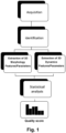
- the present invention relates, in a first aspect, to a method for sperm quality determination, comprising the computer-implemented steps of:
- step c) comprises determining the quality score for the at least one spermatozoon based on statistics performed also on at least part of the extracted three-dimensional morphological features, such as to the morphological feature(s) referring to the length of the head of the spermatozoon, or on parameters derived therefrom, such as the ellipticity of that head.
- the results of those statistics are compared with benchmarks referring to spermatozoa morphology and dynamics.
- the extracted three-dimensional features not being object of those statistics are also compared with benchmarks referring to spermatozoa morphology and/or dynamics.
- the quality score of steps c) and d) is an individual quality score for the at least one spermatozoon.
- steps a) and b) are applied to at least two spermatozoa, steps c) and d) respectively comprising determining and providing at least two respective individual health scores for the at least two spermatozoa.
- individual statistics for the spermatozoa may be performed for embodiments of the present invention, ad used to determine individual quality scores.
- steps a) and b) are applied to at least two spermatozoa, the quality score of steps c) and d) being a global quality score, wherein step c) comprises determining that global quality score based on the extracted three-dimensional features of the at least two spermatozoa, for at least part of the extracted three-dimensional dynamics features of the at least two spermatozoa after applying statistics thereon or on parameters derived therefrom, and on a comparison with benchmarks referring to spermatozoa morphology and dynamics.
- step c) comprises determining the global quality score for the at least two spermatozoa based on statistics performed also on at least part of the extracted three-dimensional morphological features of the at least two spermatozoa, such as to the morphological feature(s) referring to the length of the head of the spermatozoa, or on parameters derived therefrom, such as the ellipticity of that head.
- the results of those statistics are compared with benchmarks referring to spermatozoa morphology and dynamics.
- the extracted three-dimensional features not being object of those statistics are also compared with benchmarks referring to spermatozoa morphology and/or dynamics.
- step b) comprises:
- the method of the first aspect of the present invention further comprising, optically and in vitro, the step of sequentially acquiring the above mentioned plurality of volumetric groups of images of a sample that includes a plurality of freely swimming spermatozoa, wherein that sequential acquisition is carried out by means of non-point scanning optical sectioning microscopy.
- each of the acquired images of each volumetric group includes a plurality of spermatozoa, i.e. simultaneous imaging of multiple freely moving sperms can be performed, also due to the depth of field decoupling from the detection objective through non-point scanning optical sectioning.
- the method further comprises providing that sample, in vitro, within a three-dimensional chamber allowing the plurality of spermatozoa freely swim, with the appropriate density and arranged at the right place within the above mentioned field of view, so that the above conditions are met, and thus each of the acquired images of each volumetric group includes a plurality of spermatozoa.
- the method comprises performing the step of sequentially acquiring the volumetric groups of images at a minimum volumetric recording rate above 10 Hz, and acquiring, for each volumetric group, at least three images for three respective consecutive optical sections of the sample, although preferably at least ten images for ten respective consecutive optical sections of the sample are acquired, and, for some embodiments, even more, up to 3000 images per volumetric group.
- the method comprising performing the step of sequentially acquiring said volumetric groups of images at a minimum volumetric recording rate above 70 Hz, i.e. doubling that tail beat frequency, following the Nyquist criterion.
- the non-point scanning optical sectioning microscopy is a structured illumination microscopy (SIM, 3D-SIM), and thus the images of consecutive optical sections of the sample come from the information contained in the light emitted or scattered by the elements of the sample present at those optical sections when a structured pattern is created in the sample by means of non-uniform illumination.
- SIM structured illumination microscopy
- 3D-SIM structured illumination microscopy
- the images of consecutive optical sections of the sample come from the information contained in the light emitted or scattered by the elements of the sample present at those optical sections when a structured pattern is created in the sample by means of non-uniform illumination.
- multiple raw images with a phase shift in the illumination pattern are used to reconstruct an optically sectioned image, although any other kind of structured illumination microscopy using other types of illumination patterns can also be used according to the present invention.
- the non-point scanning optical sectioning microscopy is a light sheet microscopy (LSM), and thus the images of consecutive optical sections of the sample are acquired at consecutive image planes, at each image plane a light sheet being consecutively arranged to illuminate the sample and then the light emitted by the illuminated parts of the sample are picked up by an imaging lens and detected by a detector to form an image.
- LSM light sheet microscopy
- the technique is complemented with additional approaches to extend the depth of field, such as wavefront coding (WFC) and/or with electrotuneable lenses (ETL) and/or tuneable acoustic-gradient index refraction lenses, to compensate for a possible defocusing which could be introduced by the motion of the light sheet, if that was the case.
- WFC wavefront coding
- ETL electrotuneable lenses
- tuneable acoustic-gradient index refraction lenses to compensate for a possible defocusing which could be introduced by the motion of the light sheet, if that was the case.
- the method further comprises an automatic selection of the sperm based on the determined quality score(s).
- a second aspect of the present invention relates to a computer program, including code instructions that, when executed on at least one processor, implement the computer-implemented steps of the method of the first aspect of the present invention.
- a computer program product is further provided by the present invention, comprising a tangible medium and, stored therein, the computer program of the second aspect of the present invention.
- a third aspect of the present invention relates to a system for sperm quality determination, comprising a processor and a memory storing instructions that when executed by the processor cause the processor to implement the computer-implemented steps of the method of the first aspect of the present invention.
- the system of the third aspect of the present invention further comprises a non-point scanning optical sectioning microscope configured and arranged to implement the sequential acquisition of the plurality of volumetric groups of images of a sample that includes a plurality of freely swimming spermatozoa, and a three-dimensional chamber configured and arranged for in vitro housing said sample so that said plurality of spermatozoa can freely swim within said three-dimensional chamber.
- the three-dimensional chamber is sized to allow the plurality of spermatozoa to swim freely, with the appropriate density, and arranged at the right place within the above mentioned field of view of the microscope, so that the conditions explained above are met, and thus each of the acquired images of each volumetric group includes a plurality of spermatozoa.
- the non-point scanning optical sectioning microscope is a structured illumination microscope.
- the non-point scanning optical sectioning microscope is a light sheet microscope (LSM).
- LSM light sheet microscope
- the system further comprises additional mechanisms to extend the depth of field, such as wavefront coding (WFC) and/or electrotuneable lenses (ETL) and/or tuneable acoustic-gradient index refraction lenses, to compensate a possible defocusing which could be introduced by the motion of the light sheet, if that was the case.
- WFC wavefront coding
- ETL electrotuneable lenses
- tuneable acoustic-gradient index refraction lenses to compensate a possible defocusing which could be introduced by the motion of the light sheet, if that was the case.
- system of the third aspect of the invention further comprises a device, such as a display, speaker, haptic device, etc., for providing the quality score(s) to a user.
- a device such as a display, speaker, haptic device, etc.
- system of the third aspect of the invention further comprises automatic selector with an input for the received the quality score(s), and which is configured to automatically perform the sperm selection based on the received quality score(s).
- the present invention relies on sperm statistics based on 3D morphology and/or dynamics, and is capable of simultaneous characterisation of many sperm in a large FOV, hence, reducing the acquisition time to have enough data for meaningful statistics.
- the present inventors used the LSM technique to probe the capabilities to resolve the 3D morphology and mobility/dynamics of the head and flagellum of the spermatozoa.
- volumetric images of the sperm are obtained.
- each sperm is located and segmented in 3D, as schematically shown in Figure 2 .
- the dynamics of the spermatozoa tail are determined.
- the complete characterization of the 3D beat pattern on time is achievable by its curvature profile, ⁇ (s,t), and torsion profile, ⁇ (s,t).
- the parameter of the ellipticity of the head is measured directly in the method of the first aspect of the invention (i.e., derived from the extracted head length A, head width B, and head thickness C extracted features), in contrast to holographic and interferometric methods, where the retrieval of the parameters A, B, C is indirect since it requires the assumption of the ellipticity of the sperm head followed by the use of back propagation methods.
- Table 1 shows the minimum features/parameters necessary for the three-dimensional determination of spermatozoa morphology and mobility, for an embodiment, and which have been extracted (features) or determined (parameters) in the here explained experiment:
- each spermatozoon has specific characteristics, requiring a specialized approach for a precise extraction of their three-dimensional information. These characteristics determine the optical specifications needed to ensure precise analysis.
- the tail beat frequency of sperm typically oscillate around 35 Hz, requiring a minimum volumetric recording rate of 70 Hz (volumes/s) according to the Nyquist criterion. Additionally, the dimensions of the spermatozoa's head, particularly the smallest thickness is around 2um, Therefore the minimum optical resolution required for accurate visualization, is around 1 um.
- the physical dimensions of the visualization container i.e., the three-dimensional chamber Ch
- the physical dimensions of the visualization container must be at least double the size of the spermatozoon in all dimensions (i.e., 100x100x100 ⁇ m).
- DoF Depth of Field
- FoV Field of View
- the minimum observation volume needed to extract the three-dimensional features of a spermatozoon is 5000 ⁇ m 3 (50 ⁇ 10 ⁇ 10 ⁇ m). I.e., only part of the three-dimensional chamber Ch has to be focused in this volume of 5000 ⁇ m 3 .
- the minimum number of f images of consecutive optical sections required to extract the three-dimensional characteristics of a sperm is three per volume.
- the method of the first aspect of the present invention not only allows extracting three-dimensional parameters in normal sperm conditions, but is also useful for analyzing sperm that are outside the norm, either due to factors such as the health status of the donor, or due to alterations in physiological and/or environmental conditions, such as; temperature changes, presence of medications, among others. These conditions can alter both the morphology and dynamics of the spermatozoon, thus modifying the optical specifications needed to ensure precise analysis. Therefore, the non-point scanning optical sectioning allows decoupling the detection resolution from the depth of field, which allows multiple spermatozoa to be recorded simultaneously in three dimensions.
- Table 2 Volumetric recording rate [volumes/s] 10 to 1000 Optical resolution [ ⁇ m] 0.300 to 5 Field of View (FoV) [mm] 0.100 to 10 Depth of Field (DoF) [mm] 0.100 to 10 Frames per volume 3 to 3000
- optical 3D sperm characterization conducted in the above described experiment is described in a more detailed manner, for an embodiment, although alternative ways of conducting that characterization could be used, for other embodiments.
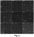
- Figure 4 only shows a particular optical section, for illustration purposes, but as explained above, the acquisition is made on volumes, i.e. a plurality of volumetric groups of images of corresponding consecutive optical sections are acquired.
- each spermatozoon available in the imaged volume is segmented individually at each time point. Then, a bounding box is determined around each spermatozoon, so that it includes the whole cell (head and flagellum, and, optionally also the midpiece). Every box is then treated separately.
- the head is localized providing the spermatozoon position in 3D (x,y,z). Moreover, a 3D analysis of the head provides its dimensions (Sx, Sy, Sz), orientation (phi, psi, theta) and shape. Finally, the method also provides the flagellum length (L) and 3D curvature (C).
- the head dimension (Sx, Sy, Sz) and angular orientation (theta, phi, psi) provide morphological and motility information in 3D, as head rotation frequency.
- the flagellum length (L) and curvature (C) it is possible to provide additional information/parameters, such as flagellum beating frequency, beating amplitude or oscillation modes.
- each spermatozoon was segmented individually and followed during its trajectory. With the parameters extracted from the position and angles of the head, as well as the curvature and size of the flagellum, the morphological and motor characteristics of each spermatozoon were determined. Table 3 shows the semen characteristics that can be extracted for healthy samples, for different embodiments. Table 3. Semen Characteristics and Sperm Morphology in healthy fresh samples 2D features Literature Normal morphology (%) 23.1 ⁇ 9.2 Concentration (10 6 /mL) 198.3 ⁇ 108.8 Motility (%) 59.9 ⁇ 16.5 Motile sperm conc.
- the 3D features/parameters could be reduced to the typical 2D motility parameters known in the art. Nevertheless, since in the present invention the features/parameters are obtained in a non-confined environment that allows the natural movement of the spermatozoa, they minimise the bias introduced by the 2D confinement of the prior art proposals.
- the head dimension (Sx, Sy, Sz) and angular orientation (theta, phi, psi) provided with the method of the invention, morphological and motility information in 3D, as head rotation frequency can be obtained.
- L flagellum length
- C3D 3D curvature
- results presented here represent a partial proof of concept of the capabilities of the present invention to extract the individual spermatozoa information, under high-density ( ⁇ 200K sperms/mm3) condition, which will feed the data for statistical analysis, and then to generate and provide the quality score(s).
- the 3D information extracted with the method of the invention provides more information than the 2D methods.
- spermatozoa motion is not constrained in any direction (dimension), the results obtained are more reliable.
- FIG. 5 schematically shows the system of the third aspect of the present invention, for an embodiment for which the system comprises:
- the microscope M is a light sheet microscopy (LSM), and comprises an illumination arm and a detection arm.
- LSM light sheet microscopy
- the illumination arm comprises a laser source, a cylindrical lens, a 1D galvo mirror, and a relay lens, to generate a light sheet and sequentially illuminate with the same different consecutive optical sections of the sample a plurality of times, each for a respective one of the plurality of volumetric groups.
- the detection arm comprises a microscope objective, a phase mask for wavefront coding (WFC), a tube lens, and a camera, to pick up and detect the light emitted, through a wall of the 3D chamber Ch, by the illuminated parts of the sample at each optical section, and thus the images formed with that emitted light.
- WFC wavefront coding
- the detected images of the plurality of volumetric groups are provided to the computing entity C, which process the same as explained above, according to the method of the first aspect of the invention.
- the detection is performed using a collection lens (20x microscope objective, 1 NA, 2 mm WD, Olympus), two achromatic lenses (AC508-200-A-ML, Thorlabs) a deformable mirror and a sCMOS camera (Hamamatsu Orca-Flash4.0 v3). Care was taken to place all the optical elements in the proper conjugated planes obtaining a total magnification of 22.2x.
- the signal was recorded for 10 min at a scanning speed of 80 volumes/second, every volume had a depth of 40 ⁇ m, containing 10 imaged planes.
- the size of each voxel was 0.3 ⁇ m ⁇ 0.3 ⁇ m ⁇ 4 ⁇ m (x, y, z).
- the collected data was structured in volumetric frames providing a sequence of 3D images. If necessary, (in the case of WFC, light field, Fourier light field) each volume was deconvolved with the corresponding 3D point spread function (PSF). Volumetric deconvolution was performed by deconvolving each frame with the PSF frame of the corresponding z (using the Richardson-Lucy-based algorithm). The volumetric PSF was generated from the distilling of the experimental PSF using the Zernike coefficients applied to the phase mask.
- deconvolution can be done following classical deconvolution methods such as Richardson-Lucy, blind, Wiener algorithms or it can be done based on Machine learning, Simulated annealing, Genetic algorithms, among others.
- the volumetric PSF necessary for the deconvolution can be measured directly in the experiment using a fluorescent bead (or an artificial star) or be generated from the simulation of the system, without it being necessary in cases like machine learning algorithms.
- the schematic shown in Figure 5 can be varied in multiple manners, for implementing different kinds of LSMs or complement the same with further mechanisms, such as those intended to extend the depth of field, such as wavefront coding (WFC) (in addition or alternatively to the above mentioned phase mask), and/or electrotuneable lenses (ETL) and/or tuneable acoustic-gradient index refraction lenses, to compensate a possible defocusing which could be introduced by the motion of the light sheet, if that was the case.
- WFC wavefront coding
- ETL electrotuneable lenses
- tuneable acoustic-gradient index refraction lenses to compensate a possible defocusing which could be introduced by the motion of the light sheet, if that was the case.
Landscapes
- Engineering & Computer Science (AREA)
- Theoretical Computer Science (AREA)
- Physics & Mathematics (AREA)
- Health & Medical Sciences (AREA)
- General Physics & Mathematics (AREA)
- Multimedia (AREA)
- Computer Vision & Pattern Recognition (AREA)
- Life Sciences & Earth Sciences (AREA)
- General Health & Medical Sciences (AREA)
- Biomedical Technology (AREA)
- Medical Informatics (AREA)
- Molecular Biology (AREA)
- Software Systems (AREA)
- Evolutionary Computation (AREA)
- Databases & Information Systems (AREA)
- Computing Systems (AREA)
- Artificial Intelligence (AREA)
- Wood Science & Technology (AREA)
- Organic Chemistry (AREA)
- Genetics & Genomics (AREA)
- Bioinformatics & Cheminformatics (AREA)
- Zoology (AREA)
- Chemical & Material Sciences (AREA)
- Biotechnology (AREA)
- Heart & Thoracic Surgery (AREA)
- Reproductive Health (AREA)
- Veterinary Medicine (AREA)
- Public Health (AREA)
- Animal Behavior & Ethology (AREA)
- Quality & Reliability (AREA)
- Radiology & Medical Imaging (AREA)
- Nuclear Medicine, Radiotherapy & Molecular Imaging (AREA)
- Gynecology & Obstetrics (AREA)
- Surgery (AREA)
- Biophysics (AREA)
- Pathology (AREA)
- Microbiology (AREA)
- General Engineering & Computer Science (AREA)
- Developmental Biology & Embryology (AREA)
- Biochemistry (AREA)
Priority Applications (2)
| Application Number | Priority Date | Filing Date | Title |
|---|---|---|---|
| EP23383385.4A EP4579604A1 (de) | 2023-12-27 | 2023-12-27 | Verfahren, computerprogramm und system zur bestimmung der spermaqualität |
| US19/000,016 US20250213178A1 (en) | 2023-12-27 | 2024-12-23 | Method, a computer program and a system for sperm quality determination |
Applications Claiming Priority (1)
| Application Number | Priority Date | Filing Date | Title |
|---|---|---|---|
| EP23383385.4A EP4579604A1 (de) | 2023-12-27 | 2023-12-27 | Verfahren, computerprogramm und system zur bestimmung der spermaqualität |
Publications (1)
| Publication Number | Publication Date |
|---|---|
| EP4579604A1 true EP4579604A1 (de) | 2025-07-02 |
Family
ID=89573536
Family Applications (1)
| Application Number | Title | Priority Date | Filing Date |
|---|---|---|---|
| EP23383385.4A Pending EP4579604A1 (de) | 2023-12-27 | 2023-12-27 | Verfahren, computerprogramm und system zur bestimmung der spermaqualität |
Country Status (2)
| Country | Link |
|---|---|
| US (1) | US20250213178A1 (de) |
| EP (1) | EP4579604A1 (de) |
Citations (3)
| Publication number | Priority date | Publication date | Assignee | Title |
|---|---|---|---|---|
| EP1054948B1 (de) | 1998-02-19 | 2014-04-30 | Medical Electronic Systems, LLC | Analysesystem für Sperma |
| EP3071704B1 (de) | 2013-11-20 | 2022-03-30 | Brigham and Women's Hospital, Inc. | System und verfahren zum sortieren von sperma |
| US20230061402A1 (en) * | 2020-01-16 | 2023-03-02 | Baibys Fertility Ltd | Automated spermatozoa candidate identification |
-
2023
- 2023-12-27 EP EP23383385.4A patent/EP4579604A1/de active Pending
-
2024
- 2024-12-23 US US19/000,016 patent/US20250213178A1/en active Pending
Patent Citations (3)
| Publication number | Priority date | Publication date | Assignee | Title |
|---|---|---|---|---|
| EP1054948B1 (de) | 1998-02-19 | 2014-04-30 | Medical Electronic Systems, LLC | Analysesystem für Sperma |
| EP3071704B1 (de) | 2013-11-20 | 2022-03-30 | Brigham and Women's Hospital, Inc. | System und verfahren zum sortieren von sperma |
| US20230061402A1 (en) * | 2020-01-16 | 2023-03-02 | Baibys Fertility Ltd | Automated spermatozoa candidate identification |
Non-Patent Citations (9)
| Title |
|---|
| BAI CHUANBIAO ET AL: "3D dynamic observation of human sperm by parallel phase-shifting digital holographic microscopy based on pixelated polarization", APL PHOTONICS, AMERICAN INSTITUTE OF PHYSICS, 2 HUNTINGTON QUADRANGLE, MELVILLE, NY 11747, vol. 8, no. 7, 14 July 2023 (2023-07-14), XP012276178, DOI: 10.1063/5.0150582 * |
| G. CORKIDI ET AL.: "Tracking sperm in three-dimensions", BIOCHEMICAL AND BIOPHYSICAL RESEARCH COMMUNICATIONS, vol. 373, no. 1, August 2008 (2008-08-01), pages 125 - 129, XP022795094, DOI: 10.1016/j.bbrc.2008.05.189 |
| G. CORKIDI: "The human sperm head spins with a conserved direction during swimming in 3D", BIORXIV, 29 November 2022 (2022-11-29), pages 1 - 43, XP093164223, Retrieved from the Internet <URL:https://www.biorxiv.org/content/10.1101/2022.11.28.517870v1> [retrieved on 20240517], DOI: 10.1101/2022.11.28.517870 * |
| GILI DARDIKMAN-YOFFE ET AL.: "High-resolution 4-D acquisition of freely swimming human sperm cells without staining", SCI. ADV., vol. 6, no. 15, April 2020 (2020-04-01), pages eaay7619 |
| H. GADELHA ET AL.: "Human sperm uses asymmetric and anisotropic flagellar controls to regulate swimming symmetry and cell steering", SCI. ADV., vol. 6, no. 31, July 2020 (2020-07-01), pages eaba5168 |
| INABA, K.MIZUNO, K.: "Sperm dysfunction and ciliopathy", REPROD. MED. BIOL., vol. 15, 2016, pages 77 - 94, XP035960667, Retrieved from the Internet <URL:https://doi.org/10.1007/s12522-015-0225-5> DOI: 10.1007/s12522-015-0225-5 |
| JAN N. HANSEN: "Multifocal imaging for precise, label-free tracking of fast biological processes in 3D", NATURE COMMUNICATIONS, vol. 12, no. 1, 28 July 2021 (2021-07-28), UK, pages 1 - 15, XP093164278, ISSN: 2041-1723, Retrieved from the Internet <URL:https://www.nature.com/articles/s41467-021-24768-4> [retrieved on 20240517], DOI: 10.1038/s41467-021-24768-4 * |
| M. U. DALOGLU ET AL.: "Label-free 3D computational imaging of spermatozoon locomotion, head spin and flagellum beating over a large volume", LIGHT: SCIENCE & APPLICATIONS, vol. 7, no. 1, January 2018 (2018-01-01), pages 17121, XP055752149, DOI: 10.1038/lsa.2017.121 |
| T. W. SU ET AL.: "High-throughput lensfree 3D tracking of human sperms reveals rare statistics of helical trajectories", PROC. NATL. ACAD. SCI. U.S.A., vol. 109, no. 40, October 2012 (2012-10-01), pages 16018 - 16022, XP055545037, DOI: 10.1073/pnas.1212506109 |
Also Published As
| Publication number | Publication date |
|---|---|
| US20250213178A1 (en) | 2025-07-03 |
Similar Documents
| Publication | Publication Date | Title |
|---|---|---|
| CN113822852B (zh) | 图像处理方法、程序以及记录介质 | |
| Sutton et al. | Strain field measurements on mouse carotid arteries using microscopic three‐dimensional digital image correlation | |
| CN111630564A (zh) | 图像处理方法、程序及记录介质 | |
| KR101871082B1 (ko) | 약효 평가방법 및 약효 평가를 위한 화상 처리 장치 | |
| Singh et al. | Quantifying three-dimensional rodent retina vascular development using optical tissue clearing and light-sheet microscopy | |
| WO2020240555A1 (en) | System and method for reconstructing morphology and dynamics of biological cells from holographic images | |
| EP2084668A1 (de) | Rekonstruktion und visualisierung neuronaler zellenstrukturen mit hellfeld-mosaik-mikroskopie | |
| JP7845610B2 (ja) | 経時的に試料中の粒子を表す入力画像のシーケンスを分類する方法 | |
| US12136211B2 (en) | Fertilized egg development stage determination method, recording medium, imaging method and imaging apparatus | |
| US12399103B2 (en) | Cell life-and-death determination method, cell life-and-death determination device, and cell life-and-death determination system | |
| EP4579604A1 (de) | Verfahren, computerprogramm und system zur bestimmung der spermaqualität | |
| WO2019125583A1 (en) | Imaging device for measuring sperm motility | |
| US20070140543A1 (en) | Systems and methods for enhanced cytological specimen review | |
| CN111971607B (zh) | 试样观察装置 | |
| JP7382289B2 (ja) | 画像処理方法、プログラムおよび記録媒体 | |
| JP6343874B2 (ja) | 観察装置、観察方法、観察システム、そのプログラム、および細胞の製造方法 | |
| EP3259631B1 (de) | Vorrichtung und verfahren zur erstellung eines optischen tomogramms einer mikroskopischen probe | |
| JP7382290B2 (ja) | 画像処理方法、プログラムおよび記録媒体 | |
| Tang et al. | Fast post-processing pipeline for optical projection tomography | |
| EP4349999A1 (de) | Automatisiertes system und verfahren zur mikromassstab-überwachung von in einer petrischale enthaltenen c.-elementen mit transparentem medium, erhaltene überwachungsaufzeichnung und verwendung davon | |
| Alanis et al. | Optical detection of Trypanosoma cruzi in blood samples for diagnosis purpose | |
| On et al. | 3D Reconstruction of Phase Contrast Images Using Focus Measures | |
| Wei et al. | Optical imaging technologies and applications | |
| Moon et al. | Holographic red blood cell image analysis | |
| Skoglund et al. | A method for 3D reconstruction of neuronal processes using semithin serial sections displayed as a cinematographic sequence |
Legal Events
| Date | Code | Title | Description |
|---|---|---|---|
| PUAI | Public reference made under article 153(3) epc to a published international application that has entered the european phase |
Free format text: ORIGINAL CODE: 0009012 |
|
| STAA | Information on the status of an ep patent application or granted ep patent |
Free format text: STATUS: THE APPLICATION HAS BEEN PUBLISHED |
|
| AK | Designated contracting states |
Kind code of ref document: A1 Designated state(s): AL AT BE BG CH CY CZ DE DK EE ES FI FR GB GR HR HU IE IS IT LI LT LU LV MC ME MK MT NL NO PL PT RO RS SE SI SK SM TR |
|
| STAA | Information on the status of an ep patent application or granted ep patent |
Free format text: STATUS: REQUEST FOR EXAMINATION WAS MADE |
|
| 17P | Request for examination filed |
Effective date: 20251218 |