ES2833046T3 - Antibodies that bind human programmed death ligand 1 (PD-L1) - Google Patents
Antibodies that bind human programmed death ligand 1 (PD-L1) Download PDFInfo
- Publication number
- ES2833046T3 ES2833046T3 ES13814812T ES13814812T ES2833046T3 ES 2833046 T3 ES2833046 T3 ES 2833046T3 ES 13814812 T ES13814812 T ES 13814812T ES 13814812 T ES13814812 T ES 13814812T ES 2833046 T3 ES2833046 T3 ES 2833046T3
- Authority
- ES
- Spain
- Prior art keywords
- antibody
- antibodies
- binding
- seq
- human
- Prior art date
- Legal status (The legal status is an assumption and is not a legal conclusion. Google has not performed a legal analysis and makes no representation as to the accuracy of the status listed.)
- Active
Links
Classifications
-
- C—CHEMISTRY; METALLURGY
- C07—ORGANIC CHEMISTRY
- C07K—PEPTIDES
- C07K16/00—Immunoglobulins [IG], e.g. monoclonal or polyclonal antibodies
- C07K16/18—Immunoglobulins [IG], e.g. monoclonal or polyclonal antibodies against material from animals or humans
- C07K16/28—Immunoglobulins [IG], e.g. monoclonal or polyclonal antibodies against material from animals or humans against receptors, cell surface antigens or cell surface determinants
- C07K16/2803—Immunoglobulins [IG], e.g. monoclonal or polyclonal antibodies against material from animals or humans against receptors, cell surface antigens or cell surface determinants against the immunoglobulin superfamily
- C07K16/2827—Immunoglobulins [IG], e.g. monoclonal or polyclonal antibodies against material from animals or humans against receptors, cell surface antigens or cell surface determinants against the immunoglobulin superfamily against B7 molecules, e.g. CD80, CD86
-
- G—PHYSICS
- G01—MEASURING; TESTING
- G01N—INVESTIGATING OR ANALYSING MATERIALS BY DETERMINING THEIR CHEMICAL OR PHYSICAL PROPERTIES
- G01N33/00—Investigating or analysing materials by specific methods not covered by groups G01N1/00 - G01N31/00
- G01N33/48—Biological material, e.g. blood, urine; Haemocytometers
- G01N33/50—Chemical analysis of biological material, e.g. blood, urine; Testing involving biospecific ligand binding methods; Immunological testing
- G01N33/53—Immunoassay; Biospecific binding assay; Materials therefor
- G01N33/575—Immunoassay; Biospecific binding assay; Materials therefor for cancer
- G01N33/5758—Immunoassay; Biospecific binding assay; Materials therefor for cancer involving compounds serving as markers for tumours, cancers or neoplasias, e.g. cellular determinants, receptors, heat shock/stress proteins, A-protein, oligosaccharides or metabolites
- G01N33/5759—Immunoassay; Biospecific binding assay; Materials therefor for cancer involving compounds serving as markers for tumours, cancers or neoplasias, e.g. cellular determinants, receptors, heat shock/stress proteins, A-protein, oligosaccharides or metabolites involving compounds localised on the membrane of tumour or cancer cells
-
- G—PHYSICS
- G01—MEASURING; TESTING
- G01N—INVESTIGATING OR ANALYSING MATERIALS BY DETERMINING THEIR CHEMICAL OR PHYSICAL PROPERTIES
- G01N33/00—Investigating or analysing materials by specific methods not covered by groups G01N1/00 - G01N31/00
- G01N33/48—Biological material, e.g. blood, urine; Haemocytometers
- G01N33/50—Chemical analysis of biological material, e.g. blood, urine; Testing involving biospecific ligand binding methods; Immunological testing
- G01N33/53—Immunoassay; Biospecific binding assay; Materials therefor
- G01N33/575—Immunoassay; Biospecific binding assay; Materials therefor for cancer
- G01N33/57557—Immunoassay; Biospecific binding assay; Materials therefor for cancer of other specific parts of the body, e.g. brain
-
- G—PHYSICS
- G01—MEASURING; TESTING
- G01N—INVESTIGATING OR ANALYSING MATERIALS BY DETERMINING THEIR CHEMICAL OR PHYSICAL PROPERTIES
- G01N33/00—Investigating or analysing materials by specific methods not covered by groups G01N1/00 - G01N31/00
- G01N33/48—Biological material, e.g. blood, urine; Haemocytometers
- G01N33/50—Chemical analysis of biological material, e.g. blood, urine; Testing involving biospecific ligand binding methods; Immunological testing
- G01N33/68—Chemical analysis of biological material, e.g. blood, urine; Testing involving biospecific ligand binding methods; Immunological testing involving proteins, peptides or amino acids
- G01N33/6872—Intracellular protein regulatory factors and their receptors, e.g. including ion channels
-
- G—PHYSICS
- G01—MEASURING; TESTING
- G01N—INVESTIGATING OR ANALYSING MATERIALS BY DETERMINING THEIR CHEMICAL OR PHYSICAL PROPERTIES
- G01N33/00—Investigating or analysing materials by specific methods not covered by groups G01N1/00 - G01N31/00
- G01N33/48—Biological material, e.g. blood, urine; Haemocytometers
- G01N33/50—Chemical analysis of biological material, e.g. blood, urine; Testing involving biospecific ligand binding methods; Immunological testing
- G01N33/68—Chemical analysis of biological material, e.g. blood, urine; Testing involving biospecific ligand binding methods; Immunological testing involving proteins, peptides or amino acids
- G01N33/6878—Chemical analysis of biological material, e.g. blood, urine; Testing involving biospecific ligand binding methods; Immunological testing involving proteins, peptides or amino acids in epitope analysis
-
- C—CHEMISTRY; METALLURGY
- C07—ORGANIC CHEMISTRY
- C07K—PEPTIDES
- C07K2317/00—Immunoglobulins specific features
- C07K2317/30—Immunoglobulins specific features characterized by aspects of specificity or valency
- C07K2317/31—Immunoglobulins specific features characterized by aspects of specificity or valency multispecific
-
- C—CHEMISTRY; METALLURGY
- C07—ORGANIC CHEMISTRY
- C07K—PEPTIDES
- C07K2317/00—Immunoglobulins specific features
- C07K2317/30—Immunoglobulins specific features characterized by aspects of specificity or valency
- C07K2317/34—Identification of a linear epitope shorter than 20 amino acid residues or of a conformational epitope defined by amino acid residues
-
- C—CHEMISTRY; METALLURGY
- C07—ORGANIC CHEMISTRY
- C07K—PEPTIDES
- C07K2317/00—Immunoglobulins specific features
- C07K2317/50—Immunoglobulins specific features characterized by immunoglobulin fragments
- C07K2317/56—Immunoglobulins specific features characterized by immunoglobulin fragments variable (Fv) region, i.e. VH and/or VL
- C07K2317/565—Complementarity determining region [CDR]
-
- G—PHYSICS
- G01—MEASURING; TESTING
- G01N—INVESTIGATING OR ANALYSING MATERIALS BY DETERMINING THEIR CHEMICAL OR PHYSICAL PROPERTIES
- G01N2333/00—Assays involving biological materials from specific organisms or of a specific nature
- G01N2333/435—Assays involving biological materials from specific organisms or of a specific nature from animals; from humans
- G01N2333/705—Assays involving receptors, cell surface antigens or cell surface determinants
- G01N2333/70503—Immunoglobulin superfamily, e.g. VCAMs, PECAM, LFA-3
- G01N2333/70532—B7 molecules, e.g. CD80, CD86
Landscapes
- Health & Medical Sciences (AREA)
- Life Sciences & Earth Sciences (AREA)
- Immunology (AREA)
- Chemical & Material Sciences (AREA)
- Engineering & Computer Science (AREA)
- Molecular Biology (AREA)
- Hematology (AREA)
- Urology & Nephrology (AREA)
- Biomedical Technology (AREA)
- Biochemistry (AREA)
- General Health & Medical Sciences (AREA)
- Medicinal Chemistry (AREA)
- Cell Biology (AREA)
- Proteomics, Peptides & Aminoacids (AREA)
- Organic Chemistry (AREA)
- Physics & Mathematics (AREA)
- Analytical Chemistry (AREA)
- Food Science & Technology (AREA)
- General Physics & Mathematics (AREA)
- Pathology (AREA)
- Microbiology (AREA)
- Biotechnology (AREA)
- Genetics & Genomics (AREA)
- Biophysics (AREA)
- Peptides Or Proteins (AREA)
- Medicines Containing Antibodies Or Antigens For Use As Internal Diagnostic Agents (AREA)
- Micro-Organisms Or Cultivation Processes Thereof (AREA)
- Oncology (AREA)
- Measuring Or Testing Involving Enzymes Or Micro-Organisms (AREA)
Abstract
Un anticuerpo o un fragmento de union a antigeno aislados que se unen especificamente al PD-L1 en donde: (a) la region variable de cadena ligera es el SEQ ID NO: 13 y la region variable de cadena pesada es el SEQ ID NO: 20; o (b) la region variable de cadena ligera es el SEQ ID NO: 25 y la region variable de cadena pesada es el SEQ ID NO: 32, en donde en el SEQ ID NO: 32, X es Q o pE.An isolated antibody or antigen-binding fragment that specifically binds to PD-L1 wherein: (a) the light chain variable region is SEQ ID NO: 13 and the heavy chain variable region is SEQ ID NO: twenty; or (b) the light chain variable region is SEQ ID NO: 25 and the heavy chain variable region is SEQ ID NO: 32, where in SEQ ID NO: 32, X is Q or pE.
Description
DESCRIPCIÓNDESCRIPTION
Anticuerpos que se unen al ligando 1 de muerte programada (PD-L1) humanoAntibodies that bind human programmed death ligand 1 (PD-L1)
Campo de la invenciónField of the invention
La presente invención se refiere a anticuerpos que tienen secuencias específicas que se unen al ligando 1 de muerte programada (PD-L1, por las siglas del inglés Programmed Death Ligand 1) humano y que son útiles para detectar la expresión del PD-L1 en muestras de tejido humano mediante análisis inmunohistoquímico (IHQ). La invención también se refiere a ensayos IHQ específicos que emplean estos Anticuerpos anti-PD-L1 humano.The present invention relates to antibodies that have specific sequences that bind to human Programmed Death Ligand 1 (PD-L1) and that are useful for detecting the expression of PD-L1 in samples. of human tissue by immunohistochemical analysis (IHC). The invention also relates to specific IHC assays employing these anti-human PD-L1 antibodies.
Antecedentes de la invenciónBackground of the invention
El PD-L1 es una glucoproteína de la superficie celular que es uno de los dos ligandos conocidos de la Muerte Programada 1 (PD-1), que se reconoce como un actor importante en la regulación inmunitaria y el mantenimiento de la tolerancia periférica. La expresión del PD-L1 se ha observado en la superficie de varios células inmunitarias, incluyendo linfocitos vírgenes y células B y T activadas, monocitos y células dendríticas (Id.). Por otro lado, el ARNm del PD-L1 se expresa en tejidos no linfoides, incluidas las células endoteliales vasculares, células epiteliales, células musculares, y en tejido amigdalino y placentario. Véase, por ejemplo, Keir, M.E. et al., Annu Rev Immunol. 26:677-704 (2008); Sharp A.H. et al., Nature Immunol. 8:239-245 (2007); Okazaki T y Honjo T, Internat. immunol. 19:813-824 (2007).PD-L1 is a cell surface glycoprotein that is one of the two known ligands of Programmed Death 1 (PD-1), which is recognized as an important actor in immune regulation and the maintenance of peripheral tolerance. PD-L1 expression has been observed on the surface of various immune cells, including naive lymphocytes and activated B and T cells, monocytes, and dendritic cells (Id.). On the other hand, PD-L1 mRNA is expressed in non-lymphoid tissues, including vascular endothelial cells, epithelial cells, muscle cells, and in tonsillar and placental tissue. See, for example, Keir, ME et al., Annu Rev Immunol. 26: 677-704 (2008); Sharp AH et al., Nature Immunol. 8: 239-245 (2007); Okazaki T and Honjo T, Internat. immunol. 19: 813-824 (2007).
La expresión del PD-L1 también se ha observado en varios cánceres humanos, y la interacción del PD-L1, expresado en células tumorales, con PD-1, puede inducir la inhibición o la apoptosis de células T específicas de tumor. En grandes conjuntos de muestras, por ejemplo, de cánceres de ovario, riñón, colorrectal, páncreas, hígado y melanoma, se demostró que la expresión del PD-L1 se correlacionaba con un mal pronóstico y una reducción de la supervivencia global independientemente del tratamiento posterior. Se ha demostrado que los anticuerpos monoclonales anti-PD-1 que bloquean la unión del PD-L1 con PD-1, tienen actividad antitumoral contra varios tipos de tumores, sugiriendo los datos clínicos tempranos en humanos que los pacientes cuyos tumores expresan PD-L1 tienen más probabilidades de responder a la terapia anti-PD-1. Véase, por ejemplo, Iwai et al., PNAS 99:12293-12297 (2002); Ohigashi et al., Clin Cancer Res 11:2947-2953 (2005); Ghebeh et al., Neoplasia 8:190-198 (2006); Hamanishi, J et al., PNAS 104:3360-3365 (2007); Yang et al., Invest Ophthalmol Vis Sci. 49(6):2518-2525 (2008); Gao et al., Clin Cancer Res 15:971-979 (2009); Brahmer J.R. et al., J Clin Oncol. 28:3167-3175 (2010).The expression of PD-L1 has also been observed in several human cancers, and the interaction of PD-L1, expressed in tumor cells, with PD-1, can induce the inhibition or apoptosis of tumor-specific T cells. In large sample sets, for example, from ovarian, kidney, colorectal, pancreas, liver and melanoma cancers, PD-L1 expression was shown to correlate with a poor prognosis and reduced overall survival regardless of subsequent treatment . Anti-PD-1 monoclonal antibodies that block the binding of PD-L1 to PD-1 have been shown to have antitumor activity against various types of tumors, suggesting early clinical data in humans that patients whose tumors express PD-L1 they are more likely to respond to anti-PD-1 therapy. See, for example, Iwai et al., PNAS 99: 12293-12297 (2002); Ohigashi et al., Clin Cancer Res 11: 2947-2953 (2005); Ghebeh et al., Neoplasia 8: 190-198 (2006); Hamanishi, J et al., PNAS 104: 3360-3365 (2007); Yang et al., Invest Ophthalmol Vis Sci. 49 (6): 2518-2525 (2008); Gao et al., Clin Cancer Res 15: 971-979 (2009); Brahmer JR et al., J Clin Oncol. 28: 3167-3175 (2010).
En un informe reciente se describe la comparación de 15 anticuerpos anti-PD-L1 humano por su utilidad en la detección de la expresión del hPD-L1 en muestras de melanoma humano fijadas en formol y embebidas en parafina (FFPE) (Gadiot, J., et al., Cancer 117(10):2192-2201 (2011)). Los criterios de utilidad evaluados en esta comparación fueron: (1) capacidad para teñir tejidos embebidos en parafina, (2) producir una tinción de fondo baja y (3) bloquear la unión con el PD-L1 mediante preincubación con una proteína de fusión PD-L1. Los autores concluyeron que el Ac n.° 4059, un anticuerpo policlonal anti-ser humano de conejo (obtenido en ProSci, Poway, CA Estados Unidos), fue el único anticuerpo anti-PD-L1 humano de los 15 analizados que cumplió aceptablemente con todos estos criterios (Id. en 2195, 2a columna).A recent report describes the comparison of 15 anti-human PD-L1 antibodies for their usefulness in detecting hPD-L1 expression in formalin-fixed and paraffin-embedded (FFPE) human melanoma samples (Gadiot, J. , et al., Cancer 117 (10): 2192-2201 (2011)). The utility criteria evaluated in this comparison were: (1) ability to stain paraffin-embedded tissues, (2) produce low background staining, and (3) block PD-L1 binding by preincubation with a PD fusion protein. -L1. The authors concluded that Ab # 4059, a rabbit anti-human polyclonal antibody (obtained from ProSci, Poway, CA USA), was the only anti-human PD-L1 antibody of the 15 tested that was acceptably compliant with all these criteria (Id. at 2195, 2nd column).
Sumario de la invenciónSummary of the invention
La presente invención se refiere a anticuerpos monoclonales anti-PD-L1 humano, que producen un patrón de tinción IHQ en tejido amigdalino FFPE que los inventores de la presente creen que es más relevante desde el punto de vista inmunológico que el producido por el Ac n.° 4059 de ProSci. Como se describe en los ejemplos más adelante, los inventores descubrieron que este anticuerpo de ProSci (PRS4059, Sigma-Aldrich lote 40590604) teñía todos los linajes hematopoyéticos en la amígdala con la misma intensidad, mientras que dos anticuerpos de la presente invención, 22C3 y 20C3, teñían selectivamente el epitelio críptico amigdalino y las células mieloides CD68+ foliculares, que morfológicamente están en consonancia con los macrófagos. Por otra parte, los anticuerpos 22C3 y 20C3 demuestran una diferencia de intensidad consistente entre estas dos poblaciones de células distintas con una intensidad de tinción en el epitelio críptico mucho mayor que en los macrófagos foliculares. Los tres anticuerpos (PRS4059, 22C3 y 20C3) se neutralizan con preincubación con el antígeno PD-L1, lo que indica que la reactividad está mediada por el dominio de unión a antígeno (las CDR). Por tanto, la invención también se refiere al uso de los anticuerpos de la presente invención en la detección de la expresión del PD-L1 en la superficie de células humanas, incluyendo en ensayos IHQ para detectar el PD-L1 en secciones de tejido FFPE.The present invention relates to human anti-PD-L1 monoclonal antibodies, which produce an IHC staining pattern in FFPE tonsil tissue that the present inventors believe is more relevant from the immunological point of view than that produced by Ac n . 4059 from ProSci. As described in the examples below, the inventors found that this ProSci antibody (PRS4059, Sigma-Aldrich lot 40590604) stained all hematopoietic lineages in the amygdala with the same intensity, whereas two antibodies of the present invention, 22C3 and 20C3, selectively stained the cryptic tonsillar epithelium and follicular CD68 + myeloid cells, which are morphologically consistent with macrophages. On the other hand, antibodies 22C3 and 20C3 demonstrate a consistent intensity difference between these two distinct cell populations with a much higher staining intensity in cryptic epithelium than in follicular macrophages. All three antibodies (PRS4059, 22C3, and 20C3) are neutralized with preincubation with the PD-L1 antigen, indicating that the reactivity is mediated by the antigen-binding domain (the CDRs). Therefore, the invention also relates to the use of the antibodies of the present invention in the detection of PD-L1 expression on the surface of human cells, including in IHC assays to detect PD-L1 in FFPE tissue sections.
En un aspecto, la invención proporciona un anticuerpo aislado, o un fragmento de unión a antígeno del mismo, que se une específicamente al PD-L1 humano en donde (a) la región variable de cadena ligera es el SEQ ID NO: 13 y la región variable de cadena pesada es el SEQ ID NO: 20; o (b) la región variable de cadena ligera es el SEQ ID NO: 25 y la región variable de cadena pesada es el SEQ ID NO: 32, en donde en el SEQ ID NO: 32, X es Q o pE.In one aspect, the invention provides an isolated antibody, or antigen-binding fragment thereof, that specifically binds to human PD-L1 wherein (a) the light chain variable region is SEQ ID NO: 13 and heavy chain variable region is SEQ ID NO: 20; or (b) the light chain variable region is SEQ ID NO: 25 and the heavy chain variable region is SEQ ID NO: 32, where in SEQ ID NO: 32, X is Q or pE.
En un anticuerpo o fragmento de unión a antígeno preferido de la invención, la región variable de cadena ligera es el SEQ ID NO: 13 y la región variable de cadena pesada es el SEQ ID NO: 20. In a preferred antibody or antigen-binding fragment of the invention, the light chain variable region is SEQ ID NO: 13 and the heavy chain variable region is SEQ ID NO: 20.
En otra realización más, el anticuerpo o fragmento de unión de la invención, comprende una región variable de cadena ligera del SEQ ID NO: 25 y una región variable de cadena pesada del SEQ ID n O: 32, en donde en el SEQ ID NO: 32, X es pE.In yet another embodiment, the antibody or binding fragment of the invention comprises a light chain variable region of SEQ ID NO: 25 and a heavy chain variable region of SEQ ID NO: 32, where in SEQ ID NO : 32, X is pE.
En otra realización adicional, el anticuerpo o fragmento de unión de la invención, comprende una región variable de cadena ligera del SEQ ID NO: 25 y una región variable de cadena pesada del SEQ ID NO: 32, en donde en el SEQ ID NO: 32, X es Q.In yet another embodiment, the antibody or binding fragment of the invention comprises a light chain variable region of SEQ ID NO: 25 and a heavy chain variable region of SEQ ID NO: 32, wherein in SEQ ID NO: 32, X is Q.
El anticuerpo es una IgGi . En una realización, el anticuerpo comprende una región constante de IgGi de ratón. The antibody is an IgGi. In one embodiment, the antibody comprises a mouse IgGi constant region.
Los anticuerpos de la presente invención son los anticuerpos monoclonales 20C3 y 22C3, que son anticuerpos de IgG1 expresados por los hibridomas MEB037.20C3 y MEB037.22C3, respectivamente.The antibodies of the present invention are monoclonal antibodies 20C3 and 22C3, which are IgG1 antibodies expressed by the hybridomas MEB037.20C3 and MEB037.22C3, respectively.
En el presente documento se describe un anticuerpo monoclonal aislado, o un fragmento de unión a antígeno del mismo, que se une específicamente al PD-L1 humano y bloquea la unión de 20C3 o 22C3 con el PD-L1 humano, o de un anticuerpo de referencia que comprendel SEQ ID NO: 25 y el SEQ ID NO: 32. En el presente documento se describe un anticuerpo o fragmento de unión a antígeno de la invención que bloquea la unión con el PD-L1 humano de cada uno de 20C3 y 22C3, o de cada uno de (a) un anticuerpo de referencia que comprendel SEQ ID NO: 13 y el SEQ ID NO: 20 y (b) un anticuerpo de referencia que comprendel SEQ ID NO: 25 y el SEQ ID NO: 32.Described herein is an isolated monoclonal antibody, or antigen-binding fragment thereof, that specifically binds to human PD-L1 and blocks the binding of 20C3 or 22C3 to human PD-L1, or of a human PD-L1 antibody. reference comprising SEQ ID NO: 25 and SEQ ID NO: 32. An antibody or antigen-binding fragment of the invention that blocks binding with human PD-L1 of each of 20C3 and 22C3 is described herein. , or each of (a) a reference antibody comprising SEQ ID NO: 13 and SEQ ID NO: 20 and (b) a reference antibody comprising SEQ ID NO: 25 and SEQ ID NO: 32.
En el presente documento se describe una composición de anticuerpos, que comprende cualquiera de los anticuerpos o fragmentos de anticuerpos descritos anteriormente en una formulación. Una formulación adecuada comprende acetato de sodio 20 mM y sacarosa al 9 % a un pH de 5,0. La composición puede comprender una mezcla de moléculas de anticuerpo, en la que una mayoría (es decir, más de cualquiera de un 60 %, 65 %, 70 %, 80 %, 85 %, 90 % o 95 %) de las moléculas de anticuerpo en la mezcla comprendel SEQ ID NO: 25 y el SEQ ID NO: 32, en donde en el SEQ ID NO: 32, X es pE, y las restantes moléculas de anticuerpo en la mezcla comprenden el SEQ ID NO: 25 y el SEQ ID NO: 32, en donde en el SEQ ID NO: 32, X es Q.Described herein is an antibody composition, comprising any of the above-described antibodies or antibody fragments in a formulation. A suitable formulation comprises 20 mM sodium acetate and 9% sucrose at a pH of 5.0. The composition may comprise a mixture of antibody molecules, wherein a majority (i.e., more than any of 60%, 65%, 70%, 80%, 85%, 90%, or 95%) of the antibody molecules The antibody in the mixture comprises SEQ ID NO: 25 and SEQ ID NO: 32, where in SEQ ID NO: 32, X is pE, and the remaining antibody molecules in the mixture comprise SEQ ID NO: 25 and SEQ ID NO: 32, where in SEQ ID NO: 32, X is Q.
En cualquiera de las realizaciones anteriores, el fragmento de unión al antígeno es un fragmento Fab, un fragmento Fab', un fragmento (Fab')2.In any of the above embodiments, the antigen-binding fragment is a Fab fragment, a Fab 'fragment, a (Fab') 2 fragment.
En cualquiera de las realizaciones anteriores, el anticuerpo o fragmento de unión a antígeno puede comprender además un marcador detectable.In any of the above embodiments, the antibody or antigen-binding fragment may further comprise a detectable marker.
La invención también proporciona un ácido nucleico aislado que codifica cualquiera de las regiones variables del anticuerpo desveladas anteriormente. En cualquiera de estas realizaciones, el ácido nucleico aislado es preferentemente un vector de expresión.The invention also provides an isolated nucleic acid encoding any of the antibody variable regions disclosed above. In any of these embodiments, the isolated nucleic acid is preferably an expression vector.
La invención también se refiere a una célula hospedadora que comprende un vector de expresión que codifica cualquiera de las regiones variables del anticuerpo desveladas anteriormente.The invention also relates to a host cell comprising an expression vector encoding any of the variable regions of the antibody disclosed above.
La invención también proporciona un método para analizar una muestra de tejido humano que se ha extraído de un ser humano para determinar la expresión del PD-L1. El método de ensayo comprende poner en contacto la muestra de tejido con un reactivo de unión al PD-L1 en condiciones que permitan la unión específica del reactivo de unión al PD-L1 con el PD-L1 humano, eliminar el reactivo de unión al PD-L1 no unido y detectar la presencia o la ausencia del agente de unión al PD-L1 unido. En una realización preferida, el método comprende además cuantificar la cantidad de reactivo de unión que se ha unido. El reactivo de unión es cualquiera de los anticuerpos monoclonales o fragmentos de unión a antígeno descritos anteriormente. El reactivo de unión es un anticuerpo que comprendel SEQ ID NO: 13 y el SEQ ID NO: 20, o comprendel SEQ ID NO: 25 y el SEQ ID NO: 32, en donde en el SEQ ID NO: 32, X es Q o pE. En una realización preferida, el reactivo de unión es una composición de anticuerpo que comprende una mezcla de moléculas de anticuerpo que comprendel SEQ ID NO: 25 y el SEQ ID NO: 32, en donde una mayoría de las moléculas (es decir, más de cualquiera de un 60 %, 65 %, 70 %, 80 %, 85 %, 90 % o 95 %) tiene pE en la posición X en el SEQ ID NO: 32 y el resto de las moléculas tienen Q en la posición X en SEQ ID NO: 32.The invention also provides a method of analyzing a human tissue sample that has been extracted from a human for expression of PD-L1. The assay method comprises contacting the tissue sample with a PD-L1 binding reagent under conditions that allow specific binding of the PD-L1 binding reagent with human PD-L1, removing the PD-binding reagent -L1 unbound and detect the presence or absence of the bound PD-L1 binding agent. In a preferred embodiment, the method further comprises quantifying the amount of binding reagent that has bound. The binding reagent is any of the monoclonal antibodies or antigen-binding fragments described above. The binding reagent is an antibody comprising SEQ ID NO: 13 and SEQ ID NO: 20, or comprising SEQ ID NO: 25 and SEQ ID NO: 32, where in SEQ ID NO: 32, X is Q or pE. In a preferred embodiment, the binding reagent is an antibody composition comprising a mixture of antibody molecules comprising SEQ ID NO: 25 and SEQ ID NO: 32, wherein a majority of the molecules (i.e., more than any of 60%, 65%, 70%, 80%, 85%, 90% or 95%) has pE in position X in SEQ ID NO: 32 and the rest of the molecules have Q in position X in SEQ ID NO: 32.
En otro aspecto, la invención proporciona un kit para analizar una muestra de tejido humano para determinar la expresión del PD-L1. El kit comprende un agente de unión al PD-L1 y un conjunto de reactivos para detectar un complejo que comprende el agente de unión unido al PD-L1 humano. El agente de unión al PD-L1 es cualquier anticuerpo monoclonal o fragmento de unión a antígeno descrito anteriormente que se una específicamente al PD-L1 humano. El anticuerpo o fragmento de unión comprendel SEQ ID NO: 13 y el s Eq ID NO: 20, o comprendel SEQ ID NO: 25 y el SEQ ID NO: 32, en donde en el SEQ Id NO: 32, X es Q o pE. En una realización preferida, el reactivo de unión es una composición de anticuerpo que comprende una mezcla de moléculas de anticuerpo que comprendel SEQ ID NO: 25 y el SEQ ID NO: 32, en donde una mayoría de las moléculas (es decir, más de cualquiera de un 60 %, 65 %, 70 %, 80 %, 85 %, 90 % o 95 %) tiene pE en la posición X en el Se Q ID NO: 32 y el resto de las moléculas tienen Q en la posición X en SEQ ID n O: 32. In another aspect, the invention provides a kit for analyzing a human tissue sample for PD-L1 expression. The kit comprises a PD-L1 binding agent and a set of reagents for detecting a complex comprising the human PD-L1 binding agent. The PD-L1 binding agent is any monoclonal antibody or antigen-binding fragment described above that specifically binds human PD-L1. The antibody or binding fragment comprises SEQ ID NO: 13 and s Eq ID NO: 20, or comprises SEQ ID NO: 25 and SEQ ID NO: 32, where in SEQ Id NO: 32, X is Q or pE. In a preferred embodiment, the binding reagent is an antibody composition comprising a mixture of antibody molecules comprising SEQ ID NO: 25 and SEQ ID NO: 32, wherein a majority of the molecules (i.e., more than any of 60%, 65%, 70%, 80%, 85%, 90% or 95%) has pE in position X in Se Q ID NO: 32 and the rest of the molecules have Q in position X in SEQ ID NO: 32.
Breve descripción de los dibujosBrief description of the drawings
La figura 1 muestra las secuencias de nucleótidos del ADNc de cadena ligera y pesada variable del anticuerpo preparado a partir de ARN total aislado del hibridoma MEB037.20C3 y las secuencias de aminoácidos predichas codificadas por el mismo (en negrita), indicando entre corchetes las secuencias de nucleótidos y aminoácidos del péptido líder y subrayando las secuencias de las CDR (regiones determinantes de complementariedad).Figure 1 shows the nucleotide sequences of the variable heavy and light chain cDNA of the antibody prepared from total RNA isolated from the hybridoma MEB037.20C3 and the predicted amino acid sequences encoded by it (in bold), indicating the sequences in square brackets. nucleotides and amino acids of the leader peptide and underlining the sequences of the CDRs (complementarity determining regions).
La figura 2 muestra las secuencias de nucleótidos del ADNc de cadena ligera y pesada variable del anticuerpo preparado a partir de ARN total aislado del hibridoma MEB037.22C3 y las secuencias de aminoácidos predichas codificadas por el mismo (en negrita), indicando entre corchetes las secuencias de nucleótidos y aminoácidos del péptido líder y subrayando las secuencias de las CDR (regiones determinantes de complementariedad).Figure 2 shows the nucleotide sequences of the variable heavy and light chain cDNA of the antibody prepared from total RNA isolated from the MEB037.22C3 hybridoma and the predicted amino acid sequences encoded by it (in bold), indicating the sequences in square brackets. nucleotides and amino acids of the leader peptide and underlining the sequences of the CDRs (complementarity determining regions).
La figura 3 muestra las secuencias de aminoácidos alineadas de las regiones variables maduras de las cadenas ligera y pesada de los anticuerpos 20C3 y 22C3, indicando en negrita las posiciones donde varían las secuencias, subrayándose las secuencias de las CDR, según lo definido por el sistema de numeración de Kabat e indicando entre corchetes la CDR1 de cadena pesada según lo definido por el sistema de numeración de Chothia.Figure 3 shows the aligned amino acid sequences of the mature variable regions of the light and heavy chains of the antibodies 20C3 and 22C3, indicating in bold the positions where the sequences vary, underlining the sequences of the CDRs, as defined by the system. Kabat numbering system and indicating in brackets the heavy chain CDR1 as defined by the Chothia numbering system.
La figura 4 muestra una tinción de secciones de amígdala producida mediante un ensayo inmunohistoquímico usando el anticuerpo PRS4059 disponible en el comercio (figura 4A) o el anticuerpo 22C3 de la invención (figura 4B), mostrando las secciones del lado derecho de las figuras 4A y 4B los resultados después de la preincubación con una proteína de fusión PD-L1-IgG1 (R&D Systems), que compite con los anticuerpos anti-PD-L1 humano por la unión con el PD-L1 humano.Figure 4 shows a staining of tonsil sections produced by an immunohistochemical assay using the commercially available PRS4059 antibody (Figure 4A) or the 22C3 antibody of the invention (Figure 4B), showing the right side sections of Figures 4A and 4B results after preincubation with a PD-L1-IgG1 fusion protein (R&D Systems), which competes with anti-human PD-L1 antibodies for binding to human PD-L1.
La figura 5 muestra fotografías de secciones de tejido amigdalino FFPE normal adyacente en las que se analizó la proteína PD-L1 humano y la expresión de ARNm por hibridación in situ (HIS) mediante un ensayo IHQ utilizando el anticuerpo 22C33 (figura 5A) e hibridación in situ (HIS) (figura 5B), respectivamente, y que demuestran tinción diferencial entre dos poblaciones de células únicas: epitelio críptico (Fig. 5A, vista ampliada a la izquierda y Fig. 5B, vista superior) y macrófagos foliculares (Fig. 5A, vista ampliada a la derecha y Fig. 5B, vista inferior).Figure 5 shows photographs of adjacent normal FFPE tonsillar tissue sections in which human PD-L1 protein and mRNA expression were analyzed by in situ hybridization (HIS) by an IHC assay using the 22C33 antibody (Figure 5A) and hybridization. in situ (HIS) (Figure 5B), respectively, and demonstrating differential staining between two single cell populations: cryptic epithelium (Fig. 5A, enlarged view on the left and Fig. 5B, top view) and follicular macrophages (Fig. 5A, enlarged view to the right and Fig. 5B, bottom view).
La figura 6 ilustra los resultados de una evaluación mediante citometría de flujo de la unión de varios anticuerpos anti-PD-L1 humano y de un anticuerpo de control de isotipo a células HT144, que se sabía que eran negativas para la expresión del hPD-L1 mediante análisis de ARNm (PCRc) (Fig. 6A) y células de melanoma LOX, que se sabía que expresaban altos niveles de ARNm de hPD-L1 (PCRc) (Fig. 6B).Figure 6 illustrates the results of a flow cytometric evaluation of the binding of various anti-human PD-L1 antibodies and an isotype control antibody to HT144 cells, which were known to be negative for hPD-L1 expression. by analysis of mRNA (cCRP) (Fig. 6A) and LOX melanoma cells, which were known to express high levels of hPD-L1 mRNA (cCRP) (Fig. 6B).
La figura 7 muestra la tinción IHQ producida por el anticuerpo 22C3 en sedimentos de células FFPE de líneas celulares CHO genomodificadas (figura 7A) y líneas celulares humanas (figura 7B, panel superior), y demuestra que la intensidad de la tinción se correlaciona bien con los niveles de expresión de ARNm de hPD-L1 medidos en las mismas líneas celulares humanas (figura 7B, panel inferior).Figure 7 shows the IHC staining produced by the 22C3 antibody in FFPE cell pellets of genomodified CHO cell lines (Figure 7A) and human cell lines (Figure 7B, top panel), and demonstrates that the intensity of the staining correlates well with hPD-L1 mRNA expression levels measured in the same human cell lines (Figure 7B, lower panel).
La figura 8 ilustra la unión selectiva y la afinidad relativa de los anticuerpos 22C3 y 20C3 por hPD-L1, mostrando los gráficos los resultados de un experimento ELISA basado en células en el que se incubaron células que no expresaban hPD-L1 (figura 8A), que expresaban hPDL-1 (Figuras 8B y 8C) o que expresaban PD-L2 humano (figura 8D) con el anticuerpo primario indicado a las concentraciones indicadas, y después, la unión del anticuerpo primario se detectó con un anticuerpo secundario de cabra anti-IgG humana, como se describe en los Ejemplos. Figure 8 illustrates the selective binding and relative affinity of antibodies 22C3 and 20C3 for hPD-L1, graphs showing the results of a cell-based ELISA experiment in which cells not expressing hPD-L1 were incubated (Figure 8A) , expressing hPDL-1 (Figures 8B and 8C) or expressing human PD-L2 (Figure 8D) with the indicated primary antibody at the indicated concentrations, and then, the binding of the primary antibody was detected with a secondary goat anti -Human IgG, as described in the Examples.
La figura 9 muestra los resultados de los ensayos de competición de unión de anticuerpos que demuestran que los anticuerpos 22C3 y 20C3 se unen a epítopos no idénticos pero solapantes.Figure 9 shows the results of antibody binding competition assays demonstrating that antibodies 22C3 and 20C3 bind to non-identical but overlapping epitopes.
La figura 10 ilustra los resultados de una puntuación de Gestalt semicuantitativa de la intensidad de la tinción IHQ con 22C3 de muestras FFPE de los tipos de tumores indicados, aumentando la extensión de la tinción al aumentar el número de puntuaciones.Figure 10 illustrates the results of a semi-quantitative Gestalt score of the intensity of IHC staining with 22C3 of FFPE samples of the indicated tumor types, the extent of staining increasing with increasing number of scores.
La figura 11 ilustra que la expresión del PD-L1 humano detectada con el anticuerpo 22C3 en un ensayo IHQ se correlaciona con la respuesta de los pacientes con melanoma a la terapia con un anticuerpo anti-PD-1 humana (MK-3475), mostrando en la figura 11A imágenes representativas de la tinción IHQ con 22C3 interpretadas como positiva, negativa o ambigua para la expresión de hPD-L1 y mostrando la figura 11B el número de pacientes que tuvieron una respuesta positiva o negativa que se calificaron como positivos o negativos (incluidos los pacientes calificados como ambiguos) para la expresión del hPD-L1 mediante ensayo IHQ.Figure 11 illustrates that the expression of human PD-L1 detected with the 22C3 antibody in an IHC assay correlates with the response of melanoma patients to therapy with an anti-human PD-1 antibody (MK-3475), showing in figure 11A representative images of the IHC staining with 22C3 interpreted as positive, negative or ambiguous for the expression of hPD-L1 and showing in figure 11B the number of patients who had a positive or negative response that were scored as positive or negative ( including patients rated as ambiguous) for the expression of hPD-L1 by the IHC assay.
Descripción detalladaDetailed description
Abreviaturas. A lo largo de la descripción detallada y de los ejemplos de la invención, se utilizarán las siguientes abreviaturas: Abbreviations Throughout the detailed description and examples of the invention, the following abbreviations will be used:
ADCC (antibody-dependent cellular cytotoxicity) citotoxicidad celular dependiente de anticuerposADCC (antibody-dependent cellular cytotoxicity ) antibody-dependent cellular cytotoxicity
CDC (complement-dependent cytotoxicity) citotoxicidad dependiente del complemento CDC (complement-dependent cytotoxicity ) complement-dependent cytotoxicity
CDR (complementarity determining región) región determinante de complementariedad en las regiones variables de inmunoglobulina, definida mediante el sistema de numeración de Kabat, a menos que se indique lo contrario CHO (chinese hamster ovary) ovario de hámster chinoCDR ( Complementarity Determining Region) Complementarity determining region in immunoglobulin variable regions, defined using the Kabat numbering system, unless otherwise stated CHO (Chinese hamster ovary) Chinese hamster ovary
Clothia, un sistema de numeración de anticuerpos descrito en Al-Lazikani et al., JMB 273:927-948 (1997) Concentración CE50 que da como resultado una eficacia o unión del 50 %Clothia, an antibody numbering system described in Al-Lazikani et al., JMB 273: 927-948 (1997) EC50 concentration resulting in 50% efficacy or binding
ELISA (enzyme-linked immunosorbant assay) enzimoinmunoanálisis de adsorciónELISA ( enzyme-linked immunosorbant assay) enzyme-linked immunosorbent assay
FFPE (formalin-fixed, paraffin-embedded) fijado en formol y embebido en parafinaFFPE ( formalin-fixed, paraffin-embedded) fixed in formalin and embedded in paraffin
FR (framework región) región estructural de un anticuerpo: las regiones variables de inmunoglobulina excluyendo las regiones CDR.FR ( framework region) structural region of an antibody: the immunoglobulin variable regions excluding the CDR regions.
HRP (horseradish peroxidase) peroxidasa de rábano picanteHRP ( horseradish peroxidase) horseradish peroxidase
IFN interferónIFN interferon
Concentración CI50 que da como resultado una inhibición del 50 % IgG Inmunoglobulina GIC50 concentration resulting in 50% inhibition IgG Immunoglobulin G
Kabat, un sistema de alineación y numeración de inmunoglobulinas liderado por Elvin A. Kabat ((1991) Sequences of Proteins of Immunological Interest, 5a Ed. Public Health Service, National Institutes of Health, Bethesda, Md.) Acm o acM o AcM, anticuerpo monoclonalKabat, an immunoglobulin alignment and numbering system led by Elvin A. Kabat ((1991) Sequences of Proteins of Immunological Interest, 5th Ed. Public Health Service, National Institutes of Health, Bethesda, Md.) Acm or acM or AcM, monoclonal antibody
MES ácido 2-(N-morfolino)etanosulfónicoMES 2- (N-morpholino) ethanesulfonic acid
MDA Mecanismo de acciónMDA Mechanism of action
SHN = suero humano normalSHN = normal human serum
PCR (polymerase chain reaction) reacción en cadena de la polimerasaPCR ( polymerase chain reaction) polymerase chain reaction
pE PiroglutamatopE Pyroglutamate
FC FarmacocinéticaFC Pharmacokinetics
SEB (Staphylococcus Enterotoxin B) enterotoxina estafilocócica BSEB ( Staphylococcus Enterotoxin B) Staphylococcal enterotoxin B
TT Toxoide tetánicoTT Tetanus Toxoid
Región V El segmento de cadenas de IgG cuya secuencia es variable entre diferentes anticuerpos. Se extiende hasta el resto 109 de Kabat en la cadena ligera y hasta el 113 en la cadena pesada.Region V The segment of IgG chains whose sequence is variable among different antibodies. It extends up to residue 109 of Kabat on the light chain and up to 113 on the heavy chain.
VH región variable de cadena pesada de inmunoglobulinaVH immunoglobulin heavy chain variable region
VK región variable de cadena ligera kappa de inmunoglobulinaVK immunoglobulin kappa light chain variable region
DefinicionesDefinitions
Para que la invención se entienda más fácilmente, a continuación se definen específicamente algunos términos técnicos y científicos. A menos que se defina específicamente en otra parte del presente documento, todos los demás términos técnicos y científicos usados en el presente documento tienen el significado comprendido habitualmente por un experto en la materia a la que pertenece la presente invención.In order to make the invention easier to understand, some technical and scientific terms are specifically defined below. Unless specifically defined elsewhere herein, all other technical and scientific terms used herein have the meaning commonly understood by one of ordinary skill in the art to which the present invention pertains.
Como se usa en el presente documento, incluyendo en las reivindicaciones adjuntas, las formas singulares de palabras tales como "un", "uno/a", "el" y "la", incluyen sus referencias plurales correspondientes a menos que el contexto dictamine claramente otra cosa.As used herein, including in the appended claims, singular forms of words such as "a", "an", "the" and "the" include their corresponding plural references unless the context dictates. clearly something else.
"Activación", de la manera en la que se aplica a células o a receptores, se refiere a la activación o al tratamiento de una célula o de un receptor con un ligando, a menos que el contexto indique lo contrario o explícitamente. "Ligando" abarca ligandos naturales y sintéticos, por ejemplo, citocinas, variantes de citocinas, análogos, muteínas y compuestos de unión derivados de anticuerpos. "Ligando" también abarca moléculas pequeñas, por ejemplo, miméticos peptídicos de citocinas y miméticos peptídicos de anticuerpos. "Activación" puede referirse a la activación celular regulada por mecanismos internos así como por factores externos o ambientales."Activation", as applied to cells or receptors, refers to the activation or treatment of a cell or receptor with a ligand, unless the context explicitly or otherwise indicates. "Ligand" encompasses natural and synthetic ligands, eg, cytokines, cytokine variants, analogs, muteins, and antibody-derived binding compounds. "Ligand" also encompasses small molecules, eg, cytokine peptide mimetics and antibody peptide mimetics. "Activation" can refer to cellular activation regulated by internal mechanisms as well as external or environmental factors.
La "actividad" de una molécula puede describir o referirse a la unión de la molécula con un ligando o con un receptor, a la actividad catalítica; a la capacidad de estimular la expresión génica o la señalización, diferenciación o maduración celular; a la actividad antigénica, a la modulación de las actividades de otras moléculas, y similares. La "actividad" de una molécula también puede referirse a la actividad para modular o conservar interacciones entre células, por ejemplo, adhesión o actividad en la conservación de la estructura de una célula, por ejemplo, membranas celulares o citoesqueleto. "Actividad" también puede significar actividad específica, por ejemplo, [actividad catalítica]/[mg de proteína] o [actividad inmunológica]/[mg de proteína], concentración en un compartimento biológico, o similar. "Actividad" puede referirse a la modulación de componentes de los sistemas inmunitarios innato o adaptativo.The "activity" of a molecule can describe or refer to the binding of the molecule with a ligand or with a receptor, to the catalytic activity; to the ability to stimulate gene expression or cell signaling, differentiation or maturation; to antigenic activity, to modulation of the activities of other molecules, and the like. The "activity" of a molecule can also refer to the activity to modulate or preserve interactions between cells, eg, adhesion or activity in preserving the structure of a cell, eg, cell membranes or cytoskeleton. "Activity" can also mean specific activity, eg, [catalytic activity] / [mg protein] or [immunological activity] / [mg protein], concentration in a biological compartment, or the like. "Activity" can refer to modulation of components of the innate or adaptive immune systems.
"Administración" y "tratamiento", de la manera en la que se aplica a un sujeto animal, humano, experimental, a una célula, a un tejido, a un órgano o a un líquido biológico, se refiere al contacto de un agente farmacéutico, terapéutico, de diagnóstico exógeno o de una composición con el animal, ser humano, sujeto, célula, tejido, órgano o líquido biológico. El tratamiento de una célula abarca el contacto de un reactivo con la célula, así como el contacto de un reactivo con un líquido, donde el líquido está en contacto con la célula. "Administración" y "tratamiento" también significa tratamientos in vitro y ex vivo, por ejemplo, de una célula, mediante un compuesto reactivo, de diagnóstico, de unión, o mediante otra célula. El término "sujeto" incluye cualquier organismo, preferentemente un animal, más preferentemente un mamífero (por ejemplo, rata, ratón, perro, gato, conejo) y, lo más preferentemente, un ser humano. "Administration" and "treatment", as applied to an experimental, human, animal subject, cell, tissue, organ or biological liquid, refers to the contact of a pharmaceutical agent, therapeutic, exogenous diagnostic or of a composition with the animal, human being, subject, cell, tissue, organ or biological fluid. Treatment of a cell encompasses the contact of a reagent with the cell, as well as the contact of a reagent with a liquid, where the liquid is in contact with the cell. "Administration" and "treatment" also mean in vitro and ex vivo treatments, for example, of a cell, by a reactive, diagnostic, binding compound, or by another cell. The term "subject" includes any organism, preferably an animal, more preferably a mammal (eg, rat, mouse, dog, cat, rabbit), and most preferably a human.
"Tratar" o "tratamiento" significa administrar un agente terapéutico, tal como una composición que contiene cualquiera de los anticuerpos o fragmentos de unión a antígeno de la presente invención, interna o externamente a un sujeto o paciente que tiene uno o más síntomas de enfermedad, o que se sospecha que tiene una enfermedad, para lo cual el agente tiene actividad terapéutica. Normalmente, para aliviar uno o más síntomas de enfermedad en el sujeto tratado o en la población tratada, el agente se administra en una cantidad eficaz, ya sea induciendo la regresión del avance de dicho(s) síntoma(s) o inhibiendo su avance en cualquier grado clínicamente medible. La cantidad de un agente terapéutico que es eficaz para aliviar cualquier síntoma de enfermedad en particular (también denominada "cantidad terapéuticamente eficaz") puede variar según factores tales como la patología de la enfermedad, la edad y el peso del paciente y la capacidad del fármaco para desencadenar una respuesta deseada en el sujeto. El alivio de un síntoma de la enfermedad puede evaluarse mediante cualquier medida clínica utilizada normalmente por los médicos u otros proveedores de atención médica capacitados para evaluar la gravedad o el estado de avance de ese síntoma. Aunque una realización de la presente divulgación (por ejemplo, un método de tratamiento o un artículo de fabricación) puede que no sea eficaz para aliviar el síntoma o los síntomas de la enfermedad específica en cada sujeto, debería aliviar el síntoma o los síntomas de la enfermedad específica en un número estadísticamente significativo de sujetos según lo determinado por cualquier prueba estadística conocida en la técnica, tal como la prueba de la t de Student, la prueba de chi2, la prueba U de Mann y Whitney, la prueba de Kruskal-Wallis (prueba H), la prueba de Jonckheere-Terpstra y la prueba de Wilcoxon. "Treat" or "treatment" means administering a therapeutic agent, such as a composition containing any of the antibodies or antigen-binding fragments of the present invention, internally or externally to a subject or patient who has one or more symptoms of disease. , or suspected of having a disease, for which the agent has therapeutic activity. Typically, to alleviate one or more symptoms of disease in the treated subject or in the treated population, the agent is administered in an effective amount, either by inducing the regression of the advancement of said symptom (s) or by inhibiting its advance in any clinically measurable grade. The amount of a therapeutic agent that is effective in alleviating any particular disease symptom (also referred to as a "therapeutically effective amount") can vary depending on factors such as the pathology of the disease, the age and weight of the patient, and the ability of the drug. to trigger a desired response in the subject. Relief of a disease symptom can be assessed by any clinical measure commonly used by physicians or other trained health care providers to assess the severity or progress of that symptom. Although an embodiment of the present disclosure (eg, a method of treatment or an article of manufacture) may not be effective in alleviating the symptom or symptoms of the specific disease in each subject, it should alleviate the symptom (s) of the disease. specific disease in a statistically significant number of subjects as determined by any statistical tests known in the art, such as Student's t test, chi2 test, Mann and Whitney U test, Kruskal-Wallis test (H test), the Jonckheere-Terpstra test, and the Wilcoxon test.
"Tratamiento", de la manera en la que se aplica a un sujeto humano, veterinario o de investigación, se refiere a tratamiento terapéutico, así como a aplicaciones de investigación y diagnóstico. "Tratamiento", de la manera en la que se aplica a un sujeto humano, veterinario o de investigación, o a una célula, a un tejido o a un órgano, abarca el contacto de los anticuerpos o fragmentos de unión a antígeno de la presente invención, con un sujeto humano o animal, una célula, tejido, compartimento fisiológico o líquido fisiológico."Treatment", as applied to a human, veterinary or research subject, refers to therapeutic treatment, as well as research and diagnostic applications. "Treatment", as applied to a human, veterinary, or research subject, or to a cell, tissue, or organ, encompasses contact with the antibodies or antigen-binding fragments of the present invention, with a human or animal subject, a cell, tissue, physiological compartment or physiological fluid.
Anticuerpos anti-PD-LIAnti-PD-LI antibodies
El anticuerpo 20C3 es el anticuerpo producido por el subclon de hibridoma MEB037.20C3.116.Antibody 20C3 is the antibody produced by the hybridoma subclone MEB037.20C3.116.
El anticuerpo 22C3 es el anticuerpo producido por el subclon de hibridoma MEB037.22C3.138, y corresponde al alotipo S414R de una IgG1 de ratón. El resto N-terminal de la cadena pesada madura de 22C3 es glutamina o piroglutamato (pE), que es una modificación postraduccional común que se observa con frecuencia en los anticuerpos monoclonales cuando la secuencia del gen codifica una glutamina N-terminal en la cadena pesada o ligera madura.Antibody 22C3 is the antibody produced by the hybridoma subclone MEB037.22C3.138, and corresponds to the S414R allotype of a mouse IgG1. The N-terminal residue of the mature 22C3 heavy chain is glutamine or pyroglutamate (pE), which is a common post-translational modification frequently seen in monoclonal antibodies when the gene sequence encodes an N-terminal glutamine in the heavy chain. or slightly ripe.
Los anticuerpos y fragmentos de unión a antígeno de la presente invención se unen a la forma madura del PD-L1 humano (que carece de la secuencia líder presecretora, también denominada péptido líder) que se expresa en la superficie de ciertas células humanas. Las expresiones "PD-L1" y "PD-L1 maduro" se usan indistintamente en el presente documento y se entenderá que se refieren a la misma molécula a menos que se indique lo contrario o se desprenda fácilmente del contexto. Una molécula del PD-L1 humano maduro consiste en los aminoácidos 19-290 de la siguiente secuencia (SEQ ID NO:37):The antibodies and antigen-binding fragments of the present invention bind to the mature form of human PD-L1 (lacking the presecretory leader sequence, also called a leader peptide) that is expressed on the surface of certain human cells. The terms "PD-L1" and "mature PD-L1" are used interchangeably herein and will be understood to refer to the same molecule unless otherwise indicated or easily taken out of context. A mature human PD-L1 molecule consists of amino acids 19-290 of the following sequence (SEQ ID NO: 37):
MRIFAVFIFMTYWHLLNAFTVTVPKDLYWEYGSNMTIECKFPVEKQLDLAALIYYWE MRIFAVFIFMTYWHLLNAFTVTVPKDLYWEYGSNMTIECKFPVEKQLDLAALIYYWE
MEDKNIIQFVHGEEDLKVQHSSYRQRARLLKDQLSLGNAALQITDVKLQDAGVYRCMI MEDKNIIQFVHGEEDLKVQHSSYRQRARLLKDQLSLGNAALQITDVKLQDAGVYRCMI
S YGGAD YKRITVKVNAP YNKINQRE.WDPVTSEHELTCQAEGYPKAEVIWT S SDHQVL S YGGAD YKRITVKVNAP YNKINQRE.WDPVTSEHELTCQAEGYPKAEVIWT S SDHQVL
SGKTTTTNSKREEKLFNVTSTLRINTTTNEIFYCTFRRLDPEENHTAELVIPELPLAHPPNESGKTTTTNSKREEKLFNVTSTLRINTTTNEIFYCTFRRLDPEENHTAELVIPELPLAHPPNE
RTHLVILGAILLCLGVALTFIFRLRKGRMMDVKKCGIQDTNSKKQSDTHLEET.RTHLVILGAILLCLGVALTFIFRLRKGRMMDVKKCGIQDTNSKKQSDTHLEET.
El dominio extracelular del PD-L1 humano maduro consiste en la siguiente secuencia (SEQ ID NO: 38):The extracellular domain of mature human PD-L1 consists of the following sequence (SEQ ID NO: 38):
FT VT VPKDL Y W E Y G SNMTIE CKFP VEKQLDLA ALIVY WEMEDKNIIQF VHGEEDLKV Q FT VT VPKDL Y W E Y G SNMTIE CKFP VEKQLDLA ALIVY WEMEDKNIIQF VHGEEDLKV Q
HSSYRQRARLLKDQLSLGNAALQITDVKLQDAGVYRCMISYGGADYKRITVKVNAPYN HSSYRQRARLLKDQLSLGNAALQITDVKLQDAGVYRCMISYGGADYKRITVKVNAPYN
KINQRILWDPVTSEHELTCQAEGYPKAEVIWTSSDHQVLSGKTTTTNSKREEKLFNVTSKINQRILWDPVTSEHELTCQAEGYPKAEVIWTSSDHQVLSGKTTTTNSKREEKLFNVTS
TLRINTTTNEIF Y CTFRRLDPEENHT AELVIPELPLAHPPNERT.TLRINTTTNEIF AND CTFRRLDPEENHT AELVIPELPLAHPPNERT.
Como se usa en el presente documento, un anticuerpo anti-PD-L1 humano o un anticuerpo anti-hPD-L1 se refiere a un anticuerpo que se une específicamente al PD-L1 humano. Un anticuerpo que "se une específicamente al PD-L1 humano", o un anticuerpo que "se une específicamente a un polipéptido que comprende la secuencia de aminoácidos del PD-L1 humano", es un anticuerpo que presenta unión preferencial con el PD-L1 humano en comparación con otros antígenos, pero esta especificidad no requiere especificidad de unión absoluta. Un anticuerpo anti-hPD-L1 se considera "específico" para el PD-L1 humano si su unión es determinante de la presencia del PD-L1 humano en una muestra, por ejemplo, sin producir resultados no deseados, tales como positivos falsos en un ensayo de diagnóstico IHQ. El grado de especificidad necesario para un anticuerpo anti-hPD-L1 de la invención puede depender del uso pretendido del anticuerpo y, en cualquier caso, se define por su idoneidad para su uso para un propósito pretendido. El anticuerpo, o fragmento de unión del mismo, de la invención, se une al PD-L1 humano con una afinidad que es al menos dos veces mayor, preferentemente al menos diez veces mayor, más preferentemente al menos 20 veces mayor y, lo más preferentemente, al menos 100 veces mayor, que la afinidad con cualquier proteína no PD-L1. Como se usa en el presente documento, se dice que un anticuerpo se une específicamente a un polipéptido que comprende una secuencia de aminoácidos dada, por ejemplo, el PD-L1 humano maduro (en este caso los aminoácidos 19-290 del SEQ ID NO: 37), si se une a polipéptidos que comprenden esa secuencia pero no se une a proteínas que carecen de esa secuencia. Por ejemplo, un anticuerpo que se une específicamente a un polipéptido que comprende los aminoácidos 19-290 del s Eq ID NO: 37 puede unirse a una forma etiquetada con FLAG® de los aminoácidos 19-290 del SEQ ID NO: 37 pero no se unirá a otras proteínas etiquetadas con FLAG®.As used herein, an anti-human PD-L1 antibody or an anti-hPD-L1 antibody refers to an antibody that specifically binds to human PD-L1. An antibody that "specifically binds to human PD-L1", or an antibody that "specifically binds to a polypeptide comprising the amino acid sequence of human PD-L1", is an antibody that exhibits preferential binding to PD-L1 human compared to other antigens, but this specificity does not require absolute binding specificity. An anti-hPD-L1 antibody is considered "specific" for human PD-L1 if its binding determines the presence of human PD-L1 in a sample, for example, without producing unwanted results, such as false positives in a SSI diagnostic assay. The degree of specificity required for an anti-hPD-L1 antibody of the invention may depend on the intended use of the antibody and, in any event, is defined by its suitability for use for an intended purpose. The antibody, or binding fragment thereof, of the invention binds to human PD-L1 with an affinity that is at least two times greater, preferably at least ten times greater, more preferably at least 20 times greater, and most preferably at least 100 times greater than the affinity for any non-PD-L1 protein. As used herein, an antibody is said to specifically bind to a polypeptide comprising a given amino acid sequence, for example, mature human PD-L1 (in this case amino acids 19-290 of SEQ ID NO: 37), if it binds to polypeptides that comprise that sequence but does not bind to proteins that lack that sequence. For example, an antibody that specifically binds to a polypeptide comprising amino acids 19-290 of s Eq ID NO: 37 can bind to a FLAG®-tagged form of amino acids 19-290 of SEQ ID NO: 37 but is not will bind to other FLAG®-tagged proteins.
Como se usa en el presente documento, el término "anticuerpo" se refiere a cualquier forma de anticuerpo que presente la actividad biológica deseada. Por tanto, se utiliza en el sentido más amplio y cubre específicamente, aunque no de forma limitativa, anticuerpos monoclonales (incluyendo anticuerpos monoclonales de longitud completa), anticuerpos policlonales, anticuerpos multiespecíficos (por ejemplo, anticuerpos biespecíficos), anticuerpos humanizados, completamente humanos, anticuerpos quiméricos y anticuerpos camelizados de dominio único. Los "anticuerpos precursores" son anticuerpos obtenidos mediante la exposición de un sistema inmunitario a un antígeno antes de la modificación de los anticuerpos para un uso previsto, tal como la humanización de un anticuerpo para su uso como anticuerpo terapéutico humano.As used herein, the term "antibody" refers to any form of antibody that exhibits the desired biological activity. Therefore, it is used in the broadest sense and covers specifically, but not limited to, monoclonal antibodies (including full-length monoclonal antibodies), polyclonal antibodies, multispecific antibodies (eg, bispecific antibodies), humanized, fully human antibodies, chimeric antibodies and camelized single domain antibodies. "Precursor antibodies" are antibodies obtained by exposing an immune system to an antigen prior to modification of the antibodies for an intended use, such as humanizing an antibody for use as a human therapeutic antibody.
Como se usa en el presente documento, a menos que se indique lo contrario, "fragmento de anticuerpo" o "fragmento de unión a antígeno" se refiere a fragmentos de unión a antígeno de anticuerpos, es decir, fragmentos de anticuerpos que conservan la capacidad de unirse específicamente al antígeno unido por el anticuerpo de longitud completa, por ejemplo, fragmentos que conservan una o más regiones CDR. Los ejemplos de fragmentos de unión de anticuerpo incluyen, aunque sin limitación, fragmentos Fab, Fab', F(ab')2 y Fv; diacuerpos; anticuerpos lineales; moléculas de anticuerpo monocatenario, por ejemplo, sc-Fv; nanocuerpos y anticuerpos multiespecíficos formados a partir de fragmentos de anticuerpo.As used herein, unless otherwise indicated, "antibody fragment" or "antigen-binding fragment" refers to antigen-binding fragments of antibodies, that is, antibody fragments that retain the ability to of specifically binding to the antigen bound by the full-length antibody, eg, fragments that retain one or more CDR regions. Examples of antibody-binding fragments include, but are not limited to, Fab, Fab ', F (ab') 2, and Fv fragments; diabodies; linear antibodies; single chain antibody molecules, eg sc-Fv; Multispecific nanobodies and antibodies formed from antibody fragments.
Un "fragmento Fab" comprende una cadena ligera y las regiones Ch 1 y variable de una cadena pesada. La cadena pesada de una molécula Fab no puede formar un enlace disulfuro con otra molécula de cadena pesada. Un "fragmento Fab" puede ser el producto de la escisión con papaína de un anticuerpo.An "Fab fragment" comprises a light chain and the C h 1 and variable regions of a heavy chain. The heavy chain of an Fab molecule cannot form a disulfide bond with another heavy chain molecule. An "Fab fragment" can be the product of the papain cleavage of an antibody.
Una región "Fc" comprende dos fragmentos de cadena pesada que comprenden los dominios Ch 1 y Ch2 de un anticuerpo. Los dos fragmentos de la cadena pesada se mantienen unidos mediante dos o más enlaces disulfuro y mediante interacciones hidrófobas de los dominios Ch3.An "Fc" region comprises two heavy chain fragments that comprise the C h 1 and C h 2 domains of an antibody. The two fragments of the heavy chain are held together by two or more disulfide bonds and by hydrophobic interactions of the C h 3 domains.
Un "fragmento Fab'" contiene una cadena ligera y una parte o un fragmento de una cadena pesada que contiene el dominio Vh y el dominio Ch 1 y también la región entre los dominios Ch 1 y Ch2, de tal forma que puede formarse un enlace disulfuro intercadena entre las dos cadenas pesadas de dos fragmentos Fab' para formar una molécula de F(ab')2.An "Fab fragment '" contains a light chain and a part or a fragment of a heavy chain that contains the V h domain and the C h 1 domain and also the region between the C h 1 and C h 2 domains, in such a way that an interchain disulfide bond can be formed between the two heavy chains of two Fab 'fragments to form a F (ab') 2 molecule.
Un "fragmento F(ab')2" contiene dos cadenas ligeras y dos cadenas pesadas que contienen una parte de la región constante entre los dominios Ch 1 y Ch2, de tal forma que se forma un enlace disulfuro intercadena entre las dos cadenas pesadas. Por tanto, un fragmento F(ab')2 está compuesto por dos fragmentos Fab' que se mantienen unidos mediante un enlace disulfuro entre las dos cadenas pesadas. Un "fragmento F(ab')2" puede ser el producto de la escisión con pepsina de un anticuerpo.An "F (ab ') 2 fragment" contains two light chains and two heavy chains that contain a part of the constant region between the C h 1 and C h 2 domains, such that an interchain disulfide bond is formed between the two heavy chains. Thus, an F (ab ') 2 fragment is composed of two Fab' fragments that are held together by a disulfide bond between the two heavy chains. An "F (ab ') 2 fragment" can be the pepsin cleavage product of an antibody.
La "región Fv" comprende las regiones variables de las cadenas tanto pesada como ligera, pero carece de las regiones constantes.The "Fv region" comprises the variable regions of both heavy and light chains, but lacks the constant regions.
La expresión anticuerpo "Fv monocatenario" o "scFv", se refiere a fragmentos de anticuerpo que comprenden los dominios Vh y V l de un anticuerpo, en donde estos dominios están presentes en una única cadena polipeptídica. En general, el polipéptido Fv comprende además un enlazador polipeptídico entre los dominios Vh y V l lo que permite que el scFv forme la estructura deseada para la unión con el antígeno. Para una revisión de scFv, véase Pluckthun (1994) THE PHARMACOLOGY OF MONOCLONAL ANTIBODIES, vol. 113, Rosenburg y Moore eds. Springer-Verlag, Nueva York, págs. 269-315. Véase también, la publicación de solicitud de patente internacional N.° WO 88/01649 y las patentes de Estados Unidos N.° 4.946, 778, 5.260.203.The term "single chain Fv" or "scFv" antibody refers to antibody fragments comprising the V h and V l domains of an antibody, where these domains are present on a single polypeptide chain. In general, the Fv polypeptide further comprises a polypeptide linker between the V h and V l domains which allows the scFv to form the desired structure for antigen binding. For a review of scFv, see Pluckthun (1994) THE PHARMACOLOGY OF MONOCLONAL ANTIBODIES, vol. 113, Rosenburg and Moore eds. Springer-Verlag, New York, pp. 269-315. See also, International Patent Application Publication No. WO 88/01649 and United States Patents No. 4,946, 778, 5,260,203.
Un "anticuerpo de dominio" es un fragmento de inmunoglobulina inmunológicamente funcional que contiene únicamente la región variable de una cadena pesada o la región variable de una cadena ligera. En algunos casos, dos o más regiones V h se unen covalentemente con un enlazador peptídico para crear un anticuerpo de dominio bivalente. Las dos regiones Vh de un anticuerpo de dominio bivalente pueden dirigirse al mismo antígeno o a antígenos diferentes. A "domain antibody" is an immunologically functional immunoglobulin fragment that contains only the variable region of a heavy chain or the variable region of a light chain. In some cases, two or more V h regions are covalently linked with a peptide linker to create a bivalent domain antibody. The two V h regions of a bivalent domain antibody can be directed to the same antigen or to different antigens.
Un "anticuerpo bivalente" comprende dos sitios de unión a antígeno. En algunos casos, los dos sitios de unión tienen las mismas especificidades antigénicas. Sin embargo, los anticuerpos bivalentes pueden ser biespecíficos (véase más adelante).A "bivalent antibody" comprises two antigen-binding sites. In some cases, the two binding sites have the same antigenic specificities. However, bivalent antibodies can be bispecific (see below).
Los anticuerpos monoclonales descritos en el presente documento también incluyen anticuerpos camelizados de dominio único. Véase, por ejemplo, Muyldermans et al. (2001) Trends Biochem. Sci. 26:230; Reichmann et al. (1999) J. Immunol. Methods 231:25; los documentos WO 94/04678 y WO 94/25591; y la patente de Estados Unidos N.° 6.005.079. La presente divulgación proporciona anticuerpos de dominio único que comprenden dos dominios V h con modificaciones tales que se forman anticuerpos de dominio único.The monoclonal antibodies described herein also include single domain camelized antibodies. See, for example, Muyldermans et al. (2001) Trends Biochem. Sci. 26: 230; Reichmann et al. (1999) J. Immunol. Methods 231: 25; WO 94/04678 and WO 94/25591; and US Patent No. 6,005,079. The present disclosure provides single domain antibodies comprising two V h domains with modifications such that single domain antibodies are formed.
Como se usa en el presente documento, el término "diacuerpos" se refiere a fragmentos de anticuerpos pequeños con dos sitios de unión a antígeno, comprendiendo dichos fragmentos un dominio variable de cadena pesada (Vh) conectado con un dominio variable de cadena ligera (Vl) en la misma cadena polipeptídica (Vh-Vl o Vl-Vh). Usando un enlazador que sea demasiado corto como para permitir el emparejamiento entre los dos dominios en la misma cadena, se obliga a los dominios a emparejarse con los dominios complementarios de otra cadena y crear dos sitios de unión al antígeno. En los documentos EP 404097 y WO 93/11161 y en Holliger et al. (1993) Proc. Natl. Acad. Sci. USA 90: 6444-6448, se describen diacuerpos de manera más completa. Para una revisión de variantes de anticuerpos genomodificadas véase, en general, Holliger y Hudson (2005) Nat. Biotechnol. 23:1126-1136.As used herein, the term "diabodies" refers to small antibody fragments with two antigen-binding sites, said fragments comprising a heavy chain variable domain (V h ) connected to a light chain variable domain ( V l ) in the same polypeptide chain (V h -V l or V l -V h ). Using a linker that is too short to allow pairing between the two domains on the same chain, the domains are forced to pair with the complementary domains on another chain and create two antigen-binding sites. In EP 404097 and WO 93/11161 and in Holliger et al. (1993) Proc. Natl. Acad. Sci. USA 90: 6444-6448, diabodies are described more fully. For a review of genomodified antibody variants see generally Holliger and Hudson (2005) Nat. Biotechnol. 23: 1126-1136.
Normalmente, un anticuerpo o fragmento de unión a antígeno de la invención conserva al menos un 10 % de su actividad de unión al PD-L1 humano (cuando se compara con el anticuerpo precursor) cuando esa actividad se expresa sobre una base molar. Preferentemente, un anticuerpo o fragmento de unión a antígeno de la invención conserva al menos un 20 %, 50 %, 70 %, 80 %, 90 %, 95 % o 100 % o más de la afinidad de unión al PD-L1 humano como el anticuerpo precursor. También se pretende que un anticuerpo o fragmento de unión a antígeno como se describe en el presente documento pueda incluir sustituciones de aminoácidos conservativas o no conservativas (denominadas "variantes conservativas o "variantes conservadas en función" del anticuerpo) que no alteren sustancialmente su actividad biológica.Typically, an antibody or antigen-binding fragment of the invention retains at least 10% of its binding activity to human PD-L1 (when compared to the parent antibody) when that activity is expressed on a molar basis. Preferably, an antibody or antigen-binding fragment of the invention retains at least 20%, 50%, 70%, 80%, 90%, 95 %, or 100 % or more of the binding affinity to human PD-L1 as the parent antibody. It is also intended that an antibody or antigen-binding fragment as described herein may include conservative or non-conservative amino acid substitutions (referred to as "conservative variants or" conserved variants in function "of the antibody) that do not substantially alter its biological activity. .
"Anticuerpo aislado" se refiere al estado de purificación y en dicho contexto significa que la molécula carece sustancialmente de otras moléculas biológicas tales como ácidos nucleicos, proteínas, lípidos, carbohidratos u otros materiales, tales como restos celulares y medios de crecimiento. En general, el término "aislado" no pretende referirse a una ausencia total de dicho material o una ausencia de agua, tampones o sales, a menos que estén presentes en cantidades que interfieran sustancialmente con el uso experimental o terapéutico del compuesto de unión como se describe en el presente documento."Isolated antibody" refers to the state of purification and in that context means that the molecule is substantially free of other biological molecules such as nucleic acids, proteins, lipids, carbohydrates, or other materials, such as cell debris and growth media. In general, the term "isolated" is not intended to refer to a complete absence of such material or an absence of water, buffers, or salts, unless they are present in amounts that substantially interfere with the experimental or therapeutic use of the binding compound as described. described in this document.
La expresión "anticuerpo monoclonal", como se usa en el presente documento, se refiere a una población de anticuerpos sustancialmente homogéneos, es decir, las moléculas de anticuerpo que comprenden la población son idénticas en cuanto a la secuencia de aminoácidos excepto por posibles mutaciones de origen natural que puede haber en cantidades minoritarias. En cambio, las preparaciones de anticuerpos (policlonales) convencionales incluyen normalmente una multitud de anticuerpos diferentes que tienen diferentes secuencias de aminoácidos en sus dominios variables, particularmente en sus CDR, que a menudo son específicos para diferentes epítopos. El modificador "monoclonal" indica el carácter del anticuerpo obtenido de una población de anticuerpos sustancialmente homogénea y no debe interpretarse como que requiere la producción del anticuerpo por ningún método concreto. Por ejemplo, los anticuerpos monoclonales para su uso de acuerdo con la presente invención, pueden prepararse mediante el método del hibridoma descrito por primera vez en Kohler et al. (1975) Nature 256: 495 o pueden prepararse mediante métodos de ADN recombinante (véase, por ejemplo, la patente de Estados Unidos N.° 4.816.567. Los "anticuerpos monoclonales" también pueden aislarse de fagotecas de anticuerpos utilizando las técnicas descritas en Clackson et al. (1991) Nature 352: 624-628 y en Marks et al. (1991) J. Mol. Biol. 222: 581-597, por ejemplo. Véase también Presta (2005) J. Allergy Clin. Immunol. 116:731.The term "monoclonal antibody", as used herein, refers to a population of substantially homogeneous antibodies, that is, the antibody molecules that comprise the population are identical in amino acid sequence except for possible mutations of natural origin that may be in minor quantities. In contrast, conventional (polyclonal) antibody preparations typically include a multitude of different antibodies that have different amino acid sequences in their variable domains, particularly in their CDRs, which are often specific for different epitopes. The modifier "monoclonal" indicates the character of the antibody derived from a substantially homogeneous population of antibodies and should not be construed as requiring production of the antibody by any particular method. For example, monoclonal antibodies for use in accordance with the present invention can be prepared by the hybridoma method first described in Kohler et al. (1975) Nature 256: 495 or they can be prepared by recombinant DNA methods (see, for example, US Patent No. 4,816,567. "Monoclonal antibodies" can also be isolated from antibody phage libraries using the techniques described in Clackson et al. (1991) Nature 352: 624-628 and in Marks et al. (1991) J. Mol. Biol. 222: 581-597, for example See also Presta (2005) J. Allergy Clin. Immunol. 116: 731.
En general, la unidad estructural básica del anticuerpo comprende un tetrámero. Cada tetrámero incluye dos pares idénticos de cadenas polipeptídicas, teniendo cada par una cadena "ligera" (de aproximadamente 25 kDa) y una "pesada" (de aproximadamente 50-70 kDa). La parte amino-terminal de cada cadena incluye una región variable de aproximadamente 100 a 110 o más aminoácidos, que son principalmente responsables del reconocimiento del antígeno. La parte carboxi-terminal de la cadena pesada puede definir una región constante que es principalmente responsable de la función efectora. Normalmente, las cadenas ligeras humanas se clasifican como cadenas ligeras kappa y lambda. Por otro lado, las cadenas pesadas humanas se clasifican normalmente como mu, delta, gamma, alfa o épsilon y definen el isotipo del anticuerpo, tal como IgM, IgD, IgG, IgA e IgE, respectivamente. Dentro de cadenas ligeras y pesadas, las regiones variable y constante están unidas por una región "J" de aproximadamente 12 o más aminoácidos, incluyendo también la cadena pesada una región "D" de aproximadamente 10 aminoácidos más. Véase, en general, Fundamental Immunology Cap. 7 (Paul, W., ed., 2a ed. Raven Press, N.Y. (1989).In general, the basic structural unit of the antibody comprises a tetramer. Each tetramer includes two identical pairs of polypeptide chains, each pair having a "light" chain (approximately 25 kDa) and a "heavy" chain (approximately 50-70 kDa). The amino-terminal part of each chain includes a variable region of about 100 to 110 or more amino acids, which are primarily responsible for antigen recognition. The carboxy-terminal part of the heavy chain can define a constant region that is primarily responsible for the effector function. Normally, human light chains are classified as kappa and lambda light chains. On the other hand, human heavy chains are normally classified as mu, delta, gamma, alpha, or epsilon and define the isotype of the antibody, such as IgM, IgD, IgG, IgA, and IgE, respectively. Within heavy and light chains, the variable and constant regions are linked by a "J" region of about 12 or more amino acids, the heavy chain also including a "D" region of about 10 more amino acids. See generally Fundamental Immunology Cap. 7 (Paul, W., ed., 2nd ed. Raven Press, N.Y. (1989).
Las regiones variables de cada par de cadena ligera/pesada forman el sitio de unión del anticuerpo. Por tanto, en general, un anticuerpo intacto tiene dos sitios de unión. Excepto en anticuerpos bifuncionales o biespecíficos, los dos sitios de unión son, en general, iguales. The variable regions of each heavy / light chain pair form the binding site of the antibody. Thus, in general, an intact antibody has two binding sites. Except for bifunctional or bispecific antibodies, the two binding sites are generally the same.
Normalmente, los dominios variables de las cadenas pesada y ligera comprenden tres regiones hipervariables, también denominadas regiones determinantes de complementariedad (CDr ), localizadas dentro de regiones estructurales (FR, del inglés framework regions) relativamente conservadas. Las CDR están normalmente alineadas mediante las regiones estructurales, lo que permite la unión a un epítopo específico. En general, del N-terminal al C-terminal, los dominios variables de las cadenas tanto ligera como pesada comprenden FR1, CDR1, FR2, CDR2, FR3, CDR3 y FR4. Generalmente, la asignación de aminoácidos a cada dominio se realiza según las definiciones de Sequences of Proteins of Immunological Interest, Kabat, et al.; National Institutes of Health, Bethesda, Md.; 5a ed.; NIH Publ. N.° 91-3242 (1991); Kabat (1978) Adv. Prot. Chem. 32:1-75; Kabat, et al., (1977) J. Biol. Chem. 252:6609-6616; Chothia, et al., (1987) J Mol. Biol. 196:901-917 o Chothia, et al., (1989) Nature 342:878-883.Typically, the variable domains of the heavy and light chains comprise three hypervariable regions, also called complementarity determining regions (CDr), located within relatively conserved framework regions (FRs). CDRs are normally aligned by framework regions, allowing binding to a specific epitope. In general, from the N-terminus to the C-terminus, the variable domains of both light and heavy chains include FR1, CDR1, FR2, CDR2, FR3, CDR3, and FR4. Generally, the assignment of amino acids to each domain is done according to the definitions of Sequences of Proteins of Immunological Interest, Kabat, et al .; National Institutes of Health, Bethesda, Md .; 5th ed .; NIH Publ. No. 91-3242 (1991); Kabat (1978) Adv. Prot. Chem. 32: 1-75; Kabat, et al., (1977) J. Biol. Chem. 252: 6609-6616; Chothia, et al., (1987) J Mol. Biol. 196: 901-917 or Chothia, et al., (1989) Nature 342: 878-883.
Como se usa en el presente documento, la expresión "región hipervariable" se refiere a los restos de aminoácidos de un anticuerpo que son responsables de la unión al antígeno. La región hipervariable comprende restos de aminoácidos de una "región determinante de complementariedad" o "CDR" (es decir, CDRL1, CDRL2 y CDRL3 en el dominio variable de cadena ligera y CDRH1, CDRH2 y CDRH3 en el dominio variable de cadena pesada). Véase Kabat et al. (1991) Sequences of Proteins of Immunological Interest, 5 a Ed. Public Health Service, National Institutes of Health, Bethesda, Md. (que definen las regiones CDR de un anticuerpo por secuencia); véase también Chothia y Lesk (1987) J. Mol. Biol. 196: 901-917 (que definen las regiones CDR de un anticuerpo por estructura). Como se usa en el presente documento, la expresión restos "estructurales" o "FR" se refiere a los restos de dominio variable distintos de los restos de la región hipervariable definidos en el presente documento como restos de CDR.As used herein, the term "hypervariable region" refers to the amino acid residues of an antibody that are responsible for antigen binding. The hypervariable region comprises amino acid residues from a "complementarity determining region" or "CDR" (ie, CDRL1, CDRL2, and CDRL3 in the light chain variable domain and CDRH1, CDRH2, and CDRH3 in the heavy chain variable domain). See Kabat et al. (1991) Sequences of Proteins of Immunological Interest, 5th Ed. Public Health Service, National Institutes of Health, Bethesda, Md. (Defining the CDR regions of an antibody by sequence); see also Chothia and Lesk (1987) J. Mol. Biol. 196: 901-917 (defining the CDR regions of an antibody by structure). As used herein, the term "structural" or "FR" residues refers to variable domain residues other than hypervariable region residues defined herein as CDR residues.
"Homología" se refiere a la similitud de secuencia entre dos secuencias de polinucleótidos o entre dos secuencias de polipéptidos cuando están alineadas de manera óptima. Cuando en las dos secuencias comparadas, una posición está ocupada por la misma base o subunidad monomérica de aminoácidos, por ejemplo, si en cada una de las dos moléculas de ADN, una posición está ocupada por adenina, entonces las moléculas son homólogas en dicha posición. El porcentaje de homología es el número de posiciones homólogas compartidas por las dos secuencias dividido entre el número total de posiciones comparadas x 100. Por ejemplo, si en dos secuencias, 8 de 10 de las posiciones coinciden o son homólogas cuando las secuencias están alineadas de manera óptima, entonces las dos secuencias son 80 % homólogas. En general, la comparación se hace cuando dos secuencias se alinean para proporcionar el máximo porcentaje de homología. Por ejemplo, la comparación se puede realizar mediante un algoritmo BLAST en el que los parámetros del algoritmo se seleccionan para dar la mayor coincidencia entre las secuencias respectivas en toda la longitud de las secuencias de referencia respectivas."Homology" refers to the sequence similarity between two polynucleotide sequences or between two polypeptide sequences when optimally aligned. When in the two compared sequences, a position is occupied by the same base or monomeric subunit of amino acids, for example, if in each of the two DNA molecules, a position is occupied by adenine, then the molecules are homologous in said position . The percentage homology is the number of homologous positions shared by the two sequences divided by the total number of positions compared x 100. For example, if in two sequences, 8 out of 10 of the positions coincide or are homologous when the sequences are aligned in optimally, then the two sequences are 80% homologous. In general, the comparison is made when two sequences are aligned to provide the maximum percent homology. For example, the comparison can be performed by a BLAST algorithm in which the algorithm parameters are selected to give the best match between the respective sequences over the entire length of the respective reference sequences.
Por "molécula de ácido nucleico aislada" se entiende un ADN o ARN de origen genómico, ARNm, ADNc o sintético o alguna combinación de los mismos, que no está asociado a la totalidad o una parte de un polinucleótido en el que el polinucleótido aislado se encuentra en la naturaleza, o está unido a un polinucleótido al que no está unido en la naturaleza. A efectos de la presente divulgación, ha de entenderse que "una molécula de ácido nucleico que comprende" una secuencia de nucleótidos particular, no abarca cromosomas intactos. Las moléculas de ácido nucleico aisladas "que comprenden" secuencias específicas de ácido nucleico pueden incluir, además de las secuencias específicas, secuencias codificantes para hasta diez o incluso hasta veinte o más proteínas distintas o partes o fragmentos de las mismas, o pueden incluir secuencias reguladoras unidas operativamente que controlan la expresión de la región codificante de las secuencias de ácido nucleico citadas y/o pueden incluir secuencias de vector.By "isolated nucleic acid molecule" is meant a DNA or RNA of genomic origin, mRNA, cDNA or synthetic or some combination thereof, which is not associated with all or part of a polynucleotide in which the isolated polynucleotide is found in nature, or is linked to a polynucleotide to which it is not linked in nature. For the purposes of the present disclosure, it is to be understood that "a nucleic acid molecule comprising" a particular nucleotide sequence does not encompass intact chromosomes. Isolated nucleic acid molecules "comprising" specific nucleic acid sequences can include, in addition to specific sequences, coding sequences for up to ten or even twenty or more different proteins or parts or fragments thereof, or they can include regulatory sequences operably linked that control the expression of the coding region of the cited nucleic acid sequences and / or may include vector sequences.
La expresión "secuencias de control" se refiere a secuencias de ADN necesarias para la expresión de una secuencia codificante unida operativamente en un organismo hospedador particular. Las secuencias de control que son adecuadas para procariotas, incluyen, por ejemplo, un promotor, opcionalmente una secuencia operadora y un sitio de unión al ribosoma. Se sabe que las células eucariotas usan promotores, señales de poliadenilación y potenciadores. The term "control sequences" refers to DNA sequences necessary for the expression of an operably linked coding sequence in a particular host organism. Control sequences that are suitable for prokaryotes include, for example, a promoter, optionally an operator sequence, and a ribosome binding site. Eukaryotic cells are known to use promoters, polyadenylation signals, and enhancers.
Un ácido nucleico está "unido operativamente" cuando se coloca en una relación funcional con otra secuencia de ácido nucleico. Por ejemplo, el ADN de una presecuencia o líder de secreción está unido operativamente con ADN de un polipéptido si se expresa como una preproteína que participa en la secreción del polipéptido; un promotor o potenciador está unido operativamente con una secuencia codificante si afecta a la transcripción de la secuencia; o un sitio de unión al ribosoma está unido operativamente con una secuencia codificante si se coloca de manera que facilite la traducción. En general, "unido operativamente" significa que las secuencias de ADN que se unen son contiguas y, en el caso de un líder de secreción, contiguas y en fase de lectura. Sin embargo, no es necesario que los potenciadores sean contiguos. La unión se realiza mediante ligamiento en sitios de restricción convenientes. Si no existen dichos sitios, se usan adaptadores o enlazadores oligonucleotídicos sintéticos de acuerdo con la práctica convencional. A nucleic acid is "operably linked" when it is placed into a functional relationship with another nucleic acid sequence. For example, DNA for a presequence or secretory leader is operably linked with DNA for a polypeptide if it is expressed as a preprotein that participates in the secretion of the polypeptide; a promoter or enhancer is operably linked to a coding sequence if it affects the transcription of the sequence; or a ribosome binding site is operably linked with a coding sequence if it is positioned so as to facilitate translation. In general, "operably linked" means that the DNA sequences being linked are contiguous and, in the case of a secretory leader, contiguous and in reading phase. However, the enhancers do not need to be contiguous. Ligation is accomplished by ligation at convenient restriction sites. If no such sites exist, synthetic oligonucleotide adapters or linkers are used in accordance with conventional practice.
Como se usa en el presente documento, las expresiones "célula", "línea celular", y "cultivo celular", se pueden utilizar indistintamente y todas estas denominaciones incluyen la descendencia. Por tanto, las palabras "transformantes" y "células transformadas" incluyen la célula objeto primaria y cultivos procedentes de la misma sin tener en cuenta el número de transferencias. También se entiende que no toda la descendencia tendrá un contenido de ADN exactamente idéntico, debido a mutaciones deliberadas o involuntarias. Se incluye descendencia mutante que tiene la misma función o actividad biológica que la explorada en la célula transformada originalmente. Cuando se pretenda hacer denominaciones distintas, estas quedaran claras a partir del contexto.As used herein, the terms "cell", "cell line", and "cell culture" can be used interchangeably and all of these designations include offspring. Thus, the words "transformants" and "transformed cells" include the primary target cell and cultures derived from it without regard to the number of transfers. It is also understood that not all offspring will have exactly identical DNA content, due to deliberate or inadvertent mutations. Mutant offspring that have the same function or biological activity as that explored in the originally transformed cell are included. When trying to make different names, these will be clear from the context.
Como se usa en el presente documento, "reacción en cadena de la polimerasa" o "PCR" se refiere a un procedimiento o a una técnica donde se amplifican secuencias específicas de ácidos nucleicos, ARN y/o ADN, como se describe, por ejemplo, en la patente de Estados Unidos N.° 4.683.195. En general, la información de secuencia de los extremos de la región de interés o más allá, se utiliza para diseñar cebadores de oligonucleótidos. Estos cebadores tendrán una secuencia idéntica o similar a las cadenas opuestas del molde que se va a amplificar. Los nucleótidos 5' terminales de los dos cebadores pueden coincidir con los extremos del material amplificado. La PCR se puede usar para amplificar secuencias de ARN específicas, secuencias de ADN específicas a partir de ADN genómico total y ADNc transcrito a partir de ARN celular total, secuencias de bacteriófago o plásmido, etc. Véase, en general, Mullis et al. (1987) Cold Spring Harbor Symp. Cuant. Biol. 51:263; Erlich, ed., (1989) PCR TECHNOLOGY (Stockton Press, N.Y.) Como se usa en el presente documento, se considera que la PCR es un ejemplo, aunque no el único, de un método de reacción de la polimerasa de ácido nucleico para amplificar una muestra de ensayo de ácido nucleico que comprende el uso de un ácido nucleico conocido como cebador y una polimerasa de ácido nucleico para amplificar o generar un trozo específico de ácido nucleico.As used herein, "polymerase chain reaction" or "PCR" refers to a procedure or to a technique where specific nucleic acid, RNA and / or DNA sequences are amplified, as described, for example, in US Patent No. 4,683,195. In general, sequence information from the ends of the region of interest or beyond is used to design oligonucleotide primers. These primers will have an identical or similar sequence to the opposite strands of the template to be amplified. The 5 'terminal nucleotides of the two primers can coincide with the ends of the amplified material. PCR can be used to amplify specific RNA sequences, specific DNA sequences from total genomic DNA and cDNA transcribed from total cellular RNA, bacteriophage or plasmid sequences, etc. See, generally, Mullis et al. (1987) Cold Spring Harbor Symp. Amount Biol. 51: 263; Erlich, ed., (1989) PCR TECHNOLOGY (Stockton Press, NY) As used herein, PCR is considered to be one example, but not the only one, of a nucleic acid polymerase reaction method for amplifying a nucleic acid test sample comprising the use of a known nucleic acid as a primer and a nucleic acid polymerase to amplify or generate a specific piece of nucleic acid.
Como se usa en el presente documento, la expresión "secuencia de línea germinal" se refiere a una secuencia de secuencias de ADN de inmunoglobulina no reordenadas. Se puede usar cualquier fuente adecuada de secuencias de inmunoglobulina no reordenadas. Se pueden obtener secuencias de la línea germinal humana, por ejemplo, de las bases de datos de la línea germinal JOINSOLVER® en el sitio web del National Institute of Arthritis and Musculoskeletal and Skin Diseases de los Institutos Nacionales de Salud de los Estados Unidos. Se pueden obtener secuencias de la línea germinal de ratón, por ejemplo, como se describe en Giudicelli et al. (2005) Nucleic Acids Res. 33:D256-D261.As used herein, the term "germline sequence" refers to a sequence of non-rearranged immunoglobulin DNA sequences. Any suitable source of non-rearranged immunoglobulin sequences can be used. Human germline sequences can be obtained, for example, from the JOINSOLVER® germline databases on the website of the National Institute of Arthritis and Musculoskeletal and Skin Diseases of the United States National Institutes of Health. Mouse germline sequences can be obtained, for example, as described in Giudicelli et al. (2005) Nucleic Acids Res. 33: D256-D261.
Propiedades físicas y funcionales de los anticuerpos anti-PD-LI ilustrativosPhysical and Functional Properties of Illustrative Anti-PD-LI Antibodies
La presente invención proporciona anticuerpos anti-PD-L1 aislados y métodos de uso de los anticuerpos o fragmentos de unión a antígeno de los mismos en la detección de la expresión del PD-L1 en la superficie de las células. Son ejemplos de anticuerpos anti-PD-L1 de la invención: los anticuerpos 20C3 y 22C3 (véanse las figuras 1 y 2). Los anticuerpos 20C3 y 22C3 se unen a epítopos no idénticos pero adyacentes (véase el ejemplo 2 y la figura 9), lo que indica que las CDR de estos dos anticuerpos pueden mezclarse para obtener anticuerpos adicionales, como se describe en el presente documento, que se unen específicamente al PD-L1 en uno o ambos de estos epítopos. Por tanto, como se describe en el presente documento, el anticuerpo o fragmento de unión a antígeno del mismo aislado que se une al PD-L1 humano puede comprender tres de las regiones determinantes de complementariedad (CDR) de cadena ligera y tres de las CDR de cadena pesada mostradas a continuación en las Tablas 1 a 3.The present invention provides isolated anti-PD-L1 antibodies and methods of using the antibodies or antigen-binding fragments thereof in detecting the expression of PD-L1 on the surface of cells. Examples of anti-PD-L1 antibodies of the invention are: antibodies 20C3 and 22C3 (see Figures 1 and 2). Antibodies 20C3 and 22C3 bind to non-identical but adjacent epitopes (see Example 2 and Figure 9), indicating that the CDRs of these two antibodies can be mixed to obtain additional antibodies, as described herein, which they specifically bind to PD-L1 at one or both of these epitopes. Thus, as described herein, the antibody or antigen-binding fragment from the same isolate that binds to human PD-L1 may comprise three of the light chain complementarity determining regions (CDRs) and three of the CDRs. heavy chain shown below in Tables 1 to 3.

(continuación)(continuation)

(continuación)(continuation)

(continuación)(continuation)
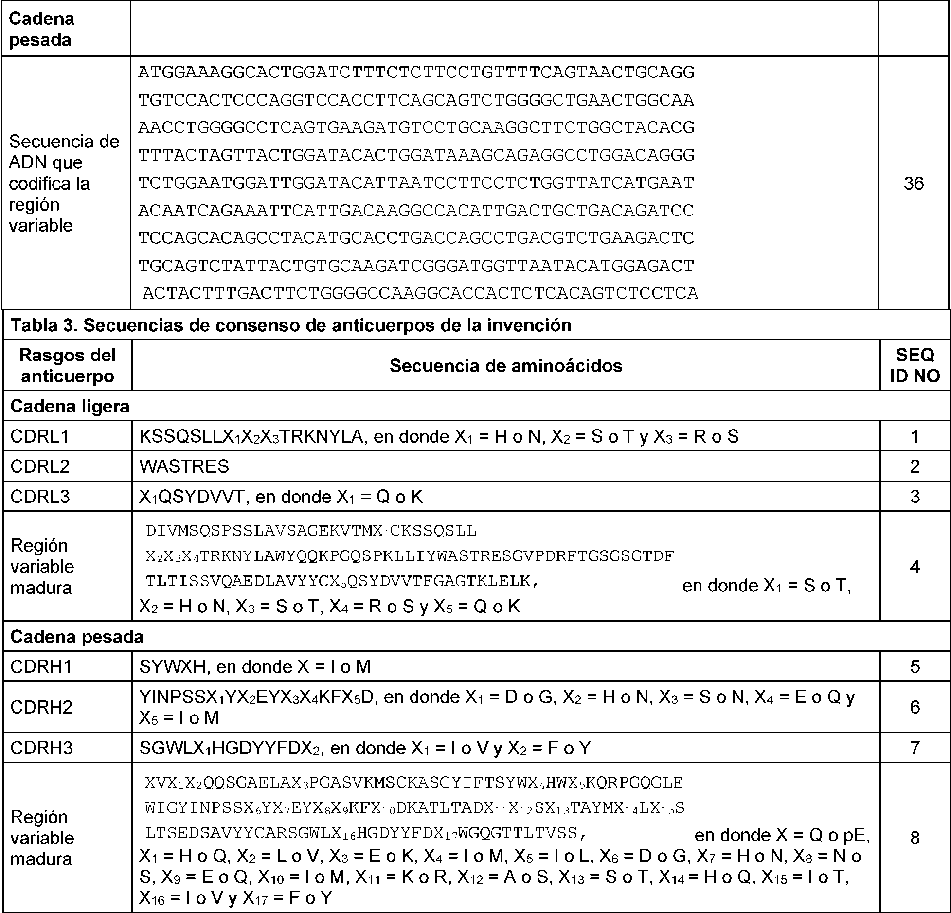
"Variantes modificadas de manera conservativa" o "sustitución conservativa" se refiere a sustituciones de aminoácidos en una proteína por otros aminoácidos que tienen características similares (por ejemplo, carga, tamaño de la cadena lateral, hidrofobicidad/hidrofilicidad, conformación y rigidez de la cadena principal, etc.), de tal manera que pueden realizarse cambios con frecuencia sin alterar la actividad biológica de la proteína. Los expertos en esta técnica reconocen que, en general, las sustituciones de aminoácidos individuales en regiones no esenciales de un polipéptido no alteran sustancialmente la actividad biológica (véase, por ejemplo, Watson et al. (1987) Molecular Biology of the Gene, The Benjamin/Cummings Pub. Co., pág. 224 (4a Ed.)). Además, las sustituciones de aminoácidos estructural o funcionalmente similares tienen menos probabilidades de alterar la actividad biológica. Las sustituciones conservativas ilustrativas de la presente divulgación se exponen en la Tabla 4."Conservatively modified variants" or "conservative substitution" refers to substitutions of amino acids in a protein for other amino acids that have similar characteristics (eg, charge, side chain size, hydrophobicity / hydrophilicity, conformation and stiffness of the chain main, etc.), such that changes can be made frequently without altering the biological activity of the protein. Those skilled in the art recognize that, in general, individual amino acid substitutions in nonessential regions of a polypeptide do not substantially alter biological activity (see, for example, Watson et al. (1987) Molecular Biology of the Gene, The Benjamin / Cummings Pub. Co., p. 224 (4th Ed.)). Furthermore, structurally or functionally similar amino acid substitutions are less likely to alter biological activity. Illustrative conservative substitutions in the present disclosure are set forth in Table 4.
TABLA 4. Sustituciones conservativas de aminoácidos ilustrativasTABLE 4. Illustrative Conservative Amino Acid Substitutions

continuacióncontinuation

En la presente divulgación también se contemplan variantes de función conservativa de los anticuerpos de la divulgación. "Variantes de función conservativa", como se usa en el presente documento, se refiere a anticuerpos o a fragmentos en los que se han cambiado uno o más restos de aminoácidos sin alterar una propiedad deseada, tal como una afinidad y/o especificidad del antígeno. Dichas variantes incluyen, aunque sin limitación, el remplazo de un aminoácido por uno que tiene propiedades similares, tal como las sustituciones de aminoácidos conservativas de la Tabla 4.Conservative function variants of the antibodies of the disclosure are also contemplated in the present disclosure. "Conservative function variants", as used herein, refers to antibodies or fragments in which one or more amino acid residues have been changed without altering a desired property, such as an affinity and / or specificity of the antigen. Such variants include, but are not limited to, the replacement of an amino acid with one that has similar properties, such as the conservative amino acid substitutions in Table 4.
En otra realización, la invención incluye un anticuerpo o fragmento de unión a antígeno del mismo que se une específicamente al PD-L1 y que tiene dominios Vl y dominios Vh y que comparte una homología de secuencia del 100 % con las regiones variables maduras de cadena ligera y pesada de las Tablas 1 o 2.In another embodiment, the invention includes an antibody or antigen-binding fragment thereof that specifically binds to PD-L1 and has V l domains and V h domains and that shares 100% sequence homology with mature variable regions. heavy and light chain from Tables 1 or 2.
Ácidos nucleicosNucleic acids
La presente invención también proporciona ácidos nucleicos que codifican las cadenas de inmunoglobulina de los anticuerpos anti-PD-L1 y fragmentos de unión a antígeno desvelados en el presente documento. La presente invención incluye ácidos nucleicos que codifican los aminoácidos de las regiones variables maduras de cadena ligera y pesada de las Tablas 1 o 2, en donde en el SEQ ID NO: 32, X es Q.The present invention also provides nucleic acids encoding the immunoglobulin chains of anti-PD-L1 antibodies and antigen-binding fragments disclosed herein. The present invention includes nucleic acids encoding the amino acids of the mature heavy and light chain variable regions of Tables 1 or 2, wherein in SEQ ID NO: 32, X is Q.
En general, los ácidos nucleicos se hibridan en condiciones de rigurosidad baja, moderada o alta, y codifican anticuerpos que conservan la capacidad de unirse específicamente al PD-L1. Una primera molécula de ácido nucleico "puede "hibridarse" con una segunda molécula de ácido nucleico cuando una forma monocatenaria de la primera molécula de ácido nucleico puede aparearse con la segunda molécula de ácido nucleico en las condiciones adecuadas de temperatura y fuerza iónica de la solución (véase Sambrook, et al., citado anteriormente). Las condiciones de temperatura y fuerza iónica determinan la "rigurosidad" de la hibridación. Las condiciones normales de hibridación de rigurosidad baja incluyen 55 °C, SSC 5X, SDS al 0,1% y sin formamida; o con formamida al 30 %, SSC 5X, SDS al 0,5 % a 42 °C. Las condiciones normales de hibridación de rigurosidad moderada son formamida al 40 %, con SSC 5X o 6X y SDS al 0,1 % a 42 °C. Las condiciones de hibridación de rigurosidad alta son formamida al 50 %, SSC 5X o 6X a 42 °C u, opcionalmente, a una temperatura más alta (por ejemplo, 57 °C, 59 °C, 60 °C, 62 °C, 63 °C, 65°°C o 68°°C). En general, el SSC es NaCl 0,15 M y citrato de Na 0,015 M. La hibridación requiere que los dos ácidos nucleicos contengan secuencias complementarias, aunque, dependiendo de la rigurosidad de la hibridación, son posibles emparejamientos erróneos entre bases. La rigurosidad apropiada para hibridar ácidos nucleicos depende de la longitud de los ácidos nucleicos y del grado de complementación, variables bien conocidas en la técnica. Cuanto mayor sea el grado de similitud u homología entre dos secuencias de nucleótidos, mayor será la rigurosidad con la que pueden hibridarse los ácidos nucleicos. Para híbridos de más de 100 nucleótidos de longitud, se han obtenido ecuaciones para calcular la temperatura de fusión (véase Sambrook, et al., citado anteriormente, 9.50-9.51). Para la hibridación con ácidos nucleicos más cortos, por ejemplo, oligonucleótidos, la posición de los emparejamientos erróneos se vuelve más importante y la longitud del oligonucleótido determina su especificidad (véase Sambrook, et al., citado anteriormente, 11.7-11.8).In general, nucleic acids hybridize under conditions of low, moderate, or high stringency, and encode antibodies that retain the ability to specifically bind to PD-L1. A first nucleic acid molecule "can" hybridize "with a second nucleic acid molecule when a single-stranded form of the first nucleic acid molecule can pair with the second nucleic acid molecule under the appropriate conditions of temperature and ionic strength of the solution. (See Sambrook, et al., cited above.) Ionic strength and temperature conditions determine the "stringency" of hybridization. Typical low stringency hybridization conditions include 55 ° C, 5X SSC, 0.1% SDS and without formamide; or with 30% formamide, 5X SSC, 0.5% SDS at 42 ° C. Normal hybridization conditions of moderate stringency are 40% formamide, with 5X or 6X SSC and 0.1 SDS % at 42 ° C. High stringency hybridization conditions are 50% formamide, 5X or 6X SSC at 42 ° C or optionally higher temperature (e.g. 57 ° C, 59 ° C, 60 ° C, 62 ° C, 63 ° C, 65 ° C or 68 ° C). In general, SSC is 0.15 M NaCl and Na 0 citrate, 015 M. Hybridization requires that the two nucleic acids contain complementary sequences, although, depending on the stringency of hybridization, base mismatches are possible. The appropriate stringency to hybridize nucleic acids depends on the length of the nucleic acids and the degree of complementation, variables well known in the art. The greater the degree of similarity or homology between two nucleotide sequences, the greater the stringency with which nucleic acids can hybridize. For hybrids greater than 100 nucleotides in length, equations have been obtained to calculate the melting temperature (see Sambrook, et al., Cited above, 9.50-9.51). For hybridization to shorter nucleic acids, eg oligonucleotides, the position of the mismatches becomes more important and the length of the oligonucleotide determines its specificity (see Sambrook, et al., Cited above, 11.7-11.8).
Las siguientes referencias se refieren a algoritmos BLAST que se utilizan a menudo para el análisis de secuencias: ALGORITMOS BLAST: Altschul, S.F., et al., (1990) J. Mol. Biol. 215:403-410; Gish, W., et al., (1993) Nature Genet.The following references refer to BLAST algorithms that are often used for sequence analysis: BLAST ALGORITHMS: Altschul, SF, et al., (1990) J. Mol. Biol. 215: 403-410; Gish, W., et al., (1993) Nature Genet.
3:266-272; Madden, T.L., et al., (1996) Meth. Enzymol. 266:131-141; Altschul, S.F., et al., (1997) Nucleic Acids Res.3: 266-272; Madden, TL, et al., (1996) Meth. Enzymol. 266: 131-141; Altschul, SF, et al., (1997) Nucleic Acids Res.
25:3389-3402; Zhang, J., et al., (1997) Genome Res. 7:649-656; Wootton, J.C., et al., (1993) Comput. Chem. 17:149-163; Hancock, J.M. et al., (1994) Comput. Appl. Biosci. 10:67-70; SISTEMAS DE PUNTUACIÓN DE ALINEACIÓN: Dayhoff, M.O., et al., "A model of evolutionary change in proteins" in Atlas of Protein Sequence and Structure, (1978) vol. 5, suppl. 3. M.O. Dayhoff (ed.), págs. 345-352, Natl Biomed. Res. Found., Washington, DC; Schwartz, R.M., et al., "Matrices for detecting distant relationships" in Atlas of Protein Sequence and Structure, (1978) vol. 5, suppl. 3". M.O. Dayhoff (ed.), págs. 353-358, Natl Biomed. Res. Found., Washington, DC; Altschul, S.F., (1991) J. Mol. Biol. 219:555-565; States, D.J., et al., (1991) Methods 3:66-70; Henikoff, S., et al., (1992) Proc. Natl. Acad. Sci. USA 89:10915-10919; Altschul, S.F., et al., (1993) J. Mol. Evol. 36:290-300; DATOS ESTADÍSTICOS DE ALINEACIÓN: Karlin, S., et al., (1990) Proc. Natl. Acad. Sci. USA 87:2264-2268; Karlin, S., et al., (1993) Proc. Natl. Acad. Sci. USA 90:5873-5877; Dembo, A., et al., (1994) Ann. Prob. 22:2022-2039; y Altschul, S.F. "Evaluating the statistical significance of multiple distinct local alignments." en Theoretical and Computational Methods in Genome Research (S. Suhai, ed.), (1997) págS. 1-14, Plenum, Nueva York.25: 3389-3402; Zhang, J., et al., (1997) Genome Res. 7: 649-656; Wootton, JC, et al., (1993) Comput. Chem. 17: 149-163; Hancock, JM et al., (1994) Comput. Appl. Biosci. 10: 67-70; ALIGNMENT SCORE SYSTEMS: Dayhoff, MO, et al., "A model of evolutionary change in proteins" in Atlas of Protein Sequence and Structure, (1978) vol. 5, suppl. 3. MO Dayhoff (ed.), Pp. 345-352, Natl Biomed. Res. Found., Washington, DC; Schwartz, RM, et al., "Matrices for detecting distant relationships" in Atlas of Protein Sequence and Structure, (1978) vol. 5, suppl. 3 ". MO Dayhoff (ed.), Pp. 353-358, Natl Biomed. Res. Found., Washington, DC; Altschul, SF, (1991) J. Mol. Biol. 219: 555-565; States, DJ , et al., (1991) Methods 3: 66-70; Henikoff, S., et al., (1992) Proc. Natl. Acad. Sci. USA 89: 10915-10919; Altschul, SF, et al., (1993) J. Mol. Evol. 36: 290-300; STATISTICAL ALIGNMENT DATA: Karlin, S., et al., (1990) Proc. Natl. Acad. Sci. USA 87: 2264-2268; Karlin, S ., et al., (1993) Proc. Natl. Acad. Sci. USA 90: 5873-5877; Dembo, A., et al., (1994) Ann. Prob. 22: 2022-2039; and Altschul, SF "Evaluating the statistical significance of multiple distinct local alignments." In Theoretical and Computational Methods in Genome Research (S. Suhai, ed.), (1997) pp. 1-14, Plenum, New York.
En algunas realizaciones, la invención proporciona un ácido nucleico aislado que codifica tanto una cadena ligera como una cadena pesada como se reivindica en una única molécula de ácido nucleico, y en otras realizaciones las cadenas ligera y pesada están codificadas en moléculas de ácido nucleico distintas. En otra realización, los ácidos nucleicos codifican además una secuencia señal.In some embodiments, the invention provides an isolated nucleic acid encoding both a light chain and a heavy chain as claimed in a single nucleic acid molecule, and in other embodiments the light and heavy chains are encoded in separate nucleic acid molecules. In another embodiment, the nucleic acids further encode a signal sequence.
La presente invención también proporciona vectores de expresión que comprenden los ácidos nucleicos aislados de la invención, en donde el ácido nucleico está unido operativamente a secuencias de control que son reconocidas por una célula hospedadora cuando la célula hospedadora se transfecta con el vector. También se proporcionan células hospedadoras que comprenden un vector de expresión de la presente invención. En el presente documento se describen métodos para producir el anticuerpo o fragmento de unión a antígeno del mismo que se desvelan en el presente documento que comprenden cultivar en un medio de cultivo una célula hospedadora que lleva un vector de expresión que codifica el anticuerpo o fragmento de unión a antígeno y aislar el antígeno o fragmento de unión a antígeno del mismo de la célula hospedadora o del medio de cultivo.The present invention also provides expression vectors comprising the isolated nucleic acids of the invention, wherein the nucleic acid is operably linked to control sequences that are recognized by a host cell when the host cell is transfected with the vector. Host cells comprising an expression vector of the present invention are also provided. Described herein are methods for producing the antibody or antigen-binding fragment thereof disclosed herein which comprise culturing in culture medium a host cell carrying an expression vector encoding the antibody or fragment of antigen binding and isolating the antigen or antigen-binding fragment thereof from the host cell or culture medium.
Unión epitópicaEpitopic union
En el presente documento se describen anticuerpos o fragmentos de unión a antígeno de los mismos que bloquean la unión del anticuerpo 20C3 o 22C3 con el PD-L1 humano uniéndose al mismo epítopo que 20C3 o 22C3, respectivamente. Dichos anticuerpos y fragmentos de unión pueden identificarse usando cualquier análisis de bloqueo cruzado o de competencia conocido en la técnica, incluyendo los análisis de competencia Octet descritos en el Ejemplo 2, seguido de la identificación del epítopo en el PD-L1 humano al que se une el anticuerpo de bloqueo cruzado. Se considera que un primer anticuerpo bloquea de forma cruzada la unión de un segundo anticuerpo si la unión previa de la diana con el primer anticuerpo a la saturación aumenta la concentración del segundo anticuerpo necesaria para lograr la unión media máxima de la diana en 2-, 3-, 4-, 5-, 10-, 20-, 50-, 100-, 200 veces o más. El epítopo de unión de un anticuerpo de bloqueo cruzado puede identificarse usando técnicas bien conocidas en la materia.Described herein are antibodies or antigen-binding fragments thereof that block the binding of antibody 20C3 or 22C3 to human PD-L1 by binding to the same epitope as 20C3 or 22C3, respectively. Such antibodies and binding fragments can be identified using any competition or cross-blocking assay known in the art, including the Octet competition assays described in Example 2, followed by identification of the epitope on the human PD-L1 to which it binds. the cross-blocking antibody. A first antibody is considered to cross-block the binding of a second antibody if prior binding of the target to the first antibody to saturation increases the concentration of the second antibody necessary to achieve the maximum mean binding of the target by 2-, 3-, 4-, 5-, 10-, 20-, 50-, 100-, 200 times or more. The binding epitope of a cross-blocking antibody can be identified using techniques well known in the art.
Una de dichas técnicas de mapeo epitópico es el intercambio de hidrógeno/deuterio acoplado con proteólisis y espectrometría de masas (HDX-MS). Este método se basa en la medición precisa y la comparación del grado de incorporación de deuterio por un antígeno cuando se incuba en agua pesada (D2O) por sí solo y en presencia de su anticuerpo en varios intervalos de tiempo. El deuterio se intercambia por hidrógeno en la cadena principal amida de las proteínas en las áreas expuestas, mientras que las regiones del antígeno unidas al anticuerpo estarán protegidas y mostrarán menos o ningún intercambio después del análisis por cromatografía líquida acoplada a espectrometría de masas en tándem (LC-MS/MS) de fragmentos proteolíticos.One such epitopic mapping technique is hydrogen / deuterium exchange coupled with proteolysis and mass spectrometry (HDX-MS). This method is based on the precise measurement and comparison of the degree of incorporation of deuterium by an antigen when it is incubated in heavy water (D2O) on its own and in the presence of its antibody at various time intervals. Deuterium is exchanged for hydrogen in the amide backbone of proteins in the exposed areas, while the regions of the antigen bound to the antibody will be protected and will show less or no exchange after analysis by liquid chromatography coupled to tandem mass spectrometry ( LC-MS / MS) of proteolytic fragments.
Basándose en el mapeo epitópico HDX-MS descrito en el Ejemplo 3, el epítopo propuesto en el PD-L1 humano maduro para el anticuerpo 22C3 comprende restos en dos segmentos de aminoácidos discontinuos en el dominio extracelular (SEQ ID NO: 38): 156 a 178 y 196 a 206. Es probable que haya restos epitópicos adicionales en los siguientes segmentos del dominio extracelular (SEQ ID NO: 38): 3 a 9; 10 a 13; 88 a 93 y 135 a 147.Based on the HDX-MS epitopic mapping described in Example 3, the proposed epitope on mature human PD-L1 for the 22C3 antibody comprises residues in two discontinuous amino acid segments in the extracellular domain (SEQ ID NO: 38): 156 to 178 and 196 to 206. It is likely that there are additional epitopic residues in the following extracellular domain segments (SEQ ID NO: 38): 3 to 9; 10 to 13; 88 to 93 and 135 to 147.
Por tanto, como se describe en el presente documento, un anticuerpo que bloquea la unión del anticuerpo 22C3 con el PD-L1 humano al unirse al mismo epítopo que 22C3 se une a los restos en un primer segmento de los aminoácidos 156 a 178 del SEQ ID NO: 38 y a los restos en un segundo segmento de los aminoácidos 196 a 206 del SEQ ID NO: 38, y también se une a los restos en cualquiera de uno, dos, o tres, o en los cuatro, de los siguientes segmentos del SEQ ID NO: 38: aminoácidos 3 a 9; aminoácidos 10 a 13; aminoácidos 88 a 93 y aminoácidos 135 a 147.Thus, as described herein, an antibody that blocks the binding of antibody 22C3 to human PD-L1 by binding to the same epitope as 22C3 binds to residues in a first segment of amino acids 156 to 178 of SEQ ID NO: 38 and to residues in a second segment of amino acids 196 to 206 of SEQ ID NO: 38, and also binds to residues in any one, two, or three, or all four, of the following segments SEQ ID NO: 38: amino acids 3 to 9; amino acids 10 to 13; amino acids 88 to 93 and amino acids 135 to 147.
Métodos de fabricación de anticuerpos y de fragmentos de unión a antígeno de los mismosMethods of manufacturing antibodies and antigen-binding fragments thereof
Las células de hibridoma que producen anticuerpos anti-X monoclonales precursores (por ejemplo, roedores) pueden producirse mediante métodos conocidos comúnmente en la materia. Estos métodos incluyen, aunque sin limitación, la técnica del hibridoma desarrollada originalmente por Kohler, et al., (1975) (Nature 256:495-497), así como la técnica del trioma (Hering, et al., (1988) Biomed. Biochim. Acta. 47:211-216 y Hagiwara, et al., (1993) Hum. Antibod. Hybridomas 4:15), la técnica del hibridoma de células B humanas (Kozbor, et al., (1983) Immunology Today 4:72 y Cote, et al., (1983) Proc. Natl. Acad. Sci. USA 80: 2026-2030), la técnica del hibridoma con el VEB (virus de Epstein Barr) (Cole, et al., en Monoclonal Antibodies and Cancer Therapy, Alan R. Liss, Inc., págs. 77-96, 1985), y electrofusión basada en campo eléctrico usando un electroporador de fusión selectiva de cámara grande Cyto Pulse (Cyto Pulse Sciences, Inc., Glen Burnie, MD). Preferentemente, esplenocitos de ratón se aíslan y se fusionan con PEG o mediante electrofusión con una línea celular de mieloma de ratón basándose en protocolos estándar.Hybridoma cells that produce precursor monoclonal anti-X antibodies (eg, rodents) can be produced by methods commonly known in the art. These methods include, but are not limited to, the hybridoma technique originally developed by Kohler, et al., (1975) (Nature 256: 495-497), as well as the trioma technique (Hering, et al., (1988) Biomed Biochim. Acta. 47: 211-216 and Hagiwara, et al., (1993) Hum. Antibod. Hybridomas 4:15), the human B-cell hybridoma technique (Kozbor, et al., (1983) Immunology Today 4:72 and Cote, et al., (1983) Proc. Natl. Acad. Sci. USA 80: 2026-2030), the hybridoma technique with EBV (Epstein Barr virus) (Cole, et al., In Monoclonal Antibodies and Cancer Therapy, Alan R. Liss, Inc., pp. 77-96, 1985), and electric field-based electrofusion using a Cyto Pulse large chamber selective fusion electroporator (Cyto Pulse Sciences, Inc., Glen Burnie, MD). Preferably, mouse splenocytes are isolated and fused with PEG or by electrofusion with a mouse myeloma cell line based on standard protocols.
A continuación, los hibridomas resultantes pueden explorarse con respecto a la producción de anticuerpos específicos de antígeno. Por ejemplo, suspensiones unicelulares de linfocitos esplénicos de ratones inmunizados pueden fusionarse a una sexta parte del número de células de mieloma de ratón no secretoras P3X63-Ag8.653 (ATCC, CRL 1580) con PEG al 50 %. Las células pueden sembrarse en placas a aproximadamente 2 x 10⁵ células/ml en una placa de microtitulación de fondo plano, seguido de dos semanas de incubación en medio selectivo que contiene suero de clon fetal al 20 %, medios acondicionados "653" al 18 %, origen (IGEN) al 5 %, L-glutamina 4 mM, L-glutamina 1 mM, piruvato sódico 1 mM, HEPES 5 mM, 2-mercaptoetanol 0,055 mM, penicilina 50 unidades/ml, estreptomicina 50 mg/ml, gentamicina 50 mg/ml y HAT 1X (Sigma; el HAT se añade 24 horas después de la fusión). Después de dos semanas, las células pueden cultivarse en medio en el que el HAT está reemplazado por HT. A continuación, los pocillos individuales pueden explorarse mediante ELISA para detectar anticuerpos de IgG monoclonales anti-X. Una vez que se produce el crecimiento extenso del hibridoma, el medio puede observarse normalmente después de 10-14 días. Los hibridomas secretores de anticuerpo pueden resembrarse en placas, explorarse de nuevo y si siguen siendo positivos a la IgG humana, los anticuerpos monoclonales anti-X, pueden subclonarse al menos dos veces mediante dilución limitante.The resulting hybridomas can then be screened for the production of antigen-specific antibodies. For example, single-cell suspensions of splenic lymphocytes from immunized mice can be fused to one-sixth the number of non-secreting mouse myeloma cells P3X63-Ag8.653 (ATCC, CRL 1580) with 50% PEG. Cells can be plated at approximately 2 x 10⁵ cells / ml in a flat-bottom microtiter plate, followed by two weeks incubation in selective medium containing 20% fetal clone serum, 18% "653" conditioned media. , 5% origin (IGEN), 4 mM L-glutamine, 1 mM L-glutamine, 1 mM sodium pyruvate, 5 mM HEPES, 0.055 mM 2-mercaptoethanol, penicillin 50 units / ml, streptomycin 50 mg / ml, gentamicin 50 mg / ml and 1X HAT (Sigma; HAT is added 24 hours after melting). After two weeks, cells can be cultured in medium in which HAT is replaced by HT. Individual wells can then be screened by ELISA for anti-X monoclonal IgG antibodies. Once extensive hybridoma growth occurs, the medium can normally be observed after 10-14 days. Antibody-secreting hybridomas can be re-plated, screened again, and if they remain positive for human IgG, anti-X monoclonal antibodies can be subcloned at least twice by limiting dilution.
A continuación, para su caracterización, los subclones estables pueden cultivarse in vitro para generar pequeñas cantidades de anticuerpo en medio de cultivo tisular. Por ejemplo, usando el siguiente procedimiento, a partir de la línea celular de hibridoma de ratón MEB037.22C3.138, puede producirse y purificarse aproximadamente 1 gramo del anticuerpo 22C3. Las células de MEB037.22C3.138 congeladas se descongelan y se adaptan en un matraz de agitación usando medio sin suero de hibridoma con L-glutamina adicional 2 mM con o sin Pluronic F-68 al 0,18 %. La presencia de Pluronic F-68 puede mejorar la viabilidad del cultivo en el matraz de agitación. Una vez que las células se han adaptado completamente en el matraz de agitación, se realiza un cultivo de producción de 20 litros en medio sin suero en un biorreactor WAVE (GE Healthcare Life Sciences) con la adición de suministro B eficiente de CHO CD al 10 % (Invitrogen, n.° de catálogo A10240-01). Para la expansión celular, se inicia un cultivo de 1 litro en una pequeña bolsa WAVE, y después el cultivo WAVE de 1 l se expande a un cultivo de 20 l en el biorreactor WAVE. El cultivo de 20 litros puede iniciarse a una densidad celular de 0,5 x 10⁶ células viables/ml, alimentarse con suministro B eficiente de CHO CD al 10 % el día 1 y el pH puede ajustarse diariamente con Na2CO3 1 N. Las células se recogen después de cuatro días. Para el análisis NOVA pueden recogerse pequeñas muestras diariamente.The stable subclones can then be cultured in vitro to generate small amounts of antibody in tissue culture medium for characterization. For example, using the following procedure, from the mouse hybridoma cell line MEB037.22C3.138, approximately 1 gram of the 22C3 antibody can be produced and purified. Frozen MEB037.22C3.138 cells are thawed and adapted in a shake flask using medium without hybridoma serum with additional 2 mM L-glutamine with or without 0.18% Pluronic F-68. The presence of Pluronic F-68 can improve the viability of the culture in the shake flask. Once the cells have fully adapted in the shake flask, a 20 liter production culture in serum-free medium is performed in a WAVE bioreactor (GE Healthcare Life Sciences) with the addition of efficient B delivery of 10 CHO CD. % (Invitrogen, Catalog # A10240-01). For cell expansion, a 1 L culture is started in a small WAVE bag, and then the 1 L WAVE culture is expanded to a 20 L culture in the WAVE bioreactor. The 20 liter culture can be started at a cell density of 0.5 x 10⁶ viable cells / ml, fed with efficient B supply of 10% CHO CD on day 1, and the pH can be adjusted daily with 1 N Na2CO3. they collect after four days. Small samples can be collected daily for NOVA analysis.
Los anticuerpos anti-hPD-L1 de la invención pueden purificarse de un cultivo de hibridoma mediante el siguiente proceso. El cultivo de hibridoma se aclara mediante filtración profunda utilizando fibra de vidrio de 1,2 micrómetros y un filtro de acetato de celulosa de 0,2 micrómetros. Al aclarado recogido se le añade un mismo volumen de tampón ProSepA 2X (ácido bórico 100 mM, NaCl 5 M, pH 8,5) y el diluido recogido se carga en una columna de proteína A con un volumen de lecho de 170 ml. La columna se lava con 5 volúmenes de columna (VC) de tampón ProSepA 1X (ácido bórico 50 mM, NaCl 2,5 M, pH 8,5), después se lava con 2VC de PBS 1X y el anticuerpo anti-hPD-L1 se eluye con 5VC de tampón de elución (glicina 0,1M, pH 3,0). Las fracciones de elución que contienen IgG se combinan y el pH se neutraliza añadiendo un volumen de 1/10 de Tris 1,0 M, tampón de pH. La composición de anticuerpo neutralizado se esteriliza después por filtración usando un casete de TFF desechable de 10 kDa. Para su conservación, el anticuerpo puede formularse mediante diafiltración frente a 10 litros de tampón de formulación (acetato de sodio 20 mM, sacarosa al 9 %, pH 5,0) y usando 20 cambios de volumen. Usando este protocolo, el anticuerpo 22C3 puede prepararse a una concentración de aproximadamente 5,0 mg/ml y con una pureza de al menos el 98 % mediante SDS-Pa Ge , Mediciones por SEC HPLC y C8 RP-HPLC, con niveles de endotoxina inferiores a 0,1 UE/ml y menos de 0,02 UE/mg.The anti-hPD-L1 antibodies of the invention can be purified from a hybridoma culture by the following process. The hybridoma culture is clarified by deep filtration using 1.2 micron fiberglass and a 0.2 micron cellulose acetate filter. To the collected rinse, the same volume of 2X ProSepA buffer (100 mM boric acid, 5 M NaCl, pH 8.5) is added and the collected dilute is loaded onto a protein A column with a bed volume of 170 ml. The column is washed with 5 column volumes (CV) of 1X ProSepA buffer (50 mM boric acid, 2.5 M NaCl, pH 8.5), then washed with 2VC of 1X PBS and the anti-hPD-L1 antibody. Elute with 5VC of elution buffer (0.1M glycine, pH 3.0). The elution fractions containing IgG are pooled and the pH is neutralized by adding a 1/10 volume of 1.0 M Tris, pH buffer. The neutralized antibody composition is then sterilized by filtration using a disposable 10 kDa TFF cassette. For preservation, the antibody can be formulated by diafiltration against 10 liters of formulation buffer (20 mM sodium acetate, 9% sucrose, pH 5.0) and using 20 volume changes. Using this protocol, the 22C3 antibody can be prepared at a concentration of approximately 5.0 mg / ml and with a purity of at least 98% by SDS-Pa Ge, SEC HPLC and C8 RP-HPLC measurements, with endotoxin levels less than 0.1 EU / ml and less than 0.02 EU / mg.
Los anticuerpos anti-PD-L1 desvelados en el presente documento también pueden producirse de manera recombinante (por ejemplo, en un sistema de expresión de E. coli/T7, como se indicó anteriormente). En esta realización, ácidos nucleicos que codifican las moléculas de anticuerpo de la invención (por ejemplo, Vh o V l) pueden insertarse en un plásmido basado en pET y expresarse en el sistema de E. coli/T7. Existen varios métodos conocidos en la materia para producir anticuerpos recombinantes. En la Patente de Estados Unidos N° 4.816.567 se desvela un ejemplo de un método de producción recombinante de anticuerpos. La transformación puede ser mediante cualquier método conocido de introducción de polinucleótidos en una célula hospedadora. En la materia se conocen bien métodos para introducir polinucleótidos heterólogos en células de mamífero e incluyen transfección mediada por dextrano, precipitación con fosfato de calcio, transfección mediada por polibreno, fusión de protoplastos, electroporación, encapsulación del polinucleótido (o de los polinucleótidos en liposomas, inyección biolística y microinyección directa del ADN en los núcleos. Además, las moléculas de ácido nucleico pueden introducirse en células de mamíferos mediante vectores víricos. En la materia se conocen bien métodos de transformación de células. Véanse, por ejemplo, las patentes de Estados Unidos N.° 4.399.216; 4.912.040; 4.740.461 y 4.959.455.The anti-PD-L1 antibodies disclosed herein can also be produced recombinantly (eg, in an E. coli / T7 expression system, as noted above). In this embodiment, nucleic acids encoding the antibody molecules of the invention (eg, V h or V l ) can be inserted into a pET-based plasmid and expressed in the E. coli / T7 system. There are several methods known in the art for producing recombinant antibodies. An example of a recombinant antibody production method is disclosed in US Patent No. 4,816,567. Transformation can be by any known method of introducing polynucleotides into a host cell. Methods for introducing heterologous polynucleotides into mammalian cells are well known in the art and include dextran-mediated transfection, calcium phosphate precipitation, polybrene-mediated transfection, protoplast fusion, electroporation, encapsulation of the polynucleotide (or polynucleotides in liposomes, Biolistic injection and direct microinjection of DNA into nuclei. In addition, nucleic acid molecules can be introduced into mammalian cells by viral vectors. Methods of cell transformation are well known in the art. See, for example, US Pat. Nos. 4,399,216; 4,912,040; 4,740,461 and 4,959,455.
Los anticuerpos anti-PD-L1 también pueden sintetizarse mediante cualquiera de los métodos expuestos en la patente de Estados Unidos N.° 6.331.415.Anti-PD-L1 antibodies can also be synthesized by any of the methods set forth in US Patent No. 6,331,415.
En la materia se conocen bien líneas celulares de mamífero disponibles como hospedadores para la expresión de los anticuerpos o fragmentos desvelados en el presente documento e incluyen muchas líneas celulares inmortalizadas disponibles en la colección americana de cultivos tipo (American Type Culture Collection, ATCC). Estas incluyen, entre otras, células de ovario de hámster chino (CHO), NSO, células SP2, células HeLa, células de riñón de cría de hámster (BHK, baby hámster kidney), células de riñón de mono (COS), células de carcinoma hepatocelular humano (por ejemplo, Hep G2), células A549, células 3T3, células h EK-293 y diversas líneas celulares distintas. Las células hospedadoras de mamífero incluyen seres humanos, ratón, rata, perro, mono, cerdo, cabra, bovinos, células de caballo y hámster. Las líneas celulares de preferencia particular se seleccionan determinando qué líneas celulares tienen altos niveles de expresión. Otras líneas celulares que pueden utilizarse son líneas celulares de insecto, tales como células Sf9, células de anfibios, células bacterianas, células vegetales y células fúngicas. Cuando los vectores de expresión recombinante codifican la cadena pesada o la parte de unión al antígeno o un fragmento de la misma, la cadena ligera y/o el fragmento de unión a antígeno de la misma se introducen en células hospedadoras de mamífero, los anticuerpos se producen cultivando las células hospedadoras durante un periodo de tiempo suficiente para permitir la expresión del anticuerpo en las células hospedadoras o, más preferentemente, la secreción del anticuerpo en el medio de cultivo en el que crecen las células hospedadoras.Mammalian cell lines available as hosts for the expression of the antibodies or fragments disclosed herein are well known in the art and include many immortalized cell lines. available from the American Type Culture Collection ( ATCC). These include, but are not limited to, Chinese Hamster Ovary (CHO) cells, NSO, SP2 cells, HeLa cells, Baby Hamster Kidney (BHK) cells, Monkey Kidney (COS) cells, human hepatocellular carcinoma (eg, Hep G2), A549 cells, 3T3 cells, EK-293 h cells, and a number of other cell lines. Mammalian host cells include human, mouse, rat, dog, monkey, pig, goat, bovine, horse, and hamster cells. Cell lines of particular preference are selected by determining which cell lines have high levels of expression. Other cell lines that can be used are insect cell lines, such as Sf9 cells, amphibian cells, bacterial cells, plant cells, and fungal cells. When recombinant expression vectors encode the heavy chain or antigen-binding part or a fragment thereof, the light chain and / or antigen-binding fragment thereof are introduced into mammalian host cells, the antibodies are They are produced by culturing the host cells for a period of time sufficient to allow the expression of the antibody in the host cells or, more preferably, the secretion of the antibody into the culture medium in which the host cells grow.
Los anticuerpos pueden recuperarse del medio de cultivo usando métodos de purificación de proteínas convencionales. Adicionalmente, usando diversas de técnicas conocidas, puede potenciarse la expresión de los anticuerpos de la invención (u otras fracciones de los mismos) a partir de líneas celulares de producción. Por ejemplo, el sistema de expresión del gen de la glutamina sintetasa (el sistema GS) es una estrategia habitual para mejorar la expresión en determinadas condiciones. El sistema GS se analiza, total o parcialmente, en relación con las patentes europeas N.° 0216846, 0256055 y 0323997 y con la solicitud de patente europea N.° 89303964.4.Antibodies can be recovered from the culture medium using standard protein purification methods. Additionally, using various of known techniques, the expression of the antibodies of the invention (or other fractions thereof) can be enhanced from production cell lines. For example, the glutamine synthetase gene expression system (the GS system) is a common strategy to enhance expression under certain conditions. The GS system is analyzed, in whole or in part, in relation to European patents Nos. 0216846, 0256055 and 0323997 and with European patent application No. 89303964.4.
Un anticuerpo policlonal es un anticuerpo que se produce entre o en presencia de uno o más, anticuerpos distintos, no idénticos. En general, los anticuerpos policlonales se producen a partir de colecciones de linfocitos B diferentes, por ejemplo, el linfocito B de un animal tratado con un inmunógeno de interés, que produce una población de anticuerpos diferentes que están todos dirigidos al inmunógeno. Habitualmente, los anticuerpos policlonales se obtienen directamente de un animal inmunizado, por ejemplo, de bazo, suero o líquido ascítico.A polyclonal antibody is an antibody that is produced between or in the presence of one or more, distinct, non-identical antibodies. In general, polyclonal antibodies are produced from different collections of B lymphocytes, for example, the B lymphocyte of an animal treated with an immunogen of interest, which produces a population of different antibodies that are all directed to the immunogen. Typically, polyclonal antibodies are obtained directly from an immunized animal, eg, spleen, serum, or ascites fluid.
La presente invención incluye además fragmentos de anticuerpo de los anticuerpos anti-PD-LI desvelados en el presente documento. Los fragmentos de anticuerpo incluyen fragmentos F(ab)2, que pueden producirse por escisión enzimática de una IgG mediante, por ejemplo, pepsina. Los fragmentos Fab pueden producirse, por ejemplo, mediante reducción de F(ab)2 con ditiotreitol o mercaptoetilamina. Un fragmento Fab es una cadena Vl-Cl unida a una cadena Vh-Chi mediante un puente disulfuro. Un fragmento F(ab)2 está constituido por dos fragmentos Fab que, a su vez, están unidos por dos puentes disulfuro. La parte Fab de una molécula F(ab)2 incluye una parte de la región Fc entre la que se encuentran los puentes disulfuro. Un fragmento Fv es una región V l o V h .The present invention further includes antibody fragments of the anti-PD-LI antibodies disclosed herein. Antibody fragments include F (ab) 2 fragments, which can be produced by enzymatic cleavage of an IgG by, for example, pepsin. Fab fragments can be produced, for example, by reduction of F (ab) 2 with dithiothreitol or mercaptoethylamine. An Fab fragment is a V l -C l chain linked to a V h -C hi chain by a disulfide bridge. An F (ab) 2 fragment is made up of two Fab fragments that, in turn, are linked by two disulfide bridges. The Fab part of an F (ab) 2 molecule includes a part of the Fc region between which the disulfide bridges lie. An Fv fragment is a V l or V h region .
Las inmunoglobulinas pueden asignarse a diferentes clases dependiendo de las secuencias de aminoácidos del dominio constante de sus cadenas pesadas. Hay al menos cinco clases principales de inmunoglobulinas: IgA, IgD, IgE, IgG e IgM, y varias de éstas pueden dividirse además en subclases (isotipos), por ejemplo, IgG-1, IgG-2, IgG-3 e IgG-4; IgA-1 e IgA-2. La invención comprende anticuerpos y fragmentos de IgG de unión a antígeno.Immunoglobulins can be assigned to different classes depending on the amino acid sequences of the constant domain of their heavy chains. There are at least five major classes of immunoglobulins: IgA, IgD, IgE, IgG, and IgM, and several of these can be further divided into subclasses (isotypes), for example, IgG-1, IgG-2, IgG-3, and IgG-4. ; IgA-1 and IgA-2. The invention encompasses antibodies and antigen-binding IgG fragments.
En una realización, el anticuerpo o el fragmento de unión a antígeno comprende una región constante de cadena pesada, por ejemplo, una región constante humana, tal como la región constante humana de cadena pesada humana Yl, y2, y3 o y4 o una de sus variantes. En otra realización, el anticuerpo o fragmento de unión a antígeno comprende una región constante de cadena ligera, por ejemplo, una región constante de cadena ligera humana, tal como la región de cadena ligera humana lambda o kappa o una de sus variantes. Como ejemplo, y sin limitación, la región constante de cadena pesada humana puede ser y1 y la región constante de cadena ligera humana puede ser kappa. En una realización alternativa, la región Fc del anticuerpo es y4 con una mutación Ser228Pro (Schuurman, J et al., Mol. Immunol. 38: 1-8, 2001).In one embodiment, the antibody or antigen-binding fragment comprises a heavy chain constant region, for example, a human constant region, such as the human Y1, y2, y3, or y4 human heavy chain constant region or one of its variants. In another embodiment, the antibody or antigen-binding fragment comprises a light chain constant region, eg, a human light chain constant region, such as the human lambda or kappa light chain region or a variant thereof. As an example, and without limitation, the human heavy chain constant region can be y1 and the human light chain constant region can be kappa. In an alternative embodiment, the Fc region of the antibody is y4 with a Ser228Pro mutation (Schuurman, J et al., Mol. Immunol. 38: 1-8, 2001).
En algunas realizaciones, se pueden añadir diferentes dominios constantes a las regiones V l y Vh humanizadas reivindicadas. Por ejemplo, si un uso particular previsto de un anticuerpo (o fragmento) de la presente invención requiriera la alteración de las funciones efectoras, podría utilizarse un dominio constante de cadena pesada distinto de la IgG1 humana, o podría utilizarse un híbrido de IgG1/IgG4.In some embodiments, different constant domains can be added to the claimed humanized V l and V h regions. For example, if a particular intended use of an antibody (or fragment) of the present invention requires alteration of effector functions, a heavy chain constant domain other than human IgG1 could be used, or an IgG1 / IgG4 hybrid could be used. .
Genomodificación de anticuerposGenomodification of antibodies
Como se describe en el presente documento, para proporcionar una mayor estabilidad química del anticuerpo final, sería deseable cambiar ciertos aminoácidos que contienen cadenas laterales expuestas por otro resto de aminoácido, como se expone a continuación. La desaminación de la asparagina puede producirse en secuencias N-G o D-G y puede dar como resultado la creación de un resto de ácido isoaspártico que puede introducir un pliegue en la cadena polipeptídica y puede disminuir su estabilidad (efecto de ácido isoaspártico). Como se describe en el presente documento, los anticuerpos de la presente divulgación no contienen sitios de isomería de asparagina.As described herein, to provide greater chemical stability for the final antibody, it would be desirable to exchange certain amino acids containing exposed side chains for another amino acid residue, as discussed below. Deamination of asparagine can occur in N-G or D-G sequences and can result in the creation of an isoaspartic acid residue that can introduce a fold in the polypeptide chain and can decrease its stability (isoaspartic acid effect). As described herein, the antibodies of the present disclosure do not contain asparagine isomerism sites.
Por ejemplo, un resto de asparagina (Asn) se puede cambiar por Gln o Ala para reducir el potencial de formación de isoaspartato en cualquier secuencia Asn-Gly, particularmente dentro de una CDR. Puede producirse un problema similar en una secuencia de Asp-Gly. Reissner y Aswad (2003) Cell. Mol. Life Sci. 60:1281. La formación de isoaspartato puede debilitar o anular completamente la unión de un anticuerpo con su antígeno diana. Véase, Presta (2005) J. Allergy Clin. Immunol. 116:731 en 734. Como se describe en el presente documento, la asparagina se cambia a glutamina (Gln). También puede ser deseable alterar un aminoácido adyacente a un resto de asparagina (Asn) o glutamina (Gln) para reducir la probabilidad de desamidación, que ocurre a mayor velocidad cuando los aminoácidos pequeños se encuentran adyacentes a la asparagina o la glutamina. Véase, Bischoff y Kolbe (1994) J. Chromatog.For example, an asparagine (Asn) residue can be changed to Gln or Ala to reduce the potential for isoaspartate formation in any Asn-Gly sequence, particularly within a CDR. A similar problem can occur in an Asp-Gly sequence. Reissner and Aswad (2003) Cell. Mol. Life Sci. 60: 1281. The formation of Isoaspartate can weaken or completely abolish the binding of an antibody to its target antigen. See, Presta (2005) J. Allergy Clin. Immunol. 116: 731 at 734. As described herein, asparagine is changed to glutamine (Gln). It may also be desirable to alter an amino acid adjacent to an asparagine (Asn) or glutamine (Gln) residue to reduce the likelihood of deamidation, which occurs at a higher rate when small amino acids are adjacent to asparagine or glutamine. See, Bischoff and Kolbe (1994) J. Chromatog.
662:261. Además, en las CDR, cualquier resto de metionina (normalmente Met expuesta a disolvente) puede cambiarse a Lys, Leu, Ala o Phe para reducir la posibilidad de que se oxide el azufre de metionina, lo que podría reducir la afinidad de unión al antígeno y también contribuir a la heterogeneidad molecular en la preparación final de anticuerpos. Id. Como se describe en el presente documento, la metionina se cambia a alanina (Ala). Adicionalmente, para prevenir o minimizar posibles enlaces peptídicos Asn-Pro escindibles, puede ser deseable alterar cualquier combinación de Asn-Pro encontrada en una CDR a Gln-Pro, Ala-Pro o Asn-Ala. Los anticuerpos con dichas sustituciones se exploran posteriormente para garantizar que las sustituciones no disminuyan la afinidad o especificidad del anticuerpo por el PD-L1 humano u otra actividad biológica deseada a niveles inaceptables.662: 261. Additionally, in CDRs, any methionine residue (typically solvent-exposed Met) can be changed to Lys, Leu, Ala, or Phe to reduce the chance of methionine sulfur oxidizing, which could reduce antigen-binding affinity. and also contribute to molecular heterogeneity in the final antibody preparation. Id. As described herein, methionine is changed to alanine (Ala). Additionally, to prevent or minimize possible cleavable Asn-Pro peptide bonds, it may be desirable to alter any combination of Asn-Pro found in a CDR to Gln-Pro, Ala-Pro or Asn-Ala. Antibodies with such substitutions are subsequently screened to ensure that the substitutions do not decrease the affinity or specificity of the antibody for human PD-L1 or other desired biological activity to unacceptable levels.
TABLA 5 Variantes de CDR estabilizadoras ilustrativas de la divul aciónTABLE 5 Illustrative Stabilizing CDR Variants of the Disclosure

Conjugados de anticuerposAntibody conjugates
Las moléculas de anticuerpo anti-PD-L1 desveladas en el presente documento también pueden conjugarse con una fracción química tal como un radionúclido u otro marcador detectable. Los radionúclidos incluyen 99Tc, 90Y, 111In, 32P, 14C, 125I, 3H, 131I, 11C, 15O, 13N, 18F, 35S ,51 Cr, 57To, 226Ra, 60Co, 59Fe, 57Se, 152Eu, 67CU, 217Ci, 211At, 212Pb, 47Sc, 109Pd, 234Th y 40K, 157Gd, 55Mn, 52Tr y 56Fe. Los marcadores fluorescentes o quimioluminiscentes incluyen fluoróforos, tales como quelatos de tierras raras, fluoresceína y sus derivados, rodamina y sus derivados, isotiocianato, ficoeritrina, ficocianina, aloficocianina, o-ftaladehído, fluorescamina, 152Eu, dansilo, umbeliferona, luciferina, marcador luminal, marcador isoluminal, un marcador de éster de acridinio aromático, un marcador de imidazol, un marcador de sal de acridinio, un marcador de éster de oxalato, un marcador de aecuorina, 2,3-dihidroftalacinadionas, biotina/avidina, marcadores de espín y radicales libres estables.The anti-PD-L1 antibody molecules disclosed herein can also be conjugated to a chemical moiety such as a radionuclide or other detectable marker. Radionuclides include 99Tc, 90Y, 111In, 32P, 14C, 125I, 3H, 131I, 11C, 15O, 13N, 18F, 35S, 51 Cr, 57To, 226Ra, 60Co, 59Fe, 57Se, 152Eu, 67CU, 217Ci, 211At, 212Pb, 47Sc, 109Pd, 234Th and 40K, 157Gd, 55Mn, 52Tr and 56Fe. Fluorescent or chemiluminescent markers include fluorophores, such as rare earth chelates, fluorescein and its derivatives, rhodamine and its derivatives, isothiocyanate, phycoerythrin, phycocyanin, allophycocyanin, o-phthaladehyde, fluorescamine, 152Eu, dansyl, umbelliferone, luciferin, marker luminal isoluminal marker, an aromatic acridinium ester marker, an imidazole marker, an acridinium salt marker, an oxalate ester marker, an aequorin marker, 2,3-dihydrophthalazinadiones, biotin / avidin, spin markers and radicals stable free.
Se puede emplear cualquier método conocido en la materia para conjugar las moléculas de anticuerpo con las diversas fracciones, incluyendo los métodos descritos por Hunter, et al., (1962) Nature 144:945; David, et al., (1974) Biochemistry 13:1014; Pain, et al., (1981) J. Immunol. Meth. 40:219; y Nygren, J., (1982) Histochem. and Cytochem.Any method known in the art can be employed to conjugate the antibody molecules to the various fractions, including the methods described by Hunter, et al., (1962) Nature 144: 945; David, et al., (1974) Biochemistry 13: 1014; Pain, et al., (1981) J. Immunol. Meth. 40: 219; and Nygren, J., (1982) Histochem. and Cytochem.
30:407. Los métodos para conjugar anticuerpos son convencionales y muy conocidos en la materia.30: 407. Methods for conjugating antibodies are conventional and well known in the art.
Usos experimentales y de diagnósticoExperimental and diagnostic uses
Los anticuerpos anti-PD-L1 y los fragmentos de anticuerpos desvelados en el presente documento pueden usarse para detectar específicamente el PD-L1 humano expresado en la superficie de una célula. La célula puede estar presente en una muestra de tejido o suero obtenida de un individuo humano y la detección de la expresión del PD-L1 se realiza utilizando cualquiera de varios métodos de ensayo in vitro conocidos en la materia.The anti-PD-L1 antibodies and antibody fragments disclosed herein can be used to specifically detect human PD-L1 expressed on the surface of a cell. The cell may be present in a tissue or serum sample obtained from a human individual and the detection of PD-L1 expression is performed using any of a number of in vitro assay methods known in the art.
Por ejemplo, realizaciones particulares incluyen ensayos ELISA (enzimoinmunoanálisis de adsorción), que normalmente comprenden las siguientes etapas:For example, particular embodiments include ELISA (enzyme-linked immunosorbent assay) assays, which typically comprise the following steps:
(a) recubrir un sustrato (p. ej., la superficie de un pocillo de una placa de microtitulación, por ejemplo, una placa de plástico) con un fragmento de unión al antígeno del anticuerpo anti-PD-L1 del mismo;(a) coating a substrate (eg, the surface of a well of a microtiter plate, eg, a plastic plate) with an anti-PD-L1 antibody antigen-binding fragment thereof;
(b) aplicar al sustrato una muestra para analizar la presencia del PD-L1 humano;(b) applying a sample to the substrate to test for the presence of human PD-L1;
(c) lavar la placa, de modo que se elimine el material no unido en la muestra;(c) washing the plate, so that unbound material in the sample is removed;
(d) aplicar anticuerpos marcados de manera detectable (por ejemplo, anticuerpos ligados a enzimas) que también son específicos del PD-L1 humano;(d) applying detectably labeled antibodies (eg, enzyme-linked antibodies) that are also specific for human PD-L1;
(e) lavar el sustrato, para eliminar los anticuerpos marcados no unidos;(e) washing the substrate, to remove unbound labeled antibodies;
(f) si los anticuerpos marcados están ligados a enzimas, aplicar una sustancia química que la enzima convierta en una señal fluorescente; y(f) if the labeled antibodies are linked to enzymes, applying a chemical that the enzyme converts into a fluorescent signal; Y
(g) detectar la presencia del anticuerpo marcado.(g) detecting the presence of the labeled antibody.
En una realización adicional, el anticuerpo marcado está marcado con peroxidasa que reacciona con ABTS (por ejemplo, ácido 2,2'-azino-bis(3-etilbenzotiazolin-6-sulfónico)) o 3,3',5,5'-tetrametilbencidina, para producir un cambio de color que sea detectable. Como alternativa, el anticuerpo marcado está marcado con un radioisótopo detectable (por ejemplo, 3H) que puede detectarse mediante un contador de centelleo en presencia de un centelleante. In a further embodiment, the labeled antibody is labeled with ABTS reactive peroxidase (e.g. 2,2'-azino-bis (3-ethylbenzothiazoline-6-sulfonic acid)) or 3,3 ', 5,5'- tetramethylbenzidine, to produce a color change that is detectable. Alternatively, the labeled antibody is labeled with a detectable radioisotope (eg, 3H) that can be detected by a scintillation counter in the presence of a scintillant.
Los anticuerpos anti-PD-LI y los fragmentos de unión a antígeno de los mismos de la invención pueden usarse en un procedimiento de transferencia Western o inmunoproteína. Dicho procedimiento forma parte de la presente invención e incluye, por ejemplo:The anti-PD-LI antibodies and antigen-binding fragments thereof of the invention can be used in a Western blot or immunoprotein procedure. Said procedure is part of the present invention and includes, for example:
(1) poner en contacto una membrana u otro sustrato sólido para analizar la presencia del PD-L1 humano del mismo con un anticuerpo o fragmento de unión a antígeno del mismo de la invención. Dicha membrana puede adoptar la forma de una membrana de nitrocelulosa o basada en vinilo (por ejemplo, una membrana de fluoruro de polivinilideno (PVDF)) en la que se han transferido proteínas para analizar la presencia de X en un gel PAGE (polyacrylamide gel electrophoresis, electroforesis en gel de poliacrilamida) no desnaturalizante o gel SDS-PAGE (sodium dodecyl sulfate polyacrylamide gel electrophoresis, electroforesis en gel de poliacrilamida con dodecil sulfato de sodio) (por ejemplo, después de la separación electroforética en el gel). Antes del contacto de la membrana con el anticuerpo o fragmento anti-PD-L1, la membrana está opcionalmente bloqueada, por ejemplo, con leche en polvo desnatada o similar para unir sitios de unión a proteínas inespecíficos en la membrana (2) lavar la membrana una o más veces para eliminar el anticuerpo o fragmento anti-PD-L1 no unido y otras sustancias no unidas; y(1) contacting a membrane or other solid substrate to test for the presence of human PD-L1 thereof with an antibody or antigen-binding fragment thereof of the invention. Such a membrane may take the form of a nitrocellulose or vinyl-based membrane (for example, a polyvinylidene fluoride (PVDF) membrane) onto which proteins have been transferred to analyze the presence of X on a PAGE gel ( polyacrylamide gel electrophoresis , non-denaturing polyacrylamide gel electrophoresis or SDS-PAGE gel ( sodium dodecyl sulfate polyacrylamide gel electrophoresis, sodium dodecyl sulfate polyacrylamide gel electrophoresis ) (eg, after gel electrophoretic separation). Before contact of the membrane with the anti-PD-L1 antibody or fragment, the membrane is optionally blocked, for example, with skim milk powder or the like to bind nonspecific protein binding sites on the membrane (2) wash the membrane one or more times to remove unbound anti-PD-L1 antibody or fragment and other unbound substances; Y
(3) detectar el anticuerpo o fragmento anti-PD-L1 unido.(3) detect bound anti-PD-L1 antibody or fragment.
El anticuerpo o fragmento unido puede detectarse incubando el anticuerpo o fragmento unido con un anticuerpo secundario (un anticuerpo anti-inmunoglobulina) que está marcado de manera detectable y, a continuación, detectando la presencia del anticuerpo secundario.The bound antibody or fragment can be detected by incubating the bound antibody or fragment with a secondary antibody (an anti-immunoglobulin antibody) that is detectably labeled and then detecting the presence of the secondary antibody.
Los anticuerpos anti-PD-L1 y los fragmentos de unión a antígeno de los mismos desvelados en el presente documento también pueden usarse en ensayos de inmunohistoquímica (IHQ), que pueden realizarse usando varios formatos IHQ conocidos en la materia y que constituyen realizaciones de la invención. En un ensayo IHQ normal se usa una sección de tejido FFPE de aproximadamente 3-4 milímetros, y preferentemente de 4 micrómetros, montada y secada en un portaobjetos de microscopio y que comprende, por ejemplo, (1) someter la sección de tejido a desparafinización e hidratación, poner en contacto la sección de tejido rehidratado con un anticuerpo anti-PD-L1 o un fragmento de unión a antígeno del mismo de la invención; y (2) detectar el anticuerpo anti-PD-L1 o su fragmento de unión a antígeno en la superficie de una o más células en el tejido. Si el anticuerpo o el propio fragmento está marcado de manera detectable, este puede detectarse directamente. Como alternativa, el anticuerpo o fragmento que se detecta puede unirse mediante un anticuerpo secundario marcado de manera detectable.Anti-PD-L1 antibodies and antigen-binding fragments thereof disclosed herein may also be used in immunohistochemical (IHC) assays, which can be performed using various IHC formats known in the art and which constitute embodiments of the invention. In a normal IHC assay a section of FFPE tissue of approximately 3-4 millimeters, and preferably 4 microns, is used, mounted and dried on a microscope slide and comprising, for example, (1) subjecting the tissue section to deparaffinization and hydration, contacting the rehydrated tissue section with an anti-PD-L1 antibody or antigen-binding fragment thereof of the invention; and (2) detecting the anti-PD-L1 antibody or its antigen-binding fragment on the surface of one or more cells in the tissue. If the antibody or the fragment itself is detectably labeled, it can be detected directly. Alternatively, the antibody or fragment that is detected can be bound by a detectably labeled secondary antibody.
En un ensayo IHQ preferido se emplea el sistema de detección FLEX de Dako EnVision ™ disponible en el comercio, que está diseñado para su uso junto con un instrumento Dako Autostainer (Dako, una empresa de tecnologías Agilent, Glostrup, Dinamarca). Cuando se emplea este sistema con el anticuerpo 22C3, o con un anticuerpo que comprende las regiones variables de cadena pesada y ligera del anticuerpo 22C3, el ensayo IHQ puede realizarse de la siguiente manera. Secciones de tejido FFPE de cuatro micras de espesor, montadas en un portaobjetos, se secan al aire durante la noche, se cuecen a 60°°C durante 45 minutos, se desparafinan y se rehidratan. Después de la desparafinación, los portaobjetos FFPE se someten a una recuperación de epítopos inducida por calor utilizando la solución de recuperación diana de pH alto FLEX de EnVision™ a 97°°C seguido de 20 minutos a temperatura ambiente. Después, los portaobjetos se lavan, se tiñen durante 60 minutos con 22C3 a 2 |jg/ml y a continuación se detectan usando reactivos FLEX de Dako EnVision™ de la siguiente manera: Enlazador FLEX+ MS de EnVision™ (15 minutos), FLEX/HRP de EnVision™ (20 minutos), FLEX DAB de EnVision™ (10 minutos) y potenciador DAB (7 minutos) con etapas de lavado intermedias.A preferred IHC assay employs the commercially available Dako EnVision ™ FLEX detection system, which is designed for use in conjunction with a Dako Autostainer instrument (Dako, an Agilent technologies company, Glostrup, Denmark). When this system is used with the 22C3 antibody, or with an antibody comprising the heavy and light chain variable regions of the 22C3 antibody, the IHC assay can be performed as follows. Four micron thick FFPE tissue sections, mounted on a slide, air dried overnight, baked at 60 ° C for 45 minutes, deparaffinized and rehydrated. Following deparaffinization, FFPE slides are subjected to heat-induced epitope retrieval using EnVision ™ FLEX High pH Target Retrieval Solution at 97 ° C followed by 20 minutes at room temperature. The slides are then washed, stained for 60 minutes with 22C3 at 2 µg / ml and then detected using Dako EnVision ™ FLEX reagents as follows: EnVision ™ FLEX Linker + MS (15 minutes), FLEX / HRP EnVision ™ (20 minutes), EnVision ™ FLEX DAB (10 minutes) and DAB Enhancer (7 minutes) with intermediate wash steps.
Como se describe en el presente documento, determinados anticuerpos anti-PD-L1 y fragmentos de unión a antígeno de los mismos desvelados en el presente documento, también pueden usarse para la formación de imágenes tumorales in vivo. Dicho método puede incluir la inyección de un anticuerpo anti-PD-L1 o de un fragmento de unión a antígeno del mismo radiomarcado en el cuerpo de un paciente humano para analizar la presencia de un tumor asociado a la expresión del PD-L1 seguido de la formación de imágenes nucleares del cuerpo del paciente para detectar la presencia del anticuerpo o fragmento marcado, por ejemplo, en lugares que comprenden una alta concentración del anticuerpo o fragmento que se une al tumor.As described herein, certain anti-PD-L1 antibodies and antigen-binding fragments thereof disclosed herein can also be used for in vivo tumor imaging. Said method may include the injection of an anti-PD-L1 antibody or a radiolabeled antigen-binding fragment thereof into the body of a human patient to analyze the presence of a tumor associated with the expression of PD-L1 followed by the nuclear imaging of the patient's body to detect the presence of the labeled antibody or fragment, eg, at sites comprising a high concentration of the tumor-binding antibody or fragment.
Las técnicas de obtención de imágenes incluyen imágenes SPECT (single-photon emission computed tomography, tomografía de emisión monofotónica) o imágenes PET (positron emission tomography, tomografía de emisión positrónica). Los marcadores incluyen, por ejemplo, yodo 123 (123I) y tecnecio 99m (99mTc), por ejemplo, junto con obtención de imágenes SPECT o 11C, 13N, 15O o 18F, por ejemplo, junto con obtención de imágenes PET o indio 111 (Véase, por ejemplo, Gordon et al., (2005) International Rev. Neurobiol. 67:385-440).Imaging techniques include SPECT ( single-photon emission computed tomography ) images or PET ( positron emission tomography ) images. Markers include, for example, iodine 123 (123I) and technetium 99m (99mTc), for example, in conjunction with SPECT imaging or 11C, 13N, 15O, or 18F, for example, in conjunction with PET imaging or Indium 111 ( See, for example, Gordon et al., (2005) International Rev. Neurobiol. 67: 385-440).
Kits de detección y kits terapéuticosDetection kits and therapeutic kits
Por cuestiones de conveniencia, un anticuerpo o agente de unión específico desvelado en el presente documento se puede proporcionar en un kit, es decir, una combinación envasada de reactivos en cantidades predeterminadas con instrucciones para realizar el ensayo de diagnóstico o detección. Cuando el anticuerpo está marcado con una enzima, el kit incluirá sustratos y cofactores requeridos por la enzima (por ejemplo, un precursor de sustrato que proporciona el cromóforo o fluoróforo detectable). Además, pueden incluirse otros aditivos tales como estabilizadores, tampones (por ejemplo, un tampón de bloqueo o tampón de lisis) y similares. Las cantidades relativas de los diversos reactivos pueden variarse ampliamente para proporcionar unas concentraciones en solución de los reactivos que optimicen sustancialmente la sensibilidad del ensayo. En particular, los reactivos pueden proporcionarse en forma de polvos secos, habitualmente liofilizados, incluyendo excipientes que, en disolución, proporcionarán una solución de reactivo que tiene la concentración apropiada.For convenience, a specific antibody or binding agent disclosed herein may be provided in a kit, ie, a packaged combination of reagents in predetermined amounts with instructions for performing the diagnostic or detection assay. When the antibody is labeled with an enzyme, the kit will include substrates and cofactors required by the enzyme (eg, a substrate precursor that provides the detectable chromophore or fluorophore). In addition, other additives such as stabilizers, buffers may be included (eg, a blocking buffer or lysis buffer) and the like. The relative amounts of the various reagents can be varied widely to provide solution concentrations of the reagents that substantially optimize the sensitivity of the assay. In particular, the reagents can be provided in the form of dry powders, usually lyophilized, including excipients which, in solution, will provide a reagent solution having the appropriate concentration.
También se proporcionan reactivos y kits de diagnóstico o detección que comprenden uno o más de dichos reactivos para su uso en varios ensayos de detección, incluyendo, por ejemplo, inmunoanálisis tales como ELISA (de tipo sándwich o con formato competitivo). Los componentes del kit se pueden unir previamente a un soporte sólido o se pueden aplicar a la superficie de un soporte sólido cuando se utiliza el kit. En algunas realizaciones, los medios de generación de señales pueden venir previamente asociados a un anticuerpo de la invención o pueden requerir la combinación con uno o más componentes, por ejemplo, tampones, conjugados de anticuerpo y enzima, sustratos de enzimas, o similares, antes de su uso. Los kits también pueden incluir reactivos adicionales, por ejemplo, reactivos de bloqueo para reducir la unión inespecífica a la superficie de la fase sólida, reactivos de lavado, sustratos de enzimas y similares. La superficie de la fase sólida puede tener forma de tubo, de perla, de una placa de microtitulación, una microesfera u otros materiales adecuados para inmovilizar proteínas, péptidos o polipéptidos. En aspectos particulares, una enzima que cataliza la formación de un producto quimioluminiscente o cromogénico o la reducción de un sustrato quimioluminiscente o cromogénico es un componente de los medios de generación de señales. Dichas enzimas se conocen bien en la materia. Los kits pueden comprender cualquiera de los agentes de captura y reactivos de detección descritos en el presente documento. Opcionalmente, el kit también puede comprender instrucciones para llevar a cabo los métodos de la invención.Diagnostic or detection reagents and kits comprising one or more of such reagents are also provided for use in various detection assays, including, for example, immunoassays such as ELISA (sandwich type or competitive format). The kit components can be pre-attached to a solid support or can be applied to the surface of a solid support when using the kit. In some embodiments, the signal generating means may be pre-associated with an antibody of the invention or may require combination with one or more components, eg, buffers, antibody-enzyme conjugates, enzyme substrates, or the like, prior to of its use. Kits can also include additional reagents, for example, blocking reagents to reduce nonspecific binding to the solid phase surface, wash reagents, enzyme substrates, and the like. The surface of the solid phase may be in the form of a tube, a bead, a microtiter plate, a microsphere, or other materials suitable for immobilizing proteins, peptides, or polypeptides. In particular aspects, an enzyme that catalyzes the formation of a chromogenic or chemiluminescent product or the reduction of a chromogenic or chemiluminescent substrate is a component of the signal generation means. Such enzymes are well known in the art. Kits can comprise any of the capture agents and detection reagents described herein. Optionally, the kit may also comprise instructions for carrying out the methods of the invention.
Los kits de detección desvelados en el presente documento también pueden prepararse para que comprendan al menos uno del anticuerpo o fragmento de unión al antígeno desvelado en el presente documento e instrucciones para usar la composición como un reactivo de detección. Los recipientes para usar en dichos kits pueden comprender normalmente al menos un vial, un tubo de ensayo, un matraz, un frasco, una jeringa u otro recipiente adecuado, en el que pueden colocarse una o más de las composiciones de detección, y preferentemente, dividirse convenientemente en alícuotas. Los kits desvelados en el presente documento también incluirán normalmente un medio para contener el vial o los viales en confinamiento cerrado para la venta comercial, tal como, por ejemplo, recipientes de plástico inyectables o moldeados por soplado en los que se guarda el vial o los viales deseados. Cuando en el kit se incluye un radiomarcador, un marcador cromogénico, fluorigénico u otro tipo de marcador detectable o medio de detección, el agente marcador puede proporcionarse en el mismo recipiente que el de la propia composición de detección, o de manera alternativa puede colocarse en un segundo medio de recipiente distinto en el que esta segunda composición puede colocarse y dividirse convenientemente en alícuotas. Como alternativa, el reactivo de detección puede prepararse en un solo medio de recipiente, y en la mayoría de los casos, el kit también incluirá normalmente un medio para contener el vial o los viales en confinamiento cerrado para la venta comercial y/o el envasado y suministro convenientes.The detection kits disclosed herein can also be prepared to comprise at least one of the antibody or antigen-binding fragment disclosed herein and instructions for using the composition as a detection reagent. Containers for use in such kits can typically comprise at least one vial, test tube, flask, vial, syringe, or other suitable container, into which one or more of the detection compositions can be placed, and preferably, conveniently divided into aliquots. The kits disclosed herein will also typically include a means for containing the vial (s) in closed containment for commercial sale, such as, for example, injectable or blow-molded plastic containers in which the vial (s) are stored. desired vials. When a radiolabel, chromogenic, fluorogenic, or other type of detectable label or detection medium is included in the kit, the labeling agent may be provided in the same container as that of the detection composition itself, or alternatively it may be placed in a second separate container means in which this second composition can be conveniently placed and aliquoted. Alternatively, the detection reagent can be prepared in a single container medium, and in most cases, the kit will also typically include a means to contain the vial (s) in closed containment for commercial sale and / or packaging. and convenient supply.
En el presente documento también se proporciona un dispositivo o aparato para llevar a cabo los métodos de detección o monitorización descritos en el presente documento. Dicho aparato puede incluir una cámara o un tubo en el que se puede introducir la muestra, un sistema de manipulación de líquidos que incluye opcionalmente válvulas o bombas para dirigir el flujo de la muestra a través del dispositivo, opcionalmente filtros para separar el plasma o el suero de la sangre, cámaras de mezclado para la adición de agentes de captura o reactivos de detección y, opcionalmente, un dispositivo de detección para detectar la cantidad de marcador detectable unido al inmunocomplejo del agente de captura. El flujo de muestra puede ser pasivo (por ejemplo, mediante fuerza capilar, hidrostática u otras fuerzas que no requieren una manipulación adicional del dispositivo una vez aplicada la muestra) o activo (por ejemplo, mediante la aplicación de una fuerza generada mediante bombas mecánicas, bombas electroosmóticas, fuerza centrífuga o aumento de la presión del aire), o mediante una combinación de fuerzas activas y pasivas.Also provided herein is a device or apparatus for carrying out the detection or monitoring methods described herein. Such apparatus may include a chamber or tube into which the sample can be introduced, a liquid handling system optionally including valves or pumps to direct the flow of the sample through the device, optionally filters to separate the plasma or the blood serum, mixing chambers for the addition of capture agents or detection reagents, and optionally a detection device to detect the amount of detectable marker bound to the capture agent immune complex. Sample flow can be passive (for example, by capillary, hydrostatic, or other forces that do not require further manipulation of the device after the sample has been applied) or active (for example, by applying a force generated by mechanical pumps, electroosmotic pumps, centrifugal force or increased air pressure), or by a combination of active and passive forces.
En el presente documento también se proporciona un procesador, una memoria legible por ordenador y una rutina almacenada en la memoria legible por ordenador y adaptada para ejecutarse en el procesador para realizar cualquiera de los métodos descritos en el presente documento. Como ejemplos de sistemas informáticos, entornos y/o configuraciones adecuados se incluyen ordenadores personales, ordenadores servidores, dispositivos manuales o portátiles, sistema multiprocesadores, sistemas basados en microprocesadores, decodificadores, electrónica de consumo programable, PC de red, miniordenadores, ordenadores centrales, entornos informáticos distribuidos que incluyen cualquiera de los sistemas o dispositivos anteriores, o cualquier otro sistema conocido en la materia.Also provided herein is a processor, a computer-readable memory, and a routine stored in the computer-readable memory and adapted to run on the processor to perform any of the methods described herein. Examples of suitable computer systems, environments, and / or configurations include personal computers, server computers, handheld or portable devices, multiprocessor systems, microprocessor-based systems, decoders, programmable consumer electronics, network PCs, minicomputers, mainframes, environments. distributed computing that includes any of the above systems or devices, or any other system known in the field.
MÉTODOS GENERALESGENERAL METHODS
Se describen métodos convencionales en biología molecular en Sambrook, Fritsch y Maniatis (1982 y 19892a edición, 2001 3 a Edición) Molecular Cloning, A Laboratory Manual, Cold Spring Harbor Laboratory Press, Cold Spring Harbor, NY; Sambrook y Russell (2001) Molecular Cloning, 3 a ed., Cold Spring Harbor Laboratory Press, Cold Spring Harbor, NY; Wu (1993) Recombinant DNA, Vol. 217, Academic Press, San Diego, CA). También aparecen métodos estándar en Ausbel, et al. (2001) Current Protocols in Molecular Biology, Vols.1-4, John Wiley y Sons, Inc. Nueva York, NY, que describen la clonación en células bacterianas y la mutagénesis de ADN (vol. 1), la clonación en células de mamífero y levadura (vol. 2), glucoconjugados y expresión de proteínas (vol. 3) y bioinformática (vol. 4). Conventional methods in molecular biology are described in Sambrook, Fritsch and Maniatis (1982 and 19892nd Edition, 2001 3rd Edition) Molecular Cloning, A Laboratory Manual, Cold Spring Harbor Laboratory Press, Cold Spring Harbor, NY; Sambrook and Russell (2001) Molecular Cloning, 3rd ed., Cold Spring Harbor Laboratory Press, Cold Spring Harbor, NY; Wu (1993) Recombinant DNA, Vol. 217, Academic Press, San Diego, CA). Standard methods also appear in Ausbel, et al. (2001) Current Protocols in Molecular Biology, Vols.1-4, John Wiley and Sons, Inc. New York, NY, describing cloning in bacterial cells and DNA mutagenesis (Vol. 1), cloning in cells of mammalian and yeast (vol. 2), glycoconjugates and protein expression (vol. 3) and bioinformatics (vol. 4).
Métodos de purificación de proteínas, incluida la inmunoprecipitación, cromatografía, electroforesis, centrifugación y cristalización, se describen en Coligan et al. (2000) Current Protocols en Protein Science, Vol. 1, John Wiley y Sons, Inc., Nueva York). Se describe el análisis químico, la modificación química, la modificación postraduccional, la producción de proteínas de fusión, la glucosilación de proteínas (véase, por ejemplo, Coligan, et al. (2000) Current Protocols in Protein Science, Vol. 2, John Wiley y Sons, Inc., Nueva York; Ausubel, et al. (2001) Current Protocols in Molecular Biology, Vol. 3, John Wiley y Sons, Inc., NY, NY, págs. 16.0.5-16.22.17; Sigma-Aldrich, Co. (2001) Products for Life Science Research, St. Louis, MO; págs.45-89; Amersham Pharmacia Biotech (2001) BioDirectory, Piscataway, N.J., págs. 384-391). Se describe la producción, purificación y fragmentación de anticuerpos policlonales y monoclonales (Coligan, et al. (2001)Current Protcols in Immunology, Vol. 1, John Wiley y Sons, Inc., Nueva York; Harlow y Lane (1999) Using Antibodies, Cold Spring Harbor Laboratory Press, Cold Spring Harbor, NY; Harlow y Lane, citado anteriormente). Se dispone de técnicas convencionales para caracterizar interacciones entre ligando/receptor véase, por ejemplo, Coligan, et al. (2001) Current Protocols in Immunology, Vol. 4, John Wiley, Inc., Nueva York). Protein purification methods, including immunoprecipitation, chromatography, electrophoresis, centrifugation, and crystallization, are described in Coligan et al. (2000) Current Protocols in Protein Science, Vol. 1, John Wiley and Sons, Inc., New York). Chemical analysis, chemical modification, post-translational modification, production of fusion proteins, glycosylation of proteins are described (see, for example, Coligan, et al. (2000) Current Protocols in Protein Science, Vol. 2, John Wiley and Sons, Inc., New York: Ausubel, et al. (2001) Current Protocols in Molecular Biology, Vol. 3, John Wiley and Sons, Inc., NY, NY, pp. 16.0.5-16.22.17; Sigma-Aldrich, Co. (2001) Products for Life Science Research, St. Louis, MO; pp. 45-89; Amersham Pharmacia Biotech (2001) BioDirectory, Piscataway, NJ, pp. 384-391). The production, purification and fragmentation of polyclonal and monoclonal antibodies is described (Coligan, et al. (2001) Current Protcols in Immunology, Vol. 1, John Wiley and Sons, Inc., New York; Harlow and Lane (1999) Using Antibodies , Cold Spring Harbor Laboratory Press, Cold Spring Harbor, NY; Harlow and Lane, cited above). Standard techniques are available to characterize ligand / receptor interactions see, for example, Coligan, et al. (2001) Current Protocols in Immunology, Vol. 4, John Wiley, Inc., New York).
Pueden prepararse anticuerpos monoclonales, policlonales y humanizados (véase, por ejemplo, Sheperd y Dean (eds.) (2000) Monoclonal Antibodies, Oxford Univ. Press, Nueva York, NY; Kontermann y Dubel (eds.) (2001) Antibody Engineering, Springer-Verlag, Nueva York; Harlow y Lane (1988) Antibodies A Laboratory Manual, Cold Spring Harbor Laboratory Press, Cold Spring Harbor, NY, págs. 139-243; Carpenter, et al. (2000) J. Immunol. 165:6205; He, et al. (1998) J. Immunol. 160:1029; Tang et al. (1999) J. Biol. Chem. 274:27371-27378; Baca et al. (1997) J. Biol. Chem.Monoclonal, polyclonal and humanized antibodies can be prepared (see, for example, Sheperd and Dean (eds.) (2000) Monoclonal Antibodies, Oxford Univ. Press, New York, NY; Kontermann and Dubel (eds.) (2001) Antibody Engineering, Springer-Verlag, New York; Harlow and Lane (1988) Antibodies A Laboratory Manual, Cold Spring Harbor Laboratory Press, Cold Spring Harbor, NY, pp. 139-243; Carpenter, et al. (2000) J. Immunol. 165: 6205; He, et al. (1998) J. Immunol. 160: 1029; Tang et al. (1999) J. Biol. Chem. 274: 27371-27378; Baca et al. (1997) J. Biol. Chem.
272:10678-10684; Chothia et al. (1989) Nature 342:877-883; Foote y Winter (1992) J. Mol. Biol. 224:487-499; la patente de Estados Unidos N.° 6.329.511.272: 10678-10684; Chothia et al. (1989) Nature 342: 877-883; Foote and Winter (1992) J. Mol. Biol. 224: 487-499; US Patent No. 6,329,511.
Una alternativa a la humanización es utilizar bibliotecas de anticuerpos humanos presentadas en fagotecas o bibliotecas de anticuerpos humanos en ratones transgénicos (Vaughan et al. (1996) Nature Biotechnol. 14:309-314; Barbas (1995) Nature Medicine 1:837-839; Mendez et al. (1997) Nature Genetics 15:146-156; Hoogenboom y Chames (2000) Immunol. Today 21:371-377; Barbas et al. (2001) Phage Display: A Laboratory Manual, Cold Spring Harbor Laboratory Press, Cold Spring Harbor, Nueva York; Kay et al. (1996) Phage Display of Peptides and Proteins: A Laboratory Manual, Academic Press, San Diego, CA; de Bruin et al. (1999) Nature Biotechnol. 17:397-399).An alternative to humanization is to use human antibody libraries presented in phage libraries or human antibody libraries in transgenic mice (Vaughan et al. (1996) Nature Biotechnol. 14: 309-314; Barbas (1995) Nature Medicine 1: 837-839 ; Mendez et al. (1997) Nature Genetics 15: 146-156; Hoogenboom and Chames (2000) Immunol. Today 21: 371-377; Barbas et al. (2001) Phage Display: A Laboratory Manual, Cold Spring Harbor Laboratory Press , Cold Spring Harbor, New York; Kay et al. (1996) Phage Display of Peptides and Proteins: A Laboratory Manual, Academic Press, San Diego, CA; de Bruin et al. (1999) Nature Biotechnol. 17: 397-399 ).
La purificación del antígeno no es necesaria para la generación de anticuerpos. Los animales pueden inmunizarse con células que llevan el antígeno de interés. Los esplenocitos pueden aislarse después de los animales inmunizados y los esplenocitos pueden fusionarse con una línea celular de mieloma para producir un hibridoma (véase, por ejemplo, Meyaard et al. (1997) Immunity 7:283-290; Wright et al. (2000) Immunity 13:233-242; Preston et al., citados anteriormente; Kaithamana et al. (1999) J. Immunol. 163:5157-5164).Purification of the antigen is not necessary for the generation of antibodies. Animals can be immunized with cells bearing the antigen of interest. Splenocytes can then be isolated from immunized animals and splenocytes can be fused with a myeloma cell line to produce a hybridoma (see, for example, Meyaard et al. (1997) Immunity 7: 283-290; Wright et al. (2000 ) Immunity 13: 233-242; Preston et al., Cited above; Kaithamana et al. (1999) J. Immunol. 163: 5157-5164).
Los anticuerpos se pueden conjugar, por ejemplo, con pequeñas moléculas de fármaco, enzimas, liposomas, polietilenglicol (PEG). Los anticuerpos son útiles para fines terapéuticos, de diagnóstico, kits u otros propósitos, e incluyen anticuerpos acoplados, por ejemplo, a colorantes, radioisótopos, enzimas o metales, por ejemplo, oro coloidal (véase, por ejemplo, Le Doussal et al. (1991) J. Immunol. 146:169-175; Gibellini et al. (1998) J. Immunol. 160:3891-3898; Hsing y Bishop (1999) J. Immunol. 162:2804-2811; Everts et al. (2002) J. Immunol. 168:883-889).Antibodies can be conjugated, for example, to small drug molecules, enzymes, liposomes, polyethylene glycol (PEG). Antibodies are useful for therapeutic, diagnostic, kit, or other purposes, and include antibodies coupled, for example, to dyes, radioisotopes, enzymes, or metals, for example, colloidal gold (see, for example, Le Doussal et al. ( 1991) J. Immunol. 146: 169-175; Gibellini et al. (1998) J. Immunol. 160: 3891-3898; Hsing and Bishop (1999) J. Immunol. 162: 2804-2811; Everts et al. ( 2002) J. Immunol. 168: 883-889).
Se dispone de métodos de citometría de flujo, incluida la clasificación de células activadas por fluorescencia (FACS, fluorescence activated cell sorting) (véase, por ejemplo, Owens, et al. (1994) Flow Cytometry Principles for Clinical Laboratory Practice, John Wiley y Sons, Hoboken, NJ; Givan (2001) Flow Cytometry, 2a ed.; Wiley-Liss, Hoboken, NJ; Shapiro (2003) Practical Flow Cytometry, John Wiley y Sons, Hoboken, Nueva Jersey). Se dispone de reactivos fluorescentes adecuados para modificar ácidos nucleicos, incluyendo cebadores y sondas de ácido nucleico, polipéptidos y anticuerpos, para su uso, por ejemplo, como reactivos de diagnóstico (Molecular Probes (2003) Catalogue, Molecular Probes, Inc., Eugene, OR; Sigma-Aldrich (2003) Catalogue, St. Louis, MO).Flow cytometry methods are available, including fluorescence activated cell sorting (FACS) (see, for example, Owens, et al. (1994) Flow Cytometry Principles for Clinical Laboratory Practice, John Wiley and Sons, Hoboken, NJ; Givan (2001) Flow Cytometry, 2nd ed .; Wiley-Liss, Hoboken, NJ; Shapiro (2003) Practical Flow Cytometry, John Wiley and Sons, Hoboken, NJ). Fluorescent reagents suitable for modifying nucleic acids, including nucleic acid primers and probes, polypeptides, and antibodies, are available for use, for example, as diagnostic reagents (Molecular Probes (2003) Catalog, Molecular Probes, Inc., Eugene, OR; Sigma-Aldrich (2003) Catalog, St. Louis, MO).
Se describen métodos convencionales de histología del sistema inmunitario (véase, por ejemplo, Muller-Harmelink (ed.) (1986) Human Thymus: Histopathology and Pathology, Springer Verlag, Nueva York, NY; Hiatt, et al. (2000) Color Atlas of Histology, Lippincott, Williams y Wilkins, Phila, PA; Louis, et al. (2002) Basic Histology: Text and Atlas, McGraw-Hill, Nueva York, NY).Conventional methods of immune system histology are described (see, for example, Muller-Harmelink (ed.) (1986) Human Thymus: Histopathology and Pathology, Springer Verlag, New York, NY; Hiatt, et al. (2000) Color Atlas of Histology, Lippincott, Williams and Wilkins, Phila, PA; Louis, et al. (2002) Basic Histology: Text and Atlas, McGraw-Hill, New York, NY).
Se dispone de paquetes informáticos y de bases de datos para determinar, por ejemplo, fragmentos antigénicos, secuencias líder, plegamiento de proteínas, dominios funcionales, sitios de glucosilación y alineaciones de secuencias (véase, por ejemplo, GenBank, Vector NTI® Suite (Informax, Inc, Bethesda, MD); GCG Wisconsin Package (Accelrys, Inc., San Diego, CA); DeCypher® (TimeLogic Corp., Crystal Bay, Nevada); Menne, et al. (2000) Bioinformatics 16: 741-742; Menne, et al. (2000) Bioinformatics Applications Note 16:741-742; Wren, et al. (2002) Comput. Methods Programs Biomed. 68:177-181; von Heijne (1983) Eur. J. Biochem. 133:17-21; von Heijne (1986) Nucleic Acids Res.Software and database packages are available to determine, for example, antigenic fragments, leader sequences, protein folding, functional domains, glycosylation sites, and sequence alignments (see, for example, GenBank, Vector NTI® Suite (Informax , Inc, Bethesda, MD); GCG Wisconsin Package (Accelrys, Inc., San Diego, CA); DeCypher® (TimeLogic Corp., Crystal Bay, Nevada); Menne, et al. (2000) Bioinformatics 16: 741-742 ; Menne, et al. (2000) Bioinformatics Applications Note 16: 741-742; Wren, et al. (2002) Comput. Methods Programs Biomed. 68: 177-181; von Heijne (1983) Eur. J. Biochem. 133 : 17-21; von Heijne (1986) Nucleic Acids Res.
14:4683-4690).14: 4683-4690).
EjemplosExamples
Ejemplo 1. Generación y exploración de hibridomas anti-PD-LI Example 1. Generation and screening of anti-PD-LI hybridomas
Ratones Balb/C se inmunizaron con una proteína de fusión PD-L1 humano-Fc (R&D Systems® Catálogo N.° 156-B7-100) en adyuvante. Esta proteína de fusión contiene el dominio extracelular de PD-L1 (Phe19-Thr239) fusionado a un fragmento de IgG1 humana (Pro100-Lys 300). Después de 12 inmunizaciones, se extrajeron los ganglios linfáticos de dos ratones con títulos altos de PD-L1 humano y se realizó una electrofusión para generar dos lotes de hibridomas, que recibieron los nombres de laboratorio MEB033 y MEB037.Balb / C mice were immunized with a human PD-L1-Fc fusion protein (R&D Systems® Catalog No. 156-B7-100) in adjuvant. This fusion protein contains the extracellular domain of PD-L1 (Phe19-Thr239) fused to a fragment of human IgG1 (Pro100-Lys 300). After 12 immunizations, the lymph nodes of two mice with high human PD-L1 titers were removed and electrofusion was performed to generate two batches of hybridomas, which were given the laboratory names MEB033 and MEB037.
Se exploraron los sobrenadantes de los lotes de hibridomas MEB033 y MEB037 para identificar hibridomas que producen anticuerpos contra el PD-L1 humano. La exploración empleó un ensayo ELISA basado en proteínas para la unión a la proteína hPD-L1 humano-Fc; y ensayos ELISA basados en células para detectar la unión a transformantes estables PD-L1 humano-CHO PD-L1 humano-CHO y células CHO precursoras como control negativo. Los sobrenadantes de 88 clones del hibridoma MEB037 y de 23 clones del hibridoma MEB033 dieron positivo a la presencia de anticuerpo anti-PD-L1 (datos no mostrados) y sus muestras se analizaron para determinar la reactividad IHQ en secciones de tejido FFPE de amígdalas humanas normales (datos no mostrados).The supernatants of the hybridoma batches MEB033 and MEB037 were screened to identify hybridomas that produce antibodies against human PD-L1. The screening employed a protein-based ELISA assay for binding to the human hPD-L1-Fc protein; and cell-based ELISA assays to detect binding to stable transformants human PD-L1-CHO human PD-L1-CHO and CHO precursor cells as negative control. The supernatants of 88 clones of the hybridoma MEB037 and of 23 clones of the hybridoma MEB033 were positive for the presence of anti-PD-L1 antibody (data not shown) and their samples were analyzed for IHC reactivity in FFPE tissue sections of human tonsils. normal (data not shown).
De los 88 clones, solo 11 clones del lote MEB037 produjeron patrones de tinción de suficiente intensidad y especificidad aparente como para justificar una evaluación adicional, basándose en la comparación con los patrones de tinción obtenidos con los anticuerpos anti-PD-L1 disponibles en el comercio que se enumeran en la siguiente tabla:Of the 88 clones, only 11 clones from lot MEB037 produced staining patterns of sufficient intensity and apparent specificity to warrant further evaluation, based on comparison with staining patterns obtained with commercially available anti-PD-L1 antibodies. which are listed in the following table:
TABLA 6 Anticuer os anti-PD-L1 humano dis onibles en el comercioTABLE 6 Commercially available anti-human PD-L1 antibodies

Cuando los inventores compararon los diversos patrones de tinción obtenidos con los anticuerpos experimentales con los patrones obtenidos con estos anticuerpos anti-PD-L1 humano disponibles en el comercio, observaron diferencias significativas entre los patrones de tinción, incluida la localización de la tinción y los tipos de células teñidas. En un intento para explicar estas diferencias, los inventores llevaron a cabo diversos experimentos adicionales y descubrieron que ninguno de estos anticuerpos disponibles en el comercio proporcionaba la combinación de atributos requeridos para su uso en IHQ de la expresión de PD-L1 en secciones de f Fp E: (1) sensibilidad - la capacidad de detectar la expresión fisiológica normal en tejidos de control positivo (por ejemplo, amígdalas humanas) así como la expresión en tejido tumoral (por ejemplo, muestras de melanoma humano); (2) especificidad - el patrón de tinción debe correlacionarse con la distribución anatómica/celular conocida de PD-L1 y debe ser neutralizable; y (3) contundente -poca o ninguna variación en los patrones de tinción cuando se usa para analizar secciones de tejido "duplicadas". When the inventors compared the various staining patterns obtained with the experimental antibodies with the patterns obtained with these commercially available anti-human PD-L1 antibodies, they observed significant differences between the staining patterns, including the location of the staining and the types. stained cells. In an attempt to explain these differences, the inventors carried out several additional experiments and found that none of these commercially available antibodies provided the combination of attributes required for use in IHC of PD-L1 expression in sections of f Fp E: (1) sensitivity - the ability to detect normal physiological expression in positive control tissues (eg, human tonsils) as well as expression in tumor tissue (eg, human melanoma samples); (2) specificity - the staining pattern must correlate with the known anatomical / cellular distribution of PD-L1 and must be neutralizable; and (3) robust - little or no variation in staining patterns when used to analyze "duplicate" tissue sections.
Por ejemplo, los inventores descubrieron que el anticuerpo PRS4059 de Sigma/ProSco mostraba múltiples bandas en lisados de amígdalas, ninguno de los cuales pudo ser confirmado para representar al PD-L1, no pudo teñir líneas celulares de melanoma LOX que habían demostrado ser positivas a PD-L1 mediante citometría de flujo y no pudo diferenciar entre líneas celulares positivas y negativas mediante IHQ.For example, the inventors discovered that the Sigma / ProSco PRS4059 antibody showed multiple bands in tonsil lysates, none of which could be confirmed to represent PD-L1, it could not stain LOX melanoma cell lines that had been shown to be positive for PD-L1 by flow cytometry and could not differentiate between positive and negative cell lines by SSI.
Algunos de estos datos se muestran en la figura 4, en la que la tinción inmunohistoquímica de las secciones de amígdalas identificó los anticuerpos 22C3 y 20C3 como dos anticuerpos con propiedades inmunohistoquímicas inusuales y útiles en tejido FFPE en comparación con el anticuerpo PRS4059 identificado por Gadiot et al (citado anteriormente) como el único candidato adecuado entre 15 anticuerpos anti-PD-L1 humano para detectar la expresión del PD-L1 en secciones de tejido FFPE. En experimentos realizados por los inventores, el anticuerpo Prosci (PRS4059, lote 40590604, utilizado principalmente a 0,4 mg/ml, seguido por el sistema de detección de polímero de conejo (DAKO Envision) tiñó todos los linajes hematopoyéticos en la amígdala con la misma intensidad (figura 4A) mientras que el anticuerpo 22C3 tiñó selectivamente el epitelio críptico amigdalino y las células mieloides CD68+ foliculares, que son morfológicamente compatibles con los macrófagos (figura 4B). Se observó sustancialmente el mismo patrón de tinción con el anticuerpo 20C3 (datos no mostrados). Por otra parte, los anticuerpos 22C3 y 20C3 demuestran una diferencia de intensidad de tinción constante entre estas dos poblaciones de células distintas con epitelio críptico mucho mayor que los macrófagos foliculares. Los tres anticuerpos podrían neutralizarse mediante preincubación con el antígeno PD-L1, lo que indica que la reactividad está mediada por el dominio de unión a antígeno (las CDR).Some of these data are shown in Figure 4, in which immunohistochemical staining of tonsil sections identified antibodies 22C3 and 20C3 as two antibodies with unusual and useful immunohistochemical properties in FFPE tissue compared to the PRS4059 antibody identified by Gadiot et al. al (cited above) as the only suitable candidate among 15 anti-human PD-L1 antibodies to detect PD-L1 expression in FFPE tissue sections. In experiments conducted by the inventors, the Prosci antibody (PRS4059, lot 40590604, used mainly at 0.4 mg / ml, followed by the Rabbit Polymer Detection System (DAKO Envision) stained all hematopoietic lineages in the amygdala with the same intensity (Figure 4A) while antibody 22C3 selectively stained the cryptic tonsillar epithelium and follicular CD68 + myeloid cells, which are morphologically compatible with macrophages (Figure 4B). Substantially the same staining pattern was observed with antibody 20C3 (data On the other hand, antibodies 22C3 and 20C3 demonstrate a constant staining intensity difference between these two distinct cell populations with cryptic epithelium much greater than follicular macrophages. All three antibodies could be neutralized by preincubation with the PD- antigen. L1, indicating that the reactivity is mediated by the antigen-binding domain (CDRs).
Ejemplo 2. Evaluación de la calidad de los anticuerpos 20C3 y 22C3 anti-PD-LIExample 2. Evaluation of the quality of anti-PD-LI antibodies 20C3 and 22C3
Este ejemplo describe experimentos adicionales que se llevaron a cabo para evaluar la utilidad de los anticuerpos 20C3 y 22C3 para su uso en ensayos IHQ de secciones de tejido FFPE.This example describes additional experiments that were carried out to evaluate the utility of antibodies 20C3 and 22C3 for use in IHC assays of FFPE tissue sections.
Un experimento evaluó la capacidad de estos dos anticuerpos para detectar un intervalo de expresión de la proteína PD-L1 humano (hPD-L1) en el ensayo IHQ de secciones de amígdalas FFPE humanas normales, y en la figura 5A se muestran imágenes representativas de 22C3. La tinción inmunohistoquímica con 22C3 marca fuertemente el epitelio críptico amigdalino y también demuestra una tinción de débil a moderada de una población mieloide folicular CD68+ (presuntos macrófagos). Ambos anticuerpos (los datos del anticuerpo 20C3 no se muestran) marcan las células en un patrón de superficie membranosa/celular bien definido en estos dos tipos de células. La idoneidad de la expresión de hPD-L1 en la amígdala (es decir, la restricción de la tinción IHQ a las dos poblaciones celulares (epitelio críptico y macrófagos foliculares) se corroboró mediante una metodología independiente (hibridación in situ [HIS] para el ARNm de hPD-L1) en secciones de tejido amigdalino FFPE adyacentes. Además, la expresión diferencial de la proteína hPD-L1 evaluada por IHQ (epitelio críptico >> macrófagos foliculares) se corresponde con la abundancia relativa de ARNm de hPD-L1 observado con HIS.An experiment evaluated the ability of these two antibodies to detect a range of expression of the human PD-L1 protein (hPD-L1) in the IHC assay of normal human FFPE tonsil sections, and representative images of 22C3 are shown in Figure 5A. . Immunohistochemical staining with 22C3 strongly marks the cryptic tonsillar epithelium and also demonstrates weak to moderate staining of a CD68 + follicular myeloid population. (suspected macrophages). Both antibodies (20C3 antibody data not shown) mark cells in a well-defined membranous / cellular surface pattern in these two cell types. The adequacy of hPD-L1 expression in the amygdala (that is, the restriction of IHC staining to the two cell populations (cryptic epithelium and follicular macrophages) was corroborated by an independent methodology (hybridization in situ [HIS] for mRNA hPD-L1) in adjacent FFPE tonsillar tissue sections.In addition, the differential expression of hPD-L1 protein assessed by IHC (cryptic epithelium >> follicular macrophages) corresponds to the relative abundance of hPD-L1 mRNA observed with HIS .
En otro experimento se evaluó la especificidad de unión de 20C3 y 22C3 en células que expresan hPD-L1. Células HT144, que se sabía que eran negativas para la expresión de hPD-L1 mediante análisis de ARNm (PCRc) y células de melanoma LOX, que se sabía que expresaban altos niveles de ARNm de hPD-L1 (PCRc), se tiñeron con 1 microgramo/ml de IgG de ratón purificada de siete hibridomas generados en los experimentos descritos en el Ejemplo 1 anterior. También se utilizó un anticuerpo de ratón de control de isotipo irrelevante a la misma concentración. Para detectar los anticuerpos primarios de ratón se usó un anticuerpo secundario anti-ratón marcado con fluorescencia. Después de teñir y lavar repetidamente, las células se analizaron mediante citometría de flujo, con intensidades fluorescentes medianas calculadas para la población (> 10.000 acontecimientos recogidos). Los resultados se muestran en la figura 6.In another experiment, the binding specificity of 20C3 and 22C3 was evaluated in cells expressing hPD-L1. HT144 cells, which were known to be negative for hPD-L1 expression by mRNA analysis (cCRP) and LOX melanoma cells, which were known to express high levels of hPD-L1 mRNA (cCRP), were stained with 1 microgram / ml purified mouse IgG from seven hybridomas generated in the experiments described in Example 1 above. An irrelevant isotype control mouse antibody was also used at the same concentration. A fluorescently labeled anti-mouse secondary antibody was used to detect mouse primary antibodies. After staining and washing repeatedly, cells were analyzed by flow cytometry, with median fluorescent intensities calculated for the population (> 10,000 events collected). The results are shown in figure 6.
Como reactivos de citometría de flujo se emplearon los anticuerpos 20C3 y 22C3, así como otros anticuerpos contra hPD-L1, para detectar el hPD-L1 en la superficie celular. El desplazamiento significativo a la derecha de las curvas del histograma de 20C3 y 22C3 (figura 6A) en comparación con la curva del anticuerpo de control de isotipo refleja la detección selectiva de hPD-L1 en la línea celular de melanoma LOX positiva a hPD-L1. En la figura 6B se muestran las intensidades de fluorescencia media asociadas a estos histogramas y a otros de este análisis. La selectividad de la unión de 20C3 y 22C3 se corrobora adicionalmente por la ausencia de unión significativa (es decir, IFM (intensidad de fluorescencia media) de 22C3 y 20C3 comparable a la del isotipo) en la línea celular negativa, HT144. En cambio, los datos de la figura 6B muestran que tanto 20C3 como 22C3 producen un aumento en la IFM de al menos 10 veces en comparación con el isotipo en la línea celular de melanoma LOX positiva a hPD-L1. Por tanto, tanto 20C3 como 22C3 (además de los clones 5F9, 7C8, 13D2 y 31D3) demuestran una unión selectiva a células que expresan hPD-L1 mediante evaluación por citometría de flujo.As flow cytometry reagents, antibodies 20C3 and 22C3, as well as other antibodies against hPD-L1, were used to detect hPD-L1 on the cell surface. The significant shift to the right of the 20C3 and 22C3 histogram curves (Figure 6A) compared to the isotype control antibody curve reflects the selective detection of hPD-L1 in the hPD-L1 positive LOX melanoma cell line. . Figure 6B shows the mean fluorescence intensities associated with these histograms and others from this analysis. The selectivity of binding of 20C3 and 22C3 is further corroborated by the absence of significant binding (i.e., MFI (mean fluorescence intensity) of 22C3 and 20C3 comparable to that of the isotype) in the negative cell line, HT144. In contrast, the data in Figure 6B show that both 20C3 and 22C3 produce an increase in MFI of at least 10-fold compared to the isotype in the hPD-L1 positive LOX melanoma cell line. Thus, both 20C3 and 22C3 (in addition to clones 5F9, 7C8, 13D2 and 31D3) demonstrate selective binding to cells expressing hPD-L1 by flow cytometric evaluation.
Otro experimento evaluó la capacidad del anticuerpo 22C3 para detectar la expresión de hPD-L1 en líneas celulares genomodificadas y humanas. Una línea celular de ovario de hámster chino (CHO), que era negativa para hPD-L1, se transfectó con un vector de expresión que codificaba el PD-L1 humano para crear una línea celular de control positiva genomodificada. Como se muestra en la figura 7B, la tinción inmunohistoquímica con 22C3 de sedimentos de células fijadas en formol y embebidas en parafina (FFPE) de la línea celular CHO precursora (control negativo) y la línea celular CHO transfectada (control positivo) demuestra tinción positiva y negativa apropiada.Another experiment evaluated the ability of the 22C3 antibody to detect hPD-L1 expression in genomodified and human cell lines. A Chinese hamster ovary (CHO) cell line, which was negative for hPD-L1, was transfected with an expression vector encoding human PD-L1 to create a genomodified positive control cell line. As shown in Figure 7B, 22C3 immunohistochemical staining of formalin-fixed and paraffin-embedded (FFPE) cell pellets of precursor CHO cell line (negative control) and transfected CHO cell line (positive control) demonstrates positive staining. and appropriate negative.
Se tiñeron sedimentos de líneas celulares humanas FFPE adicionales (A375, HS578T y melanoma LOX) con 22C3 y se demostró un intervalo de patrones e intensidades de tinción, como se muestra en la figura 7B. La tinción con 22C3 fue fuerte y uniforme en las células del melanoma Lox, aunque en los sedimentos celulares A375 y HS578T solo se mostraron células positivas, poco frecuentes e individuales. Se observó una tinción similar con el anticuerpo 20C3 (datos no mostrados). Los niveles de expresión del hPD-L1 detectados en estas 3 líneas celulares por 22C3 en el ensayo IHQ, se correlacionaron bien con los niveles de ARNm de PD-L1 en estas líneas celulares, evaluados usando PCRc con ARNm de ubiquitina como valor de referencia.Additional FFPE human cell line pellets (A375, HS578T and melanoma LOX) were stained with 22C3 and a range of staining patterns and intensities were demonstrated, as shown in Figure 7B. 22C3 staining was strong and uniform in Lox melanoma cells, although only rare, single, positive cells were shown in cell pellets A375 and HS578T. Similar staining was observed with antibody 20C3 (data not shown). The hPD-L1 expression levels detected in these 3 cell lines by 22C3 in the IHC assay, correlated well with the PD-L1 mRNA levels in these cell lines, evaluated using cCRP with ubiquitin mRNA as a reference value.
La unión selectiva y la afinidad relativa de 22C3 por células que expresan el hPD-L1 se evaluaron en un experimento ELISA basado en células. Varias líneas celulares (células CHOK1 hPDL1, células CHOK1 precursoras, células CHOK1 hPDL2 y células LOX) se sembraron en placas en pocillos individuales de placas de 96 pocillos revestidas con colágeno y se cultivaron hasta la confluencia. El medio se eliminó y se reemplazó con medio CHOK1 reciente (DMEM/F12 que contiene BCS al 10 %) que contiene un anticuerpo primario a concentraciones crecientes de entre 1,4 y 3000 ng/ml. Se utilizaron los siguientes anticuerpos primarios: dos lotes de producción diferentes de cada uno de los anticuerpos 20C3 y 22C3, con un isotipo de IgG1 de ratón, un anticuerpo anti-PD-L1 (BioLegend) y un anticuerpo anti-PD-L2 que sirven como controles. El anticuerpo primario se incubó durante 1 hora a 37°°C, se lavó 3X con PBS/Tween 20 al 0,01 % y el anticuerpo secundario, anti-IgG humana de cabra, conjugado con HRP específico de Fc (Southern Biotech, n° de cat. 1030-05) se añadió a una dilución de 1:2000 en medio CHOK1. El anticuerpo secundario se incubó durante 1 hora a 37°°C y se lavó 5X como se ha indicado anteriormente. El ELISA se reveló utilizando TMB, se detuvo con ácido fosfórico 0,1 N, y la absorbancia se leyó a 450 nm con sustracción de fondo a 650 nm. Los resultados, mostrados en la figura 8, demuestran unión selectiva de 22C3 y 20C3 con células que expresan el hPD-L1, siendo la afinidad de unión de 22C3 mayor que la de 20C3 tanto para las células genomodificadas CHOK1 hPD-L1 como para las células LOX.Selective binding and relative affinity of 22C3 for cells expressing hPD-L1 were evaluated in a cell-based ELISA experiment. Several cell lines (CHOK1 hPDL1 cells, CHOK1 precursor cells, CHOK1 hPDL2 cells, and LOX cells) were plated in individual wells of collagen-coated 96-well plates and grown to confluence. The medium was removed and replaced with fresh CHOK1 medium (DMEM / F12 containing 10% BCS) containing a primary antibody at increasing concentrations between 1.4 and 3000 ng / ml. The following primary antibodies were used: two different production lots of each of the 20C3 and 22C3 antibodies, with a mouse IgG1 isotype, an anti-PD-L1 antibody (BioLegend) and an anti-PD-L2 antibody serving as controls. The primary antibody was incubated for 1 hour at 37 ° C, washed 3X with PBS / 0.01% Tween 20 and the secondary antibody, goat anti-human IgG, conjugated with Fc-specific HRP (Southern Biotech, n Cat. # 1030-05) was added at a 1: 2000 dilution in CHOK1 medium. The secondary antibody was incubated for 1 hour at 37 ° C and washed 5X as above. The ELISA was developed using TMB, stopped with 0.1 N phosphoric acid, and the absorbance was read at 450 nm with background subtraction at 650 nm. The results, shown in Figure 8, demonstrate selective binding of 22C3 and 20C3 with cells that express hPD-L1, with the binding affinity of 22C3 being greater than that of 20C3 for both the genomodified CHOK1 hPD-L1 cells and the cells. LOX.
Se realizó un experimento ELISA similar para evaluar si alguno de los anticuerpos 20C3 o 22C3 se unía al PD-L1 de ratón. No se observó unión significativa de ninguno de los anticuerpos con el PD-L1 de ratón (datos no mostrados). A similar ELISA experiment was performed to assess whether any of the 20C3 or 22C3 antibodies bound to mouse PD-L1. No significant binding of any of the antibodies was observed to mouse PD-L1 (data not shown).
Se realizaron ensayos de competición de unión de anticuerpos ("bloqueo cruzado") entre diferentes pares de anticuerpos anti-hPD-L1 identificados en los experimentos descritos en el Ejemplo 1. Los ensayos emplean la plataforma ForteBio® Octet®, que se basa en la interferometría de biocapa. En resumen, esta técnica mide la unión de un anticuerpo inicial (Acm1) con la superficie de la punta del biodetector como un cambio de longitud de onda (AA) debido a que el anticuerpo unido aumenta el espesor óptico (eje Y) en la punta del biodetector a lo largo del tiempo (eje X). La punta consta de un detector anti-huIgG al que se une hPD-L1-Fc. El cambio del espesor óptico después de la unión del anticuerpo anti-PD-LI se refleja en la curva de pendiente ascendente que comienza en la primera línea roja de puntos vertical (véanse los gráficos en las figuras 9A, 9B y 9C), que representa la adición de una concentración saturante de Acm1 (10 microgramos/ml) en la solución. Después de permitir el equilibrio (-1 000 segundos), se inyecta un segundo anticuerpo en la solución de ensayo (indicado por la segunda línea de puntos roja vertical en las figuras 9A, 9B y 9C). La unión del segundo Acm2, indicada por una variación adicional de la curva, sugiere que los dos anticuerpos se unen a epítopos no solapantes, mientras que poca o ninguna variación de la curva sugiere que los dos anticuerpos se unen a epítopos solapantes o idénticos.Antibody binding competition ("cross-blocking") assays were performed between different pairs of anti-hPD-L1 antibodies identified in the experiments described in Example 1. The assays employ the ForteBio® Octet® platform, which is based on biolayer interferometry. In summary, this technique measures the binding of an initial antibody (mAb1) to the surface of the biosensor tip as a wavelength change (AA) because the bound antibody increases the optical thickness (Y-axis) at the tip. of the biosensor over time (X-axis). The tip consists of an anti-huIgG detector to which hPD-L1-Fc binds. The change in optical thickness after anti-PD-LI antibody binding is reflected by the upward sloping curve starting at the first vertical red dotted line (see graphs in Figures 9A, 9B and 9C), which represents adding a saturating concentration of mAb1 (10 micrograms / ml) in the solution. After allowing equilibration (-1000 seconds), a second antibody is injected into the test solution (indicated by the second vertical red dotted line in Figures 9A, 9B and 9C). The binding of the second Acm2, indicated by a further variation of the curve, suggests that the two antibodies bind to non-overlapping epitopes, while little or no variation of the curve suggests that the two antibodies bind to identical or overlapping epitopes.
Resumiendo, los resultados demuestran que la unión de 22C3 compite con la unión adicional de todos los demás clones anti-hPDL-1 analizados como Acm2, excepto para 5H9 (figura 9A). De manera similar, 20C3 tampoco compite con la unión a 5H9, pero muestra un grado intermedio de unión adicional con 22C3 así como con 4B7 (figura B). Considerados en conjunto, estos datos indican que 20C3 y 22C3 se unen a epítopos solapantes.In summary, the results demonstrate that the binding of 22C3 competes with the additional binding of all other anti-hPDL-1 clones analyzed as Acm2, except for 5H9 (Figure 9A). Similarly, 20C3 also does not compete with 5H9 binding, but shows an additional intermediate degree of binding with 22C3 as well as 4B7 (Figure B). Taken together, these data indicate that 20C3 and 22C3 bind to overlapping epitopes.
La capacidad del anticuerpo 22C3 para detectar un intervalo de expresión de hPD-L1 en diferentes tipos de tumores se evaluó realizando análisis IHQ en secciones FFPE preparadas a partir de los siguientes tumores: vejiga, esofágico, cabeza y cuello, renal, HCC, mama, pulmón, ovárico y gástrico. Se realizó una exploración preliminar de la reactividad de 22C3 con estas secciones de tejido tumoral usando una interpretación "gestalt" semicuantitativa del grado de tinción. Como se muestra en la figura 10, el anticuerpo 22C3 es capaz de detectar un intervalo de expresión de PD-L1 desde esencialmente ninguna tinción (Puntuación = 0) hasta una expresión prominente y fuerte (Puntuación = 4), lo que demuestra la utilidad del ensayo IHQ con 22C3 para guiar futuros tipos de tumores que pueden responder a la interdicción de las interacciones inmunosupresoras de PD-1/PD-L1.The ability of the 22C3 antibody to detect a range of hPD-L1 expression in different tumor types was evaluated by performing IHC analysis on FFPE sections prepared from the following tumors: bladder, esophageal, head and neck, renal, HCC, breast, lung, ovarian and gastric. A preliminary screening for 22C3 reactivity was performed with these tumor tissue sections using a semi-quantitative "gestalt" interpretation of the degree of staining. As shown in Figure 10, the 22C3 antibody is able to detect a range of PD-L1 expression from essentially no staining (Score = 0) to prominent and strong expression (Score = 4), demonstrating the utility of IHC assay with 22C3 to guide future tumor types that may respond to interdiction of PD-1 / PD-L1 immunosuppressive interactions.
La utilidad del anticuerpo 22C3 para estratificar pacientes que tienen más probabilidades de responder a la terapia que bloquea la interacción de p D-1 y PD-L1, se evaluó en estudios que utilizaban la evaluación inmunohistoquímica con 22C3 de muestras de archivo obtenidas de 18 pacientes con melanoma inscritos en un ensayo clínico en fase 1 (P001) con MK-3475, un anticuerpo terapéutico anti-PDl desarrollado por Merck and Co., Inc. Los casos se evaluaron de manera independiente por dos patólogos y se asignaron como "positivos", "negativos" o "ambiguos" y en la figura 11A se muestran imágenes representativas de estas tres categorías. La concordancia interpatológica en este conjunto de muestras (n = 18) fue del 100 %.The utility of the 22C3 antibody in stratifying patients who are more likely to respond to therapy that blocks the interaction of p D-1 and PD-L1 was evaluated in studies using 22C3 immunohistochemical evaluation of archival samples obtained from 18 patients. patients with melanoma enrolled in a phase 1 clinical trial (P001) with MK-3475, an anti-PDl therapeutic antibody developed by Merck and Co., Inc. Cases were independently evaluated by two pathologists and assigned as "positive" , "negative" or "ambiguous" and representative images of these three categories are shown in Figure 11A. The interpathological concordance in this set of samples (n = 18) was 100%.
Las respuestas clínicas se evaluaron utilizando criterios de respuesta relacionados con la inmunidad (irRC, immune related response entena) y se correlacionaron con los resultados IHQ. Para este análisis, los casos "ambiguos" se consideraron como negativos, dando como resultado una sensibilidad de ensayo del 72 % y una especificidad del 86 %. Los resultados, mostrados en la figura 11B, sugieren que la tinción inmunohistoquímica con 22C3 en tejido FFPE tendrá utilidad como un biomarcador de selección de pacientes.Clinical responses were evaluated using immune related response criteria (irRC) and were correlated with IHC results. For this analysis, "ambiguous" cases were considered negative, resulting in an assay sensitivity of 72% and a specificity of 86%. The results, shown in Figure 11B, suggest that 22C3 immunohistochemical staining in FFPE tissue will have utility as a patient selection biomarker.
Basándose en los resultados de los experimentos descritos anteriormente, los inventores del presente documento determinaron que los anticuerpos producidos por dos de los 88 hibridomas experimentales, MEB037.20C3.138 y MEB037.20C3.116, tenían la combinación necesaria de sensibilidad, especificidad y robusted para ser considerados para el desarrollo como reactivos de diagnóstico IHQ reactivos a FFPE candidatos.Based on the results of the experiments described above, the present inventors determined that the antibodies produced by two of the 88 experimental hybridomas, MEB037.20C3.138 and MEB037.20C3.116, had the necessary combination of sensitivity, specificity and robustness. to be considered for development as candidate FFPE-reactive IHC diagnostic reagents.
Ejemplo 3. Mapeo del epítopo en el PD-L1 humano para el anticuerpo 22C3 anti-PD-LIExample 3. Epitope mapping on human PD-L1 for anti-PD-LI antibody 22C3
El mapeo epitópico HDX-MS se realizó utilizando el anticuerpo 22C3 y una proteína PD-L1-His, que contenía el dominio extracelular del PD-L1 humano maduro (SEQ ID NO: 38) fusionado a una etiqueta de histidina de 11 unidades monoméricas. Los segmentos 156 a 178 y 196 a 206 en el dominio extracelular del PD-L1 humano (SEQ ID NO: 38) mostraron fuerte protección (una diferencia promedio del nivel de deuteración de > 10 %) al unirse al anticuerpo 22C3. Además, los segmentos 3 a 9, 10 a 13, 88 a 93 y 135 a 147, mostraron protección marginal aunque significativa (una diferencia promedio del nivel de deuteración del 5 % al 10 %).HDX-MS epitope mapping was performed using the 22C3 antibody and a PD-L1-His protein, which contained the extracellular domain of mature human PD-L1 (SEQ ID NO: 38) fused to a histidine tag of 11 monomer units. Segments 156 to 178 and 196 to 206 in the extracellular domain of human PD-L1 (SEQ ID NO: 38) showed strong protection (average deuteration level difference of> 10%) when binding to antibody 22C3. In addition, segments 3 to 9, 10 to 13, 88 to 93 and 135 to 147, showed marginal but significant protection (an average difference of the level of deuteration of 5% to 10%).
La mención de las referencias del presente documento no pretende ser una admisión de que la referencia sea una técnica anterior pertinente, ni constituye una admisión en cuanto al contenido o la fecha de estas publicaciones o documentos.Mention of references herein is not intended to be an admission that the reference is relevant prior art, nor does it constitute an admission as to the content or the date of these publications or documents.
El alcance de la presente invención no debe limitarse a las realizaciones específicas que se describen en el presente documento. De hecho, a partir de la descripción anterior y de las figuras adjuntas, diversas modificaciones de la invención, además de las que se describen en el presente documento, serán obvias para los expertos en la materia. Se pretende incluir dichas modificaciones dentro del alcance de las reivindicaciones adjuntas.The scope of the present invention should not be limited to the specific embodiments described herein. In fact, from the foregoing description and the accompanying figures, various modifications of the invention, in addition to those described herein, will be obvious to those skilled in the art. Such modifications are intended to be included within the scope of the appended claims.
La memoria descriptiva escrita anteriormente se considera que es suficiente como para permitir a un experto en la materia poner en práctica la invención. Varias modificaciones de la invención, además de las mostradas y descritas en el presente documento, serán obvias para los expertos en la materia a partir de la descripción anterior y se incluirán The above-written specification is considered to be sufficient to enable one skilled in the art to practice the invention. Various modifications of the invention, in addition to those shown and described herein, will be obvious to those skilled in the art from the foregoing description and will be included
Claims (9)
Applications Claiming Priority (2)
| Application Number | Priority Date | Filing Date | Title |
|---|---|---|---|
| US201261745386P | 2012-12-21 | 2012-12-21 | |
| PCT/US2013/075932 WO2014100079A1 (en) | 2012-12-21 | 2013-12-18 | Antibodies that bind to human programmed death ligand 1 (pd-l1) |
Publications (1)
| Publication Number | Publication Date |
|---|---|
| ES2833046T3 true ES2833046T3 (en) | 2021-06-14 |
Family
ID=49885509
Family Applications (1)
| Application Number | Title | Priority Date | Filing Date |
|---|---|---|---|
| ES13814812T Active ES2833046T3 (en) | 2012-12-21 | 2013-12-18 | Antibodies that bind human programmed death ligand 1 (PD-L1) |
Country Status (24)
| Country | Link |
|---|---|
| US (1) | US9709568B2 (en) |
| EP (2) | EP3783030A1 (en) |
| JP (4) | JP6586014B2 (en) |
| KR (1) | KR101780877B1 (en) |
| CN (2) | CN105579471B (en) |
| AR (2) | AR093984A1 (en) |
| AU (1) | AU2013361617B2 (en) |
| BR (1) | BR112015014833B8 (en) |
| CA (1) | CA2895191C (en) |
| CY (1) | CY1123708T1 (en) |
| DK (1) | DK2935329T3 (en) |
| ES (1) | ES2833046T3 (en) |
| HR (1) | HRP20201856T1 (en) |
| HU (1) | HUE052134T2 (en) |
| JO (1) | JO3812B1 (en) |
| LT (1) | LT2935329T (en) |
| MX (1) | MX361502B (en) |
| PL (1) | PL2935329T3 (en) |
| PT (1) | PT2935329T (en) |
| RS (1) | RS61083B1 (en) |
| RU (1) | RU2689953C2 (en) |
| SI (1) | SI2935329T1 (en) |
| TW (1) | TWI655209B (en) |
| WO (1) | WO2014100079A1 (en) |
Families Citing this family (244)
| Publication number | Priority date | Publication date | Assignee | Title |
|---|---|---|---|---|
| US9650639B2 (en) | 2008-05-19 | 2017-05-16 | Advaxis, Inc. | Dual delivery system for heterologous antigens |
| US9017660B2 (en) | 2009-11-11 | 2015-04-28 | Advaxis, Inc. | Compositions and methods for prevention of escape mutation in the treatment of Her2/neu over-expressing tumors |
| US20110129499A1 (en) | 2008-05-19 | 2011-06-02 | Paulo Maciag | Dual delivery system for heterologous antigens |
| AU2009333580B2 (en) | 2008-12-09 | 2016-07-07 | Genentech, Inc. | Anti-PD-L1 antibodies and their use to enhance T-cell function |
| US10016617B2 (en) | 2009-11-11 | 2018-07-10 | The Trustees Of The University Of Pennsylvania | Combination immuno therapy and radiotherapy for the treatment of Her-2-positive cancers |
| US9226958B2 (en) | 2010-10-01 | 2016-01-05 | University Of Georgia Research Foundation, Inc. | Use of Listeria vaccine vectors to reverse vaccine unresponsiveness in parasitically infected individuals |
| AU2012229218B2 (en) | 2011-03-11 | 2017-03-02 | Advaxis, Inc. | Listeria-based adjuvants |
| EP2734205B1 (en) | 2011-07-21 | 2018-03-21 | Tolero Pharmaceuticals, Inc. | Heterocyclic protein kinase inhibitors |
| CN104411327A (en) | 2012-03-12 | 2015-03-11 | 阿德瓦希斯公司 | Suppressor cell function inhibition following listeria vaccine treatment |
| AR093984A1 (en) * | 2012-12-21 | 2015-07-01 | Merck Sharp & Dohme | ANTIBODIES THAT JOIN LEGEND 1 OF SCHEDULED DEATH (PD-L1) HUMAN |
| KR102389677B1 (en) | 2013-03-15 | 2022-04-21 | 제넨테크, 인크. | Biomarkers and methods of treating pd-1 and pd-l1 related conditions |
| EP2981821B2 (en) | 2013-04-02 | 2021-11-03 | Merck Sharp & Dohme Corp. | Immunohistochemical assay for detecting expression of programmed death ligand 1 (pd-l1) in tumor tissue |
| HRP20181052T1 (en) | 2013-09-06 | 2018-09-07 | Aurigene Discovery Technologies Limited | 1,2,4-OXADIAZOL DERIVATIVES AS IMMUNOMODULATORS |
| EP3041468B1 (en) | 2013-09-06 | 2018-06-13 | Aurigene Discovery Technologies Limited | Cyclic peptidomimetic compounds as immunomodulators |
| SI3041828T1 (en) | 2013-09-06 | 2018-10-30 | Aurigene Discovery Technologies Limited | 1,3,4-oxadiazole and 1,3,4-thiadiazole derivatives as immunomodulators |
| EA034666B1 (en) | 2013-09-13 | 2020-03-04 | Бейджин Свитзерланд Гмбх | Anti-pd1 antibody and use thereof for treating cancer or viral infection, and antibody fragment |
| US10570204B2 (en) | 2013-09-26 | 2020-02-25 | The Medical College Of Wisconsin, Inc. | Methods for treating hematologic cancers |
| SG10201900002QA (en) | 2014-01-24 | 2019-02-27 | Dana Farber Cancer Institue Inc | Antibody molecules to pd-1 and uses thereof |
| EP3099717B1 (en) | 2014-01-31 | 2019-03-27 | Novartis AG | Antibody molecules to tim-3 and uses thereof |
| EA201691376A1 (en) | 2014-02-04 | 2017-01-30 | Пфайзер Инк. | COMBINATION OF ANTAGONIST PD-1 AND VEGFR INHIBITOR FOR CANCER TREATMENT |
| GB201403775D0 (en) | 2014-03-04 | 2014-04-16 | Kymab Ltd | Antibodies, uses & methods |
| US20150259420A1 (en) | 2014-03-14 | 2015-09-17 | Novartis Ag | Antibody molecules to lag-3 and uses thereof |
| EP3149042B1 (en) | 2014-05-29 | 2019-08-28 | Spring Bioscience Corporation | Pd-l1 antibodies and uses thereof |
| TWI726608B (en) * | 2014-07-03 | 2021-05-01 | 英屬開曼群島商百濟神州有限公司 | Anti-pd-l1 antibodies and their use as therapeutics and diagnostics |
| ES2916923T3 (en) * | 2014-07-11 | 2022-07-06 | Ventana Med Syst Inc | Anti-PD-L1 antibodies and diagnostic uses thereof |
| CA2955612C (en) | 2014-07-18 | 2022-05-17 | Advaxis, Inc. | Combination of a pd-1 antagonist and a listeria-based vaccine for treating prostate cancer |
| KR102524920B1 (en) | 2014-07-22 | 2023-04-25 | 아폴로믹스 인코포레이티드 | Anti-pd-1 antibodies |
| US10435470B2 (en) * | 2014-08-05 | 2019-10-08 | Cb Therapeutics, Inc. | Anti-PD-L1 antibodies |
| CA2955676A1 (en) * | 2014-08-25 | 2016-03-03 | Pfizer Inc. | Combination of a pd-1 antagonist and an alk inhibitor for treating cancer |
| US9535074B2 (en) | 2014-09-08 | 2017-01-03 | Merck Sharp & Dohme Corp. | Immunoassay for soluble PD-L1 |
| ES2771926T3 (en) | 2014-09-13 | 2020-07-07 | Novartis Ag | Combination therapies |
| TWI716362B (en) | 2014-10-14 | 2021-01-21 | 瑞士商諾華公司 | Antibody molecules to pd-l1 and uses thereof |
| JP6908531B2 (en) * | 2015-01-27 | 2021-07-28 | ベンタナ メディカル システムズ, インコーポレイテッド | Tissue sample fixation method using long-term immersion in an aldehyde-based fixation solution |
| ES2791950T3 (en) | 2015-02-03 | 2020-11-06 | Ventana Med Syst Inc | Histochemical assay to evaluate the expression of programmed death ligand 1 (PD-L1) |
| US10202444B2 (en) | 2015-02-24 | 2019-02-12 | Rpeptide, Llc | Anti-tau antibodies |
| NZ733854A (en) | 2015-02-26 | 2022-07-01 | Merck Patent Gmbh | Pd-1 / pd-l1 inhibitors for the treatment of cancer |
| KR102662228B1 (en) | 2015-03-04 | 2024-05-02 | 머크 샤프 앤드 돔 코포레이션 | Combination of PD-1 antagonists and VEGFR/FGFR/RET tyrosine kinase inhibitors to treat cancer |
| CA2978311A1 (en) | 2015-03-04 | 2016-09-09 | Merck Sharp & Dohme Corp. | Combination of a pd-1 antagonist and eribulin for treating cancer |
| NZ734650A (en) | 2015-03-10 | 2023-07-28 | Aurigene Oncology Ltd | 1,2,4-oxadiazole and thiadiazole compounds as immunomodulators |
| CN108112254B (en) | 2015-03-13 | 2022-01-28 | 西托姆克斯治疗公司 | anti-PDL 1 antibodies, activatable anti-PDL 1 antibodies, and methods of use thereof |
| WO2016162368A1 (en) * | 2015-04-07 | 2016-10-13 | INSERM (Institut National de la Santé et de la Recherche Médicale) | Non-invasive imaging of tumor pd-l1 expression |
| EP3283508B1 (en) | 2015-04-17 | 2021-03-17 | Alpine Immune Sciences, Inc. | Immunomodulatory proteins with tunable affinities |
| ES2739749T3 (en) | 2015-05-18 | 2020-02-03 | Tolero Pharmaceuticals Inc | Alvocidib prodrugs that have increased bioavailability |
| MA44594B1 (en) | 2015-05-29 | 2020-09-30 | Memorial Sloan Kettering Cancer Center | Anti-ctla-4 Antibodies and Methods of Use thereof |
| US10751412B2 (en) | 2015-05-29 | 2020-08-25 | Merck Sharp & Dohme Corp. | Combination of a PD-1 antagonist and CPG-C type oligonucleotide for treating cancer |
| WO2016197367A1 (en) | 2015-06-11 | 2016-12-15 | Wuxi Biologics (Shanghai) Co. Ltd. | Novel anti-pd-l1 antibodies |
| PH12017501857B1 (en) | 2015-06-16 | 2024-01-17 | Merck Patent Gmbh | Pd-l1 antagonist combination treatments |
| CN116059219A (en) | 2015-07-16 | 2023-05-05 | 比奥克斯塞尔医疗股份有限公司 | Novel method for treating cancer using immunomodulation |
| DK3317301T3 (en) | 2015-07-29 | 2021-06-28 | Immutep Sas | COMBINATION THERAPIES INCLUDING ANTIBODY MOLECULES AGAINST LAYER-3 |
| WO2017020291A1 (en) | 2015-08-06 | 2017-02-09 | Wuxi Biologics (Shanghai) Co. Ltd. | Novel anti-pd-l1 antibodies |
| US11453697B1 (en) | 2015-08-13 | 2022-09-27 | Merck Sharp & Dohme Llc | Cyclic di-nucleotide compounds as sting agonists |
| AR105654A1 (en) | 2015-08-24 | 2017-10-25 | Lilly Co Eli | ANTIBODIES PD-L1 (LINKING 1 OF PROGRAMMED CELL DEATH) |
| CA2995502A1 (en) | 2015-09-21 | 2017-03-30 | Merck Sharp & Dohme Corp. | Antibody that binds to human programmed death ligand 2 (pd-l2) and uses thereof |
| CN109608544B (en) | 2015-11-17 | 2022-09-16 | 苏州盛迪亚生物医药有限公司 | PD-L1 antibody, antigen binding fragment thereof and medical application thereof |
| US20180346571A1 (en) * | 2015-11-17 | 2018-12-06 | Oncomed Pharmaceuticals, Inc. | Pd-l1-binding agents and uses thereof |
| KR20180096789A (en) | 2016-01-11 | 2018-08-29 | 인히브릭스, 인크. | Multivalent and multispecific 41BB-binding fusion proteins |
| WO2017123673A2 (en) | 2016-01-11 | 2017-07-20 | Inhibrx Lp | Multivalent and multispecific ox40-binding fusion proteins |
| EA037855B1 (en) * | 2016-01-29 | 2021-05-27 | Сорренто Терапьютикс, Инк. | FULLY HUMAN IGG CLASS ANTIBODY BINDING EPITOPE PD-L1 |
| JP6432121B2 (en) | 2016-03-04 | 2018-12-05 | シチュアン ケルン−バイオテック バイオファーマシューティカル カンパニー リミテッド | PDL-1 antibody, pharmaceutical composition thereof and use thereof |
| CN108779180B (en) | 2016-03-23 | 2020-10-16 | 迈博斯生物医药(苏州)有限公司 | Novel anti-PD-L1 antibodies |
| CN105968200B (en) * | 2016-05-20 | 2019-03-15 | 瑞阳(苏州)生物科技有限公司 | Anti-human PD-L1 humanized monoclonal antibody and its application |
| TWI781934B (en) | 2016-05-27 | 2022-11-01 | 美商艾吉納斯公司 | Anti-tim-3 antibodies and methods of use thereof |
| JP7185530B2 (en) | 2016-06-13 | 2022-12-07 | トルク セラピューティクス, インコーポレイテッド | Methods and compositions for promoting immune cell function |
| CN108350082B (en) * | 2016-06-13 | 2021-09-24 | 天境生物科技(上海)有限公司 | PD-L1 antibody and its use |
| US9567399B1 (en) | 2016-06-20 | 2017-02-14 | Kymab Limited | Antibodies and immunocytokines |
| KR102379464B1 (en) | 2016-06-20 | 2022-03-29 | 키맵 리미티드 | anti-PD-L1 antibody |
| WO2018029474A2 (en) | 2016-08-09 | 2018-02-15 | Kymab Limited | Anti-icos antibodies |
| US10864203B2 (en) | 2016-07-05 | 2020-12-15 | Beigene, Ltd. | Combination of a PD-1 antagonist and a RAF inhibitor for treating cancer |
| KR20260030924A (en) | 2016-08-09 | 2026-03-06 | 키맵 리미티드 | Anti-icos antibodies |
| PT3500299T (en) | 2016-08-19 | 2024-02-21 | Beigene Switzerland Gmbh | Use of a combination comprising a btk inhibitor for treating cancers |
| AU2017329780B2 (en) * | 2016-09-20 | 2024-11-14 | Merck Patent Gmbh | Diagnostic anti-PD-L1 antibody and use thereof |
| KR20190062515A (en) | 2016-10-06 | 2019-06-05 | 화이자 인코포레이티드 | Usage of Abelipab for the Treatment of Cancer |
| SG10201912663YA (en) | 2016-10-11 | 2020-03-30 | Agenus Inc | Anti-lag-3 antibodies and methods of use thereof |
| BR112019007145A2 (en) | 2016-10-14 | 2019-07-02 | Eisai R&D Man Co Ltd | combination of a pd-1 antagonist and eribulin for the treatment of urothelial cancer |
| EP3534947A1 (en) | 2016-11-03 | 2019-09-11 | Kymab Limited | Antibodies, combinations comprising antibodies, biomarkers, uses & methods |
| WO2018094275A1 (en) | 2016-11-18 | 2018-05-24 | Tolero Pharmaceuticals, Inc. | Alvocidib prodrugs and their use as protein kinase inhibitors |
| WO2018106862A1 (en) | 2016-12-07 | 2018-06-14 | Agenus Inc. | Anti-ctla-4 antibodies and methods of use thereof |
| JP7106538B2 (en) | 2016-12-07 | 2022-07-26 | アジェナス インコーポレイテッド | Antibodies and methods of their use |
| NZ754187A (en) | 2016-12-14 | 2026-02-27 | Janssen Biotech Inc | Cd8a-binding fibronectin type iii domains |
| US10611823B2 (en) | 2016-12-14 | 2020-04-07 | Hanssen Biotech, Inc | CD137 binding fibronectin type III domains |
| WO2018111976A1 (en) | 2016-12-14 | 2018-06-21 | Janssen Biotech, Inc. | Pd-l1 binding fibronectin type iii domains |
| CN110198954A (en) | 2017-01-13 | 2019-09-03 | 艾吉纳斯公司 | T cell receptor and its application method in conjunction with NY-ESO-1 |
| EP3573989A4 (en) | 2017-01-25 | 2020-11-18 | Beigene, Ltd. | CRYSTALLINE FORMS OF (S) -7- (1- (BUT-2-YNOYL) -PIPERIDINE-4-YL) -2- (4-PHENOXYPHENYL) -4,5,6,7-TETRAHYDROPYRAZOLO [1,5-A ] PYRIMIDINE-3-CARBOXAMIDE, MANUFACTURING AND USES THEREOF |
| CN106699891B (en) * | 2017-01-25 | 2019-04-09 | 北京天广实生物技术股份有限公司 | A kind of anti-PD-L1 antibody, its pharmaceutical composition and use |
| WO2018150224A1 (en) | 2017-02-16 | 2018-08-23 | Shenzhen Runshin Bioscience | Anti-programmed death-ligand 1 (pd-l1) antibodies and therapeutic uses thereof |
| PT3596116T (en) * | 2017-03-16 | 2023-12-04 | Alpine Immune Sciences Inc | Pd-l1 variant immunomodulatory proteins and uses thereof |
| SG10202110707UA (en) | 2017-03-30 | 2021-11-29 | Merck Patent Gmbh | Combination of an anti-pd-l1 antibody and a dna-pk inhibitor for the treatment of cancer |
| JP7297672B2 (en) | 2017-04-13 | 2023-06-26 | アジェナス インコーポレイテッド | ANTI-CD137 ANTIBODY AND METHODS OF USE THEREOF |
| AR111651A1 (en) | 2017-04-28 | 2019-08-07 | Novartis Ag | CONJUGATES OF ANTIBODIES THAT INCLUDE TOLL TYPE RECEIVER AGONISTS AND COMBINATION THERAPIES |
| RS64576B1 (en) | 2017-05-01 | 2023-10-31 | Agenus Inc | Anti-tigit antibodies and methods of use thereof |
| CN107192817B (en) * | 2017-05-22 | 2018-04-24 | 广州华弘生物科技有限公司 | A kind of quick detection kit and its detection method for being used to detect PD-L1 |
| JP2020522486A (en) | 2017-06-01 | 2020-07-30 | サイトメックス セラピューティクス インコーポレイテッド | Activatable anti-PDL1 antibody and method of using the same |
| WO2018229715A1 (en) | 2017-06-16 | 2018-12-20 | Novartis Ag | Compositions comprising anti-cd32b antibodies and methods of use thereof |
| GB201709808D0 (en) | 2017-06-20 | 2017-08-02 | Kymab Ltd | Antibodies |
| WO2018235056A1 (en) | 2017-06-22 | 2018-12-27 | Novartis Ag | IL-1BETA BINDING ANTIBODIES FOR USE IN THE TREATMENT OF CANCER |
| WO2018237173A1 (en) | 2017-06-22 | 2018-12-27 | Novartis Ag | ANTIBODY MOLECULES DIRECTED AGAINST CD73 AND CORRESPONDING USES |
| CN118307674A (en) | 2017-06-22 | 2024-07-09 | 诺华股份有限公司 | Antibody molecules targeting CD73 and uses thereof |
| JP2020524694A (en) | 2017-06-22 | 2020-08-20 | ノバルティス アーゲー | IL-1β binding antibodies for use in the treatment of cancer |
| TW202515616A (en) | 2017-06-26 | 2025-04-16 | 英屬開曼群島商百濟神州有限公司 | Use of anti-pd-1 antibody or antigen-binding fragment thereof in preparation of medicine for treatment of hepatocellular carcinoma (hcc) |
| AU2018292618A1 (en) | 2017-06-27 | 2019-12-19 | Novartis Ag | Dosage regimens for anti-TIM-3 antibodies and uses thereof |
| CN111163798A (en) | 2017-07-20 | 2020-05-15 | 诺华股份有限公司 | Dosing regimens for anti-LAG-3 antibodies and uses thereof |
| CN107286243A (en) * | 2017-07-27 | 2017-10-24 | 北京大学第医院 | Radiate iodine labeling PD L1 monoclonal antibodies and preparation method and application |
| SG11202000198QA (en) | 2017-08-04 | 2020-02-27 | Genmab As | Binding agents binding to pd-l1 and cd137 and use thereof |
| KR20200052327A (en) | 2017-09-04 | 2020-05-14 | 아게누스 인코포레이티드 | T cell receptor binding to mixed lineage leukemia (MLL) -specific phosphopeptides and methods of use thereof |
| WO2019055579A1 (en) | 2017-09-12 | 2019-03-21 | Tolero Pharmaceuticals, Inc. | Treatment regimen for cancers that are insensitive to bcl-2 inhibitors using the mcl-1 inhibitor alvocidib |
| WO2019061324A1 (en) | 2017-09-29 | 2019-04-04 | Curis Inc. | Crystal forms of immunomodulators |
| IL273896B2 (en) | 2017-10-11 | 2025-08-01 | Aurigene Oncology Ltd | Crystalline forms of 1,2,4-oxadiazole-3-methyl-1,2-dione |
| KR20200072507A (en) | 2017-10-19 | 2020-06-22 | 데비오팜 인터네셔날 에스 에이 | Combination products for cancer treatment |
| EP3700933A1 (en) | 2017-10-25 | 2020-09-02 | Novartis AG | Antibodies targeting cd32b and methods of use thereof |
| AU2018360386B2 (en) | 2017-11-03 | 2023-11-09 | Aurigene Oncology Limited | Dual inhibitors of TIM-3 and PD-1 pathways |
| CN111386128A (en) | 2017-11-06 | 2020-07-07 | 奥瑞基尼探索技术有限公司 | Combination therapy for immunomodulation |
| CN111801334B (en) | 2017-11-29 | 2023-06-09 | 百济神州瑞士有限责任公司 | Treatment of Indolent or Aggressive B-Cell Lymphoma Using Combinations Containing BTK Inhibitors |
| BR112020010579A2 (en) | 2017-11-30 | 2020-11-10 | Novartis Ag | bcma targeting chimeric antigen receptor and uses thereof |
| EP3728314A1 (en) | 2017-12-19 | 2020-10-28 | Kymab Limited | Bispecific antibody for icos and pd-l1 |
| GB201721338D0 (en) | 2017-12-19 | 2018-01-31 | Kymab Ltd | Anti-icos Antibodies |
| CN115925943A (en) | 2017-12-27 | 2023-04-07 | 信达生物制药(苏州)有限公司 | Anti-PD-L1 antibodies and uses thereof |
| WO2019129136A1 (en) * | 2017-12-27 | 2019-07-04 | 信达生物制药(苏州)有限公司 | Anti-pd-l1 antibody and uses thereof |
| SG11202006148UA (en) | 2018-01-03 | 2020-07-29 | Alpine Immune Sciences Inc | Multi-domain immunomodulatory proteins and methods of use thereof |
| KR102914621B1 (en) | 2018-01-03 | 2026-01-21 | 팔레온 파마슈티칼스 인크. | Recombinant human sialidase, sialidase fusion protein, and method of using same |
| TW201930591A (en) | 2018-01-08 | 2019-08-01 | 瑞士商諾華公司 | Immune-enhancing RNAs for combination with chimeric antigen receptor therapy |
| EP3741777A4 (en) | 2018-01-10 | 2021-10-13 | Jiangsu Hengrui Medicine Co., Ltd. | PD-L1 ANTIBODIES, ANTIGEN BINDING FRAGMENT AND ASSOCIATED PHARMACEUTICAL USE |
| AU2019207535B2 (en) * | 2018-01-15 | 2021-12-23 | Epiaxis Therapeutics Pty Ltd | Agents and methods for predicting response to therapy |
| EP3746116A1 (en) | 2018-01-31 | 2020-12-09 | Novartis AG | Combination therapy using a chimeric antigen receptor |
| MA52789A (en) | 2018-02-13 | 2021-04-14 | Merck Sharp & Dohme | CANCER TREATMENT METHODS WITH ANTI-PD-1 ANTIBODIES |
| WO2019160956A1 (en) | 2018-02-13 | 2019-08-22 | Novartis Ag | Chimeric antigen receptor therapy in combination with il-15r and il15 |
| AU2019233596A1 (en) | 2018-03-14 | 2020-10-08 | Merck Patent Gmbh | Compounds and uses thereof to treat tumors in a subject |
| US20210147547A1 (en) | 2018-04-13 | 2021-05-20 | Novartis Ag | Dosage Regimens For Anti-Pd-L1 Antibodies And Uses Thereof |
| MA52363A (en) | 2018-04-26 | 2021-03-03 | Agenus Inc | THERMAL SHOCK PROTEIN (HSP) PEPTIDIC COMPOSITIONS AND THEIR METHODS OF USE |
| CN112512576A (en) | 2018-05-04 | 2021-03-16 | 默克专利有限公司 | Combined inhibition of PD-1/PD-L1, TGF beta and DNA-PK for the treatment of cancer |
| AR126019A1 (en) | 2018-05-30 | 2023-09-06 | Novartis Ag | ANTIBODIES AGAINST ENTPD2, COMBINATION THERAPIES AND METHODS OF USE OF ANTIBODIES AND COMBINATION THERAPIES |
| EP3810109B1 (en) | 2018-05-31 | 2024-08-07 | Peloton Therapeutics, Inc. | Compounds and compositions for inhibiting cd73 |
| US20210214459A1 (en) | 2018-05-31 | 2021-07-15 | Novartis Ag | Antibody molecules to cd73 and uses thereof |
| UY38251A (en) | 2018-06-01 | 2019-12-31 | Novartis Ag | BINDING MOLECULES AGAINST BCMA AND USES OF THEM |
| CN112638375A (en) | 2018-06-15 | 2021-04-09 | 旗舰创业创新五公司 | Increasing immune activity through modulation of post-cellular signaling factors |
| DK3820573T3 (en) | 2018-07-10 | 2023-10-23 | Novartis Ag | 3-(5-HYDROXY-1-OXOISOINDOLIN-2-YL)PIPERIDINE-2,6-DIONE DERIVATIVES AND THEIR USE IN THE TREATMENT OF IKAROS FAMILY ZINC FINGER 2 (IKZF2)-DEPENDENT DISEASES |
| AR116109A1 (en) | 2018-07-10 | 2021-03-31 | Novartis Ag | DERIVATIVES OF 3- (5-AMINO-1-OXOISOINDOLIN-2-IL) PIPERIDINE-2,6-DIONA AND USES OF THE SAME |
| TWI822815B (en) * | 2018-07-14 | 2023-11-21 | 財團法人生物技術開發中心 | Anti- human pd-l1 antibodies and their uses |
| WO2020020307A1 (en) | 2018-07-25 | 2020-01-30 | I-Mab Biopharma Co., Ltd. | Anti-cd73 anti-pd-l1 bispecific antibodies |
| AR114552A1 (en) | 2018-08-13 | 2020-09-16 | Inhibrx Inc | OX40 BINDING POLYPEPTIDES AND THEIR USES |
| US12391758B2 (en) | 2018-08-20 | 2025-08-19 | 1Globe Biomedical Co., Ltd. | Anti-PD-L1 cancer immunotherapy antibodies |
| AU2019337547A1 (en) | 2018-09-13 | 2021-03-18 | Merck Sharp & Dohme Llc | Combination of PD-1 antagonist and LAG3 antagonist for treating non-microsatellite instablity-high/proficient mismatch repair colorectal cancer |
| WO2020064971A1 (en) | 2018-09-26 | 2020-04-02 | Merck Patent Gmbh | Combination of a pd-1 antagonist, an atr inhibitor and a platinating agent for the treatment of cancer |
| EP3873532A1 (en) | 2018-10-31 | 2021-09-08 | Novartis AG | Dc-sign antibody drug conjugates |
| CN113453678A (en) | 2018-11-26 | 2021-09-28 | 德彪药业国际股份公司 | Combination therapy for HIV infection |
| WO2020113141A2 (en) | 2018-11-30 | 2020-06-04 | Alpine Immune Sciences, Inc. | Cd86 variant immunomodulatory proteins and uses thereof |
| US11034710B2 (en) | 2018-12-04 | 2021-06-15 | Sumitomo Dainippon Pharma Oncology, Inc. | CDK9 inhibitors and polymorphs thereof for use as agents for treatment of cancer |
| SG11202104331YA (en) | 2018-12-11 | 2021-05-28 | Theravance Biopharma R&D Ip Llc | Naphthyridine and quinoline derivatives useful as alk5 inhibitors |
| KR20210106437A (en) | 2018-12-20 | 2021-08-30 | 노파르티스 아게 | Dosage regimens and pharmaceutical combinations comprising 3-(1-oxoisoindolin-2-yl)piperidine-2,6-dione derivatives |
| CN113227138A (en) | 2018-12-21 | 2021-08-06 | 诺华股份有限公司 | Use of IL-1 beta binding antibodies |
| NL2024544B1 (en) | 2018-12-21 | 2021-03-15 | Aim Immunotech Inc | Compositions And Methods For Cancer Therapy |
| EP3897613A1 (en) | 2018-12-21 | 2021-10-27 | Novartis AG | Use of il-1beta binding antibodies |
| WO2020127965A1 (en) | 2018-12-21 | 2020-06-25 | Onxeo | New conjugated nucleic acid molecules and their uses |
| WO2020128893A1 (en) | 2018-12-21 | 2020-06-25 | Pfizer Inc. | Combination treatments of cancer comprising a tlr agonist |
| WO2020128637A1 (en) | 2018-12-21 | 2020-06-25 | Novartis Ag | Use of il-1 binding antibodies in the treatment of a msi-h cancer |
| BR112021011351A2 (en) | 2018-12-21 | 2021-11-16 | Novartis Ag | Use of IL-1 beta antibodies in the treatment or prevention of myelodysplastic syndrome |
| NZ778055A (en) | 2019-02-12 | 2025-11-28 | Sumitomo Pharma America Inc | Formulations comprising heterocyclic protein kinase inhibitors |
| CN113490528B (en) | 2019-02-15 | 2024-12-03 | 诺华股份有限公司 | 3-(1-oxo-5-(piperidin-4-yl)isoindolin-2-yl)piperidine-2,6-dione derivatives and uses thereof |
| EP3924055B1 (en) | 2019-02-15 | 2024-04-03 | Novartis AG | Substituted 3-(1-oxoisoindolin-2-yl)piperidine-2,6-dione derivatives and uses thereof |
| US11793802B2 (en) | 2019-03-20 | 2023-10-24 | Sumitomo Pharma Oncology, Inc. | Treatment of acute myeloid leukemia (AML) with venetoclax failure |
| CA3133460A1 (en) | 2019-03-22 | 2020-10-01 | Sumitomo Dainippon Pharma Oncology, Inc. | Compositions comprising pkm2 modulators and methods of treatment using the same |
| WO2020227159A2 (en) | 2019-05-03 | 2020-11-12 | Flagship Pioneering Innovations V, Inc. | Methods of modulating immune activity |
| AU2020281535A1 (en) | 2019-05-24 | 2022-01-27 | Merck Patent Gmbh | Combination therapies using CDK inhibitors |
| JP2022539208A (en) | 2019-07-03 | 2022-09-07 | スミトモ ファーマ オンコロジー, インコーポレイテッド | Tyrosine kinase non-receptor 1 (TNK1) inhibitors and uses thereof |
| WO2021024020A1 (en) | 2019-08-06 | 2021-02-11 | Astellas Pharma Inc. | Combination therapy involving antibodies against claudin 18.2 and immune checkpoint inhibitors for treatment of cancer |
| TW202122420A (en) | 2019-08-30 | 2021-06-16 | 美商艾吉納斯公司 | Anti-cd96 antibodies and methods of use thereof |
| US20220348651A1 (en) | 2019-09-18 | 2022-11-03 | Novartis Ag | Entpd2 antibodies, combination therapies, and methods of using the antibodies and combination therapies |
| TW202124446A (en) | 2019-09-18 | 2021-07-01 | 瑞士商諾華公司 | Combination therapies with entpd2 antibodies |
| JP7732977B2 (en) | 2019-09-20 | 2025-09-02 | トランスジーン | Combination of poxvirus encoding HPV polypeptides and IL-2 with anti-PD-L1 antibodies |
| CN114786682B (en) | 2019-10-14 | 2024-07-16 | Aro生物疗法公司 | Fibronectin type III domain that binds CD71 |
| WO2021076574A2 (en) | 2019-10-14 | 2021-04-22 | Aro Biotherapeutics Company | Fn3 domain-sirna conjugates and uses thereof |
| WO2021076543A1 (en) | 2019-10-14 | 2021-04-22 | Aro Biotherapeutics Company | Epcam binding fibronectin type iii domains |
| TW202128191A (en) | 2019-10-21 | 2021-08-01 | 瑞士商諾華公司 | Tim-3 inhibitors and uses thereof |
| CN114786679A (en) | 2019-10-21 | 2022-07-22 | 诺华股份有限公司 | Combination therapy with Vernetork and TIM-3 inhibitors |
| MX2022005056A (en) | 2019-10-29 | 2022-05-18 | Eisai R&D Man Co Ltd | COMBINATION OF A PD-1 ANTAGONIST, A VEGFR/FGFR/RET TYROSINE KINASE INHIBITOR AND A CBP/BETA-CATENIN INHIBITOR FOR THE TREATMENT OF CANCER. |
| CN112745391B (en) * | 2019-10-30 | 2022-11-22 | 三优生物医药(上海)有限公司 | PD-L1 binding molecules |
| CN114728941A (en) | 2019-11-22 | 2022-07-08 | 施万生物制药研发Ip有限责任公司 | Substituted 1,5-naphthyridines or quinolines as ALK5 inhibitors |
| WO2021102343A1 (en) | 2019-11-22 | 2021-05-27 | Sumitomo Dainippon Pharma Oncology, Inc. | Solid dose pharmaceutical composition |
| JP2023509359A (en) | 2019-12-17 | 2023-03-08 | フラグシップ パイオニアリング イノベーションズ ブイ,インコーポレーテッド | Combination anticancer therapy with inducers of iron-dependent cell degradation |
| JP7369297B2 (en) | 2019-12-17 | 2023-10-25 | ファイザー・インク | Antibodies specific for CD47, PD-L1 and uses thereof |
| MX2022007759A (en) | 2019-12-20 | 2022-07-19 | Novartis Ag | Combination of anti tim-3 antibody mbg453 and anti tgf-beta antibody nis793, with or without decitabine or the anti pd-1 antibody spartalizumab, for treating myelofibrosis and myelodysplastic syndrome. |
| CN110964111B (en) * | 2019-12-25 | 2020-07-24 | 北京东方百泰生物科技有限公司 | anti-PD-L1 monoclonal antibody, antigen binding fragment thereof and application thereof |
| JP2023510393A (en) | 2020-01-17 | 2023-03-13 | ノバルティス アーゲー | A combination comprising a TIM-3 inhibitor and a hypomethylating agent for use in treating myelodysplastic syndrome or chronic myelomonocytic leukemia |
| EP4097234A1 (en) | 2020-01-28 | 2022-12-07 | Université de Strasbourg | Antisense oligonucleotide targeting linc00518 for treating melanoma |
| TW202204339A (en) | 2020-03-31 | 2022-02-01 | 美商施萬生物製藥研發 Ip有限責任公司 | Substituted pyrimidines and methods of use |
| CN116323607A (en) | 2020-06-10 | 2023-06-23 | 施万生物制药研发Ip有限责任公司 | Naphthyridine derivatives useful as ALK5 inhibitors |
| WO2021249969A1 (en) | 2020-06-10 | 2021-12-16 | Merck Patent Gmbh | Combination product for the treatment of cancer diseases |
| AR122644A1 (en) | 2020-06-19 | 2022-09-28 | Onxeo | NEW CONJUGATED NUCLEIC ACID MOLECULES AND THEIR USES |
| CN115916199A (en) | 2020-06-23 | 2023-04-04 | 诺华股份有限公司 | Dosage regimens comprising 3-(1-oxoisoindolin-2-yl)piperidine-2,6-dione derivatives |
| JP2023532339A (en) | 2020-06-29 | 2023-07-27 | フラグシップ パイオニアリング イノベーションズ ブイ,インコーポレーテッド | Viruses engineered to promote sanotransmission and their use in treating cancer |
| US20230250173A1 (en) | 2020-07-01 | 2023-08-10 | Pfizer Inc. | Biomarkers for pd-1 axis binding antagonist therapy |
| MX2023000197A (en) | 2020-07-07 | 2023-02-22 | BioNTech SE | THERAPEUTIC RNA FOR HPV POSITIVE CANCER. |
| US20230271940A1 (en) | 2020-08-03 | 2023-08-31 | Novartis Ag | Heteroaryl substituted 3-(1-oxoisoindolin-2-yl)piperidine-2,6-dione derivatives and uses thereof |
| CN112079928B (en) * | 2020-09-19 | 2023-08-01 | 北京华大蛋白质研发中心有限公司 | anti-PD-L1 monoclonal antibody |
| US20240025993A1 (en) | 2020-11-06 | 2024-01-25 | Novartis Ag | Cd19 binding molecules and uses thereof |
| TW202237119A (en) | 2020-12-10 | 2022-10-01 | 美商住友製藥腫瘤公司 | Alk-5 inhibitors and uses thereof |
| WO2022135667A1 (en) | 2020-12-21 | 2022-06-30 | BioNTech SE | Therapeutic rna for treating cancer |
| TW202245808A (en) | 2020-12-21 | 2022-12-01 | 德商拜恩迪克公司 | Therapeutic rna for treating cancer |
| WO2022135666A1 (en) | 2020-12-21 | 2022-06-30 | BioNTech SE | Treatment schedule for cytokine proteins |
| US20240067729A1 (en) * | 2021-01-06 | 2024-02-29 | Palleon Pharmaceuticals Inc. | Anti-pd-l1 antibodies and fusion proteins thereof |
| WO2022162569A1 (en) | 2021-01-29 | 2022-08-04 | Novartis Ag | Dosage regimes for anti-cd73 and anti-entpd2 antibodies and uses thereof |
| CN117321418A (en) | 2021-03-18 | 2023-12-29 | 诺华股份有限公司 | Cancer biomarkers and how to use them |
| TW202304506A (en) | 2021-03-25 | 2023-02-01 | 日商安斯泰來製藥公司 | Combination therapy involving antibodies against claudin 18.2 for treatment of cancer |
| JP2024512669A (en) | 2021-03-31 | 2024-03-19 | フラグシップ パイオニアリング イノベーションズ ブイ,インコーポレーテッド | Tanotransmission polypeptides and their use in the treatment of cancer |
| CN113087796B (en) * | 2021-04-02 | 2022-03-18 | 河南省肿瘤医院 | An anti-PD-L1 antibody and its application |
| TW202304979A (en) | 2021-04-07 | 2023-02-01 | 瑞士商諾華公司 | USES OF ANTI-TGFβ ANTIBODIES AND OTHER THERAPEUTIC AGENTS FOR THE TREATMENT OF PROLIFERATIVE DISEASES |
| TW202305009A (en) | 2021-04-08 | 2023-02-01 | 美商默沙東有限責任公司 | Methods for treating cancer with subcutaneous administration of anti-pd1 antibodies |
| EP4320156A1 (en) | 2021-04-09 | 2024-02-14 | Ose Immunotherapeutics | Scaffold for bifunctioanl molecules comprising pd-1 or cd28 and sirp binding domains |
| AU2022253351A1 (en) | 2021-04-09 | 2023-10-12 | Boehringer Ingelheim International Gmbh | New scaffold for bifunctional molecules with improved properties |
| EP4323356A1 (en) | 2021-04-13 | 2024-02-21 | Nuvalent, Inc. | Amino-substituted heterocycles for treating cancers with egfr mutations |
| CA3214552A1 (en) | 2021-04-14 | 2022-10-20 | Russell C. Addis | Cd71 binding fibronectin type iii domains |
| BR112023021318A2 (en) | 2021-04-14 | 2023-12-19 | Aro Biotherapeutics Company | FN3-SIRNA DOMAIN CONJUGATES AND USES THEREOF |
| AR125874A1 (en) | 2021-05-18 | 2023-08-23 | Novartis Ag | COMBINATION THERAPIES |
| US20220396623A1 (en) | 2021-05-18 | 2022-12-15 | Kymab Limited | Uses of anti-icos antibodies |
| WO2022251359A1 (en) | 2021-05-26 | 2022-12-01 | Theravance Biopharma R&D Ip, Llc | Bicyclic inhibitors of alk5 and methods of use |
| GB202107994D0 (en) | 2021-06-04 | 2021-07-21 | Kymab Ltd | Treatment of cancer |
| US20240316104A1 (en) | 2021-06-29 | 2024-09-26 | Flagship Pioneering Innovations V, Inc. | Immune cells engineered to promote thanotransmission and uses thereof |
| JP2024525758A (en) | 2021-07-13 | 2024-07-12 | ビオンテック・ソシエタス・エウロパエア | Multispecific binding agents to CD40 and CD137 in combination therapy for cancer - Patents.com |
| WO2023051926A1 (en) | 2021-09-30 | 2023-04-06 | BioNTech SE | Treatment involving non-immunogenic rna for antigen vaccination and pd-1 axis binding antagonists |
| EP4412714A1 (en) | 2021-10-06 | 2024-08-14 | Genmab A/S | Multispecific binding agents against pd-l1 and cd137 in combination with anti pd-1 antibodies for treating cancers |
| TW202333802A (en) | 2021-10-11 | 2023-09-01 | 德商拜恩迪克公司 | Therapeutic rna for lung cancer |
| WO2023083439A1 (en) | 2021-11-09 | 2023-05-19 | BioNTech SE | Tlr7 agonist and combinations for cancer treatment |
| JP2025500922A (en) | 2021-12-16 | 2025-01-15 | ヴァレリオ・セラピューティクス | Novel conjugated nucleic acid molecules and uses thereof |
| US20240117030A1 (en) | 2022-03-03 | 2024-04-11 | Pfizer Inc. | Multispecific antibodies and uses thereof |
| US20250177352A1 (en) | 2022-03-14 | 2025-06-05 | Laekna Limited | Combination treatment for cancer |
| WO2023214325A1 (en) | 2022-05-05 | 2023-11-09 | Novartis Ag | Pyrazolopyrimidine derivatives and uses thereof as tet2 inhibitors |
| US20250340641A1 (en) | 2022-05-18 | 2025-11-06 | Kymab Limited | Uses of anti-icos antibodies |
| JP2025531738A (en) | 2022-09-01 | 2025-09-25 | ジェネンテック, インコーポレイテッド | Bladder cancer treatment and diagnosis methods |
| WO2024077191A1 (en) | 2022-10-05 | 2024-04-11 | Flagship Pioneering Innovations V, Inc. | Nucleic acid molecules encoding trif and additionalpolypeptides and their use in treating cancer |
| JP2025541738A (en) | 2022-12-01 | 2025-12-23 | ビオンテック・ソシエタス・エウロパエア | Multispecific antibodies against CD40 and CD137 in combination with anti-PD1AB and chemotherapy |
| KR20250120305A (en) | 2022-12-14 | 2025-08-08 | 아스텔라스 파마 유럽 비.브이. | Combination therapy involving a dual-specificity binding agent that binds to CLDN18.2 and CD3 and an immune checkpoint inhibitor |
| WO2024151687A1 (en) | 2023-01-09 | 2024-07-18 | Flagship Pioneering Innovations V, Inc. | Genetic switches and their use in treating cancer |
| WO2024160721A1 (en) | 2023-01-30 | 2024-08-08 | Kymab Limited | Antibodies |
| WO2024216028A1 (en) | 2023-04-12 | 2024-10-17 | Agenus Inc. | Methods of treating cancer using an anti-ctla4 antibody and an enpp1 inhibitor |
| WO2025056180A1 (en) | 2023-09-15 | 2025-03-20 | BioNTech SE | Methods of treatment using agents binding to epcam and cd137 in combination with pd-1 axis binding antagonists |
| WO2026033885A1 (en) | 2024-08-08 | 2026-02-12 | Astellas Pharma Inc. | Combination therapy involving bispecific binding agents binding to cldn18.2 and cd3 and agents stabilizing or increasing expression of cldn18.2 |
| AR134560A1 (en) | 2023-12-08 | 2026-01-28 | Astellas Pharma Inc | COMBINATION THERAPY INVOLVING BISPECIFIC BINDING AGENTS THAT BIND TO CLDN18.2 AND CD3 AND AGENTS THAT STABILIZE OR INCREASE CLDN18.2 EXPRESSION |
| WO2025120866A1 (en) | 2023-12-08 | 2025-06-12 | Astellas Pharma Inc. | Combination therapy involving bispecific binding agents binding to cldn18.2 and cd3 and agents stabilizing or increasing expression of cldn18.2 |
| WO2025120867A1 (en) | 2023-12-08 | 2025-06-12 | Astellas Pharma Inc. | Combination therapy involving bispecific binding agents binding to cldn18.2 and cd3 and anti-vegfr2 antibodies |
| WO2025185733A1 (en) | 2024-03-07 | 2025-09-12 | Biotheus Inc. | Combination therapy comprising a pd-1/pd-l1 and vegf/vegfr binding agent and a chemotherapy for cancer treatment |
| WO2025185161A1 (en) | 2024-03-07 | 2025-09-12 | Biotheus Inc. | Combination therapy comprising a bispecific anti-vegf-a and anti-pd-l1 antibody and a chemotherapy for cancer treatment |
| WO2025184858A1 (en) | 2024-03-07 | 2025-09-12 | Biotheus Inc. | Combination therapy comprising a bispecific anti-vegf-a and anti-pd-l1 antibody and a chemotherapy for cancer treatment |
| WO2025213154A1 (en) | 2024-04-05 | 2025-10-09 | Amgen Inc. | Gastrointestinal cancer treatments using mta-cooperative prmt5 inhibitors |
| WO2025242827A1 (en) | 2024-05-22 | 2025-11-27 | Genmab A/S | Bispecific antibody against pd-l1 and cd137 in combination therapy |
| WO2026057739A1 (en) | 2024-09-12 | 2026-03-19 | BioNTech SE | Combination therapy comprising anti-ctla4 antibodies and anti-pd-1 antibodies for platinum-resistant ovarian cancer treatment |
Family Cites Families (28)
| Publication number | Priority date | Publication date | Assignee | Title |
|---|---|---|---|---|
| US4399216A (en) | 1980-02-25 | 1983-08-16 | The Trustees Of Columbia University | Processes for inserting DNA into eucaryotic cells and for producing proteinaceous materials |
| US4816567A (en) | 1983-04-08 | 1989-03-28 | Genentech, Inc. | Recombinant immunoglobin preparations |
| US4740461A (en) | 1983-12-27 | 1988-04-26 | Genetics Institute, Inc. | Vectors and methods for transformation of eucaryotic cells |
| US4683195A (en) | 1986-01-30 | 1987-07-28 | Cetus Corporation | Process for amplifying, detecting, and/or-cloning nucleic acid sequences |
| AU584417B2 (en) | 1985-04-01 | 1989-05-25 | Lonza Group Ag | Transformed myeloma cell-line and a process for the expression of a gene coding for a eukaryotic polypeptide employing same |
| GB8601597D0 (en) | 1986-01-23 | 1986-02-26 | Wilson R H | Nucleotide sequences |
| US4959455A (en) | 1986-07-14 | 1990-09-25 | Genetics Institute, Inc. | Primate hematopoietic growth factors IL-3 and pharmaceutical compositions |
| US5260203A (en) | 1986-09-02 | 1993-11-09 | Enzon, Inc. | Single polypeptide chain binding molecules |
| US4946778A (en) | 1987-09-21 | 1990-08-07 | Genex Corporation | Single polypeptide chain binding molecules |
| ATE87659T1 (en) | 1986-09-02 | 1993-04-15 | Enzon Lab Inc | BINDING MOLECULES WITH SINGLE POLYPEPTIDE CHAIN. |
| US4912040A (en) | 1986-11-14 | 1990-03-27 | Genetics Institute, Inc. | Eucaryotic expression system |
| GB8717430D0 (en) | 1987-07-23 | 1987-08-26 | Celltech Ltd | Recombinant dna product |
| DE3920358A1 (en) | 1989-06-22 | 1991-01-17 | Behringwerke Ag | BISPECIFIC AND OLIGO-SPECIFIC, MONO- AND OLIGOVALENT ANTI-BODY CONSTRUCTS, THEIR PRODUCTION AND USE |
| DE69232137T2 (en) | 1991-11-25 | 2002-05-29 | Enzon Inc | MULTIVALENT ANTI-BINDING PROTEINS |
| AU701578B2 (en) | 1992-08-21 | 1999-02-04 | Vrije Universiteit Brussel | Immunoglobulins devoid of light chains |
| US6005079A (en) | 1992-08-21 | 1999-12-21 | Vrije Universiteit Brussels | Immunoglobulins devoid of light chains |
| WO1994025591A1 (en) | 1993-04-29 | 1994-11-10 | Unilever N.V. | PRODUCTION OF ANTIBODIES OR (FUNCTIONALIZED) FRAGMENTS THEREOF DERIVED FROM HEAVY CHAIN IMMUNOGLOBULINS OF $i(CAMELIDAE) |
| JP2002531466A (en) | 1998-12-01 | 2002-09-24 | プロテイン デザイン ラブス, インコーポレイテッド | Humanized antibodies to gamma interferon |
| ZA200703482B (en) * | 2004-10-06 | 2008-09-25 | Mayo Foundation | B7-H1 and methods of diagnosis, prognosis, and treatment of cancer |
| WO2007082144A2 (en) | 2006-01-05 | 2007-07-19 | Mayo Foundation For Medical Education And Research | B7-h1 and survivin in cancer |
| US20090215084A1 (en) | 2006-01-05 | 2009-08-27 | Mayo Foundation For Medical Education And Research | B7-h1 and b7-h4 in cancer |
| CN101104640A (en) * | 2006-07-10 | 2008-01-16 | 苏州大学 | Preparation and application of anti-human PD-L1 monoclonal antibody |
| HRP20131167T1 (en) * | 2007-06-18 | 2014-01-03 | Merck Sharp & Dohme B.V. | Antibodies to human programmed death receptor pd-1 |
| KR101814408B1 (en) | 2008-09-26 | 2018-01-04 | 다나-파버 캔서 인스티튜트 인크. | Human anti-pd-1, pd-l1, and pd-l2 antibodies and uses therefor |
| NZ628923A (en) * | 2009-11-24 | 2016-02-26 | Medimmune Ltd | Targeted binding agents against b7-h1 |
| CN113967253A (en) * | 2012-05-15 | 2022-01-25 | 百时美施贵宝公司 | Immunotherapy by disrupting PD-1/PD-L1 signaling |
| AR093984A1 (en) * | 2012-12-21 | 2015-07-01 | Merck Sharp & Dohme | ANTIBODIES THAT JOIN LEGEND 1 OF SCHEDULED DEATH (PD-L1) HUMAN |
| EP2981821B2 (en) | 2013-04-02 | 2021-11-03 | Merck Sharp & Dohme Corp. | Immunohistochemical assay for detecting expression of programmed death ligand 1 (pd-l1) in tumor tissue |
-
2013
- 2013-12-13 AR ARP130104701A patent/AR093984A1/en active IP Right Grant
- 2013-12-16 TW TW102146450A patent/TWI655209B/en active
- 2013-12-18 EP EP20193314.0A patent/EP3783030A1/en active Pending
- 2013-12-18 AU AU2013361617A patent/AU2013361617B2/en active Active
- 2013-12-18 EP EP13814812.7A patent/EP2935329B1/en active Active
- 2013-12-18 RU RU2015129813A patent/RU2689953C2/en active
- 2013-12-18 SI SI201331817T patent/SI2935329T1/en unknown
- 2013-12-18 HR HRP20201856TT patent/HRP20201856T1/en unknown
- 2013-12-18 CN CN201380065289.9A patent/CN105579471B/en active Active
- 2013-12-18 WO PCT/US2013/075932 patent/WO2014100079A1/en not_active Ceased
- 2013-12-18 PL PL13814812T patent/PL2935329T3/en unknown
- 2013-12-18 DK DK13814812.7T patent/DK2935329T3/en active
- 2013-12-18 HU HUE13814812A patent/HUE052134T2/en unknown
- 2013-12-18 LT LTEP13814812.7T patent/LT2935329T/en unknown
- 2013-12-18 KR KR1020157019108A patent/KR101780877B1/en active Active
- 2013-12-18 RS RS20201401A patent/RS61083B1/en unknown
- 2013-12-18 ES ES13814812T patent/ES2833046T3/en active Active
- 2013-12-18 JO JOP/2013/0368A patent/JO3812B1/en active
- 2013-12-18 PT PT138148127T patent/PT2935329T/en unknown
- 2013-12-18 BR BR112015014833A patent/BR112015014833B8/en active Search and Examination
- 2013-12-18 US US14/653,868 patent/US9709568B2/en active Active
- 2013-12-18 CA CA2895191A patent/CA2895191C/en active Active
- 2013-12-18 MX MX2015008045A patent/MX361502B/en active IP Right Grant
- 2013-12-18 CN CN202110381996.5A patent/CN113321731B/en active Active
- 2013-12-18 JP JP2015549600A patent/JP6586014B2/en active Active
-
2019
- 2019-07-18 JP JP2019132342A patent/JP6854322B2/en active Active
-
2020
- 2020-11-25 CY CY20201101119T patent/CY1123708T1/en unknown
-
2021
- 2021-03-15 JP JP2021041308A patent/JP2021097705A/en active Pending
-
2023
- 2023-03-08 JP JP2023035810A patent/JP2023071948A/en not_active Withdrawn
-
2024
- 2024-12-06 AR ARP240103378A patent/AR134583A2/en unknown
Also Published As
Similar Documents
| Publication | Publication Date | Title |
|---|---|---|
| ES2833046T3 (en) | Antibodies that bind human programmed death ligand 1 (PD-L1) | |
| ES2952132T3 (en) | New anti-pad4 antibody | |
| ES2698606T3 (en) | Anti-CXCR3 antibodies | |
| ES2469369T3 (en) | Humanized anti-CXCR4 antibodies for cancer treatment | |
| KR20140008298A (en) | Peptide or peptide complex binding to a2 integrin and methods and uses involving the same | |
| TW201305213A (en) | Novel antigen binding proteins | |
| ES2994127T3 (en) | Anti-pd-1 antibodies | |
| US20100151486A1 (en) | Osteopontin functional epitopes, monoclonal antibodies against the epitopes and uses thereof | |
| ES2779412T3 (en) | Enhanced Alpha-V Beta-8 Antibodies | |
| CN110386981A (en) | Anti- GITR antibody and application thereof | |
| CN115996951A (en) | anti-CD 103 antibodies | |
| WO2025106361A1 (en) | Human nectin-4 binders | |
| WO2024079074A1 (en) | ANTI-sCD146 ANTIBODIES AND USES THEREOF | |
| AU2017425111B2 (en) | Antibody specifically binding to PAUF protein, and use thereof | |
| HK40039049A (en) | Antibodies that bind to human programmed death ligand 1 (pd-l1) | |
| HK1210785B (en) | Antibodies that bind to human programmed death ligand 1 (pd-l1) | |
| HK40008779B (en) | NEUTRALIZING ANTIBODIES TO THE αvβ8 INTEGRIN COMPLEX FOR IMMUNOTHERAPY | |
| HK40008779A (en) | NEUTRALIZING ANTIBODIES TO THE αVβ8 INTEGRIN COMPLEX FOR IMMUNOTHERAPY |