JP2012509729A - 画像化システム - Google Patents
画像化システム Download PDFInfo
- Publication number
- JP2012509729A JP2012509729A JP2011537886A JP2011537886A JP2012509729A JP 2012509729 A JP2012509729 A JP 2012509729A JP 2011537886 A JP2011537886 A JP 2011537886A JP 2011537886 A JP2011537886 A JP 2011537886A JP 2012509729 A JP2012509729 A JP 2012509729A
- Authority
- JP
- Japan
- Prior art keywords
- oct
- measurement light
- light
- oct measurement
- imaging system
- Prior art date
- Legal status (The legal status is an assumption and is not a legal conclusion. Google has not performed a legal analysis and makes no representation as to the accuracy of the status listed.)
- Granted
Links
Images


Classifications
-
- A—HUMAN NECESSITIES
- A61—MEDICAL OR VETERINARY SCIENCE; HYGIENE
- A61B—DIAGNOSIS; SURGERY; IDENTIFICATION
- A61B3/00—Apparatus for testing the eyes; Instruments for examining the eyes
- A61B3/10—Objective types, i.e. instruments for examining the eyes independent of the patients' perceptions or reactions
- A61B3/102—Objective types, i.e. instruments for examining the eyes independent of the patients' perceptions or reactions for optical coherence tomography [OCT]
-
- A—HUMAN NECESSITIES
- A61—MEDICAL OR VETERINARY SCIENCE; HYGIENE
- A61B—DIAGNOSIS; SURGERY; IDENTIFICATION
- A61B3/00—Apparatus for testing the eyes; Instruments for examining the eyes
- A61B3/0016—Operational features thereof
- A61B3/0041—Operational features thereof characterised by display arrangements
- A61B3/005—Constructional features of the display
-
- A—HUMAN NECESSITIES
- A61—MEDICAL OR VETERINARY SCIENCE; HYGIENE
- A61B—DIAGNOSIS; SURGERY; IDENTIFICATION
- A61B3/00—Apparatus for testing the eyes; Instruments for examining the eyes
- A61B3/10—Objective types, i.e. instruments for examining the eyes independent of the patients' perceptions or reactions
- A61B3/117—Objective types, i.e. instruments for examining the eyes independent of the patients' perceptions or reactions for examining the anterior chamber or the anterior chamber angle, e.g. gonioscopes
-
- A—HUMAN NECESSITIES
- A61—MEDICAL OR VETERINARY SCIENCE; HYGIENE
- A61B—DIAGNOSIS; SURGERY; IDENTIFICATION
- A61B3/00—Apparatus for testing the eyes; Instruments for examining the eyes
- A61B3/10—Objective types, i.e. instruments for examining the eyes independent of the patients' perceptions or reactions
- A61B3/12—Objective types, i.e. instruments for examining the eyes independent of the patients' perceptions or reactions for looking at the eye fundus, e.g. ophthalmoscopes
-
- A—HUMAN NECESSITIES
- A61—MEDICAL OR VETERINARY SCIENCE; HYGIENE
- A61B—DIAGNOSIS; SURGERY; IDENTIFICATION
- A61B3/00—Apparatus for testing the eyes; Instruments for examining the eyes
- A61B3/10—Objective types, i.e. instruments for examining the eyes independent of the patients' perceptions or reactions
- A61B3/12—Objective types, i.e. instruments for examining the eyes independent of the patients' perceptions or reactions for looking at the eye fundus, e.g. ophthalmoscopes
- A61B3/1225—Objective types, i.e. instruments for examining the eyes independent of the patients' perceptions or reactions for looking at the eye fundus, e.g. ophthalmoscopes using coherent radiation
-
- A—HUMAN NECESSITIES
- A61—MEDICAL OR VETERINARY SCIENCE; HYGIENE
- A61B—DIAGNOSIS; SURGERY; IDENTIFICATION
- A61B3/00—Apparatus for testing the eyes; Instruments for examining the eyes
- A61B3/10—Objective types, i.e. instruments for examining the eyes independent of the patients' perceptions or reactions
- A61B3/13—Ophthalmic microscopes
-
- A—HUMAN NECESSITIES
- A61—MEDICAL OR VETERINARY SCIENCE; HYGIENE
- A61B—DIAGNOSIS; SURGERY; IDENTIFICATION
- A61B3/00—Apparatus for testing the eyes; Instruments for examining the eyes
- A61B3/10—Objective types, i.e. instruments for examining the eyes independent of the patients' perceptions or reactions
- A61B3/14—Arrangements specially adapted for eye photography
-
- G—PHYSICS
- G01—MEASURING; TESTING
- G01B—MEASURING LENGTH, THICKNESS OR SIMILAR LINEAR DIMENSIONS; MEASURING ANGLES; MEASURING AREAS; MEASURING IRREGULARITIES OF SURFACES OR CONTOURS
- G01B9/00—Measuring instruments characterised by the use of optical techniques
- G01B9/02—Interferometers
- G01B9/02001—Interferometers characterised by controlling or generating intrinsic radiation properties
- G01B9/02002—Interferometers characterised by controlling or generating intrinsic radiation properties using two or more frequencies
- G01B9/02004—Interferometers characterised by controlling or generating intrinsic radiation properties using two or more frequencies using frequency scans
-
- G—PHYSICS
- G01—MEASURING; TESTING
- G01B—MEASURING LENGTH, THICKNESS OR SIMILAR LINEAR DIMENSIONS; MEASURING ANGLES; MEASURING AREAS; MEASURING IRREGULARITIES OF SURFACES OR CONTOURS
- G01B9/00—Measuring instruments characterised by the use of optical techniques
- G01B9/02—Interferometers
- G01B9/02015—Interferometers characterised by the beam path configuration
- G01B9/02029—Combination with non-interferometric systems, i.e. for measuring the object
- G01B9/0203—With imaging systems
-
- G—PHYSICS
- G01—MEASURING; TESTING
- G01B—MEASURING LENGTH, THICKNESS OR SIMILAR LINEAR DIMENSIONS; MEASURING ANGLES; MEASURING AREAS; MEASURING IRREGULARITIES OF SURFACES OR CONTOURS
- G01B9/00—Measuring instruments characterised by the use of optical techniques
- G01B9/02—Interferometers
- G01B9/02034—Interferometers characterised by particularly shaped beams or wavefronts
- G01B9/02035—Shaping the focal point, e.g. elongated focus
-
- G—PHYSICS
- G01—MEASURING; TESTING
- G01B—MEASURING LENGTH, THICKNESS OR SIMILAR LINEAR DIMENSIONS; MEASURING ANGLES; MEASURING AREAS; MEASURING IRREGULARITIES OF SURFACES OR CONTOURS
- G01B9/00—Measuring instruments characterised by the use of optical techniques
- G01B9/02—Interferometers
- G01B9/02041—Interferometers characterised by particular imaging or detection techniques
- G01B9/02044—Imaging in the frequency domain, e.g. by using a spectrometer
-
- G—PHYSICS
- G01—MEASURING; TESTING
- G01B—MEASURING LENGTH, THICKNESS OR SIMILAR LINEAR DIMENSIONS; MEASURING ANGLES; MEASURING AREAS; MEASURING IRREGULARITIES OF SURFACES OR CONTOURS
- G01B9/00—Measuring instruments characterised by the use of optical techniques
- G01B9/02—Interferometers
- G01B9/0209—Low-coherence interferometers
- G01B9/02091—Tomographic interferometers, e.g. based on optical coherence
-
- G—PHYSICS
- G02—OPTICS
- G02B—OPTICAL ELEMENTS, SYSTEMS OR APPARATUS
- G02B27/00—Optical systems or apparatus not provided for by any of the groups G02B1/00 - G02B26/00, G02B30/00
- G02B27/09—Beam shaping, e.g. changing the cross-sectional area, not otherwise provided for
- G02B27/0938—Using specific optical elements
- G02B27/095—Refractive optical elements
- G02B27/0955—Lenses
-
- G—PHYSICS
- G02—OPTICS
- G02B—OPTICAL ELEMENTS, SYSTEMS OR APPARATUS
- G02B7/00—Mountings, adjusting means, or light-tight connections, for optical elements
- G02B7/02—Mountings, adjusting means, or light-tight connections, for optical elements for lenses
- G02B7/14—Mountings, adjusting means, or light-tight connections, for optical elements for lenses adapted to interchange lenses
Landscapes
- Health & Medical Sciences (AREA)
- Life Sciences & Earth Sciences (AREA)
- Physics & Mathematics (AREA)
- General Health & Medical Sciences (AREA)
- General Physics & Mathematics (AREA)
- Medical Informatics (AREA)
- Veterinary Medicine (AREA)
- Engineering & Computer Science (AREA)
- Biomedical Technology (AREA)
- Heart & Thoracic Surgery (AREA)
- Biophysics (AREA)
- Molecular Biology (AREA)
- Surgery (AREA)
- Animal Behavior & Ethology (AREA)
- Ophthalmology & Optometry (AREA)
- Public Health (AREA)
- Radiology & Medical Imaging (AREA)
- Nuclear Medicine, Radiotherapy & Molecular Imaging (AREA)
- Optics & Photonics (AREA)
- Investigating Or Analysing Materials By Optical Means (AREA)
- Eye Examination Apparatus (AREA)
- Instruments For Measurement Of Length By Optical Means (AREA)
- Length Measuring Devices By Optical Means (AREA)
Abstract
Description
本発明は光干渉断層法(optical coherence tomography: OCT)システム、およびOCTシステムを含む画像化システムに関する。本発明は特に、OCTシステムおよび顕微鏡システムを含む画像化システムに関する。
光干渉断層法(OCT)は、対象の構造情報を得るための光学干渉計法である。対象は干渉計の測定アームに配置されて測定光で照らされ、対象から戻った測定光が干渉計の参照アームを横切った参照光に重畳されることによって、光の重畳部分が互いに干渉し得る。干渉光の輝度が検出される。対象の異なる部分から戻る測定光は、参照光に対して異なる位相差を受けるため、参照光との重畳の後に異なる検出光輝度が得られる。このような測定によって得られた干渉パターンを分析することによって、対象の構造に関する情報を得ることができる。
目の前部および目の後部を画像化するための異なる種類のOCT装置が開発されている。
本発明は上記の問題に鑑みてなされた。
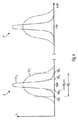
検査対象に含まれる材料、OCT測定光のスペクトルの平均波長、および他の物理的特性に依存して、対象を貫通するOCT測定光の輝度は、特定の貫通深さによって特徴付けられるように指数関数的に減少する。貫通深さは、OCT測定光の平均波長がたとえば800nmから1300nmの範囲内にある場合、組織などの生物学的対象についてはたとえば1mmから3mmになり得る。対象の内部に特定の深さに至るまで貫通したOCT測定光は、散乱および反射プロセスを含む当該深さにおける容量内に存在する材料と相互に作用する。特に、ある容量の対象の内部の反射率は、対象内部の特定の容量の内部の屈折率および/または材料の屈折率の勾配に依存する。特に、対象内部の特定の深さで発出するOCT測定光の輝度は、対象内部のこの深さにおける反射率に依存する。
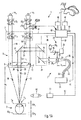
図1Aは、第1の動作モードにおける本発明の例示的な実施例に係る画像化システム1を概略的に示す。画像化システム1は、顕微鏡部3として具体化される顕微鏡特徴、およびOCT部5として具体化されるOCT特徴を提供する。示される例では、画像化システム1を用いて人間の目7を検査する。顕微鏡部3によって目7の目視検査、特に目7の目視双眼検査が可能である。特に、目7の顕微鏡画像を取得することができる。このため、目7は、可視波長範囲内の波長を有する顕微鏡法照明光を生成する顕微鏡法照明光源(図1Aには図示せず)によって照らされる。用途に依存して、顕微鏡法照明光の入射光角、および顕微鏡法照明光の色温度が変化し得る。顕微鏡法照明光に関するこれらの適応があり得るにもかかわらず、目7を目視検査のみで検査することは容易ではない。なぜなら、目は、分解または認識が困難であり得る多数の透明な解剖学的構造を含むからである。したがって、目7を完全に検査するために、画像化システム1はOCT部5によるOCT特徴を提供する。OCT特徴によって、目7の構造容量データを取得することができる。したがって、OCT機構によって、目7の横延長部および深さ(または軸方向)延長部を横断してデータを取得することができる。
Claims (19)
- OCTシステムであって、
測定対象に向かって延在する測定アームおよび参照アームを提供する干渉計を備え、前記測定アームは、OCT測定光のビームを発するためのOCT測定ビームエミッタと、前記測定アームの前記ビーム経路内の前記OCT測定ビームエミッタの下流に配置されたOCTビーム成形光学部品とを含み、前記OCTビーム成形光学部品は、OCT測定光の前記ビームの焦点をビーム焦点に合わすように構成され、前記OCTビーム成形光学部品は、前記ビーム焦点におけるOCT測定光の前記ビームのビームウエストの横幅を変更するように構成される、OCTシステム。 - 前記OCTビーム成形光学部品は、ビームウエストの前記横幅を最大幅から最小幅に変更するように構成され、前記最大幅は前記最小幅よりも少なくとも2倍大きい、請求項1に記載のOCTシステム。
- 前記OCTビーム成形光学部品は可変焦点距離を有する、請求項1または2に記載の画像化システム。
- 前記OCT測定ビームエミッタは、前記OCTビーム成形光学部品の焦点に配置される、請求項1から3のいずれかに記載の画像化システム。
- 前記OCTビーム成形光学部品は、少なくとも2つのレンズと、前記少なくとも2つのレンズが装着されるキャリアと、少なくとも2つの焦点レンズの一方が前記測定アームの前記ビーム経路に配置されるように前記キャリアを動かすアクチュエータとを含む、請求項1から4のいずれかに記載の画像化システム。
- 特に請求項1から5のいずれかと組合されたOCTシステムであって、
測定対象に向かって延在する測定アームおよび参照アームを提供する干渉計であって、OCT測定光源と、少なくとも1つのビームスプリッタと、前記測定アームから戻って、前記参照アームから戻ったOCT測定光に重畳されたOCT測定光の複数のスペクトル部分を検出するように構成された検出系とを含む干渉計を備え、
以下の少なくとも一方が当てはまる:
前記OCT測定光源から発せられる前記OCT測定光のスペクトル幅を、1.5倍よりも大きく変更可能である;および、
検出器によって検出されるOCT測定光の前記複数のスペクトル部分のスペクトル幅を、1.5倍よりも大きく変更可能である、OCTシステム。 - 検出系は、分散を提供する分光計を含み、前記分光計は、少なくとも最大値および最小値を提供するように構成され、前記最大値と前記最小値の比率は1.5よりも大きい、請求項6に記載のOCTシステム。
- 前記検出系は空間分解能検出器を含む、請求項7に記載のOCTシステム。
- 前記分光計は、重畳光を前記検出器上に導く可変光パワーを有するレンズを含む、請求項7に記載のOCTシステム。
- 前記OCT測定光源は、前記OCT測定光源のビーム経路に交互に配置可能な少なくとも2つのスペクトルフィルタを含む、請求項1から9のいずれかに記載のOCTシステム。
- 顕微鏡システムおよびOCTシステムを備える画像化システムであって、
前記顕微鏡システムは、対物レンズを横切る画像化ビーム経路を介して、対象平面を画像平面上に画像化するように構成され、
前記OCTシステムは、OCT測定光を、OCT測定ビームエミッタから延在してOCTビーム成形光学部品および前記対物レンズを横切るOCTビーム経路を介して前記対象平面に向けて導くように構成され、前記OCTビーム成形光学部品は、前記画像化ビーム経路の外部に配置され、前記対象平面におけるOCT測定光の前記ビームのビームウエストの横幅を変更するように構成される、画像化システム。 - 前記画像化ビーム経路に配置された少なくとも1つの接眼レンズをさらに備える、請求項11に記載の画像化システム。
- 前記画像化ビーム経路に配置される少なくとも1つの画像センサをさらに備える、請求項11または12に記載の画像化システム。
- 少なくとも1つの検出器によって検出される光輝度を表示するように構成され、前記対象平面に配置された対象を表わす画像ディスプレイをさらに備える、請求項13に記載の画像化システム。
- 前記ディスプレイは頭部装着型ディスプレイを含む、請求項14に記載の画像化システム。
- 前記対物レンズは可変焦点距離を有する、請求項11から15のいずれかに記載の画像化システム。
- 請求項1から10のいずれかに記載のOCTシステムを備える、請求項11から15のいずれかに記載の画像化システム。
- 目を検査する方法であって、
第1の動作モードおよび第2の動作モードのいずれかにおいて、請求項11から17のいずれかに記載の画像化システムを動作させるステップを備え、
前記第1の動作モードでは、前記対物レンズの対象平面は目の前部の領域に配置され、前記第2の動作モードでは、前記対象平面または前記対象平面の画像は目の網膜の領域に配置され、
前記第1の動作モードにおける前記画像平面のOCT測定ビームの横幅は、前記第2の動作モードにおける前記対象平面または前記対象平面の前記画像のOCT測定ビームの横幅よりも大きい、方法。 - 特に請求項18に記載の目を検査する方法であって、
第1の動作モードおよび第2の動作モードのいずれかにおいて、請求項11から17のいずれかに記載の画像化システムを動作させるステップを備え、
前記第1の動作モードでは、前記対物レンズの対象平面は目の前部の領域に配置され、前記第2の動作モードでは、前記対象平面または前記対象平面の画像は目の網膜の領域に配置され、
前記第1の動作モードにおけるOCTシステムの軸方向視野は、前記第2の動作モードにおける前記OCTシステムの軸方向視野よりも大きい、方法。
Applications Claiming Priority (3)
| Application Number | Priority Date | Filing Date | Title |
|---|---|---|---|
| US11821308P | 2008-11-26 | 2008-11-26 | |
| US61/118,213 | 2008-11-26 | ||
| PCT/EP2009/008433 WO2010060622A2 (en) | 2008-11-26 | 2009-11-26 | Imaging system |
Related Child Applications (1)
| Application Number | Title | Priority Date | Filing Date |
|---|---|---|---|
| JP2014059716A Division JP5941085B2 (ja) | 2008-11-26 | 2014-03-24 | 画像化システム |
Publications (3)
| Publication Number | Publication Date |
|---|---|
| JP2012509729A true JP2012509729A (ja) | 2012-04-26 |
| JP2012509729A5 JP2012509729A5 (ja) | 2012-12-13 |
| JP5632386B2 JP5632386B2 (ja) | 2014-11-26 |
Family
ID=41650451
Family Applications (3)
| Application Number | Title | Priority Date | Filing Date |
|---|---|---|---|
| JP2011537886A Active JP5632386B2 (ja) | 2008-11-26 | 2009-11-26 | 画像化システム |
| JP2014059716A Active JP5941085B2 (ja) | 2008-11-26 | 2014-03-24 | 画像化システム |
| JP2016100036A Pending JP2016165534A (ja) | 2008-11-26 | 2016-05-19 | 画像化システム |
Family Applications After (2)
| Application Number | Title | Priority Date | Filing Date |
|---|---|---|---|
| JP2014059716A Active JP5941085B2 (ja) | 2008-11-26 | 2014-03-24 | 画像化システム |
| JP2016100036A Pending JP2016165534A (ja) | 2008-11-26 | 2016-05-19 | 画像化システム |
Country Status (3)
| Country | Link |
|---|---|
| US (5) | US8922882B2 (ja) |
| JP (3) | JP5632386B2 (ja) |
| WO (1) | WO2010060622A2 (ja) |
Cited By (17)
| Publication number | Priority date | Publication date | Assignee | Title |
|---|---|---|---|---|
| JP2013230234A (ja) * | 2012-04-27 | 2013-11-14 | Nidek Co Ltd | 眼底撮影装置、及び眼底撮影装置用アタッチメント |
| JP2014138904A (ja) * | 2014-05-02 | 2014-07-31 | Nidek Co Ltd | 眼科撮影装置 |
| JP2016533796A (ja) * | 2013-08-28 | 2016-11-04 | バイオプティジェン, インコーポレイテッドBioptigen, Inc. | 光干渉断層撮影機能が組み込まれた手術用顕微鏡用のヘッドアップディスプレイ |
| JP2016209577A (ja) * | 2015-05-01 | 2016-12-15 | 株式会社ニデック | 眼科撮像装置 |
| WO2017002383A1 (ja) * | 2015-06-30 | 2017-01-05 | 株式会社トプコン | 眼科用顕微鏡システム |
| WO2017002378A1 (ja) * | 2015-06-30 | 2017-01-05 | 株式会社トプコン | 眼科用顕微鏡システム |
| WO2017002379A1 (ja) * | 2015-06-30 | 2017-01-05 | 株式会社トプコン | 眼科用顕微鏡システム |
| JP2017012535A (ja) * | 2015-07-02 | 2017-01-19 | 株式会社トプコン | 眼科用顕微鏡 |
| JP2017012431A (ja) * | 2015-06-30 | 2017-01-19 | 株式会社トプコン | 眼科用顕微鏡 |
| JP2017012536A (ja) * | 2015-07-02 | 2017-01-19 | 株式会社トプコン | 眼科用顕微鏡 |
| JP2017012433A (ja) * | 2015-06-30 | 2017-01-19 | 株式会社トプコン | 眼科用顕微鏡システム |
| JP2017514659A (ja) * | 2014-05-08 | 2017-06-08 | ミーモ アクチエンゲゼルシャフトMimo AG | 被験者の網膜を撮像する光干渉断層法撮像装置 |
| JP2017533004A (ja) * | 2014-09-19 | 2017-11-09 | カール・ツアイス・メディテック・アーゲー | ズーム可能なケプラーシステムを備える光学コヒーレンストモグラフィのシステム |
| JP2018114230A (ja) * | 2017-01-20 | 2018-07-26 | 株式会社トプコン | 眼科装置 |
| JP2019084411A (ja) * | 2019-03-14 | 2019-06-06 | 株式会社トプコン | 眼科用顕微鏡システム |
| JP2020515368A (ja) * | 2017-04-03 | 2020-05-28 | ホーガン,ジョシュア,ノエル | ホームモニタリング光コヒーレンストモグラフィシステム |
| JP2023073345A (ja) * | 2017-05-02 | 2023-05-25 | アルコン インコーポレイティド | 再構成可能な光干渉断層撮影(oct)システム |
Families Citing this family (40)
| Publication number | Priority date | Publication date | Assignee | Title |
|---|---|---|---|---|
| WO2010060622A2 (en) | 2008-11-26 | 2010-06-03 | Carl Zeiss Surgical Gmbh | Imaging system |
| JP5054072B2 (ja) * | 2009-07-28 | 2012-10-24 | キヤノン株式会社 | 光断層画像撮像装置 |
| JP5426339B2 (ja) * | 2009-12-02 | 2014-02-26 | 株式会社ニデック | 眼寸法測定装置 |
| DE102011114523A1 (de) * | 2011-09-29 | 2013-04-04 | Carl Zeiss Meditec Ag | Modul zur Weiterleitung eines Lichtstrahls |
| DE102011119899A1 (de) * | 2011-11-30 | 2013-06-06 | Carl Zeiss Meditec Ag | Mikroskopiesystem zur augenuntersuchung und oct-system |
| JP6140932B2 (ja) * | 2012-03-30 | 2017-06-07 | キヤノン株式会社 | 光干渉断層撮影装置及び制御方法 |
| CN203643682U (zh) * | 2012-04-24 | 2014-06-11 | 深圳市斯尔顿科技有限公司 | 一种眼科光学相干断层成像系统 |
| DE102013010844A1 (de) * | 2013-06-28 | 2014-12-31 | Carl Zeiss Meditec Ag | Mikroskopiesystem zur Abbildung von Phasenobjekten, Verfahren zum Betreiben des Mikroskopiesystems und Computerprogrammprodukt |
| JP6080128B2 (ja) | 2014-02-28 | 2017-02-15 | 株式会社トプコン | 眼科撮影装置およびこれに装着可能な光学ユニット |
| WO2015183994A1 (en) | 2014-05-28 | 2015-12-03 | Santec Corporation | Non-invasive optical measurement of blood analyte |
| JP6808618B2 (ja) * | 2014-10-17 | 2021-01-06 | オプティメディカ・コーポレイションOptimedica Corporation | レーザ眼手術システムの自動患者位置決め |
| JP6490469B2 (ja) | 2015-03-27 | 2019-03-27 | 株式会社トプコン | 眼科用顕微鏡システム |
| US10548520B2 (en) | 2015-04-01 | 2020-02-04 | Santec Corporation | Non-invasive optical measurement of blood analyte |
| JP2016202453A (ja) * | 2015-04-20 | 2016-12-08 | 株式会社トプコン | 眼科手術用顕微鏡 |
| JP6713149B2 (ja) | 2015-06-01 | 2020-06-24 | サンテック株式会社 | 2つの波長を合成する光コヒーレンストモグラフィーシステム |
| JP6505539B2 (ja) * | 2015-07-27 | 2019-04-24 | 株式会社トプコン | 眼科用顕微鏡 |
| US11033186B2 (en) * | 2016-02-26 | 2021-06-15 | Alcon Inc. | Methods and system for imaging an inner limiting membrane using a stain |
| DE102016205370B4 (de) * | 2016-03-31 | 2022-08-18 | Optomedical Technologies Gmbh | OCT-System |
| US10677580B2 (en) | 2016-04-27 | 2020-06-09 | Santec Corporation | Optical coherence tomography system using polarization switching |
| KR101855816B1 (ko) | 2016-05-13 | 2018-05-10 | 주식회사 고영테크놀러지 | 생체 조직 검사 장치 및 그 방법 |
| JP6776076B2 (ja) * | 2016-09-23 | 2020-10-28 | 株式会社トプコン | Oct装置 |
| DE102017105580B4 (de) * | 2016-11-04 | 2025-10-02 | Carl Zeiss Meditec Ag | Operationsmikroskop |
| CA3048197A1 (en) | 2016-12-21 | 2018-06-28 | Acucela Inc. | Miniaturized mobile, low cost optical coherence tomography system for home based ophthalmic applications |
| US10408600B2 (en) | 2017-06-22 | 2019-09-10 | Santec Corporation | Optical coherence tomography with a fizeau-type interferometer |
| US10416429B2 (en) | 2017-09-15 | 2019-09-17 | Agile Focus Designs, LLC | Dynamic focus and zoom system for use with wide-field, confocal and multiphoton microscopes |
| US10466461B2 (en) | 2017-09-15 | 2019-11-05 | Agile Focus Designs, LLC | Dynamic focus and zoom system for use with wide-field, confocal and multiphoton microscopes |
| US10502546B2 (en) * | 2017-11-07 | 2019-12-10 | Santec Corporation | Systems and methods for variable-range fourier domain imaging |
| US11213200B2 (en) | 2018-03-22 | 2022-01-04 | Santec Corporation | Topographical imaging using combined sensing inputs |
| US10838047B2 (en) | 2018-04-17 | 2020-11-17 | Santec Corporation | Systems and methods for LIDAR scanning of an environment over a sweep of wavelengths |
| US11067671B2 (en) | 2018-04-17 | 2021-07-20 | Santec Corporation | LIDAR sensing arrangements |
| CN118576146A (zh) | 2018-06-20 | 2024-09-03 | 奥克塞拉有限公司 | 基于家庭的眼科应用的微型移动低成本光学相干断层扫描系统 |
| US11730363B2 (en) | 2019-12-26 | 2023-08-22 | Acucela Inc. | Optical coherence tomography patient alignment system for home based ophthalmic applications |
| US10959613B1 (en) | 2020-08-04 | 2021-03-30 | Acucela Inc. | Scan pattern and signal processing for optical coherence tomography |
| WO2022035809A1 (en) | 2020-08-14 | 2022-02-17 | Acucela Inc. | System and method for optical coherence tomography a-scan decurving |
| US11393094B2 (en) | 2020-09-11 | 2022-07-19 | Acucela Inc. | Artificial intelligence for evaluation of optical coherence tomography images |
| JP2023544704A (ja) | 2020-09-30 | 2023-10-25 | アキュセラ インコーポレイテッド | 近視の予測、診断、計画、および監視デバイス |
| WO2022204622A1 (en) | 2021-03-24 | 2022-09-29 | Acucela Inc. | Axial length measurement monitor |
| US12135373B2 (en) | 2021-04-16 | 2024-11-05 | santec Holdings Corporation | Systems and methods for LIDAR sensing |
| CN115399728B (zh) * | 2022-08-01 | 2025-03-18 | 浙江大学嘉兴研究院 | 一种基于可变焦oct的眼轴测量方法及装置 |
| US12313883B2 (en) | 2023-03-03 | 2025-05-27 | santec Holdings Corporation | Photonic beam steering device with wavelength sweep |
Citations (8)
| Publication number | Priority date | Publication date | Assignee | Title |
|---|---|---|---|---|
| JP2006095318A (ja) * | 1994-08-18 | 2006-04-13 | Carl Zeiss:Fa | 眼科手術装置 |
| JP2007508558A (ja) * | 2003-10-14 | 2007-04-05 | ユニヴァーシティ・オブ・ケント | スペクトル干渉法及びスペクトル干渉装置 |
| JP2007121262A (ja) * | 2005-09-30 | 2007-05-17 | Fujifilm Corp | 光断層画像化装置 |
| JP2007212376A (ja) * | 2006-02-13 | 2007-08-23 | Fujifilm Corp | 光断層画像化装置 |
| US20070263226A1 (en) * | 2006-05-15 | 2007-11-15 | Eastman Kodak Company | Tissue imaging system |
| US20080117504A1 (en) * | 2006-11-06 | 2008-05-22 | Peter Reimer | Surgical microscope having an OCT-system |
| JP2008520992A (ja) * | 2004-11-18 | 2008-06-19 | ミケルソン ダイアグノスティックス リミテッド | 干渉装置及び方法並びにプローブ |
| JP2008529669A (ja) * | 2005-02-10 | 2008-08-07 | ライトラボ・イメージング・インコーポレーテッド | 光学コヒーレンス断層撮影法の装置及び方法 |
Family Cites Families (23)
| Publication number | Priority date | Publication date | Assignee | Title |
|---|---|---|---|---|
| US5493109A (en) | 1994-08-18 | 1996-02-20 | Carl Zeiss, Inc. | Optical coherence tomography assisted ophthalmologic surgical microscope |
| WO1998038907A1 (en) * | 1997-03-06 | 1998-09-11 | Massachusetts Institute Of Technology | Instrument for optically scanning of living tissue |
| US5975697A (en) * | 1998-11-25 | 1999-11-02 | Oti Ophthalmic Technologies, Inc. | Optical mapping apparatus with adjustable depth resolution |
| DE29905969U1 (de) | 1999-04-08 | 1999-07-08 | Oculus Optikgeräte GmbH, 35582 Wetzlar | Stereoskopisches Mikroskop |
| US6741359B2 (en) * | 2002-05-22 | 2004-05-25 | Carl Zeiss Meditec, Inc. | Optical coherence tomography optical scanner |
| US7580185B2 (en) * | 2002-08-28 | 2009-08-25 | Carl Zeiss Surgical Gmbh | Microscopy system, microscopy method and a method of treating an aneurysm |
| EP1447698B1 (en) * | 2003-02-17 | 2007-04-11 | Kabushiki Kaisha TOPCON | Ophtalmic operation microscope |
| US7876974B2 (en) * | 2003-08-29 | 2011-01-25 | Vladimir Brajovic | Method for improving digital images and an image sensor for sensing the same |
| WO2006019181A1 (ja) * | 2004-08-18 | 2006-02-23 | National University Corporation Tokyo University Of Agriculture And Technology | 形状測定方法、形状測定装置および周波数コム光発生装置 |
| WO2006022045A1 (ja) * | 2004-08-26 | 2006-03-02 | National University Corporation Nagoya University | 光干渉断層装置 |
| JP4546209B2 (ja) | 2004-09-30 | 2010-09-15 | 株式会社ニデック | 眼科装置 |
| ES2669059T3 (es) | 2005-07-28 | 2018-05-23 | Bioptigen, Inc. | Sistema de formación de imagen de tomografía de coherencia óptica en el dominio de la frecuencia |
| US20070291277A1 (en) * | 2006-06-20 | 2007-12-20 | Everett Matthew J | Spectral domain optical coherence tomography system |
| DE102007019680A1 (de) * | 2006-11-06 | 2008-05-08 | Carl Zeiss Surgical Gmbh | Ophthalmo-Operationsmikroskop mit OCT-System |
| DE102007019677A1 (de) | 2006-11-06 | 2008-05-08 | Carl Zeiss Surgical Gmbh | Operationsmikroskop mit OCT-System und Operationsmikroskop-Beleuchtungsmodul mit OCT-System |
| DE102007019679B4 (de) | 2006-11-06 | 2025-04-30 | Carl Zeiss Meditec Ag | Operationsmikroskop mit OCT-System |
| JP5213417B2 (ja) * | 2007-04-24 | 2013-06-19 | カール・ツアイス・メディテック・アーゲー | Octシステムを備える手術用顕微鏡 |
| JP5188146B2 (ja) * | 2007-04-24 | 2013-04-24 | カール・ツアイス・メディテック・アーゲー | Octシステムを備える手術用顕微鏡 |
| DE502008001698D1 (de) * | 2007-10-18 | 2010-12-16 | Nectar Imaging S R L | Vorrichtung zur tomografischen Erfassung von Objekten |
| PL384779A1 (pl) * | 2008-03-25 | 2009-09-28 | Optopol Technology Spółka Akcyjna | Urządzenie do badania obiektów za pomocą tomografii optycznej z przystawką, przystawka do urządzenia do badania obiektów za pomocą tomografii optycznej i sposób badania obiektów za pomocą tomografii optycznej |
| US8348429B2 (en) * | 2008-03-27 | 2013-01-08 | Doheny Eye Institute | Optical coherence tomography device, method, and system |
| WO2010060622A2 (en) * | 2008-11-26 | 2010-06-03 | Carl Zeiss Surgical Gmbh | Imaging system |
| US8425037B2 (en) * | 2010-07-30 | 2013-04-23 | Adventus Technologies, Inc. | Intraoperative imaging system and apparatus |
-
2009
- 2009-11-26 WO PCT/EP2009/008433 patent/WO2010060622A2/en not_active Ceased
- 2009-11-26 JP JP2011537886A patent/JP5632386B2/ja active Active
-
2011
- 2011-05-26 US US13/116,358 patent/US8922882B2/en active Active
-
2014
- 2014-03-24 JP JP2014059716A patent/JP5941085B2/ja active Active
- 2014-11-20 US US14/549,040 patent/US9377293B2/en active Active
- 2014-11-20 US US14/549,058 patent/US9618326B2/en active Active
-
2016
- 2016-05-19 JP JP2016100036A patent/JP2016165534A/ja active Pending
-
2017
- 2017-03-23 US US15/467,308 patent/US9962076B2/en active Active
- 2017-03-23 US US15/467,325 patent/US10098538B2/en active Active
Patent Citations (8)
| Publication number | Priority date | Publication date | Assignee | Title |
|---|---|---|---|---|
| JP2006095318A (ja) * | 1994-08-18 | 2006-04-13 | Carl Zeiss:Fa | 眼科手術装置 |
| JP2007508558A (ja) * | 2003-10-14 | 2007-04-05 | ユニヴァーシティ・オブ・ケント | スペクトル干渉法及びスペクトル干渉装置 |
| JP2008520992A (ja) * | 2004-11-18 | 2008-06-19 | ミケルソン ダイアグノスティックス リミテッド | 干渉装置及び方法並びにプローブ |
| JP2008529669A (ja) * | 2005-02-10 | 2008-08-07 | ライトラボ・イメージング・インコーポレーテッド | 光学コヒーレンス断層撮影法の装置及び方法 |
| JP2007121262A (ja) * | 2005-09-30 | 2007-05-17 | Fujifilm Corp | 光断層画像化装置 |
| JP2007212376A (ja) * | 2006-02-13 | 2007-08-23 | Fujifilm Corp | 光断層画像化装置 |
| US20070263226A1 (en) * | 2006-05-15 | 2007-11-15 | Eastman Kodak Company | Tissue imaging system |
| US20080117504A1 (en) * | 2006-11-06 | 2008-05-22 | Peter Reimer | Surgical microscope having an OCT-system |
Cited By (22)
| Publication number | Priority date | Publication date | Assignee | Title |
|---|---|---|---|---|
| JP2013230234A (ja) * | 2012-04-27 | 2013-11-14 | Nidek Co Ltd | 眼底撮影装置、及び眼底撮影装置用アタッチメント |
| JP2016533796A (ja) * | 2013-08-28 | 2016-11-04 | バイオプティジェン, インコーポレイテッドBioptigen, Inc. | 光干渉断層撮影機能が組み込まれた手術用顕微鏡用のヘッドアップディスプレイ |
| JP2014138904A (ja) * | 2014-05-02 | 2014-07-31 | Nidek Co Ltd | 眼科撮影装置 |
| JP2017514659A (ja) * | 2014-05-08 | 2017-06-08 | ミーモ アクチエンゲゼルシャフトMimo AG | 被験者の網膜を撮像する光干渉断層法撮像装置 |
| JP2017533004A (ja) * | 2014-09-19 | 2017-11-09 | カール・ツアイス・メディテック・アーゲー | ズーム可能なケプラーシステムを備える光学コヒーレンストモグラフィのシステム |
| JP2016209577A (ja) * | 2015-05-01 | 2016-12-15 | 株式会社ニデック | 眼科撮像装置 |
| JP2017012429A (ja) * | 2015-06-30 | 2017-01-19 | 株式会社トプコン | 眼科用顕微鏡システム |
| WO2017002378A1 (ja) * | 2015-06-30 | 2017-01-05 | 株式会社トプコン | 眼科用顕微鏡システム |
| JP2017012431A (ja) * | 2015-06-30 | 2017-01-19 | 株式会社トプコン | 眼科用顕微鏡 |
| US10932665B2 (en) | 2015-06-30 | 2021-03-02 | Kabushiki Kaisha Topcon | Ophthalmologic microscope system |
| WO2017002379A1 (ja) * | 2015-06-30 | 2017-01-05 | 株式会社トプコン | 眼科用顕微鏡システム |
| JP2017012430A (ja) * | 2015-06-30 | 2017-01-19 | 株式会社トプコン | 眼科用顕微鏡システム |
| JP2017012433A (ja) * | 2015-06-30 | 2017-01-19 | 株式会社トプコン | 眼科用顕微鏡システム |
| US10531984B2 (en) | 2015-06-30 | 2020-01-14 | Kabushiki Kaisha Topcon | Ophthalmologic microscope system |
| WO2017002383A1 (ja) * | 2015-06-30 | 2017-01-05 | 株式会社トプコン | 眼科用顕微鏡システム |
| JP2017012535A (ja) * | 2015-07-02 | 2017-01-19 | 株式会社トプコン | 眼科用顕微鏡 |
| JP2017012536A (ja) * | 2015-07-02 | 2017-01-19 | 株式会社トプコン | 眼科用顕微鏡 |
| JP2018114230A (ja) * | 2017-01-20 | 2018-07-26 | 株式会社トプコン | 眼科装置 |
| JP2020515368A (ja) * | 2017-04-03 | 2020-05-28 | ホーガン,ジョシュア,ノエル | ホームモニタリング光コヒーレンストモグラフィシステム |
| JP2023073345A (ja) * | 2017-05-02 | 2023-05-25 | アルコン インコーポレイティド | 再構成可能な光干渉断層撮影(oct)システム |
| JP7505074B2 (ja) | 2017-05-02 | 2024-06-24 | アルコン インコーポレイティド | 再構成可能な光干渉断層撮影(oct)システム |
| JP2019084411A (ja) * | 2019-03-14 | 2019-06-06 | 株式会社トプコン | 眼科用顕微鏡システム |
Also Published As
| Publication number | Publication date |
|---|---|
| US10098538B2 (en) | 2018-10-16 |
| JP2014121647A (ja) | 2014-07-03 |
| US20110228218A1 (en) | 2011-09-22 |
| US20150077708A1 (en) | 2015-03-19 |
| US20150077707A1 (en) | 2015-03-19 |
| US9618326B2 (en) | 2017-04-11 |
| WO2010060622A3 (en) | 2010-07-29 |
| US20170188819A1 (en) | 2017-07-06 |
| JP5941085B2 (ja) | 2016-06-29 |
| US8922882B2 (en) | 2014-12-30 |
| WO2010060622A2 (en) | 2010-06-03 |
| US20170188820A1 (en) | 2017-07-06 |
| US9377293B2 (en) | 2016-06-28 |
| US9962076B2 (en) | 2018-05-08 |
| JP2016165534A (ja) | 2016-09-15 |
| JP5632386B2 (ja) | 2014-11-26 |
Similar Documents
| Publication | Publication Date | Title |
|---|---|---|
| JP5941085B2 (ja) | 画像化システム | |
| US8427653B2 (en) | Optical coherence tomography methods and systems | |
| US10337995B2 (en) | Systems and methods for oblique laser scanning | |
| US10028656B2 (en) | Optical coherence tomographic apparatus | |
| CN105980810B (zh) | 光学断层摄影装置和方法 | |
| JP6805539B2 (ja) | 眼科撮像装置 | |
| JP7370493B2 (ja) | 試料の顕微鏡分析を行うためのシステムおよび方法 | |
| JP6207221B2 (ja) | 光断層撮像装置 | |
| JP6207220B2 (ja) | 光断層撮像装置 | |
| KR20150043115A (ko) | Oct 장치 | |
| JP5213417B2 (ja) | Octシステムを備える手術用顕微鏡 | |
| JP6898716B2 (ja) | 光断層撮像装置 | |
| JP2010035949A (ja) | 眼科撮影装置 | |
| JP5214216B2 (ja) | Octシステムを備える手術用顕微鏡、およびoctシステムを備える手術用顕微鏡照明モジュール | |
| JP2007212376A (ja) | 光断層画像化装置 | |
| JP6456444B2 (ja) | 眼底撮影装置 | |
| JP2021524330A (ja) | Octシステム及びoct方法 | |
| Langevin et al. | New setups for optical coherence tomography | |
| JP2016013188A (ja) | 断層画像撮像装置及びその制御方法 |
Legal Events
| Date | Code | Title | Description |
|---|---|---|---|
| A521 | Request for written amendment filed |
Free format text: JAPANESE INTERMEDIATE CODE: A523 Effective date: 20121025 |
|
| A621 | Written request for application examination |
Free format text: JAPANESE INTERMEDIATE CODE: A621 Effective date: 20121025 |
|
| A711 | Notification of change in applicant |
Free format text: JAPANESE INTERMEDIATE CODE: A712 Effective date: 20130214 |
|
| A977 | Report on retrieval |
Free format text: JAPANESE INTERMEDIATE CODE: A971007 Effective date: 20130919 |
|
| A131 | Notification of reasons for refusal |
Free format text: JAPANESE INTERMEDIATE CODE: A131 Effective date: 20130924 |
|
| A601 | Written request for extension of time |
Free format text: JAPANESE INTERMEDIATE CODE: A601 Effective date: 20131220 |
|
| A602 | Written permission of extension of time |
Free format text: JAPANESE INTERMEDIATE CODE: A602 Effective date: 20140106 |
|
| A601 | Written request for extension of time |
Free format text: JAPANESE INTERMEDIATE CODE: A601 Effective date: 20140123 |
|
| A602 | Written permission of extension of time |
Free format text: JAPANESE INTERMEDIATE CODE: A602 Effective date: 20140130 |
|
| A601 | Written request for extension of time |
Free format text: JAPANESE INTERMEDIATE CODE: A601 Effective date: 20140221 |
|
| A602 | Written permission of extension of time |
Free format text: JAPANESE INTERMEDIATE CODE: A602 Effective date: 20140228 |
|
| A521 | Request for written amendment filed |
Free format text: JAPANESE INTERMEDIATE CODE: A523 Effective date: 20140324 |
|
| TRDD | Decision of grant or rejection written | ||
| A01 | Written decision to grant a patent or to grant a registration (utility model) |
Free format text: JAPANESE INTERMEDIATE CODE: A01 Effective date: 20140916 |
|
| A61 | First payment of annual fees (during grant procedure) |
Free format text: JAPANESE INTERMEDIATE CODE: A61 Effective date: 20141009 |
|
| R150 | Certificate of patent or registration of utility model |
Ref document number: 5632386 Country of ref document: JP Free format text: JAPANESE INTERMEDIATE CODE: R150 |
|
| R250 | Receipt of annual fees |
Free format text: JAPANESE INTERMEDIATE CODE: R250 |
|
| R250 | Receipt of annual fees |
Free format text: JAPANESE INTERMEDIATE CODE: R250 |
|
| R250 | Receipt of annual fees |
Free format text: JAPANESE INTERMEDIATE CODE: R250 |
|
| R250 | Receipt of annual fees |
Free format text: JAPANESE INTERMEDIATE CODE: R250 |
|
| R250 | Receipt of annual fees |
Free format text: JAPANESE INTERMEDIATE CODE: R250 |
|
| R250 | Receipt of annual fees |
Free format text: JAPANESE INTERMEDIATE CODE: R250 |
|
| R250 | Receipt of annual fees |
Free format text: JAPANESE INTERMEDIATE CODE: R250 |
|
| R250 | Receipt of annual fees |
Free format text: JAPANESE INTERMEDIATE CODE: R250 |
|
| R250 | Receipt of annual fees |
Free format text: JAPANESE INTERMEDIATE CODE: R250 |