JP2014155694A - 眼科装置及び眼科方法 - Google Patents
眼科装置及び眼科方法 Download PDFInfo
- Publication number
- JP2014155694A JP2014155694A JP2014001524A JP2014001524A JP2014155694A JP 2014155694 A JP2014155694 A JP 2014155694A JP 2014001524 A JP2014001524 A JP 2014001524A JP 2014001524 A JP2014001524 A JP 2014001524A JP 2014155694 A JP2014155694 A JP 2014155694A
- Authority
- JP
- Japan
- Prior art keywords
- fundus
- image
- images
- polarization
- tomographic
- Prior art date
- Legal status (The legal status is an assumption and is not a legal conclusion. Google has not performed a legal analysis and makes no representation as to the accuracy of the status listed.)
- Pending
Links
Images


Classifications
-
- A—HUMAN NECESSITIES
- A61—MEDICAL OR VETERINARY SCIENCE; HYGIENE
- A61B—DIAGNOSIS; SURGERY; IDENTIFICATION
- A61B3/00—Apparatus for testing the eyes; Instruments for examining the eyes
- A61B3/0016—Operational features thereof
- A61B3/0025—Operational features thereof characterised by electronic signal processing, e.g. eye models
-
- A—HUMAN NECESSITIES
- A61—MEDICAL OR VETERINARY SCIENCE; HYGIENE
- A61B—DIAGNOSIS; SURGERY; IDENTIFICATION
- A61B3/00—Apparatus for testing the eyes; Instruments for examining the eyes
- A61B3/0016—Operational features thereof
- A61B3/0041—Operational features thereof characterised by display arrangements
- A61B3/0058—Operational features thereof characterised by display arrangements for multiple images
-
- A—HUMAN NECESSITIES
- A61—MEDICAL OR VETERINARY SCIENCE; HYGIENE
- A61B—DIAGNOSIS; SURGERY; IDENTIFICATION
- A61B3/00—Apparatus for testing the eyes; Instruments for examining the eyes
- A61B3/10—Objective types, i.e. instruments for examining the eyes independent of the patients' perceptions or reactions
- A61B3/102—Objective types, i.e. instruments for examining the eyes independent of the patients' perceptions or reactions for optical coherence tomography [OCT]
-
- A—HUMAN NECESSITIES
- A61—MEDICAL OR VETERINARY SCIENCE; HYGIENE
- A61B—DIAGNOSIS; SURGERY; IDENTIFICATION
- A61B3/00—Apparatus for testing the eyes; Instruments for examining the eyes
- A61B3/10—Objective types, i.e. instruments for examining the eyes independent of the patients' perceptions or reactions
- A61B3/12—Objective types, i.e. instruments for examining the eyes independent of the patients' perceptions or reactions for looking at the eye fundus, e.g. ophthalmoscopes
- A61B3/1225—Objective types, i.e. instruments for examining the eyes independent of the patients' perceptions or reactions for looking at the eye fundus, e.g. ophthalmoscopes using coherent radiation
-
- A—HUMAN NECESSITIES
- A61—MEDICAL OR VETERINARY SCIENCE; HYGIENE
- A61B—DIAGNOSIS; SURGERY; IDENTIFICATION
- A61B5/00—Measuring for diagnostic purposes; Identification of persons
- A61B5/0059—Measuring for diagnostic purposes; Identification of persons using light, e.g. diagnosis by transillumination, diascopy, fluorescence
- A61B5/0062—Arrangements for scanning
- A61B5/0066—Optical coherence imaging
-
- G—PHYSICS
- G01—MEASURING; TESTING
- G01B—MEASURING LENGTH, THICKNESS OR SIMILAR LINEAR DIMENSIONS; MEASURING ANGLES; MEASURING AREAS; MEASURING IRREGULARITIES OF SURFACES OR CONTOURS
- G01B9/00—Measuring instruments characterised by the use of optical techniques
- G01B9/02—Interferometers
- G01B9/02015—Interferometers characterised by the beam path configuration
- G01B9/02027—Two or more interferometric channels or interferometers
-
- G—PHYSICS
- G01—MEASURING; TESTING
- G01B—MEASURING LENGTH, THICKNESS OR SIMILAR LINEAR DIMENSIONS; MEASURING ANGLES; MEASURING AREAS; MEASURING IRREGULARITIES OF SURFACES OR CONTOURS
- G01B9/00—Measuring instruments characterised by the use of optical techniques
- G01B9/02—Interferometers
- G01B9/02015—Interferometers characterised by the beam path configuration
- G01B9/02029—Combination with non-interferometric systems, i.e. for measuring the object
- G01B9/0203—With imaging systems
-
- G—PHYSICS
- G01—MEASURING; TESTING
- G01B—MEASURING LENGTH, THICKNESS OR SIMILAR LINEAR DIMENSIONS; MEASURING ANGLES; MEASURING AREAS; MEASURING IRREGULARITIES OF SURFACES OR CONTOURS
- G01B9/00—Measuring instruments characterised by the use of optical techniques
- G01B9/02—Interferometers
- G01B9/02041—Interferometers characterised by particular imaging or detection techniques
- G01B9/02044—Imaging in the frequency domain, e.g. by using a spectrometer
-
- G—PHYSICS
- G01—MEASURING; TESTING
- G01B—MEASURING LENGTH, THICKNESS OR SIMILAR LINEAR DIMENSIONS; MEASURING ANGLES; MEASURING AREAS; MEASURING IRREGULARITIES OF SURFACES OR CONTOURS
- G01B9/00—Measuring instruments characterised by the use of optical techniques
- G01B9/02—Interferometers
- G01B9/02055—Reduction or prevention of errors; Testing; Calibration
- G01B9/02075—Reduction or prevention of errors; Testing; Calibration of particular errors
- G01B9/02076—Caused by motion
-
- G—PHYSICS
- G01—MEASURING; TESTING
- G01B—MEASURING LENGTH, THICKNESS OR SIMILAR LINEAR DIMENSIONS; MEASURING ANGLES; MEASURING AREAS; MEASURING IRREGULARITIES OF SURFACES OR CONTOURS
- G01B9/00—Measuring instruments characterised by the use of optical techniques
- G01B9/02—Interferometers
- G01B9/0209—Low-coherence interferometers
- G01B9/02091—Tomographic interferometers, e.g. based on optical coherence
-
- G—PHYSICS
- G01—MEASURING; TESTING
- G01B—MEASURING LENGTH, THICKNESS OR SIMILAR LINEAR DIMENSIONS; MEASURING ANGLES; MEASURING AREAS; MEASURING IRREGULARITIES OF SURFACES OR CONTOURS
- G01B2290/00—Aspects of interferometers not specifically covered by any group under G01B9/02
- G01B2290/45—Multiple detectors for detecting interferometer signals
-
- G—PHYSICS
- G01—MEASURING; TESTING
- G01B—MEASURING LENGTH, THICKNESS OR SIMILAR LINEAR DIMENSIONS; MEASURING ANGLES; MEASURING AREAS; MEASURING IRREGULARITIES OF SURFACES OR CONTOURS
- G01B2290/00—Aspects of interferometers not specifically covered by any group under G01B9/02
- G01B2290/70—Using polarization in the interferometer
Landscapes
- Health & Medical Sciences (AREA)
- Life Sciences & Earth Sciences (AREA)
- Physics & Mathematics (AREA)
- Engineering & Computer Science (AREA)
- General Health & Medical Sciences (AREA)
- General Physics & Mathematics (AREA)
- Biophysics (AREA)
- Public Health (AREA)
- Medical Informatics (AREA)
- Molecular Biology (AREA)
- Surgery (AREA)
- Animal Behavior & Ethology (AREA)
- Heart & Thoracic Surgery (AREA)
- Veterinary Medicine (AREA)
- Biomedical Technology (AREA)
- Ophthalmology & Optometry (AREA)
- Nuclear Medicine, Radiotherapy & Molecular Imaging (AREA)
- Radiology & Medical Imaging (AREA)
- Signal Processing (AREA)
- Pathology (AREA)
- Eye Examination Apparatus (AREA)
Abstract
【解決手段】 異なる時刻に被検眼の眼底を撮像して得た複数の眼底画像と、該複数の眼底画像よりも少ない数であり且つ該複数の眼底画像とは異なる時刻に被検眼の眼底を撮像して得た少なくとも1つの眼底画像とを取得し、該複数の眼底画像を平均化して新たな眼底画像を生成し、該生成された新たな眼底画像から特徴領域を抽出し、該抽出された特徴領域と該少なくとも1つの眼底画像とに基づいて、該新たな眼底画像に対応する眼底の第1の偏光断層画像と該少なくとも1つの眼底画像に対応する眼底の第2の偏光断層画像との位置が補正されるように、眼底を追尾する。
【選択図】 図10
Description
異なる時刻に被検眼の眼底を撮像して得た複数の眼底画像と、前記複数の眼底画像よりも少ない数であり且つ前記複数の眼底画像とは異なる時刻に前記被検眼の眼底を撮像して得た少なくとも1つの眼底画像とを取得する眼底画像取得手段と、
前記複数の眼底画像を平均化して新たな眼底画像を生成する手段と、
前記生成された新たな眼底画像から特徴領域を抽出する抽出手段と、
前記抽出された特徴領域と前記少なくとも1つの眼底画像とに基づいて、前記新たな眼底画像に対応する前記眼底の第1の偏光断層画像と前記少なくとも1つの眼底画像に対応する前記眼底の第2の偏光断層画像との位置が補正されるように、前記眼底を追尾する手段と、を有する。
また、本発明に係る眼科方法は、
異なる時刻に被検眼の眼底を撮像して得た複数の眼底画像と、前記複数の眼底画像よりも少ない数であり且つ前記複数の眼底画像とは異なる時刻に前記被検眼の眼底を撮像して得た少なくとも1つの眼底画像とを取得する工程と、
前記複数の眼底画像を平均化して新たな眼底画像を生成する工程と、
前記生成された新たな眼底画像から特徴領域を抽出する工程と、
前記抽出された特徴領域と前記少なくとも1つの眼底画像とに基づいて、前記新たな眼底画像に対応する前記眼底の第1の偏光断層画像と前記少なくとも1つの眼底画像に対応する前記眼底の第2の偏光断層画像との位置が補正されるように、前記眼底を追尾する工程と、を有する。
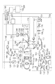
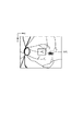
図1は、本実施形態における撮影装置の一例である「眼科装置」の全体構成の概略図である。なお、後述する信号処理部190の少なくとも一部を「画像処理装置」とみなすことができ、また、この場合、「眼科装置」全体を「眼科システム」、あるいは「撮影装置」全体を「撮影システム」とみなすこともできる。
PS−OCT100の構成について説明する。
PS−SLO140の構成について説明する。
前眼部撮像部160について説明する。
内部固視灯170について説明する。
本装置全体を制御するための制御部200について説明する。
次に、信号処理部190を構成する画像生成部193における画像生成について説明する。
画像生成部193は、前述した2つの断層信号から断層輝度画像を生成する。
断層輝度画像は、従来のOCTにおける断層画像と基本的に同じもので、その画素値rは各ラインセンサ129、133から得られた断層信号AHおよびAVから(式1)によって計算される。

画像生成部193は、互いに直行する偏光成分の断層画像からリターデーション画像を生成する。
画像生成部193は、複数のBスキャン像に対して得たリターデーション(Retardation)画像からリターデーションマップを生成する。
画像生成部193は、先に生成されたリターデーション画像の各Aスキャン画像において、ILMからRNFLの範囲でリターデーションδの値を線形近似し、その傾きを当該Aスキャン画像の網膜上の位置における複屈折として決定する。すなわち、リターデーションはRNFLにおける距離と複屈折と積であるため、各Aスキャン画像において深さとリターデーションの値をプロットすると線形の関係が得られる。したがって、このプロットに対して最小二乗法等により線形近似を行い、その傾きを求めればそれが当該Aスキャン画像におけるRNFLの複屈折の値となる。この処理を取得した全てのリターデーション画像に対して行うことで、複屈折を表すマップを生成する。
画像生成部193は、取得した断層信号AH、AVとそれらの間の位相差ΔΦから、各画素毎にストークスベクトルSを(式3)により計算する。
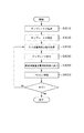
次に本画像処理装置による処理動作について説明する。
まず、ステップS101において、被検眼を本装置に配置した状態で、本装置と被検眼のアライメントを行う。アライメントの説明に関して、本実施形態に特有な処理について説明し、ワーキングディスタンス等のXYZ方向のアライメント、フォーカス、コヒーレンスゲートの調整等は一般的であるのでその説明は省略する。
PS−OCTの撮像中の固視微動または固視不良などによる被検眼の動きは、PS−OCT画像の歪みの原因となる。これを防ぐため、S102のステップではOCTのスキャナの動きを眼の動きに追従させる追尾を行いつつPS−OCTの撮像をしている。
S102において、撮像で網膜Erからの戻り光の信号を画像生成部193に出力し、前述の通り各画像を生成する。
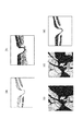
健常眼の断層画像と比べて疾病眼の断層画像では、病気の影響により断層画像の輝度値が暗くなってしまう場合があり、その影響で網膜層の見落としや誤検出をしてしまう。そのため、ステップS104においては、画像解析部194は、ステップS103で画像生成部193が計算した偏光状態を解消する箇所の情報を用いて網膜の各層を検出する。
次に、生成した各画像及び解析した結果の出力処理ステップS105について説明する。本実施形態おける出力処理は、ステップS104で求めた情報を効果的に表示する。
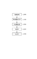
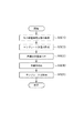
図9は、滲出物等の偏光を解消する領域情報を抽出する変形例の処理の流れを示す図である。
また、本発明は、以下の処理を実行することによっても実現される。即ち、上述した実施形態の機能を実現するソフトウェア(プログラム)を、ネットワーク又は各種記憶媒体を介してシステム或いは装置に供給し、そのシステム或いは装置のコンピュータ(またはCPUやMPU等)がプログラムを読み出して実行する処理である。
Claims (8)
- 異なる時刻に被検眼の眼底を撮像して得た複数の眼底画像と、前記複数の眼底画像よりも少ない数であり且つ前記複数の眼底画像とは異なる時刻に前記被検眼の眼底を撮像して得た少なくとも1つの眼底画像とを取得する眼底画像取得手段と、
前記複数の眼底画像を平均化して新たな眼底画像を生成する手段と、
前記生成された新たな眼底画像から特徴領域を抽出する抽出手段と、
前記抽出された特徴領域と前記少なくとも1つの眼底画像とに基づいて、前記新たな眼底画像に対応する前記眼底の第1の偏光断層画像と前記少なくとも1つの眼底画像に対応する前記眼底の第2の偏光断層画像との位置が補正されるように、前記眼底を追尾する手段と、
を有することを特徴とする眼科装置。 - 前記眼底画像取得手段が、前記複数の眼底画像及び前記少なくとも1つの眼底画像を60Hz以上のフレームレートで取得することを特徴とする請求項1に記載の眼科装置。
- 異なる時刻に被検眼の眼底を撮像して得た複数の眼底画像と、前記複数の眼底画像よりも少ない数であり且つ前記複数の眼底画像とは異なる時刻に前記被検眼の眼底を撮像して得た少なくとも1つの眼底画像とを取得する工程と、
前記複数の眼底画像を平均化して新たな眼底画像を生成する工程と、
前記生成された新たな眼底画像から特徴領域を抽出する工程と、
前記抽出された特徴領域と前記少なくとも1つの眼底画像とに基づいて、前記新たな眼底画像に対応する前記眼底の第1の偏光断層画像と前記少なくとも1つの眼底画像に対応する前記眼底の第2の偏光断層画像との位置が補正されるように、前記眼底を追尾する工程と、
を有することを特徴とする眼科方法。 - 前記取得する工程において、前記複数の眼底画像及び前記少なくとも1つの眼底画像を60Hz以上のフレームレートで取得することを特徴とする請求項3に記載の眼科方法。
- 請求項3または4に記載の眼科方法の各工程をコンピュータに実行させることを特徴とするプログラム。
- 異なる時間で撮像された被検体の偏光状態を示す複数の断層画像を取得する取得手段と、
前記複数の断層画像から偏光が解消された領域をそれぞれ抽出する抽出手段と、
前記偏光が解消されたそれぞれの領域の情報を関連付けて表示手段に表示させる表示制御手段と、
前記複数の断層画像に対応する複数の平面画像を60Hz以上のフレームレートで取得する平面画像取得手段と、
を有することを特徴とする撮影装置。 - 異なる時間で撮像された被検体の偏光状態を示す複数の断層画像を取得する工程と、
前記複数の断層画像から前記偏光が解消された領域をそれぞれ抽出する工程と、
前記偏光が解消されたそれぞれの領域の情報を関連付けて表示手段に表示させる工程と、
前記複数の断層画像に対応する複数の平面画像を60Hz以上のフレームレートで取得する工程と、
を有することを特徴とする撮影方法。 - 請求項7に記載の撮影方法の各工程をコンピュータに実行させることを特徴とするプログラム。
Priority Applications (3)
| Application Number | Priority Date | Filing Date | Title |
|---|---|---|---|
| JP2014001524A JP2014155694A (ja) | 2013-01-16 | 2014-01-08 | 眼科装置及び眼科方法 |
| US14/155,222 US9232887B2 (en) | 2013-01-16 | 2014-01-14 | Ophthalmic apparatus and ophthalmic method |
| EP14151390.3A EP2756796B1 (en) | 2013-01-16 | 2014-01-16 | Ophthalmic apparatus and ophthalmic method |
Applications Claiming Priority (3)
| Application Number | Priority Date | Filing Date | Title |
|---|---|---|---|
| JP2013005393 | 2013-01-16 | ||
| JP2013005393 | 2013-01-16 | ||
| JP2014001524A JP2014155694A (ja) | 2013-01-16 | 2014-01-08 | 眼科装置及び眼科方法 |
Publications (2)
| Publication Number | Publication Date |
|---|---|
| JP2014155694A true JP2014155694A (ja) | 2014-08-28 |
| JP2014155694A5 JP2014155694A5 (ja) | 2017-03-16 |
Family
ID=49943250
Family Applications (1)
| Application Number | Title | Priority Date | Filing Date |
|---|---|---|---|
| JP2014001524A Pending JP2014155694A (ja) | 2013-01-16 | 2014-01-08 | 眼科装置及び眼科方法 |
Country Status (3)
| Country | Link |
|---|---|
| US (1) | US9232887B2 (ja) |
| EP (1) | EP2756796B1 (ja) |
| JP (1) | JP2014155694A (ja) |
Cited By (3)
| Publication number | Priority date | Publication date | Assignee | Title |
|---|---|---|---|---|
| JP2016075585A (ja) * | 2014-10-07 | 2016-05-12 | キヤノン株式会社 | 撮像装置、断層画像のノイズ低減方法、及びプログラム |
| JP2019205912A (ja) * | 2019-09-03 | 2019-12-05 | キヤノン株式会社 | 眼科装置、画像生成方法およびプログラム |
| JP2024537988A (ja) * | 2021-11-05 | 2024-10-18 | アーエムエス インターナショナル アーゲー | 網膜撮像 |
Families Citing this family (10)
| Publication number | Priority date | Publication date | Assignee | Title |
|---|---|---|---|---|
| JP6742691B2 (ja) * | 2015-01-30 | 2020-08-19 | キヤノン株式会社 | 眼科装置、画像処理方法およびプログラム |
| JP6594033B2 (ja) * | 2015-05-14 | 2019-10-23 | キヤノン株式会社 | 画像処理装置、画像処理方法及びプログラム |
| JP2017080344A (ja) * | 2015-10-30 | 2017-05-18 | キヤノン株式会社 | 画像処理装置、画像処理方法及び光干渉断層撮影装置 |
| US10335026B2 (en) * | 2017-01-11 | 2019-07-02 | Canon Kabushiki Kaisha | Image processing apparatus that generates a tomographic image using an estimation value of pixel values, and related optical coherence tomography apparatus, image processing method, and computer-readable storage medium |
| ES2899428T3 (es) * | 2017-10-20 | 2022-03-11 | Svision Imaging Ltd | Sistema de diagnóstico de obtención de imágenes oftálmicas |
| EP3653987A1 (en) * | 2018-11-16 | 2020-05-20 | Nokia Technologies Oy | Apparatus and method for detecting light |
| JP7677885B2 (ja) * | 2018-11-21 | 2025-05-15 | ユニバーシティ オブ ワシントン | 遠隔眼科学における網膜テンプレート合致のためのシステムおよび方法 |
| JP7199236B2 (ja) * | 2019-01-24 | 2023-01-05 | 株式会社トプコン | 眼科装置 |
| EP3714770B1 (en) * | 2019-03-29 | 2023-05-10 | Nidek Co., Ltd. | Ophthalmological image processing apparatus |
| TWI749531B (zh) * | 2020-04-22 | 2021-12-11 | 晉弘科技股份有限公司 | 掃描裝置以及光學同調斷層掃描系統 |
Citations (2)
| Publication number | Priority date | Publication date | Assignee | Title |
|---|---|---|---|---|
| JP2011212103A (ja) * | 2010-03-31 | 2011-10-27 | Topcon Corp | レーザ走査型撮影装置 |
| JP2011229835A (ja) * | 2010-04-30 | 2011-11-17 | Canon Inc | 特徴点抽出方法及び眼科装置 |
Family Cites Families (10)
| Publication number | Priority date | Publication date | Assignee | Title |
|---|---|---|---|---|
| WO2007127291A2 (en) | 2006-04-24 | 2007-11-08 | Physical Sciences, Inc. | Stabilized retinal imaging with adaptive optics |
| JP2010175448A (ja) | 2009-01-30 | 2010-08-12 | Kowa Co | 光学撮像装置 |
| EP2243420A1 (en) | 2009-04-24 | 2010-10-27 | Schmidt-Erfurth, Ursula | Method for determining exudates in the retina |
| GB0907277D0 (en) | 2009-04-29 | 2009-06-10 | Univ Kent Kanterbury | Method for depth resolved wavefront sensing, depth resolved wavefront sensors and method and apparatus for optical imaging |
| JP5017328B2 (ja) * | 2009-08-11 | 2012-09-05 | キヤノン株式会社 | 断層像撮像装置およびその制御方法、プログラム、記憶媒体 |
| EP2563206B1 (en) | 2010-04-29 | 2018-08-29 | Massachusetts Institute of Technology | Method and apparatus for motion correction and image enhancement for optical coherence tomography |
| JP5297415B2 (ja) * | 2010-04-30 | 2013-09-25 | キヤノン株式会社 | 眼科装置及び眼科方法 |
| US8801178B2 (en) | 2010-11-04 | 2014-08-12 | Nidek Co., Ltd. | Fundus photographing apparatus |
| JP2012161382A (ja) | 2011-02-03 | 2012-08-30 | Nidek Co Ltd | 眼科装置 |
| EP2574273B1 (en) | 2011-06-23 | 2014-09-24 | Nidek Co., Ltd. | Optical coherence tomography apparatus |
-
2014
- 2014-01-08 JP JP2014001524A patent/JP2014155694A/ja active Pending
- 2014-01-14 US US14/155,222 patent/US9232887B2/en not_active Expired - Fee Related
- 2014-01-16 EP EP14151390.3A patent/EP2756796B1/en not_active Not-in-force
Patent Citations (2)
| Publication number | Priority date | Publication date | Assignee | Title |
|---|---|---|---|---|
| JP2011212103A (ja) * | 2010-03-31 | 2011-10-27 | Topcon Corp | レーザ走査型撮影装置 |
| JP2011229835A (ja) * | 2010-04-30 | 2011-11-17 | Canon Inc | 特徴点抽出方法及び眼科装置 |
Cited By (6)
| Publication number | Priority date | Publication date | Assignee | Title |
|---|---|---|---|---|
| JP2016075585A (ja) * | 2014-10-07 | 2016-05-12 | キヤノン株式会社 | 撮像装置、断層画像のノイズ低減方法、及びプログラム |
| US10126112B2 (en) | 2014-10-07 | 2018-11-13 | Canon Kabushiki Kaisha | Tomographic image capturing apparatus and method with noise reduction technique |
| JP2019205912A (ja) * | 2019-09-03 | 2019-12-05 | キヤノン株式会社 | 眼科装置、画像生成方法およびプログラム |
| JP7077283B2 (ja) | 2019-09-03 | 2022-05-30 | キヤノン株式会社 | 眼科装置、画像生成方法およびプログラム |
| JP2024537988A (ja) * | 2021-11-05 | 2024-10-18 | アーエムエス インターナショナル アーゲー | 網膜撮像 |
| JP7760718B2 (ja) | 2021-11-05 | 2025-10-27 | アーエムエス インターナショナル アーゲー | 網膜撮像 |
Also Published As
| Publication number | Publication date |
|---|---|
| EP2756796B1 (en) | 2017-09-13 |
| EP2756796A1 (en) | 2014-07-23 |
| US20140198300A1 (en) | 2014-07-17 |
| US9232887B2 (en) | 2016-01-12 |
Similar Documents
| Publication | Publication Date | Title |
|---|---|---|
| JP6071331B2 (ja) | 画像処理装置及び画像処理方法 | |
| JP5988772B2 (ja) | 画像処理装置及び画像処理方法 | |
| JP2014155694A (ja) | 眼科装置及び眼科方法 | |
| JP6184232B2 (ja) | 画像処理装置及び画像処理方法 | |
| JP6184231B2 (ja) | 画像処理装置および画像処理方法 | |
| JP6061554B2 (ja) | 画像処理装置及び画像処理方法 | |
| JP6202924B2 (ja) | 撮影装置及び撮影方法 | |
| JP6143422B2 (ja) | 画像処理装置及びその方法 | |
| JP6061555B2 (ja) | 画像処理装置及び画像処理方法 | |
| JP2014110883A (ja) | 画像処理装置及び画像処理方法 | |
| JP2017131550A (ja) | 画像処理装置及び画像処理方法 | |
| JP2014110884A (ja) | 画像処理装置及び画像処理方法 | |
| US9068812B2 (en) | Imaging apparatus, imaging method, and storage medium | |
| JP2014083285A (ja) | 画像処理装置及び画像処理方法 | |
| JP6146951B2 (ja) | 画像処理装置、画像処理方法、撮影装置及び撮影方法 | |
| JP5988883B2 (ja) | 画像処理装置及び画像処理方法 | |
| JP6647013B2 (ja) | 画像処理装置、画像処理方法及び光干渉断層撮影装置 | |
| JP6381622B2 (ja) | 画像処理装置及び画像処理方法 | |
| JP6437055B2 (ja) | 画像処理装置及び画像処理方法 | |
| JP6039185B2 (ja) | 撮影装置 | |
| JP5936368B2 (ja) | 光干渉断層撮影装置及びその作動方法 | |
| JP6505072B2 (ja) | 画像処理装置及び画像処理方法 | |
| JP2013148509A (ja) | 画像処理装置及び画像処理方法 | |
| JP2018000687A (ja) | 画像処理装置、画像処理方法及びプログラム |
Legal Events
| Date | Code | Title | Description |
|---|---|---|---|
| A621 | Written request for application examination |
Free format text: JAPANESE INTERMEDIATE CODE: A621 Effective date: 20170106 |
|
| A521 | Request for written amendment filed |
Free format text: JAPANESE INTERMEDIATE CODE: A523 Effective date: 20170206 |
|
| A977 | Report on retrieval |
Free format text: JAPANESE INTERMEDIATE CODE: A971007 Effective date: 20171027 |
|
| A131 | Notification of reasons for refusal |
Free format text: JAPANESE INTERMEDIATE CODE: A131 Effective date: 20171114 |
|
| A521 | Request for written amendment filed |
Free format text: JAPANESE INTERMEDIATE CODE: A523 Effective date: 20180109 |
|
| A131 | Notification of reasons for refusal |
Free format text: JAPANESE INTERMEDIATE CODE: A131 Effective date: 20180626 |
|
| A02 | Decision of refusal |
Free format text: JAPANESE INTERMEDIATE CODE: A02 Effective date: 20181218 |