JP5708643B2 - プローブ - Google Patents
プローブ Download PDFInfo
- Publication number
- JP5708643B2 JP5708643B2 JP2012511660A JP2012511660A JP5708643B2 JP 5708643 B2 JP5708643 B2 JP 5708643B2 JP 2012511660 A JP2012511660 A JP 2012511660A JP 2012511660 A JP2012511660 A JP 2012511660A JP 5708643 B2 JP5708643 B2 JP 5708643B2
- Authority
- JP
- Japan
- Prior art keywords
- probe
- light
- imaging device
- view
- observation target
- Prior art date
- Legal status (The legal status is an assumption and is not a legal conclusion. Google has not performed a legal analysis and makes no representation as to the accuracy of the status listed.)
- Expired - Fee Related
Links
- 239000000523 sample Substances 0.000 title claims description 141
- 238000003384 imaging method Methods 0.000 claims description 93
- 230000003287 optical effect Effects 0.000 claims description 35
- 239000013307 optical fiber Substances 0.000 claims description 31
- 230000000007 visual effect Effects 0.000 claims description 15
- 230000001678 irradiating effect Effects 0.000 claims description 11
- 230000005855 radiation Effects 0.000 claims description 8
- 238000001069 Raman spectroscopy Methods 0.000 claims description 3
- 230000005284 excitation Effects 0.000 description 21
- 238000010586 diagram Methods 0.000 description 11
- 238000003745 diagnosis Methods 0.000 description 6
- 238000001917 fluorescence detection Methods 0.000 description 6
- 238000005452 bending Methods 0.000 description 4
- 201000010099 disease Diseases 0.000 description 3
- 208000037265 diseases, disorders, signs and symptoms Diseases 0.000 description 3
- 238000005259 measurement Methods 0.000 description 3
- 206010028980 Neoplasm Diseases 0.000 description 2
- 201000011510 cancer Diseases 0.000 description 2
- 230000007850 degeneration Effects 0.000 description 2
- 230000000694 effects Effects 0.000 description 2
- 230000003902 lesion Effects 0.000 description 2
- 230000002093 peripheral effect Effects 0.000 description 2
- 230000008054 signal transmission Effects 0.000 description 2
- 230000002411 adverse Effects 0.000 description 1
- 230000008602 contraction Effects 0.000 description 1
- 230000003111 delayed effect Effects 0.000 description 1
- 238000001514 detection method Methods 0.000 description 1
- 239000003814 drug Substances 0.000 description 1
- 229940079593 drug Drugs 0.000 description 1
- 238000005516 engineering process Methods 0.000 description 1
- 238000000799 fluorescence microscopy Methods 0.000 description 1
- 230000005283 ground state Effects 0.000 description 1
- 238000005286 illumination Methods 0.000 description 1
- 230000008595 infiltration Effects 0.000 description 1
- 238000001764 infiltration Methods 0.000 description 1
- 230000010365 information processing Effects 0.000 description 1
- 239000007788 liquid Substances 0.000 description 1
- 239000011347 resin Substances 0.000 description 1
- 229920005989 resin Polymers 0.000 description 1
- 238000004611 spectroscopical analysis Methods 0.000 description 1
Images











Classifications
-
- A—HUMAN NECESSITIES
- A61—MEDICAL OR VETERINARY SCIENCE; HYGIENE
- A61B—DIAGNOSIS; SURGERY; IDENTIFICATION
- A61B1/00—Instruments for performing medical examinations of the interior of cavities or tubes of the body by visual or photographical inspection, e.g. endoscopes; Illuminating arrangements therefor
- A61B1/00163—Optical arrangements
- A61B1/00174—Optical arrangements characterised by the viewing angles
- A61B1/00177—Optical arrangements characterised by the viewing angles for 90 degrees side-viewing
-
- A—HUMAN NECESSITIES
- A61—MEDICAL OR VETERINARY SCIENCE; HYGIENE
- A61B—DIAGNOSIS; SURGERY; IDENTIFICATION
- A61B1/00—Instruments for performing medical examinations of the interior of cavities or tubes of the body by visual or photographical inspection, e.g. endoscopes; Illuminating arrangements therefor
- A61B1/00163—Optical arrangements
- A61B1/00174—Optical arrangements characterised by the viewing angles
- A61B1/00179—Optical arrangements characterised by the viewing angles for off-axis viewing
-
- A—HUMAN NECESSITIES
- A61—MEDICAL OR VETERINARY SCIENCE; HYGIENE
- A61B—DIAGNOSIS; SURGERY; IDENTIFICATION
- A61B1/00—Instruments for performing medical examinations of the interior of cavities or tubes of the body by visual or photographical inspection, e.g. endoscopes; Illuminating arrangements therefor
- A61B1/00163—Optical arrangements
- A61B1/00174—Optical arrangements characterised by the viewing angles
- A61B1/00183—Optical arrangements characterised by the viewing angles for variable viewing angles
-
- A—HUMAN NECESSITIES
- A61—MEDICAL OR VETERINARY SCIENCE; HYGIENE
- A61B—DIAGNOSIS; SURGERY; IDENTIFICATION
- A61B5/00—Measuring for diagnostic purposes; Identification of persons
- A61B5/0059—Measuring for diagnostic purposes; Identification of persons using light, e.g. diagnosis by transillumination, diascopy, fluorescence
- A61B5/0082—Measuring for diagnostic purposes; Identification of persons using light, e.g. diagnosis by transillumination, diascopy, fluorescence adapted for particular medical purposes
- A61B5/0084—Measuring for diagnostic purposes; Identification of persons using light, e.g. diagnosis by transillumination, diascopy, fluorescence adapted for particular medical purposes for introduction into the body, e.g. by catheters
-
- G—PHYSICS
- G01—MEASURING; TESTING
- G01J—MEASUREMENT OF INTENSITY, VELOCITY, SPECTRAL CONTENT, POLARISATION, PHASE OR PULSE CHARACTERISTICS OF INFRARED, VISIBLE OR ULTRAVIOLET LIGHT; COLORIMETRY; RADIATION PYROMETRY
- G01J3/00—Spectrometry; Spectrophotometry; Monochromators; Measuring colours
- G01J3/02—Details
-
- G—PHYSICS
- G01—MEASURING; TESTING
- G01J—MEASUREMENT OF INTENSITY, VELOCITY, SPECTRAL CONTENT, POLARISATION, PHASE OR PULSE CHARACTERISTICS OF INFRARED, VISIBLE OR ULTRAVIOLET LIGHT; COLORIMETRY; RADIATION PYROMETRY
- G01J3/00—Spectrometry; Spectrophotometry; Monochromators; Measuring colours
- G01J3/02—Details
- G01J3/0205—Optical elements not provided otherwise, e.g. optical manifolds, diffusers, windows
- G01J3/021—Optical elements not provided otherwise, e.g. optical manifolds, diffusers, windows using plane or convex mirrors, parallel phase plates, or particular reflectors
-
- G—PHYSICS
- G01—MEASURING; TESTING
- G01J—MEASUREMENT OF INTENSITY, VELOCITY, SPECTRAL CONTENT, POLARISATION, PHASE OR PULSE CHARACTERISTICS OF INFRARED, VISIBLE OR ULTRAVIOLET LIGHT; COLORIMETRY; RADIATION PYROMETRY
- G01J3/00—Spectrometry; Spectrophotometry; Monochromators; Measuring colours
- G01J3/02—Details
- G01J3/0205—Optical elements not provided otherwise, e.g. optical manifolds, diffusers, windows
- G01J3/0218—Optical elements not provided otherwise, e.g. optical manifolds, diffusers, windows using optical fibers
-
- G—PHYSICS
- G01—MEASURING; TESTING
- G01J—MEASUREMENT OF INTENSITY, VELOCITY, SPECTRAL CONTENT, POLARISATION, PHASE OR PULSE CHARACTERISTICS OF INFRARED, VISIBLE OR ULTRAVIOLET LIGHT; COLORIMETRY; RADIATION PYROMETRY
- G01J3/00—Spectrometry; Spectrophotometry; Monochromators; Measuring colours
- G01J3/02—Details
- G01J3/0256—Compact construction
-
- G—PHYSICS
- G01—MEASURING; TESTING
- G01J—MEASUREMENT OF INTENSITY, VELOCITY, SPECTRAL CONTENT, POLARISATION, PHASE OR PULSE CHARACTERISTICS OF INFRARED, VISIBLE OR ULTRAVIOLET LIGHT; COLORIMETRY; RADIATION PYROMETRY
- G01J3/00—Spectrometry; Spectrophotometry; Monochromators; Measuring colours
- G01J3/02—Details
- G01J3/06—Scanning arrangements arrangements for order-selection
-
- G—PHYSICS
- G01—MEASURING; TESTING
- G01N—INVESTIGATING OR ANALYSING MATERIALS BY DETERMINING THEIR CHEMICAL OR PHYSICAL PROPERTIES
- G01N21/00—Investigating or analysing materials by the use of optical means, i.e. using sub-millimetre waves, infrared, visible or ultraviolet light
- G01N21/62—Systems in which the material investigated is excited whereby it emits light or causes a change in wavelength of the incident light
- G01N21/63—Systems in which the material investigated is excited whereby it emits light or causes a change in wavelength of the incident light optically excited
- G01N21/64—Fluorescence; Phosphorescence
- G01N21/645—Specially adapted constructive features of fluorimeters
- G01N21/6456—Spatial resolved fluorescence measurements; Imaging
-
- G—PHYSICS
- G01—MEASURING; TESTING
- G01N—INVESTIGATING OR ANALYSING MATERIALS BY DETERMINING THEIR CHEMICAL OR PHYSICAL PROPERTIES
- G01N21/00—Investigating or analysing materials by the use of optical means, i.e. using sub-millimetre waves, infrared, visible or ultraviolet light
- G01N21/62—Systems in which the material investigated is excited whereby it emits light or causes a change in wavelength of the incident light
- G01N21/63—Systems in which the material investigated is excited whereby it emits light or causes a change in wavelength of the incident light optically excited
- G01N21/65—Raman scattering
-
- G—PHYSICS
- G02—OPTICS
- G02B—OPTICAL ELEMENTS, SYSTEMS OR APPARATUS
- G02B23/00—Telescopes, e.g. binoculars; Periscopes; Instruments for viewing the inside of hollow bodies; Viewfinders; Optical aiming or sighting devices
- G02B23/24—Instruments or systems for viewing the inside of hollow bodies, e.g. fibrescopes
- G02B23/2407—Optical details
- G02B23/2423—Optical details of the distal end
-
- G—PHYSICS
- G02—OPTICS
- G02B—OPTICAL ELEMENTS, SYSTEMS OR APPARATUS
- G02B23/00—Telescopes, e.g. binoculars; Periscopes; Instruments for viewing the inside of hollow bodies; Viewfinders; Optical aiming or sighting devices
- G02B23/24—Instruments or systems for viewing the inside of hollow bodies, e.g. fibrescopes
- G02B23/2407—Optical details
- G02B23/2461—Illumination
- G02B23/2469—Illumination using optical fibres
-
- A—HUMAN NECESSITIES
- A61—MEDICAL OR VETERINARY SCIENCE; HYGIENE
- A61B—DIAGNOSIS; SURGERY; IDENTIFICATION
- A61B1/00—Instruments for performing medical examinations of the interior of cavities or tubes of the body by visual or photographical inspection, e.g. endoscopes; Illuminating arrangements therefor
- A61B1/005—Flexible endoscopes
- A61B1/0051—Flexible endoscopes with controlled bending of insertion part
-
- A—HUMAN NECESSITIES
- A61—MEDICAL OR VETERINARY SCIENCE; HYGIENE
- A61B—DIAGNOSIS; SURGERY; IDENTIFICATION
- A61B1/00—Instruments for performing medical examinations of the interior of cavities or tubes of the body by visual or photographical inspection, e.g. endoscopes; Illuminating arrangements therefor
- A61B1/04—Instruments for performing medical examinations of the interior of cavities or tubes of the body by visual or photographical inspection, e.g. endoscopes; Illuminating arrangements therefor combined with photographic or television appliances
- A61B1/043—Instruments for performing medical examinations of the interior of cavities or tubes of the body by visual or photographical inspection, e.g. endoscopes; Illuminating arrangements therefor combined with photographic or television appliances for fluorescence imaging
-
- A—HUMAN NECESSITIES
- A61—MEDICAL OR VETERINARY SCIENCE; HYGIENE
- A61B—DIAGNOSIS; SURGERY; IDENTIFICATION
- A61B1/00—Instruments for performing medical examinations of the interior of cavities or tubes of the body by visual or photographical inspection, e.g. endoscopes; Illuminating arrangements therefor
- A61B1/04—Instruments for performing medical examinations of the interior of cavities or tubes of the body by visual or photographical inspection, e.g. endoscopes; Illuminating arrangements therefor combined with photographic or television appliances
- A61B1/05—Instruments for performing medical examinations of the interior of cavities or tubes of the body by visual or photographical inspection, e.g. endoscopes; Illuminating arrangements therefor combined with photographic or television appliances characterised by the image sensor, e.g. camera, being in the distal end portion
-
- A—HUMAN NECESSITIES
- A61—MEDICAL OR VETERINARY SCIENCE; HYGIENE
- A61B—DIAGNOSIS; SURGERY; IDENTIFICATION
- A61B1/00—Instruments for performing medical examinations of the interior of cavities or tubes of the body by visual or photographical inspection, e.g. endoscopes; Illuminating arrangements therefor
- A61B1/06—Instruments for performing medical examinations of the interior of cavities or tubes of the body by visual or photographical inspection, e.g. endoscopes; Illuminating arrangements therefor with illuminating arrangements
- A61B1/0661—Endoscope light sources
- A61B1/0676—Endoscope light sources at distal tip of an endoscope
-
- A—HUMAN NECESSITIES
- A61—MEDICAL OR VETERINARY SCIENCE; HYGIENE
- A61B—DIAGNOSIS; SURGERY; IDENTIFICATION
- A61B5/00—Measuring for diagnostic purposes; Identification of persons
- A61B5/68—Arrangements of detecting, measuring or recording means, e.g. sensors, in relation to patient
- A61B5/6846—Arrangements of detecting, measuring or recording means, e.g. sensors, in relation to patient specially adapted to be brought in contact with an internal body part, i.e. invasive
- A61B5/6847—Arrangements of detecting, measuring or recording means, e.g. sensors, in relation to patient specially adapted to be brought in contact with an internal body part, i.e. invasive mounted on an invasive device
- A61B5/6852—Catheters
- A61B5/6853—Catheters with a balloon
-
- G—PHYSICS
- G01—MEASURING; TESTING
- G01J—MEASUREMENT OF INTENSITY, VELOCITY, SPECTRAL CONTENT, POLARISATION, PHASE OR PULSE CHARACTERISTICS OF INFRARED, VISIBLE OR ULTRAVIOLET LIGHT; COLORIMETRY; RADIATION PYROMETRY
- G01J3/00—Spectrometry; Spectrophotometry; Monochromators; Measuring colours
- G01J3/28—Investigating the spectrum
- G01J3/44—Raman spectrometry; Scattering spectrometry ; Fluorescence spectrometry
- G01J3/4406—Fluorescence spectrometry
Landscapes
- Physics & Mathematics (AREA)
- Health & Medical Sciences (AREA)
- Life Sciences & Earth Sciences (AREA)
- Spectroscopy & Molecular Physics (AREA)
- General Physics & Mathematics (AREA)
- Surgery (AREA)
- General Health & Medical Sciences (AREA)
- Pathology (AREA)
- Optics & Photonics (AREA)
- Nuclear Medicine, Radiotherapy & Molecular Imaging (AREA)
- Heart & Thoracic Surgery (AREA)
- Public Health (AREA)
- Engineering & Computer Science (AREA)
- Biomedical Technology (AREA)
- Biophysics (AREA)
- Medical Informatics (AREA)
- Molecular Biology (AREA)
- Animal Behavior & Ethology (AREA)
- Veterinary Medicine (AREA)
- Radiology & Medical Imaging (AREA)
- Astronomy & Astrophysics (AREA)
- Chemical & Material Sciences (AREA)
- Analytical Chemistry (AREA)
- Biochemistry (AREA)
- Immunology (AREA)
- Investigating, Analyzing Materials By Fluorescence Or Luminescence (AREA)
- Endoscopes (AREA)
- Instruments For Viewing The Inside Of Hollow Bodies (AREA)
Description
このようなプローブには、照射光を生体の観察対象部位に照射し病変部から放射される放射光を受光する光ファイバー等の手段が備えられている。また、観察対象部位の周囲を視覚的に確認することも求められるため、観察対象部位の画像を撮像する撮像装置を併せ持つものが提案されている。
特許文献1記載のプローブにあっては、蛍光観察方向及び撮像方向がプローブの先端方向を向いているとともに、観察対象部位から反射した光を同一光路で受光し、光分岐手段で蛍光検出及び撮像のために分光する。
特許文献1,2記載のプローブにあっては、励起光を生体の観察対象部位に照射し観察対象部位からの反射光を受光する光学系と、撮像素子とがプローブの径方向に並んでおり、蛍光観察方向及び撮影方向はプローブ先端方向を向いている。
一方、特許文献4記載のプローブにあっては、励起光は円周方向および長手方向に走査し、複数の光検出素子が円周状に配設され、上記励起光による生体観察部位が発する蛍光を受光する。
特許文献1,2記載のプローブにあっては、励起光を生体の観察対象部位に照射し観察対象部位からの反射光を受光する光学系と、撮像素子とがプローブの径方向に並んでおり、蛍光観察方向及び撮影方向はプローブ先端方向を向いている。
そのため、プローブの細径化や、プローブの側方の撮像や蛍光観察が困難である。
特許文献4記載のプローブにあっては、蛍光を受光する手段のほかに観察対象部位の表面画像を撮像する撮像手段が備えられておらず、観察対象部位の表面を視覚的に観察することができない。
前記光学系に対して、当該プローブの先端側に前記撮像装置が配置されてなり、
当該プローブの長手方向に延在する軸を回動軸として、受光する前記放射光の当該プローブへの入射方向と前記撮像装置の視野方向とを、前記回動軸周りの相対角度を固定して回動し、
前記回動軸に対する垂直方向又は傾斜した方向に沿って当該プローブに入射する前記放射光を受光し、
同時に前記撮像装置による視野内にあるか又は前記回動によって前記撮像装置による視野内に時間差をもって入る観察対象部位から放射される前記放射光を受光するプローブである。
前記光学素子は、前記照射用光ファイバー及び前記受光用光ファイバーより当該プローブの先端側に配置され、
前記反射面を光路に介在させることにより、当該プローブの長手方向に対する垂直方向又は傾斜した方向に沿って当該プローブに入射する前記放射光を受光する請求項1に記載のプローブである。
前記回動軸周りに前記ユニットを回動させるための回動手段を有する請求項1に記載のプローブである。
また、当該プローブの先端側に撮像装置が配置されるため、当該プローブの先端方向の撮像も容易であるという効果がある。
ユニットフレーム基端部4aは円盤状に形成され、トルクコイル3の先端に固定される。また、ユニットフレーム基端部4aは、照射用光ファイバー5及び受光用光ファイバー6を保持している。ユニットフレームは、ユニットフレーム基端部4aの周部に連続した図示しない側壁部を有して、集光レンズ7、ミラー8及び撮像カメラ9を保持している。そして、トルクコイルが回動することにより、ユニットフレーム全体が回動する。
照射用光ファイバー5及び受光用光ファイバー6の軸は、本プローブの先端方向を向いており、更に先端側に、光ファイバー側からみて、集光レンズ7、ミラー8、撮像カメラ9の順で配置されている。撮像カメラ9には撮像時に用いる図示しない照明装置も設けられている。
すなわち、励起光を生体組織の観察対象部位に照射し観察対象部位からの反射光を受光する光学系に対して、当該プローブの先端側に撮像装置が配置される。撮像カメラ9を迂回して照射用光ファイバー5及び受光用光ファイバー6を配置することは非常に困難であるから、撮像カメラ9を先端側とすることにより、照射用光ファイバー5、受光用光ファイバー6、集光レンズ7及びミラー8からなる光学系とほぼ同軸に撮像カメラ9を配置することができ、本プローブを細径化することができる。また、本プローブの先端側に撮像カメラ9が配置されるため、本プローブの先端方向の撮像も容易である。
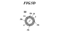
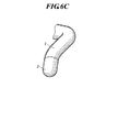
図5A,図5B,図5C,図5Dに示すようにユニットフレーム4は円筒状に形成される。ユニットフレーム4の内部に集光レンズ7、ミラー8及び撮像カメラ9が固定され、回動ユニットMが構成される。ユニットフレーム4の周面に窓4bが設けられる。窓4bは、透明部材で構成されるか、又は開口により形成される。窓4bは励起光の出射、反射光の入射、撮像カメラ9による撮像のための照明光の出射、及び撮像カメラ9による撮像のための窓である。軸Xは、本プローブの長手方向に延在する回動軸である。
図5A,図5Bに示す構成では、先端外套2は全部が透明とされ、回動ユニットMはトルクコイル3を介して伝達される動力により先端外套2内で回動軸X周りに回動する。
図5C,図5Dに示す構成では、ユニットフレーム4は、先端外套を兼ねている。したがって、窓4bは開口でなく透明部材で構成される。図5C,図5Dに示す構成では、ユニットフレーム4は、トルク伝達可能なトルクチューブ1Aに接続され、トルクチューブ1Aの基端がアクチュエータに接続されており、回動ユニットMはトルクチューブ1Aを介して伝達される動力により回動軸X周りに回動する。トルクチューブ1Aは、例えば、トルクコイルを被覆したチューブにより構成される。
いずれの構成にあっても、特定範囲を窓4bとせず全体を透明としてもよい。
回動ユニットMに光ファイバー5,6が接続されているため、回動ユニットMの回動は、所定の回転角で規制されるようにしてある。後述する回動走査も所定の回転角(例えば、360°に達するか又は360°を超えた時点)で反転して行われる。
上述した蛍光の検出と、撮像カメラ9による撮影を行いながら、回動ユニットMを回動走査して蛍光強度分布情報と画像情報を得る。ベースユニット等に搭載されるメモリにこれを記録する。本プローブが励起光を照射した観察対象部位から反射し本プローブに入射して本プローブが受光して検出する反射光の本プローブへの入射方向は、以上の構成にあってはミラー8が決定する。この反射光の入射方向と、撮像カメラ9の視野方向との相対角が回動走査中も一定しているのでこれを特定することができ、予め定数として情報処理装置に設定しておくことで蛍光強度分布情報と画像情報とを座標の一致させた重ね合わせ画像として表示出力することができる。
蛍光強度分布情報と画像情報とを取得するための回動ユニットMの回動走査中は、バルーン10aを膨張させて回動ユニットMの回動軸Xを固定して行う。また、回動ユニットMを含むプローブ本体は、アウターチューブ10に対して軸方向に移動し、軸X方向についても連続して走査可能である。
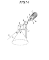
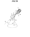
図7A,図7Bに分解斜視図として示す構成にあっては、励起光の出射光路L1及び反射光の入射光路L2は、本プローブの回動軸Xに対して垂直であり、撮像カメラ9の視野Yと同方向を向いている。なお、図7Aではミラー8と撮像カメラ9とを離して図示しているが、図7Bに示すようにミラー8と撮像カメラ9とを回動ユニットM上に搭載されたときの間隔で描くと、蛍光検出による観察対象部位は視野Y内にある。そして、図8A,図8B,図8Cに示すように回動走査が行われる。なお、図12Aに本構成の配置模式図を示した。
図9A示す構成では、励起光の出射光路L1及び反射光の入射光路L2は撮像カメラ9の視野中心YAと180°異なっている。
図9B示す構成では、励起光の出射光路L1及び反射光の入射光路L2は撮像カメラ9の視野中心YAの軸線に対して傾斜している。
すなわち、図9A,図9Bのいずれの構成にあっても、反射光の入射方向と、撮像カメラ9による視野角θとが、回動ユニットMの回動軸X周りに相対角を有する。視野角θと相対角を有するとは、視野角θ範囲内に含まれないことを意味する。これにより、蛍光検出による観察対象部位は視野Y外にあり、回動ユニットMの回動によって視野Y内に時間差をもって入ることとなる。
本構成を用いて、回動走査中に画像情報を先に得て保持し、撮像が済んだ観察対象部位の蛍光強度分布情報を後で得て、両情報を合成して表示出力すると、リアルタイムの撮影画像に対して出力画像が遅れてしまい、検査者に対して違和感を与えるおそれがある。
したがって、本構成を用いて蛍光強度分布情報を先に得て保持し、蛍光測定が済んだ観察対象部位の画像情報を後で得て、両情報を合成することが好ましい。これにより、検査者に対して撮像時に対して時間差が小さく違和感のない画像を表示することができるという利点がある。
また、図9A,図9Bに示したように、ミラー8の撮像カメラ9に対する角度を選択することができることによって、撮像カメラ9から延出するケーブルとミラー8とが干渉しない配置を選択でき、よりミラー8と撮像カメラ9とを近接配置して、回動ユニットMを短くコンパクトにすることができるという利点がある。
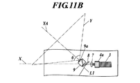
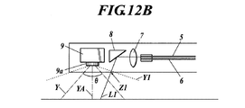
図10A,図10B及び図11A,図11Bに示すように、励起光の出射光路L1及び反射光の入射光路L2を、回動軸Xに対する垂直方向よりも撮像カメラ9の視野Y側に傾斜した方向とする。これにより、視野中心YAがプローブ先端側に傾いていても、図10A,図10Bに示すように蛍光検出による観察対象部位を視野Y内に入れることができ、図11A,図11Bに示すように、蛍光検出による観察対象部位を回動ユニットMの回動によって視野Y内に時間差をもって入れることができる。図12Bには、視野中心YAが回動軸Xに対して垂直な場合において励起光の出射光路L1及び反射光の入射光路L2を視野Y側に傾斜させた構成の配置模式図を示した。ミラー8の向きを調整することで、蛍光観察用光路を垂直方向から傾け、カメラ視野に重なるようにすることができる。
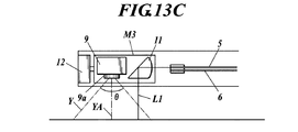
回動ユニットに搭載すべき最小要素は、撮像カメラ9と、受光する反射光の当該プローブへの入射方向、すなわち、蛍光観察方向を決定する光学素子である。したがって、図13Aに示すようなミラー8と撮像カメラ9とを搭載した回動ユニットM1や、図13Bに示すような集光レンズ7とミラー8と撮像カメラ9とを搭載した回動ユニットM2、図13Cに示すような光学素子11と撮像カメラ9とを搭載した回動ユニットM3など、様々な搭載形態を選択できる。これらの回動ユニットM1,M2,M3を、例えばプローブ先端側に設置したサーボモータ12の出力端に接続することによって、回動走査を実施することができる。
すなわち、図12Bに破線で示した視野Y1のように、撮像カメラ9の視野Y1がプローブの外表面上の励起光及び受光する反射光の通過部Z1を含むように構成することも有効である。この場合、蛍光測定に悪影響を与える外表面の汚れの有無を検出することが可能となり、測定精度を維持することに役立たせることができる。
1A トルクチューブ
2 先端外套
3 トルクコイル
4 ユニットフレーム
4a ユニットフレーム基端部
4b 窓
5 照射用光ファイバー
6 受光用光ファイバー
7 集光レンズ
8 ミラー(又はプリズム)
9 撮像カメラ
10 アウターチューブ
10a バルーン
11 光学素子
11a 集光面
11b 反射面
12 サーボモータ
L1 出射光路
L2 入射光路
M 回動ユニット
M1 回動ユニット
M2 回動ユニット
M3 回動ユニット
X 回動軸
Y 視野
Y1 視野
YA 視野中心
Z1 通過部
θ 視野角
Claims (10)
- 生体組織の観察対象部位に照射光を照射して観察対象部位から放射される放射光を受光するための光学系と、観察対象部位の表面画像を撮像する撮像装置とを備えるプローブにおいて、
前記光学系に対して、当該プローブの先端側に前記撮像装置が配置されてなり、
当該プローブの長手方向に延在する軸を回動軸として、受光する前記放射光の当該プローブへの入射方向と前記撮像装置の視野方向とを、前記回動軸周りの相対角度を固定して回動し、
前記回動軸に対する垂直方向又は傾斜した方向に沿って当該プローブに入射する前記放射光を受光し、
同時に前記撮像装置による視野内にあるか又は前記回動によって前記撮像装置による視野内に時間差をもって入る観察対象部位から放射される前記放射光を受光するプローブ。 - 前記光学系は、前記照射光の照射用光ファイバーと、前記放射光の受光用光ファイバーと、反射面を有した光学素子とを備え、
前記光学素子は、前記照射用光ファイバー及び前記受光用光ファイバーより当該プローブの先端側に配置され、
前記反射面を光路に介在させることにより、当該プローブの長手方向に対する垂直方向又は傾斜した方向に沿って当該プローブに入射する前記放射光を受光する請求項1に記載のプローブ。 - 前記撮像装置の視野角は、当該プローブの先端方向に垂直な方向を含む請求項1又は請求項2に記載のプローブ。
- 前記撮像装置の視野角は、当該プローブの先端方向を含む請求項3に記載のプローブ。
- 前記撮像装置の視野は、当該プローブの外表面上の前記照射光及び前記放射光の通過部を含む請求項3又は請求項4に記載のプローブ。
- 前記放射光の当該プローブへの入射方向を決定する光学素子及び前記撮像装置がユニットに取り付けられ、
前記回動軸周りに前記ユニットを回動させるための回動手段を有する請求項1に記載のプローブ。 - 前記入射方向が、前記回動軸に対する垂直方向よりも前記撮像装置の視野側に傾斜した方向とされることにより、同時に前記撮像装置による視野内にあるか又は前記回動によって前記撮像装置による視野内に時間差をもって入る観察対象部位から放射される前記放射光を受光する請求項1に記載のプローブ。
- 同時において前記撮像装置による視野外にあり、前記回動によって前記撮像装置による視野内に時間差をもって入る観察対象部位から放射される前記放射光を受光する請求項1に記載のプローブ。
- 前記入射方向と、前記撮像装置による視野角とが、前記回動軸周りに相対角を有することにより、同時において前記撮像装置による視野外にあり、前記回動によって前記撮像装置による視野内に時間差をもって入る観察対象部位から放射される前記放射光を受光する請求項8に記載のプローブ。
- 前記光学系は、前記照射光に起因して生じる蛍光、散乱光又はラマン散乱光を受光することを特徴とする請求項1から請求項9のうちいずれか一に記載のプローブ。
Priority Applications (1)
| Application Number | Priority Date | Filing Date | Title |
|---|---|---|---|
| JP2012511660A JP5708643B2 (ja) | 2010-04-23 | 2011-04-19 | プローブ |
Applications Claiming Priority (6)
| Application Number | Priority Date | Filing Date | Title |
|---|---|---|---|
| JP2010099868 | 2010-04-23 | ||
| JP2010099868 | 2010-04-23 | ||
| JP2010099867 | 2010-04-23 | ||
| JP2010099867 | 2010-04-23 | ||
| JP2012511660A JP5708643B2 (ja) | 2010-04-23 | 2011-04-19 | プローブ |
| PCT/JP2011/059600 WO2011132661A1 (ja) | 2010-04-23 | 2011-04-19 | プローブ |
Publications (2)
| Publication Number | Publication Date |
|---|---|
| JPWO2011132661A1 JPWO2011132661A1 (ja) | 2013-07-18 |
| JP5708643B2 true JP5708643B2 (ja) | 2015-04-30 |
Family
ID=44834180
Family Applications (1)
| Application Number | Title | Priority Date | Filing Date |
|---|---|---|---|
| JP2012511660A Expired - Fee Related JP5708643B2 (ja) | 2010-04-23 | 2011-04-19 | プローブ |
Country Status (5)
| Country | Link |
|---|---|
| US (1) | US9277852B2 (ja) |
| EP (1) | EP2561795A4 (ja) |
| JP (1) | JP5708643B2 (ja) |
| CN (1) | CN102858224B (ja) |
| WO (1) | WO2011132661A1 (ja) |
Families Citing this family (13)
| Publication number | Priority date | Publication date | Assignee | Title |
|---|---|---|---|---|
| CN102551677B (zh) * | 2012-03-06 | 2013-11-27 | 天津大学 | 用于漫射光断层成像的内窥式旋转探头 |
| MX382655B (es) * | 2013-02-01 | 2025-03-13 | Deka Products Lp | Endoscopio con camara que puede moverse en planos horizontal y vertical. |
| US9194690B2 (en) * | 2013-03-04 | 2015-11-24 | Corning Incorporated | Power transmission and sensing device |
| US9518895B2 (en) * | 2013-08-15 | 2016-12-13 | Siemens Energy, Inc. | Optical probe with improved affixing structure for supporting a light-redirecting element |
| US9182285B2 (en) * | 2013-08-15 | 2015-11-10 | Siemens Energy, Inc. | Methods regarding optical probe having an inner tube with separable tube sections to house optical elements |
| US9195044B2 (en) | 2013-08-15 | 2015-11-24 | Siemens Energy, Inc | Optical probe having an inner tube with separable tube sections to house optical elements |
| DE112014006391T5 (de) * | 2014-02-21 | 2016-11-17 | Honda Motor Co., Ltd. | Optische Messsonde und mit selbiger versehene optische Messvorrichtung |
| JPWO2017168703A1 (ja) * | 2016-03-31 | 2018-10-11 | 株式会社島津製作所 | 光学測定用プローブ及びこれを備えた光学測定装置 |
| CN107255632A (zh) * | 2017-06-27 | 2017-10-17 | 上海化工研究院有限公司 | 一种用于探测较深层固体样品的拉曼光谱探头延伸装置 |
| JP7320620B2 (ja) * | 2019-12-26 | 2023-08-03 | 富士フイルム株式会社 | 内視鏡及び内視鏡システム |
| CN113952010A (zh) * | 2021-12-06 | 2022-01-21 | 中国科学院长春光学精密机械与物理研究所 | 一种新型可视化产钳系统 |
| EP4249984A1 (en) * | 2022-03-25 | 2023-09-27 | General Electric Company | Tool stabilization mechanism and related methods |
| US12326553B2 (en) | 2022-03-25 | 2025-06-10 | General Electric Company | Tool stabilization mechanism and related methods |
Citations (5)
| Publication number | Priority date | Publication date | Assignee | Title |
|---|---|---|---|---|
| JPS63164932A (ja) * | 1986-12-27 | 1988-07-08 | オリンパス光学工業株式会社 | 内視鏡装置 |
| JPH04341232A (ja) * | 1991-03-11 | 1992-11-27 | Olympus Optical Co Ltd | 電子内視鏡システム |
| JP2001079007A (ja) * | 1999-09-13 | 2001-03-27 | Olympus Optical Co Ltd | 光プローブ装置 |
| JP2003204926A (ja) * | 2002-01-11 | 2003-07-22 | Olympus Optical Co Ltd | 内視鏡 |
| JP2006087447A (ja) * | 2004-09-21 | 2006-04-06 | Michiaki Nagai | スキャナシステム |
Family Cites Families (12)
| Publication number | Priority date | Publication date | Assignee | Title |
|---|---|---|---|---|
| JPS58216212A (ja) * | 1982-06-11 | 1983-12-15 | Sumitomo Electric Ind Ltd | 視野回転機構付イメ−ジフアイバ |
| JPH0211117A (ja) * | 1988-06-30 | 1990-01-16 | Toshiba Corp | 電子内視鏡装置 |
| JPH07327916A (ja) * | 1994-06-02 | 1995-12-19 | Olympus Optical Co Ltd | 視野方向可変型内視鏡 |
| US5797836A (en) * | 1995-06-07 | 1998-08-25 | Smith & Nephew, Inc. | Endoscope with relative rotation and axial motion between an optical element and an imaging device |
| US6428470B1 (en) * | 1995-09-15 | 2002-08-06 | Pinotage, Llc | Imaging system and components thereof |
| JP3552844B2 (ja) | 1996-04-30 | 2004-08-11 | 富士写真フイルム株式会社 | 内視鏡 |
| JPH10127562A (ja) | 1996-10-28 | 1998-05-19 | Fuji Photo Film Co Ltd | 蛍光内視鏡 |
| US7175593B2 (en) * | 2000-08-30 | 2007-02-13 | Durell & Gitelis, Inc. | Variable view arthroscope with charge coupled device |
| US7018309B2 (en) | 2004-05-04 | 2006-03-28 | Bridgestone Sports Co., Ltd. | Golf ball |
| JP4495513B2 (ja) | 2004-05-11 | 2010-07-07 | Hoya株式会社 | 蛍光内視鏡装置 |
| JP5314841B2 (ja) | 2006-08-22 | 2013-10-16 | オリンパス株式会社 | 内視鏡装置、及び内視鏡プローブ |
| CN101686827B (zh) * | 2007-01-19 | 2014-08-13 | 桑尼布鲁克健康科学中心 | 具有组合的超声和光学成像装置的成像探头 |
-
2011
- 2011-04-19 WO PCT/JP2011/059600 patent/WO2011132661A1/ja not_active Ceased
- 2011-04-19 US US13/643,029 patent/US9277852B2/en not_active Expired - Fee Related
- 2011-04-19 JP JP2012511660A patent/JP5708643B2/ja not_active Expired - Fee Related
- 2011-04-19 EP EP11771996.3A patent/EP2561795A4/en not_active Withdrawn
- 2011-04-19 CN CN201180019771.XA patent/CN102858224B/zh not_active Expired - Fee Related
Patent Citations (5)
| Publication number | Priority date | Publication date | Assignee | Title |
|---|---|---|---|---|
| JPS63164932A (ja) * | 1986-12-27 | 1988-07-08 | オリンパス光学工業株式会社 | 内視鏡装置 |
| JPH04341232A (ja) * | 1991-03-11 | 1992-11-27 | Olympus Optical Co Ltd | 電子内視鏡システム |
| JP2001079007A (ja) * | 1999-09-13 | 2001-03-27 | Olympus Optical Co Ltd | 光プローブ装置 |
| JP2003204926A (ja) * | 2002-01-11 | 2003-07-22 | Olympus Optical Co Ltd | 内視鏡 |
| JP2006087447A (ja) * | 2004-09-21 | 2006-04-06 | Michiaki Nagai | スキャナシステム |
Also Published As
| Publication number | Publication date |
|---|---|
| CN102858224A (zh) | 2013-01-02 |
| US20130038872A1 (en) | 2013-02-14 |
| EP2561795A4 (en) | 2013-09-25 |
| US9277852B2 (en) | 2016-03-08 |
| JPWO2011132661A1 (ja) | 2013-07-18 |
| CN102858224B (zh) | 2015-04-01 |
| WO2011132661A1 (ja) | 2011-10-27 |
| EP2561795A1 (en) | 2013-02-27 |
Similar Documents
| Publication | Publication Date | Title |
|---|---|---|
| JP5708643B2 (ja) | プローブ | |
| JP5118867B2 (ja) | 内視鏡観察装置および内視鏡の作動方法 | |
| US10314490B2 (en) | Method and device for multi-spectral photonic imaging | |
| EP2046188B1 (en) | Capsule camera with variable illumination of the surrounding tissue | |
| JP6254121B2 (ja) | 胃腸管の異常の診断に使える情報を提供する経口機器 | |
| AU759282B2 (en) | Systems and methods for optical examination of samples | |
| US20130070255A1 (en) | Probe for optical tomograpic image measurement device and method for adjusting probe | |
| JP2003535659A (ja) | 走査型単一光ファイバシステムを用いる医療用画像化、診断および治療 | |
| JP5314841B2 (ja) | 内視鏡装置、及び内視鏡プローブ | |
| JP2013223635A (ja) | プローブ | |
| US20070293766A1 (en) | Transmission Based Imaging for Spectroscopic Analysis | |
| JP7160093B2 (ja) | 医用画像撮像装置 | |
| WO2010061471A1 (ja) | 生体観察装置 | |
| KR101887033B1 (ko) | 치아 구조/기능 영상을 위한 복합영상장치 | |
| JP2012085805A (ja) | 光断層画像化装置 | |
| JP5928461B2 (ja) | プローブ | |
| JP5708659B2 (ja) | プローブ及び測定システム | |
| JP5648456B2 (ja) | プローブ | |
| JP2011127924A (ja) | イメージングプローブ | |
| JP5811049B2 (ja) | プローブ | |
| JP2008295744A (ja) | 生体観察装置 |
Legal Events
| Date | Code | Title | Description |
|---|---|---|---|
| A711 | Notification of change in applicant |
Free format text: JAPANESE INTERMEDIATE CODE: A712 Effective date: 20130416 |
|
| A621 | Written request for application examination |
Free format text: JAPANESE INTERMEDIATE CODE: A621 Effective date: 20140118 |
|
| TRDD | Decision of grant or rejection written | ||
| A01 | Written decision to grant a patent or to grant a registration (utility model) |
Free format text: JAPANESE INTERMEDIATE CODE: A01 Effective date: 20150203 |
|
| A61 | First payment of annual fees (during grant procedure) |
Free format text: JAPANESE INTERMEDIATE CODE: A61 Effective date: 20150216 |
|
| R150 | Certificate of patent or registration of utility model |
Ref document number: 5708643 Country of ref document: JP Free format text: JAPANESE INTERMEDIATE CODE: R150 |
|
| LAPS | Cancellation because of no payment of annual fees |