JP7242536B2 - High-throughput 3D assays for immune cell and drug homing, migration and tumor cytotoxicity - Google Patents
High-throughput 3D assays for immune cell and drug homing, migration and tumor cytotoxicity Download PDFInfo
- Publication number
- JP7242536B2 JP7242536B2 JP2019542516A JP2019542516A JP7242536B2 JP 7242536 B2 JP7242536 B2 JP 7242536B2 JP 2019542516 A JP2019542516 A JP 2019542516A JP 2019542516 A JP2019542516 A JP 2019542516A JP 7242536 B2 JP7242536 B2 JP 7242536B2
- Authority
- JP
- Japan
- Prior art keywords
- cells
- tumor
- assay method
- chamber
- cell
- Prior art date
- Legal status (The legal status is an assumption and is not a legal conclusion. Google has not performed a legal analysis and makes no representation as to the accuracy of the status listed.)
- Active
Links
Images


Classifications
-
- G—PHYSICS
- G01—MEASURING; TESTING
- G01N—INVESTIGATING OR ANALYSING MATERIALS BY DETERMINING THEIR CHEMICAL OR PHYSICAL PROPERTIES
- G01N33/00—Investigating or analysing materials by specific methods not covered by groups G01N1/00 - G01N31/00
- G01N33/48—Biological material, e.g. blood, urine; Haemocytometers
- G01N33/50—Chemical analysis of biological material, e.g. blood, urine; Testing involving biospecific ligand binding methods; Immunological testing
- G01N33/5005—Chemical analysis of biological material, e.g. blood, urine; Testing involving biospecific ligand binding methods; Immunological testing involving human or animal cells
- G01N33/5008—Chemical analysis of biological material, e.g. blood, urine; Testing involving biospecific ligand binding methods; Immunological testing involving human or animal cells for testing or evaluating the effect of chemical or biological compounds, e.g. drugs, cosmetics
- G01N33/5011—Chemical analysis of biological material, e.g. blood, urine; Testing involving biospecific ligand binding methods; Immunological testing involving human or animal cells for testing or evaluating the effect of chemical or biological compounds, e.g. drugs, cosmetics for testing antineoplastic activity
-
- C—CHEMISTRY; METALLURGY
- C12—BIOCHEMISTRY; BEER; SPIRITS; WINE; VINEGAR; MICROBIOLOGY; ENZYMOLOGY; MUTATION OR GENETIC ENGINEERING
- C12N—MICROORGANISMS OR ENZYMES; COMPOSITIONS THEREOF; PROPAGATING, PRESERVING, OR MAINTAINING MICROORGANISMS; MUTATION OR GENETIC ENGINEERING; CULTURE MEDIA
- C12N5/00—Undifferentiated human, animal or plant cells, e.g. cell lines; Tissues; Cultivation or maintenance thereof; Culture media therefor
- C12N5/06—Animal cells or tissues; Human cells or tissues
- C12N5/0602—Vertebrate cells
- C12N5/0693—Tumour cells; Cancer cells
-
- G—PHYSICS
- G01—MEASURING; TESTING
- G01N—INVESTIGATING OR ANALYSING MATERIALS BY DETERMINING THEIR CHEMICAL OR PHYSICAL PROPERTIES
- G01N33/00—Investigating or analysing materials by specific methods not covered by groups G01N1/00 - G01N31/00
- G01N33/48—Biological material, e.g. blood, urine; Haemocytometers
- G01N33/50—Chemical analysis of biological material, e.g. blood, urine; Testing involving biospecific ligand binding methods; Immunological testing
- G01N33/5005—Chemical analysis of biological material, e.g. blood, urine; Testing involving biospecific ligand binding methods; Immunological testing involving human or animal cells
- G01N33/5008—Chemical analysis of biological material, e.g. blood, urine; Testing involving biospecific ligand binding methods; Immunological testing involving human or animal cells for testing or evaluating the effect of chemical or biological compounds, e.g. drugs, cosmetics
- G01N33/5014—Chemical analysis of biological material, e.g. blood, urine; Testing involving biospecific ligand binding methods; Immunological testing involving human or animal cells for testing or evaluating the effect of chemical or biological compounds, e.g. drugs, cosmetics for testing toxicity
-
- G—PHYSICS
- G01—MEASURING; TESTING
- G01N—INVESTIGATING OR ANALYSING MATERIALS BY DETERMINING THEIR CHEMICAL OR PHYSICAL PROPERTIES
- G01N33/00—Investigating or analysing materials by specific methods not covered by groups G01N1/00 - G01N31/00
- G01N33/48—Biological material, e.g. blood, urine; Haemocytometers
- G01N33/50—Chemical analysis of biological material, e.g. blood, urine; Testing involving biospecific ligand binding methods; Immunological testing
- G01N33/5005—Chemical analysis of biological material, e.g. blood, urine; Testing involving biospecific ligand binding methods; Immunological testing involving human or animal cells
- G01N33/5008—Chemical analysis of biological material, e.g. blood, urine; Testing involving biospecific ligand binding methods; Immunological testing involving human or animal cells for testing or evaluating the effect of chemical or biological compounds, e.g. drugs, cosmetics
- G01N33/502—Chemical analysis of biological material, e.g. blood, urine; Testing involving biospecific ligand binding methods; Immunological testing involving human or animal cells for testing or evaluating the effect of chemical or biological compounds, e.g. drugs, cosmetics for testing non-proliferative effects
- G01N33/5029—Chemical analysis of biological material, e.g. blood, urine; Testing involving biospecific ligand binding methods; Immunological testing involving human or animal cells for testing or evaluating the effect of chemical or biological compounds, e.g. drugs, cosmetics for testing non-proliferative effects on cell motility
-
- G—PHYSICS
- G01—MEASURING; TESTING
- G01N—INVESTIGATING OR ANALYSING MATERIALS BY DETERMINING THEIR CHEMICAL OR PHYSICAL PROPERTIES
- G01N33/00—Investigating or analysing materials by specific methods not covered by groups G01N1/00 - G01N31/00
- G01N33/48—Biological material, e.g. blood, urine; Haemocytometers
- G01N33/50—Chemical analysis of biological material, e.g. blood, urine; Testing involving biospecific ligand binding methods; Immunological testing
- G01N33/5005—Chemical analysis of biological material, e.g. blood, urine; Testing involving biospecific ligand binding methods; Immunological testing involving human or animal cells
- G01N33/5008—Chemical analysis of biological material, e.g. blood, urine; Testing involving biospecific ligand binding methods; Immunological testing involving human or animal cells for testing or evaluating the effect of chemical or biological compounds, e.g. drugs, cosmetics
- G01N33/5044—Chemical analysis of biological material, e.g. blood, urine; Testing involving biospecific ligand binding methods; Immunological testing involving human or animal cells for testing or evaluating the effect of chemical or biological compounds, e.g. drugs, cosmetics involving specific cell types
-
- G—PHYSICS
- G01—MEASURING; TESTING
- G01N—INVESTIGATING OR ANALYSING MATERIALS BY DETERMINING THEIR CHEMICAL OR PHYSICAL PROPERTIES
- G01N33/00—Investigating or analysing materials by specific methods not covered by groups G01N1/00 - G01N31/00
- G01N33/48—Biological material, e.g. blood, urine; Haemocytometers
- G01N33/50—Chemical analysis of biological material, e.g. blood, urine; Testing involving biospecific ligand binding methods; Immunological testing
- G01N33/5005—Chemical analysis of biological material, e.g. blood, urine; Testing involving biospecific ligand binding methods; Immunological testing involving human or animal cells
- G01N33/5008—Chemical analysis of biological material, e.g. blood, urine; Testing involving biospecific ligand binding methods; Immunological testing involving human or animal cells for testing or evaluating the effect of chemical or biological compounds, e.g. drugs, cosmetics
- G01N33/5082—Supracellular entities, e.g. tissue, organisms
- G01N33/5088—Supracellular entities, e.g. tissue, organisms of vertebrates
-
- C—CHEMISTRY; METALLURGY
- C12—BIOCHEMISTRY; BEER; SPIRITS; WINE; VINEGAR; MICROBIOLOGY; ENZYMOLOGY; MUTATION OR GENETIC ENGINEERING
- C12N—MICROORGANISMS OR ENZYMES; COMPOSITIONS THEREOF; PROPAGATING, PRESERVING, OR MAINTAINING MICROORGANISMS; MUTATION OR GENETIC ENGINEERING; CULTURE MEDIA
- C12N2501/00—Active agents used in cell culture processes, e.g. differentation
- C12N2501/02—Compounds of the arachidonic acid pathway, e.g. prostaglandins, leukotrienes
-
- C—CHEMISTRY; METALLURGY
- C12—BIOCHEMISTRY; BEER; SPIRITS; WINE; VINEGAR; MICROBIOLOGY; ENZYMOLOGY; MUTATION OR GENETIC ENGINEERING
- C12N—MICROORGANISMS OR ENZYMES; COMPOSITIONS THEREOF; PROPAGATING, PRESERVING, OR MAINTAINING MICROORGANISMS; MUTATION OR GENETIC ENGINEERING; CULTURE MEDIA
- C12N2501/00—Active agents used in cell culture processes, e.g. differentation
- C12N2501/20—Cytokines; Chemokines
- C12N2501/21—Chemokines, e.g. MIP-1, MIP-2, RANTES, MCP, PF-4
-
- C—CHEMISTRY; METALLURGY
- C12—BIOCHEMISTRY; BEER; SPIRITS; WINE; VINEGAR; MICROBIOLOGY; ENZYMOLOGY; MUTATION OR GENETIC ENGINEERING
- C12N—MICROORGANISMS OR ENZYMES; COMPOSITIONS THEREOF; PROPAGATING, PRESERVING, OR MAINTAINING MICROORGANISMS; MUTATION OR GENETIC ENGINEERING; CULTURE MEDIA
- C12N2502/00—Coculture with; Conditioned medium produced by
- C12N2502/28—Vascular endothelial cells
-
- C—CHEMISTRY; METALLURGY
- C12—BIOCHEMISTRY; BEER; SPIRITS; WINE; VINEGAR; MICROBIOLOGY; ENZYMOLOGY; MUTATION OR GENETIC ENGINEERING
- C12N—MICROORGANISMS OR ENZYMES; COMPOSITIONS THEREOF; PROPAGATING, PRESERVING, OR MAINTAINING MICROORGANISMS; MUTATION OR GENETIC ENGINEERING; CULTURE MEDIA
- C12N2503/00—Use of cells in diagnostics
- C12N2503/02—Drug screening
-
- C—CHEMISTRY; METALLURGY
- C12—BIOCHEMISTRY; BEER; SPIRITS; WINE; VINEGAR; MICROBIOLOGY; ENZYMOLOGY; MUTATION OR GENETIC ENGINEERING
- C12N—MICROORGANISMS OR ENZYMES; COMPOSITIONS THEREOF; PROPAGATING, PRESERVING, OR MAINTAINING MICROORGANISMS; MUTATION OR GENETIC ENGINEERING; CULTURE MEDIA
- C12N2513/00—3D culture
-
- G—PHYSICS
- G01—MEASURING; TESTING
- G01N—INVESTIGATING OR ANALYSING MATERIALS BY DETERMINING THEIR CHEMICAL OR PHYSICAL PROPERTIES
- G01N2500/00—Screening for compounds of potential therapeutic value
- G01N2500/10—Screening for compounds of potential therapeutic value involving cells
Landscapes
- Health & Medical Sciences (AREA)
- Life Sciences & Earth Sciences (AREA)
- Engineering & Computer Science (AREA)
- Biomedical Technology (AREA)
- Immunology (AREA)
- Chemical & Material Sciences (AREA)
- Hematology (AREA)
- Urology & Nephrology (AREA)
- Molecular Biology (AREA)
- Cell Biology (AREA)
- Bioinformatics & Cheminformatics (AREA)
- Biotechnology (AREA)
- Microbiology (AREA)
- General Health & Medical Sciences (AREA)
- Toxicology (AREA)
- Biochemistry (AREA)
- Medicinal Chemistry (AREA)
- Physics & Mathematics (AREA)
- Analytical Chemistry (AREA)
- Food Science & Technology (AREA)
- Tropical Medicine & Parasitology (AREA)
- General Physics & Mathematics (AREA)
- Pathology (AREA)
- Wood Science & Technology (AREA)
- Genetics & Genomics (AREA)
- Zoology (AREA)
- Organic Chemistry (AREA)
- Oncology (AREA)
- General Engineering & Computer Science (AREA)
- Measuring Or Testing Involving Enzymes Or Micro-Organisms (AREA)
- Apparatus Associated With Microorganisms And Enzymes (AREA)
- Medicines Containing Material From Animals Or Micro-Organisms (AREA)
- Medicines That Contain Protein Lipid Enzymes And Other Medicines (AREA)
- Pharmaceuticals Containing Other Organic And Inorganic Compounds (AREA)
- Acyclic And Carbocyclic Compounds In Medicinal Compositions (AREA)
Description
本願は、2017年2月7日に出願された米国特許仮出願第62/455,881号を引用し、優先権を主張し、その内容全体が参照により本明細書中に援用される。 This application cites and claims priority from US Provisional Patent Application No. 62/455,881, filed February 7, 2017, the entire contents of which are incorporated herein by reference.
本開示は概して、腫瘍細胞対する、治療薬の移動能(active migration)および細胞毒性、例えば免疫細胞にならびに/または薬物ホーミング、移動、および腫瘍細胞毒性、についてアッセイを実施するための方法に関する。この方法は、免疫細胞または薬物などの治療薬が、3Dスフェロイドコンフォメーションに成長する腫瘍細胞を含む腫瘍細胞へ向かって移動する機会を提供する実験機器中で実施される。この方法は、他の使用の中でも、免疫細胞または薬物などの治療薬の腫瘍細胞に対する効果の調査を可能にし、またよりインビボ様な試験のための単一で使いやすい高スループットシステムにおけるホーミング、腫瘍細胞毒性、および腫瘍免疫回避の調査を可能にする。 The present disclosure generally relates to methods for performing assays for active migration and cytotoxicity of therapeutic agents to tumor cells, such as to immune cells and/or drug homing, migration, and tumor cytotoxicity. This method is performed in an experimental setup that provides an opportunity for therapeutic agents, such as immune cells or drugs, to migrate toward tumor cells, including tumor cells growing in a 3D spheroid conformation. This method allows, among other uses, the investigation of the effects of therapeutic agents, such as immune cells or drugs, on tumor cells, as well as for more in vivo-like studies, in a single, easy-to-use, high-throughput system for homing, tumor homing, and tumor cell homing in a single, easy-to-use high-throughput system. Allows investigation of cytotoxicity and tumor immune evasion.
伝統的に、治療薬のホーミングおよび殺腫瘍活性ならびに腫瘍の免疫回避を調査するインビトロモデルは、三次元(3D)腫瘍の複雑さを正確に反映していない可能性がある、腫瘍細胞の二次元システム(2D)を利用することによって、または3D腫瘍細胞モデルを用いるが、移動成分を用いないで細胞毒性を研究することによって、別々に研究されてきた。重要なことに、免疫細胞および薬物が3D腫瘍細胞システムにおいて克服する必要がある障壁は、2D腫瘍細胞システムのものよりもはるかに大きい。例えば、免疫細胞は腫瘍部位に移動する必要があるだけでなく、標的腫瘍細胞を攻撃するために3D腫瘍構造に浸潤する必要もある。2Dシステムと3Dシステムとの物理的違いを越えて、腫瘍細胞を3Dで培養する場合は表現型の違いも生じ、これらの表現型の違いによって細胞毒性に対するより高い抵抗性が可能になることが示されている。したがって、がん治療のために免疫細胞を利用することへの関心が高まりつつあるが、患者自身の免疫細胞(例えば、T細胞、NK細胞、B細胞など)を活性化して、自身の腫瘍を攻撃することを含む治療では、免疫療法の有効性は、全ての患者またはがん型について同等というわけではなく、そのため科学者や研究者にとってさらに良好なモデルが必要となっている。 Traditionally, in vitro models investigating the homing and tumoricidal activity of therapeutics and the immune escape of tumors may not accurately reflect the complexity of three-dimensional (3D) tumors. It has been studied separately by utilizing the system (2D) or by studying cytotoxicity using a 3D tumor cell model but without a moving component. Importantly, the barriers that immune cells and drugs need to overcome in 3D tumor cell systems are much greater than those in 2D tumor cell systems. For example, immune cells not only need to migrate to the tumor site, but also need to infiltrate the 3D tumor structure to attack the target tumor cells. Beyond the physical differences between 2D and 3D systems, phenotypic differences also arise when tumor cells are cultured in 3D, and these phenotypic differences may allow greater resistance to cytotoxicity. It is shown. Therefore, while there is growing interest in utilizing immune cells for cancer therapy, it is important to activate the patient's own immune cells (e.g., T cells, NK cells, B cells, etc.) to fight their own tumors. For treatments that involve attack, the efficacy of immunotherapy is not the same for all patients or cancer types, so scientists and researchers need better models.
したがって、よりインビボ様な試験のための単一で使いやすい高スループットシステムにおいて、免疫細胞および薬物ホーミング、腫瘍細胞毒性、ならびに腫瘍免疫回避の調査を可能にするための代替的モデルおよび方法が引き続き必要とされている。 Therefore, there is a continuing need for alternative models and methods to enable investigation of immune cell and drug homing, tumor cytotoxicity, and tumor immune escape in a single, easy-to-use, high-throughput system for more in vivo-like studies. It is said that
本開示の様々な実施形態によると、免疫細胞および薬物を含む治療薬、ならびに培養において3Dスフェロイドコンフォメーションに成長する腫瘍細胞に対するそれらの効果を分析するための方法および実験機器が本明細書中で開示されている。この方法は、他の使用の中でも、免疫細胞または薬物などの治療薬の腫瘍細胞に対する効果の調査を可能にし、またよりインビボ様な試験のための単一で使いやすい高スループットシステムにおけるホーミング、腫瘍細胞毒性、および腫瘍免疫回避の調査を可能にする。 According to various embodiments of the present disclosure, herein are therapeutic agents, including immune cells and drugs, and methods and experimental instrumentation for analyzing their effects on tumor cells growing in 3D spheroid conformation in culture. disclosed. This method allows, among other uses, the investigation of the effects of therapeutic agents, such as immune cells or drugs, on tumor cells, as well as for more in vivo-like studies, in a single, easy-to-use, high-throughput system for homing, tumor homing, etc. Allows investigation of cytotoxicity and tumor immune evasion.
様々な実施形態において、治療薬の移動能および細胞毒性を検出するためのアッセイ法が開示されている。このアッセイ法には、細胞培養製品中で腫瘍細胞を培養してスフェロイドを形成することが含まれ、ここで、細胞培養製品は、腫瘍細胞を3Dスフェロイドコンフォメーションに成長させる構造のチャンバーを含む。アッセイ法はさらに、多孔質膜を含むインサートを細胞培養製品に入れ、治療薬をこのインサートに導入することを含む。このアッセイ法はまた、インサートから細胞培養製品チャンバーへの治療薬の移動能を検出し、腫瘍細胞応答を検出することも含む。実施形態において、腫瘍細胞応答は、腫瘍細胞溶解、腫瘍細胞スフェロイドへの治療薬の浸透、または腫瘍細胞生理学における変化の測定であり得る。 In various embodiments, assays are disclosed for detecting the translocation capacity and cytotoxicity of therapeutic agents. The assay involves culturing tumor cells to form spheroids in a cell culture article, where the cell culture article contains structured chambers that allow tumor cells to grow into a 3D spheroid conformation. The assay method further includes placing an insert containing the porous membrane into the cell culture article and introducing the therapeutic agent into the insert. The assay also includes detecting the ability of the therapeutic agent to move from the insert into the cell culture product chamber and detecting tumor cell responses. In embodiments, a tumor cell response may be tumor cell lysis, penetration of a therapeutic agent into a tumor cell spheroid , or measurement of changes in tumor cell physiology.
いくつかの実施形態において、治療薬の移動能および腫瘍細胞溶解はどちらもフローサイトメトリーによって検出される。いくつかの実施形態において、アッセイ法はさらに、免疫細胞などの治療薬の腫瘍細胞スフェロイドへの浸透を検出することを含む。 In some embodiments, both the therapeutic agent's ability to migrate and tumor cell lysis are detected by flow cytometry. In some embodiments, the assay further comprises detecting penetration of therapeutic agents, such as immune cells, into the tumor cell spheroids .
いくつかの実施形態において、治療薬は細胞治療薬および/または薬物である。いくつかの実施形態において、この細胞治療薬は免疫細胞を含む。いくつかの実施形態において、免疫細胞は白血球である。いくつかの実施形態において、この免疫細胞はリンパ球である。 In some embodiments, the therapeutic agent is a cytotherapeutic agent and/or drug. In some embodiments, the cell therapeutic agent comprises immune cells. In some embodiments, immune cells are white blood cells. In some embodiments, the immune cells are lymphocytes.
治療薬の移動能および細胞毒性を検出するためのアッセイ法のいくつかの実施形態において、細胞培養製品のチャンバーは、側壁と、上部開口部と、少なくとも1つの陥凹面を含む液体不透過性底部とを含む。実施形態において、底面の少なくとも一部は少なくとも1つの陥凹面中または少なくとも1つの陥凹面上に低接着性または非接着性材料を含む。いくつかの実施形態において、少なくとも1つの陥凹面を含む液体不透過性底部はガス透過性である。いくつかの実施形態において、側壁は不透明である。いくつかの実施形態において、底部の少なくとも一部は透明である。 In some embodiments of assays for detecting mobilization and cytotoxicity of therapeutic agents, the chamber of the cell culture article has a liquid impermeable bottom comprising side walls, a top opening, and at least one recessed surface. including. In embodiments, at least a portion of the bottom surface comprises a low-adhesion or non-adhesion material in or on at least one recessed surface. In some embodiments, the liquid impermeable bottom including at least one recessed surface is gas permeable. In some embodiments the sidewalls are opaque. In some embodiments, at least a portion of the bottom is transparent.
治療薬の移動能および細胞毒性を検出するためのアッセイ法のいくつかの実施形態において、細胞培養製品は1から約2,000までの前記チャンバーを含み、各チャンバーは任意の他のチャンバーから物理的に分離されている。いくつかの実施形態において、チャンバーの少なくとも1つの陥凹面は同じチャンバー内で複数の陥凹面を含む。 In some embodiments of assays for detecting mobilization and cytotoxicity of therapeutic agents, the cell culture article comprises from 1 to about 2,000 said chambers, each chamber being physically separated from any other chamber. physically separated. In some embodiments, at least one recessed surface of the chamber includes multiple recessed surfaces within the same chamber.
治療薬の移動能および細胞毒性を検出するためのアッセイ法のいくつかの実施形態において、細胞培養製品のチャンバーの少なくとも1つの陥凹面は、半球状面、側壁から底面へと30から約60度のテーパーを有する円錐面、またはその組み合わせを含む。 In some embodiments of assays for detecting mobilization and cytotoxicity of therapeutic agents, at least one recessed surface of the chamber of the cell culture article is a hemispherical surface, 30 to about 60 degrees from sidewall to bottom. or a combination of conical surfaces with a taper of
治療薬の移動能および細胞毒性を検出するためのアッセイ法のいくつかの実施形態において、細胞培養製品のチャンバーの側壁面は、垂直円筒、チャンバーの頂部から底面へと直径が減少する垂直円錐(vertical conic)の一部、少なくとも1つの陥凹底面への円錐形遷移部(conical transition)を有する垂直方形シャフト(vertical square shaft)、またはその組み合わせを包含する。 In some embodiments of assays for detecting mobilization and cytotoxicity of therapeutic agents, the side wall surface of the chamber of the cell culture product is a vertical cylinder, a vertical cone with decreasing diameter from the top to the bottom of the chamber ( a vertical conic, a vertical square shaft having a conical transition to at least one recessed bottom surface, or combinations thereof.
治療薬の移動能および細胞毒性を検出するためのアッセイ法のいくつかの実施形態において、細胞培養製品はさらに、吸引のためにピペットチップを受容するチャンバー付属物を包含し、このチャンバー付属物は、チャンバーに隣接し、チャンバーと流体連通した表面を包含し、このチャンバー付属物は、底面の上方に離間した第二の底部を有し、この第二の底部はピペットから分配された流体を底面からそらせる。 In some embodiments of assays for detecting mobilization and cytotoxicity of therapeutic agents, the cell culture article further includes a chamber attachment that receives a pipette tip for aspiration, the chamber attachment comprising: , which includes a surface adjacent to and in fluid communication with the chamber, the chamber appendage having a second bottom spaced above the bottom surface, the second bottom receiving fluid dispensed from the pipette; distract from
治療薬の移動能および細胞毒性を検出するためのアッセイ法のいくつかの実施形態において、インサートはインサートプレートを包含する。 In some embodiments of assays for detecting mobilization and cytotoxicity of therapeutic agents, the inserts include insert plates.
治療薬の移動能および細胞毒性を検出するためのアッセイ法のいくつかの実施形態において、多孔質膜の少なくとも一部は生物学的障壁をシミュレートするように構成される。いくつかの実施形態では、この生物学的障壁は血液脳関門である。いくつかの実施形態において、多孔質膜の少なくとも一部は、微小血管内皮細胞の本質的にコンフルエントな単層を包含する。 In some embodiments of assays for detecting migration ability and cytotoxicity of therapeutic agents, at least a portion of the porous membrane is configured to simulate a biological barrier. In some embodiments, this biological barrier is the blood-brain barrier. In some embodiments, at least a portion of the porous membrane comprises an essentially confluent monolayer of microvascular endothelial cells.
本開示の主題のさらなる特徴および利益は、以下の詳細な説明に記載され、その記載から当業者には容易に明らかになるか、または後述する詳細な説明、特許請求の範囲、ならびに添付の図面を包含する本明細書中で記載するような本開示の主題を実施することによって認識されるであろう。 Additional features and benefits of the disclosed subject matter will be set forth in, and will be readily apparent to those skilled in the art from, the following detailed description, or from the detailed description, claims, and accompanying drawings set forth below. It will be appreciated by practicing the subject matter of this disclosure as described herein, including the
前記一般的記載および以下の詳細な説明はどちらも本開示の主題の実施形態を提示し、請求されるとおりの本開示の主題の特質および性質を理解するための概観または構想を提供することが意図されると理解すべきである。添付の図面は本開示の主題をさらに理解するために包含され、本明細書に組み込まれ、本明細書の一部を構成する。図面は本開示の主題の様々な実施形態を示し、説明と併せて本開示の主題の原理および作用を説明する役割を果たす。さらに、図面および説明は単なる例示に過ぎず、特許請求の範囲をなんら限定することを意図しない。 Both the foregoing general description and the following detailed description present embodiments of the disclosed subject matter and may provide an overview or concept for understanding the nature and nature of the disclosed subject matter as claimed. should be understood to be intended. The accompanying drawings are included to provide a further understanding of the subject matter of this disclosure, and are incorporated in and constitute a part of this specification. The drawings illustrate various embodiments of the disclosed subject matter and, together with the description, serve to explain the principles and operation of the disclosed subject matter. Furthermore, the drawings and description are merely exemplary and are not intended to limit the scope of the claims in any way.
本開示の具体的な実施形態の以下の詳細な説明は、以下の図面とあわせて読むと最もよく理解でき、図中、同様の構造は同様の参照番号で示される。
ここで、本開示の主題の様々な実施形態をさらに詳細に参照するが、そのいくつかの実施形態を添付の図面に示す。図で用いられている同様の番号は、同様の成分、ステップなどを指す。しかしながら、所与の図中の成分を指すための数字の使用は、同じ数字で表示された別の図中の成分を限定することを意図しないと理解される。加えて、成分を指し示すための異なる数字の使用は、その異なる数字を付した成分が他の数字を付した成分と同じまたは類似したものであり得ないことを示すことを意図するものではない。 Reference will now be made in more detail to various embodiments of the disclosed subject matter, several embodiments of which are illustrated in the accompanying drawings. Like numbers used in the figures refer to like components, steps, and the like. It will be understood, however, that the use of numbers to refer to components in a given figure is not intended to limit components in other figures labeled with the same number. Additionally, the use of a different number to designate an ingredient is not intended to indicate that the different numbered ingredient cannot be the same or similar to other numbered ingredients.
特定の実施形態の以下の説明は本質的に例示に過ぎず、本発明の範囲、その適用、または使用を何ら限定することを意図するものではなく、これらはもちろん様々であり得る。本発明を本明細書中に包含される非限定的定義および用語に関連して記載する。これらの定義および用語は本発明の範囲または実施に対する制限として機能するようにデザインされたものではなく、例示および説明の目的でのみ提示されている。別段の定めのない限り、本明細書中で使用されるすべての用語(技術用語および科学用語を包含する)は本開示が属する分野の当業者により通常理解されるのと同じ意味を有する。通常使用される辞書で定義されるような用語は、関連する技術分野および本開示に関連したそれらの意味と一致する意味を有すると解釈されるべきであり、本明細書中でそのように明示されていない限り、理想化されたまたは過度に形式的な意味で解釈されない。 The following descriptions of particular embodiments are merely exemplary in nature and are not intended to in any way limit the scope, its application, or uses of the invention, which, of course, may vary. The present invention is described with reference to the non-limiting definitions and terms contained herein. These definitions and terms are not designed to act as a limitation on the scope or practice of this invention, but are presented for purposes of illustration and description only. Unless defined otherwise, all terms (including technical and scientific terms) used herein have the same meaning as commonly understood by one of ordinary skill in the art to which this disclosure belongs. Terms as defined in commonly used dictionaries are to be construed to have a meaning consistent with their meaning in connection with the relevant technical field and this disclosure, and are so expressly defined herein. not be interpreted in an idealized or overly formal sense unless
定義
本明細書で使用する場合、文脈でそうでないことが明示されていない限り、単数形「a」、「an」、および「the」は複数の指示対象を包含する。したがって、例えば「~の構造の底面」に対する言及は、文脈でそうでないことが明示されていない限り、2つ以上のそのような「構造の底面」を有する例を包含する。
DEFINITIONS As used herein, the singular forms “a,” “an,” and “the” include plural referents unless the context clearly dictates otherwise. Thus, for example, reference to "the base of a structure" includes examples having two or more such "bases of structures" unless the context clearly indicates otherwise.
本明細書および添付の特許請求の範囲において使用する場合、「または」という語は、文脈でそうでないことが明示されていない限り、概して、「および/または」を含む意味で使用される。「および/または」という語は、列挙された要素の1つまたは全部または列挙された要素のいずれか2つ以上の組み合わせを意味する。 As used in this specification and the appended claims, the word "or" is generally used in its sense including "and/or" unless the context clearly dictates otherwise. The term "and/or" means one or all of the listed elements or any combination of two or more of the listed elements.
本明細書中で使用する場合、「有する(have、has、having)」、「包含する(include、includes、including)」、「含む(comprise、comprises、comprising)」などの語は、それらのオープンエンドで包括的な意味で使用され、概して、「限定されるものではないが包含する」ことを意味する。 As used herein, the terms “have, has, having,” “include, includes, including,” “comprise, comprises, comprising,” etc. refer to those open Used in an inclusive sense at the end, it generally means "including but not limited to."
「任意」または「任意に」とは、その後に記載された事象、状況、または成分が起こる可能性があるかまたは起こらない可能性があり、その記載が、その事象、状況、または成分が起こる場合と、起こらない場合とを包含することを意味する。 "Optional" or "optionally" means that the subsequently described event, circumstance, or component may or may not occur, and the description indicates that the event, circumstance, or component occurs. It is meant to include the case and the case that does not occur.
「好ましい」および「好ましくは」という語は、ある特定の状況下である特定の利益をもたらし得る本開示の実施形態を指す。しかしながら、同じ状況または他の状況下で他の実施形態も好ましい場合がある。さらに、1つ以上の好ましい実施形態の記載は、他の実施形態が有用でないことを意味するのではなく、本発明の技術範囲から他の実施形態を排除することを意図しない。 The terms "preferred" and "preferably" refer to embodiments of the disclosure that may afford certain benefits, under certain circumstances. However, other embodiments may be preferred under the same or other circumstances. Furthermore, a description of one or more preferred embodiments does not imply that other embodiments are not useful, nor is it intended to exclude other embodiments from the scope of the present invention.
範囲は、本明細書では、「約」1つの特定の値から、および/または「約」別の特定の値までとして表すことができる。そのような範囲が表される場合、例は、1つの特定の値から、および/または他の特定の値までを包含する。同様に、先行詞「約」の使用によって値が近似値として表される場合、特定の値が別の態様を形成すると理解される。さらに、範囲の各々の端点は、他の端点に関連してと、他の端点とは無関係との両方で有意であると理解される。 Ranges can be expressed herein as from "about" one particular value, and/or to "about" another particular value. When such a range is expressed, an example includes from the one particular value and/or to the other particular value. Similarly, when values are expressed as approximations by the use of the antecedent “about,” it is understood that the particular value forms another aspect. Further, each endpoint of a range is understood to be significant both relative to the other endpoint and independently of the other endpoint.
また本明細書中では、端点による数値範囲の列挙には、その範囲内に組み込まれる全ての数を包含する(例えば、1から5は、1、1.5、2、2.75、3、3.80、4、5などを包含する)。さらに、本明細書全体にわたって記載される全ての数値範囲はそのようなより狭い数値範囲がすべて本明細書中で明らかに記載されているかのように、そのようなより広い数値範囲内にある全てのより狭い数値範囲を包含すると理解すべきである。値の範囲が特定の値「よりも大きい」、「よりも小さい」などである場合、その値はその範囲内に包含される。 Also herein, the recitation of numerical ranges by endpoints includes all numbers subsumed within that range (e.g., 1 to 5 is 1, 1.5, 2, 2.75, 3, 3. including 80, 4, 5, etc.). Moreover, all numerical ranges given throughout this specification will be subject to all sub-ranges within such broader numerical ranges, as if all such narrower numerical ranges were expressly written herein. should be understood to encompass the narrower numerical ranges of . When a range of values is "greater than", "less than", etc. a particular value, that value is encompassed within that range.
本明細書中で言及される任意の方向、例えば「頂部」、「底部」、「左」、「右」、「上」、「下」、「上方」、「下方」、および他の方向ならびに方位は図に関連して明確にするために本明細書中で記載し、実際のデバイスもしくはシステム、またはそのデバイスもしくはシステムの使用を限定しない。本明細書中に記載するデバイス、製品またはシステムの多くは、多くの方向および方位で使用することができる。本明細書中で細胞培養装置に関して使用される方向の記述子は、多くの場合、その装置の中で細胞を培養する目的でその装置が向けられる場合の方向を指す。 Any direction referred to herein, such as "top", "bottom", "left", "right", "up", "down", "up", "down", and other directions and Orientations are provided herein for clarity in connection with the figures and do not limit the actual device or system or use of that device or system. Many of the devices, products or systems described herein can be used in many directions and orientations. The directional descriptors used herein with respect to a cell culture device often refer to the direction in which the device is oriented for the purpose of culturing cells within the device.
本明細書中での記述は、ある成分が特定の方法で機能するように「構成」または「適用」されていることを指すことにも留意されたい。これに関して、そのような成分は、特定の特性を具現化するように、または特定の方法で機能するように「構成」または「適用」され、そのような記述は、使用目的の記述とは対照的な構造的記述である。さらに具体的には、ある成分が「構成」または「適用」される方法についての本明細書中での言及は、その成分の既存の物理的状態を示し、したがって、その成分の構造的特徴の明らかな記述と解釈されるべきである。 It should also be noted that statements herein refer to a component being "configured" or "adapted" to function in a particular way. In this regard, such ingredients are "configured" or "adapted" to embody certain properties or to function in a certain way, and such statements are in contrast to statements of intended use. is a structural description. More specifically, references herein to how a component is "constructed" or "applied" refer to the existing physical state of the component and thus the structural characteristics of the component. should be construed as an explicit statement.
本明細書中で用いられる場合、「細胞培養」という語は、細胞をインビトロで生き続けさせることを指す。この用語に包含されるのは、連続細胞株(例えば、不死表現型を有するもの)、初代細胞培養、有限細胞株(例えば、非形質転換細胞)、ならびに卵母細胞および胚をはじめとするインビトロで維持される任意の他の細胞集団である。 As used herein, the term "cell culture" refers to keeping cells alive in vitro. Included in the term are continuous cell lines (eg, those with an immortal phenotype), primary cell cultures, finite cell lines (eg, non-transformed cells), and in vitro cells, including oocytes and embryos. Any other cell population maintained in
本明細書中で用いられる場合、「インビトロ」という語は、人工環境および人工環境内で起こるプロセスまたは反応を指す。インビトロ環境は、限定されるものではないが、試験管および細胞培養から構成され得る。「インビボ」という語は、自然環境(例えば、動物または細胞)および自然環境内で起こるプロセスまたは反応を指す。 As used herein, the term "in vitro" refers to an artificial environment and to processes or reactions that occur within an artificial environment. In vitro environments can consist of, but are not limited to, test tubes and cell cultures. The term "in vivo" refers to the natural environment (eg, an animal or a cell) and to processes or reactions that occur within a natural environment.
本明細書中で使用する場合、「細胞培養製品」という語は、細胞を培養するために有用な任意の容器を意味し、細胞培養のための環境を提供する、プレート、ウェル、フラスコ、マルチウェルプレート、多層式フラスコ、灌流システムを包含する。 As used herein, the term "cell culture article" means any vessel useful for culturing cells, including plates, wells, flasks, multi-purpose containers, which provide an environment for cell culture. Includes well plates, multi-layer flasks, perfusion system.
実施形態において、「ウェル」は、マルチウェルプレート様式において提供される個別の細胞培養環境である。実施形態において、ウェルは、4ウェルプレート、5ウェルプレ
ート、6ウェルプレート、12ウェルプレート、24ウェルプレート、96ウェルプレート、1536ウェルプレート、または任意の他のマルチウェルプレート形態であり得る。
In embodiments, a "well" is an individual cell culture environment provided in a multiwell plate format. In embodiments, the wells may be 4-well plates, 5-well plates, 6-well plates, 12-well plates, 24-well plates, 96-well plates, 1536-well plates, or any other multi-well plate format.
本明細書中で使用する場合、「関心対象の細胞を3Dコンフォメーションで成長させるよう構造の」チャンバーまたはウェル等とは、培養において細胞の2次元シートとしてではなく3Dもしくはスフェロイドコンフォメーションでの細胞の成長を促進する、次元もしくは処理、または次元と処理の組み合わせを有するウェルを意味する。処理には、例えば低結合溶液での処理、表面を低疎水性にする処理、または滅菌処理が包含される。 As used herein, a chamber or well or the like "structured to grow cells of interest in a 3D conformation" refers to growing cells in a 3D or spheroid conformation rather than as a two-dimensional sheet of cells in culture. means a well having a dimension or treatment, or a combination of dimensions and treatment, that promotes the growth of . Treatments include, for example, treatment with a low binding solution, treatment to render the surface less hydrophobic, or sterilization.
本明細書中で用いられる場合、「~を提供するような構造の」または「~を提供するように構成された」とは、その製品が記載された結果を提供する特徴を有することを意味する。 As used herein, "structured to provide" or "configured to provide" means that the product has the characteristics to provide the stated result. do.
実施形態において、単一の「スフェロイドウェル」は、その単一スフェロイドウェル中で、関心対象の細胞を単一の3D細胞塊として、または単一のスフェロイドとして成長させるような構造のマルチウェルプレートのウェルであり得る。例えば、96ウェルプレートのウェル(伝統的な96ウェルプレートのウェルは、深さ約10.67mmであり、約6.86mmの上部開口部と約6.35mmのウェル底部直径を有する。 In embodiments, a single " spheroid well" is a multiwell plate structured to allow the cells of interest to grow as a single 3D cell cluster or as a single spheroid in that single spheroid well. can be wells. For example, the wells of a 96-well plate (wells of a traditional 96-well plate are about 10.67 mm deep, with top openings of about 6.86 mm and well bottom diameters of about 6.35 mm).
実施形態において、「スフェロイドプレート」とは、単一スフェロイドウェルのアレイを有するマルチウェルプレートを意味する。 In embodiments, " spheroid plate" refers to a multiwell plate having an array of single spheroid wells.
実施形態において、ウェルは「マイクロキャビティ」のアレイを有していてもよい。実施形態において、「マイクロキャビティ」は、例えば、上部開口部および最下点と、上部開口部の中心と、最下点と上部開口部の中心との間の中心軸とを画定するマイクロウェルであり得る。実施形態において、ウェルはその軸の周りに回転対称である(すなわち、側壁は円筒状である)。あるいは実施形態において、ウェルは図1Cに示すような六角形、または他の形状であってよい。いくつかの実施形態において、上部開口部は、250μmから1mm、またはそれらの測定値内の任意の範囲の上部開口部の直径を画定する。いくつかの実施形態において、上部開口部から最下点までの距離(深さ「d」)は200μmと900μmの間、または400と600μmの間である。マイクロキャビティのアレイは、異なる形状、例えば、放物型、双曲型、山型、および断面形状、またはそれらの組み合わせを有していてもよい。 In embodiments, a well may have an array of "microcavities." In embodiments, a "microcavity" is, for example, a microwell that defines a top opening and a lowest point, a center of the top opening, and a central axis between the lowest point and the center of the top opening. could be. In embodiments, the well is rotationally symmetrical about its axis (ie, the sidewalls are cylindrical). Alternatively, in embodiments, the wells may be hexagonal, as shown in FIG. 1C, or other shapes. In some embodiments, the top opening defines a top opening diameter of 250 μm to 1 mm, or any range within those measurements. In some embodiments, the distance from the top opening to the lowest point (depth “d”) is between 200 μm and 900 μm, or between 400 and 600 μm. The array of microcavities may have different shapes, such as parabolic, hyperbolic, chevron, and cross-sectional shapes, or combinations thereof.
実施形態において、「マイクロキャビティスフェロイドプレート」とは、ウェルのアレイを有するマルチウェルプレートであって、各ウェルがマイクロキャビティのアレイを有するものを意味する。 In embodiments, "microcavity spheroid plate" means a multiwell plate having an array of wells, each well having an array of microcavities.
実施形態において、ウェルまたはマイクロキャビティウェルの「丸底」は、例えば、半球、またはウェルもしくはマイクロキャビティの底部を形成する半球の水平断面もしくはスライスなどの半球の一部であり得る。 In embodiments, a “round bottom” of a well or microcavity well can be, for example, a hemisphere or a portion of a hemisphere, such as a horizontal section or slice of a hemisphere that forms the bottom of the well or microcavity.
実施形態において、「3Dスフェロイド」または「スフェロイド」という語は、例えば、細胞の平坦な二次元シートではない、培養における細胞のボールであり得る。「3Dスフェロイド」および「スフェロイド」という語はここでは交換可能に用いられる。実施形態において、スフェロイドは、例えばスフェロイド中の細胞の種類に応じて、例えば、約100から約500マイクロメートル、さらに好ましくは約150から約400マイクロメートル、なおいっそう好ましくは約150から約300マイクロメートル、そして最も好ましくは約200から約250マイクロメートルで、その間の値および範囲を含む直径を有する、単一の細胞型または複数の細胞型から構成される。スフェロイド直径は、例えば、約200から約400マイクロメートルで
あり得る。スフェロイドの最大サイズは、一般に、拡散の検討事項によって制約を受ける(例えば、スフェロイド及びスフェロイド容器の概説については、Achilli,T-M,et.al.Expert Opin.Biol.Ther.(2012)12(10)を参照のこと)。
In embodiments, the term "3D spheroid " or " spheroid " can be, for example, a ball of cells in culture that is not a flat two-dimensional sheet of cells. The terms "3D spheroid " and " spheroid " are used interchangeably herein. In embodiments, the spheroids are, for example, about 100 to about 500 micrometers, more preferably about 150 to about 400 micrometers, even more preferably about 150 to about 300 micrometers, depending on, for example, the type of cells in the spheroids . , and most preferably composed of a single cell type or multiple cell types, having a diameter of from about 200 to about 250 micrometers, including values and ranges therebetween. Spheroid diameters can be, for example, from about 200 to about 400 microns. The maximum size of spheroids is generally constrained by diffusion considerations (e.g., for a review of spheroids and spheroid vessels see Achilli, TM, et. al. Expert Opin. Biol. Ther. (2012) 12 ( 10)).
本明細書中で使用する場合、「腫瘍細胞」とは、腫瘍から単離されるか、腫瘍に由来するか、または動物に注射された場合に腫瘍を引き起こすか、または動物に注射された場合に腫瘍を引き起こす細胞に由来する、任意の細胞を意味する。腫瘍細胞は、ヒトをはじめとする動物から得られる原発腫瘍細胞であり得るか、または腫瘍細胞は細胞株もしくは遺伝子操作された細胞であり得る。 As used herein, a "tumor cell" is one that is isolated from a tumor, derived from a tumor, or causes a tumor when injected into an animal, or It refers to any cell derived from a tumor-causing cell. Tumor cells can be primary tumor cells obtained from animals, including humans, or tumor cells can be cell lines or genetically engineered cells.
本明細書中で使用する場合、「インサート」とは、スフェロイドプレートまたはマイクロキャビティスフェロイドプレートのウェル中にぴったりとはまる細胞培養ウェルを意味する。このインサートは細胞を培養するためのキャビティを画定する側壁と底面とを有する。底面は多孔性であり、細胞または化学薬品が多孔性底面を通って移動し、3Dコンフォメーションに成長している関心対象の細胞に作用するのを可能にする。 As used herein, "insert" means a cell culture well that fits into a well of a spheroid plate or microcavity spheroid plate. The insert has sidewalls and a bottom surface that define a cavity for culturing cells. The bottom surface is porous, allowing cells or chemicals to migrate through the porous bottom surface and affect cells of interest growing in a 3D conformation.
本明細書中で使用される場合、「インサートプレート」とは、マルチウェルプレートのウェルのアレイにぴったりとはまるような構造のインサートのアレイを含むインサートプレートを意味する。 As used herein, "insert plate" means an insert plate containing an array of inserts structured to fit into an array of wells of a multiwell plate.
本明細書中で使用する場合、「治療薬」とは、所望の、そして通常は有益であるかまたは治療的な効果から選択される任意の生体活性材料を意味する。治療薬としては、例えば、限定されるものではないが:抗腫瘍薬、免疫抑制剤、免疫刺激剤(immune-stimulant)、抗増殖剤、アンチトロンビン、抗血小板物質、抗脂質、抗炎症剤、抗生物質、血管新生、抗血管新生、ビタミン類、ACE阻害剤、血管作動性物質、抗有糸分裂薬、メタロプロテイナーゼ阻害剤、NOドナー、エストラジオール、抗硬化薬(anti-sclerosing agent)、ホルモン、フリーラジカルスカベンジャー、毒素、アルキル化剤を単独または組み合わせで包含するがこれらに限定されないあらゆるクラスを包含する「薬物」と通常称される低分子量治療薬を含み得る。治療薬は、例えば、限定されるものではないが、ペプチド、脂質、タンパク質薬物、タンパク質複合体薬物、酵素、オリゴヌクレオチド、リボザイム、遺伝物質、プリオン、ウイルス、および細菌を含む生物剤も含み得る。 As used herein, "therapeutic agent" means any bioactive material selected for its desired and usually beneficial or therapeutic effect. Therapeutic agents include, but are not limited to: antineoplastic agents, immunosuppressive agents, immune-stimulants, antiproliferative agents, antithrombins, antiplatelet agents, antilipids, anti-inflammatory agents, antibiotics, angiogenesis, antiangiogenesis, vitamins, ACE inhibitors, vasoactive agents, antimitotics, metalloproteinase inhibitors, NO donors, estradiol, anti-sclerosing agents, hormones, It may include low molecular weight therapeutic agents commonly referred to as "drugs" which encompass all classes including but not limited to free radical scavengers, toxins, alkylating agents alone or in combination. Therapeutic agents can also include biological agents including, but not limited to, peptides, lipids, protein drugs, protein complex drugs, enzymes, oligonucleotides, ribozymes, genetic material, prions, viruses, and bacteria.
本明細書中で使用する場合、「細胞治療薬」とは、別の細胞に対して影響を及ぼし得る任意の細胞を意味する。細胞治療薬は、関心対象の細胞を接触させる、関心対象の細胞に影響を及ぼす環境を提供する、関心対象の細胞を飲み込む(貪食作用)、関心対象の細胞に影響を及ぼす化学物質を排出または他の方法で提供する、3D細胞集団を破壊することによって作用することができるか、または他の方法で関心対象の細胞に影響を及ぼす。 As used herein, "cell therapeutic agent" means any cell that can have an effect on another cell. Cell therapy agents contact the cells of interest, provide an environment that affects the cells of interest, engulf the cells of interest (phagocytosis), excrete or release chemicals that affect the cells of interest. It can act by disrupting 3D cell populations that otherwise provide or otherwise affect the cells of interest.
特に明示されない限り、本明細書中で記載するいずれの方法もそのステップを特定の順序で実施することを必要とすると解釈されることを何ら意図しない。したがって、ある方法クレームが、そのステップがしたがうべき順序を実際に記載していないか、また特定の順序に限定されることをクレームまたは明細書で他の方法で具体的に記載していない場合、任意の特定の順序が推測されることを何ら意図しない。いずれか1つのクレームにおける単一または複数の記載された特徴または態様はいずれも、任意の他のクレームにおける任意の他の記載された特徴または態様と組み合わせるかまたは置換することができる。 Unless otherwise specified, it is not intended that any method described herein be construed as requiring its steps to be performed in any particular order. Thus, if a method claim does not actually recite the order in which its steps are to be followed, or otherwise specifically recite in the claim or specification that it is limited to a particular order, No particular order is intended to be inferred. Any single or multiple stated feature or aspect in any one claim may be combined or substituted for any other stated feature or aspect in any other claim.
特定の実施形態の様々な特徴、要素またはステップは移行句「含む(comprising)」を用いて開示することができるが、移行句「~からなる」または「~から実質的
になる」を使用して記載することができるものを包含する別の実施形態を暗示すると理解されるべきである。
Although various features, elements or steps of a particular embodiment may be disclosed using the transitional phrase "comprising," using the transitional phrases "consisting of" or "consisting essentially of" It should be understood to imply other embodiments encompassing what may be described as
本開示は、他のものの中でも、免疫細胞および薬物を含む治療薬、ならびに培養において3Dスフェロイドコンフォメーションで成長する腫瘍細胞に対するそれらの効果を分析するための方法および実験機器を記載する。方法は、他の使用のなかでも、免疫細胞または薬物などの治療薬の、腫瘍細胞に対する効果の調査を可能にし、よりインビボ様の試験のために、単一の、使いやすい高スループットシステムにおけるホーミング、腫瘍細胞毒性、および腫瘍免疫回避の調査を可能にする。2Dで培養された腫瘍細胞を利用する、免疫細胞移動および侵襲性アッセイのためのほとんどの現行の高スループットモデルとは異なり、このモデルはよりインビボ様の試験のための3D腫瘍スフェロイド成分を可能にする。 This disclosure describes, among other things, therapeutic agents, including immune cells and drugs, and methods and experimental instrumentation for analyzing their effects on tumor cells growing in 3D spheroid conformation in culture. The method allows, among other uses, investigation of the effects of therapeutic agents, such as immune cells or drugs, on tumor cells, homing in a single, easy-to-use, high-throughput system for more in vivo-like testing. , tumor cytotoxicity, and tumor immune evasion. Unlike most current high-throughput models for immune cell migration and invasiveness assays that utilize tumor cells cultured in 2D, this model allows 3D tumor spheroid components for more in vivo-like testing. do.
様々な実施形態において、治療薬の移動能および細胞毒性を検出するためのアッセイ法を開示する。アッセイ法は、細胞培養製品中で腫瘍細胞を培養してスフェロイドを形成することを含み、この細胞培養製品は、腫瘍細胞を3Dスフェロイドコンフォメーションで成長させる構造のチャンバー、例えばウェルを含む。アッセイ法は、多孔質膜を含むインサートをこの細胞培養製品に入れ、そしてこのインサート中に治療薬を導入することをさらに含む。アッセイ法は、ある期間の後、インサートから細胞培養製品チャンバー中への治療薬の移動能を検出し、そして腫瘍細胞溶解を検出することをさらに含む。そのような細胞培養製品と多孔質膜を含むインサートとを組み合わせることによって、単一の高スループットアッセイにおける3Dがんおよび免疫細胞相互作用を研究するためのモデルを作製する。2Dにおいて培養された腫瘍細胞を利用する、免疫細胞移動および侵襲性アッセイのためのほとんどの現行の高スループットモデルとは異なり、このモデルはよりインビボ様の試験のための3D腫瘍スフェロイド成分を可能にする。高スループット3D腫瘍スフェロイド形成および細胞毒性アッセイのための別のモデルは、これらのアッセイの移動および侵襲成分の透過性支持体と互換性がない。結果として、このモデルは、単一の使いやすい高スループットシステムにおいて免疫細胞および/または薬物ホーミング、腫瘍細胞毒性、および腫瘍免疫回避の調査を可能にする。
In various embodiments, assays are disclosed for detecting the translocation capacity and cytotoxicity of therapeutic agents. The assay involves culturing tumor cells to form spheroids in a cell culture article, which includes chambers, eg, wells, structured to allow tumor cells to grow in a 3D spheroid conformation. The assay further comprises placing an insert containing the porous membrane into the cell culture article and introducing a therapeutic agent into the insert. The assay further comprises detecting the ability of the therapeutic agent to migrate from the insert into the cell culture product chamber after a period of time and detecting tumor cell lysis. Combining such cell culture products with inserts containing porous membranes creates a model for studying 3D cancer and immune cell interactions in a single high-throughput assay. Unlike most current high-throughput models for immune cell migration and invasiveness assays that utilize tumor cells cultured in 2D, this model allows 3D tumor spheroid components for more in vivo-like testing. do. Another model for high-
スフェロイドなどの三次元で培養された細胞は、単層として二次元で培養されたそれらのカウンターパートよりもインビボ様の機能性を示し得る。二次元細胞培養システムにおいて、細胞は培養される基体上に付着し得る。しかしながら、細胞をスフェロイドなどの三次元で成長させる場合、その細胞は基体に付着するのではなく、互いに相互作用する。三次元に培養された細胞は、細胞連通および細胞外マトリックスの発生の点でインビボ組織により密接に類似している。例えば、伝統的には、殺腫瘍活性と免疫回避は、3Dシステムにおける腫瘍の複雑さを正確に反映していない可能性がある二次元(2D)で成長させた細胞を利用することによって独立して研究されてきた。免疫細胞が3Dシステム、およびインビボシステムにおいて克服する必要がある障壁は3Dシステムであり、2Dシステムにおける障壁よりもはるかに大きい。免疫細胞は腫瘍部位に移動する必要があるだけでなく、標的細胞を攻撃するために3D構造に浸潤する必要がある。さらに、2Dシステムと3Dシステムとの間の物理的違いを越えて、腫瘍細胞が3Dにおいて培養される場合に表現型の違いが生じ、これによって細胞毒性に対するより高い抵抗性が可能になることが示された。腫瘍細胞スフェロイドはしたがって細胞移動、分化、生存、および成長のより優れたモデルを提供し、したがって、薬効、薬効薬理、および毒性試験をはじめとする診断に関連する研究のためのより良好なシステムを提供する。 Cells cultured in three dimensions, such as spheroids , may exhibit more in vivo-like functionality than their counterparts cultured in two dimensions as monolayers. In two-dimensional cell culture systems, cells can adhere to the substrate on which they are cultured. However, when cells are grown in three dimensions, such as spheroids , the cells interact with each other rather than adhere to a substrate. Cells cultured in three dimensions more closely resemble in vivo tissue in terms of cell communication and extracellular matrix development. For example, traditionally, tumoricidal activity and immune evasion were independent by utilizing cells grown in two dimensions (2D), which may not accurately reflect the complexity of tumors in 3D systems. have been studied. The barriers that immune cells need to overcome in 3D systems, and in vivo systems, are much greater than in 2D systems. Immune cells not only need to migrate to the tumor site, they need to infiltrate the 3D structure to attack the target cells. Furthermore, beyond the physical differences between 2D and 3D systems, phenotypic differences occur when tumor cells are cultured in 3D, allowing greater resistance to cytotoxicity. shown. Tumor cell spheroids thus provide a better model of cell migration, differentiation, survival, and growth, and thus a better system for diagnostic-related studies, including drug efficacy, pharmacology, and toxicity testing. offer.
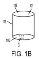
ここで、図3Aおよび図3Bを参照すると、腫瘍細胞を3Dスフェロイドコンフォメーションで成長させる構造のチャンバーを含む細胞培養製品の実施形態、例えば、スフェロイドプレートが示されている。図3Aは、スフェロイドプレート11の実施形態、この場合は、96ウェルの各々に単一のスフェロイドを含むように構成された丸みを帯びた底部119を有する96ウェルプレートを示す。通常、これらのプレートは、ウェル101の上部開口部118が上向きで使用されるが、図3Aでは、プレートは、ウェル101の底部の構造を示すために上下逆で示されている。図3Bは、フレーム130、複数のウェル101であって、各々が上部開口部118、側壁121、および液体不透過性陥凹弓状底面119を有するスフェロイドマイクロプレートの実施形態の図である。3D腫瘍細胞スフェロイド25はそれぞれ個別のウェル101の底部に示されている。実施形態において、フレーム130は、実験台またはテーブルなどの表面の上方でウェルの底部を保持し得る。いくつかの実施形態において、ウェルの底部119とプレートの下の表面との間に空間が設けられていてもよい。実施形態において、その空間は、外部環境と連通していてもよいし、または閉じられていてもよい。
Referring now to Figures 3A and 3B, there is shown an embodiment of a cell culture article, eg, a spheroid plate, comprising chambers structured to grow tumor cells in a 3D spheroid conformation. FIG. 3A shows an embodiment of
実施形態において、チャンバーの少なくとも1つの陥凹弓状底面は、例えば、同じウェル内に複数の隣接する陥凹弓状底面を有し得る。または、図1に示すように、マルチウェルプレートは、同じウェル内に隣接する陥凹弓状底面またはマイクロキャビティのアレイを有する平坦な底面を有するウェルを有し得る。実施形態において、細胞培養製品は、例えば、各ウェルの底部または基部に複数のディンプルまたはピットなどの多くの「スフェロイドウェル」を有する単一のウェルまたはマルチウェルプレート構造、例えば、マイクロキャビティスフェロイドプレートであり得る。チャンバー当たりの複数のスフェロイドまたはスフェロイドウェルは、好ましくは、例えば、スフェロイドウェルあたり単一または1つのスフェロイドを収容し得る。 In embodiments, at least one recessed arcuate bottom of the chamber can have multiple adjacent recessed arcuate bottoms, eg, within the same well. Alternatively, as shown in FIG. 1, a multiwell plate can have flat-bottomed wells with adjacent recessed arcuate bottoms or arrays of microcavities within the same well. In embodiments, the cell culture product is a single well or multiwell plate structure, e.g., a microcavity spheroid plate, having many " spheroid wells", e.g., multiple dimples or pits at the bottom or base of each well. could be. Multiple spheroids or spheroid wells per chamber can preferably accommodate, for example, single or one spheroid per spheroid well.
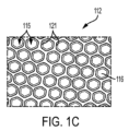
次に図1A、図1Bおよび図1Cを参照すると、マイクロキャビティスフェロイドプレートの一実施形態、この場合は、各ウェルの底面上にマイクロキャビティのアレイを有して、96ウェルの各々において複数のスフェロイドを提供している、96ウェルマイクロキャビティスフェロイドプレートが示されている。図1Aは、ウェル101のアレイを有するマルチウェルプレート10を示す。図1Bは、図1Aのマルチウェルプレート10の単一のウェル101を示す。単一のウェル101は、上部開口部118と、底面106と、側壁113とを有する。図1Cは、図1Bに示す単一のウェルの底面におけるマイクロキャビティ112のアレイを示す図1B中のボックスC中に示されるウェル101の底面106の部分の分解組立図である。マイクロキャビティ112のアレイにおける各マイクロキャビティ115は側壁121と底面116とを有する。各個別のウェル101の底部においてマイクロキャビティ112のアレイを提供する図1A、図1Bおよび図1Cに示されるマイクロキャビティスフェロイドプレートは、マルチウェルプレートの各個別のウェルの各マイクロキャビティにおいて3Dスフェロイドを成長させるために使用することができる。このタイプの容器を使用することによって、ユーザーは、マルチウェルプレートの各ウェル中で多数のスフェロイドを成長させることができ、それによって、本明細書中で提供するようなアッセイにおいて使用するために同じ培養および実験条件下で処理することができる多数のスフェロイドを提供することができる。
1A, 1B and 1C, one embodiment of a microcavity spheroid plate, in this case having an array of microcavities on the bottom surface of each well, with a plurality of spheroids in each of 96 wells. Shown is a 96-well microcavity spheroid plate providing . FIG. 1A shows a
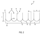
次に図2を参照すると、マイクロキャビティ112のアレイの図が示されている。図2は、各々が上部開口部118、底面119、深さd、および側壁121によって画定される幅wを有するウェル115を示す。図2に示すように、マイクロキャビティのアレイは、丸みを帯びた底部119を有する。実施形態において、マイクロキャビティの底面は、丸みを帯びているかもしくは円錐形、角度のある、平坦な底部の、または3D腫瘍スフェロイドを形成するために適した任意の形状であり得る。実施形態において、マイクロキャビティは丸みを帯びた底部を有する。丸みを帯びた底部119は、丸みを帯びた底部119に移行する垂直な側壁としての遷移ゾーン120を有し得る。これは、平滑または傾斜した遷移ゾーンであり得る。実施形態において、「マイクロキャビティ」は、例えば、上部開口部118および最下点116と、上部開口部の中心と、最下点と上部開口部の中心との間の中心軸105とを画定するマイクロウェル115であり得る。実施形態において、ウェルは、その軸の周りに回転対称である(すなわち、側壁は円筒状である)。あるいは、ウェルは、例えば、六角形などの他の形状を有していてもよい。いくつかの実施形態におい
て、上部開口部は、250μmから1mm、またはそれらの測定値内の任意の範囲の上部開口部(幅w)の直径を画定する。いくつかの実施形態において、上部開口部から最下点までの距離(深さ「d」)は200μmから900μm、または400から600μmである。マイクロキャビティのアレイは、様々な形状,例えば、放物型、双曲型、山型、および断面形状、またはそれらの組み合わせを有していてもよい。実施形態において、マイクロキャビティは、それらの下に、それらが実験台またはテーブルなどの表面と直接接触することから保護する保護層130を有していてもよい。いくつかの実施形態において、ウェルの底部119と保護層との間に空間110が設けられていてもよい。実施形態において、空間110は外部環境と連通していてもよいし、または閉じられていてもよい。
Referring now to FIG. 2, a diagram of an array of
実施形態において、少なくとも1つの陥凹弓状底面または「カップ」を有するチャンバーの底面は、例えば、半球状面、丸みを帯びた底部を有する円錐面、および同様の表面形状、またはその組み合わせであり得る。チャンバー(例えば、ウェル)およびチャンバー底部(例えば、ウェル底部またはマイクロキャビティ底部)は最終的に、ディンプル、ピット、および同様の陥凹円錐台形レリーフ面、またはそれらの組み合わせなどのスフェロイドの「やさしく」丸みを帯びたまたは湾曲した表面で、終結する、終端する、または底に達する。実施形態において、細胞培養製品のチャンバーの少なくとも1つの陥凹面は、半球状面、側壁から底面まで30から約60のテーパーを有する円錐面、またはその組み合わせを包含する。いくつかの実施形態において、少なくとも1つの陥凹弓状底面は、例えば選択されたウェル形状、各ウェル内の陥凹弓状面の数、プレート中のウェルの数、および同様の検討事項に応じて、例えば中間の値および範囲を含む約250から約5,000マイクロメートル(すなわち、0.010から0.200インチ(254から5080マイクロメートル))の直径を有する半球の水平断面またはスライスなどの半球の一部であり得る。他の陥凹弓状面は、例えば、放物型、双曲型、山型、および同様の断面形状、またはそれらの組み合わせを有し得る。 In embodiments, the bottom surface of the chamber with at least one concave arcuate bottom surface or "cup" is, for example, a hemispherical surface, a conical surface with a rounded bottom, and similar surface shapes, or combinations thereof. obtain. The chambers (e.g., wells) and chamber bottoms (e.g., well bottoms or microcavity bottoms) are ultimately spheroid "gently" rounded, such as dimples, pits, and similar depressed frusto-conical relief surfaces, or combinations thereof. Terminate, terminate or bottom out in a ridged or curved surface. In embodiments, at least one recessed surface of the chamber of the cell culture article includes a hemispherical surface, a conical surface having a taper of 30 to about 60 from the sidewall to the bottom surface, or a combination thereof. In some embodiments, the at least one recessed arcuate bottom surface is dependent, for example, on the selected well shape, the number of recessed arcuate surfaces in each well, the number of wells in the plate, and similar considerations. such as a horizontal section or slice of a hemisphere having a diameter of about 250 to about 5,000 micrometers (i.e., 0.010 to 0.200 inches (254 to 5080 micrometers)), including intermediate values and ranges. It can be part of a hemisphere. Other concave arcuate surfaces may have, for example, parabolic, hyperbolic, chevron, and similar cross-sectional shapes, or combinations thereof.
実施形態において、チャンバー、例えば、スフェロイドプレートまたはマイクロキャビティスフェロイドプレートを含む細胞培養製品は、チャンバーの一部の上、例えば少なくとも1つの陥凹面および/または1以上の側壁上に低接着性、超低接着性、または非接着性コーティングをさらに含み得る。非接着材料の例としては、パーフルオロポリマー、オレフィン、または同様のポリマー、またはそれらの混合物が挙げられる。他の例としては、アガロース、ポリアクリルアミドなどの非イオン性ヒドロゲル、またはポリエチレンオキシドなどのポリエーテルもしくはポリビニルアルコールなどのポリオール、または同様の材料、またはそれらの混合物が挙げられる。 In embodiments, a cell culture product comprising a chamber, e.g., a spheroid plate or a microcavity spheroid plate, has a low adhesion, ultra-low adhesion layer on a portion of the chamber, e.g., on at least one recessed surface and/or one or more sidewalls. Adhesive or non-adhesive coatings may be further included. Examples of non-adhesive materials include perfluoropolymers, olefins, or similar polymers, or mixtures thereof. Other examples include agarose, non-ionic hydrogels such as polyacrylamide, or polyethers such as polyethylene oxide or polyols such as polyvinyl alcohol, or similar materials, or mixtures thereof.
実施形態において、側壁面(すなわち、周囲)は、例えば、垂直円筒またはシャフト、チャンバー頂部からチャンバー底部まで直径が減少する垂直円錐の一部、円錐形遷移部を有する、すなわちウェルの頂部で方形または楕円形で、円錐に遷移し、少なくとも1つの陥凹弓状面を有する、すなわち丸みを帯びたもしくは湾曲した底部で終端する垂直方形シャフトまたは垂直楕円形シャフト、またはその組み合わせであり得る。他の例示的形状例としては、有孔円筒、有孔円錐円筒、最初は円筒で後に円錐、および他の同様の形状、またはそれらの組み合わせが挙げられる。 In embodiments, the side wall surface (i.e., the perimeter) has, for example, a vertical cylinder or shaft, a portion of a vertical cone decreasing in diameter from the top of the chamber to the bottom of the chamber, a conical transition, i.e., square or square at the top of the well. It can be an elliptical, conically transitioning, vertical square shaft or vertical elliptical shaft having at least one concave arcuate surface, ie terminating in a rounded or curved bottom, or a combination thereof. Other exemplary shapes include perforated cylinders, perforated conical cylinders, first cylindrical and later conical, and other similar shapes, or combinations thereof.
例えば、低接着(low-attachment)基体、細胞培養製品チャンバーの本体および基部におけるウェルの湾曲、ならびに重力のうちの1つ以上によって、腫瘍細胞を自己集合させてスフェロイドにすることができる。腫瘍細胞は、単層で成長した細胞と比べてよりインビボ様の応答であることを示す分化した細胞機能を維持する。実施形態において、スフェロイドは、例えばスフェロイドにおける細胞型に応じて、例えば、約100から約500マイクロメートル、さらに好ましくは約150から約400マイクロメートル、なお一層好ましくは約150から約300マイクロメートル、そして最も好ましくは約200から約250マイクロメートルであって、その間の値および範囲を含む直径を有する、例えば実質的に球であり得る。スフェロイド直径は、例えば、約200から約400マイクロメートルであり得、上側直径は拡散の検討事項によって制約を受ける。 For example, one or more of a low-attachment substrate, curvature of the wells in the body and base of the cell culture product chamber, and gravity can cause tumor cells to self-assemble into spheroids . Tumor cells maintain differentiated cellular functions indicating a more in vivo-like response compared to cells grown in monolayers. In embodiments, the spheroids are, for example, about 100 to about 500 micrometers, more preferably about 150 to about 400 micrometers, even more preferably about 150 to about 300 micrometers, and Most preferably they can be, eg, substantially spherical, with a diameter of about 200 to about 250 micrometers, including values and ranges therebetween. Spheroid diameters can be, for example, from about 200 to about 400 microns, with upper diameters constrained by diffusion considerations.
実施形態において、細胞培養製品は、不透明な側壁および/または少なくとも1つの陥凹面を含むガス透過性かつ液体不透過性底部をさらに含み得る。いくつかの実施形態において、少なくとも1つの陥凹面を含む底部の少なくとも一部は透明である。そのような特徴を有するウェルプレートは、腫瘍細胞スフェロイドを1つのマルチウェルプレート(その中でスフェロイドが形成され、可視化することができる)からアッセイ(例えば、治療薬の溶解および移動の測定)を実施するための別のプレートへ移す必要性を排除し、したがって、時間を節約し、スフェロイドのいかなる不必要な破壊も回避することをはじめとする、本開示の方法にとってのいくつかの利益を提供することができる。さらに、ガス透過性底部(例えば、特定の所与の厚さでガス透過性の特性を有するポリマーから作られたウェル底部)は、腫瘍スフェロイドが増大した酸素供給を受容することを可能にすることができる。例示的ガス透過性底部はある特定の厚さでパーフルオロポリマーまたはポリ4-メチルペンタンなどのポリマーから形成することができる。ガス透過性ポリマーの代表的な厚さおよび範囲は、例えば、約0.001インチ(25.4マイクロメートル)から約0.025インチ(635マイクロメートル)、0.0015インチ(38.1マイクロメートル)から約0.03インチ(762マイクロメートル)であり得、中間の値および範囲を含む(ここで、1インチ=25,400マイクロメートル;0.000039インチ=1マイクロメートル)。付加的または代替的に、ポリジメチルシロキサンポリマーなどの高いガス透過性を有する他の材料は、例えば、約1インチ(25,400マイクロメートル)までの厚さで十分なガス拡散を提供することができる。 In embodiments, the cell culture product may further comprise a gas permeable and liquid impermeable bottom comprising opaque sidewalls and/or at least one recessed surface. In some embodiments, at least a portion of the bottom including at least one recessed surface is transparent. Well plates with such features allow tumor cell spheroids to be assayed (e.g., measuring lysis and migration of therapeutic agents) from a single multiwell plate in which spheroids are formed and visualized. provides several benefits to the disclosed method, including eliminating the need to transfer to a separate plate for be able to. Furthermore, gas permeable bottoms (e.g., well bottoms made from polymers with gas permeable properties at a given thickness) allow tumor spheroids to receive increased oxygen supply. can be done. An exemplary gas permeable bottom can be formed from a perfluoropolymer or a polymer such as poly-4-methylpentane at a certain thickness. Typical thicknesses and ranges for gas permeable polymers are, for example, from about 0.001 inches (25.4 microns) to about 0.025 inches (635 microns), 0.0015 inches (38.1 microns) ) to about 0.03 inch (762 micrometers), including intermediate values and ranges (where 1 inch = 25,400 micrometers; 0.000039 inch = 1 micrometer). Additionally or alternatively, other materials with high gas permeability such as polydimethylsiloxane polymers can provide sufficient gas diffusion at thicknesses up to about 1 inch (25,400 micrometers), for example. can.
実施形態において、細胞培養製品は、吸引のためにピペットチップを受容するためのチャンバー付属物、チャンバー拡張領域、もしくは補助的サイドチャンバーをさらに含むことができ、このチャンバー付属物またはチャンバー拡張部(例えば、サイドポケット)は、例えば、チャンバーに隣接し、かつチャンバーと流体連通した一体型表面であり得る。チャンバー付属物は、チャンバーの液体不透過性底部から離間した第二の底部を有し得る。チャンバー付属物およびこのチャンバー付属物の第二の底部は、例えば、さらに高所にまたは相対的な高所などで、チャンバーの液体不透過性底部から離間させることができる。チャンバー付属物の第二の底部は、ピペットから分配される流体をチャンバーの液体不透過性底部からそらせて、スフェロイドの破壊またはかく乱を回避する。 In embodiments, the cell culture article may further comprise a chamber appendage, chamber extension region, or auxiliary side chamber for receiving a pipette tip for aspiration, the chamber appendage or chamber extension (e.g. , side pockets) can be, for example, a unitary surface adjacent to and in fluid communication with the chamber. The chamber appendage may have a second bottom spaced apart from the liquid impermeable bottom of the chamber. The chamber appendage and the second bottom of the chamber appendage can be spaced apart from the liquid impermeable bottom of the chamber, eg, at a higher or relative elevation. The second bottom of the chamber appendage diverts fluid dispensed from the pipette away from the liquid-impermeable bottom of the chamber to avoid breaking or disturbing the spheroids .
治療薬の移動能および細胞毒性を検出するためのアッセイ法は、細胞培養製品中に多孔質膜を含むインサートを入れ、治療薬をインサート中に導入することをさらに含む。実施形態において、多孔質膜を含むインサートは、チャンバーの一部の中に、チャンバー付属物の一部の中に、またはチャンバーおよびチャンバー付属物部分の両方に入れることができる。多孔質膜を含むインサートは、チャンバーの上部中、多孔質膜によって形成されたチャンバーの上部中、または両チャンバーに入れた免疫細胞または薬物など治療薬の、透明底部近くの一方または両方のチャンバーの下部中の腫瘍細胞スフェロイドからの単離または分離(少なくとも最初に)を提供する。 Assays for detecting migration ability and cytotoxicity of therapeutic agents further comprise placing inserts comprising porous membranes in cell culture articles and introducing therapeutic agents into the inserts. In embodiments, the insert containing the porous membrane can be placed in a portion of the chamber, in a chamber appendage portion, or in both the chamber and chamber appendage portion. Inserts containing porous membranes can be placed in the upper part of the chamber, in the upper part of the chamber formed by the porous membrane, or in one or both chambers near the transparent bottom for therapeutic agents such as immune cells or drugs placed in both chambers. Provides isolation or separation (at least initially) from tumor cell spheroids in the bottom.
図4Aおよび図4Bはインサート400の透視図である。図4Aおよび図4Bで示すインサート400はCorning Transwell(登録商標)インサートである。図4Aおよび図4Bに示されるように、インサートは、キャビティ420を形成する上部開口部418、側壁421および底面419を有する。図4Cに示されるように、これらのインサート400はインサートプレート401形態で提供することができ、この場合、単一のプレート401は複数のインサート400を含み、マルチウェルインサートプレートは、マルチウェルプレート11中のウェルの相補的(complimentary)アレイ中に挿入するような構造である。インサートは多くの形態で入手可能である。実施形態において、これらのインサートは、小分子、例えば薬物、タンパク質、ベクター、または他の材料は底面119を通過させるが、細胞は通過させないために充分な多孔性である多孔性底面を有する。さらなる実施形態において、インサートは、免疫細胞を含む細胞治療薬などの細胞が底面を通って移動するために充分な直径の細孔を有する多孔性底面419を有する。
4A and 4B are perspective views of
インサートの多孔質膜は、限定されるものではないが、トラック蝕刻(track etch)された膜または製織もしくは不織多孔性材料をはじめとする様々な材料で作ることができる。多孔質膜の材料は、細胞に対してより接着性または非接着性となるように処理もしくはコーティングすることができるか、または細胞培養を支持するための任意の他の望ましいコーティングのための処理もしくはコーティングをすることができる。処理は、プラズマ放電、コロナ放電、ガスプラズマ放電、イオン衝撃、イオン化照射、および高強度UV光を含む、当該技術分野で公知の多数の方法で実施することができる。コーティングは、印刷、噴霧、凝縮、放射エネルギー、イオン化技術または浸漬をはじめとする当該技術分野で公知の任意の好適な方法によって導入することができる。コーティングは次に共有または非共有結合部位のいずれかを提供し得る。そのような部位は、細胞培養成分(例えば、成長または接着を促進するタンパク質)などの部分を結合させるために使用できる。さらに、コーティングは、細胞の付着を増進するために使用することもできる(例えば、ポリリジン)。あるいは、上記のような細胞非付着性コーティングを使用して細胞結合を防止または阻害することができる。多孔質膜はガンマ線滅菌してもよい。そのようなインサートは、Corning(「Transwell」)またはMillipore(登録商標)(Millicell(登録商標)もしくはUltracell(登録商標))から一般に入手可能である。 The porous membrane of the insert can be made of a variety of materials including, but not limited to, track etched membranes or woven or non-woven porous materials. The material of the porous membrane can be treated or coated to be more adherent or non-adherent to cells, or treated or coated for any other desired coating to support cell culture. can be coated. Treatment can be carried out in a number of ways known in the art, including plasma discharge, corona discharge, gas plasma discharge, ion bombardment, ionizing irradiation, and high intensity UV light. Coatings can be introduced by any suitable method known in the art, including printing, spraying, condensation, radiant energy, ionization techniques or dipping. The coating can then provide either covalent or non-covalent binding sites. Such sites can be used to attach moieties such as cell culture components (eg, proteins that promote growth or adhesion). Additionally, coatings can be used to enhance cell attachment (eg, polylysine). Alternatively, a cell non-adhesive coating as described above can be used to prevent or inhibit cell binding. The porous membrane may be gamma sterilized. Such inserts are commonly available from Corning (“Transwell”) or Millipore® (Millicell® or Ultracell®).
ある態様において、多孔質膜は、当該技術分野で公知の様に、多孔質膜の少なくとも一部が血液脳関門(BBB)をシミュレートするような構造であるように処理またはコーティングしてもよい。いくつかの実施形態において、多孔質膜の少なくとも一部は、限定されるものではないが、微小血管内皮細胞などの内皮細胞の本質的にコンフルエントな単層を含む。いくつかの実施形態において、微小血管内皮細胞は脳内皮細胞である。いくつかの実施形態において、微小血管内皮細胞に含まれる内皮細胞は、星状細胞および/または周皮細胞と組み合わせて用いられる。そのようなモデルは、脳自体の防御システムとして作用するBBBのために治療するのが最も困難ながんの1つである脳癌を研究するために有用である。脳を潜在的な毒素から保護するはずのBBBは、多くの場合、化学療法などの通常の療法が脳腫瘍に到達するのを妨害する。伝統的に、放射性標識またはフルオロフォア標識化合物が透過性支持体システム上で成長する細胞単層を通過するので、インビトロでのBBBによる化合物透過性高スループット試験は放射性標識またはフルオロフォア標識化合物のアッセイに限定されてきた。残念なことに、標識自体がアッセイに影響を及ぼす可能性があり、結果としての腫瘍細胞毒性を判定する能力を独立して調査しなければならない。ここで開示される方法およびデータは、治療薬(例えば、細胞治療薬、薬物、または生物剤)のBBB輸送ならびに結果としての治療薬の脳腫瘍細胞毒性を研究するための三次元(3D)モデルを示し、よりインビボ様の試験のための単一で使いやすい高スループットシステムにおけるホーミング、腫瘍細胞毒性、および腫瘍免疫回避の調査を可能にする。2Dで培養した腫瘍細胞を利用する免疫細胞移動および侵襲性アッセイのためのほとんどの現行の高スループットモデルとは異なり、このモデルはよりインビボ様の試験のための3D腫瘍スフェロイド成分を可能にする。 In some embodiments, the porous membrane may be treated or coated such that at least a portion of the porous membrane is structured to simulate the blood-brain barrier (BBB), as known in the art. . In some embodiments, at least a portion of the porous membrane comprises an essentially confluent monolayer of endothelial cells, such as but not limited to microvascular endothelial cells. In some embodiments, the microvascular endothelial cells are brain endothelial cells. In some embodiments, endothelial cells, including microvascular endothelial cells, are used in combination with astrocytes and/or pericytes. Such models are useful for studying brain cancer, one of the most difficult cancers to treat due to the BBB acting as the brain's own defense system. The BBB, which is supposed to protect the brain from potential toxins, often prevents conventional therapies such as chemotherapy from reaching brain tumors. High-throughput testing of compound permeability by BBB in vitro is an assay for radiolabeled or fluorophore-labeled compounds, as traditionally radiolabeled or fluorophore-labeled compounds pass through cell monolayers grown on permeable support systems. has been limited to Unfortunately, the label itself can affect the assay and the ability to determine the resulting tumor cytotoxicity must be independently investigated. The methods and data disclosed herein provide a three-dimensional (3D) model for studying BBB transport of therapeutics (e.g., cell therapeutics, drugs, or biological agents) and the resulting brain tumor cytotoxicity of therapeutics. demonstrate and allow investigation of homing, tumor cytotoxicity, and tumor immune evasion in a single, easy-to-use, high-throughput system for more in vivo-like studies. Unlike most current high-throughput models for immune cell migration and invasiveness assays that utilize tumor cells cultured in 2D, this model allows 3D tumor spheroid components for more in vivo-like studies.
治療薬の移動能および細胞毒性を検出するための本開示のアッセイ法の特定の態様において、本開示は、アッセイを行うための環境を提供するための、Corning 96HTS Transwell透過性支持体システムと組み合わせたCorningスフェロイドマイクロプレートの使用を提供する。Corningスフェロイドマイクロプレートは丸底ウェル形状を有する複数ウェル細胞培養マイクロプレートであって、Corning Ultra-Low Attachment表面でコーティングされているので、各ウェルの中心に置かれた高度に再現性のある単一多細胞腫瘍スフェロイドを形成することができる。Corning 96 HTS Transwellは、高スループット薬物輸送、ならびに細胞移動および侵襲研究のために使用される透過性支持体である。 In certain aspects of assays of the present disclosure for detecting mobilization and cytotoxicity of therapeutic agents, the present disclosure is combined with the Corning 96HTS Transwell permeable support system to provide an environment for performing the assays. We provide the use of Corning spheroid microplates. Corning spheroid microplates are multi-well cell culture microplates with a round-bottom well geometry that are coated with a Corning Ultra-Low Attachment surface to provide a highly reproducible single cell centered in each well. Multicellular tumor spheroids can be formed. The Corning 96 HTS Transwell is a permeable support used for high-throughput drug delivery and cell migration and invasion studies.
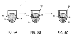
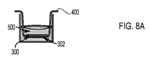
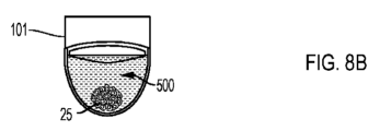
図5Aから5Cは、治療薬の移動能および細胞毒性を検出するための本開示方法の実施形態を示す。図5Aで示されるように、腫瘍細胞などの関心対象の細胞525を3Dコンフォメーションで成長させる構造のウェル101中の培地500において関心対象の細胞525を成長させる。この場合ウェル101は、単一の単一スフェロイドウェルである。実施形態において、ウェル101は例えば、Corning96ウェルスフェロイドプレートの1つのウェルなどの、96ウェルプレートの1つのウェルである。実施形態において、関心対象の細胞525、例えば腫瘍細胞は、マルチウェルプレート(図1Cを参照)のウェル中で成長させることができ、各ウェル101はマイクロキャビティ112のアレイを有し、各マイクロキャビティは、関心対象の細胞を3Dコンフォメーションで成長させる構造であり、その結果、マルチウェルプレートのウェルの底面上のマイクロキャビティのアレイ中のマイクロキャビティの各々に1つずつ、スフェロイドのアレイが生じる。細胞は成長して培養中で繁殖するにつれ、それらはスフェロイド25として成長させられる。時間とともに、スフェロイド25が発生する。図5Aはスフェロイド25の形成を示す。腫瘍細胞がスフェロイド25に発達したら、図5Bに示すように、細胞培養インサート400をウェル101に入れる。実施形態では、インサートは図4Bに示すようなインサートプレートであってよい。治療薬(例えば、細胞治療薬、薬物、または生物剤)、この場合では細胞治療薬30をインサート400のキャビティ420に添加する。この形態で、治療薬細胞30などの治療薬(例えば、細胞治療薬、薬物、または生物剤)を含むインサート、および3D腫瘍スフェロイドなどの3Dコンフォメーションの関心対象の細胞525を含むウェル101を一緒にインキュベートする。次に、好適な時間の後、治療薬(例えば、細胞治療薬、薬物、または生物剤)、この場合は細胞治療薬30の、3D腫瘍スフェロイドなどの関心対象の細胞に対する効果、ならびにインサートから細胞培養製品のアッセイチャンバーへの治療薬の移動をアッセイで測定する。このインキュベーション期間は、実施するアッセイのタイプ、このアッセイで使用する治療薬、例えば、細胞治療薬、薬物、または生物剤に応じて変わる。このインキュベーション期間はまた、インサート400中の膜について用いられる細孔サイズによっても変わる。細孔サイズが大きいほど治療薬は迅速に分配され、したがってインキュベーション期間は短くなるが、非特異的結果となる可能性がある。インキュベーション時間の調節は、事前の実験で実施することができ、当業者の一般的な技術範囲内である。図5Cで示すように、治療薬30の腫瘍スフェロイド25に対する効果を次に測定することができる。例えば、図5C中、実施形態において、アッセイはスフェロイド25の溶解を測定することができる。
Figures 5A through 5C show embodiments of the disclosed method for detecting mobilization and cytotoxicity of therapeutic agents. As shown in FIG. 5A, cells of
図8A、8B、8C、8D、8E、8Fおよび8Gは、インサート400の一部である多孔質膜302上の細胞300の層の使用を含むモデルにおいて治療薬30の移動能および細胞毒性を検出するための本開示の方法の実施形態の図である。これらの実施形態において、インサート400の多孔質膜302上の細胞300の層は、血液脳関門(BBB)をシミュレートするように構成されている。
8A, 8B, 8C, 8D, 8E, 8F and 8G detect the mobilization and cytotoxicity of
まず、図8Aに示すように、BBBをシミュレートするために使用する細胞300を培養培地500中の細胞培養インサート400の多孔質膜302上に播種する。これらの細胞は、例えば、内皮細胞である。ある時間の後、播種した細胞300、例えば内皮細胞は、BBBをシミュレートする細胞306のコンフルエントな単層(2D層)を形成する。一旦、細胞306のコンフルエントな単層が形成されたら、インサート400は、図8Cに示すようにスフェロイドウェル101に入れることができる。
First, the
図5と同様に、腫瘍細胞などの関心対象の細胞はまた、図8Bに示すように、関心対象の細胞を3Dコンフォメーションで成長させてスフェロイド25を形成させるような構造のウェル101中の培地500中で成長させる。図8Bで示すように、ウェル101は、単一の、単一スフェロイドウェルである。実施形態において、腫瘍細胞などの関心対象の細胞は、各々のマイクロキャビティが関心対象の細胞を3Dコンフォメーションで成長させる構造の(図1Cを参照)、マイクロキャビティのアレイを有するマルチウェルプレートのウェル中で成長させることができ(図1A、1Bおよび1Cに示す通り)、その結果、マルチウェルプレートのウェルの底面上のマイクロキャビティのアレイのマイクロキャビティのそれぞれに1つずつ、多数のスフェロイドが生じる。細胞が成長し、培養中で増殖する際、図8Bに示すように、それらはスフェロイドとして成長させられ、スフェロイド25が生じる。腫瘍細胞がスフェロイド25になり、BBBをシミュレートする内皮細胞306のコンフルエントな単層が形成されたら、図8Cに示すように細胞培養インサート400をウェル101に入れる。
Similar to FIG. 5, cells of interest, such as tumor cells, may also be placed in media in
また図8Cを参照して、図5と同様に、治療薬(例えば、細胞治療薬、薬物、または生物剤)、この場合は薬物310をインサート400のキャビティ312に添加する。この形態で、治療薬310(例えば、細胞治療薬、薬物、または生物剤)、この場合は薬物310を含むインサート400と、3D腫瘍スフェロイド25などの3Dコンフォメーションの関心対象の細胞を含むウェルを一緒にインキュベートする。これを図8Dおよび図8Eに示す。インキュベーション期間は、アッセイで使用する治療薬、例えば、細胞治療薬、薬物、または生物剤によって変わる。これはまた、インサート400中の膜302に使用する細孔サイズによっても変わる。細孔サイズが大きいほど治療薬310の分配が迅速になり、したがってインキュベーション期間が短くなるが、非特異的結果となる可能性がある。インキュベーション時間の調節は、事前の実験で行うことができ、当業者の通常の技術範囲内である。血液脳関門(BBB)をシミュレートするように構成された多孔質膜302上の細胞306の層を使用して、治療薬が血液脳関門を越える能力を評価することができる。
8C, a therapeutic agent (eg, a cytotherapeutic agent, drug, or biological agent), in this
次に、好適な時間ののち、治療薬310の、3D腫瘍スフェロイド25などの関心対象の細胞に対する効果を測定することができる。例えば、図8Fに示すように、スフェロイド25の破壊または溶解が示される。この破壊は、細胞培養製品のアッセイチャンバー315中のスフェロイド25の一体性における変化を測定することによって測定することができる。あるいは、図8Gに示すようにスフェロイド25は、インキュベーション期間後にインタクトであることが示される一方で、細胞機能の測定を行って、スフェロイド25を構成する細胞の生理機能における変化を測定することができる。例えば、薬物310の細胞毒性効果または薬物310の3D腫瘍スフェロイド25への浸透は、腫瘍細胞生理学における変化を測定することによって測定することができる。
After a suitable period of time, the effect of
インサート400から細胞培養製品のアッセイチャンバー315への治療薬310の移動を測定することができる。例えば、治療薬310をデバイスのインサートチャンバー312に導入し、次いでアッセイチャンバー315から測定される薬物310の濃度に関して評価を行い、どれほど多くの薬物310が血液脳関門を通過するかを判定する。
The migration of
3D腫瘍スフェロイドに対する効果およびインサートから細胞培養製品のチャンバーへの治療薬の移動は、細胞、細胞抽出物、または培地の可視化、蛍光測定、遺伝子、代謝またはタンパク質分析をはじめとする当該技術分野で公知の任意の手段によって測定することができる。例えば、免疫細胞の移動(治療薬細胞が免疫細胞である場合)および細胞毒性(または腫瘍細胞の生理機能における変化)を、例えば限定されるものではないが、当該技術分野で公知の様な適切な染色を使用することによるフローサイトメトリーによって評価することができ、本明細書中の実施例で使用することができる。さらに一例として、放射標識された薬物もしくは生物剤または蛍光標識された薬物もしくは生物剤などを含む標識された薬物もしくは生物剤を使用することによって薬物もしくは生物剤の移動を測定することができる。治療薬、例えば、細胞治療薬、薬物、または生物剤の、3D腫瘍スフェロイドへの浸透または腫瘍細胞溶解は、例えば限定されるものではないが、可視化および/または蛍光測定によって検出することができる。例えば、3D腫瘍スフェロイドおよび浸潤細胞を固定し分割することができ、細胞を、当該技術分野で公知の様に従来型組織学的技術によって染色することができる。 Effects on 3D tumor spheroids and transfer of therapeutic agents from inserts to chambers of cell culture articles are known in the art, including visualization of cells, cell extracts, or media, fluorescence measurements, genetic, metabolic or protein analysis. can be measured by any means of For example, immune cell migration (if the therapeutic cell is an immune cell) and cytotoxicity (or changes in tumor cell physiology) can be mediated by suitable methods such as, but not limited to, those known in the art. staining can be assessed by flow cytometry and can be used in the Examples herein. Further by way of example, drug or biological agent translocation can be measured by using labeled drugs or biological agents, including radiolabeled drugs or biological agents, fluorescently labeled drugs or biological agents, and the like. Penetration or tumor cell lysis of a therapeutic agent, such as a cytotherapeutic agent, drug, or biological agent, into a 3D tumor spheroid can be detected, for example, but not limited to, by visualization and/or fluorometry. For example, 3D tumor spheroids and infiltrating cells can be fixed and divided, and the cells can be stained by conventional histological techniques as known in the art.
様々な腫瘍細胞タイプを培養して3D腫瘍スフェロイド25を形成することができる。培養して3D腫瘍スフェロイド25を形成することができる腫瘍細胞タイプに用いられるがん細胞としては、腫瘍、新生物、がん、前がん状態、細胞株に由来する任意の細胞、または無制限に拡大し成長する可能性を有する任意の他の細胞源が挙げられる。がん細胞は自然発生源由来であるか、または人工的に生成される。がん細胞は動物宿主に入れられた場合に他の組織に浸潤し転移することができる。がん細胞は、他の組織を侵害し、および/または転移した任意の悪性細胞をさらに包含する。生命体の関連での1以上のがん細胞はまた、がん、腫瘍、新生物、成長、悪性腫瘍、または癌性状態の細胞を記述するために当該技術分野で使用される任意の他の用語でも呼ぶことができる。
Various tumor cell types can be cultured to form 3D tumor spheroids25 . Cancer cells used for tumor cell types that can be cultured to form
培養して3D腫瘍スフェロイド25を形成することができる腫瘍細胞型源としての働きをするがんとしては、限定されるものではないが、固形腫瘍、例えば線維肉腫、粘液肉腫、脂肪肉腫、軟骨肉腫、骨肉腫、脊索腫、血管肉腫、内皮肉腫(endotheliosarcoma)、リンパ管肉腫、リンパ管内皮肉腫(lymphangioendotheliosarcoma)、滑液腫瘍、中皮腫、ユーイング腫瘍、平滑筋肉腫、横紋筋肉腫、結腸がん、結腸直腸がん、腎臓がん、膵臓がん、骨がん、脳癌、乳がん、卵巣がん、前立腺がん、食道がん、胃がん、口腔がん、鼻腔がん、咽喉がん、扁平上皮がん、基底細胞がん、腺がん,汗腺がん、皮脂腺がん、乳頭がん、乳頭腺がん、嚢胞腺がん、髄様がん、気管支がん、腎細胞がん、肝がん、胆管がん、絨毛腫、セミノーマ、胎生期がん、ウィルムス腫瘍、子宮頸がん、子宮がん、精巣がん、小細胞肺がん、膀胱がん、肺がん、上皮がん、神経膠腫、多形成膠芽腫、星状細胞腫、髄芽腫、頭蓋咽頭腫、上衣腫、松果体腫、血管芽細胞腫、聴神経腫、乏突起膠腫、髄膜腫、皮膚がん、黒色腫、神経芽細胞腫、および網膜芽腫が挙げられる。
Cancers that serve as sources of tumor cell types that can be cultured to form
がん細胞源としての働きをするさらなるがんには、限定されるものではないが、血液感染性がん、例えば急性リンパ芽球性白血病、急性リンパ芽級性B細胞白血病、急性リンパ芽級性T細胞白血病、急性骨髄芽球性白血病、急性前骨髄性白血病、急性単芽球性白血病、急性赤白血病性白血病(erythroleukemic leukemia)、急性巨核芽球性白血病、急性骨髄単球性白血病、急性非リンパ球性白血病(nonlymphocyctic leukemia)、急性未分化白血病、慢性骨髄性白血病、慢性リンパ性白血病、有毛細胞白血病、多発性骨髄腫,リンパ芽球性白血病、骨髄性白血病、リンパ性白血病、骨髄性白血病、ホジキン病、非ホジキンリンパ腫、ヴァルデンストレームマクログロブリン血症、重鎖病、および真性赤血球増加症が含まれる。本明細書中で開示する治療薬の移動能および細胞毒性を検出するための方法のいくつかの実施形態において、3D腫瘍スフェロイドは1以上のがん細胞を含む。いくつかの実施形態では、がん細胞を凍結保存する。いくつかの実施形態において、1以上の細胞は活発に分裂している。 Additional cancers that serve as cancer cell sources include, but are not limited to, blood-borne cancers such as acute lymphoblastic leukemia, acute lymphoblastic grade B-cell leukemia, acute lymphoblastic grade acute myeloblastic leukemia, acute promyelocytic leukemia, acute monoblastic leukemia, erythroleukemic leukemia, acute megakaryoblastic leukemia, acute myelomonocytic leukemia, acute Nonlymphocytic leukemia, acute undifferentiated leukemia, chronic myeloid leukemia, chronic lymphocytic leukemia, hairy cell leukemia, multiple myeloma, lymphoblastic leukemia, myeloid leukemia, lymphocytic leukemia, bone marrow leukemia, Hodgkin's disease, non-Hodgkin's lymphoma, Waldenström's macroglobulinemia, heavy chain disease, and polycythemia vera. In some embodiments of the methods for detecting migration ability and cytotoxicity of therapeutic agents disclosed herein, the 3D tumor spheroids comprise one or more cancer cells. In some embodiments, cancer cells are cryopreserved. In some embodiments, one or more cells are actively dividing.
いくつかの実施形態において、方法は、培養培地(例えば、当該技術分野で公知の栄養素(例えば、タンパク質、ペプチド、アミノ酸)、エネルギー(例えば、炭水化物)、必須金属およびミネラル(例えば、カルシウム、マグネシウム、鉄、リン酸塩、硫酸塩)、緩衝剤(例えば、リン酸塩、酢酸塩)、pH変化の指示薬(例えば、フェノールレッド、
ブロモクレゾールパープル)、選択剤(例えば、化学薬品、抗菌薬)などを含む)を含む。
In some embodiments, the methods include culture media (e.g., nutrients known in the art (e.g., proteins, peptides, amino acids), energy (e.g., carbohydrates), essential metals and minerals (e.g., calcium, magnesium, iron, phosphates, sulfates), buffers (e.g. phosphates, acetates), pH change indicators (e.g. phenol red,
bromocresol purple), selective agents (eg, chemicals, antimicrobials), etc.).
治療薬の移動能および細胞毒性を検出するための本開示の方法のいくつかの実施形態において、治療薬は細胞治療薬、薬物,および/または生物剤である。 In some embodiments of the disclosed methods for detecting mobilization and cytotoxicity of a therapeutic agent, the therapeutic agent is a cytotherapeutic agent, drug, and/or biological agent.
いくつかの実施形態において、細胞治療薬は免疫細胞を含む。いくつかの実施形態において、免疫細胞は白血球(例えば、好中球、マクロファージ、樹状細胞、または単球)である。いくつかの実施形態において、免疫細胞はリンパ球(例えば、ナチュラルキラー細胞、またはリンパ球、例えばT細胞、B細胞、そしてさらに詳細には細胞傷害性T細胞)である。 In some embodiments, cell therapeutic agents comprise immune cells. In some embodiments, immune cells are leukocytes (eg, neutrophils, macrophages, dendritic cells, or monocytes). In some embodiments, the immune cells are lymphocytes (eg, natural killer cells, or lymphocytes such as T cells, B cells, and more particularly cytotoxic T cells).
公知抗がん薬または新規分子的実体もしくは新規化学物質などの潜在的な抗がん活性があると思われる物質を含む治療薬の移動能および細胞毒性を検出するための本開示の方法において、様々な薬物310を試験することができる。抗がん薬としては、限定されるものではないが:アシビシン;アクラルビシン;塩酸アコダゾール(acodazole hydrochloride);アクロニン;アドゼレシン;アルデスロイキン;アルトレタミン;アンボマイシン;酢酸アメタントロン;アミノグルテチミド;アムサクリン;アナストロゾール;アントラマイシン;アスパラギナーゼ;アスペルリン;アザシチジン;アゼテパ(azetepa);アゾトマイシン;バチマスタット;ベンゾデパ(benzodepa);ビカルタミド;塩酸ビスアントレン;ジメシル酸ビスナフィド(ビスナフィド dimesylate);ビゼレシン;硫酸ブレオマイシン;ブレキナルナトリウム(brequinar sodium);ブロピリミン(bropirimine);ブスルファン;カクチノマイシン;カルステロン;カラセミド;カルベチマー;カルボプラチン;カルムスチン;塩酸カルビシン;カルゼルシン;セデフィンゴール;クロラムブシル;シロレマイシン(cirolemycin);シスプラチン;クラドリビン;メシル酸クリスナトール;シクロホスファミド;シタラビン;ダカルバジン;ダクチノマイシン;塩酸ダウノルビシン;デシタビン;デキソルマプラチン;デザグアニン;メシル酸デザグアニン;ジアジクオン;ドセタキセル;ドキソルビシン;塩酸ドキソルビシン;ドロロキシフェン;クエン酸ドロロキシフェン;プロピオン酸ドロモスタノロン;デュアゾマイシン(duazomycin);エダトレキサート;塩酸エフロミチン(eflomithine);エルサミトルシン(elsamitrucin);エンロプラチン;エンプロマート;エピプロピジン;塩酸エピルビシン;エルブロゾール;塩酸エソルビシン;エストラムスチン;リン酸エストラムスチンナトリウム;エタニダゾール;エトポシド;リン酸エトポシド;エトプリン;塩酸ファドラゾール(ファドロゾール);ファザラビン;フェンレチニド(fenretinide);フロクスウリジン(floxuridine);リン酸フルダラビン;フルオロウラシル;フルロシタビン;ホスキドン;ホストリエシン(fostriecin)ナトリウム;ゲムシタビン;塩酸ゲムシタビン;ヒドロキシウレア;塩酸イダルビシン;イホスファミド;イルモホシン;インターロイキンII(組換えインターロイキンII、またはrIL2を含む)、インターフェロンα-2a;インターフェロンα-2b;インターフェロンα-n1;インターフェロンα-n3;インターフェロンβ-Ia;インターフェロンγ-Ib;イプロプラチン;塩酸イリノテカン;酢酸ランレオチド;レトロゾール;酢酸リュープロレリン;塩酸リアロゾール;ロメトレキソールナトリウム;ロムスチン;塩酸ロソキサントロン;マソプロコール;マイタンシン;塩酸メクロレタミン;酢酸メゲストロール;酢酸メレンゲストロール;メルファラン;メノガリル;メルカプトプリン;メトトレキサート;メトトレキサートナトリウム;メトプリン;メツレデパ;ミチンドミド;ミトカルシン(mitocarcin);マイトクロミン(mitocromin);マイトジリン;マイトマルシン(mitomalcin);マイトマイシン;マイトスパー(mitosper);ミトタン;塩酸ミトキサントロン;ミコフェノール酸;ノコダゾール;ノガラマイシン;オルマプラチン;オキシスラン;パクリタキセル;ペガスパルカーゼ;ペリオマイシン(peliomycin);ペンタムスチン(pentamustine);硫酸ペプロマイシン;ペルフォスファミド;ピポブロマン;ピポスルファン;塩酸ピロキサントロン(piroxantrone);プリカマイシン;プロメスタン;ポルフィマーナトリウム;ポルフィロマイシン;プレドニムスチン;塩酸プロカルバジン;ピューロマイシン;塩酸ピューロマイシン;ピラゾフリン;リボプリン;ログレチミド(rogletimide);サフィンゴール;塩酸サフィンゴール;セムスチン;シムトラゼン;スパルフォサーテ(sparfosate)ナトリウム;スパルソマイシン;塩酸スピロゲルマニウム;スピロムスチン;スピロプラチン;ストレプトニグリン;ストレプトゾシン;スルフェヌル;タリソマイシン;テコガランナトリウム;テガフール;塩酸テロキサントロン;テモポルフィン;テニポシド;テロキシロン;テストラクトン;チアミプリン;チオグアニン;チオテパ;チアゾフリン;チラパザミン;クエン酸トレミフェン;酢酸トレストロン;リン酸トリシリビン;トリメトレキサート;グルクロン酸トリメトレキサート;トリプトレリン;塩酸ツブロゾール;ウラシルマスタード;ウレデパ;バプレオチド;ベルテポルフィン(verteporfin);硫酸ビンブラスチン;硫酸ビンクリスチン;ビンデシン;硫酸ビンデシン;硫酸ビネピジン;硫酸ビングリシナート;硫酸ビンロイロシン(vinleurosine);酒石酸ビノレルビン;硫酸ビンロシジン(vinrosidine);硫酸ビンゾリジン;ボロゾール;ゼニプラチン;ジノスタチン;塩酸ゾルビシンが挙げられる。他の抗がん薬としては、限定されるものではないが、20-エピ-1,25ジヒドロキシビタミンD3;5-エチニルウラシル;アビラテロン;アクラルビシン;アシルフルベン;アデシペノール;アドゼレシン;アルデスロイキン;ALL-TKアンタゴニスト;アルトレタミン;アンバムスチン;アミドックス;アミフォスチン;アミノレブリン酸;アムルビシン;アムサクリン;アナグレリド;アナストロゾール;アンドログラホリド;血管新生阻害剤;アンタゴニストD;アンタゴニストG;アンタレリックス;抗背側化(anti-dorsalizing)形態形成タンパク質-1;抗アンドロゲン、前立腺がん;抗エストロゲン;抗新生物薬;アンチセンスオリゴヌクレオチド;グリシン酸アフィジコリン;アポトーシス遺伝子モジュレータ;アポトーシス調節因子;アプリン酸;アラ-CDP-DL-PTBA;アルギニンデアミナーゼ;アスラクリン;アタメスタン;アトリムスチン;アキシナスタチン1;アキシナスタチン2;アキシナスタチン3;アザセトロン;アザトキシン;アザチロシン;バッカチンIII誘導体;バラノール;バチマスタット;BCR/ABLアンタゴニスト;ベンゾクロリン(benzochlorin);ベンゾイルスタウロスポリン;βラクタム誘導体;β-アレチン;ベタクラマイシンB;ベツリン酸;bFGF阻害剤;ビカルタミド;ビスアントレン;ビスアジリジニルスペルミン;ビスナフィド;ビストラテンA;ビゼレシン;ブレフラート;ブロピリミン;ブドチタン;ブチオニンスルホキシミン;カルシポトリオール;カルホスチンC;カンプトテシン誘導体;カナリポックスIL-2;カペシタビン;カルボキサミド-アミノ-トリアゾール;カルボキシアミドトリアゾール;CaRest M3;CARN 700;軟骨由来阻害剤;カルゼルシン;カゼインキナーゼ阻害剤(ICOS);カスタノスペルミン;セクロピンB;セトロレリクス;クロリン;クロロキノキサリンスルホンアミド;シカプロスト;シス-ポルフィリン;クラドリビン;クロミフェンアナログ;クロトリマゾール;コリスマイシン(collismycin)A;コリスマイシンB;コンブレタスタチンA4;コンブレタスタチンアナログ;コナゲニン;クランベシジン816;クリスタノール;クリプトフィシン8;クリプトフィシンA誘導体;クラシンA;シクロペンタアントラキノン(cyclopentanthraquinones);シクロプラタム;シペマイシン(絨毛膜);シタラビンオクホスファート;細胞溶解因子;シトスタチン;ダクリキシマブ;デシタビン;デヒドロジデムニンB;デスロレリン;デキサメタゾン;デキシホスファミド;デキソラゾキサン;デクスベラパミル;ジアジクオン;ジデムニンB;ジドクス(didox);ジエチルノルスペルミン;ジヒドロ-5-アザシチジン;9-ジヒドロタキソール;ジオキサマイシン;ジフェニルスピロムスチン;ドセタキセル;ドコサノール;ドラセトロン;ドキシフルリジン;ドロロキシフェン;ドロナビノール;デュオカルマイシンSA;エブセレン;エコムスチン;エデルホシン;エドレコロマブ;エフロルニチン;エレメン;エミテフル;エピルビシン;エプリステリド;エストラムスチンアナログ;エストロゲンアゴニスト;エストロゲンアンタゴニスト;エタニダゾール;リン酸エトポシド;エキセメスタン;ファドロゾール;ファザラビン;フェンレチニド;フィルグラスチム;フィナステリド;フラボピリドール;フレゼラスチン;フルアステロン;フルダラビン;塩酸フルオロダウノルニシン;ホルフェニメクス;フォルメスタン;ホストリエシン;フォテムスチン;ガドリニウムテキサフィリン;硝酸ガリウム;ガロシタビン(galocitabine);ガニレリクス;ゼラチナーゼ阻害剤;ゲムシタビン;グルタチオン阻害剤;ヘプスルファム;ヘレグリン;ヘキサメチレンビスアセトアミド;ヒペリシン;イバンドロン酸;イダルビシン;イドキシフェン;イドラマントン(idramantone);イルモホシン;イロマスタット;イミダゾアクリドン(imidazoacridones);イミキモド;免疫賦活薬ペプチド;インスリン様成長因子1受容体阻害剤;インターフェロンアゴニスト;インターフェロン;インターロイキン;ヨーベングアン;ヨードドキソルビシン;4-イポメアナール;イロプラクト(iroplact);イルソグラジン;イソベンガゾール;イソホモハリコドリン(isohomohalicondrin)B;イタセトロン;ジャスプラキノリド(jasplakinolide);カハラリド(kahalalide)F;ラメラリン-Nトリアセテート;ランレオチド;レイナマイシン;レノグラスチム;硫酸レンチナン;レプトルスタチン;レトロゾール;白血病阻止因子;白血球αインターフェロン;リュープロレリン+エストロゲン+プロゲステロン;リュープロレリン;レバミゾール;リアロゾール;線状ポリアミンアナログ;親油性二糖ペプチド;親油性白金化合物;リソクリナミド(lissoclinamide)7;ロバプラチン;ロンブリシン;ロメトレキソール;ロニダミン;ロソキサントロン;ロバスタチン;ロキソルビン(loxoribine);ラルトテカン;ルテチウムテキサフィリン;リソフィリン(lysofylline);溶解性ペプチド;マイタンシン(maitansine);マンノスタチンA;マリマスタット(marimastat);マソプロコール;マスピン(maspin);マトリリシン(matrilysin)阻害剤;マトリックスメタロプロテイナーゼ阻害剤;メノガリル;メルバロン;メテレリン;メチオニナーゼ;メトクロプラミド;MIF阻害剤;ミフェプリストン;ミルテホシン;ミリモスチム;ミスマッチ二本鎖RNA;ミトグアゾン;ミトラクトール;マイトマイシンアナログ;ミトナフィド(mitonafide);マイトトキシン線維芽細胞成長因子-サポリン;ミトキサントロン;モファロテン(mofarotene);モルグラモスチム;モノクローナル抗体ヒト絨毛膜ゴナドトロピン;モノホスホリル脂質A+ミオバクテリア(myobacterium)細胞壁抽出物;モピダモール;多剤耐性遺伝子阻害剤;多腫瘍抑制因子1系療法;マスタード抗がん剤;ミカペルオキシドB;マイコバクテリア細胞壁抽出物;ミリアポロン;N-アセチルジナリン;N-置換ベンズアミド;ナファレリン;ナグレスチップ;ナロキソン+ペンタゾシン;ナパビン(napavin);ナフテルピン(naphterpin);ナルトグラスチム;ネダプラチン;ネモルビシン;ネリドロン酸;中性エンドペプチダーゼ;ニルタミド;ニサマイシン;一酸化窒素モジュレータ;ニトロオキシド抗酸化剤;ニトルリン(nitrullyn);O6-ベンジルグアニン;オクトレオチド;
オキセノン;オリゴヌクレオチド;オナプリストン;オンダンセトロン;オンダンセトロン;オラシン(oracin);口腔サイトカインインデューサー;オルマプラチン;オサテロン;オキサリプラチン;オキサウノマイシン;パクリタキセル;パクリタキセルアナログ;パクリタキセル誘導体;パラウアミン;パルミトイルリゾキシン;パミドロン酸;パナキシトリオール;パノミフェン;パラバクチン;パゼリプチン(pazelliptine);ペガスパルカーゼ;ペルデシン;ペントサンポリスルフェートナトリウム;ペントスタチン;ペントロゾール;パーフルブロン;ペルフォスファミド;ペリリルアルコール;フェナジノマイシン;フェニルアセテート;ホスファターゼ阻害剤;ピシバニール;塩酸ピロカルピン;ピラルビシン;ピリトレキシム;プラセチンA;プラセチンB;プラスミノーゲンアクチベータ阻害剤;白金錯体;白金化合物;白金-トリアミン複合体;ポルフィマーナトリウム;ポルフィロマイシン;プレドニゾン;プロピルビス-アクリドン;プロスタグランジンJ2;プロテアソーム阻害剤;タンパク質A系免疫モジュレータ;タンパク質キナーゼC阻害剤;タンパク質キナーゼC阻害剤、微細藻類;タンパク質チロシンホスファターゼ阻害剤;プリンヌクレオシドホスホリラーゼ阻害剤;プルプリン;ピラゾロアクリジン;ピリドキシル化ヘモグロビンポリオキシエチレンコンジュゲート;rafアンタゴニスト;ラルチトレキセド;ラモセトロン;rasファルネシルタンパク質トランスフェラーゼ阻害剤;ras阻害剤;ras-GAP阻害剤;脱メチル化レテリプチン;レニウムRe186エチドロネート;リゾキシン;リボザイム;RIIレチナミド;ログレチミド;ロヒツキン(rohitukine);ロムルチド;ロキニメックス;ルビギノンB1;ルボキシル;サフィンゴール;サイントピン;SarCNU;サルコフィトール(sarcophytol)A;サルグラモスチム;Sdi1模倣物;セムスチン;老化由来(senescence derived)阻害剤1;センスオリゴヌクレオチド;シグナル変換阻害剤;シグナル変換モジュレータ;単鎖抗原結合タンパク質;シゾフィラン;ソブゾキサン;ボロカプチン酸ナトリウム(sodium borocaptate);フェニル酢酸ナトリウム;ソルベロール;ソマトメジン結合タンパク質;ソネルミン;スパルフォス酸;スピカマイシンD;スピロムスチン;スプレノペンチン;スポンジスタチン1;スクワラミン;幹細胞阻害剤;幹細胞分裂阻害剤;スチピアミド;ストロメリシン阻害剤;スルフィノシン;超活性血管作動性腸ペプチドアンタゴニスト;スラジスタ;スラミン;スワインソニン;合成グリコサミングリカン;タリムスチン;タモキシフェンメチオジド;タウロムスチン;タザロテン;テコガランナトリウム;テガフール;テルラピリリウム;テロメラーゼ阻害剤;テモポルフィン;テモゾロマイド;テニポシド;テトラクロロデカオキシド;テトラゾミン(tetrazomine);タリブラスチン(thaliblastine);チオコラリン;トロンボポエチン;トロンボポエチン模倣物;チマルファシン;チモポエチン受容体アゴニスト;チモトリナン;甲状腺刺激ホルモン;エチルエチオプルプリンスズ;チラパザミン;チタノセンジクロリド;トプセチン;トレミフェン;分化全能性幹細胞因子;翻訳阻害剤;トレチノイン;トリアセチルウリジン;トリシリビン;トリメトレキサート;トリプトレリン;トロピセトロン;ツロステリド;チロシンキナーゼ阻害剤;チルホスチン(tyrphostins);UBC阻害剤;ウベニメクス;尿生殖洞由来の成長阻害因子;ウロキナーゼ受容体アンタゴニスト;バプレオチド;バリオリン(variolin)B;ベクターシステム、赤血球遺伝子療法;ベラレソール(velaresol);べラミン(veramine);ベルディンス(verdins);ベルテポルフィン;ビノレルビン;ビンキサルチン;ビタキシン(vitaxin);ボロゾール;ザノテロン;ゼニプラチン;ジラスコルブ;およびジノスタチンスチマラマーが挙げられる。
In the methods of the present disclosure for detecting the locomotion and cytotoxicity of therapeutic agents, including agents suspected of potential anticancer activity such as known anticancer agents or novel molecular entities or novel chemical entities,
Oxenone; Oligonucleotide; Onapristone; Ondansetron; Ondansetron; Oracin; Oral Cytokine Inducer; pamidronic acid; panaxytriol; panomiphene; parabactin; pazelliptine; phosphatase inhibitors; picibanil; pilocarpine hydrochloride; pirarubicin; pyritrexime; placetin A; protein A-based immune modulators; protein kinase C inhibitors; protein kinase C inhibitors, microalgae; protein tyrosine phosphatase inhibitors; purine nucleoside phosphorylase inhibitors; ras farnesyl-protein transferase inhibitor; ras inhibitor; ras-GAP inhibitor; demethylated leteriptin; rhenium Re186 etidronate; sarcophytol A; sargramostim; Sdi1 mimetic; semustine; senescence derived
本開示の方法および概念は、さらなる同時培養モデルに拡張することができると理解されるべきである。例えば、シミュレートされた血液脳関門を記載してきたが、多孔質膜上で成長させた細胞は、任意の生物学的障壁をシミュレートするように構成することができる。「生物学的障壁」とは生理学的保護障壁に関連する生体膜であり、限定されるものではないが、血液脳関門、肺障壁、胎盤障壁、表皮性障壁、眼障壁、嗅覚障壁、胃食道障壁、粘膜、血液-尿障壁、空気-組織障壁、血液-胆汁障壁、口腔障壁、肛門直腸障壁、膣障壁、および尿道障壁を含み得る。さらなる生物学的障壁としては、血液-乳障壁、血液-睾丸障壁、および膣粘膜、尿道粘膜、肛門粘膜、頬粘膜、舌下粘膜、直腸粘膜をはじめとする粘膜性障壁などが挙げられる。 It should be appreciated that the methods and concepts of the present disclosure can be extended to additional co-culture models. For example, although a simulated blood-brain barrier has been described, cells grown on porous membranes can be configured to simulate any biological barrier. A "biological barrier" is a biological membrane associated with a physiological protective barrier, including but not limited to blood-brain barrier, lung barrier, placental barrier, epidermal barrier, ocular barrier, olfactory barrier, gastroesophageal barrier. Barriers may include mucosal barriers, blood-urinary barriers, air-tissue barriers, blood-biliary barriers, oral barriers, anorectal barriers, vaginal barriers, and urethral barriers. Additional biological barriers include blood-milk barriers, blood-testicular barriers, and mucosal barriers including vaginal, urethral, anal, buccal, sublingual, and rectal mucosa.
さらに、細胞培養製品で培養されてスフェロイド25を形成する細胞は腫瘍またはがん細胞に限定されない。様々な細胞型を培養することができる。いくつかの実施形態では、スフェロイドは単一の細胞型を含む。いくつかの実施形態では、スフェロイドは複数の細胞型を含む。複数のスフェロイドを成長させるいくつかの実施形態では、各スフェロイドは同じ型のものであり、一方、他の実施形態では、2以上の異なる型のスフェロイドを成長させる。スフェロイドで成長させた細胞は、天然の細胞であってもよいし、または改変された細胞であってもよい(例えば、1以上の非天然遺伝子改変を含む細胞)。いくつかの実施形態において、細胞は体細胞である。いくつかの実施形態において、細胞は、分化の任意の望ましい状態(例えば、多能性(pluripotent)、多能性(multi-potent)、運命決定(fate determined)、不死化など)の幹細胞または前駆細胞(例えば、胚性幹細胞、iPS細胞)である。いくつかの実施形態において、細胞は疾患細胞または疾患モデル細胞である。細胞は、限定されるものではないが、副腎、膀胱、血管、骨、骨髄、脳、軟骨、子宮頸部、角膜、子宮内膜、食道、胃腸、免疫系(例えば、Tリンパ球、Bリンパ球、白血球、マクロファージ、および樹状細胞)、肝臓、肺、リンパ管、筋肉(例えば、心筋)、神経、卵巣、膵臓(例えば、島細胞)、下垂体、前立腺、腎臓、唾液腺、皮膚、腱、精巣、および甲状腺をはじめとする任意の所望の組織または臓器タイプ由来であり得るかまたは誘導することができる。いくつかの実施形態において、細胞は哺乳動物細胞(例えば、ヒト、マウス、ネズミ、ウサギ、イヌ、ネコ、ウシ、ブタ、ニワトリ、ヤギ、ウマなど)である。
Furthermore, the cells cultured with the cell culture product to form
さらに、細胞培養チャンバー中のスフェロイドに対する治療薬の試験した効果は、細胞毒性(cytoxity)(例えば、細胞溶解)に限定されない。本方法および概念は、創薬、特性評価、有効性試験、および毒性試験に適用することができる。そのような試験としては、限定されるものではないが、薬理学的効果評価、発がん性評価、医学的造影剤特性評価、半減期評価、放射線安全評価、遺伝毒性試験、免疫毒性試験、繁殖および発育試験、薬物相互作用評価、線量評価、吸着評価、素質評価、代謝評価、放出研究などが挙げられる。当業者には公知の様に、特異的試験に関して(例えば、肝臓毒性に関して肝細胞、腎臓毒性に関して腎臓近位尿細管上皮細胞、血管毒性に関して血管内皮細胞、神経毒性に関してニューロンおよびグリア細胞、心臓毒性に関して心筋細胞、横紋筋融解症に関して骨格筋細胞など)特異的細胞タイプを使用することができる。スフェロイド細胞に対する治療薬の効果は、限定されるものではないが、膜完全性、細胞内代謝内容物、ミトコンドリア機能、リソソーム機能、アポトーシス、遺伝子変化、遺伝子発現差異などをはじめとする、任意の数の所望のパラメータについて評価することができる。 Furthermore, the tested effects of therapeutic agents on spheroids in cell culture chambers are not limited to cytotoxicity (eg, cell lysis). The methods and concepts can be applied to drug discovery, characterization, efficacy testing, and toxicity testing. Such studies include, but are not limited to, pharmacological efficacy assessments, carcinogenicity assessments, medical contrast agent characterizations, half-life assessments, radiation safety assessments, genotoxicity studies, immunotoxicity studies, reproductive and Development studies, drug interaction assessments, dose assessments, adsorption assessments, diathesis assessments, metabolism assessments, release studies, etc. As known to those skilled in the art, for specific tests (e.g. hepatocytes for liver toxicity, kidney proximal tubular epithelial cells for nephrotoxicity, vascular endothelial cells for vascular toxicity, neuronal and glial cells for neurotoxicity, cardiotoxicity). Cardiomyocytes for rhabdomyolysis, skeletal muscle cells for rhabdomyolysis, etc.) specific cell types can be used. The effects of therapeutic agents on spheroid cells can include, but are not limited to, any number of effects, including membrane integrity, intracellular metabolic content, mitochondrial function, lysosomal function, apoptosis, genetic alterations, differential gene expression, and the like. can be evaluated for the desired parameters of
したがって、本開示の方法および概念を使用して、例えば、また限定するものではないが、薬物またはウイルス(治療薬が薬物またはウイルスである場合)のいずれかが血液脳関門または胎盤障壁(多孔質膜が血液脳関門または胎盤障壁をシミュレートするように構成されている場合)を横切る能力、ならびにその後の効果(例えば、細胞毒性)および細胞培養製品チャンバー中の腫瘍スフェロイドへの浸透を研究することができる。さらに、一例として、本開示の方法および概念を使用して、スフェロイド細胞のウイルス感染(治療薬がウイルスである場合)または胚発生(例えば、スフェロイドが胚性幹細胞から構成される場合)に対する治療薬の効果を研究することができる。 Thus, using the methods and concepts of the present disclosure, for example and without limitation, either a drug or virus (where the therapeutic agent is a drug or virus) can be applied to the blood-brain barrier or the placental barrier (porous to study the ability to cross the blood-brain barrier or placental barrier (if the membrane is configured to simulate the blood-brain barrier or placental barrier), as well as subsequent effects (e.g., cytotoxicity) and penetration of tumor spheroids in cell culture product chambers can be done. Additionally, by way of example, the methods and concepts of the present disclosure can be used to treat viral infection of spheroid cells (where the therapeutic agent is a virus) or embryonic development (e.g., where the spheroids are composed of embryonic stem cells). can study the effects of
以下の実施例は、本開示のある特定の好ましい実施形態および態様を説明する役割を果たし、その範囲を限定するものと解釈されるべきではない。 The following examples serve to illustrate certain preferred embodiments and aspects of this disclosure and should not be construed as limiting its scope.
実施例1
細胞培養
A549/GFP細胞(Cell Biolabs,Inc.カタログ番号AKR-209)は、10%ウシ胎仔血清(FBS)(Corningカタログ番号35-010-CV)を追加したIscoveの修飾DMEM(IMDM)(Corningカタログ番号10-016-CM)中にウェルあたり2,000個の細胞で96ウェルスフェロイドマイクロプレート(Corningカタログ番号4515)に播種した。翌日、培地を、30ng/mLのヒトSDF-1α(SDF-1α)/CXCL12(Shenandoah Biotechnology Inc(商標)カタログ番号100-20)またはビヒクル対照を含む200μLのIMDM10%FBSと置換した。NK92-MI細胞を80μM細胞Tracker(商標)Blue CMHC Dye(Molecular Probes(商標)カタログ番号C2110)で1時間染色し、同時に血清を含まないIMDM中2μg/mLのプロスタグランジンE2(PGE2)(Tocrisカタログ番号2296)またはビヒクル対照で1時間処理した。HTS Transwell-96Well Permeable Supportを96ウェルスフェロイドプレートに入れた(概略図を図1Aおよび図4Cに示す)。NK-92MI細胞を次に無血清IMDM中に再懸濁させ、インサートのアピカルチャンバーに50,000細胞/インサートで播種した。24時間後、インサートを取り出し、スフェロイドマイクロプレートをフローサイトメトリーのために処理した。簡単に言うと、培地を除去し、150μLのTrypLE(商標)Select Enzyme(10X)(Gibco(商標)カタログ番号A1217701)で置換し、スフェロイドを最小のピペッティングで単細胞に分解することができるようになるまで37℃でインキュベートした。細胞を次にMiltenyi Biotec MacsQuant(登録商標)を利用してフローサイトメトリーにより分析した。
Example 1
Cell Culture A549/GFP cells (Cell Biolabs, Inc. Catalog No. AKR-209) were cultured in Iscove's modified DMEM (IMDM) (Corning 96-well spheroid microplates (Corning Cat. No. 4515) were seeded at 2,000 cells per well in Cat. No. 10-016-CM). The next day, the medium was replaced with 200 μL of
NK-92MI(NK)細胞移動の評価
A549/GFP細胞のNK-92MI移動および殺腫瘍活性は、図4および図5で示すように、Corning 96ウェルスフェロイドマイクロプレートとともに市販のCorning HTS 96Transwellインサートを利用して評価した。悪性腫瘍構造中のある特定の免疫細胞の存在は、患者生存率の増加と相関することが示されている。免疫細胞毒性を研究するためにより一般的に用いられる2Dインビトロモデルとは異なり、腫瘍スフェロイドへの免疫細胞浸透を観察するために3Dモデルを使用することができる。図6は、図5で示す実施形態にしたがって、3D中に培養されるA549/GFP腫瘍細胞へのNK細胞の移動を示すグラフである。データは2つの独立した研究の平均で示し、ボンフェローニポストテストを伴う一元配置分散分析でN=24。***=p<0.0001そして**=p<0.001。図6は、免疫細胞移動は、どのようにしてSDF-1αなどのケモカインの添加によって増強することができ、またPGE2などの阻害剤の添加によって抑制できるかを示す。NK移動は、化学誘引物質SDF-1αがスフェロイドマイクロプレート中に腫瘍スフェロイドとともに存在する場合、有意に増大した。図7は、図4A、図4B、および図5で示す実施形態にしたがって3Dに培養されたA549/GFP腫瘍細胞のNK誘発性細胞毒性を示すグラフである。反対に、免疫回避の形態としてがん細胞によって多くの場合分泌される免疫細胞機能の公知阻害剤であるPGE2にNK細胞が曝露された場合、移動が有意に減少した。殺腫瘍活性は、移動データとよく相関し、PGE2を含まないSDF-1αに曝露されたNK細胞で観察されたA549/GFP腫瘍細胞の生存率は最低である(図7)。
Evaluation of NK-92MI (NK) cell migration NK-92MI migration and tumoricidal activity of A549/GFP cells was assessed using commercially available Corning HTS 96 Transwell inserts with Corning 96-well spheroid microplates, as shown in Figures 4 and 5. and evaluated. The presence of certain immune cells in malignant structures has been shown to correlate with increased patient survival. Unlike the more commonly used 2D in vitro models to study immune cytotoxicity, 3D models can be used to observe immune cell penetration into tumor spheroids . FIG. 6 is a graph showing NK cell migration to A549/GFP tumor cells cultured in 3D according to the embodiment shown in FIG. Data represent the mean of two independent studies, N=24 by one-way ANOVA with Bonferroni post-test. ***=p<0.0001 and **=p<0.001. FIG. 6 shows how immune cell migration can be enhanced by the addition of chemokines such as SDF-1α and suppressed by the addition of inhibitors such as PGE2. NK migration was significantly increased when the chemoattractant SDF-1α was present with tumor spheroids in spheroid microplates. FIG. 7 is a graph showing NK-induced cytotoxicity of A549/GFP tumor cells cultured in 3D according to the embodiment shown in FIGS. 4A, 4B, and 5; Conversely, migration was significantly reduced when NK cells were exposed to PGE2, a known inhibitor of immune cell function that is often secreted by cancer cells as a form of immune evasion. Tumorcidal activity correlated well with migration data, with the lowest A549/GFP tumor cell viability observed in NK cells exposed to SDF-1α without PGE2 (FIG. 7).
実施例2
血液脳関門モデル
MDCKII/MDR1細胞をPiet Borst博士(Netherlands Cancer Institute、オランダ国アムステルダム)から入手し、10%ウシ胎仔血清(FBS)(Corningカタログ番号35-010-CV)を追加した100μLのDulbecco’s Modification of Eagle’s Medium(DMEM)(Corningカタログ番号10-013-CM)中で1cm2あたり100,000個の細胞でHTS96-ウェルTranswell(Corningカタログ番号3391または3977)中に播種した。アッセイの24時間前に培地を交換してそれらを5日間培養した。単層完全性をルシファーイエロー透過性(図10A)(シグマカタログ番号L0144)およびローダミン123輸送(図10B)(シグマカタログ番号R8004)により評価した。製造業者のプロトコル(データは不掲載)どおりに密着結合タンパク質ZO1(Thermo Fisherカタログ番号339188)およびオクルディン(Thermo Fisherカタログ番号331588)の存在を確認するために、MDCKII/MDR1単層の免疫染色を実施した。
Example 2
Blood-Brain Barrier Model MDCKII/MDR1 cells were obtained from Dr. Piet Borst (Netherlands Cancer Institute, Amsterdam, The Netherlands) and added to 100 μL of Dulbecco' supplemented with 10% fetal bovine serum (FBS) (Corning Catalog No. 35-010-CV). HTS 96-well Transwells (Corning Catalog #3391 or 3977) were seeded at 100,000 cells per cm2 in Modification of Eagle's Medium (DMEM) (Corning Catalog #10-013-CM). They were cultured for 5 days with medium change 24 hours prior to assay. Monolayer integrity was assessed by lucifer yellow permeability (Fig. 10A) (Sigma Cat. No. L0144) and rhodamine 123 transport (Fig. 10B) (Sigma Cat. No. R8004). Immunostaining of MDCKII/MDR1 monolayers was performed to confirm the presence of the tight junction proteins ZO1 (Thermo Fisher Cat#339188) and Occludin (Thermo Fisher Cat#331588) as per the manufacturer's protocol (data not shown). bottom.
神経膠腫球形成
LN229細胞(ATCC(登録商標)カタログ番号CRL-2611(商標))を、10%FBSを含むDMEM中でルーチン的に培養した。細胞をAccutase(登録商標)細胞分離溶液(Corningカタログ番号25-058-CI)で収集し、96ウェルスフェロイドマイクロプレートにウェルあたり1,000細胞でアッセイ前24時間播種した(図8B)。
Glioma sphere formation LN229 cells (ATCC® Catalog No. CRL-2611™) were routinely cultured in DMEM containing 10% FBS. Cells were harvested with Accutase® Cell Dissociation Solution (Corning Catalog No. 25-058-CI) and seeded into 96-well spheroid microplates at 1,000 cells per well for 24 hours prior to assay (FIG. 8B).
血液脳関門/神経膠腫球モデル試験
図8Aから8Gに示すように、血液脳関門/神経膠腫球モデル試験を、血液脳関門をシミュレートするように構成された多孔質膜の使用を含むモデルにおいて治療薬の移動能および細胞毒性を検出するための本開示の方法の一実施形態で実施した。まず、図8Aに示すように、BBBをシミュレートするために使用するMDCKII/MDR1細胞を細胞培養インサート304の多孔質膜302上に播種した。LN229腫瘍細胞をCorning96ウェルスフェロイドプレート中で成長させた(図8Bに示すとおり)。スフェロイドに発生したLN229腫瘍細胞(図8Bに示すとおり)および細胞培養インサートの多孔質膜上のBBBをシミュレートするMDCKII/MDR1内皮細胞のコンフルエントな単層(図8Aに示すとおり)が形成されたら、細胞培養インサートをウェルに入れた。図8Cを参照して、薬物、シスプラチンまたはピペロングミンなどの薬物をインサートのキャビティに2時間添加した。薬物インキュベーション後、細胞培養インサートを取り出し、単層完全性について試験した(データは不掲載)。スフェロイドをさらに2日間培養し(図8Dおよび図8Eに示すとおり)、次いで、例えばCellTiter-Glo(登録商標)3Dなどの染色によって、腫瘍細胞溶解について分析した(図8Fに示すとおり。処理後のスフェロイド生理機能(図8Gに示すとおり)も試験した。図9は、化合物シスプラチンおよびピペロングミンとともに直接培養した48時間後のLN229スフェロイドの用量依存性細胞毒性を示すグラフである。濃度あたりN=12ウェルで2回の独立した研究を行う。図11は血液脳関門サロゲートの有無でのシスプラチンまたはピペロングミンのLN229 細胞毒性を示すグラフである。BBBの有無でのTranswellによる2時間の薬物曝露後48時間のLN229スフェロイドの生存百分率を示す。薬物なしの対照を100%生存率に標準化することによって生存率を評価した。データは3つの独立した研究の平均として示し、ボンフェローニポストテストを伴う一元配置分散分析でN=30。***=p<0.0001。図12Aおよび図12Bは、BBB有(図12B)およびBBB無(図12A)で見られるヒットを示すTocrisライブラリからの代表的なスクリーンを示すグラフである。上の点線は平均緩衝液対照であり、下の点線は緩衝液応答より低い3シグマを表す。図13Aおよび13Bは、BBBの有無で見られるヒットのコンピレーションを示す図12Aおよび図12Bのスクリーンの概要のグラフである。ヒットは、それらが3つの独立したスクリーンのうち少なくとも2つにおいて、緩衝液応答よりも低い3シグマであるか否かを考慮した。横縞のハッチングを施したボックス(緩衝液単独以外に)はBBBなしでのみ見られるヒットである。斜めのハッチングを施したボックスは、BBBの存在下および非存在下で見られるヒットであった。したがって、本明細書中で提示するデータは、CorningスフェロイドマイクロプレートとHTS Transwell96-ウェル透過性支持体との組み合わせを含み得る本開示の方法によって、結果として得られる神経膠腫球(または他の3D腫瘍スフェロイド)細胞毒性も調査しながら、BBBを通過することができる化合物と通過できない化合物とを区別することができる新規3Dモデルが可能になることを示す。
Blood-Brain Barrier/Glioma Sphere Model Testing As shown in FIGS. 8A-8G, the blood-brain barrier/glioma sphere model testing involves the use of a porous membrane configured to simulate the blood-brain barrier. An embodiment of the method of the present disclosure for detecting mobilization and cytotoxicity of therapeutic agents in a model was performed. First, the MDCKII/MDR1 cells used to simulate the BBB were seeded onto the
前記明細書中で言及される全ての刊行物および特許は参照により本明細書中に組み込まれる。様々な変更および変動は開示の主旨および範囲から逸脱することなく本発明技術に対してなすことができることは当業者には明らかであろう。開示を特定の好ましい実施形態に関連して記載してきたが、請求する開示はそのような特定の実施形態に必要以上に限定されるべきでないと理解すべきである。発明技術の主旨および要旨を組み入れた開示された実施形態の変更、組み合わせ、副結合および変動を当業者は思いつくことができるので、発明技術は添付の特許請求の範囲およびそれらの均等物の範囲内の全てのものを含むと解釈すべきである。 All publications and patents mentioned in the above specification are herein incorporated by reference. It will be apparent to those skilled in the art that various modifications and variations can be made to the present technology without departing from the spirit and scope of the disclosure. Although the disclosure has been described in connection with specific preferred embodiments, it should be understood that the claimed disclosure should not be unduly limited to such specific embodiments. Modifications, combinations, subcombinations, and variations of the disclosed embodiments incorporating the spirit and essence of the inventive technology may occur to those skilled in the art, so that the inventive technology falls within the scope of the appended claims and their equivalents. should be construed to include all of
以下、本発明の好ましい実施形態を項分け記載する。 Hereinafter, preferred embodiments of the present invention will be described item by item.
実施形態1
治療薬の移動能および細胞毒性を検出するためのアッセイ法であって:
a)腫瘍細胞を3Dスフェロイドコンフォメーションに成長させるような構造のチャンバーを含む細胞培養製品中で腫瘍細胞を培養してスフェロイドを形成し、
b)多孔質膜を含むインサートを前記細胞培養製品に入れ、治療薬を当該インサート中に導入し、
c)前記インサートから前記細胞培養製品チャンバーへの前記治療薬の移動能を検出し、そして
d)腫瘍細胞応答を検出すること
を含む、アッセイ法。
An assay for detecting mobilization and cytotoxicity of therapeutic agents comprising:
a) culturing the tumor cells to form spheroids in a cell culture ware comprising a chamber structured to allow the tumor cells to grow in a 3D spheroid conformation;
b) placing an insert comprising a porous membrane into said cell culture product and introducing a therapeutic agent into said insert;
c) detecting the ability of said therapeutic agent to migrate from said insert into said cell culture product chamber; and d) detecting a tumor cell response.
実施形態2
前記チャンバーが:
側壁、
上部開口部、および
少なくとも1つの陥凹面を含む液体不透過性底部であって、当該底部の少なくとも一部が当該少なくとも1つの陥凹面中または上に低接着性または非接着性材料を含む、液体不透過性底部
を含む、実施形態1に記載のアッセイ法。
said chamber:
sidewall,
a top opening; and a liquid impervious bottom comprising at least one recessed surface, at least a portion of said bottom comprising a low adhesion or non-adhesion material in or on said at least one recessed surface. The assay method of
実施形態3
少なくとも1つの陥凹面を含む前記液体不透過性底部がガス透過性である、実施形態1または2に記載のアッセイ法。
3. The assay method of
実施形態4
前記側壁が不透明である、実施形態2に記載のアッセイ法。
3. The assay method of
実施形態5
前記底部の少なくとも一部が透明である、実施形態2から4のいずれか1つに記載のアッセイ法。
5. The assay method of any one of embodiments 2-4, wherein at least a portion of said bottom is transparent.
実施形態6
前記細胞培養製品が、1から約2000の前記チャンバーを含み、各チャンバーは互いに物理的に離間されている、実施形態2から5のいずれか1つに記載のアッセイ法。
6. The assay method of any one of embodiments 2-5, wherein said cell culture article comprises from 1 to about 2000 said chambers, each chamber being physically separated from each other.
実施形態7
前記少なくとも1つの陥凹面が同じチャンバー内で複数の陥凹面を含む、実施形態2から6のいずれか1つに記載のアッセイ法。
7. The assay method of any one of embodiments 2-6, wherein said at least one recessed surface comprises a plurality of recessed surfaces within the same chamber.
実施形態8
前記少なくとも1つの陥凹面が半球状面、前記側壁から前記底面へと30から約60度のテーパーを有する円錐面、またはそれらの組み合わせを含む、実施形態2から7のいずれか1つに記載のアッセイ法。
8. Any one of embodiments 2-7, wherein the at least one recessed surface comprises a hemispherical surface, a conical surface having a taper of 30 to about 60 degrees from the sidewall to the bottom surface, or combinations thereof. assay method.
実施形態9
前記側壁面が、垂直円筒、前記チャンバー頂部から底面へと直径が減少する垂直円錐の一部、前記少なくとも1つの陥凹底面に対して、円錐遷移部を有する垂直な方形シャフト、またはそれらの組み合わせを含む、実施形態2に記載のアッセイ法。
said sidewall faces being a vertical cylinder, a portion of a vertical cone decreasing in diameter from the top to the bottom of said chamber, a vertical rectangular shaft having a conical transition to said at least one recessed bottom surface, or combinations thereof The assay method of
実施形態10
前記製品が吸引のためにピペットチップを受容するためのチャンバー付属物をさらに含み、当該チャンバー付属物は前記チャンバーに隣接し、前記チャンバーと流体連通した表面を含み、当該チャンバー付属物は前記底面より上方に離間した第二底部を有し、当該第二底部はピペットから分配された流体を当該底面からそれさせる、実施形態2に記載のアッセイ法。
The article of manufacture further comprises a chamber attachment for receiving a pipette tip for aspiration, the chamber attachment including a surface adjacent to and in fluid communication with the chamber, the chamber attachment extending from the bottom surface. 3. The assay method of
実施形態11
前記インサートがインサートプレートを含む、実施形態1から10のいずれか1つに記載のアッセイ法。
11. The assay method of any one of embodiments 1-10, wherein said insert comprises an insert plate.
実施形態12
前記多孔質膜の少なくとも一部が血液脳関門をシミュレートするように構成される、実施形態1から11のいずれか1つに記載のアッセイ法。
12. The assay method of any one of embodiments 1-11, wherein at least a portion of said porous membrane is configured to simulate the blood-brain barrier.
実施形態13
前記多孔質膜の少なくとも一部が微小血管内皮細胞の本質的にコンフルエントな単層を含む、実施形態12に記載のアッセイ法。
13. The assay method of
実施形態14
前記治療薬の移動能および腫瘍細胞応答の両方がフローサイトメトリーによって検出される、実施形態1から13のいずれか1つに記載のアッセイ法。
Embodiment 14
14. The assay of any one of embodiments 1-13, wherein both the migratory potential and tumor cell response of said therapeutic agent are detected by flow cytometry.
実施形態15
腫瘍細胞応答の検出が、前記治療薬の前記腫瘍細胞スフェロイドへの浸透を検出することを含む、実施形態1から14のいずれか1つに記載のアッセイ法。
15. The assay method of any one of embodiments 1-14, wherein detecting a tumor cell response comprises detecting penetration of said therapeutic agent into said tumor cell spheroid .
実施形態16
腫瘍細胞応答の検出が、腫瘍細胞溶解を測定することを含む、実施形態1から15のいずれか一項に記載のアッセイ法。
16. The assay method of any one of embodiments 1-15, wherein detecting a tumor cell response comprises measuring tumor cell lysis.
実施形態17
前記治療薬が薬物または細胞治療薬を含む、実施形態1から16のいずれか1つに記載のアッセイ法。
Embodiment 17
17. The assay method of any one of embodiments 1-16, wherein said therapeutic agent comprises a drug or a cytotherapeutic agent.
実施形態18
前記治療薬が白血球またはリンパ球を含む、実施形態17に記載のアッセイ法。
Embodiment 18
18. The assay of embodiment 17, wherein said therapeutic agent comprises white blood cells or lymphocytes.
実施形態19
前記治療薬が薬物である、実施形態10から18のいずれか一項に記載のアッセイ法。
19. The assay method of any one of embodiments 10-18, wherein said therapeutic agent is a drug.
10 マルチウェルプレート
11 スフェロイドプレート
25 3D腫瘍細胞スフェロイド
30、310 治療薬
101 ウェル
105 中心軸
106 底面
110 空間
112 マイクロキャビティ
113 側壁
115 マイクロウェル
116 底面、最下点
118、418 上部開口部
119、419 底部
120 遷移ゾーン
121、421 側壁
130 フレーム、保護層
300、306、525 細胞
302 多孔質膜
304、400 インサート
312、キャビティ、インサートチェンバー
315 アッセイチャンバー
401 インサートプレート
420 キャビティ
500 培地
w 幅
d 深さ
10
30, 310
Claims (16)
a)腫瘍細胞を3Dスフェロイドコンフォメーションに成長させるような構造のチャンバーを含む細胞培養製品中で腫瘍細胞を培養してスフェロイドを形成し、
b)少なくとも一部が微小血管内皮細胞の本質的にコンフルエントな単層を含み、血液脳関門をシミュレートするように構成された多孔質膜を含むインサートを前記細胞培養製品に入れ、治療薬を該インサート中に導入し、
c)前記インサートから前記細胞培養製品チャンバーへの前記治療薬の移動能を検出し、そして
d)腫瘍細胞応答を検出すること
を含む、アッセイ法。 An assay for detecting mobilization and cytotoxicity of therapeutic agents comprising:
a) culturing the tumor cells to form spheroids in a cell culture ware comprising a chamber structured to allow the tumor cells to grow in a 3D spheroid conformation;
b) placing an insert comprising a porous membrane configured to simulate the blood-brain barrier, at least a portion of which comprises an essentially confluent monolayer of microvascular endothelial cells, into said cell culture article; introduced into the insert;
c) detecting the ability of said therapeutic agent to migrate from said insert into said cell culture product chamber; and d) detecting a tumor cell response.
側壁、
上部開口部、および
少なくとも1つの陥凹面を含む液体不透過性底部を含み、
前記少なくとも1つの陥凹面および/または側壁上に低接着性または非接着性コーティングを有する、請求項1に記載のアッセイ法。 the chamber is
sidewall,
a top opening and a liquid impermeable bottom including at least one recessed surface ;
2. The assay method of claim 1 , having a low-adhesion or non-adhesion coating on said at least one recessed surface and/or sidewalls .
Applications Claiming Priority (3)
| Application Number | Priority Date | Filing Date | Title |
|---|---|---|---|
| US201762455881P | 2017-02-07 | 2017-02-07 | |
| US62/455,881 | 2017-02-07 | ||
| PCT/US2018/017081 WO2018148208A1 (en) | 2017-02-07 | 2018-02-06 | A high throughput 3d assay for immune cell and drug homing, migration and tumor cytotoxicity |
Publications (3)
| Publication Number | Publication Date |
|---|---|
| JP2020510414A JP2020510414A (en) | 2020-04-09 |
| JPWO2018148208A5 JPWO2018148208A5 (en) | 2022-05-27 |
| JP7242536B2 true JP7242536B2 (en) | 2023-03-20 |
Family
ID=61244768
Family Applications (1)
| Application Number | Title | Priority Date | Filing Date |
|---|---|---|---|
| JP2019542516A Active JP7242536B2 (en) | 2017-02-07 | 2018-02-06 | High-throughput 3D assays for immune cell and drug homing, migration and tumor cytotoxicity |
Country Status (8)
| Country | Link |
|---|---|
| US (1) | US10859565B2 (en) |
| EP (1) | EP3580333B1 (en) |
| JP (1) | JP7242536B2 (en) |
| CN (1) | CN110312791A (en) |
| CA (1) | CA3052970A1 (en) |
| PL (1) | PL3580333T3 (en) |
| SG (1) | SG11201907277XA (en) |
| WO (1) | WO2018148208A1 (en) |
Families Citing this family (2)
| Publication number | Priority date | Publication date | Assignee | Title |
|---|---|---|---|---|
| WO2022177250A2 (en) * | 2021-02-16 | 2022-08-25 | 연세대학교 산학협력단 | High-purity cancer organoids and construction method therefor |
| US20240269678A1 (en) * | 2021-06-07 | 2024-08-15 | Plexium, Inc. | Transfer dispensers for assay devices with bead size exclusion |
Citations (1)
| Publication number | Priority date | Publication date | Assignee | Title |
|---|---|---|---|---|
| JP2016520307A (en) | 2013-04-30 | 2016-07-14 | コーニング インコーポレイテッド | Spheroid cell culture well product and method thereof |
Family Cites Families (4)
| Publication number | Priority date | Publication date | Assignee | Title |
|---|---|---|---|---|
| KR20090094814A (en) * | 2006-10-10 | 2009-09-08 | 스퀴코어 | Compositions and methods for treating and diagnosing cancers |
| WO2009126524A2 (en) * | 2008-04-08 | 2009-10-15 | Massachusetts Institute Of Technology | Three-dimensional microfluidic platforms and methods of use thereof |
| US8163537B2 (en) * | 2009-01-27 | 2012-04-24 | Corning Incorporated | Nested permeable support device and method for using the nested permeable support device |
| JP6832042B2 (en) * | 2014-10-29 | 2021-02-24 | コーニング インコーポレイテッド | Insert for spheroid capture |
-
2018
- 2018-02-06 PL PL18706096T patent/PL3580333T3/en unknown
- 2018-02-06 EP EP18706096.7A patent/EP3580333B1/en active Active
- 2018-02-06 CA CA3052970A patent/CA3052970A1/en not_active Abandoned
- 2018-02-06 CN CN201880010756.0A patent/CN110312791A/en active Pending
- 2018-02-06 US US15/889,766 patent/US10859565B2/en active Active
- 2018-02-06 JP JP2019542516A patent/JP7242536B2/en active Active
- 2018-02-06 SG SG11201907277XA patent/SG11201907277XA/en unknown
- 2018-02-06 WO PCT/US2018/017081 patent/WO2018148208A1/en not_active Ceased
Patent Citations (1)
| Publication number | Priority date | Publication date | Assignee | Title |
|---|---|---|---|---|
| JP2016520307A (en) | 2013-04-30 | 2016-07-14 | コーニング インコーポレイテッド | Spheroid cell culture well product and method thereof |
Non-Patent Citations (3)
| Title |
|---|
| Frontiers in Bioengineering and Biotechnology ,Volume 4, Article 12,pp.1-14 |
| MOLECULAR ONCOLOGY 9(2015)44-57 |
| PNAS, Vol. 113, No. 3, pp.710-715 |
Also Published As
| Publication number | Publication date |
|---|---|
| US10859565B2 (en) | 2020-12-08 |
| PL3580333T3 (en) | 2021-08-02 |
| SG11201907277XA (en) | 2019-09-27 |
| WO2018148208A1 (en) | 2018-08-16 |
| CN110312791A (en) | 2019-10-08 |
| JP2020510414A (en) | 2020-04-09 |
| CA3052970A1 (en) | 2018-08-16 |
| US20180224428A1 (en) | 2018-08-09 |
| EP3580333B1 (en) | 2021-04-21 |
| EP3580333A1 (en) | 2019-12-18 |
Similar Documents
| Publication | Publication Date | Title |
|---|---|---|
| EP2138571B1 (en) | Process for the preparation of multicellular spheroids | |
| EP2526978B1 (en) | Device and method of generating in vitro blood vessels | |
| US11555180B2 (en) | Methods and apparatuses for patient-derived micro-organospheres | |
| Rodrigues et al. | Osteosarcoma tumor microenvironment: the key for the successful development of biologically relevant 3D in vitro models | |
| JP5881031B2 (en) | Biochip for drug sensitivity test | |
| US20260077355A1 (en) | Vascular flow-based microfluidic platform | |
| Lim et al. | Formation of size-controllable tumour spheroids using a microfluidic pillar array (μFPA) device | |
| Silberman et al. | Modeled vascular microenvironments: immune-endothelial cell interactions in vitro | |
| JP7242536B2 (en) | High-throughput 3D assays for immune cell and drug homing, migration and tumor cytotoxicity | |
| Liu et al. | A tumor-on-a-chip for in vitro study of CAR-T cell immunotherapy in solid tumors | |
| US20150377863A1 (en) | Method for Testing the Response of Cells to Exposure with Therapeutics | |
| Adriani et al. | Studying TCR T cell anti-tumor activity in a microfluidic intrahepatic tumor model | |
| Wei et al. | A microfluidic platform culturing two cell lines paralleled under in-vivo like fluidic microenvironment for testing the tumor targeting of nanoparticles | |
| JP2022010049A (en) | Method for preparing cell having drug resistance, and method for screening anti-cancer drug | |
| EP4444863A1 (en) | Method of production of organoids and uses thereof | |
| JPWO2018148208A5 (en) | ||
| US20250320462A1 (en) | Methods and apparatuses for testing hepatocyte toxicity using microorganospheres | |
| US20260055431A1 (en) | Methods of delivering components to microorganospheres | |
| 風間 et al. | Studies on Cell Dome as a cell culture and | |
| Dey | Reconstituting the 3D Breast Cancer Microenvironment for Chemo and Immunotherapy | |
| Rodrigues | Tumor-on-a-chip for osteosarcoma drug screening: development of the tumor microenvironment compartment | |
| Ali | Engineering a Segregated Co-Culture Microdevice Platform for Studying Tumor Mediated Pro-Angiogenic Endothelial Dysregulations | |
| Carter et al. | A Superhydrophobic 3D Cell Culture System Reveals the Mechanobiological Role of Cancer‐Associated Fibroblasts in Prostate Cancer Metastasis | |
| Geyer | Development of Novel Microfluidic-Based Pancreaticcancer-On-A-Chip Models | |
| WO2025117593A1 (en) | Bioprinted 3d culture scaffolds |
Legal Events
| Date | Code | Title | Description |
|---|---|---|---|
| A621 | Written request for application examination |
Free format text: JAPANESE INTERMEDIATE CODE: A621 Effective date: 20210208 |
|
| A977 | Report on retrieval |
Free format text: JAPANESE INTERMEDIATE CODE: A971007 Effective date: 20211020 |
|
| A131 | Notification of reasons for refusal |
Free format text: JAPANESE INTERMEDIATE CODE: A131 Effective date: 20211027 |
|
| A601 | Written request for extension of time |
Free format text: JAPANESE INTERMEDIATE CODE: A601 Effective date: 20220127 |
|
| A524 | Written submission of copy of amendment under article 19 pct |
Free format text: JAPANESE INTERMEDIATE CODE: A524 Effective date: 20220328 |
|
| A131 | Notification of reasons for refusal |
Free format text: JAPANESE INTERMEDIATE CODE: A131 Effective date: 20220803 |
|
| A601 | Written request for extension of time |
Free format text: JAPANESE INTERMEDIATE CODE: A601 Effective date: 20221104 |
|
| A521 | Request for written amendment filed |
Free format text: JAPANESE INTERMEDIATE CODE: A523 Effective date: 20221130 |
|
| TRDD | Decision of grant or rejection written | ||
| A01 | Written decision to grant a patent or to grant a registration (utility model) |
Free format text: JAPANESE INTERMEDIATE CODE: A01 Effective date: 20230208 |
|
| A61 | First payment of annual fees (during grant procedure) |
Free format text: JAPANESE INTERMEDIATE CODE: A61 Effective date: 20230308 |
|
| R150 | Certificate of patent or registration of utility model |
Ref document number: 7242536 Country of ref document: JP Free format text: JAPANESE INTERMEDIATE CODE: R150 |
|
| R250 | Receipt of annual fees |
Free format text: JAPANESE INTERMEDIATE CODE: R250 |