JP7601394B2 - HTLV-I specific CTL activator - Google Patents
HTLV-I specific CTL activator Download PDFInfo
- Publication number
- JP7601394B2 JP7601394B2 JP2021524895A JP2021524895A JP7601394B2 JP 7601394 B2 JP7601394 B2 JP 7601394B2 JP 2021524895 A JP2021524895 A JP 2021524895A JP 2021524895 A JP2021524895 A JP 2021524895A JP 7601394 B2 JP7601394 B2 JP 7601394B2
- Authority
- JP
- Japan
- Prior art keywords
- cells
- htlv
- peripheral blood
- blood mononuclear
- infected
- Prior art date
- Legal status (The legal status is an assumption and is not a legal conclusion. Google has not performed a legal analysis and makes no representation as to the accuracy of the status listed.)
- Active
Links
Images




Classifications
-
- A—HUMAN NECESSITIES
- A61—MEDICAL OR VETERINARY SCIENCE; HYGIENE
- A61K—PREPARATIONS FOR MEDICAL, DENTAL OR TOILETRY PURPOSES
- A61K35/00—Medicinal preparations containing materials or reaction products thereof with undetermined constitution
- A61K35/12—Materials from mammals; Compositions comprising non-specified tissues or cells; Compositions comprising non-embryonic stem cells; Genetically modified cells
- A61K35/14—Blood; Artificial blood
-
- A—HUMAN NECESSITIES
- A61—MEDICAL OR VETERINARY SCIENCE; HYGIENE
- A61P—SPECIFIC THERAPEUTIC ACTIVITY OF CHEMICAL COMPOUNDS OR MEDICINAL PREPARATIONS
- A61P35/00—Antineoplastic agents
- A61P35/02—Antineoplastic agents specific for leukemia
-
- A—HUMAN NECESSITIES
- A61—MEDICAL OR VETERINARY SCIENCE; HYGIENE
- A61P—SPECIFIC THERAPEUTIC ACTIVITY OF CHEMICAL COMPOUNDS OR MEDICINAL PREPARATIONS
- A61P37/00—Drugs for immunological or allergic disorders
- A61P37/02—Immunomodulators
- A61P37/04—Immunostimulants
Landscapes
- Health & Medical Sciences (AREA)
- Immunology (AREA)
- Life Sciences & Earth Sciences (AREA)
- Pharmacology & Pharmacy (AREA)
- Veterinary Medicine (AREA)
- Public Health (AREA)
- Chemical & Material Sciences (AREA)
- General Health & Medical Sciences (AREA)
- Medicinal Chemistry (AREA)
- Animal Behavior & Ethology (AREA)
- General Chemical & Material Sciences (AREA)
- Hematology (AREA)
- Nuclear Medicine, Radiotherapy & Molecular Imaging (AREA)
- Chemical Kinetics & Catalysis (AREA)
- Organic Chemistry (AREA)
- Engineering & Computer Science (AREA)
- Biotechnology (AREA)
- Oncology (AREA)
- Biomedical Technology (AREA)
- Bioinformatics & Cheminformatics (AREA)
- Cell Biology (AREA)
- Developmental Biology & Embryology (AREA)
- Virology (AREA)
- Zoology (AREA)
- Epidemiology (AREA)
- Micro-Organisms Or Cultivation Processes Thereof (AREA)
- Medicines Containing Material From Animals Or Micro-Organisms (AREA)
Description
本発明は、HTLV-I特異的CTL活性化剤、及びその製造方法等に関する。The present invention relates to an HTLV-I-specific CTL activator and a method for producing the same.
成人T細胞白血病(Adult T cell leukemia;ATL)は、ヒトT細胞白血病ウイルス1型(human T cell leukemia virus type-1;HTLV-1)を原因とする悪性リンパ性腫瘍であり、HTLV-1感染者の約5%に発症する。ATLのうち、急性型やリンパ腫型(両型併せて「アグレッシブATL(aggressive ATL)」とも呼ばれる)は、急激な発症と高頻度の再発のため、予後不良である。アグレッシブATLの3年生存率は、化学療法では24%、造血幹細胞移植では33%であり、日本で最近臨床適用が可能となったモガムリズマブ(Mogamulizumab)、レナリドミド(Lenalidomide)等の新薬による全生存期間(overall survival)の中央値も2年以内に留まっている。したがって、アグレッシブATLに対する安全かつ有効な治療方法の確立が求められている。Adult T cell leukemia (ATL) is a malignant lymphoid tumor caused by human T cell leukemia virus type-1 (HTLV-1), and occurs in approximately 5% of HTLV-1-infected individuals. Among ATL, the acute and lymphoma types (both types are collectively called "aggressive ATL") have a poor prognosis due to their rapid onset and high recurrence rate. The 3-year survival rate of aggressive ATL is 24% with chemotherapy and 33% with hematopoietic stem cell transplantation, and the median overall survival with new drugs such as mogamulizumab and lenalidomide, which have recently become available for clinical use in Japan, is also less than 2 years. Therefore, there is a need to establish a safe and effective treatment method for aggressive ATL.
一方、ATLのうち、くすぶり型や慢性型(両型併せてインドレントATL(indolent ATL)とも呼ばれる)は、アグレッシブATLと比較して病状の進行は遅いものの、数年以内に増悪するケースが多く、長期の予後はやはり不良である。ただし、インドレントATLに対しては、化学療法はかえって病状の進行を早めることが指摘されているため、アグレッシブATLへの急性転化の兆候(血液中の乳酸脱水素酵素(LDH)濃度の増加等)が現れて、実際に急性転化するまで、無治療で経過観察することが臨床上、原則となっている。ATLは早期診断が可能であるにもかかわらず、インドレントATLは病期が進行するまで為す術が無く、インドレントATLに対して安全かつ有効で、しかも早期に適用できる治療方法の確立が求められている。On the other hand, the smoldering and chronic types of ATL (both types are collectively referred to as indolent ATL) progress more slowly than aggressive ATL, but many cases worsen within a few years, and the long-term prognosis is still poor. However, it has been pointed out that chemotherapy for indolent ATL can actually hasten the progression of the disease, so the clinical principle is to observe the disease without treatment until signs of transformation to aggressive ATL (such as an increase in blood lactate dehydrogenase (LDH) concentration) appear and the disease actually transforms into an aggressive form. Although early diagnosis of ATL is possible, there is nothing that can be done about indolent ATL until the disease has progressed, and there is a need to establish a treatment method for indolent ATL that is safe, effective, and can be applied early.
また、HTLV-1は、ATL以外にも、HTLV-1関連脊髄症(HTLV-I-associated myelopathy:HAM)又は熱帯性痙性対麻痺(Tropical spastic paraparesis:TSP)、HTLV-1ぶどう膜炎(HTLV-1 uveitis:HU)の原因ウイルスとしても知られている。In addition to ATL, HTLV-1 is also known as the causative virus of HTLV-I-associated myelopathy (HAM), tropical spastic paraparesis (TSP), and HTLV-1 uveitis (HU).
さらに、2010年から妊婦検診におけるHTLV-1抗体検査の推奨レベルが上がり、抗体陽性者には感染告知を行うことが開始されている。これは、抗体陽性者には乳児への授乳を制限することによって、母乳を介した乳児へのHTLV-1の垂直感染の頻度を減少させるためである。しかし、人工乳で乳児を保育しても、数%の乳児にはHTLV-1の感染が成立してしまう。また、HTLV-1感染者の発症リスクを減少させる方法や発症予防方法は確立していないため、感染妊婦自身に対してはカウンセリングのみで対応しているのが実情であり、感染告知による心理的、社会的問題は計り知れない。この問題は献血時にHTLV-1感染が判明した者への感染告知においても同様である。従って、HTLV-1感染者に対する発症予防方法の確立は急務である。 Furthermore, since 2010, the recommended level for HTLV-1 antibody testing in prenatal checkups has been raised, and those who test positive for antibodies are now being notified of their infection. This is to reduce the frequency of vertical transmission of HTLV-1 to infants via breast milk by restricting breastfeeding for those who test positive for antibodies. However, even if infants are fed formula, a few percent of infants will still be infected with HTLV-1. Furthermore, because there are no established methods for reducing the risk of developing the disease in HTLV-1-infected individuals or for preventing the disease, the reality is that only counseling is provided to infected pregnant women themselves, and the psychological and social problems associated with being notified of the infection are immeasurable. The same problem applies to notifying those who are found to be HTLV-1-infected during blood donation. Therefore, it is urgent to establish a method for preventing the disease from developing in HTLV-1-infected individuals.
これまでに、本発明者らは、抗腫瘍効果を担うHTLV-1 Tax特異的CTL活性がATL患者では低下していることを確認し、HTLV-1 Tax特異的CTLを活性化させる方法について研究を行ってきた。例えば、特許文献1には、HTLV-1 Tax特異的CTLを活性化させることができる、Tax特異的CTLのエピトープ部位のペプチドや、かかるペプチドを含む免疫応答誘導用ワクチンが開示されている。本発明者らは、かかるペプチドを抗原とするTaxペプチドパルス樹状細胞ワクチンの開発を実際に行った。かかる樹状細胞ワクチン療法の予備的な臨床研究では、3例中2例で4年以上生存の好成績が得られ、免疫療法の有効性が示唆された(非特許文献1)。このような免疫療法は、化学療法等に比べて副作用が少なく、今後、より早期のATL患者への適応拡大が期待される。しかし、現時点では、使用可能なエピトープの種類が限られているため、適応となる患者は、HLA-A0201、HLA-A2402又はHLA-A1101というヒト白血球抗原(Human Leukocyte Antigen:HLA)のタイプを有する患者に限定されている。日本では、かかるHLA型を有するATL患者は、ATL患者全体の約65%であり、残りの約35%のATL患者に対してはかかるペプチドワクチンを使用することができない。そのため、より多くのATL患者に適用することができる、より普遍的な治療方法の確立が望まれている。The present inventors have confirmed that HTLV-1 Tax-specific CTL activity, which is responsible for the antitumor effect, is reduced in ATL patients, and have conducted research into methods for activating HTLV-1 Tax-specific CTL. For example, Patent Document 1 discloses a peptide of the epitope site of Tax-specific CTL that can activate HTLV-1 Tax-specific CTL, and a vaccine for inducing an immune response that contains such a peptide. The present inventors have actually developed a Tax peptide-pulsed dendritic cell vaccine that uses such a peptide as an antigen. In a preliminary clinical study of such dendritic cell vaccine therapy, two out of three cases achieved favorable results of surviving for more than four years, suggesting the effectiveness of immunotherapy (Non-Patent Document 1). Such immunotherapy has fewer side effects than chemotherapy, and is expected to be applied to ATL patients at an earlier stage in the future. However, at present, the types of epitopes that can be used are limited, and therefore the applicable patients are limited to those with human leukocyte antigen (HLA) types HLA-A0201, HLA-A2402, or HLA-A1101. In Japan, ATL patients with such HLA types account for approximately 65% of all ATL patients, and such peptide vaccines cannot be used for the remaining approximately 35% of ATL patients. Therefore, there is a need to establish a more universal therapeutic method that can be applied to a larger number of ATL patients.
T細胞には、主に、CD4陽性T細胞とCD8陽性T細胞が存在し、CD4陽性のT細胞としては、CD4陽性ヘルパーT細胞が挙げられ、CD8陽性のT細胞としては、CD8陽性細胞傷害性T細胞(CD8+CTL)が挙げられる。一般に、CD8+CTLの応答を活性化させるためには、抗原を貪食した抗原提示細胞(antigen presenting cell;APC)が活性化し、副刺激分子(CD86等)の発現やIL-12等のサイトカイン産生を行うと共に、抗原ペプチドをMHCクラスII分子だけでなくMHCクラスI分子にも提示すること(すなわち、「抗原のクロスプレゼンテーション」)が必要である。しかし、抗原として、タンパク抗原を免疫した場合、かかるタンパク抗原は主にMHCクラスII分子に提示され、MHCクラスI分子にはほとんど提示されない。そのため、CD8+CTL応答を活性化する場合には、MHCクラスIに結合可能な合成オリゴペプチドが抗原として使われることが多いが、この場合には抗原提示細胞を活性化するためのアジュバントが別途必要となる。現在、ヒトに使用可能なアジュバントは限られており、CD8+CTL応答の誘導効率も不確実である。T cells mainly include CD4-positive T cells and CD8-positive T cells. CD4-positive T cells include CD4-positive helper T cells, and CD8-positive T cells include CD8-positive cytotoxic T cells (CD8+CTL). In general, to activate the response of CD8+CTL, antigen-presenting cells (APCs) that have phagocytosed antigens must be activated to express costimulatory molecules (CD86, etc.) and produce cytokines such as IL-12, and present antigen peptides not only to MHC class II molecules but also to MHC class I molecules (i.e., "antigen cross-presentation"). However, when a protein antigen is immunized as an antigen, the protein antigen is mainly presented to MHC class II molecules and is rarely presented to MHC class I molecules. Therefore, when activating the CD8+CTL response, synthetic oligopeptides capable of binding to MHC class I are often used as antigens, but in this case, a separate adjuvant is required to activate the antigen-presenting cells. Currently, adjuvants available for use in humans are limited, and the efficiency of inducing CD8+ CTL responses is uncertain.
ところで、本発明者らは、これまでに、HTLV-1の経口感染モデルラットが、HTLV-1特異的T細胞の免疫寛容を示すこと、前述のモデルラットにおけるHTLV-1のプロウイルス量がHTLV-1特異的T細胞応答と逆相関すること、及び、前述のモデルラットへの同系のHTLV-1感染細胞の接種により、HTLV-1特異的T細胞応答が回復したこと等を見いだしている(非特許文献2及び3)。しかし、これら非特許文献2及び3では、かかるT細胞応答が、CD4陽性T細胞の応答か、CD8陽性T細胞の応答かを区別しておらず、HTLV-1感染細胞の接種がCD8陽性T細胞の応答を活性化することは開示されていない。また、本発明者らは、ATL患者由来の末梢血単核細胞(PBMC)を培養し、かかる末梢血リンパ球におけるTax発現を確認したところ、約半数のATL患者においてTax発現が誘導されると共に、CD80等の副刺激分子が誘導されることを見いだしている(非特許文献4)。また、非特許文献4には、ATL患者由来の末梢血リンパ球を培養後にホルマリン固定したものを健常ラットに投与したところ、HTLV-1特異的T細胞応答がCD4優位で惹起されることが記載されている。The present inventors have previously found that a rat model of oral infection with HTLV-1 exhibits immune tolerance to HTLV-1-specific T cells, that the amount of HTLV-1 provirus in the aforementioned model rat is inversely correlated with the HTLV-1-specific T cell response, and that the inoculation of syngeneic HTLV-1-infected cells into the aforementioned model rat restored the HTLV-1-specific T cell response (Non-Patent Documents 2 and 3). However, Non-Patent Documents 2 and 3 do not distinguish whether the T cell response is a response of CD4-positive T cells or a response of CD8-positive T cells, and do not disclose that the inoculation of HTLV-1-infected cells activates the response of CD8-positive T cells. In addition, the present inventors cultured peripheral blood mononuclear cells (PBMCs) derived from ATL patients and confirmed Tax expression in the peripheral blood lymphocytes, and found that Tax expression was induced in approximately half of the ATL patients, as well as costimulatory molecules such as CD80 (Non-Patent Document 4). In addition, Non-Patent Document 4 describes that when peripheral blood lymphocytes derived from an ATL patient were cultured and then formalin-fixed, and then administered to healthy rats, an HTLV-1-specific T cell response was induced in a CD4-predominant manner.
しかし、ATL患者等のHTLV-1感染者から採取されたPBMCを培養して得られるPBMCを抗がん剤で処理して得られるPBMCを対象に投与すると、その対象において、HTLV-1特異的なCD8陽性CTLを効率的に活性化することができることは、これまでに知られていなかった。However, it was not previously known that administering PBMCs obtained by culturing PBMCs collected from HTLV-1-infected individuals, such as ATL patients, to a subject and treating the PBMCs with an anticancer drug can efficiently activate HTLV-1-specific CD8-positive CTLs in the subject.
本発明の課題は、HTLV-I特異的CTL活性化剤、及びその製造方法等を提供することにある。 The object of the present invention is to provide an HTLV-I-specific CTL activator and a method for producing the same.
本発明者らは、上記課題を解決すべく、以下のような研究を鋭意行った。
まず、CD8陽性Tax特異的CTLと、かかるCTLとMHC-Iの一致する抗原提示細胞を用意した。次に、これら両細胞(CD8陽性Tax特異的CTL及び抗原提示細胞)とはMHC-Iが一致しないHTLV-1感染T細胞を培養した後、抗がん剤で処理した。前述の培養後抗がん剤で処理したHTLV-1感染T細胞(以下、「抗がん剤処理した培養感染T細胞」とも言う。)を、前述の抗原提示細胞と共培養し、さらにこれをホルマリン固定した後、前述のCTLと共培養したところ、IFN-γの産生が認められた。このことから、その共培養中に抗原提示細胞が「抗がん剤処理した培養感染T細胞」を取り込み、MHC-I上にHTLV-1由来のTax抗原を提示し、CTLがそのTax抗原を認識してIFN-γを産生したことが示された。また、「抗がん剤処理した培養感染T細胞」と、健常者の単球由来の未熟樹状細胞とを共培養したところ、その未熟樹状細胞からのCD86(CD8陽性CTLの応答を活性化するために必要であることが知られる副刺激分子の1種)の発現が大幅に増加した。これらのことから、「抗がん剤処理した培養感染T細胞」は、HTLV-I特異的CTL活性化剤として用い得ることが示された。
In order to solve the above problems, the present inventors have intensively conducted the following research.
First, CD8-positive Tax-specific CTL and antigen-presenting cells with MHC-I matching with the CTL were prepared. Next, HTLV-1-infected T cells with MHC-I not matching with both of these cells (CD8-positive Tax-specific CTL and antigen-presenting cells) were cultured and then treated with an anticancer drug. The HTLV-1-infected T cells treated with the anticancer drug after the above-mentioned culture (hereinafter also referred to as "cultured infected T cells treated with an anticancer drug") were co-cultured with the above-mentioned antigen-presenting cells, and further fixed with formalin, and then co-cultured with the above-mentioned CTL, whereby IFN-γ production was observed. This showed that during the co-culture, the antigen-presenting cells took in the "cultured infected T cells treated with an anticancer drug" and presented the Tax antigen derived from HTLV-1 on MHC-I, and the CTL recognized the Tax antigen and produced IFN-γ. In addition, when the "cultured infected T cells treated with anticancer drugs" were co-cultured with immature dendritic cells derived from monocytes of healthy subjects, the expression of CD86 (a type of costimulatory molecule known to be necessary for activating the response of CD8 positive CTLs) from the immature dendritic cells was significantly increased. These results indicate that the "cultured infected T cells treated with anticancer drugs" can be used as an activator of HTLV-I-specific CTLs.
また、HTLV-1感染T細胞を培養する際の培地にヒストン脱アセチル化酵素阻害剤を添加すると、抗原提示細胞(好ましくは樹状細胞)からのIL-12産生が増加し、また、クロスプレゼンテーション効率も向上することが示された。 It has also been shown that the addition of a histone deacetylase inhibitor to the culture medium when culturing HTLV-1-infected T cells increases IL-12 production from antigen-presenting cells (preferably dendritic cells) and also improves cross-presentation efficiency.
また、HTLV-1に感染した対象から採取された末梢血単核細胞からCD8陽性細胞を除去した末梢血単核細胞を培養して得られる末梢血単核細胞を、抗がん剤で処理して得られた末梢血単核細胞では、HTLV-1由来の抗原であるTaxタンパク質を発現する細胞の割合が高くなることが示された。In addition, it was shown that peripheral blood mononuclear cells obtained by removing CD8-positive cells from peripheral blood mononuclear cells collected from subjects infected with HTLV-1 and culturing the resulting cells were treated with an anticancer drug, resulting in a higher proportion of cells expressing Tax protein, an antigen derived from HTLV-1.
本発明者らは、これらのことを見いだし、本発明を完成するに至った。The inventors discovered these things and completed the present invention.
すなわち、本発明は、
(1)ヒトT細胞白血病ウイルスI型(HTLV-1)に感染した対象から採取された末梢血単核細胞を動物細胞培養用培地で培養して得られる末梢血単核細胞を抗がん剤で処理して得られる末梢血単核細胞を含有する、前記対象に投与するための、HTLV-I特異的CTL活性化剤;や、
(2)末梢血単核細胞が、HTLV-1に感染した対象から採取された末梢血単核細胞を、1日以上培養して得られる末梢血単核細胞である、上記(1)に記載のHTLV-I特異的CTL活性化剤や;
(3)動物細胞培養用培地が、IL-2、IL-15又はその両方を含有する、上記(1)又は(2)に記載のHTLV-I特異的CTL活性化剤や;
(4)動物細胞培養用培地が、ヒストン脱アセチル化酵素阻害剤を含有する、上記(1)~(3)のいずれかに記載のHTLV-I特異的CTL活性化剤や;
(5)HTLV-1に感染した対象から採取された末梢血単核細胞が、HTLV-1に感染した対象から採取された末梢血単核細胞からCD8陽性細胞を除去した末梢血単核細胞である、上記(1)~(4)のいずれかに記載のHTLV-I特異的CTL活性化剤や;
(6)HTLV-1に感染した対象から採取された末梢血単核細胞が、HTLV-1に感染した対象から採取された末梢血単核細胞からCD8陽性細胞を除去した後、CD3抗体、又は、CD3抗体及びCD28抗体の両抗体で活性化された末梢血単核細胞である、上記(1)~(5)のいずれかに記載のHTLV-I特異的CTL活性化剤や;
(7)HTLV-1に感染した対象から採取された末梢血単核細胞を動物細胞培養用培地で培養して得られる末梢血単核細胞を抗がん剤で処理して得られる末梢血単核細胞が、HTLV-1に感染した対象から採取された末梢血単核細胞を動物細胞培養用培地で培養して得られる末梢血単核細胞を抗がん剤で処理した後に、HTLV-1感染細胞に結合し得る抗体を接触させた末梢血単核細胞である、上記(1)~(6)のいずれかに記載のHTLV-I特異的CTL活性化剤;や、
(8)(a)ヒトT細胞白血病ウイルスI型(HTLV-1)に感染した対象から採取された末梢血単核細胞を動物細胞培養用培地で培養する工程;
(b)前記工程(a)の培養で得られた末梢血単核細胞を抗がん剤で処理した後、末梢血単核細胞を採取する工程;及び、
(c)前記工程(b)で採取された末梢血単核細胞を薬学的に許容される担体と混合して製剤化する工程;
を有する、前記対象に投与するための、HTLV-I特異的CTL活性化剤の製造方法;や、
(9)動物細胞培養用培地が、IL-2、IL-15又はその両方を含有する、上記(8)に記載のHTLV-I特異的CTL活性化剤の製造方法;や、
(10)動物細胞培養用培地が、ヒストン脱アセチル化酵素阻害剤を含有する、上記(8)又は(9)に記載のHTLV-I特異的CTL活性化剤の製造方法;や、
(11)工程(a)が、HTLV-1に感染した対象から採取された末梢血単核細胞からCD8陽性細胞を除去した末梢血単核細胞を動物細胞培養用培地で培養する工程である、上記(8)~(10)のいずれかに記載のHTLV-I特異的CTL活性化剤の製造方法;や、
(12)工程(a)が、HTLV-1に感染した対象から採取された末梢血単核細胞からCD8陽性細胞を除去した後、CD3抗体、又は、CD3抗体及びCD28抗体の両抗体で活性化した末梢血単核細胞を動物細胞培養用培地で培養する工程である、上記(8)~(11)のいずれかに記載のHTLV-I特異的CTL活性化剤の製造方法;
に関する。
That is, the present invention provides
(1) An HTLV-I-specific CTL activator for administration to a subject infected with human T-cell leukemia virus type I (HTLV-1), comprising peripheral blood mononuclear cells obtained by culturing peripheral blood mononuclear cells collected from the subject in an animal cell culture medium and treating the peripheral blood mononuclear cells with an anticancer drug; or
(2) The HTLV-I-specific CTL activator according to (1) above, wherein the peripheral blood mononuclear cells are obtained by culturing peripheral blood mononuclear cells collected from a subject infected with HTLV-I for one day or more;
(3) The HTLV-I-specific CTL activator according to (1) or (2) above, wherein the animal cell culture medium contains IL-2, IL-15, or both;
(4) The HTLV-I-specific CTL activator according to any one of (1) to (3) above, wherein the animal cell culture medium contains a histone deacetylase inhibitor;
(5) The HTLV-I-specific CTL activator according to any one of (1) to (4) above, wherein the peripheral blood mononuclear cells collected from a subject infected with HTLV-1 are peripheral blood mononuclear cells obtained by removing CD8-positive cells from peripheral blood mononuclear cells collected from a subject infected with HTLV-1;
(6) The HTLV-I-specific CTL activator according to any one of (1) to (5) above, wherein the peripheral blood mononuclear cells collected from a subject infected with HTLV-1 are peripheral blood mononuclear cells activated with a CD3 antibody, or with both a CD3 antibody and a CD28 antibody, after removing CD8-positive cells from the peripheral blood mononuclear cells collected from the subject infected with HTLV-1;
(7) The HTLV-I-specific CTL activator according to any one of (1) to (6) above, wherein the peripheral blood mononuclear cells obtained by culturing peripheral blood mononuclear cells collected from a subject infected with HTLV-1 in an animal cell culture medium and treating the peripheral blood mononuclear cells with an anticancer drug are peripheral blood mononuclear cells obtained by culturing peripheral blood mononuclear cells collected from a subject infected with HTLV-1 in an animal cell culture medium, treating the peripheral blood mononuclear cells with an anticancer drug, and then contacting the peripheral blood mononuclear cells with an antibody capable of binding to HTLV-1-infected cells; or
(8) (a) culturing peripheral blood mononuclear cells collected from a subject infected with human T-cell leukemia virus type I (HTLV-1) in an animal cell culture medium;
(b) treating the peripheral blood mononuclear cells obtained by the culture in the step (a) with an anticancer drug and then collecting the peripheral blood mononuclear cells; and
(c) mixing the peripheral blood mononuclear cells collected in step (b) with a pharma- ceutically acceptable carrier to prepare a formulation;
A method for producing an HTLV-I-specific CTL activator for administration to a subject, comprising the steps of:
(9) The method for producing an HTLV-I-specific CTL activator according to (8) above, wherein the animal cell culture medium contains IL-2, IL-15, or both; or
(10) The method for producing an HTLV-I-specific CTL activator according to (8) or (9) above, wherein the animal cell culture medium contains a histone deacetylase inhibitor; or
(11) The method for producing an HTLV-I-specific CTL activator according to any one of (8) to (10) above, wherein the step (a) is a step of culturing peripheral blood mononuclear cells obtained by removing CD8-positive cells from peripheral blood mononuclear cells collected from a subject infected with HTLV-1 in an animal cell culture medium; or
(12) The method for producing an HTLV-I-specific CTL activator according to any one of (8) to (11) above, wherein the step (a) is a step of removing CD8-positive cells from peripheral blood mononuclear cells collected from a subject infected with HTLV-1, and then culturing the peripheral blood mononuclear cells activated with a CD3 antibody or with both CD3 and CD28 antibodies in an animal cell culture medium;
Regarding.
本発明によれば、HTLV-I特異的CTL活性化剤、及びその製造方法等を提供することができる。また、本発明によれば、HTLV-1の感染に起因する疾患の患者のHLA型に制限されずに、多くの患者に比較的安価で効果的な免疫療法を提供できることが期待される。また、本発明によれば、現在、安全で有効な治療法が存在しないため無治療観察が原則となっているくすぶり型や慢性型のATL(インドレントATL)に対して早期段階で適用が可能な治療法を提供できることが期待される。 According to the present invention, it is possible to provide an HTLV-I-specific CTL activator and a method for producing the same. Furthermore, according to the present invention, it is expected that it will be possible to provide a relatively inexpensive and effective immunotherapy to many patients, regardless of the HLA type of the patient suffering from a disease caused by HTLV-1 infection. Furthermore, according to the present invention, it is expected that it will be possible to provide a treatment that can be applied at an early stage to smoldering and chronic ATL (indolent ATL), which is currently treated as a rule by observation without treatment due to the lack of a safe and effective treatment.
本発明は、
[1]ヒトT細胞白血病ウイルスI型(HTLV-1)に感染した対象から採取された末梢血単核細胞を動物細胞培養用培地で培養して得られる末梢血単核細胞を抗がん剤で処理して得られる末梢血単核細胞を含有する、前記対象に投与するための、HTLV-I特異的CTL活性化剤(又は、HTLV-I特異的CTL活性化用組成物);や、
[2](a)ヒトT細胞白血病ウイルスI型(HTLV-1)に感染した対象から採取された末梢血単核細胞を動物細胞培養用培地で培養する工程;
(b)前記工程(a)の培養で得られた末梢血単核細胞を抗がん剤で処理した後、末梢血単核細胞を採取する工程;及び、
(c)前記工程(b)で採取された末梢血単核細胞を薬学的に許容される担体と混合して製剤化する工程;
を有する、前記対象に投与するための、HTLV-I特異的CTL活性化剤(又は、HTLV-I特異的CTL活性化剤又は活性化用組成物)の製造方法;
などの態様を含んでいる。なお、本明細書において、HTLV-I特異的CTL活性化剤は、HTLV-I特異的CTL活性化用組成物として用いることができる。
The present invention relates to
[1] An HTLV-I-specific CTL activator (or a composition for activating HTLV-I-specific CTL) for administration to a subject infected with human T-cell leukemia virus type I (HTLV-1), comprising peripheral blood mononuclear cells obtained by culturing peripheral blood mononuclear cells collected from the subject in an animal cell culture medium and treating the peripheral blood mononuclear cells with an anticancer drug; or
[2] (a) culturing peripheral blood mononuclear cells collected from a subject infected with human T-cell leukemia virus type I (HTLV-1) in an animal cell culture medium;
(b) treating the peripheral blood mononuclear cells obtained by the culture in the step (a) with an anticancer drug and then collecting the peripheral blood mononuclear cells; and
(c) mixing the peripheral blood mononuclear cells collected in step (b) with a pharma- ceutically acceptable carrier to prepare a formulation;
a method for producing an HTLV-I-specific CTL activator (or an HTLV-I-specific CTL activator or composition for activating HTLV-I) for administration to a subject, comprising:
The present invention includes the following embodiments. In the present specification, the HTLV-I-specific CTL activator can be used as a composition for activating HTLV-I-specific CTL.
また、本発明はその他の態様として、
[3](a)ヒトT細胞白血病ウイルスI型(HTLV-1)に感染した対象から採取された末梢血単核細胞を動物細胞培養用培地で培養する工程;
(b)前記工程(a)の培養で得られた末梢血単核細胞を抗がん剤で処理した後、末梢血単核細胞を採取する工程;及び、
(c)前記工程(b)で採取した末梢血単核細胞(又は、かかる末梢血単核細胞を含むHTLV-I特異的CTL活性化剤又は活性化用組成物)を前記対象に投与する工程;
を有する、前記対象におけるHTLV-1感染に起因する疾患の予防又は治療方法;や、
[4](a)ヒトT細胞白血病ウイルスI型(HTLV-1)に感染した対象から採取された末梢血単核細胞を動物細胞培養用培地で培養する工程;
(b)前記工程(a)の培養で得られた末梢血単核細胞を抗がん剤で処理した後、末梢血単核細胞を採取する工程;及び、
(c)前記工程(b)で採取した末梢血単核細胞(又は、かかる末梢血単核細胞を含むHTLV-I特異的CTL活性化剤又は活性化用組成物)を前記対象に投与する工程;
を有する、前記対象における抗HTLV-1特異的CTLの活性化方法;や、
[5]HTLV-1感染に起因する疾患の予防又は治療における使用のための、「ヒトT細胞白血病ウイルスI型(HTLV-1)に感染した対象から採取された末梢血単核細胞を動物細胞培養用培地で培養して得られる末梢血単核細胞を抗がん剤で処理して得られる末梢血単核細胞」;や、
[6]HTLV-1感染に起因する疾患の予防又は治療剤(又は、予防又は治療用医薬組成物)の製造のための、「ヒトT細胞白血病ウイルスI型(HTLV-1)に感染した対象から採取された末梢血単核細胞を動物細胞培養用培地で培養して得られる末梢血単核細胞を抗がん剤で処理して得られる末梢血単核細胞」の使用;
などの態様も含んでいる。
In addition, the present invention provides, as another aspect,
[3] (a) culturing peripheral blood mononuclear cells collected from a subject infected with human T-cell leukemia virus type I (HTLV-1) in an animal cell culture medium;
(b) treating the peripheral blood mononuclear cells obtained by the culture in the step (a) with an anticancer drug and then collecting the peripheral blood mononuclear cells; and
(c) administering the peripheral blood mononuclear cells collected in the step (b) (or an HTLV-I-specific CTL activator or composition for activating the HTLV-I-specific CTLs containing the peripheral blood mononuclear cells) to the subject;
A method for preventing or treating a disease caused by HTLV-1 infection in a subject, comprising the steps of:
[4] (a) culturing peripheral blood mononuclear cells collected from a subject infected with human T-cell leukemia virus type I (HTLV-1) in an animal cell culture medium;
(b) treating the peripheral blood mononuclear cells obtained by the culture in the step (a) with an anticancer drug and then collecting the peripheral blood mononuclear cells; and
(c) administering the peripheral blood mononuclear cells collected in the step (b) (or an HTLV-I-specific CTL activator or composition for activating the HTLV-I-specific CTLs containing the peripheral blood mononuclear cells) to the subject;
A method for activating anti-HTLV-1 specific CTL in the subject, comprising the steps of:
[5] "Peripheral blood mononuclear cells obtained by culturing peripheral blood mononuclear cells collected from a subject infected with human T-cell leukemia virus type I (HTLV-1) in an animal cell culture medium and treating the resulting peripheral blood mononuclear cells with an anticancer drug" for use in the prevention or treatment of a disease caused by HTLV-1 infection; or
[6] Use of "peripheral blood mononuclear cells obtained by culturing peripheral blood mononuclear cells collected from a subject infected with human T-cell leukemia virus type I (HTLV-1) in an animal cell culture medium, and treating the resulting peripheral blood mononuclear cells with an anticancer agent" for the manufacture of a preventive or therapeutic agent (or a pharmaceutical composition for the prevention or treatment) for a disease caused by HTLV-1 infection;
The above aspects are also included.
(末梢血単核細胞)
本発明における「末梢血単核細胞」としては、ヒトT細胞白血病ウイルスI型(HTLV-1)に感染した対象から採取された末梢血単核細胞(以下、「感染対象由来PBMC」とも表示する。)を動物細胞培養用培地で培養して得られる末梢血単核細胞(以下、「培養感染PBMC(cultured infected PBMC)」とも表示する。)を抗がん剤で処理して得られる末梢血単核細胞(以下、「抗がん剤処理した培養感染PBMC」とも表示する。)である限り特に制限されない。
(Peripheral blood mononuclear cells)
The "peripheral blood mononuclear cells" in the present invention are not particularly limited, so long as they are peripheral blood mononuclear cells obtained by culturing peripheral blood mononuclear cells (hereinafter also referred to as "cultured infected PBMCs") obtained by collecting peripheral blood mononuclear cells from a subject infected with human T-cell leukemia virus type I (HTLV-1) (hereinafter also referred to as "PBMCs derived from an infected subject") in an animal cell culture medium, and treating the resulting peripheral blood mononuclear cells with an anticancer agent (hereinafter also referred to as "cultured infected PBMCs treated with an anticancer agent").
本発明における末梢血単核細胞(PBMC)の種類としては、末梢血由来のT細胞、B細胞、NK細胞、単球が挙げられ、中でも、末梢血由来のT細胞が特に好ましく挙げられる。 Types of peripheral blood mononuclear cells (PBMCs) in the present invention include peripheral blood-derived T cells, B cells, NK cells, and monocytes, of which peripheral blood-derived T cells are particularly preferred.
上記の「ヒトT細胞白血病ウイルスI型(HTLV-1)に感染した対象」として具体的には、HTLV-1に起因する疾患(以下、「HTLV-1疾患」とも表示する。)に罹患している対象や、HTLV-1疾患に罹患していない(すなわち、HTLV-1疾患を未発症である)がHTLV-1に感染している対象を挙げることができる。なかでも、本発明の効果をより多く享受する観点から、Tax特異的CTL活性の低い対象を好ましく挙げる事ができる。上記のHTLV-1疾患としては、成人T細胞白血病(ATL)、HAM、TSP、HUを挙げることができ、ATLを好ましく挙げることができる。 Specific examples of the above "subject infected with human T-cell leukemia virus type I (HTLV-1)" include subjects suffering from a disease caused by HTLV-1 (hereinafter also referred to as "HTLV-1 disease") and subjects infected with HTLV-1 but not suffering from HTLV-1 disease (i.e., not yet developing HTLV-1 disease). Of these, from the viewpoint of enjoying the effects of the present invention to the fullest extent, subjects with low Tax-specific CTL activity are preferred. Examples of the above HTLV-1 disease include adult T-cell leukemia (ATL), HAM, TSP, and HU, with ATL being preferred.
ある対象がHTLV-1に感染しているかどうかは、公知の方法を用いることができる。かかる公知の方法としては、その対象の血液中に抗HTLV-1抗体が含まれているかを調べるウエスタンブロット法又はラインブロット法、あるいは、その対象のPBMCのゲノム中のHTLV-1ウイルスDNA(プロウイルスDNA)を特異的に検出する核酸検出法(PCR法)を挙げることができる。Known methods can be used to determine whether a subject is infected with HTLV-1. Such known methods include the Western blot method or line blot method, which examines whether anti-HTLV-1 antibodies are present in the subject's blood, and nucleic acid detection methods (PCR methods) that specifically detect HTLV-1 viral DNA (proviral DNA) in the genome of the subject's PBMC.
HTLV-1に感染した対象からPBMCを採取する方法としては、特に制限されず、対象から採取した末梢血について公知の密度勾配遠心法などを適用することにより、PBMCを採取する方法を好ましく挙げることができる。上記の密度勾配遠心法には、市販のヒトリンパ球分離液を好適に用いることができる。The method for collecting PBMCs from a subject infected with HTLV-1 is not particularly limited, and a preferred example is a method for collecting PBMCs by applying a known density gradient centrifugation method to peripheral blood collected from the subject. A commercially available human lymphocyte separation solution can be suitably used for the above density gradient centrifugation method.
HTLV-1に感染した対象から採取された末梢血単核細胞(「感染対象由来PBMC」)としては、HTLV-1に感染したPBMCを好ましく挙げることができる。HTLV-1に感染した対象から採取されたPBMCの細胞集団は、HTLV-1に感染したPBMCを含んでいればよく、その細胞集団の個々のすべての細胞がHTLV-1に感染していなくてもよいが、感染細胞の割合は高いほど良く、かかる細胞集団のPBMCのうち、5%以上、15%以上、40%以上の割合の個数の細胞がHTLV-1に感染していることが挙げられる。A preferred example of peripheral blood mononuclear cells collected from a subject infected with HTLV-1 ("PBMCs derived from an infected subject") is PBMCs infected with HTLV-1. A cell population of PBMCs collected from a subject infected with HTLV-1 needs only to contain PBMCs infected with HTLV-1, and it is not necessary for all individual cells in the cell population to be infected with HTLV-1, but the higher the proportion of infected cells, the better, and examples of cells in the PBMCs of such a cell population that are infected with HTLV-1 include 5% or more, 15% or more, and 40% or more of the cells.
また、感染対象由来PBMCとしては、HTLV-1に感染した対象から採取されたPBMC(PBMCの細胞集団)からCD8陽性細胞を除去したPBMC(PBMCの細胞集団)を好ましく挙げることができる。CD8陽性細胞を除去したPBMC(PBMCの細胞集団)を感染対象由来PBMCとして用いると、該PBMCを培養したときに、HTLV-1由来の抗原であるTaxタンパク質を発現する細胞の割合が高くなるため好ましい。かかる割合としては、20%以上、35%以上、50%以上などが好ましく挙げられる。 As a PBMC derived from an infected subject, a preferred example is a PBMC (cell population of PBMC) obtained by removing CD8-positive cells from a PBMC (cell population of PBMC) collected from a subject infected with HTLV-1. When a PBMC (cell population of PBMC) from which CD8-positive cells have been removed is used as a PBMC derived from an infected subject, it is preferable because when the PBMC is cultured, the proportion of cells expressing Tax protein, an antigen derived from HTLV-1, is high. Such a proportion is preferably 20% or more, 35% or more, 50% or more, etc.
また、感染対象由来PBMCとしては、HTLV-1に感染した対象から採取されたPBMC(PBMCの細胞集団)からCD8陽性細胞を除去したPBMC(PBMCの細胞集団)をCD3抗体、又は、CD3抗体及びCD28抗体の両抗体で刺激したPBMC(PBMCの細胞集団)を好ましく挙げることができる。かかる刺激したPBMC(PBMCの細胞集団)を感染対象由来PBMCとして用いると、該PBMCを培養したときに、HTLV-1由来の抗原であるTaxタンパク質を発現する細胞の割合が高くなるため好ましい。 As a preferred example of PBMCs derived from an infected subject, there can be mentioned PBMCs (PBMC cell populations) obtained by removing CD8-positive cells from PBMCs (PBMC cell populations) collected from a subject infected with HTLV-1 and stimulating the PBMCs (PBMC cell populations) with a CD3 antibody or with both CD3 and CD28 antibodies. When such stimulated PBMCs (PBMC cell populations) are used as PBMCs derived from an infected subject, the proportion of cells expressing Tax protein, an antigen derived from HTLV-1, is increased when the PBMCs are cultured, which is preferable.
(動物細胞培養用培地)
本発明に用いる「動物細胞培養用培地」としては、末梢血単核細胞(好ましくは末梢血T細胞)の増殖に適している液体培地である限り特に制限されず、RPMI1640培地、RPMI培地、DMEM培地、IMEM培地、MEM培地などの動物細胞培養用基本培地を使用することができる。また、これらの動物細胞培養用基本培地の混合培地を用いることもできる。これらの動物細胞培養用基本培地は市販されているものを用いることができる。
(Animal cell culture medium)
The "animal cell culture medium" used in the present invention is not particularly limited as long as it is a liquid medium suitable for the growth of peripheral blood mononuclear cells (preferably peripheral blood T cells), and basal animal cell culture media such as RPMI 1640 medium, RPMI medium, DMEM medium, IMEM medium, and MEM medium can be used. A mixed medium of these basal animal cell culture media can also be used. Commercially available basal animal cell culture media can be used.
本発明における動物細胞培養用培地は、無血清培地であってもよいが、血清含有培地であってもよく、かかる血清含有培地としては、前述の動物細胞培養用基本培地にさらに5~20重量%の血清(例えばFCS)を添加した培地を好ましく挙げることができる。The animal cell culture medium of the present invention may be a serum-free medium, but may also be a serum-containing medium. A preferred example of such a serum-containing medium is a medium in which 5 to 20% by weight of serum (e.g., FCS) has been further added to the above-mentioned basic animal cell culture medium.
また、本発明における動物細胞培養用培地は、IL-2(インターロイキン-2)及びIL-15(インターロイキン-15)のいずれも含有していなくてもよいが、IL-2(インターロイキン-2)及びIL-15(インターロイキン-15)からなる群から選択される1種又は2種の物質をさらに含有していることが好ましい。IL-2やIL-15は、末梢血単核細胞の培養を長期間継続させるために好適であるため、感染者対象由来PBMCを例えば2日間以上培養する場合に特に好適に用いることができる。本発明における動物細胞培養用培地中のIL-2の濃度としては、例えば3~300u/mL、好ましくは6~150u/mL、より好ましくは15~60u/mLを挙げることができ、また、IL-15の濃度としては、例えば1~100ng/mL、好ましくは2~50ng/mL、より好ましくは5~20ng/mLを挙げることができる。なお、IL-2やIL-15は市販されているものを用いることができる。In addition, the animal cell culture medium of the present invention may not contain either IL-2 (interleukin-2) or IL-15 (interleukin-15), but preferably further contains one or two substances selected from the group consisting of IL-2 (interleukin-2) and IL-15 (interleukin-15). IL-2 and IL-15 are suitable for continuing the culture of peripheral blood mononuclear cells for a long period of time, and therefore can be particularly preferably used when PBMCs derived from infected subjects are cultured for, for example, 2 days or more. The concentration of IL-2 in the animal cell culture medium of the present invention can be, for example, 3 to 300 u/mL, preferably 6 to 150 u/mL, and more preferably 15 to 60 u/mL, and the concentration of IL-15 can be, for example, 1 to 100 ng/mL, preferably 2 to 50 ng/mL, and more preferably 5 to 20 ng/mL. Commercially available IL-2 and IL-15 can be used.
また、本発明における動物細胞培養用培地は、ヒストン脱アセチル化酵素(HDAC)阻害剤を含有していなくてもよいが、さらにHDAC阻害剤を含有していることが好ましい。感染対象由来PBMCを培養する培地にHDAC阻害剤が含まれていると、かかるPBMCを抗原提示細胞(APC)と共培養した際の、APCからのIL-12産生が増加し、また、APCのクロスプレゼンテーション効率も向上するため好ましい。In addition, the animal cell culture medium of the present invention may not contain a histone deacetylase (HDAC) inhibitor, but preferably further contains an HDAC inhibitor. If an HDAC inhibitor is contained in a medium for culturing PBMCs derived from an infected subject, when such PBMCs are co-cultured with antigen-presenting cells (APCs), IL-12 production from the APCs increases, and the cross-presentation efficiency of the APCs is also improved, which is preferable.
HDAC阻害剤としては、HDACへの阻害作用を有する物質である限り特に制限されず、スベロイルアニリドヒドロキサム酸(SAHA)、バルプロ酸(Valproic acid, VPA)、トリコスタチンA(TSA)、スクリプタイド(Scriptaid)、オキサムフラチン(Oxamflatin)、トラポキシン(trapoxin)、フェニル酪酸(phenylbutyrate)、MS-275(Entinostat)、ピロキシアミド(pyroxamide)等を挙げることができ、中でも、SAHAやVPAを好ましく挙げることができる。また、HDAC阻害剤は2種類以上を併用してもよい。なお、これらのHDAC阻害剤は市販されているものを用いることができる。 The HDAC inhibitor is not particularly limited as long as it is a substance that has an inhibitory effect on HDAC, and examples thereof include suberoylanilide hydroxamic acid (SAHA), valproic acid (VPA), trichostatin A (TSA), scriptaid, oxamflatin, trapoxin, phenylbutyrate, MS-275 (Entinostat), pyroxamide, etc., of which SAHA and VPA are preferred. Two or more types of HDAC inhibitors may be used in combination. Commercially available HDAC inhibitors can be used.
本発明における動物細胞培養用培地中のHDAC阻害剤の濃度としては、用いるHDAC阻害剤の種類に応じて適宜設定することができるが、例えばSAHAの場合、例えば0.1~20μM、好ましくは0.25~10μM、より好ましくは0.5~2μMを挙げることができ、VPAの場合は、例えば0.1~20mM、好ましくは0.25~10mM、より好ましくは0.5~2mMを挙げることができる。The concentration of the HDAC inhibitor in the animal cell culture medium of the present invention can be appropriately set depending on the type of HDAC inhibitor used, but in the case of SAHA, for example, it can be 0.1 to 20 μM, preferably 0.25 to 10 μM, and more preferably 0.5 to 2 μM, and in the case of VPA, it can be 0.1 to 20 mM, preferably 0.25 to 10 mM, and more preferably 0.5 to 2 mM.
本発明におけるPBMCを、CD3抗体、又は、CD3抗体及びCD28抗体で刺激する場合、本発明における動物細胞培養用培地中に添加するCD3抗体やCD28抗体の量は特に制限されないが、本発明における動物細胞培養用培地中に、CD3抗体を付着したイムノビーズをPBMCの細胞数に対して0.3~2倍(好ましくは0.5~1.5倍、より好ましくは0.8~1.2倍)の個数となるように添加すること、あるいは、CD3抗体及びCD28抗体を付着したイムノビーズをPBMCの細胞数に対して0.3~2倍(好ましくは0.5~1.5倍、より好ましくは0.8~1.2倍)の個数となるように添加することができる。When PBMCs of the present invention are stimulated with a CD3 antibody, or a CD3 antibody and a CD28 antibody, the amount of CD3 antibody or CD28 antibody added to the animal cell culture medium of the present invention is not particularly limited, but immunobeads having CD3 antibodies attached thereto can be added in a number that is 0.3 to 2 times (preferably 0.5 to 1.5 times, more preferably 0.8 to 1.2 times) the number of PBMC cells, or immunobeads having CD3 antibodies and CD28 antibodies attached can be added in a number that is 0.3 to 2 times (preferably 0.5 to 1.5 times, more preferably 0.8 to 1.2 times) the number of PBMC cells, to the animal cell culture medium of the present invention.
(感染対象由来PBMCの培養方法)
感染対象由来PBMCの培養方法としては、該細胞を動物細胞培養用培地で培養する方法である限り特に制限されない。上記の動物細胞培養用培地を用いること以外は、PBMCの通常の培養条件(例えば温度条件、二酸化炭素濃度条件、培養期間等)を用いることができる。
(Method of culturing PBMCs from infected subjects)
The method for culturing PBMCs derived from an infected subject is not particularly limited as long as the cells are cultured in a medium for animal cell culture. Except for using the above-mentioned medium for animal cell culture, normal culture conditions for PBMCs (e.g., temperature conditions, carbon dioxide concentration conditions, culture period, etc.) can be used.
前述の温度条件としては、例えば30~39℃、好ましくは35~39℃を挙げることができる。 The aforementioned temperature conditions can be, for example, 30 to 39°C, preferably 35 to 39°C.
前述の二酸化炭素濃度条件としては、例えば3~7%、4~6%などを挙げることができる。 Examples of the aforementioned carbon dioxide concentration conditions include 3 to 7%, 4 to 6%, etc.
前述の培養期間の下限としては、例えば1日間以上を挙げることができ、HTLV-1由来のTaxタンパク質を発現する細胞の割合がより高くなる観点から、好ましくは3日間以上、より好ましくは5日間以上を挙げることができる。培養期間の上限としては、特に制限されないが、6か月間以下、3か月間以下、1か月間以下を挙げることができる。前述の培養期間としてより具体的には、1日間~6か月間、1日間~3か月間、1日間~1か月間、2日間~6か月間、2日間~3か月間、2日間~1か月間、3日間~6か月間、3日間~3か月間、3日間~1か月間、5日間~6か月間、5日間~3か月間、5日~1か月間などを挙げることができる。The lower limit of the aforementioned culture period can be, for example, 1 day or more, and from the viewpoint of increasing the proportion of cells expressing the Tax protein derived from HTLV-1, preferably 3 days or more, more preferably 5 days or more. The upper limit of the culture period is not particularly limited, but can be 6 months or less, 3 months or less, or 1 month or less. More specific examples of the aforementioned culture period can include 1 day to 6 months, 1 day to 3 months, 1 day to 1 month, 2 days to 6 months, 2 days to 3 months, 2 days to 1 month, 3 days to 6 months, 3 days to 3 months, 3 days to 1 month, 5 days to 6 months, 5 days to 3 months, 5 days to 1 month, etc.
感染対象由来PBMCを動物細胞培養用培地で培養する際に、培養開始時に用いる、培地1mLあたりの感染対象由来PBMCの個数としては、例えば、105~107個/mL、5×105~5×106個/mLなどを挙げることができる。 When PBMCs derived from an infected subject are cultured in an animal cell culture medium, the number of PBMCs derived from an infected subject per mL of medium used at the start of culture can be, for example, 10 to 10 cells/mL, 5 x 10 to 5 x 10 cells/mL, etc.
(抗がん剤)
培養感染PBMCでは、感染対象由来PBMCと比較して、HTLV-1抗原の発現が増加している。かかる培養感染PBMCをHTLV-I特異的CTL活性化剤としてHTLV-1感染者に投与した場合、その感染者が体液中に既に有しているHTLV-1中和抗体が働き、培養感染PBMCに基づく新たなHTLV-I感染は基本的に生じないと考えられる。さらに、本発明では培養感染PBMCを抗がん剤で処理することにより、培養感染PBMC自体をほとんど死滅させるため、新たなHTLV-I感染が生じる可能性はほとんどなく、あったとしても極めて限定的且つ一過性と考えられる。
(Anticancer drug)
In cultured infected PBMCs, expression of HTLV-1 antigens is increased compared to PBMCs derived from infected subjects. When such cultured infected PBMCs are administered to an HTLV-1-infected person as an HTLV-I-specific CTL activator, it is believed that HTLV-1 neutralizing antibodies already present in the body fluids of the infected person will act, and new HTLV-I infection based on the cultured infected PBMCs will not essentially occur. Furthermore, in the present invention, the cultured infected PBMCs themselves are almost completely killed by treating the cultured infected PBMCs with an anticancer drug, so that there is almost no possibility of new HTLV-I infection occurring, and even if there is any, it is believed to be extremely limited and transient.
本明細書における「抗がん剤」とは、培養感染PBMCの生存及び/又は増殖を阻害する活性を有する物質である限り特に制限されず、例えば通常、抗がん剤として用いられない物質についても便宜上含まれるが、抗がん剤を好ましく挙げることができる。In this specification, the term "anticancer agent" is not particularly limited as long as it is a substance that has the activity of inhibiting the survival and/or proliferation of cultured infected PBMCs, and for convenience, it also includes substances that are not normally used as anticancer agents, but anticancer agents are preferred.
かかる抗がん剤としては、抗腫瘍性抗生物質、アルキル化薬、白金製剤、トポイソメラーゼ阻害薬などのDNA複製阻害薬;葉酸代謝拮抗薬、ピリミジン代謝阻害薬、プリン代謝阻害薬などの代謝拮抗薬;微小管重合阻害薬、微小管脱重合阻害薬などの微小管阻害薬;分子標的治療薬;などを挙げることができる。Such anticancer agents include DNA replication inhibitors such as antitumor antibiotics, alkylating agents, platinum preparations, and topoisomerase inhibitors; metabolic antagonists such as folate metabolic antagonists, pyrimidine metabolic inhibitors, and purine metabolic inhibitors; microtubule inhibitors such as microtubule polymerization inhibitors and microtubule depolymerization inhibitors; molecular targeted therapeutic agents; and the like.
上記の抗腫瘍性抗生物質としては、マイトマイシンC(MMC)、ドキソルビシン、エピルビシン、ダウノルビシン、ブレオマイシン、ザルコマイシンなどを挙げることができ、中でも、マイトマイシンCを好ましく挙げることができる。 Examples of the above-mentioned antitumor antibiotics include mitomycin C (MMC), doxorubicin, epirubicin, daunorubicin, bleomycin, sarkomycin, etc., of which mitomycin C is preferred.
上記のアルキル化薬としては、シクロホスファミド、イホスファミド、メルファラン、ブスルファン、チオテパなどのナイトロジェンマスタード類;ニムスチン、ラニムスチン、ダカルバジン、プロカルバシン、テモゾロマイド、カルムスチン、ストレプトゾトシン、ベンダムスチンなどのニトロソウレア類を挙げることができる。 Examples of the alkylating agents include nitrogen mustards such as cyclophosphamide, ifosfamide, melphalan, busulfan, and thiotepa; and nitrosoureas such as nimustine, ranimustine, dacarbazine, procarbazine, temozolomide, carmustine, streptozotocin, and bendamustine.
上記の白金製剤としては、シスプラチン、カルボプラチン、オキサリプラチン、ネダプラチンなどを挙げることができる。 Examples of platinum agents include cisplatin, carboplatin, oxaliplatin, and nedaplatin.
上記のトポイソメラーゼ阻害薬としては、イリノテカン、エトポシド、ドキソルビシン、エピルビシン、オフロキサシン、シプロフロキサシン、レボフロキサシンなどを挙げることができる。 Examples of the above topoisomerase inhibitors include irinotecan, etoposide, doxorubicin, epirubicin, ofloxacin, ciprofloxacin, and levofloxacin.
上記の葉酸代謝拮抗薬としては、スルファジアジン、スルファメトキサゾール、ジアフェニルスルホンなどのジヒドロプテロイン酸シンターゼ阻害薬;メソトレキセート、トリメトプリム、ピリメタミンなどのジヒドロ葉酸レダクターゼ阻害薬;などを挙げることができる。 Examples of the above-mentioned folate metabolic antagonists include dihydropteroate synthase inhibitors such as sulfadiazine, sulfamethoxazole, and diaphenylsulfone; dihydrofolate reductase inhibitors such as methotrexate, trimethoprim, and pyrimethamine; and the like.
上記のピリミジン代謝阻害薬としては、フルオロウラシル(5-FU)、フルシトシン(FC)などのチミジル酸シンターゼ阻害薬を挙げることができる。 Examples of the above-mentioned pyrimidine metabolism inhibitors include thymidylate synthase inhibitors such as fluorouracil (5-FU) and flucytosine (FC).
上記のプリン代謝阻害薬としては、6-メルカプトプリン、アザチオプリンなどのIMPDH阻害薬;ペントスタチンなどのアデノシンデアミナーゼ阻害薬;ヒドロキシウレアなどのリボヌクレオチドレダクターゼ阻害薬;チオグアニン、リン酸フルダラビン、クラドリビンなどのプリンアナログや、シタラビン、ゲムシタビンなどのピリミジンアナログなどのヌクレオチドアナログ;などを挙げることができる。 Examples of the above-mentioned purine metabolism inhibitors include IMPDH inhibitors such as 6-mercaptopurine and azathioprine; adenosine deaminase inhibitors such as pentostatin; ribonucleotide reductase inhibitors such as hydroxyurea; purine analogs such as thioguanine, fludarabine phosphate, and cladribine, and nucleotide analogs such as pyrimidine analogs such as cytarabine and gemcitabine; and the like.
上記の微小管重合阻害薬としては、ビンブラスチン、ビンクリスチン、ビンデシン、フォルデシンを挙げることができる。 Examples of the above-mentioned microtubule polymerization inhibitors include vinblastine, vincristine, vindesine, and fordesine.
上記の微小管脱重合阻害薬としては、パクリタキセル、ドセタキセルなどを挙げることができる。 Examples of the above-mentioned microtubule depolymerization inhibitors include paclitaxel and docetaxel.
上記の分子標的治療薬としては、モガムリズマブなどを挙げることができる。 Examples of the above molecular targeted therapeutic drugs include mogamulizumab.
なお、これらの抗がん剤は2種類以上を併用してもよい。また、これらの抗がん剤は市販されているものを用いることができる。Two or more of these anticancer drugs may be used in combination. Commercially available anticancer drugs may also be used.
(培養感染PBMCを抗がん剤で処理する方法)
本発明において、培養感染PBMCを抗がん剤で処理する方法としては、培養感染PBMCと抗がん剤を接触させる方法である限り特に制限されず、例えば、培養感染PBMCを含む溶液(例えば培地)中に抗がん剤を添加したのち、一定時間静置する方法を好ましく挙げることができる。
(Method of treating cultured infected PBMC with anticancer drugs)
In the present invention, the method for treating cultured infected PBMCs with an anticancer agent is not particularly limited as long as it is a method for contacting cultured infected PBMCs with an anticancer agent, and a preferred example is a method in which an anticancer agent is added to a solution (e.g., a medium) containing cultured infected PBMCs and then allowed to stand for a certain period of time.
培地等の溶液中の抗がん剤濃度としては、培養感染PBMCの生存及び/又は増殖を阻害することができる限り特に制限されず、当業者は用いる抗がん剤の種類に応じて適宜設定することができる。例えばマイトマイシンCを用いる場合のマイトマイシンCの培地中の濃度としては、例えば5~500μg/mL、好ましくは10~250μg/mL、より好ましくは25~100μg/mLを挙げることができる。The concentration of the anticancer agent in a solution such as a medium is not particularly limited as long as it is capable of inhibiting the survival and/or proliferation of cultured infected PBMCs, and can be appropriately set by a person skilled in the art depending on the type of anticancer agent used. For example, when mitomycin C is used, the concentration of mitomycin C in the medium can be, for example, 5 to 500 μg/mL, preferably 10 to 250 μg/mL, and more preferably 25 to 100 μg/mL.
培養感染PBMCを抗がん剤に接触させる時間としては、培養感染PBMCの生存及び/又は増殖を阻害することができる限り特に制限されず、例えば、30分間~24時間、30分間~8時間、30分間~2時間などを挙げることができる。The time for which the cultured, infected PBMCs are contacted with the anticancer drug is not particularly limited as long as it is possible to inhibit the survival and/or proliferation of the cultured, infected PBMCs, and examples of the time include 30 minutes to 24 hours, 30 minutes to 8 hours, and 30 minutes to 2 hours.
培養感染PBMCを抗がん剤で処理した後は、培養感染PBMCから抗がん剤を除去するために、培養感染PBMCを培地等で洗浄することが好ましい。かかる洗浄の後、遠心分離するなどして、培養感染PBMCを採取することができる。After treating the cultured infected PBMCs with the anticancer drug, it is preferable to wash the cultured infected PBMCs with a medium or the like in order to remove the anticancer drug from the cultured infected PBMCs. After such washing, the cultured infected PBMCs can be collected by centrifugation or the like.
(HTLV-1感染細胞に結合し得る抗体を接触させること)
抗がん剤処理した培養感染PBMCは、さらに、HTLV-1感染細胞に結合し得る抗体を接触させなくてもよいが、抗原提示細胞による貪食作用が高まる場合があるため、さらに、HTLV-1感染細胞に結合し得る抗体を接触させることが好ましい。
(Contacting HTLV-1-infected cells with an antibody capable of binding to the cells)
The cultured infected PBMCs treated with an anticancer drug do not need to be further contacted with an antibody capable of binding to HTLV-1-infected cells, but since this may enhance phagocytosis by antigen-presenting cells, it is preferable to further contact them with an antibody capable of binding to HTLV-1-infected cells.
本発明において「HTLV-1感染細胞に結合し得る抗体」としては、モガムリズマブや、HTLV-I感染対象の血液中に含まれる抗体を挙げることができる。In the present invention, examples of "antibodies capable of binding to HTLV-I-infected cells" include mogamulizumab and antibodies contained in the blood of subjects infected with HTLV-I.
抗がん剤処理した培養感染PBMCを、HTLV-1感染細胞に結合し得る抗体に接触させる方法としては、該抗体を含む溶液中に、抗がん剤処理した培養感染PBMCを入れる方法が挙げられる。なお、HTLV-I感染対象の血液中に含まれる抗体を用いる場合、かかる感染対象の血漿を用いることができる。An example of a method for contacting cultured, infected PBMCs treated with an anticancer drug with an antibody capable of binding to HTLV-1-infected cells is to place the cultured, infected PBMCs treated with an anticancer drug in a solution containing the antibody. When using an antibody contained in the blood of a subject infected with HTLV-I, the plasma of the subject can be used.
(HTLV-I特異的CTL活性化剤)
本発明のHTLV-I特異的CTL活性化剤は、抗がん剤処理した培養感染PBMC(すなわち、ヒトT細胞白血病ウイルスI型(HTLV-1)に感染した対象から採取された末梢血単核細胞を動物細胞培養用培地で培養して得られる末梢血単核細胞を抗がん剤で処理して得られる末梢血単核細胞)のみを含んでいてもよいし、他の成分(すなわち任意成分)をさらに含んでいてもよい。
(HTLV-I specific CTL activator)
The HTLV-I-specific CTL activator of the present invention may contain only cultured infected PBMCs treated with an anticancer agent (i.e., peripheral blood mononuclear cells obtained by culturing peripheral blood mononuclear cells collected from a subject infected with human T-cell leukemia virus type I (HTLV-1) in an animal cell culture medium, and treating the resulting peripheral blood mononuclear cells with an anticancer agent), or may further contain other components (i.e., optional components).
本発明のHTLV-I特異的CTL活性化剤は、抗がん剤処理した培養感染PBMCを、薬学的に許容される担体と混合する等の常法にしたがって製剤化したものであってもよい。かかる製剤としては、固体製剤であってもよいし、液体製剤であってもよいが、液体製剤を好ましく挙げることができる。The HTLV-I-specific CTL activator of the present invention may be prepared by a conventional method, such as by mixing cultured infected PBMCs treated with an anticancer agent with a pharma- ceutically acceptable carrier. Such a preparation may be either a solid preparation or a liquid preparation, with liquid preparations being preferred.
かかる液体製剤としては、液剤、懸濁剤、シロップ剤、スラリー、乳剤などを挙げることができる。かかる液体製剤に用い得る液体担体としては、任意の適当な有機又は無機溶媒、例えば食塩水、緩衝生理食塩水(例えばリン酸緩衝生理食塩水、トリス緩衝生理食塩水)、生理食塩水溶液などを挙げることができる。これらの溶媒は無菌化処理されたものが好ましい。Such liquid formulations include solutions, suspensions, syrups, slurries, emulsions, etc. Liquid carriers that can be used in such liquid formulations include any suitable organic or inorganic solvent, such as saline, buffered saline (e.g., phosphate buffered saline, Tris buffered saline), saline solution, etc. These solvents are preferably sterilized.
本発明のHTLV-I特異的CTL活性化剤中の「抗がん剤処理した培養感染PBMC」の含有量としては、活性剤全体の約0.1~100重量%、好ましくは約1~99重量%、より好ましくは10~90重量%を挙げることができる。また、本発明のHTLV-I特異的CTL活性化剤中の「抗がん剤処理した培養感染PBMC」の含有量を、HTLV-I特異的CTL活性化剤1mL当たりの「抗がん剤処理した培養感染PBMC」の個数で述べると、例えば105~107個/mL、5×105~5×106個/mLなどを挙げることができる。 The content of "cultured infected PBMCs treated with an anticancer agent" in the HTLV-I-specific CTL activator of the present invention can be about 0.1 to 100% by weight, preferably about 1 to 99% by weight, and more preferably 10 to 90% by weight of the total activator. Furthermore, the content of "cultured infected PBMCs treated with an anticancer agent" in the HTLV-I-specific CTL activator of the present invention can be, for example, 10 to 10 cells/mL, 5 x 10 to 5 x 10 cells/mL, etc., expressed in terms of the number of "cultured infected PBMCs treated with an anticancer agent" per mL of HTLV-I-specific CTL activator.
(投与量)
本発明のHTLV-I特異的CTL活性化剤の投与量は、疾患の状態、個々の患者の年齢、体重等により適宜調節されるが、対象への1回の投与における薬剤中の「抗がん剤処理した培養感染PBMC」の個数として、例えば105~107個、好ましくは5×10⁵~5×10⁶個を挙げることができる。このような投与を数日間、数週間又は数か月に1回ずつ、合計2回以上(例えば2~10回)、反復投与することが好ましい。
(Dosage)
The dose of the HTLV-I-specific CTL activator of the present invention is adjusted appropriately depending on the state of the disease, the age, weight, etc. of each patient, but the number of "cultured infected PBMCs treated with an anticancer agent" in a single administration to a subject may be, for example, 10 to 10 , preferably 5 x 10 to 5 x 10. It is preferable to repeatedly administer such an administration once every few days, weeks, or months, for a total of two or more times (e.g., 2 to 10 times).
(投与方法)
本発明のHTLV-I特異的CTL活性化剤の投与方法としては、HTLV-I特異的CTL活性化効果が得られる限り特に制限されないが、例えば皮下投与、皮内投与、筋肉内投与などを挙げることができ、中でも、皮下投与を好ましく挙げることができる。
(Administration Method)
The method of administration of the HTLV-I-specific CTL activator of the present invention is not particularly limited as long as the HTLV-I-specific CTL activating effect can be obtained, and examples thereof include subcutaneous administration, intradermal administration, and intramuscular administration, with subcutaneous administration being preferred.
(投与対象)
本発明のHTLV-I特異的CTL活性化剤の投与対象は、感染対象由来PBMCを採取した対象と同じであることが望ましい。この場合、HTLV-I特異的CTL活性化剤は、拒絶反応を起こさずに、投与対象のHTLV-I特異的CTLを活性化することができる。
(Target of administration)
The subject to which the HTLV-I-specific CTL activator of the present invention is administered is preferably the same subject from which the PBMCs derived from the infected subject were collected. In this case, the HTLV-I-specific CTL activator can activate HTLV-I-specific CTLs in the subject without causing a rejection reaction.
(本発明の用途)
本発明のHTLV-I特異的CTL活性化剤は、HTLV-1疾患の患者において、HTLV-I特異的CTLを活性化することができ、その結果、HTLV-1疾患に対して治療効果を有すると考えられる。かかる治療効果には、HTLV-1疾患の症状を改善する効果や、HTLV-1疾患(特にATL)が急性転化することを抑制する効果、化学療法後の再発を防止する効果などが含まれる。また、本発明のHTLV-I特異的CTL活性化剤は、HTLV-1疾患を発症していないものの、HTLV-Iに感染している対象において、HTLV-I特異的CTLを活性化することにより、HTLV-1疾患の発症抑制効果を有すると考えられる。
(Application of the present invention)
The HTLV-I-specific CTL activator of the present invention is capable of activating HTLV-I-specific CTL in patients with HTLV-1 disease, and is therefore considered to have a therapeutic effect against HTLV-1 disease. Such therapeutic effects include an effect of improving symptoms of HTLV-1 disease, an effect of suppressing the transformation of HTLV-1 disease (particularly ATL) into blast crisis, an effect of preventing recurrence after chemotherapy, etc. Furthermore, the HTLV-I-specific CTL activator of the present invention is considered to have an effect of suppressing the onset of HTLV-1 disease in subjects who have not developed HTLV-1 disease but are infected with HTLV-I, by activating HTLV-I-specific CTL.
したがって、本発明のHTLV-I特異的CTL活性化剤(HTLV-I特異的CTL活性化用組成物)又は抗がん剤処理した培養感染PBMCは、HTLV-1感染に起因する疾患の改善剤(又は改善用医薬組成物)、HTLV-1感染に起因する疾患の急性転化抑制剤(又は急性転化抑制用医薬組成物)、HTLV-1感染に起因する疾患の発症抑制剤(又は発症抑制用医薬組成物)、HTLV-1感染に起因する疾患の予防又は治療剤(又は予防又は治療用医薬組成物)などとしても用いることができる。Therefore, the HTLV-I-specific CTL activator (composition for activating HTLV-I-specific CTL) of the present invention or cultured infected PBMCs treated with an anticancer agent can also be used as an agent for improving diseases caused by HTLV-1 infection (or a pharmaceutical composition for improving), an agent for suppressing the progression of diseases caused by HTLV-1 infection (or a pharmaceutical composition for suppressing the progression), an agent for suppressing the onset of diseases caused by HTLV-1 infection (or a pharmaceutical composition for suppressing the onset), an agent for preventing or treating diseases caused by HTLV-1 infection (or a pharmaceutical composition for preventing or treating), etc.
(HTLV-I特異的CTL活性化)
本発明において、「HTLV-I特異的CTL活性化」(HTLV-I特異的CTLを活性化すること)とは、HTLV-I特異的CTLのHTLV-1に対する細胞傷害活性を向上させることを意味し、例えば、本発明のHTLV-I特異的CTL活性化剤又は「抗がん剤処理した培養感染PBMC」を投与した対象の体内において、抗原提示細胞(好ましくは樹状細胞)のクロスプレゼンテーションが誘導され、次いで、細胞傷害活性を持たないナイーブCD8陽性T細胞からHTLV-I特異的CTLが誘導されるなどして、前記対象の体内におけるHTLV-I特異的CTLのHTLV-1に対する細胞傷害活性を向上させることが含まれる。
(HTLV-I specific CTL activation)
In the present invention, "HTLV-I-specific CTL activation" (activating HTLV-I-specific CTL) means improving the cytotoxic activity of HTLV-I-specific CTL against HTLV-1, and includes, for example, improving the cytotoxic activity of HTLV-I-specific CTL against HTLV-1 in the body of a subject to which the HTLV-I-specific CTL activator of the present invention or "cultured infected PBMC treated with an anticancer agent" is administered, by inducing cross-presentation of antigen-presenting cells (preferably dendritic cells) in the body of the subject, and then inducing HTLV-I-specific CTL from naive CD8-positive T cells that have no cytotoxic activity.
HTLV-I特異的CTLのHTLV-1に対する細胞傷害活性が向上しているとは、本来あるべきHTLV-I特異的CTL活性が減弱している感染対象に対して、抗がん剤で処理した末梢血単核細胞を投与した場合、投与前と比較して、対象におけるHTLV-I特異的CTLのHTLV-1に対する細胞傷害活性が向上していることを意味する。 Improved cytotoxic activity of HTLV-I-specific CTL against HTLV-1 means that when peripheral blood mononuclear cells treated with an anticancer drug are administered to an infected subject in which normal HTLV-I-specific CTL activity is weakened, the cytotoxic activity of HTLV-I-specific CTL against HTLV-1 in the subject is improved compared to before administration.
(HTLV-I特異的CTL活性化剤の製造方法)
本発明のHTLV-I特異的CTL活性化剤の製造方法としては、
(a)ヒトT細胞白血病ウイルスI型(HTLV-1)に感染した対象から採取された末梢血単核細胞を動物細胞培養用培地で培養する工程;
(b)前記工程(a)の培養で得られた末梢血単核細胞を抗がん剤で処理した後、末梢血単核細胞を採取する工程;及び、
(c)前記工程(b)で採取された末梢血単核細胞を薬学的に許容される担体と混合して製剤化する工程;
を有している限り特に制限されない。
(Method for producing HTLV-I specific CTL activator)
The method for producing the HTLV-I-specific CTL activator of the present invention includes the following steps:
(a) culturing peripheral blood mononuclear cells collected from a subject infected with human T-cell leukemia virus type I (HTLV-1) in an animal cell culture medium;
(b) treating the peripheral blood mononuclear cells obtained by the culture in the step (a) with an anticancer drug and then collecting the peripheral blood mononuclear cells; and
(c) mixing the peripheral blood mononuclear cells collected in step (b) with a pharma- ceutically acceptable carrier to prepare a formulation;
There is no particular limitation as long as it has the above structure.
上記の(a)の工程としては、ヒトT細胞白血病ウイルスI型(HTLV-1)に感染した対象から採取された末梢血単核細胞を動物細胞培養用培地で培養する工程である限り特に制限されない。かかる培養の方法は前述したとおりである。なお、かかる工程(a)の中でも、HTLV-1に感染した対象から採取された末梢血単核細胞からCD8陽性細胞を除去した末梢血単核細胞をCD3抗体、又は、CD3抗体及びCD28抗体の両抗体で刺激した後、動物細胞培養用培地で培養する工程を好ましく挙げることができる。また、HTLV-1に感染した対象から採取された末梢血単核細胞からCD8陽性細胞を除去した末梢血単核細胞をCD3抗体、又は、CD3抗体及びCD28抗体の両抗体で刺激する方法としては、CD3抗体を付着したイムノビーズ又はCD3抗体及びCD28抗体を付着したイムノビーズをPBMC(CD8陽性細胞を除去した末梢血単核細胞)の細胞数に対して0.3~2倍(好ましくは0.5~1.5倍、より好ましくは0.8~1.2倍)の個数となるように添加した本発明における動物細胞培養用培地中にて、CD8陽性細胞を除去した末梢血単核細胞を培養する方法が挙げられる。The above step (a) is not particularly limited as long as it is a step of culturing peripheral blood mononuclear cells collected from a subject infected with human T-cell leukemia virus type I (HTLV-1) in an animal cell culture medium. The method of such culturing is as described above. Among such step (a), a preferred example is a step of stimulating peripheral blood mononuclear cells, obtained by removing CD8-positive cells from peripheral blood mononuclear cells collected from a subject infected with HTLV-1, with a CD3 antibody or with both CD3 and CD28 antibodies, and then culturing the resulting cells in an animal cell culture medium. Furthermore, examples of a method for stimulating peripheral blood mononuclear cells obtained by removing CD8-positive cells from peripheral blood mononuclear cells collected from a subject infected with HTLV-1 with a CD3 antibody or with both CD3 and CD28 antibodies include a method of culturing peripheral blood mononuclear cells from which CD8-positive cells have been removed in the animal cell culture medium of the present invention to which immunobeads having CD3 antibody attached or immunobeads having CD3 and CD28 antibodies attached have been added in an amount of 0.3 to 2 times (preferably 0.5 to 1.5 times, more preferably 0.8 to 1.2 times) the number of PBMCs (peripheral blood mononuclear cells from which CD8-positive cells have been removed).
上記の(b)の工程としては、前記工程(a)の培養で得られた末梢血単核細胞を抗がん剤で処理した後、末梢血単核細胞を採取する工程である限り特に制限されない。かかる抗がん剤での処理方法、採取方法は前述したとおりである。The above step (b) is not particularly limited as long as it is a step of treating the peripheral blood mononuclear cells obtained by the culture in step (a) with an anticancer drug and then harvesting the peripheral blood mononuclear cells. The method of treatment with the anticancer drug and the method of harvesting are as described above.
上記の(b)の工程としては、前記工程(a)の培養で得られた末梢血単核細胞を抗がん剤で処理した後に、HTLV-1感染細胞に結合し得る抗体を接触させ、次いで、かかる末梢血単核細胞を採取する工程を好ましく挙げることができる。A preferred example of step (b) above is a step of treating the peripheral blood mononuclear cells obtained by the culture in step (a) with an anticancer drug, contacting them with an antibody capable of binding to HTLV-1-infected cells, and then harvesting the peripheral blood mononuclear cells.
上記の(c)の工程としては、前記工程(b)で採取された末梢血単核細胞を薬学的に許容される担体と混合して製剤化する工程である限り特に制限されない。かかる方法は前述したとおりである。The above step (c) is not particularly limited as long as it is a step of mixing the peripheral blood mononuclear cells collected in the step (b) with a pharma- ceutically acceptable carrier to prepare a formulation. Such a method is as described above.
以下、実施例により本発明をより具体的に説明するが、本発明の技術的範囲はこれらの例示に限定されるものではない。The present invention will be explained in more detail below with reference to examples, but the technical scope of the present invention is not limited to these examples.
試験1.[HTLV-1感染T細胞の培養によるTax抗原発現誘導試験]
HTLV-1感染者(ATL患者を含む)の末梢血中にはHTLV-1に感染した単核細胞が存在する。しかし、末梢血から分離した直後の感染単核細胞では、HTLV-1抗原は全く検出されない。HTLV-1感染末梢血単核細胞をワクチン抗原として使うためには、HTLV-1のTax抗原の発現が必要である。そこで、HTLV-1感染末梢血単核細胞を培養することにより、Tax抗原の発現がどのようになるかを調べるために以下の試験を行った。
Test 1. [Test for induction of Tax antigen expression by culturing HTLV-1-infected T cells]
HTLV-1-infected mononuclear cells are present in the peripheral blood of HTLV-1-infected individuals (including ATL patients). However, no HTLV-1 antigen is detected in infected mononuclear cells immediately after isolation from peripheral blood. In order to use HTLV-1-infected peripheral blood mononuclear cells as a vaccine antigen, it is necessary to express the HTLV-1 Tax antigen. Therefore, the following test was carried out to investigate how the Tax antigen is expressed by culturing HTLV-1-infected peripheral blood mononuclear cells.
(1)PBMCの培養方法
2人の急性型ATL(aATL)患者からそれぞれ末梢血を採取した。ヒトリンパ球比重分離液であるFicoll-Paque(登録商標)を用いた密度勾配法により、前述の末梢血から末梢血単核細胞(PBMC)をそれぞれ分離した。10u/mLとなるようにrhIL-2(「イムネース(Immunace)」(登録商標)、シオノギ製薬社製)を添加した10%FCS(ウシ胎仔血清)添加 RPMI 1640培地(以下、「IL-2添加培地」と言う。)に、前述のPBMCをそのまま5×10⁵個/mL添加し、数日間~数週間、CO2インキュベーターで37℃にて培養を行った。
(1) Method of culturing PBMC Peripheral blood was collected from two patients with acute ATL (aATL). Peripheral blood mononuclear cells (PBMCs) were separated from the peripheral blood by density gradient method using Ficoll-Paque (registered trademark), a human lymphocyte density separation solution. The PBMCs were added directly at 5 x 10⁵ cells/mL to RPMI 1640 medium (hereinafter referred to as "IL-2-added medium") containing 10% FCS (fetal calf serum) and rhIL-2 (Immunace (registered trademark), manufactured by Shionogi Pharmaceutical Co., Ltd.) at 10 u/mL, and cultured at 37°C in a CO2 incubator for several days to several weeks.
(2)CD8陽性細胞を除去したPBMCの培養方法
また、前述のPBMCをそのままIL-2添加培地に添加することに代えて、前述のPBMCからCD8陽性細胞を除去したPBMCをIL-2添加培地に添加して、数日間~数週間、CO2インキュベーターで37℃にて培養を行った。なお、PBMCからのCD8陽性細胞の除去には、CD8抗体を付着させたイムノビーズ(「Dynabeads」(登録商標)、ThermoFisher社製)を用いた。
(2) Method for culturing PBMCs from which CD8-positive cells have been removed Instead of adding the above-mentioned PBMCs directly to an IL-2-supplemented medium, the above-mentioned PBMCs from which CD8-positive cells have been removed were added to an IL-2-supplemented medium and cultured in a CO2 incubator at 37° C. for several days to several weeks. For the removal of CD8-positive cells from the PBMCs, immunobeads (Dynabeads (registered trademark), ThermoFisher) to which CD8 antibodies have been attached were used.
(3)培養後のPBMCにおけるTax抗原の発現の確認
上記(1)の方法でaATL患者由来のPBMCを3日間培養した後、そのPBMCについてフローサイトメトリーを行い、PBMCの細胞内のTaxタンパク質を検出した。フローサイトメトリーによるTaxタンパク質の検出方法は以下のとおりである。
(3) Confirmation of Tax antigen expression in PBMCs after culture After culturing PBMCs derived from aATL patients for 3 days using the method described in (1) above, the PBMCs were subjected to flow cytometry to detect intracellular Tax protein in the PBMCs. The method for detecting Tax protein by flow cytometry is as follows.
3日間培養後のPBMCをPermeabilization/Fixation Buffer(eBioscience社製)で30分間固定した後、Permeabilizing buffer(eBioscience社製)で洗浄した。これらのPBMCに一次抗体としてマウス抗HTLV-1 Tax抗体LT-4(Lee B, Tanaka Y, Tozawa H. 1989. Monoclonal antibody defining tax protein of human T-cell leukemia virus type-I. Tohoku J Exp Med 157:1-11.)を加え、氷上で30分間インキュベートした。これらのPBMCを洗浄した後、二次抗体としてAlexa488-抗マウスIgG抗体を添加して、PBMCを30分間染色した。これらのPBMCを洗浄した後、細胞解析装置MACSQuant(Miltenyi Biotec社製)を用いてPBMC内のHTLV-1 Taxタンパク質を検出した(図1の右パネル(Day3))。After 3 days of culture, the PBMCs were fixed with Permeabilization/Fixation Buffer (eBioscience) for 30 minutes and then washed with Permeabilizing buffer (eBioscience). Mouse anti-HTLV-1 Tax antibody LT-4 (Lee B, Tanaka Y, Tozawa H. 1989. Monoclonal antibody defining tax protein of human T-cell leukemia virus type-I. Tohoku J Exp Med 157:1-11.) was added to these PBMCs as a primary antibody and incubated on ice for 30 minutes. After washing these PBMCs, Alexa488-anti-mouse IgG antibody was added as a secondary antibody and the PBMCs were stained for 30 minutes. After washing these PBMCs, HTLV-1 Tax protein in the PBMCs was detected using a cell analyzer MACSQuant (Miltenyi Biotec) (right panel of Figure 1 (Day 3)).
一方、コントロールとして、上記(1)の方法におけるaATL患者由来の、培養前のPBMCについて、同様のフローサイトメトリーを行って、PBMC内のHTLV-1 Taxタンパク質を検出した(図1の左パネル(Day0))。On the other hand, as a control, similar flow cytometry was performed on pre-culture PBMCs derived from an aATL patient using the method (1) above to detect HTLV-1 Tax protein in the PBMCs (left panel of Figure 1 (Day 0)).
図1の結果から分かるように、培養前のPBMCからはTaxタンパク質を全く検出することができなかったが、3日間培養後には約半数以上のPBMCからTaxタンパク質を検出することができた。これらの結果から、aATL患者のPBMCを培養すると、細胞内でのTax抗原の発現が誘導されることが示された。As can be seen from the results in Figure 1, Tax protein was not detectable at all in the PBMCs before culture, but after 3 days of culture, Tax protein was detectable in more than half of the PBMCs. These results indicate that culturing PBMCs from aATL patients induces intracellular expression of the Tax antigen.
(4)PBMCからのCD8陽性細胞を除去することの影響
上記(1)の方法でaATL患者由来のPBMCを1日間培養した後、そのPBMCについて、上記(3)に記載の方法でフローサイトメトリーを行い、PBMCの細胞内のTaxタンパク質を検出した。その結果を図2の左パネルに示す。
(4) Effect of removing CD8 positive cells from PBMCs After culturing PBMCs from aATL patients for 1 day using the method described in (1) above, the PBMCs were subjected to flow cytometry using the method described in (3) above to detect intracellular Tax protein in the PBMCs. The results are shown in the left panel of Figure 2.
また、上記(2)の方法でaATL患者由来のPBMCからCD8陽性細胞を除去した後、かかるPBMCを上記(1)の方法で1日間培養した。かかるPBMCについて、上記(3)に記載の方法でフローサイトメトリーを行い、PBMCの細胞内のTaxタンパク質を検出した。その結果を図2の右パネルに示す。In addition, after removing CD8 positive cells from PBMCs derived from aATL patients using the method described in (2) above, the PBMCs were cultured for one day using the method described in (1) above. The PBMCs were subjected to flow cytometry using the method described in (3) above to detect intracellular Tax protein in the PBMCs. The results are shown in the right panel of Figure 2.
図2の結果から分かるように、PBMCからCD8陽性細胞を除去してから1日間培養を行った場合の、Taxタンパク質が検出される細胞の割合(7.15%)は、PBMCをそのまま1日間培養を行った場合のその割合(5.33%)よりも高かった。この結果から、感染対象PBMCからCD8陽性細胞を除去してから培養すると、HTLV-1由来の抗原であるTaxタンパク質を発現する細胞の割合が高くなる点で好ましいことが示された。As can be seen from the results in Figure 2, the percentage of cells in which Tax protein was detected (7.15%) when CD8-positive cells were removed from PBMCs and then cultured for one day was higher than the percentage (5.33%) when PBMCs were cultured as is for one day. These results demonstrate that removing CD8-positive cells from infected PBMCs before culturing is preferable in that it increases the percentage of cells expressing Tax protein, an antigen derived from HTLV-1.
(5)CD8陽性細胞を除去したPBMCのより長期間の培養
上記(2)と同様の方法で慢性ATL患者由来のPBMCからCD8陽性細胞を除去した後、CD3抗体及びCD28抗体を付着させたイムノビーズ(Dynabeads, ThermoFisher社製)を培地にPBMCと同数加えてPBMCを刺激した後、かかるPBMCを上記(1)の方法で24日間培養した。かかるPBMCについて、上記(3)に記載の方法でフローサイトメトリーを行い、PBMCの細胞内のTaxタンパク質を検出した。その結果を図3に示す。
(5) Longer-term culture of PBMCs from which CD8-positive cells have been removed CD8-positive cells were removed from PBMCs derived from chronic ATL patients by the same method as in (2) above, and then immunobeads (Dynabeads, ThermoFisher) to which CD3 and CD28 antibodies had been attached were added to the medium in the same number as the PBMCs to stimulate the PBMCs, and the PBMCs were cultured for 24 days by the method in (1) above. The PBMCs were subjected to flow cytometry by the method in (3) above to detect intracellular Tax protein in the PBMCs. The results are shown in Figure 3.
図3の結果から分かるように、PBMCからCD8陽性細胞を除去してから24日間培養を行うと、約60%以上のPBMCからTaxタンパク質を検出することができた。As can be seen from the results in Figure 3, when CD8 positive cells were removed from PBMCs and then cultured for 24 days, Tax protein could be detected in approximately 60% or more of the PBMCs.
(6)ヒストン脱アセチル化酵素(HDAC)阻害剤の影響
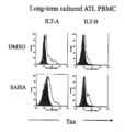
これまでに、本発明者らは、ATL患者のPBMCを、30u/mLのIL-2(rhIL-2;Peprotec社製)又は10ng/mLのIL-15(rhIL-15;Peprotec社製)存在下で長期培養することにより、Taxタンパク質を発現したT細胞株を樹立した(J Virol 79:10088-10092や、PLoS Pathog. 2017 Sep 14;13(9):e1006597.)。本発明者らが樹立したそのような細胞株のうち、ILT-A株とILT-B株を用意した。ILT-A株は急性ATL患者のPBMC由来のIL-15依存性細胞株であり、ILT-B株は慢性ATL患者のPBMC由来のIL-2依存性細胞株である。抗がん作用が知られているヒストン脱アセチル化酵素(HDAC)阻害剤が、ILT-A細胞やILT-B細胞を培養した際のTaxタンパク質の発現にどのような影響を与えるか確認を試みた。
(6) Effect of histone deacetylase (HDAC) inhibitors The present inventors have previously established T cell lines expressing Tax protein by long-term culture of PBMCs from ATL patients in the presence of 30 u/mL IL-2 (rhIL-2; manufactured by Peprotec) or 10 ng/mL IL-15 (rhIL-15; manufactured by Peprotec) (J Virol 79:10088-10092 and PLoS Pathog. 2017 Sep 14;13(9):e1006597.). Among such cell lines established by the present inventors, the ILT-A line and the ILT-B line were prepared. The ILT-A line is an IL-15-dependent cell line derived from PBMCs from an acute ATL patient, and the ILT-B line is an IL-2-dependent cell line derived from PBMCs from a chronic ATL patient. We attempted to confirm how histone deacetylase (HDAC) inhibitors, which are known to have anticancer effects, affect the expression of Tax protein when ILT-A cells or ILT-B cells are cultured.
上記(1)又は(6)に記載のIL-2またはIL-15添加培地に、スベロイルアニリドヒドロキサム酸(SAHA)を添加して、0.5μMのSAHAを含むSAHA含有培地を調製した。また、上記(1)又は(6)に記載のIL-2またはIL-15添加培地に、ジメチルスルホキシド(DMSO)を添加して、0.001重量%のDMSOを含むDMSO含有培地(コントロール培地)を調製した。Suberoylanilide hydroxamic acid (SAHA) was added to the IL-2 or IL-15-added medium described in (1) or (6) above to prepare a SAHA-containing medium containing 0.5 μM SAHA. Dimethyl sulfoxide (DMSO) was added to the IL-2 or IL-15-added medium described in (1) or (6) above to prepare a DMSO-containing medium (control medium) containing 0.001% by weight DMSO.
ILT-A細胞を、SAHA含有培地とコントロール培地にてそれぞれ2日間、CO2インキュベーターで37℃にて培養を行った。かかるILT-A細胞について上記(3)に記載の方法でフローサイトメトリーを行い、ILT-A細胞内のTaxタンパク質を検出した。ILT-A細胞に代えて、ILT-B細胞を用いて同様の実験を行い、ILT-B細胞内のTaxタンパク質を検出した。これらの結果を図4に示す。図4の結果から、PBMC細胞株をHDAC阻害剤の存在下で培養すると、HDAC阻害剤非存在下で培養した場合と比較して、Taxタンパク質発現の誘導効率が向上することが示された。 ILT-A cells were cultured in a SAHA-containing medium and a control medium for 2 days each at 37°C in a CO2 incubator. The ILT-A cells were subjected to flow cytometry by the method described in (3) above to detect Tax protein in the ILT-A cells. A similar experiment was performed using ILT-B cells instead of ILT-A cells to detect Tax protein in the ILT-B cells. These results are shown in Figure 4. The results in Figure 4 indicate that when the PBMC cell line was cultured in the presence of an HDAC inhibitor, the efficiency of induction of Tax protein expression was improved compared to when the PBMC cell line was cultured in the absence of an HDAC inhibitor.
試験2.[HTLV-1感染細胞による抗原のクロスプレゼンテーションの誘導]
一般に、CD8+CTLの応答を活性化させるためには、抗原を貪食した抗原提示細胞が活性化し、副刺激分子(CD86等)の発現やIL-12等のサイトカイン産生を行うと共に、抗原ペプチドをMHCクラスII分子だけでなくMHCクラスI分子にも提示すること(すなわち、「抗原のクロスプレゼンテーション」)が必要である。そこで、本発明者らは、クロスプレゼンテーションが生じているかのin vitroでの評価系について検討し、かかる評価系を構築した(図5)。この評価系では、CD8陽性Tax特異的CTL(Tc-M1)とMHC-Iの一致する抗原提示細胞(THP1)を用いる。そして、そのCTLやその抗原提示細胞とは、MHCの一致しないHTLV-1感染T細胞(ILT-A又はILT-B)を、マイトマイシンC(MMC)(抗がん剤の1種)で処理したものを、前述のCTL及び抗原提示細胞と共培養する。かかる共培養中に、抗原提示細胞がHTLV-1感染T細胞を取り込み、HTLV-1由来のTax抗原をMHC-I上に提示(クロスプレゼンテーション)した場合には、CD8陽性Tax特異的CTLが提示されたTax抗原を認識し、培養液中にIFN-γを産生する。培養液の上清中のIFN-γ濃度の高さで、プレゼンテーションによりCTLが活性化した程度を評価することができる。この評価系によるクロスプレゼンテーションの評価は、具体的に以下の方法で行った。
Test 2. [Induction of antigen cross-presentation by HTLV-1-infected cells]
In general, in order to activate the response of CD8+ CTL, it is necessary that antigen-presenting cells that have phagocytosed antigens are activated, and that they express costimulatory molecules (CD86, etc.) and produce cytokines such as IL-12, while presenting antigen peptides not only to MHC class II molecules but also to MHC class I molecules (i.e., "antigen cross-presentation"). Therefore, the present inventors have investigated an in vitro evaluation system for whether cross-presentation occurs, and constructed such an evaluation system (Figure 5). In this evaluation system, CD8-positive Tax-specific CTLs (Tc-M1) and antigen-presenting cells (THP1) that match MHC-I are used. Then, HTLV-1-infected T cells (ILT-A or ILT-B), which do not match MHC to the CTLs or the antigen-presenting cells, are treated with mitomycin C (MMC) (a type of anticancer drug) and co-cultured with the above-mentioned CTLs and antigen-presenting cells. During such co-culture, when the antigen-presenting cells take up the HTLV-1-infected T cells and present the HTLV-1-derived Tax antigen on MHC-I (cross-presentation), the CD8-positive Tax-specific CTL recognizes the presented Tax antigen and produces IFN-γ in the culture medium. The level of activation of CTL by presentation can be evaluated based on the level of IFN-γ concentration in the culture medium supernatant. Evaluation of cross-presentation using this evaluation system was specifically performed by the following method.
CD8陽性Tax特異的CTLとしては、HLA-A2拘束性CD8陽性Tax特異的CTLであるTc-M1株を用いた。Tc-M1株は、HAM/TSP患者のPBMCを100u/mL rhIL-2存在下で培養し、自家HTLV-1感染細胞で2週間隔にて繰り返し刺激することにより誘導して得られた細胞株である(International immunology 3:761-767)。HLA-A2陽性の抗原提示細胞としては、ヒト単球由来THP1細胞株(Virology 318:17-23)を用いた。抗原細胞としては、「HLA-A2を持たないILT-A細胞(ATL患者由来HTLV-1感染T細胞株)をIL-15添加培地で2日間培養後、50μg/mLのMMC(協和発酵キリン社製)(抗がん剤の1種)で37℃、30分間処理した後、洗浄した細胞」(以下、単に「MMC処理したILT-A細胞」とも表示する。)、あるいは、「HLA-A2を持たないILT-A細胞(ATL患者由来HTLV-1感染T細胞株)をIL-15添加培地で2日間培養後、1重量%ホルマリンで15分間処理した後、洗浄した細胞」を用いた。The Tc-M1 line, which is an HLA-A2-restricted CD8-positive Tax-specific CTL, was used as the CD8-positive Tax-specific CTL. The Tc-M1 line was derived by culturing PBMCs from a HAM/TSP patient in the presence of 100 u/mL rhIL-2 and repeatedly stimulating them with autologous HTLV-1-infected cells at two-week intervals (International immunology 3:761-767). The human monocyte-derived THP1 cell line (Virology 318:17-23) was used as the HLA-A2-positive antigen-presenting cell. The antigen cells used were "ILT-A cells (HTLV-1-infected T cell line derived from an ATL patient) not having HLA-A2 cultured in an IL-15-supplemented medium for 2 days, treated with 50 μg/mL MMC (manufactured by Kyowa Hakko Kirin Co., Ltd.) (a type of anticancer drug) at 37° C. for 30 minutes, and then washed" (hereinafter, also referred to as "MMC-treated ILT-A cells"), or "ILT-A cells (HTLV-1-infected T cell line derived from an ATL patient) not having HLA-A2 cultured in an IL-15-supplemented medium for 2 days, treated with 1% by weight formalin for 15 minutes, and then washed."
HLA-A2を持たない前述の抗原細胞を、HLA-A2陽性のTHP1細胞株と16~20時間共培養した。かかる共培養で得られた全細胞を1重量%ホルマリン水溶液で室温にて15分間固定した後、洗浄した細胞を、25u/mLのIL-2と10%FCSを含むRPMI1640培地中で、前述のHLA-A2拘束性CD8陽性Tax特異的CTL(Tc-M1)と16時間共培養した。共培養して得られた培養液の上清を回収し、かかる上清中のIFN-γ濃度をELISA(BD OptiEIA(登録商標))で測定した。そしてそのIFN-γ濃度を、CTL活性の指標とした。The aforementioned antigen cells not bearing HLA-A2 were co-cultured with the HLA-A2-positive THP1 cell line for 16 to 20 hours. All cells obtained from the co-culture were fixed with 1% by weight aqueous formalin solution at room temperature for 15 minutes, and the washed cells were then co-cultured with the aforementioned HLA-A2-restricted CD8-positive Tax-specific CTL (Tc-M1) for 16 hours in RPMI1640 medium containing 25 u/mL IL-2 and 10% FCS. The supernatant of the culture solution obtained by the co-culture was collected, and the IFN-γ concentration in the supernatant was measured by ELISA (BD OptiEIA (registered trademark)). The IFN-γ concentration was used as an index of CTL activity.
なお、コントロール実験の1つとして、抗原細胞と抗原提示細胞との共培養を行わずに、抗原細胞を前述のHLA-A2拘束性CD8陽性Tax特異的CTL(Tc-M1)と16時間共培養し、得られた培養液の上清中のIFN-γ濃度を測定した。As a control experiment, the antigen cells were not co-cultured with the antigen-presenting cells, but were co-cultured with the aforementioned HLA-A2-restricted CD8-positive Tax-specific CTL (Tc-M1) for 16 hours, and the IFN-γ concentration in the supernatant of the resulting culture medium was measured.
これらのIFN-γ濃度の測定結果を図6に示す。図6において、「ILT-MMC alone」は、始めの培養として、MMC処理したILT-A細胞(ATL患者由来HTLV-1感染T細胞株)のみをCTLと共培養した場合の培養液上清中のIFN-γ濃度を表し、「ILT-Formalin alone」は、始めの培養として、ホルマリン処理したILT-A細胞(ATL患者由来HTLV-1感染T細胞株)のみをCTLと共培養した場合のIFN-γ濃度を表し、「THP1 alone」は、始めの培養として、抗原提示細胞であるTHP1細胞のみをCTLと共培養した場合のIFN-γ濃度を表し、「ILT-MMC+THP1」は、始めの培養として、MMC処理したILT-A細胞と、THP1細胞(抗原提示細胞)を培養した場合のCTLが産生するIFN-γ濃度を表し、「ILT-Formalin+THP1」は、始めの培養として、ホルマリン処理したILT-A細胞と、THP1細胞(抗原提示細胞)を培養した場合のCTLが産生するIFN-γ濃度を表し、「None (CLT alone)」は、始めの培養の際にILT-A細胞もTHP1細胞(抗原提示細胞)も培養しなかった場合(すなわち、CTLであるTc-M1のみを培養した場合)のIFN-γ濃度を表す。The measurement results of these IFN-γ concentrations are shown in Figure 6. In Figure 6, "ILT-MMC alone" represents the IFN-γ concentration in the culture supernatant when only MMC-treated ILT-A cells (HTLV-1-infected T cell line derived from an ATL patient) were co-cultured with CTLs as the initial culture, "ILT-Formalin alone" represents the IFN-γ concentration in the culture supernatant when only formalin-treated ILT-A cells (HTLV-1-infected T cell line derived from an ATL patient) were co-cultured with CTLs as the initial culture, and "THP1 "Alone" represents the IFN-γ concentration when only THP1 cells, which are antigen-presenting cells, were co-cultured with CTLs in the initial culture, "ILT-MMC+THP1" represents the IFN-γ concentration produced by CTLs when MMC-treated ILT-A cells and THP1 cells (antigen-presenting cells) were cultured in the initial culture, "ILT-Formalin+THP1" represents the IFN-γ concentration produced by CTLs when formalin-treated ILT-A cells and THP1 cells (antigen-presenting cells) were cultured in the initial culture, and "None (CLT alone)" represents the IFN-γ concentration when neither ILT-A cells nor THP1 cells (antigen-presenting cells) were cultured in the initial culture (i.e., when only Tc-M1 CTLs were cultured).
図6の結果から以下のことが示された。
CD8陽性Tax特異的CTL(Tc-M1)は、MMC処理したILT-A細胞とのみ共培養しても、ほとんど活性化しない。また、CD8陽性Tax特異的CTL(Tc-M1)は、THP1細胞(抗原提示細胞)とのみ共培養しても、ほとんど活性化しない。しかし、CD8陽性Tax特異的CTL(Tc-M1)は、「ILT-A細胞とTHP1細胞を共培養した細胞」と共培養すると活性化する(IFN-γを産生する)。これらのことから、MMC処理したHTLV-1感染T細胞(例えばMMC処理したILT-A細胞)を、抗原提示細胞(例えばTHP1細胞)と共培養すると、抗原提示細胞はHTLV-1感染T細胞を取り込み、HTLV-1由来のTax抗原をMHC-I上に提示していると考えられた。すなわち、抗原提示細胞は、Tax抗原をクロスプレゼンテーションしていると考えられた。
なお、図6の「ILT-Formalin+THP1」の結果から分かるように、「ホルマリン固定したHTLV-1感染T細胞と抗原提示細胞を共培養した細胞」と、CTLとを共培養しても、CTLはほとんど活性化しなかった。
The results in FIG. 6 indicate the following.
CD8-positive Tax-specific CTL (Tc-M1) is hardly activated even when co-cultured only with MMC-treated ILT-A cells. Also, CD8-positive Tax-specific CTL (Tc-M1) is hardly activated even when co-cultured only with THP1 cells (antigen-presenting cells). However, CD8-positive Tax-specific CTL (Tc-M1) is activated (produces IFN-γ) when co-cultured with "cells obtained by co-culture of ILT-A cells and THP1 cells". From these findings, it was considered that when MMC-treated HTLV-1-infected T cells (e.g., MMC-treated ILT-A cells) are co-cultured with antigen-presenting cells (e.g., THP1 cells), the antigen-presenting cells take up the HTLV-1-infected T cells and present the Tax antigen derived from HTLV-1 on MHC-I. In other words, it was considered that the antigen-presenting cells cross-present the Tax antigen.
As can be seen from the results of "ILT-Formalin+THP1" in Figure 6, even when CTLs were co-cultured with "cells co-cultured with formalin-fixed HTLV-1-infected T cells and antigen-presenting cells," the CTLs were hardly activated.
試験3.[HTLV-1中和抗体及び逆転写酵素阻害剤の影響]
前述の試験2において、CD8陽性Tax特異的CTL(Tc-M1)は、「ILT-A細胞とTHP1細胞を共培養した細胞」と共培養すると活性化する(IFN-γを産生する)ことが示された。試験2におけるこの現象が、HTLV-1感染細胞であるILT-A細胞から抗原提示細胞へHTLV-1が新たに感染し、その新たな感染細胞から生じるHTLV-1抗原の結果ではないことを確認するために、HTLV-1の新たな感染をHTLV-1中和抗体又は逆転写酵素阻害剤で阻害した上で、試験2のクロスプレゼンテーションの評価試験を行った。具体的には以下のような実験を行い、HTLV-1感染細胞によるTax抗原のクロスプレゼンテーション惹起効率に対して、HTLV-1中和抗体や逆転写酵素阻害剤がどのような影響を与えるかを調べた。
Test 3. [Effects of HTLV-1 neutralizing antibodies and reverse transcriptase inhibitors]
In the above-mentioned Test 2, it was shown that CD8-positive Tax-specific CTL (Tc-M1) was activated (produced IFN-γ) when co-cultured with "cells obtained by co-culture of ILT-A cells and THP1 cells". In order to confirm that this phenomenon in Test 2 is not the result of HTLV-1 antigens generated from newly infected cells infected with HTLV-1 from ILT-A cells, which are HTLV-1-infected cells, newly infected with HTLV-1 were inhibited with HTLV-1 neutralizing antibodies or reverse transcriptase inhibitors, and then a cross-presentation evaluation test in Test 2 was performed. Specifically, the following experiment was performed to examine the effect of HTLV-1 neutralizing antibodies and reverse transcriptase inhibitors on the efficiency of cross-presentation induction of Tax antigen by HTLV-1-infected cells.
(1)HTLV-1中和抗体による影響
HTLV-1感染者の血中には高濃度のHTLV-1中和抗体が含まれており、HTLV-1抗原を発現した細胞に結合することが予想される。抗CCR4抗体であるモガムリズマブ(商品名ポテリジオ(登録商標);協和発酵キリン社製)も同様にHTLV-1感染細胞に結合する。HTLV-1感染細胞に結合するこれらの抗体による影響を調べるために以下の実験を行った。
(1) Effect of HTLV-1 neutralizing antibodies High concentrations of HTLV-1 neutralizing antibodies are contained in the blood of HTLV-1 infected individuals, and it is expected that these antibodies will bind to cells expressing HTLV-1 antigens. Mogamulizumab (trade name Poteligeo (registered trademark); manufactured by Kyowa Hakko Kirin Co., Ltd.), an anti-CCR4 antibody, also binds to HTLV-1 infected cells. The following experiment was carried out to examine the effect of these antibodies binding to HTLV-1 infected cells.
ILT-A細胞をIL-15添加培地で2日間培養後、50μg/mLのMMC(協和発酵キリン社製)(抗がん剤の1種)で37℃、30分間処理した後、洗浄することによって、「MMC処理したILT-A細胞(MMC処理ILT-A細胞)」を得た。一方、ATL患者3例、HAM/TSP患者3例及び非感染者2例の血漿(plasma)をそれぞれ0.5重量%含む水溶液を調製し、血漿水溶液とした。かかる血漿水溶液中に前述の「MMC処理ILT-A細胞」をそれぞれ約105cellずつ添加して、血漿中の抗体とILT-A細胞を接触させた。MMC処理ILT-A細胞を洗浄した後、約105cellずつのTHP1細胞と共培養した。かかる共培養で得られた全細胞を1重量%ホルマリン水溶液で室温にて15分間固定した後、洗浄した細胞を、前述のHLA-A2拘束性CD8陽性Tax特異的CTL(Tc-M1)と16時間共培養した。共培養して得られた培養液の上清を回収し、かかる上清中のIFN-γ濃度をELISA(BD OptiEIA(登録商標))で測定した。そしてそのIFN-γ濃度を、CTL活性の指標とした。 ILT-A cells were cultured in IL-15-added medium for 2 days, and then treated with 50 μg/mL MMC (Kyowa Hakko Kirin Co., Ltd.) (a type of anticancer drug) at 37° C. for 30 minutes, followed by washing to obtain "MMC-treated ILT-A cells (MMC-treated ILT-A cells)." Meanwhile, aqueous solutions containing 0.5% by weight of plasma from three ATL patients, three HAM/TSP patients, and two non-infected individuals were prepared to obtain plasma aqueous solutions. Approximately 10 5 cells of the aforementioned "MMC-treated ILT-A cells" were added to the plasma aqueous solutions to bring the antibodies in the plasma into contact with the ILT-A cells. After washing, the MMC-treated ILT-A cells were co-cultured with approximately 10 5 cells of THP1 cells. All cells obtained by this co-culture were fixed with 1% by weight aqueous formalin solution at room temperature for 15 minutes, and the washed cells were then co-cultured with the above-mentioned HLA-A2-restricted CD8-positive Tax-specific CTL (Tc-M1) for 16 hours. The supernatant of the culture solution obtained by the co-culture was collected, and the IFN-γ concentration in the supernatant was measured by ELISA (BD OptiEIA (registered trademark)). The IFN-γ concentration was used as an index of CTL activity.
また、患者等の血漿を0.5重量%含む水溶液に代えて、抗CCR4抗体であるモガムリズマブ(商品名ポテリジオ(登録商標);協和発酵キリン社製)を10μg/mL含む水溶液を用いて同様の実験を行い、上清中のIFN-γ濃度を測定した。In addition, a similar experiment was performed using an aqueous solution containing 10 μg/mL of the anti-CCR4 antibody mogamulizumab (trade name Poteligeo (registered trademark); manufactured by Kyowa Hakko Kirin Co., Ltd.) instead of an aqueous solution containing 0.5% by weight of patient plasma, and the IFN-γ concentration in the supernatant was measured.
これらの実験でIFN-γ濃度を測定した結果を図7に示す。図7の結果から分かるように、HTLV-1感染者の血漿で処理した場合(図7の「ATL-1」、「ATL-2」、「ATL-3」、「HAM-1」、「HAM-2」、「HAM-3」)や、抗CCR4抗体で処理した場合(図7の「Poteligeo」)に、IFN-γ濃度が有意に低下するということはなかった。これらの結果から、HTLV-1感染細胞によるTax抗原のクロスプレゼンテーションの誘導は、HTLV-1の新たな感染によるものではないことが示された。また、HTLV-1感染者の血漿で処理した場合(図7の「ATL-1」、「ATL-2」、「ATL-3」、「HAM-1」、「HAM-2」、「HAM-3」)と、非感染者の血漿で処理した場合(図7の「SN-1」、「SN-2」)と、抗CCR4抗体で処理した場合(図7の「Poteligeo」)とで、有意な差までは認められなかった。The results of measuring IFN-γ concentrations in these experiments are shown in Figure 7. As can be seen from the results in Figure 7, IFN-γ concentrations were not significantly reduced when cells were treated with plasma from HTLV-1-infected individuals ("ATL-1," "ATL-2," "ATL-3," "HAM-1," "HAM-2," and "HAM-3" in Figure 7) or when cells were treated with anti-CCR4 antibody ("Poteligeo" in Figure 7). These results indicate that the induction of Tax antigen cross-presentation by HTLV-1-infected cells is not due to new infection with HTLV-1. Furthermore, no significant differences were observed between treatment with plasma from HTLV-1-infected individuals ("ATL-1," "ATL-2," "ATL-3," "HAM-1," "HAM-2," and "HAM-3" in Figure 7), treatment with plasma from non-infected individuals ("SN-1" and "SN-2" in Figure 7), and treatment with anti-CCR4 antibody ("Poteligeo" in Figure 7).
(2)「MMC処理ILT-A細胞」とTHP1細胞の細胞数の比率の影響
ILT-A細胞をIL-15添加培地で2日間培養後(前述)、50μg/mLのMMC(協和発酵キリン社製)(抗がん剤の1種)で37℃、30分間処理した後、洗浄することによって、「MMC処理したILT-A細胞(MMC処理ILT-A細胞)」を得た。
「MMC処理ILT-A細胞」とTHP1細胞を共培養する際の、そのILT-A細胞とTHP1細胞の細胞数の比率を、1:1又は0.5:1又は0.25:1として、上記の「(1)HTLV-1中和抗体による影響」と同様の方法でIFN-γ濃度を測定した。その際、ALT患者の血漿として、ATL-1の血漿を用いた。
(2) Effect of the ratio of the number of cells between “MMC-treated ILT-A cells” and THP1 cells
The ILT-A cells were cultured in IL-15-supplemented medium for 2 days (as described above), and then treated with 50 μg/mL MMC (Kyowa Hakko Kirin Co., Ltd.) (a type of anticancer drug) at 37° C. for 30 minutes, followed by washing to obtain "MMC-treated ILT-A cells."
When "MMC-treated ILT-A cells" and THP1 cells were co-cultured, the ratio of the number of ILT-A cells to that of THP1 cells was set to 1:1, 0.5:1, or 0.25:1, and the IFN-γ concentration was measured in the same manner as in "(1) Effect of HTLV-1 neutralizing antibody" above. In this case, ATL-1 plasma was used as the plasma of the ALT patient.
これらの実験の結果を図8に示す。横軸は、HTLV-1感染T細胞数を表し、左から1~3番目の棒グラフはHTLV-1感染T細胞が105個(cell)(すなわち、前述の比率が1:1)の場合の結果を表し、左から4~6番目の棒グラフはHTLV-1感染T細胞5×10⁴個(cell)(すなわち、前述の比率が0.5:1)の場合の結果を表し、右から1~3番目の棒グラフが2.5×10⁴個(cell)(すなわち、前述の比率が0.25:1)の場合の結果を表す。また、図8において、黒塗りの棒グラフ(「MMC-ILT/(+)plasma」)は、「MMC処理ILT-A細胞」とHTLV-1感染者の血漿を接触させた場合の結果を表し、グレーの棒グラフ(「MMC-ILT/(-)plasma」)は、「MMC処理ILT-A細胞」に非感染者の血漿を接触させた場合の結果を表し、白抜きの棒グラフ(「MMC-ILT」)は、「MMC処理ILT-A細胞」にHTLV-1感染者の血漿を接触させなかった場合の結果を表す。 The results of these experiments are shown in Figure 8. The horizontal axis represents the number of HTLV-1-infected T cells, and the first to third bars from the left represent the results when the number of HTLV-1-infected T cells was 105 (i.e., the aforementioned ratio was 1:1), the fourth to sixth bars from the left represent the results when the number of HTLV-1-infected T cells was 5x10⁴ (i.e., the aforementioned ratio was 0.5:1), and the first to third bars from the right represent the results when the number of HTLV-1-infected T cells was 2.5x10⁴ (i.e., the aforementioned ratio was 0.25:1). In addition, in Figure 8, the black bar graph ("MMC-ILT/(+)plasma") represents the results when "MMC-treated ILT-A cells" were contacted with plasma from an HTLV-1-infected individual, the gray bar graph ("MMC-ILT/(-)plasma") represents the results when "MMC-treated ILT-A cells" were contacted with plasma from a non-infected individual, and the open bar graph ("MMC-ILT") represents the results when "MMC-treated ILT-A cells" were not contacted with plasma from an HTLV-1-infected individual.
図8の結果から、ILT-A細胞をHTLV-1感染者の血漿により処理すると、CTL活性化効果がある程度増加する傾向が認められた。また、この傾向は、標的細胞である「MMC処理ILT-A細胞」の数が、抗原提示細胞の数に対して比較的少ない場合(図8の「5~2.5×10⁴」)により強く認められた。なお、ILT-A細胞をHTLV-1感染者の血漿により処理するとCTL活性化効果が増加することは、ILT-A細胞に感染者の血漿中の抗HTLV-1抗体が結合することによるオプソニン効果の結果であると考えられた。 From the results in Figure 8, it was observed that the CTL activation effect tends to increase to some extent when ILT-A cells are treated with plasma from HTLV-1-infected individuals. This tendency was more pronounced when the number of target cells, "MMC-treated ILT-A cells," was relatively small compared to the number of antigen-presenting cells ("5-2.5 x 10 4 " in Figure 8). The increase in the CTL activation effect when ILT-A cells are treated with plasma from HTLV-1-infected individuals was considered to be the result of an opsonization effect caused by the binding of anti-HTLV-1 antibodies in the plasma of infected individuals to ILT-A cells.
(3)逆転写酵素阻害剤による影響
逆転写酵素阻害剤として、ジドブジン(Zidovudine:AZT)(商品名レトロビル(登録商標)、グラクソスミスクライン社製)を用いた。20μMのAZTを含む培地中で、抗原提示細胞であるTHP1細胞を37℃で1時間、前培養した。一方、ILT-A細胞をIL-15添加培地で2日間培養後、50μg/mLのMMC(協和発酵キリン社製)(抗がん剤の1種)で37℃、30分間処理した後、洗浄することによって、「MMC処理したILT-A細胞(MMC処理ILT-A細胞)」を得た。前述の前培養を行ったTHP1細胞の培地中に、MMC処理ILT-A細胞を添加して37℃で16時間共培養した。なお、共培養の際の培地中のAZT濃度は10μMであった。かかる共培養で得られた全細胞を1重量%ホルマリン水溶液で室温にて15分間固定した後、洗浄した細胞を、HLA-A2拘束性CD8陽性Tax特異的CTL(Tc-M1)と16時間共培養した。共培養して得られた培養液の上清を回収し、かかる上清中のIFN-γ濃度をELISA(BD OptiEIA(登録商標))で測定した。そしてそのIFN-γ濃度を、CTL活性の指標とした。
(3) Effect of reverse transcriptase inhibitor Zidovudine (AZT) (trade name Retrovir (registered trademark), manufactured by GlaxoSmithKline) was used as a reverse transcriptase inhibitor. THP1 cells, which are antigen-presenting cells, were pre-cultured in a medium containing 20 μM AZT at 37° C. for 1 hour. Meanwhile, ILT-A cells were cultured in an IL-15-added medium for 2 days, and then treated with 50 μg/mL MMC (manufactured by Kyowa Hakko Kirin Co., Ltd.) (a type of anticancer drug) at 37° C. for 30 minutes, and then washed to obtain "MMC-treated ILT-A cells (MMC-treated ILT-A cells)". MMC-treated ILT-A cells were added to the medium of the THP1 cells that had been pre-cultured as described above, and co-cultured at 37° C. for 16 hours. The AZT concentration in the medium during co-culture was 10 μM. All cells obtained by this co-culture were fixed with 1% by weight aqueous formalin solution at room temperature for 15 minutes, and the washed cells were then co-cultured with HLA-A2-restricted CD8-positive Tax-specific CTL (Tc-M1) for 16 hours. The supernatant of the culture solution obtained by the co-culture was collected, and the IFN-γ concentration in the supernatant was measured by ELISA (BD OptiEIA (registered trademark)). The IFN-γ concentration was used as an index of CTL activity.
これらのIFN-γ濃度の測定結果を図9に示す。右から1~3番目の棒グラフはHTLV-1感染T細胞(ILT-A細胞)が105個(cell)(すなわち、前述の比率が1:1)の場合の結果を表し、右から4~6番目の棒グラフはHTLV-1感染T細胞(ILT-A細胞)が3×10⁴個(cell)(すなわち、前述の比率が0.3:1)の場合の結果を表し、左から1~3番目の棒グラフはHTLV-1感染T細胞(ILT-A細胞)を用いなかった場合の結果を表す。また、図9において、黒塗りの棒グラフ(「THP1-AZT」)は、培地にAZTを添加してTHP1細胞等を培養した場合の結果を表し、グレーの棒グラフ(「THP1」)は、培地にAZTを添加しなかった場合の結果を表し、白抜きの棒グラフ(「Medium」)はTHP1細胞を用いなかった場合の結果を表す。 The measurement results of these IFN-γ concentrations are shown in FIG. 9. The first to third bars from the right show the results when HTLV-1-infected T cells (ILT-A cells) were 10 5 cells (i.e., the aforementioned ratio was 1:1), the fourth to sixth bars from the right show the results when HTLV-1-infected T cells (ILT-A cells) were 3×10 4 cells (i.e., the aforementioned ratio was 0.3:1), and the first to third bars from the left show the results when HTLV-1-infected T cells (ILT-A cells) were not used. In FIG. 9, the black bars ("THP1-AZT") show the results when AZT was added to the medium and THP1 cells or the like were cultured, the gray bars ("THP1") show the results when AZT was not added to the medium, and the open bars ("Medium") show the results when THP1 cells were not used.
図9の結果から分かるように、AZT処理の有無により、CTL活性には大きな影響は認められなかった。これらの結果から、HTLV-1感染細胞によるTax抗原のクロスプレゼンテーションの誘導は、HTLV-1の新たな感染によるものではないことが示された。As can be seen from the results in Figure 9, AZT treatment had no significant effect on CTL activity. These results indicate that the induction of Tax antigen cross-presentation by HTLV-1-infected cells is not due to new infection with HTLV-1.
試験4.[抗原提示細胞における副刺激分子の発現の、HTLV-1感染T細胞による増強]
樹状細胞等の抗原提示細胞がT細胞を活性化させるためには、CD83、CD86等の副刺激シグナルを表出することが必要とされている(例えば、Leukemia & lymphoma 38:247-263)。そこで、MMC処理ILT-C細胞(ATL患者PBMCを長期培養したHTLV-1感染細胞株)が、抗原提示細胞上の副刺激分子の発現を増強するかを調べるために、以下の実験を行った。
Test 4. [Enhancement of expression of costimulatory molecules in antigen-presenting cells by HTLV-1-infected T cells]
In order for antigen-presenting cells such as dendritic cells to activate T cells, it is necessary for them to express costimulatory signals such as CD83 and CD86 (e.g., Leukemia & lymphoma 38:247-263). Therefore, to examine whether MMC-treated ILT-C cells (HTLV-1-infected cell line obtained by long-term culture of PBMCs from ATL patients) enhance the expression of costimulatory molecules on antigen-presenting cells, the following experiment was performed.
(1)健常人のPBMC由来の未熟樹状細胞
抗原提示細胞としては、抗原を取り込む能力を未だ保持している未熟樹状細胞を用いた。この未熟樹状細胞は、10%FCS添加 RPMI1640培地をプラスチックプレートに入れ、その培地中で健常人のPBMCを2時間培養した後、プラスチックプレートに接着した単球細胞にrhGM-CSF(Miltenyi Biotec社製)(培地中の最終濃度1000u/mL)及びrhIL-4(Miltenyi Biotec社製)(培地中の最終濃度500u/mL)を添加し、5日間培養して得られた未熟樹状細胞(以下、「健常人未熟樹状細胞」とも表示する。)である。
(1) Immature dendritic cells derived from PBMCs of healthy subjects Immature dendritic cells that still retain the ability to take up antigens were used as antigen-presenting cells. These immature dendritic cells were obtained by placing RPMI1640 medium supplemented with 10% FCS in a plastic plate, culturing PBMCs of a healthy subject in the medium for 2 hours, adding rhGM-CSF (manufactured by Miltenyi Biotec) (final concentration in the medium: 1000 u/mL) and rhIL-4 (manufactured by Miltenyi Biotec) (final concentration in the medium: 500 u/mL) to monocytes attached to the plastic plate, and culturing for 5 days (hereinafter, also referred to as "healthy subject immature dendritic cells").
(2)副刺激分子の発現の評価
ILT-C22細胞(HLA-A2陽性)をIL-2添加培地で2日間培養後、50μg/mLのMMC(協和発酵キリン社製)(抗がん剤の1種)で37℃、30分間処理した後、洗浄することによって、「MMC処理したILT-C細胞(MMC処理ILT-C細胞)」を得た。次に、前述の健常人未熟樹状細胞(HLA-A2陰性)とMMC処理ILT-C株とを、10%FCS添加 RPMI1640培地で24時間共培養した後、FITC標識HLA-A2抗体(「BD 551285」;ベクトン・ディッキンソン社製)と、PerCP/Cy5.5標識抗ヒトCD83抗体(「305320」;Biolegend社製)又はPerCP/Cy5.5標識抗ヒトCD86抗体(「BD 561129」;ベクトン・ディッキンソン社製)とを用いて氷上で30分間染色し、洗浄後、フローサイトメーターMACSQuant(登録商標)(Miltenyi Biotec社製)を用いたフローサイトメトリーでHLA-A2陰性樹状細胞分画のCD83及びCD86の発現強度を測定した。また、MMC処理ILT-C株を用いないこと以外は同じ方法(すなわち、MMC処理ILT-C株と共培養しない健常人未熟樹状細胞を用いた方法)で、CD83及びCD86の発現強度を測定した。なお、HLA-A2発現の有無により、樹状細胞とILT-C細胞を区別した。
(2) Evaluation of expression of costimulatory molecules ILT-C22 cells (HLA-A2 positive) were cultured in IL-2-added medium for 2 days, and then treated with 50 μg/mL MMC (Kyowa Hakko Kirin Co., Ltd.) (a type of anticancer drug) at 37° C. for 30 minutes and washed to obtain "MMC-treated ILT-C cells." Next, the above-mentioned healthy donor immature dendritic cells (HLA-A2 negative) and the MMC-treated ILT-C line were co-cultured for 24 hours in RPMI 1640 medium supplemented with 10% FCS, and then stained for 30 minutes on ice using an FITC-labeled HLA-A2 antibody (BD 551285; Becton Dickinson and Company), a PerCP/Cy5.5-labeled anti-human CD83 antibody (305320; Biolegend) or a PerCP/Cy5.5-labeled anti-human CD86 antibody (BD 561129; Becton Dickinson and Company). After washing, the expression intensities of CD83 and CD86 in the HLA-A2 negative dendritic cell fraction were measured by flow cytometry using a flow cytometer MACSQuant (registered trademark) (Miltenyi Biotec). In addition, the expression intensities of CD83 and CD86 were measured using the same method except that the MMC-treated ILT-C line was not used (i.e., using healthy immature dendritic cells not co-cultured with the MMC-treated ILT-C line). Dendritic cells and ILT-C cells were distinguished based on the presence or absence of HLA-A2 expression.
これらの測定結果を図10に示す。MMC処理ILT-C株を用いなかった場合の健常人未熟樹状細胞と比較して、MMC処理ILT-C株と共培養した健常人未熟樹状細胞では、CD83の発現が軽度に増加し(図10の左上パネル及び左下パネル)、CD86の発現は大幅に増加した(図10の右上パネル及び右下パネル)。これらの結果から、MMC処理HTLV-1感染T細胞と未熟樹状細胞を共培養すると、MMC処理HTLV-1感染T細胞が未熟樹状細胞を刺激して、CD83やCD86等の副刺激分子の発現を増強させることが示された。These measurement results are shown in Figure 10. Compared to healthy donor immature dendritic cells without the MMC-treated ILT-C line, healthy donor immature dendritic cells co-cultured with the MMC-treated ILT-C line showed a mild increase in CD83 expression (upper left and lower left panels of Figure 10) and a significant increase in CD86 expression (upper right and lower right panels of Figure 10). These results demonstrated that when MMC-treated HTLV-1-infected T cells and immature dendritic cells were co-cultured, the MMC-treated HTLV-1-infected T cells stimulated the immature dendritic cells and enhanced the expression of costimulatory molecules such as CD83 and CD86.
試験5.[抗原提示細胞におけるIL-12の発現の、HTLV-1感染T細胞による増強]
抗原提示細胞からIL-12が産生されることは、抗原提示の際に免疫環境をTh1型に傾け、CTLを誘導する上で重要であるとされている(Therapeutic immunology 1:187-196.)。そこで、抗原提示細胞を、MMC処理したILT-A細胞又はILT-B細胞と共培養することにより、抗原提示細胞におけるIL-12産生にどのような影響が生じるかを調べるために、以下の実験を行った。
Test 5. [Enhancement of IL-12 expression in antigen-presenting cells by HTLV-1-infected T cells]
It is believed that the production of IL-12 from antigen-presenting cells is important for inclining the immune environment to Th1 type during antigen presentation and inducing CTL (Therapeutic immunology 1:187-196.) Therefore, the following experiment was performed to examine the effect of co-culturing antigen-presenting cells with MMC-treated ILT-A cells or ILT-B cells on IL-12 production in antigen-presenting cells.
(1)抗原提示細胞におけるIL-12の発現の、HTLV-1感染T細胞の薬剤処理による影響
IL-15またはIL-2を含む10%FCS(ウシ胎仔血清)添加 RPMI 1640培地に、ヒストン脱アセチル化酵素(HDAC)阻害剤であるスベロイルアニリドヒドロキサム酸(SAHA)を添加して、0.5μMのSAHAを含むSAHA含有培地を調製した。また、10%FCS(ウシ胎仔血清)添加 RPMI 1640培地に、ジメチルスルホキシド(DMSO)を添加して、0.001重量%のDMSOを含むDMSO含有培地(コントロール培地)を調製した。
(1) Effect of drug treatment of HTLV-1-infected T cells on IL-12 expression in antigen-presenting cells Suberoylanilide hydroxamic acid (SAHA), a histone deacetylase (HDAC) inhibitor, was added to RPMI 1640 medium supplemented with 10% FCS (fetal calf serum) containing IL-15 or IL-2 to prepare a SAHA-containing medium containing 0.5 μM SAHA. Dimethyl sulfoxide (DMSO) was also added to RPMI 1640 medium supplemented with 10% FCS (fetal calf serum) to prepare a DMSO-containing medium (control medium) containing 0.001 wt % DMSO.
ILT-A細胞又はILT-B細胞を、SAHA含有培地又はDMSO含有培地(コントロール培地)で24時間、CO2インキュベーターで37℃にて培養を行った。培養して得られた細胞を、50μg/mLのMMC(協和発酵キリン社製)で37℃30分間処理した後、洗浄した。このMMC処理ILT細胞(すなわち、MMC処理したILT-A細胞又はILT-B細胞)をHLA-A2陽性健常人未熟樹状細胞(DC)と37℃で20時間共培養した。かかる共培養後の培地の上清を分取し、上清中のIL-12濃度をELISA(BD OptiEIA(登録商標))で測定した。これらの結果を図11の「DC」にそれぞれ示す。
また、コントロールとして、前述のMMC処理ILT細胞(すなわち、MMC処理したILT-A細胞又はILT-B細胞)を、健常人未熟樹状細胞(DC)と共にではなく、37℃で20時間培養した。かかる培養後の培地の上清を分取し、上清中のIL-12濃度(pg/mL)をELISA(BD OptiEIA(登録商標))で測定した。これらの結果を図11の「Medium」にそれぞれ示す。
ILT-A cells or ILT-B cells were cultured in SAHA-containing medium or DMSO-containing medium (control medium) for 24 hours at 37°C in a CO2 incubator. The cultured cells were treated with 50 μg/mL MMC (Kyowa Hakko Kirin) for 30 minutes at 37°C and then washed. These MMC-treated ILT cells (i.e., MMC-treated ILT-A cells or ILT-B cells) were co-cultured with HLA-A2-positive healthy immature dendritic cells (DCs) for 20 hours at 37°C. The supernatant of the medium after the co-culture was collected, and the IL-12 concentration in the supernatant was measured by ELISA (BD OptiEIA (registered trademark)). These results are shown in "DC" in Figure 11.
As a control, the above-mentioned MMC-treated ILT cells (i.e., MMC-treated ILT-A cells or ILT-B cells) were cultured at 37° C. for 20 hours, not together with healthy donor immature dendritic cells (DCs). After the culture, the supernatant of the medium was separated, and the IL-12 concentration (pg/mL) in the supernatant was measured by ELISA (BD OptiEIA (registered trademark)). These results are shown in "Medium" in FIG. 11.
DMSO含有培地で培養したILT-B細胞をMMC処理した後、洗浄し、樹状細胞と共培養した場合は、培地の上清に約120pg/mLのIL-12産生が検出されたが(図11の「ILT-B」の「DMSO」の黒塗りの棒グラフ(「DC」))、DMSO含有培地で培養したILT-A細胞をMMC処理した後、洗浄し、樹状細胞と共培養した場合は、培地の上清中のIL-12濃度は感度以下であった(図11の「ILT-A」の「DMSO」のうちの右側)。一方、0.5μMのSAHA(HDAC阻害剤の一種)を含むSAHA含有培地で培養したILT細胞(ILT-A細胞又はILT-B細胞)を用いると、DMSO含有培地で培養したILT細胞(ILT-A細胞又はILT-B細胞)を用いた場合と比較して、培地へのIL-12産生が有意に増加した(図11の「ILT-A」の「SAHA」の黒塗りの棒グラフ(「DC」)及び「ILT-B」の「SAHA」の黒塗りの棒グラフ(「DC」)。When ILT-B cells cultured in DMSO-containing medium were treated with MMC, washed, and co-cultured with dendritic cells, approximately 120 pg/mL of IL-12 production was detected in the culture medium supernatant (solid bars ("DC") in "ILT-B" in Figure 11), whereas when ILT-A cells cultured in DMSO-containing medium were treated with MMC, washed, and co-cultured with dendritic cells, the IL-12 concentration in the culture medium supernatant was below the sensitivity level (right side of "DMSO" in "ILT-A" in Figure 11). On the other hand, when ILT cells (ILT-A cells or ILT-B cells) cultured in a SAHA-containing medium containing 0.5 μM SAHA (a type of HDAC inhibitor) were used, IL-12 production into the medium was significantly increased compared to when ILT cells (ILT-A cells or ILT-B cells) cultured in a DMSO-containing medium were used (solid bars for "SAHA" in "ILT-A" ("DC") and solid bars for "SAHA" in "ILT-B" ("DC") in Figure 11).
SAHAの替わりに別のHDAC阻害剤であるバルプロ酸(VPA)を用いた場合の結果を図12に示す。ILT-B細胞をVPA含有培地又はDMSO含有培地(コントロール培地)で24時間培養した。この細胞をMMC処理した後、洗浄し、樹状細胞と共培養し、上清中のIL-12濃度を測定した。図12の「DC」にそれぞれ示すように、1mMのVPAを含む培地で培養したILT-B細胞を用いると、DMSO含有培地で培養したILT-B細胞を用いた場合と比較して、培地へのIL-12産生が有意に増加した(図12の黒塗りの棒グラフ(「DC」))。Figure 12 shows the results when another HDAC inhibitor, valproic acid (VPA), was used instead of SAHA. ILT-B cells were cultured in VPA-containing medium or DMSO-containing medium (control medium) for 24 hours. The cells were treated with MMC, washed, and co-cultured with dendritic cells, and the IL-12 concentration in the supernatant was measured. As shown by "DC" in Figure 12, when ILT-B cells were cultured in medium containing 1 mM VPA, IL-12 production into the medium was significantly increased compared to when ILT-B cells were cultured in DMSO-containing medium (solid bars in Figure 12 ("DC")).
(2)抗原提示細胞による抗原クロスプレゼンテーションへの、HTLV-1感染T細胞の薬剤処理による影響
図11における共培養後の各細胞を1重量%ホルマリンで15分間処理した後、洗浄した細胞を、HLA-A2拘束性CD8陽性Tax特異的CTL(Tc-M1)と16時間共培養した。共培養して得られた培養液の上清を回収し、かかる上清中のIFN-γ濃度(pg/mL)をELISA(BD OptiEIA(登録商標))で測定した。これらの結果を図13に示す。
(2) Effect of drug treatment of HTLV-1-infected T cells on antigen cross-presentation by antigen-presenting cells After each cell was treated with 1% formalin by weight for 15 minutes after co-culture in FIG. 11, the washed cells were co-cultured with HLA-A2-restricted CD8-positive Tax-specific CTL (Tc-M1) for 16 hours. The supernatant of the culture solution obtained by co-culture was collected, and the IFN-γ concentration (pg/mL) in the supernatant was measured by ELISA (BD OptiEIA®). These results are shown in FIG. 13.
図13から分かるように、SAHA処理されたILT細胞を用いた場合は、SAHA処理されていないILT細胞を用いた場合よりも、CTLからのIFN-γ産生量が高かった。これらの結果から、HTLV-1感染T細胞を、SAHA等のHDAC阻害剤で処理すると、抗原提示細胞がTax特異的CTLへ抗原をクロスプレゼンテーションする効率も向上することが分かった。As can be seen from Figure 13, when SAHA-treated ILT cells were used, the amount of IFN-γ produced by CTLs was higher than when non-SAHA-treated ILT cells were used. These results indicate that treating HTLV-1-infected T cells with an HDAC inhibitor such as SAHA also improves the efficiency of antigen-presenting cells cross-presenting antigens to Tax-specific CTLs.
本発明によれば、HTLV-I特異的CTL活性化剤、及びその製造方法等を提供することができる。また、本発明によれば、HTLV-1の感染に起因する疾患の患者のHLA型に制限されずに、多くの患者に比較的安価で効果的な免疫療法を提供できることが期待される。また、本発明によれば、現在、安全で有効な治療法が存在しないため無治療観察が原則となっているくすぶり型や慢性型のATL(インドレントATL)に対して早期段階で適用が可能な治療法を提供できることが期待される。 According to the present invention, it is possible to provide an HTLV-I-specific CTL activator and a method for producing the same. Furthermore, according to the present invention, it is expected that it will be possible to provide a relatively inexpensive and effective immunotherapy to many patients, regardless of the HLA type of the patient suffering from a disease caused by HTLV-1 infection. Furthermore, according to the present invention, it is expected that it will be possible to provide a treatment that can be applied at an early stage to smoldering and chronic ATL (indolent ATL), which is currently treated as a rule by observation without treatment due to the lack of a safe and effective treatment.
Claims (12)
(b)前記工程(a)の培養で得られた末梢血単核細胞を抗がん剤で処理した後、末梢血単核細胞を採取する工程;及び、
(c)前記工程(b)で採取された末梢血単核細胞を薬学的に許容される担体と混合して製剤化する工程;
を有する、前記対象に投与するための、HTLV-I特異的CTL活性化剤の製造方法。 (a) culturing peripheral blood mononuclear cells collected from a subject infected with human T-cell leukemia virus type I (HTLV-1) in an animal cell culture medium;
(b) treating the peripheral blood mononuclear cells obtained by the culture in the step (a) with an anticancer drug and then collecting the peripheral blood mononuclear cells; and
(c) mixing the peripheral blood mononuclear cells collected in step (b) with a pharma- ceutically acceptable carrier to prepare a formulation;
A method for producing an HTLV-I-specific CTL activator for administration to a subject, comprising the steps of:
Applications Claiming Priority (3)
| Application Number | Priority Date | Filing Date | Title |
|---|---|---|---|
| JP2019105453 | 2019-06-05 | ||
| JP2019105453 | 2019-06-05 | ||
| PCT/JP2020/022065 WO2020246535A1 (en) | 2019-06-05 | 2020-06-04 | Htlv-i-specific ctl activator |
Publications (2)
| Publication Number | Publication Date |
|---|---|
| JPWO2020246535A1 JPWO2020246535A1 (en) | 2020-12-10 |
| JP7601394B2 true JP7601394B2 (en) | 2024-12-17 |
Family
ID=73653235
Family Applications (1)
| Application Number | Title | Priority Date | Filing Date |
|---|---|---|---|
| JP2021524895A Active JP7601394B2 (en) | 2019-06-05 | 2020-06-04 | HTLV-I specific CTL activator |
Country Status (2)
| Country | Link |
|---|---|
| JP (1) | JP7601394B2 (en) |
| WO (1) | WO2020246535A1 (en) |
Citations (4)
| Publication number | Priority date | Publication date | Assignee | Title |
|---|---|---|---|---|
| WO2004092373A1 (en) | 2003-04-16 | 2004-10-28 | Japan Science And Technology Agency | Peptide having htlv-1-specific ctl-inducing activity |
| WO2006035681A1 (en) | 2004-09-27 | 2006-04-06 | National University Corporation Tokyo Medical And Dental University | HLA-A11-RESTRICTED Tax ANTITUMOR EPITOPES |
| JP2012090574A (en) | 2010-10-27 | 2012-05-17 | St Marianna Univ School Of Medicine | METHOD FOR PRODUCING γδT-CELL, AND PHARMACEUTICAL |
| JP2016077185A (en) | 2014-10-14 | 2016-05-16 | 学校法人 聖マリアンナ医科大学 | PRODUCTION METHOD AND MEDICINE OF γδT CELLS |
-
2020
- 2020-06-04 JP JP2021524895A patent/JP7601394B2/en active Active
- 2020-06-04 WO PCT/JP2020/022065 patent/WO2020246535A1/en not_active Ceased
Patent Citations (4)
| Publication number | Priority date | Publication date | Assignee | Title |
|---|---|---|---|---|
| WO2004092373A1 (en) | 2003-04-16 | 2004-10-28 | Japan Science And Technology Agency | Peptide having htlv-1-specific ctl-inducing activity |
| WO2006035681A1 (en) | 2004-09-27 | 2006-04-06 | National University Corporation Tokyo Medical And Dental University | HLA-A11-RESTRICTED Tax ANTITUMOR EPITOPES |
| JP2012090574A (en) | 2010-10-27 | 2012-05-17 | St Marianna Univ School Of Medicine | METHOD FOR PRODUCING γδT-CELL, AND PHARMACEUTICAL |
| JP2016077185A (en) | 2014-10-14 | 2016-05-16 | 学校法人 聖マリアンナ医科大学 | PRODUCTION METHOD AND MEDICINE OF γδT CELLS |
Also Published As
| Publication number | Publication date |
|---|---|
| JPWO2020246535A1 (en) | 2020-12-10 |
| WO2020246535A1 (en) | 2020-12-10 |
Similar Documents
| Publication | Publication Date | Title |
|---|---|---|
| US20250034223A1 (en) | Compositions and methods for expanding ex vivo natural killer cells and therapeutic uses thereof | |
| Chang et al. | Strategies for enhancing and preserving anti-leukemia effects without aggravating graft-versus-host disease | |
| JP6422344B2 (en) | Methods for increasing allogeneic antigen-reactive regulatory T cells | |
| CN108697734B (en) | NK cells exhibiting adaptive phenotype and methods of making and using | |
| Kawai et al. | Generation of highly proliferative, rejuvenated cytotoxic T cell clones through pluripotency reprogramming for adoptive immunotherapy | |
| JP2015513403A5 (en) | ||
| US20180161366A1 (en) | Methods of obtaining mononuclear blood cells and uses thereof | |
| Nakazawa et al. | Establishment of an efficient ex vivo expansion strategy for human natural killer cells stimulated by defined cytokine cocktail and antibodies against natural killer cell activating receptors | |
| Copland et al. | Bone marrow mesenchymal stromal cells from patients with acute and chronic graft-versus-host disease deploy normal phenotype, differentiation plasticity, and immune-suppressive activity | |
| Sundin et al. | HSCT recipients have specific tolerance to MSC but not to the MSC donor | |
| US11096967B2 (en) | Pharmaceutical composition for preventing or treating regulatory T cell-mediated diseases | |
| CA2804914A1 (en) | Regulatory immune cells with enhanced targeted cell death effect | |
| JP2021502796A (en) | A composition for culturing NK cells and a method for culturing NK cells using the composition. | |
| JP7601394B2 (en) | HTLV-I specific CTL activator | |
| Zhang et al. | The immunosuppressant protosappanin a promotes dendritic cell-mediated expansion of alloantigen-specific tregs and prolongs allograft survival in rats | |
| Guan et al. | A strategy to reconstitute immunity without GVHD via adoptive allogeneic Tscm therapy | |
| Shao et al. | Combination of monoclonal antibodies with DST inhibits accelerated rejection mediated by memory T cells to induce long-lived heart allograft acceptance in mice | |
| Bennett | Glucocorticoid Induced Leucine Zipper: Gatekeeper for CD8+ T cell activation and anti-viral responses | |
| Wald et al. | Novel strategy to enhance NK cell activity | |
| HK40086176A (en) | Nk cells exhibiting an adaptive phenotype and methods for preparing and for using | |
| Kim | SIGNALING AND TRANSCRIPTIONAL BASES OF T CELL HOMEOSTASIS AND DIFFERENTIATION | |
| Wang et al. | Prolongation of rat renal allograft survival by CD4+ CD25‑T cells induced by recipient dendritic cells transfected with IKK2dn | |
| Semple | Generation and Application of Antigen-Specific Induced Regulatory T cells in Allogeneic Bone Marrow Transplantation | |
| LaCasse | Negative Modulation of Regulatory T Cells and Promotion of the Tumoricidal Activity of Dendritic Cells in Cancer: a Double-edged Strategy | |
| HK1256113B (en) | Nk cells exhibiting an adaptive phenotype and methods for preparing and for using |
Legal Events
| Date | Code | Title | Description |
|---|---|---|---|
| A621 | Written request for application examination |
Free format text: JAPANESE INTERMEDIATE CODE: A621 Effective date: 20230602 |
|
| A131 | Notification of reasons for refusal |
Free format text: JAPANESE INTERMEDIATE CODE: A131 Effective date: 20240708 |
|
| A711 | Notification of change in applicant |
Free format text: JAPANESE INTERMEDIATE CODE: A711 Effective date: 20240927 |
|
| A711 | Notification of change in applicant |
Free format text: JAPANESE INTERMEDIATE CODE: A712 Effective date: 20241011 |
|
| A521 | Request for written amendment filed |
Free format text: JAPANESE INTERMEDIATE CODE: A821 Effective date: 20240927 |
|
| TRDD | Decision of grant or rejection written | ||
| A01 | Written decision to grant a patent or to grant a registration (utility model) |
Free format text: JAPANESE INTERMEDIATE CODE: A01 Effective date: 20241125 |
|
| A61 | First payment of annual fees (during grant procedure) |
Free format text: JAPANESE INTERMEDIATE CODE: A61 Effective date: 20241128 |
|
| R150 | Certificate of patent or registration of utility model |
Ref document number: 7601394 Country of ref document: JP Free format text: JAPANESE INTERMEDIATE CODE: R150 |