JP7660077B2 - Diagnostic methods - Google Patents
Diagnostic methods Download PDFInfo
- Publication number
- JP7660077B2 JP7660077B2 JP2021572701A JP2021572701A JP7660077B2 JP 7660077 B2 JP7660077 B2 JP 7660077B2 JP 2021572701 A JP2021572701 A JP 2021572701A JP 2021572701 A JP2021572701 A JP 2021572701A JP 7660077 B2 JP7660077 B2 JP 7660077B2
- Authority
- JP
- Japan
- Prior art keywords
- adiponectin
- antibody
- polypeptide
- levels
- pancreatitis
- Prior art date
- Legal status (The legal status is an assumption and is not a legal conclusion. Google has not performed a legal analysis and makes no representation as to the accuracy of the status listed.)
- Active
Links
Images


Classifications
-
- G—PHYSICS
- G01—MEASURING; TESTING
- G01N—INVESTIGATING OR ANALYSING MATERIALS BY DETERMINING THEIR CHEMICAL OR PHYSICAL PROPERTIES
- G01N33/00—Investigating or analysing materials by specific methods not covered by groups G01N1/00 - G01N31/00
- G01N33/48—Biological material, e.g. blood, urine; Haemocytometers
- G01N33/50—Chemical analysis of biological material, e.g. blood, urine; Testing involving biospecific ligand binding methods; Immunological testing
- G01N33/74—Chemical analysis of biological material, e.g. blood, urine; Testing involving biospecific ligand binding methods; Immunological testing involving hormones or other non-cytokine intercellular protein regulatory factors such as growth factors, including receptors to hormones and growth factors
-
- G—PHYSICS
- G01—MEASURING; TESTING
- G01N—INVESTIGATING OR ANALYSING MATERIALS BY DETERMINING THEIR CHEMICAL OR PHYSICAL PROPERTIES
- G01N33/00—Investigating or analysing materials by specific methods not covered by groups G01N1/00 - G01N31/00
- G01N33/48—Biological material, e.g. blood, urine; Haemocytometers
- G01N33/50—Chemical analysis of biological material, e.g. blood, urine; Testing involving biospecific ligand binding methods; Immunological testing
- G01N33/53—Immunoassay; Biospecific binding assay; Materials therefor
- G01N33/575—Immunoassay; Biospecific binding assay; Materials therefor for cancer
- G01N33/57525—Immunoassay; Biospecific binding assay; Materials therefor for cancer of the liver or pancreas
-
- G—PHYSICS
- G01—MEASURING; TESTING
- G01N—INVESTIGATING OR ANALYSING MATERIALS BY DETERMINING THEIR CHEMICAL OR PHYSICAL PROPERTIES
- G01N33/00—Investigating or analysing materials by specific methods not covered by groups G01N1/00 - G01N31/00
- G01N33/48—Biological material, e.g. blood, urine; Haemocytometers
- G01N33/50—Chemical analysis of biological material, e.g. blood, urine; Testing involving biospecific ligand binding methods; Immunological testing
- G01N33/53—Immunoassay; Biospecific binding assay; Materials therefor
- G01N33/577—Immunoassay; Biospecific binding assay; Materials therefor involving monoclonal antibodies binding reaction mechanisms characterised by the use of monoclonal antibodies
-
- G—PHYSICS
- G01—MEASURING; TESTING
- G01N—INVESTIGATING OR ANALYSING MATERIALS BY DETERMINING THEIR CHEMICAL OR PHYSICAL PROPERTIES
- G01N33/00—Investigating or analysing materials by specific methods not covered by groups G01N1/00 - G01N31/00
- G01N33/48—Biological material, e.g. blood, urine; Haemocytometers
- G01N33/50—Chemical analysis of biological material, e.g. blood, urine; Testing involving biospecific ligand binding methods; Immunological testing
- G01N33/58—Chemical analysis of biological material, e.g. blood, urine; Testing involving biospecific ligand binding methods; Immunological testing involving labelled substances
-
- G—PHYSICS
- G01—MEASURING; TESTING
- G01N—INVESTIGATING OR ANALYSING MATERIALS BY DETERMINING THEIR CHEMICAL OR PHYSICAL PROPERTIES
- G01N33/00—Investigating or analysing materials by specific methods not covered by groups G01N1/00 - G01N31/00
- G01N33/48—Biological material, e.g. blood, urine; Haemocytometers
- G01N33/50—Chemical analysis of biological material, e.g. blood, urine; Testing involving biospecific ligand binding methods; Immunological testing
- G01N33/68—Chemical analysis of biological material, e.g. blood, urine; Testing involving biospecific ligand binding methods; Immunological testing involving proteins, peptides or amino acids
- G01N33/6863—Cytokines, i.e. immune system proteins modifying a biological response such as cell growth proliferation or differentiation, e.g. TNF, CNF, GM-CSF, lymphotoxin, MIF or their receptors
-
- G—PHYSICS
- G01—MEASURING; TESTING
- G01N—INVESTIGATING OR ANALYSING MATERIALS BY DETERMINING THEIR CHEMICAL OR PHYSICAL PROPERTIES
- G01N33/00—Investigating or analysing materials by specific methods not covered by groups G01N1/00 - G01N31/00
- G01N33/48—Biological material, e.g. blood, urine; Haemocytometers
- G01N33/50—Chemical analysis of biological material, e.g. blood, urine; Testing involving biospecific ligand binding methods; Immunological testing
- G01N33/68—Chemical analysis of biological material, e.g. blood, urine; Testing involving biospecific ligand binding methods; Immunological testing involving proteins, peptides or amino acids
- G01N33/6863—Cytokines, i.e. immune system proteins modifying a biological response such as cell growth proliferation or differentiation, e.g. TNF, CNF, GM-CSF, lymphotoxin, MIF or their receptors
- G01N33/6869—Interleukin
-
- G—PHYSICS
- G01—MEASURING; TESTING
- G01N—INVESTIGATING OR ANALYSING MATERIALS BY DETERMINING THEIR CHEMICAL OR PHYSICAL PROPERTIES
- G01N2800/00—Detection or diagnosis of diseases
- G01N2800/06—Gastro-intestinal diseases
- G01N2800/067—Pancreatitis or colitis
Landscapes
- Health & Medical Sciences (AREA)
- Life Sciences & Earth Sciences (AREA)
- Engineering & Computer Science (AREA)
- Immunology (AREA)
- Molecular Biology (AREA)
- Chemical & Material Sciences (AREA)
- Urology & Nephrology (AREA)
- Biomedical Technology (AREA)
- Hematology (AREA)
- Cell Biology (AREA)
- Physics & Mathematics (AREA)
- Pathology (AREA)
- Food Science & Technology (AREA)
- Medicinal Chemistry (AREA)
- Biotechnology (AREA)
- Analytical Chemistry (AREA)
- Biochemistry (AREA)
- General Health & Medical Sciences (AREA)
- General Physics & Mathematics (AREA)
- Microbiology (AREA)
- Proteomics, Peptides & Aminoacids (AREA)
- Endocrinology (AREA)
- Chemical Kinetics & Catalysis (AREA)
- Measuring Or Testing Involving Enzymes Or Micro-Organisms (AREA)
- Investigating Or Analysing Biological Materials (AREA)
- Medicines That Contain Protein Lipid Enzymes And Other Medicines (AREA)
- Pharmaceuticals Containing Other Organic And Inorganic Compounds (AREA)
- Acyclic And Carbocyclic Compounds In Medicinal Compositions (AREA)
- Peptides Or Proteins (AREA)
Description
本開示は、糖尿病(DM)に関連付けられたPDAC発症のリスク増加のために選択された患者コホートにおいて、膵癌[すなわち、膵管腺癌(PDAC)]または膵炎を早期検出するための診断検査に関する。 The present disclosure relates to diagnostic tests for the early detection of pancreatic cancer [i.e., pancreatic ductal adenocarcinoma (PDAC)] or pancreatitis in patient cohorts selected for increased risk of developing PDAC associated with diabetes mellitus (DM).
正常細胞の癌細胞への変質には、しばしばいくつかの遺伝子のDNA配列における連続する体細胞変異および/または遺伝的変異を伴い、その結果、細胞タンパク質に増殖優位性をもたらす機能的変化をもたらし、また、それ自体は変異していないが、癌細胞の増殖優位性に寄与するさらなる遺伝子の発現レベル(mRNAおよびタンパク質レベル)の変化をもたらす。また、(一方または両方の親に由来する)生殖系列に受け継がれる遺伝子突然変異は、個体における癌の発症もしやすくする場合がある。しかしながら、一般的に癌、および特に膵管腺癌(PDAC)は、古典的な意味では、大部分は「遺伝的に決定」はされない。 The transformation of a normal cell into a cancer cell often involves successive somatic and/or genetic mutations in the DNA sequence of several genes, resulting in functional changes in cellular proteins that confer a growth advantage, and in changes in the expression levels (mRNA and protein levels) of additional genes that are not themselves mutated but that contribute to the growth advantage of the cancer cell. Gene mutations inherited in the germline (from one or both parents) may also predispose an individual to the development of cancer. However, cancer in general, and pancreatic ductal adenocarcinoma (PDAC) in particular, are largely not "genetically determined" in the classical sense.
膵臓は、いくつかの重要なホルモン(インスリン、グルカゴン、およびソマトスタチンなど)を血液中に放出する内分泌細胞と、消化を助けるためにリパーゼおよびプロテアーゼを腸内に分泌する外分泌細胞とを含む、複雑な解剖学的構成および細胞構成を有する。PDACの起源組織であると考えられるのは、膵外分泌部である。PDACは、典型的には不明瞭な症状を有するか、または後期まで無症候性とみなされ、診断される前に他の器官への転移を許すことがしばしばある。未治療の転移性PDACの生存期間中央値は3~5か月である。局所進行性疾患の生存期間は6~10か月である。ところが、大部分の症例は進行期に診断され、治癒の可能性がある切除には遅すぎる。その上、化学療法は治癒的ではなく、それによりPDACの死亡率は発症率に近づく。 The pancreas has a complex anatomical and cellular organization, including endocrine cells that release several important hormones (such as insulin, glucagon, and somatostatin) into the blood, and exocrine cells that secrete lipases and proteases into the intestine to aid in digestion. It is the exocrine pancreas that is thought to be the tissue of origin for PDAC. PDAC typically has vague symptoms or is considered asymptomatic until late stages, often allowing metastasis to other organs before being diagnosed. The median survival time for untreated metastatic PDAC is 3-5 months. The survival time for locally advanced disease is 6-10 months. However, the majority of cases are diagnosed at an advanced stage, too late for potentially curative resection. Moreover, chemotherapy is not curative, thereby causing the mortality rate of PDAC to approach the incidence rate.
臨床診断に至る無症候性期間内に、原発腫瘍は潜在的に転移性の「シード」細胞を循環中に放つ。腫瘍が1cm未満のときに切除が行われる場合、腫瘍がより大きい場合よりも5年生存率が非常に良好である(約5%と比較して75%)。腫瘍が1cm未満のうちにすべての患者が診断できるとすれば、外科的切除によって生存率は劇的に改善される可能性がある。ところが、現時点で無症候性PDACを検出する信頼できる手段は存在しない。 During the asymptomatic period leading up to clinical diagnosis, the primary tumor releases potentially metastatic "seed" cells into the circulation. If resection is performed when tumors are less than 1 cm, 5-year survival rates are much better than for larger tumors (75% compared with approximately 5%). Surgical resection could dramatically improve survival if all patients could be diagnosed when their tumors were less than 1 cm. However, there are currently no reliable means of detecting asymptomatic PDAC.
PDACの発生率は増加しており、2030年までに乳癌を抜いて癌関連死の2番目の主因となることが予測されている。PDACは典型的に進行期に検出されるため、PDAC患者の大部分は潜在的に根治手術を受ける資格がないが、手術が可能である場合、全生存期間は有意に改善されることが示されてきた。 The incidence of PDAC is increasing and is predicted to surpass breast cancer as the second leading cause of cancer-related death by 2030. Because PDAC is typically detected at an advanced stage, the majority of PDAC patients are potentially ineligible for curative surgery, but when surgery is possible, overall survival has been shown to be significantly improved.
PDACの一般大衆のスクリーニングには、現実的な障壁がある。この疾病は癌死の主因であるが、比較的希少であり、わずか0.014%の発生率で発生する。それ故に、スクリーニング検査は、極めて特異的なものとなり、また多数の偽陽性を回避するために、ほぼ100%に近づける必要があることになる。ところが、そのようなスクリーニング検査は、現時点では存在しない。CA19-9は、PDACの管理のための日常的に臨床使用されている唯一のバイオマーカーである。ところが、その感度と特異性は低く、スクリーニング検査における偽陽性率は極めて高いため、また診断を確認するための追加検査は高価であるため、スクリーニングを非効率的で高価なものにしている。 There are practical barriers to screening the general public for PDAC. Although the disease is a leading cause of cancer death, it is relatively rare, occurring at an incidence of only 0.014%. Therefore, a screening test would need to be highly specific and approach 100% to avoid a large number of false positives. However, no such screening test currently exists. CA19-9 is the only biomarker in routine clinical use for the management of PDAC. However, its sensitivity and specificity are low, the false positive rate in screening tests is very high, and follow-up testing to confirm the diagnosis is expensive, making screening ineffective and expensive.
他のマーカーは、当該技術分野で公知である。米国特許第9863960号は、インターロイキン‐1受容体拮抗薬(IL-1Ra)の存在を測定することによって膵癌を診断する方法を開示している。国際特許公開広報第2017109518号は、対象が膵癌に罹患しているかどうかを判定するために、標的タンパク質のタンパク質グリコシル化シグネチャを判定する方法を開示している。CA-19-9およびCEAの高グリコシル化は、膵癌と関連していることが示された。さらに、米国特許公開第20170003294号および国際特許公開広報第2015157557号は、多数の範囲のバイオマーカーの有無を判定することを含む、膵癌または膵炎症性状態などの疾患を検出するための診断検査を開示している。 Other markers are known in the art. US Patent Publication No. 9,863,960 discloses a method for diagnosing pancreatic cancer by measuring the presence of interleukin-1 receptor antagonist (IL-1Ra). International Patent Publication No. WO 2017109518 discloses a method for determining the protein glycosylation signature of a target protein to determine whether a subject is afflicted with pancreatic cancer. Hyperglycosylation of CA-19-9 and CEA has been shown to be associated with pancreatic cancer. Additionally, US Patent Publication No. 20170003294 and International Patent Publication No. WO 2015157557 disclose diagnostic tests for detecting diseases such as pancreatic cancer or pancreatic inflammatory conditions, including determining the presence or absence of a range of biomarkers.
研究によると、PDAC診断の時点で、PDAC患者の大多数が糖尿病(DM)に罹患していることが示されてきた1、2。対照的に、肺癌、乳癌、前立腺癌、および結腸直腸癌を有する個体におけるDMの有病率は、非癌対照よりも高くはない3。PDACとDMの関係は複雑である。長期罹患DMは、PDACのリスクを増大させるが、約2倍(喫煙とほぼ同等)だけである。ところが、疫学データは、PDACがDMを引き起こす可能性があり、新規発症DMはPDACの存在の早期警告徴候であることを示している4。PDAC症例のおよそ50%で、糖尿病は最近の発症(3年以内)であり、新規発症DMを有する個体を最大のPDAC高リスク群にしている。新規発症DMの診断からその後のPDACの診断までの平均期間は、13ヵ月である。 Studies have shown that at the time of PDAC diagnosis, the majority of PDAC patients suffer from diabetes mellitus (DM). 1, 2 In contrast, the prevalence of DM in individuals with lung, breast, prostate, and colorectal cancer is not higher than in non-cancer controls. 3 The relationship between PDAC and DM is complex. Long-standing DM increases the risk of PDAC, but only by about twofold (roughly equivalent to smoking). However, epidemiological data indicate that PDAC can cause DM and that new-onset DM is an early warning sign of the presence of PDAC. 4 In approximately 50% of PDAC cases, diabetes is recent-onset (within 3 years), making individuals with new-onset DM the highest PDAC high-risk group. The average time between the diagnosis of new-onset DM and the subsequent diagnosis of PDAC is 13 months.
これは、より早期にPDACを検出するための実質的な機会を表すが、課題がある。一般集団におけるDMの発生率は上昇しており、英国では毎年推定20万人の2型DM(T2DM)の新規症例が診断されている。新規発症DM症例の10%では、DMは膵疾患(PDAC、慢性膵炎、その他)の二次性のもので、3cDM型として知られているが、ほとんどの場合、T2DMとして誤診される5。 This represents a substantial opportunity for earlier detection of PDAC, but there are challenges. The incidence of DM in the general population is rising, with an estimated 200,000 new cases of type 2 DM (T2DM) diagnosed each year in the UK. In 10% of new-onset DM cases, DM is secondary to pancreatic disease (PDAC, chronic pancreatitis, etc.), known as type 3cDM , but most often misdiagnosed as T2DM.
本開示は、新規発症糖尿病を有する個体におけるPDACまたは膵炎の検出のための、高度に特異的な検査の特徴付けに関するものであり、これは生体試料中のアディポネクチンおよびIL-1Raのレベルを測定し、膵外分泌部の糖尿病(PDACおよび膵炎随伴性のDMを含む3c型)をT2DMと区別して、前者がPDACのスクリーニングに参加できるようにする。 The present disclosure relates to the characterization of a highly specific test for the detection of PDAC or pancreatitis in individuals with new-onset diabetes that measures levels of adiponectin and IL-1Ra in biological samples and distinguishes exocrine pancreatic diabetes (type 3c, including PDAC and parapancreatitis-associated DM) from T2DM, allowing the former to participate in screening for PDAC.
本発明の一つの態様によれば、対象が、高レベルのアディポネクチンポリペプチドおよびIL-1Raポリペプチドを有するかどうかを判定するイムノアッセイが提供されており、これは、
i)被験対象から生体試料を取得する工程と、
ii)当該試料と、アディポネクチンに結合する抗体(単数または複数)と、IL-1Raに結合する抗体(単数または複数)とを含む調製物を形成して、抗体/アディポネクチンポリペプチド複合体および抗体/IL-1Raポリペプチド複合体を形成する工程と、
iii)各複合体を検出する工程と、
iv)アディポネクチンおよびIL-1Raのレベルを、関連する対応対照と比較する工程とを含む。
According to one aspect of the invention, there is provided an immunoassay for determining whether a subject has elevated levels of adiponectin polypeptide and IL-1Ra polypeptide, comprising:
i) obtaining a biological sample from a subject;
ii) forming a preparation comprising the sample, an antibody or antibodies that bind adiponectin, and an antibody or antibodies that bind IL-1Ra to form antibody/adiponectin polypeptide complexes and antibody/IL-1Ra polypeptide complexes;
iii) detecting each complex;
and iv) comparing the levels of adiponectin and IL-1Ra with relevant matched controls.
本開示は、被験対象から取得した試料を、関連する対応対照と比較する。例えば、関連する対応対照は、随伴性の膵癌を伴わない新規発症DMに罹患しているか、またはその疑いのある対象とすることが可能である。 The present disclosure compares a sample obtained from a test subject to a relevant matched control. For example, the relevant matched control can be a subject who has or is suspected of having new-onset DM without concomitant pancreatic cancer.
本発明の一つの態様によれば、対象が早期の膵管腺癌または膵炎に罹患している疑いがあるかどうかを判定するためのイムノアッセイが提供されており、これは、
i)被験対象から生体試料を取得する工程と、
ii)当該試料と、アディポネクチンに結合する抗体(単数または複数)と、IL-1Raに結合する抗体(単数または複数)とを含む調製物を形成して、抗体/アディポネクチンポリペプチド複合体および抗体/IL-1Raポリペプチド複合体を形成する工程と、
iii)各複合体を検出する工程と、
iv)アディポネクチンポリペプチドおよびIL-1Raポリペプチドのレベルを、関連する対応対照と比較する工程と、を含む。
According to one aspect of the present invention, there is provided an immunoassay for determining whether a subject is suspected of having early stage pancreatic ductal adenocarcinoma or pancreatitis, comprising:
i) obtaining a biological sample from a subject;
ii) forming a preparation comprising the sample, an antibody or antibodies that bind adiponectin, and an antibody or antibodies that bind IL-1Ra to form antibody/adiponectin polypeptide complexes and antibody/IL-1Ra polypeptide complexes;
iii) detecting each complex;
and iv) comparing the levels of adiponectin and IL-1Ra polypeptides with relevant matched controls.
膵炎は、膵臓の炎症であり、急性または慢性である可能性がある。急性膵炎では、膵臓は短期間だけ炎症を起こし、しばしば持続的な損傷がない。慢性膵炎では、膵臓は永久的に損傷を受け、消化液を全く生成できないか、または十分な量の消化液を生成することができない。慢性膵炎の症状は、しばしば膵癌と非常に類似している。 Pancreatitis is inflammation of the pancreas and can be acute or chronic. In acute pancreatitis, the pancreas is only inflamed for a short period of time and there is often no lasting damage. In chronic pancreatitis, the pancreas is permanently damaged and either cannot produce any digestive juices or cannot produce sufficient amounts of digestive juices. The symptoms of chronic pancreatitis are often very similar to pancreatic cancer.
本発明の好ましい方法では、当該膵炎は、急性または慢性の膵炎、好ましくは慢性膵炎である。 In a preferred method of the present invention, the pancreatitis is acute or chronic pancreatitis, preferably chronic pancreatitis.
本発明の好ましい方法では、アディポネクチンポリペプチドは、配列番号1に記載されるアミノ酸配列またはその多型配列バリアントを含む。 In a preferred method of the invention, the adiponectin polypeptide comprises the amino acid sequence set forth in SEQ ID NO:1 or a polymorphic sequence variant thereof.
本発明の好ましい方法では、アディポネクチンポリペプチドのレベルが、当該正常対応対照と比較して少なくとも2倍増加する。 In a preferred method of the present invention, the level of adiponectin polypeptide is increased by at least 2-fold compared to the normal matched control.
本発明の好ましい方法では、IL-1Raポリペプチドは、配列番号2に記載されるアミノ酸配列またはその多型配列バリアントを含む。 In a preferred method of the invention, the IL-1Ra polypeptide comprises the amino acid sequence set forth in SEQ ID NO:2 or a polymorphic sequence variant thereof.
本発明の好ましい方法では、IL-1Raポリペプチドのレベルは、当該正常対応対照と比較して少なくとも2倍増加する。 In a preferred method of the present invention, the level of IL-1Ra polypeptide is increased by at least 2-fold compared to the normal matched control.
本発明の好ましい方法では、当該対象は、対象が前糖尿病性であるか、または早期T2DMに罹患しているかを判定するために、事前スクリーニングを受ける。 In a preferred method of the invention, the subject is pre-screened to determine whether the subject is pre-diabetic or suffers from early T2DM.
糖尿病の症状はさまざまなものである可能性があるが、これには、多渇、頻尿、疲労、体重減少および筋肉量の喪失、鵝口瘡の頻発、治りの遅い切り傷または創傷、ならびにかすみ目が含まれる。糖尿病は通常、血中の血糖値を測定することによって診断される。 Symptoms of diabetes can vary, but include excessive thirst, frequent urination, fatigue, weight and muscle loss, frequent bouts of thrush, slow-healing cuts or wounds, and blurred vision. Diabetes is usually diagnosed by measuring the level of glucose in the blood.
本発明の好ましい方法では、当該対象は、糖尿病について検査を受ける。 In a preferred method of the invention, the subject is tested for diabetes.
本発明の好ましい方法では、対象がPDACに罹患しているか、またはPDACに罹患しやすいかの尺度として、CA19-9レベルが判定される。 In a preferred method of the invention, CA19-9 levels are determined as a measure of whether a subject has or is susceptible to PDAC.
本発明の代替的な方法では、対象が膵炎に罹患しているか、または膵炎に罹患しやすいかの尺度として、CA19-9レベルが判定される。 In an alternative method of the invention, CA19-9 levels are determined as a measure of whether a subject has or is susceptible to pancreatitis.
本発明の好ましい方法では、当該膵炎は、急性または慢性の膵炎、好ましくは慢性膵炎である。 In a preferred method of the present invention, the pancreatitis is acute or chronic pancreatitis, preferably chronic pancreatitis.
多くのモノクローナル抗体が、配列Neu5Acα2,3Galβ1,3(Fucα1,4)GlcNAcを有する、シアリルルイスA(ルイスファミリーの血液型抗原の一部)と呼ばれる炭水化物構造であるCA19-9抗原に対して開発されてきた。シアリルルイスAは、単糖前駆体をN結合型グリカンおよびO結合型グリカンの両方に順次連結するグリコシルトランスフェラーゼによって合成される。 Many monoclonal antibodies have been developed against the CA19-9 antigen, a carbohydrate structure called sialyl Lewis A (part of the Lewis family of blood group antigens) that has the sequence Neu5Acα2,3Galβ1,3(Fucα1,4)GlcNAc. Sialyl Lewis A is synthesized by glycosyltransferases that sequentially link monosaccharide precursors to both N-linked and O-linked glycans.
本発明の好ましい方法では、CA19-9レベルは、イムノアッセイによって判定される。 In a preferred method of the invention, CA19-9 levels are determined by immunoassay.
本発明の好ましい方法では、当該イムノアッセイは、ELISAまたはビーズベースのイムノアッセイである。 In a preferred method of the invention, the immunoassay is an ELISA or a bead-based immunoassay.
本開示のイムノアッセイの代替的なフォーマット、例えば、サンドイッチアッセイ、ウェスタンブロット、質量分析ベースの検出、ELISAまたはビーズベースのイムノアッセイ、例えば、Luminex(登録商標)が利用可能である。ところが、本発明の実施は、特定のイムノアッセイフォーマットに限定されず、当業者に利用可能な他のフォーマットも利用可能である。 Alternative formats of the immunoassays of the present disclosure may be used, such as sandwich assays, Western blots, mass spectrometry-based detection, ELISA, or bead-based immunoassays, such as Luminex®. However, the practice of the present invention is not limited to any particular immunoassay format, and other formats available to one of skill in the art may be used.
本発明の好ましい方法では、当該生体試料は、尿、血液、血漿、または血清からなる群から選択される。 In a preferred method of the present invention, the biological sample is selected from the group consisting of urine, blood, plasma, or serum.
尿はバイオマーカーの重要な供給源であり、また非侵襲的な様式で連続的に採取することができ、かつ通常採血よりも好ましい。尿中のIL-1Ra検出は標準的な手順(21)であり、また尿中のIL-1Raの検出用の市販キットが広範に入手可能である(表1)。アディポネクチンは、Luminexアッセイキットなどの市販キットを用いて、健康な対象から採取した尿中で検出することができる。 Urine is an important source of biomarkers and can be collected continuously in a noninvasive manner and is preferable to regular blood sampling. Urinary IL-1Ra detection is a standard procedure (21) and commercial kits for the detection of urinary IL-1Ra are widely available (Table 1). Adiponectin can be detected in urine from healthy subjects using commercial kits such as the Luminex assay kit.
本発明の好ましい方法では、当該生体試料は尿である。 In a preferred method of the present invention, the biological sample is urine.
本発明の好ましい方法では、当該抗体はポリクローナル血清である。 In a preferred method of the invention, the antibody is a polyclonal serum.
本発明の代替的な方法では、当該抗体はモノクローナル抗体である。 In an alternative method of the invention, the antibody is a monoclonal antibody.
本発明の好ましい方法では、対象は、例えば、断層撮影または磁気共鳴撮像によって撮像されて、対象が早期の膵管腺癌または膵炎に罹患しているかどうか判定される。 In a preferred method of the invention, the subject is imaged, for example, by tomography or magnetic resonance imaging, to determine whether the subject is afflicted with early stage pancreatic ductal adenocarcinoma or pancreatitis.
本発明のさらなる態様によれば、早期の膵管腺癌または膵炎についての治療レジメンが提供されており、これは、
i)早期の膵管腺癌または膵炎に罹患している疑いのある対象に対して、本発明による方法を実施することと、
ii)当該対象が早期の膵管腺癌または膵炎に罹患しているか、または早期の膵管腺癌または膵炎にかかりやすいと本方法で判断される場合に、早期の膵管腺癌または膵炎に対する対象を治療することと、を含む。
According to a further aspect of the present invention, there is provided a treatment regimen for early stage pancreatic ductal adenocarcinoma or pancreatitis, comprising:
i) carrying out a method according to the invention on a subject suspected of suffering from early stage pancreatic ductal adenocarcinoma or pancreatitis;
ii) treating the subject for early stage pancreatic ductal adenocarcinoma or pancreatitis if the subject is determined by the method to be suffering from or susceptible to early stage pancreatic ductal adenocarcinoma or pancreatitis.
本発明の好ましい実施形態では、当該治療は、腫瘍組織の切除である。 In a preferred embodiment of the invention, the treatment is resection of tumor tissue.
本発明の代替的な実施形態では、当該治療は、1つ以上の化学療法剤の投与である。 In an alternative embodiment of the invention, the treatment is the administration of one or more chemotherapeutic agents.
本発明の好ましい実施形態では、当該療法剤は、FOLFIRINOX(オキサリプラチン、ロイコビリン、イリノテカン、およびフルオロウラシル)、ゲムシタビン、GemCap(ゲムシタビンおよびカペシタビン)、FOLFOX(オキサリプラチン、フルオロウラシルおよびフォリン酸)、およびNab-パクリタキセルゲムシタビンからなる群から選択される。 In a preferred embodiment of the invention, the therapeutic agent is selected from the group consisting of FOLFIRINOX (oxaliplatin, leucovirin, irinotecan, and fluorouracil), gemcitabine, GemCap (gemcitabine and capecitabine), FOLFOX (oxaliplatin, fluorouracil, and folinic acid), and Nab-paclitaxel gemcitabine.
本発明のさらなる態様によれば、アディポネクチンおよび/またはIL-1Raに特異的に結合する固定化抗体を含む固体担体が提供されている。 According to a further aspect of the present invention, there is provided a solid support comprising an immobilized antibody that specifically binds to adiponectin and/or IL-1Ra.
本発明の好ましい実施形態では、当該固体担体は、CA19-9に特異的に結合する固定化抗体をさらに含む。 In a preferred embodiment of the invention, the solid support further comprises an immobilized antibody that specifically binds to CA19-9.
アディポネクチン、IL-1Ra、およびCA19-9に特異的に結合する抗体は、モノクローナル抗体であることが好ましい。 The antibodies that specifically bind to adiponectin, IL-1Ra, and CA19-9 are preferably monoclonal antibodies.
本発明のさらなる態様によれば、本発明による固体担体を含むポイントオブケア装置が提供されている。 According to a further aspect of the present invention, there is provided a point-of-care device comprising a solid carrier according to the present invention.
本発明のさらなる態様によれば、
アディポネクチンポリペプチドに特異的に結合する抗体と、
IL-1Raポリペプチドに特異的に結合する抗体と、
アディポネクチンポリペプチドに結合する当該モノクローナル抗体に結合する二次抗体、およびIL-1Raポリペプチドに結合する二次抗体であって、当該二次抗体が別個の検出可能な標識を含むか、またはそれらからなるキットが提供されている。
According to a further aspect of the present invention,
An antibody that specifically binds to an adiponectin polypeptide;
an antibody that specifically binds to an IL-1Ra polypeptide;
Kits are provided that comprise or consist of a second antibody that binds to the monoclonal antibody that binds to an adiponectin polypeptide, and a second antibody that binds to an IL-1Ra polypeptide, where the second antibody comprises or is a separate detectable label.
本発明の好ましい実施形態では、CA-19に結合する抗体、および別個に検出可能な標識を含むCA19-9に結合する二次抗体が提供されている。 In a preferred embodiment of the present invention, an antibody that binds to CA-19 and a secondary antibody that binds to CA19-9 that includes a separately detectable label are provided.
アディポネクチン、IL-1Ra、およびCA19-9に特異的に結合する当該抗体は、モノクローナル抗体であることが好ましい。 The antibody that specifically binds to adiponectin, IL-1Ra, and CA19-9 is preferably a monoclonal antibody.
本発明のさらなる態様によれば、早期の膵管腺癌または膵炎について対象が撮像されるべきかどうかを判定するための方法が提供されており、これは、
i)被験対象から生体試料を取得する工程と、
ii)当該試料と、アディポネクチンに結合する抗体(単数または複数)と、IL-1Raに結合する抗体(単数または複数)とを含む調製物を形成して、抗体/アディポネクチンポリペプチド複合体および抗体/IL-1Raポリペプチド複合体を形成する工程と、
iii)各複合体を検出する工程と、
iv)アディポネクチンポリペプチドおよびIL-1Raポリペプチドのレベルを、関連する対応対照と比較する工程と、
v)対象が早期の膵管腺癌または膵炎に罹患しているかどうかを判定するために、対象を撮像するべきかどうかを判定する工程と、を含むか、またはそれらの工程からなる。
According to a further aspect of the present invention, there is provided a method for determining whether a subject should be imaged for early stage pancreatic ductal adenocarcinoma or pancreatitis, comprising:
i) obtaining a biological sample from a subject;
ii) forming a preparation comprising the sample, an antibody or antibodies that bind adiponectin, and an antibody or antibodies that bind IL-1Ra to form antibody/adiponectin polypeptide complexes and antibody/IL-1Ra polypeptide complexes;
iii) detecting each complex;
iv) comparing the levels of adiponectin and IL-1Ra polypeptides with relevant matched controls;
v) determining whether the subject should be imaged to determine whether the subject is suffering from early stage pancreatic ductal adenocarcinoma or pancreatitis.
本発明の好ましい方法では、対象は、例えば、断層撮影または磁気共鳴撮像によって撮像されて、対象が早期の膵管腺癌または膵炎に罹患しているかどうか判定される。 In a preferred method of the invention, the subject is imaged, for example, by tomography or magnetic resonance imaging, to determine whether the subject is afflicted with early stage pancreatic ductal adenocarcinoma or pancreatitis.
本明細書の説明および特許請求の範囲全体を通して、「含む(comprise)」および「含有する(contain)」という単語、ならびに単語の変化形、例えば「含む(comprising)」および「含む(comprises)」は、「含むが、それらに限定されない」ことを意味し、その他の部分、追加物、構成要素、完全体、または工程の除外は意図しない(かつ除外しない)。「から本質的になる」とは、不可欠の完全体を有するが、不可欠の完全体の機能には実質的に影響を与えない完全体を含むことを意味する。 Throughout the description and claims of this specification, the words "comprise" and "contain," as well as variations of words such as "comprising" and "comprises," mean "including, but not limited to," and do not intend (and do not exclude) the exclusion of other parts, addenda, components, wholes, or steps. "Consisting essentially of" means including wholes that have an essential whole, but do not substantially affect the function of the essential whole.
本明細書の説明および特許請求の範囲全体を通して、文脈上別段の解釈が要求されない限り、単数形は複数形を包含する。特に、不定冠詞が使用される場合、明細書は、文脈上別段の解釈を必要としない限り、複数性だけでなく単一性も企図するものとして理解されるべきである。 Throughout the description and claims of this specification, the singular encompasses the plural, unless the context requires otherwise. In particular, where indefinite articles are used, the specification should be understood as contemplating singularity as well as plurality, unless the context requires otherwise.
本発明の特定の態様、実施形態、または実施例に関連して説明される特徴、完全体、特性、化合物、化学的部分、または基は、それらと不適合でない限り、本明細書に説明される任意の他の態様、実施形態、または実施例に適用可能であるものと理解されるべきである。 It should be understood that any feature, integer, property, compound, chemical moiety, or group described in connection with a particular aspect, embodiment, or example of the invention is applicable to any other aspect, embodiment, or example described herein, unless incompatible therewith.
ここで、本発明の実施形態を、以下の図を参照しながら、例示の目的のみで説明する。 Embodiments of the present invention will now be described, by way of example only, with reference to the following figures:
材料および方法
患者群
膵癌を有する個体(術前)および健康な対象に由来する血清および血漿の試料を、リバプール大学GCP研究施設バイオバンクから取得した。糖尿病に罹患した個体に由来する血清および血漿の試料は、糖尿病クリニックおよびプライマリケアセンターからの照会後、ロイヤルリバプール大学病院で採取した。すべての参加者に、ロイヤルリバプール大学病院で承認された倫理プロトコルを使用する書面によるインフォームドコンセントを渡した(倫理ID:11/NW/0083および16/LO/1630)。
Materials and Methods Patient Groups Serum and plasma samples from individuals with pancreatic cancer (pre-surgery) and healthy subjects were obtained from the University of Liverpool GCP Laboratory Biobank. Serum and plasma samples from individuals with diabetes were collected at the Royal Liverpool University Hospital after referral from the Diabetes Clinic and Primary Care Center. All participants gave written informed consent using the ethical protocol approved at the Royal Liverpool University Hospital (Ethics ID: 11/NW/0083 and 16/LO/1630).
試料コホート
独立した3つの試料セットを分析した。第一の発見セット(n=60)は、組織学的にPDACが確認された個体40名と、健康な対照個体20名からなる。PDACを有する個体は、糖尿病の陽性診断を有する者(患者12名、HbA1cが48mmol/mol以上)、および糖尿病診断の陰性者または画定されない者(患者28名、HbA1cが47mmol/mol未満またはデータなし)へとさらに下位分類された。2型糖尿病を有する個体における候補マーカーのレベルを決定するために、第二の独立した訓練試料セット(n=135)を解析した。これには、組織学的に確認されたPDACを有する個体80名(40名は糖尿病が診断され、40名は糖尿病陰性が診断された)、慢性膵炎を有する個体20名(10名は糖尿病が診断され、10名は糖尿病陰性が診断された)、長期罹患(診断後3年超)2型糖尿病を有する個体20名、および対象15名が含まれる。第三の独立した検証セット(n=175)を使用して、新規発症2型糖尿病(診断後3年未満)を有する個体における候補マーカーのレベルを評価した。検証セットは、組織学的に確認されたPDACを有する個体78名(37名は糖尿病が診断され、また41名は糖尿病陰性が診断された)、慢性膵炎を有する個体39名(19名は糖尿病が診断され、また20名は糖尿病陰性が診断された)、長期罹患(診断後3年超)2型糖尿病を有する個体20名、新規発症2型糖尿病(診断後3年以内)を有する個体18名、および健康な対象20名からなる。
Sample cohorts Three independent sets of samples were analyzed. The first discovery set (n=60) consisted of 40 individuals with histologically confirmed PDAC and 20 healthy control individuals. Individuals with PDAC were further subclassified into those with a positive diagnosis of diabetes (12 patients, HbA1c ≥ 48 mmol/mol) and those with a negative or undefined diagnosis of diabetes (28 patients, HbA1c < 47 mmol/mol or no data). To determine the levels of candidate markers in individuals with type 2 diabetes, a second independent training sample set (n=135) was analyzed, including 80 individuals with histologically confirmed PDAC (40 diagnosed with diabetes and 40 diagnosed with diabetes negative), 20 individuals with chronic pancreatitis (10 diagnosed with diabetes and 10 diagnosed with diabetes negative), 20 individuals with long-term (> 3 years after diagnosis) type 2 diabetes, and 15 subjects. A third independent validation set (n=175) was used to evaluate the levels of candidate markers in individuals with new-onset type 2 diabetes (diagnosed less than 3 years) consisting of 78 individuals with histologically confirmed PDAC (37 diagnosed with diabetes and 41 diagnosed with diabetes negative), 39 individuals with chronic pancreatitis (19 diagnosed with diabetes and 20 diagnosed with diabetes negative), 20 individuals with long-standing type 2 diabetes (>3 years after diagnosis), 18 individuals with new-onset type 2 diabetes (diagnosed less than 3 years), and 20 healthy subjects.
PDACを有する個体は切除可能な疾患を有し、そして治癒目的で手術を受けた。 Individuals with PDAC had resectable disease and underwent surgery with curative intent.
試料採取
Sarstedt Monovette Serum ZチューブまたはK+EDTAチューブ(Sarstedt Ltd、英国レスター)内に血液を採集し、室温で30分間静置した後、血清分画については800×gで10分間、血漿分画については16000×gで1分間遠心分離した。血清分画および血漿分画をクライオチューブの中へと分注し、そして使用前は-80℃で保存した。
Blood was collected into Sarstedt Monovette Serum Z or K+EDTA tubes (Sarstedt Ltd, Leicester, UK) and left at room temperature for 30 min before centrifugation at 800×g for 10 min for serum fraction and 16000×g for 1 min for plasma fraction. Serum and plasma fractions were aliquoted into cryotubes and stored at −80° C. before use.
発見分析
ルミネックスおよび質量分析ベースの相対および絶対定量用の等圧タグ(iTRAQ)を、前述のように、試料の発見サブセット、PDAC(n=40)および健康な対照(n=20、合計n=60)を使用して実施した ADDIN EN.CITE ADDIN EN.CITE.DATA 6、7。当方のiTRAQデータから生成した著しく改変されたタンパク質のリストを、Ingenuity Pathway Analysis(IPA)ソフトウェア(http://www.ingenuity.com)へとアップロードした。コア分析およびバイオマーカーフィルターの両方を実施して、代謝疾患経路および糖尿病に関連するタンパク質を特定した。
Discovery Analysis Luminex and mass spectrometry-based isobaric tags for relative and absolute quantification (iTRAQ) were performed using the discovery subset of samples, PDAC (n=40) and healthy controls (n=20, total n=60), as previously described ADDIN EN.CITE ADDIN EN.CITE.DATA 6, 7. Lists of significantly altered proteins generated from our iTRAQ data were uploaded into Ingenuity Pathway Analysis (IPA) software (http://www.ingenuity.com). Both core analysis and biomarker filters were performed to identify proteins associated with metabolic disease pathways and diabetes.
訓練分析
IPAを介して生成された候補バイオマーカーを、血清および血漿の試料135個;PDAC(n=40は糖尿病を有し、n=40は糖尿病を有しない)、慢性膵炎(n=10は糖尿病を有し、n=10は糖尿病を有しない)、長期罹患2型糖尿病(n=20)、および健康な対照(n=20)について、イムノアッセイを使用して評価した。
Training Analysis Candidate biomarkers generated through IPA were evaluated using immunoassays on 135 serum and plasma samples; PDAC (n=40 with diabetes, n=40 without diabetes), chronic pancreatitis (n=10 with diabetes, n=10 without diabetes), long-standing type 2 diabetes (n=20), and healthy controls (n=20).
検証分析
血清および血漿の試料175個;PDAC(n=37は糖尿病を有し、n=41は糖尿病を有しない)、慢性膵炎(n=19は糖尿病を有し、n=20は糖尿病を有しない)、長期罹患2型糖尿病(n=20)、新規発症2型糖尿病(n=18)、および健康な対照(n=20)について、イムノアッセイを使用して、訓練分析から浮かび上がった最も有望なマーカーを評価した。
Validation Analysis The most promising markers that emerged from the training analysis were evaluated using immunoassays on 175 serum and plasma samples; PDAC (n=37 with diabetes, n=41 without diabetes), chronic pancreatitis (n=19 with diabetes, n=20 without diabetes), long-standing type 2 diabetes (n=20), new-onset type 2 diabetes (n=18), and healthy controls (n=20).
バイオマーカー測定およびデータフィルタリング
血清アディポネクチンおよび血漿IL-1Raレベルを、Bio-Plex 200システム(Bio-Rad、英国)上で市販のルミネックス(それぞれBio-Plex Pro糖尿病アディポネクチンアッセイおよびBio-Plex Proヒトサイトカイン27-Plexアッセイ、Bio-Rad、英国)を使用して測定した。すべての試料を、製造業者の説明書に従って2回ずつ測定し、プレート当たり3つの品質対照を使用して評価したプレート間の変動性を評価した。
Biomarker measurements and data filtering Serum adiponectin and plasma IL-1Ra levels were measured using commercial Luminex assays (Bio-Plex Pro Diabetic Adiponectin Assay and Bio-Plex Pro Human Cytokine 27-Plex Assay, respectively, Bio-Rad, UK) on a Bio-Plex 200 system (Bio-Rad, UK). All samples were measured in duplicate according to the manufacturer's instructions, with interplate variability assessed using three quality controls per plate.
バイオマーカー濃度は、4パラメータまたは5パラメータのロジスティック回帰モデルを使用して、陽性対照タンパク質の標準曲線から判定された。15%以下のプレート間の変動が許容可能であるとみなされ、この範囲に収まらないプレートがあれば反復した。定量化されたバイオマーカーのうち、濃度が線形範囲に収まらないもの、および変動係数(CV)が20%を超える2回の測定値を有するものは、データセットから除去した。 Biomarker concentrations were determined from standard curves of positive control proteins using a four- or five-parameter logistic regression model. Plate-to-plate variation of 15% or less was considered acceptable, and any plates outside this range were repeated. Quantified biomarkers whose concentrations did not fall within the linear range and those with duplicate measurements with a coefficient of variation (CV) greater than 20% were removed from the dataset.
血糖値(HbA1c mmol/mol)は、ロイヤルリバプール大学病院臨床生化学科によって、国際臨床化学連合が承認した方法を使用して測定された。 Blood glucose levels (HbA1c mmol/mol) were measured by the Department of Clinical Biochemistry at the Royal Liverpool University Hospital using a method approved by the International Federation of Clinical Chemistry.
統計分析
JMPバージョン14(SAS Institute Inc.、米国ノースカロライナ州ケーリー)およびRStudioバージョン1.1.463(Integrated Development for R,RStudio Inc.、米国マサチューセッツ州ボストン、http://rstudio.com/)が使用された。タンパク質発現データを、両側のマン・ホイットニーのU検定を使用して分析し、段階的回帰モデルを使用して、最も有望なマーカーの組み合わせを選択した。各候補マーカー(アディポネクチンおよびIL-1Ra)の単独および併用の両方の診断精度を、ROC解析によって評価した。
Statistical Analysis JMP version 14 (SAS Institute Inc., Cary, NC, USA) and RStudio version 1.1.463 (Integrated Development for R, RStudio Inc., Boston, MA, USA, http://rstudio.com/) were used. Protein expression data were analyzed using a two-tailed Mann-Whitney U test, and a stepwise regression model was used to select the most promising marker combinations. The diagnostic accuracy of each candidate marker (adiponectin and IL-1Ra) both alone and in combination was assessed by ROC analysis.
尿試料採取
尿試料を、2名の健康な対象から取得し、そしてプロテアーゼ阻害剤を含んで、および含まないで処理した。尿試料を以下の希釈に供した:(1:1)、(1:2)、(1:4)、(1:5)、(1:10)、(1:20)、(1:100)。血漿対照は、1名の健康な対象および1名の膵癌患者から取得し、(1:400)の希釈へと調製した。アディポネクチンのレベルを、Bio-Plex Proヒト糖尿病アディポネクチンアッセイ(Bio-Rad、#171A7003M)を使用して測定した。
Urine Sample Collection Urine samples were obtained from two healthy subjects and processed with and without protease inhibitors. Urine samples were subjected to the following dilutions: (1:1), (1:2), (1:4), (1:5), (1:10), (1:20), (1:100). Plasma controls were obtained from one healthy subject and one pancreatic cancer patient and prepared to a dilution of (1:400). Adiponectin levels were measured using the Bio-Plex Pro Human Diabetic Adiponectin Assay (Bio-Rad, #171A7003M).
データを、Bio-Plex Managerソフトウェアを使用して処理した。アディポネクチン濃度を平均し、そして計算した変動係数(CV)値は15%未満であり、標準物質および試料のために許容可能であるとみなされた。CVが15%超のデータポイントは外れ値としてマークされ、結果として、さらなる分析のために8点の検量線を生成した。 Data were processed using Bio-Plex Manager software. Adiponectin concentrations were averaged and calculated coefficient of variation (CV) values were less than 15% and considered acceptable for standards and samples. Data points with CVs greater than 15% were marked as outliers, resulting in the generation of an 8-point calibration curve for further analysis.
血漿対照のアディポネクチン濃度を使用して品質管理チェックも実施され、ここで15%未満のCV値が許容可能であると見なされた。

実施例1
PDACによって引き起こされる新規発症DMを、T2DMから区別することは、PDACの早期診断を可能にすることになる。
Example 1
Differentiating new-onset DM caused by PDAC from T2DM will allow for earlier diagnosis of PDAC.
一般集団におけるDMの発生率は上昇しており、英国では毎年推定20万人の2型糖尿病(T2DM)の新たな症例が診断されている。新規発症DM症例の10%では、DMは膵疾患(PDAC、慢性膵炎、その他)の二次性のもので、3c型DM(T3cDM)として知られているが、大多数の症例ではT2DMとして誤診されている(図1)8。PDAC随伴性のDMは、誤診された新規発症T3cDMの症例の8~10%を占める(T2DMの新規診断の0.8~1%に相当)。 The incidence of DM in the general population is rising, with an estimated 200,000 new cases of type 2 diabetes mellitus (T2DM) diagnosed each year in the UK. In 10% of new-onset DM cases, DM is secondary to pancreatic disease (PDAC, chronic pancreatitis, etc.) and is known as type 3cDM (T3cDM), although the majority of cases are misdiagnosed as T2DM (Figure 1). 8 Concomitant PDAC DM accounts for 8-10% of misdiagnosed new-onset T3cDM cases (equivalent to 0.8-1% of new diagnoses of T2DM).
根底にPDAC随伴性のDMを有する新規発症糖尿病を有する個体の0.8~1%を特定することは、現在のスクリーニング様式を使用して実現可能ではない。T3cDMをT2DMと区別することは、PDACが多い集団を特定することになり、この部分集団のスクリーニングを実行可能にする。診断バイオマーカーが確立されれば、スクリーニングが容易になり、他の臨床的特徴と組み合わせて、これらの高リスクの個体を特定するのを助けることになる。 Identifying the 0.8-1% of individuals with new-onset diabetes who have underlying PDAC-associated DM is not feasible using current screening modalities. Differentiating T3cDM from T2DM would identify a population rich in PDAC, making screening of this subpopulation feasible. Establishment of diagnostic biomarkers would facilitate screening and, in combination with other clinical features, help identify these high-risk individuals.
PDACの早期診断を改善するために、タンパク質バイオマーカーの検出および開発のために血清および血漿を広範に分析した ADDIN EN.CITE ADDIN EN.CITE.DATA 6、7、9~15。相対および絶対定量用の等圧タグ(iTRAQ)を、液体クロマトグラフィー・タンデム質量分析法(LC-MS/MS)およびイムノアッセイ(ルミネックス、Rules Based Medicine、ELISA、およびウェスタンブロット)と併用して、包括的な発見プログラムを実施した。 To improve early diagnosis of PDAC, serum and plasma have been extensively analyzed for the detection and development of protein biomarkers. 6, 7, 9-15 A comprehensive discovery program was carried out using isobaric tags for relative and absolute quantification (iTRAQ) in combination with liquid chromatography-tandem mass spectrometry (LC-MS/MS) and immunoassays (Luminex, Rules Based Medicine, ELISA, and Western blot).
リバプールから入手した500個を超える診断用試料および対照用試料を使用して、発見作業では、PDACで379個の示差的に調節された血清タンパク質が特定された。Ingenuity Pathway Analysis(IPA)は、代謝疾患経路および糖尿病に関連するこれらの示差的に発現したタンパク質を選択した。DMステータスが既知であったこれらの試料のサブセットの分析は、膵癌(DMステータスに無関係)と健康な対照との間の差異を観察することを可能にした。独立した血清および血漿の訓練セットおよび検証セットを使用した19個の候補バイオマーカーのその後の解析により、PDAC-DMを含むT3cDMの検出のための新規パネルの重要な潜在的構成要素として、アディポネクチンおよびインターロイキン‐1受容体拮抗薬(IL-1Ra)が強調された。 Using over 500 diagnostic and control samples obtained from Liverpool, the discovery work identified 379 differentially regulated serum proteins in PDAC. Ingenuity Pathway Analysis (IPA) selected these differentially expressed proteins related to metabolic disease pathways and diabetes. Analysis of a subset of these samples in which DM status was known allowed to observe differences between pancreatic cancer (irrelevant to DM status) and healthy controls. Subsequent analysis of 19 candidate biomarkers using independent serum and plasma training and validation sets highlighted adiponectin and interleukin-1 receptor antagonist (IL-1Ra) as important potential components of a novel panel for the detection of T3cDM, including PDAC-DM.
低循環のアディポネクチン(<4μg/mL)はT2DMと関連付けられている16。ところが、当方は、独立した発見、訓練、および検証の試料セットにおいて、PDAC患者における(DMステータスに関わらない)このアディポカインの血清レベルの健康な対照と比較した有意な上昇を観察した(図2)。当方の訓練セットでは、PDACおよびDMの患者(n=89)について、アディポネクチンは、糖尿病および非糖尿病のPDAC患者の両方で、長期罹患DM対照と比較して上昇することが示された(それぞれp=0.001およびp=0.0004、図2B)。アディポネクチンのレベルは、慢性膵炎患者とPDAC患者との間で類似しており、黄疸の影響を受けなかった(データは示さず)。これは、以前には、血中の特定のタンパク質の測定レベルに対するビリルビン上昇の影響を実証したことがあるため、重要な考慮事項である 12。 Low circulating adiponectin (<4 μg/mL) has been associated with T2DM.16 However, we observed significant elevation of serum levels of this adipokine in PDAC patients (regardless of DM status) compared to healthy controls in independent discovery, training, and validation sample sets (Figure 2). In our training set, for PDAC and DM patients (n=89), adiponectin was shown to be elevated in both diabetic and nondiabetic PDAC patients compared to long-term DM controls (p=0.001 and p=0.0004, respectively, Figure 2B). Adiponectin levels were similar between chronic pancreatitis and PDAC patients and were not affected by jaundice (data not shown ) . This is an important consideration, as we have previously demonstrated the effect of elevated bilirubin on measured levels of certain proteins in the blood.12
独立したコホート(n=175)における検証により、PDACを有する個体においてアディポネクチンのレベルが、T2DMを有する個体と比較して、有意に上昇していることが確認された。DM(長期罹患T2DMおよび新規発症T2DMの両方)の持続期間に関わらず、血清アディポネクチンの上昇が観察された(図2C)。アディポネクチンは、T2DM対照(n=39、データは示さず)と比較して、糖尿病(n=19)および非糖尿病(n=20)の慢性膵炎(CP)患者においても上昇することが見出された。 Validation in an independent cohort (n=175) confirmed that adiponectin levels were significantly elevated in individuals with PDAC compared to those with T2DM. Elevated serum adiponectin was observed regardless of duration of DM (both long-standing and new-onset T2DM) (Figure 2C). Adiponectin was also found to be elevated in diabetic (n=19) and non-diabetic (n=20) chronic pancreatitis (CP) patients compared to T2DM controls (n=39, data not shown).
DMが新規に診断された個体では、診断時にアディポネクチンレベルの顕著な低減があったが17、より高いレベルのアディポネクチンが、PDACの存在を強調している可能性がある。さらに、アディポネクチンのより低い循環レベルは、T2DMに対する予測因子であり、アディポネクチン測定が、切迫した糖尿病の早期の特定手段を提供することになることが、ますます示唆される18。DM診断におけるアディポネクチンの役割に対する認識が高まれば、PDAC随伴性のDMを検出する機会が生まれる。 In individuals with newly diagnosed DM, there was a marked reduction in adiponectin levels at the time of diagnosis, 17 but higher levels of adiponectin may underscore the presence of PDAC. Furthermore, lower circulating levels of adiponectin are predictive for T2DM, and it is increasingly suggested that adiponectin measurement may provide a means for early identification of impending diabetes.18 With increased awareness of the role of adiponectin in DM diagnosis, opportunities exist for detecting PDAC-associated DM.
当方のデータは、DMの臨床指標を示すが、アディポネクチンが正常から高い個体は、膵癌について検査すべきであることを示唆している。 Our data suggest that individuals with clinical indications of DM but normal to high adiponectin should be screened for pancreatic cancer.
インターロイキン‐1受容体拮抗薬(IL-1Ra)は、炎症誘発性サイトカインのIL-1群の内因性活性を低下させ、高グルコース曝露の破壊的影響からβ細胞を保護する。またIL-1Raのβ細胞発現はT2DM患者で低減する一方 19、血中濃度は増加する。IL-1Ra発現は黄疸の影響を受けず、またルミネックスベースの発見プログラムにおける血清および血漿の両方において、健康な対照と比較して、PDACでは上昇することが見出された20)。この観察は、独立した訓練セットおよび検証セットにおいて血漿中で確認され、レベルは、PDACおよびPDAC-DMの両方において、T2DM(それぞれp<0.006および<0.03、図3Aおよび図3B)および新規発症T2DM(p<0.0001、図3B)と比較して、有意に上昇することが示された。当方のデータは、DMが新規に診断された高リスクの個体において、より早期にPDACを検出するための貴重なマーカーとしての循環IL-1Raの使用を裏付けるものである。 Interleukin-1 receptor antagonists (IL-1Ra) reduce the endogenous activity of the IL-1 family of proinflammatory cytokines and protect β-cells from the destructive effects of high glucose exposure. β-cell expression of IL-1Ra is also reduced in T2DM patients19 , whereas circulating levels are increased. IL-1Ra expression is not affected by jaundice and was found to be elevated in PDAC compared to healthy controls in both serum and plasma in a Luminex-based discovery program20 ). This observation was confirmed in plasma in independent training and validation sets, where levels were shown to be significantly elevated in both PDAC and PDAC-DM compared to T2DM (p<0.006 and <0.03, respectively, Figure 3A and Figure 3B) and new-onset T2DM (p<0.0001, Figure 3B). Our data support the use of circulating IL-1Ra as a valuable marker for earlier detection of PDAC in high-risk individuals with newly diagnosed DM.
アディポネクチンおよびIL-1Raの血中濃度は、DMのステータスに関わらず、PDACで、またCP(図示せず)でも上昇することが見出されたが、T2DMを有すると診断された個体におけるT3cDM(PDAC随伴性およびCP随伴性)の特定のためのマーカーとしてのそれらの潜在的な役割が強調され、2つの群の間でバイオマーカーレベルの中央値の有意な分離が観察された(p<0.0001、図4Aおよび図4B)。 Although circulating concentrations of adiponectin and IL-1Ra were found to be elevated in PDAC, regardless of DM status, and also in CP (not shown), a significant separation of median biomarker levels was observed between the two groups (p<0.0001, Figures 4A and 4B), highlighting their potential role as markers for the identification of T3cDM (PDAC-associated and CP-associated) in individuals diagnosed with T2DM.
DMステータスに関わらず、PDACおよび慢性膵炎(CP)でのアディポネクチンおよびIL-1Raの両方の発現の増加は、T2DM対照と比較して、3c型特異的効果を示す(図4)。さらに、アディポネクチンとIL-Raの併用は、T3cDM(PDAC随伴性およびCP随伴性)をT2DM(AUC 0.90、図5A)と区別することにおいて良好な性能を示し、感度/特異性は73.7%/83.7%である。より具体的には、新規発症T2DMを有する個体の中からT3cDMを区別するにあたり、アディポネクチンとIL-1Raの併用により、0.91のAUCを達成し、感度および特異性は83.7%であった(図5B)。 Regardless of DM status, increased expression of both adiponectin and IL-1Ra in PDAC and chronic pancreatitis (CP) compared to T2DM controls indicates a type 3c-specific effect (Figure 4). Furthermore, the combination of adiponectin and IL-Ra showed good performance in distinguishing T3cDM (PDAC-associated and CP-associated) from T2DM (AUC 0.90, Figure 5A), with a sensitivity/specificity of 73.7%/83.7%. More specifically, the combination of adiponectin and IL-1Ra achieved an AUC of 0.91, with a sensitivity and specificity of 83.7% in distinguishing T3cDM from individuals with new-onset T2DM (Figure 5B).
当方の検査は、英国で毎年推定20万人のT2DMを有すると診断された新規個体を標的としている。当方のマーカーは、一般的なT2DMによって生じた新規発症DMを、T3cDMによって生じた新規発症DMと区別することを可能にする。T3cDMを有する個体(PDACおよびCPの両方を含む)を特定することは、PDACが非常に多い部分集団を選択することになる(図6)。次いで、PDAC診断のリスクが最も高い者を特定することは、既存の様式(EUS、CT/MRIスキャン、生化学パネル)を使用する(この部分集団の)将来的なスクリーニングを実行可能にする(図6)。また、当方の検査では、CPを有する患者も選び出すことになる。 Our test targets the estimated 200,000 new individuals diagnosed with T2DM each year in the UK. Our marker allows for new onset DM caused by common T2DM to be differentiated from new onset DM caused by T3cDM. Identifying individuals with T3cDM (including both PDAC and CP) will select a subpopulation where PDAC is highly prevalent (Figure 6). Identifying those at highest risk of PDAC diagnosis will then make prospective screening (of this subpopulation) feasible using existing modalities (EUS, CT/MRI scans, biochemistry panels) (Figure 6). Our test will also select out patients with CP.
実施例2
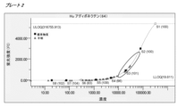
アディポネクチンは、ルミネックスアッセイ(表2)を使用して尿中で順調に検出され、濃度は生成された検量線に従って測定可能である(図7および図8)。尿の最適な希釈は、これらの試料が標準曲線の線形部分の間に位置するため、(1:1)および(1:2)であった。プロテアーゼ阻害剤を用いて処理した尿、およびプロテアーゼ阻害剤を用いずに処理した尿については、アディポネクチン濃度の差異はほとんど観察されなかった。アディポネクチンのレベルは、尿中で測定可能である。それ故に尿は、アディポネクチンおよびIL-1RAの測定に適している。
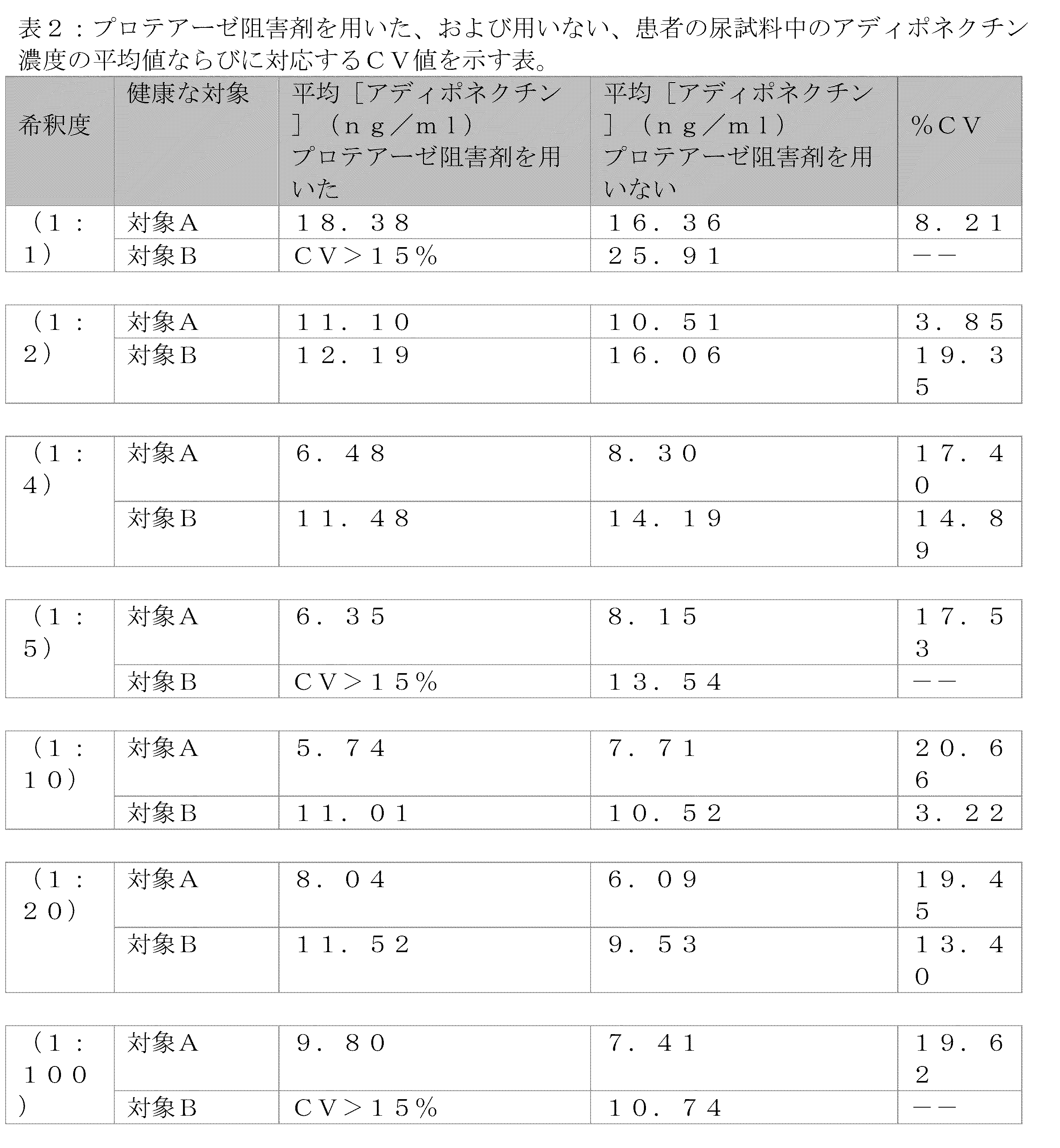
Adiponectin was successfully detected in urine using the Luminex assay (Table 2) and concentrations were measurable according to the generated calibration curves (Figures 7 and 8). The optimal dilutions of urine were (1:1) and (1:2) as these samples were located between the linear parts of the standard curve. Little difference in adiponectin concentrations was observed for urine treated with and without protease inhibitors. Adiponectin levels are measurable in urine. Urine is therefore suitable for the measurement of adiponectin and IL-1RA.
参考文献
ADDIN EN.REFLIST 1.Permert J,Ihse I,Jorfeldt L,et al.Pancreatic cancer is associated with impaired glucose metabolism.Eur J Surg 1993;159:101-7.
2.Pannala R,Leirness JB,Bamlet WR,et al.Prevalence and clinical profile of pancreatic cancer-associated diabetes mellitus.Gastroenterology 2008;134:981-7.
3.Aggarwal G,Kamada P,Chari ST.Prevalence of diabetes mellitus in pancreatic cancer compared to common cancers.Pancreas 2013;42:198-201.
4.Andersen DK,Korc M,Petersen GM,et al.Diabetes,Pancreatogenic Diabetes,and Pancreatic Cancer. Diabetes 2017;66:1103-1110.
5.Sah RP,Nagpal SJ,Mukhopadhyay D,et al.New insights into pancreatic cancer-induced paraneoplastic diabetes. Nat Rev Gastroenterol Hepatol 2013;10:423-33.
6.Jenkinson C,Elliott VL,Evans A,et al.Decreased Serum Thrombospondin-1 Levels in Pancreatic Cancer Patients Up to 24 Months Prior to Clinical Diagnosis: Association with Diabetes Mellitus.Clin Cancer Res 2016;22:1734-43.
7.Shaw VE,Lane B,Jenkinson C,et al.Serum cytokine biomarker panels for discriminating pancreatic cancer from benign pancreatic disease.Mol Cancer 2014;13:114.
8.Woodmansey C,McGovern AP,McCullough KA,et al.Incidence, Demographics,and Clinical Characteristics of Diabetes of the Exocrine Pancreas(Type 3c):A Retrospective Cohort Study.Diabetes Care 2017;40:1486-1493.
9.Jenkinson C,Elliott V,Menon U,et al.Evaluation in pre-diagnosis samples discounts ICAM-1 and TIMP-1 as biomarkers for earlier diagnosis of pancreatic cancer.J Proteomics 2014;113C:400-402.
10.O’Brien DP,Sandanayake NS,Jenkinson C,et al.Serum CA19-9 is significantly upregulated up to 2 years before diagnosis with pancreatic cancer:implications for early disease detection.Clin Cancer Res 2015;21:622-31.
11.Sinclair J,Timms JF.Quantitative profiling of serum samples using TMT protein labelling,fractionation and LC-MS/MS.Methods 2011;54:361-9.
12.Tonack S,Jenkinson C,Cox T,et al.iTRAQ reveals candidate pancreatic cancer serum biomarkers:influence of obstructive jaundice on their performance.Br J Cancer 2013;108:1846-53.
13.Tonack S,Neoptolemos JP,Costello E.Analysis of serum proteins by LC-MS/MS.Methods Mol Biol 2010;658:281-91.
14.Tonack S,Aspinall-O’Dea M,Jenkins RE,et al.A technically detailed and pragmatic protocol for quantitative serum proteomics using iTRAQ.J Proteomics 2009;73:352-6.
15.Yan L,Tonack S,Smith R,et al.Confounding effect of obstructive jaundice in the interpretation of proteomic plasma profiling data for pancreatic cancer.J Proteome Res 2009;8:142-8.
16.Kawada T,Hasegawa M.Predictive ability of serum high-molecular-weight adiponectin in combination with serum insulin and serum C-reactive protein for the presence of metabolic syndrome.Ann Hum Biol 2012;39:108-12.
17.Herder C,Carstensen M,Ouwens DM.Anti-inflammatory cytokines and risk of type 2 diabetes.Diabetes Obesity & Metabolism 2013;15:39-50.
18.Anderson SG,Dunn WB,Banerjee M,et al.Evidence That Multiple Defects in Lipid Regulation Occur before Hyperglycemia during the Prodrome of Type-2 Diabetes.Plos One 2014;9.
19.Marculescu R,Endler G,Schillinger M,et al.Interleukin-1 receptor antagonist genotype is associated with coronary atherosclerosis in patients with type 2 diabetes. Diabetes 2002;51:3582-5.
20.Shaw VE,Lane B,Jenkinson C,et al.Serum cytokine biomarker panels for discriminating pancreatic cancer from benign pancreatic disease.Molecular Cancer 2014;13.
ADDIN EN.REFLIST 21.Zwiech R.Predictive value of conjointly examined IL-1ra,TNF-R I,TNF-R II,and RANTES in patients with primary glomerulonephritis.J Korean Med Sci.2013;28(2):261-7.
ADDIN EN.REFLIST 22.Karpenko MN,Vasilishina AA,Gromova EA,Muruzheva ZM,Miliukhina IV,Bernadotte A.Interleukin-1beta,interleukin-1 receptor antagonist,interleukin-6,interleukin-10,and tumor necrosis factor-alpha levels in CSF and serum in relation to the clinical diversity of Parkinson’s disease.Cell Immunol.2018;327:77-82.
23.Arnalich F,Lopez J,Codoceo R,Jim nez M,Madero R,Montiel C.Relationship of plasma leptin to plasma cytokines and human survivalin sepsis and septic shock.The Journal of infectious diseases.1999;180(3):908-11.
24.Zwiech R.Predictive value of conjointly examined IL-1ra,TNF-R I,TNF-R II,and RANTES in patients with primary glomerulonephritis.J Korean Med Sci.2013;28(2):261-7.
References
2. Pannala R, Leiness JB, Bamlet WR, et al. Prevalence and clinical profile of pancreatic cancer-associated diabetes mellitus. Gastroenterology 2008;134:981-7.
3. Aggarwal G, Kamada P, Chari ST. Prevalence of diabetes mellitus in pancreatic cancer compared to common cancers. Pancreas 2013;42:198-201.
4. Andersen DK, Korc M, Petersen GM, et al. Diabetes, Pancreatogenic Diabetes, and Pancreatic Cancer. Diabetes 2017;66:1103-1110.
5. Sah RP, Nagpal SJ, Mukhopadhyay D, et al. New insights into pancreatic cancer-induced paraneoplastic diabetes. Nat Rev Gastroenterol Hepatol 2013;10:423-33.
6. Jenkinson C, Elliott VL, Evans A, et al. Decreased Serum Thrombospondin-1 Levels in Pancreatic Cancer Patients Up to 24 Months Prior to Clinical Diagnosis: Association with Diabetes Mellitus. Clin Cancer Res 2016;22:1734-43.
7. Shaw VE, Lane B, Jenkinson C, et al. Serum cytokine biomarker panels for discriminating pancreatic cancer from benign pancreatic disease. Mol Cancer 2014;13:114.
8. Woodmansey C, McGovern AP, McCullough KA, et al. Incidence, Demographics, and Clinical Characteristics of Diabetes of the Exocrine Pancreas (Type 3c): A Retrospective Cohort Study. Diabetes Care 2017;40:1486-1493.
9. Jenkinson C, Elliott V, Menon U, et al. Evaluation in pre-diagnosis samples discounts ICAM-1 and TIMP-1 as biomarkers for earlier diagnosis of pancreatic cancer. J Proteomics 2014;113C:400-402.
10. O'Brien DP, Sandanayake NS, Jenkinson C, et al. Serum CA19-9 is significantly upregulated up to 2 years before diagnosis with pancreatic cancer: implications for early disease detection. Clin Cancer Res 2015;21:622-31.
11. Sinclair J, Timms JF. Quantitative profiling of serum samples using TMT protein labeling, fractionation and LC-MS/MS. Methods 2011;54:361-9.
12. Tonack S, Jenkinson C, Cox T, et al. iTRAQ reveals candidates pancreatic cancer serum biomarkers: influence of obstructive jaundice on their performance. Br J Cancer 2013;108:1846-53.
13. Tonack S, Neoptolemos JP, Costello E. Analysis of serum proteins by LC-MS/MS. Methods Mol Biol 2010;658:281-91.
14. Tonack S, Aspinall-O'Dea M, Jenkins RE, et al. A technically detailed and pragmatic protocol for quantitative serum proteomics using iTRAQ. J Proteomics 2009;73:352-6.
15. Yan L, Tonack S, Smith R, et al. Confounding effect of obstructive jarundice in the interpretation of proteomic plasma profiling data for pancreatic cancer. J Proteome Res 2009;8:142-8.
16. Kawada T, Hasegawa M. Predictive ability of serum high-molecular-weight adiponectin in combination with serum insulin and serum C-reactive protein for the presence of metabolic syndrome. Ann Hum Biol 2012;39:108-12.
17. Herder C, Carstensen M, Owens DM. Anti-inflammatory cytokines and risk of type 2 diabetes. Diabetes Obesity & Metabolism 2013;15:39-50.
18. Anderson SG, Dunn WB, Banerjee M, et al. Evidence That Multiple Defects in Lipid Regulation Occur before Hyperglycemia during the Prodrome of Type-2 Diabetes. Plos One 2014;9.
19. Marcuse R, Endler G, Schillinger M, et al. Interleukin-1 receptor antagonist genotype is associated with coronary atherosclerosis in patients with type 2 diabetes. Diabetes 2002;51:3582-5.
20. Shaw VE, Lane B, Jenkinson C, et al. Serum cytokine biomarker panels for discriminating pancreatic cancer from benign pancreatic disease. Molecular Cancer 2014;13.
ADDIN EN.REFLIST 21. Zwiech R. Predictive value of conjointly examined IL-1ra, TNF-R I, TNF-R II, and RANTES in patients with primary glomerulonephritis. J Korean Med Sci. 2013;28(2):261-7.
ADDIN EN.REFLIST 22. Karpenko MN, Vasilishina AA, Gromova EA, Muruzheva ZM, Miliukhina IV, Bernadotte A. Interleukin-1beta, interleukin-1 receptor antagonist, interleukin-6, interleukin-10, and tumor necrosis factor-alpha levels in CSF and serum in relation to the clinical diversity of Parkinson's disease. Cell Immunol. 2018;327:77-82.
23. Arnalich F, Lopez J, Codoceo R, Jimnez M, Madero R, Montiel C. Relationship of plasma leptin to plasma cytokines and human survivalin sepsis and septic shock. The Journal of infectious diseases. 1999;180(3):908-11.
24. Zwiech R. Predictive value of conjointly examined IL-1ra, TNF-R I, TNF-R II, and RANTES in patients with primary glomerulonephritis. J Korean Med Sci. 2013;28(2):261-7.
Claims (17)
i)被験対象から取得した血漿または血清の試料と、アディポネクチンに結合する抗体(単数または複数)と、IL-1Raに結合する抗体(単数または複数)とを含む調製物を形成して、抗体/アディポネクチンポリペプチド複合体および抗体/IL-1Raポリペプチド複合体を形成する工程と、
ii)各複合体におけるアディポネクチンおよびIL-1Raのレベルを検出する工程と、
iii)工程ii)で得られたアディポネクチンおよびIL-1Raのレベルを、関連する対応対照におけるアディポネクチンおよびIL-1Raのレベルと比較する工程であって、前記関連する対応対照が、随伴性の膵癌および随伴性の膵炎のいずれかを伴わない新規発症糖尿病(DM)に罹患しているか、またはその疑いのある対象である工程と、を含むか、またはそれらの工程からなるイムノアッセイ。 1. An immunoassay method for testing levels of adiponectin and IL-1Ra polypeptides, comprising:
i) forming a preparation comprising a plasma or serum sample obtained from a subject, an antibody or antibodies that bind adiponectin, and an antibody or antibodies that bind IL-1Ra to form antibody/adiponectin polypeptide complexes and antibody/IL-1Ra polypeptide complexes;
ii) detecting the levels of adiponectin and IL-1Ra in each complex;
and iii) comparing the levels of adiponectin and IL-1Ra obtained in step ii) with the levels of adiponectin and IL-1Ra in a relevant matched control, said relevant matched control being a subject suffering from or suspected of suffering from new-onset diabetes mellitus (DM) without either concomitant pancreatic cancer or concomitant pancreatitis.
i)被験対象から取得した血漿または血清の試料と、アディポネクチンに結合する抗体(単数または複数)と、IL-1Raに結合する抗体(単数または複数)とを含む調製物を形成して、抗体/アディポネクチンポリペプチド複合体および抗体/IL-1Raポリペプチド複合体を形成する工程と、
ii)各複合体におけるアディポネクチンおよびIL-1Raのレベルを検出する工程と、
iii)工程ii)で得られたアディポネクチンポリペプチドおよびIL-1Raポリペプチドのレベルを、関連する対応対照におけるアディポネクチンポリペプチドおよびIL-1Raポリペプチドのレベルと比較する工程であって、前記関連する対応対照が、随伴性の膵癌および随伴性の膵炎のいずれかを伴わない新規発症糖尿病(DM)に罹患しているか、またはその疑いのある対象である工程と、を含むか、またはそれらの工程からなるイムノアッセイの方法。 1. An immunoassay method for testing whether a subject is suspected of having early stage pancreatic ductal adenocarcinoma (PDAC) or pancreatitis, comprising:
i) forming a preparation comprising a plasma or serum sample obtained from a subject, an antibody or antibodies that bind adiponectin, and an antibody or antibodies that bind IL-1Ra to form antibody/adiponectin polypeptide complexes and antibody/IL-1Ra polypeptide complexes;
ii ) detecting the levels of adiponectin and IL-1Ra in each complex;
iii ) comparing the levels of adiponectin polypeptide and IL-1Ra polypeptide obtained in step ii) with the levels of adiponectin polypeptide and IL-1Ra polypeptide in a relevant matched control, wherein the relevant matched control is a subject suffering from or suspected of suffering from new-onset diabetes mellitus (DM) without either concomitant pancreatic cancer or concomitant pancreatitis.
アディポネクチンポリペプチドに特異的に結合する抗体と、
IL-1Raポリペプチドに特異的に結合する抗体と、
アディポネクチンポリペプチドに結合する前記抗体に結合する二次抗体、およびIL-1Raポリペプチドに結合する前記抗体に結合する二次抗体であって、別個の検出可能な標識を含む前記二次抗体と、を含む、またはそれらからなる、キット。 A kit for use in the method of any one of claims 1 to 9, comprising :
An antibody that specifically binds to an adiponectin polypeptide;
an antibody that specifically binds to an IL-1Ra polypeptide;
A kit comprising or consisting of a secondary antibody that binds to the antibody that binds to an adiponectin polypeptide, and a secondary antibody that binds to the antibody that binds to an IL-1Ra polypeptide, wherein the secondary antibody comprises a separate detectable label.
i)被験対象から取得した血漿または血清の試料と、アディポネクチンに結合する抗体(単数または複数)と、IL-1Raに結合する抗体(単数または複数)とを含む調製物を形成して、抗体/アディポネクチンポリペプチド複合体および抗体/IL-1Raポリペプチド複合体を形成する工程と、
ii)各複合体におけるアディポネクチンおよびIL-1Raのレベルを検出する工程と、
iii)工程ii)で得られたアディポネクチンポリペプチドおよびIL-1Raポリペプチドのレベルを、関連する対応対照におけるアディポネクチンポリペプチドおよびIL-1Raポリペプチドのレベルと比較する工程であって、前記関連する対応対照が、随伴性の膵癌および随伴性の膵炎のいずれかを伴わない新規発症糖尿病(DM)に罹患しているか、またはその疑いのある対象である工程と、を含むか、またはそれらの工程からなるイムノアッセイの方法。 1. An immunoassay method for testing whether a subject should be imaged for early stage pancreatic ductal adenocarcinoma or pancreatitis, comprising:
i) forming a preparation comprising a plasma or serum sample obtained from a subject, an antibody or antibodies that bind adiponectin, and an antibody or antibodies that bind IL-1Ra to form antibody/adiponectin polypeptide complexes and antibody/IL-1Ra polypeptide complexes;
ii ) detecting the levels of adiponectin and IL-1Ra in each complex;
iii ) comparing the levels of adiponectin polypeptide and IL-1Ra polypeptide obtained in step ii) with the levels of adiponectin polypeptide and IL-1Ra polypeptide in a relevant matched control, wherein the relevant matched control is a subject suffering from or suspected of suffering from new-onset diabetes mellitus (DM) without either concomitant pancreatic cancer or concomitant pancreatitis.
Applications Claiming Priority (5)
| Application Number | Priority Date | Filing Date | Title |
|---|---|---|---|
| GBGB1902193.0A GB201902193D0 (en) | 2019-02-18 | 2019-02-18 | Method of diagnosis |
| GB1902193.0 | 2019-02-18 | ||
| GB1903424.8 | 2019-03-13 | ||
| GBGB1903424.8A GB201903424D0 (en) | 2019-03-13 | 2019-03-13 | Method of diagnosis |
| PCT/EP2020/054046 WO2020169511A1 (en) | 2019-02-18 | 2020-02-17 | Method of diagnosis |
Publications (3)
| Publication Number | Publication Date |
|---|---|
| JP2022529073A JP2022529073A (en) | 2022-06-16 |
| JP2022529073A5 JP2022529073A5 (en) | 2023-02-17 |
| JP7660077B2 true JP7660077B2 (en) | 2025-04-10 |
Family
ID=69591656
Family Applications (1)
| Application Number | Title | Priority Date | Filing Date |
|---|---|---|---|
| JP2021572701A Active JP7660077B2 (en) | 2019-02-18 | 2020-02-17 | Diagnostic methods |
Country Status (7)
| Country | Link |
|---|---|
| US (1) | US20220091140A1 (en) |
| EP (1) | EP3903105A1 (en) |
| JP (1) | JP7660077B2 (en) |
| CN (1) | CN113454459A (en) |
| AU (1) | AU2020223826A1 (en) |
| CA (1) | CA3127569A1 (en) |
| WO (1) | WO2020169511A1 (en) |
Citations (7)
| Publication number | Priority date | Publication date | Assignee | Title |
|---|---|---|---|---|
| WO2003016906A1 (en) | 2001-08-17 | 2003-02-27 | Rebio Gen, Inc. | Method of diagnosing or monitoring saccharometabolic error |
| JP2007525410A (en) | 2003-01-17 | 2007-09-06 | ザ リサーチ ファンデーション オブ ステイト ユニバーシティ オブ ニューヨーク | Antigen related to pancreatic cancer, antibody against them, diagnostic method and treatment method |
| JP2009528507A (en) | 2006-03-02 | 2009-08-06 | オンコセラピー・サイエンス株式会社 | Method for diagnosing pancreatic cancer using REG4 protein |
| JP2014508298A (en) | 2011-03-04 | 2014-04-03 | イムノヴィア・アクチエボラーグ | Methods, arrays and uses thereof |
| JP2014512808A (en) | 2011-03-04 | 2014-05-29 | イントレキソン コーポレーション | Vectors that conditionally express proteins |
| JP2017532633A (en) | 2014-08-14 | 2017-11-02 | メメド ダイアグノスティクス リミテッド | Computer analysis of biological data using manifolds and hyperplanes |
| US20170356908A1 (en) | 2014-10-22 | 2017-12-14 | The George Washington University a Congressionally Chartered Not-for-Profit Corporation | Blood biomarkers for appendicitis and diagnostics methods using biomarkers |
Family Cites Families (10)
| Publication number | Priority date | Publication date | Assignee | Title |
|---|---|---|---|---|
| US20060105419A1 (en) * | 2004-08-16 | 2006-05-18 | Biosite, Inc. | Use of a glutathione peroxidase 1 as a marker in cardiovascular conditions |
| US8632983B2 (en) * | 2005-04-15 | 2014-01-21 | Van Andel Research Institute | Biomarkers for pancreatic cancer and diagnostic methods |
| CN102939541B (en) * | 2010-06-15 | 2015-12-02 | 弗·哈夫曼-拉罗切有限公司 | Prediction and recognition of postoperative acute kidney injury |
| JP2014510516A (en) * | 2011-02-15 | 2014-05-01 | ノヴォ ノルディスク アー/エス | Long acting IL-1 receptor antagonist |
| US20160231334A1 (en) * | 2013-10-28 | 2016-08-11 | The Forsyth Institute | Compositions and Methods for Evaluating Metabolic Syndrome and Related Diseases |
| WO2015112429A1 (en) | 2014-01-23 | 2015-07-30 | Newomics Inc | Methods and systems for diagnosing diseases |
| WO2015157557A1 (en) | 2014-04-09 | 2015-10-15 | Myriad Rbm, Inc. | Methods of diagnosing pancreatic cancer and methods related thereto |
| WO2017019588A1 (en) * | 2015-07-28 | 2017-02-02 | The Johns Hopkins University | Compositions and methods for detecting pancreatic cancer |
| GB201522839D0 (en) | 2015-12-23 | 2016-02-03 | Randox Lab Ltd And Randox Teoranta | Method |
| US12416636B2 (en) * | 2018-05-31 | 2025-09-16 | University Of Florida Research Foundation, Inc. | Personalized treatment of pancreatic cancer |
-
2020
- 2020-02-17 CN CN202080015187.6A patent/CN113454459A/en active Pending
- 2020-02-17 AU AU2020223826A patent/AU2020223826A1/en not_active Abandoned
- 2020-02-17 CA CA3127569A patent/CA3127569A1/en active Pending
- 2020-02-17 JP JP2021572701A patent/JP7660077B2/en active Active
- 2020-02-17 EP EP20704770.5A patent/EP3903105A1/en active Pending
- 2020-02-17 US US17/431,637 patent/US20220091140A1/en active Pending
- 2020-02-17 WO PCT/EP2020/054046 patent/WO2020169511A1/en not_active Ceased
Patent Citations (7)
| Publication number | Priority date | Publication date | Assignee | Title |
|---|---|---|---|---|
| WO2003016906A1 (en) | 2001-08-17 | 2003-02-27 | Rebio Gen, Inc. | Method of diagnosing or monitoring saccharometabolic error |
| JP2007525410A (en) | 2003-01-17 | 2007-09-06 | ザ リサーチ ファンデーション オブ ステイト ユニバーシティ オブ ニューヨーク | Antigen related to pancreatic cancer, antibody against them, diagnostic method and treatment method |
| JP2009528507A (en) | 2006-03-02 | 2009-08-06 | オンコセラピー・サイエンス株式会社 | Method for diagnosing pancreatic cancer using REG4 protein |
| JP2014508298A (en) | 2011-03-04 | 2014-04-03 | イムノヴィア・アクチエボラーグ | Methods, arrays and uses thereof |
| JP2014512808A (en) | 2011-03-04 | 2014-05-29 | イントレキソン コーポレーション | Vectors that conditionally express proteins |
| JP2017532633A (en) | 2014-08-14 | 2017-11-02 | メメド ダイアグノスティクス リミテッド | Computer analysis of biological data using manifolds and hyperplanes |
| US20170356908A1 (en) | 2014-10-22 | 2017-12-14 | The George Washington University a Congressionally Chartered Not-for-Profit Corporation | Blood biomarkers for appendicitis and diagnostics methods using biomarkers |
Non-Patent Citations (9)
| Title |
|---|
| C. HERDER et al.,Anti-inflammatory cytokines and risk of type-2 diabetes,Diabetes, Obesity and Metabolism,2013年,Vol.15, Suppl.3,pp.39-50 |
| D. DRANKA-BOJAROWSKA et al.,The assessment of serum concentration of adiponectin, leptin and serum carbohydrate antigen 19.9 in patients with pancreatic cancer and chronic pancreatitis,Journal of Physiology and Pharmacology,2015年,Vol.66, No.5,pp.653-663 |
| Hye Won CHUNG et al.,Clinical significance of elevated serum soluble CD40 ligand levels as diagnostic and prognostic tumor marker for pancreatic ductal adenocarcinoma,Journal of Translational Medicine,2014年,Vol.12, No.1,Article No.102 |
| Irini CHATZIRALLI et al.,Model for Risk-Based Screening of Diabetic Retinopathy in People With Newly-Diagnosed Type 2 Diabetes Mellitus,Investigative ophthalmology & visual science,2017年05月01日,Vol.58, No.6,BIO99-BIO105 |
| Jan SKRHA et al.,Lower plasma levels of glucose-dependent insulinotropic peptide (GIP) and pancreatic polypeptide (PP) in patients with ductal adenocarcinoma of the pancreas and their relation to the presence of impaired glucoregulation and weight loss,Pancreatology,2016年12月15日,Vol.17,pp.89-94 |
| Kadri COLAKIGLU et al.,Role of adiponectin and leptin as diagnostic markers in pancreatic cancer,Bratislava Medical Journal,2017年07月24日,Vol.118, No.7,pp.394-398 |
| Ming-Chu CHANG et al.,Adiponectin as a Potential Differential Marker to Distinguish pancreatic Cancer and Chronic Pancreatitis,Pancreas,2007年07月,Vol.35, No.1,pp.16-21 |
| Ramesh KHADKA et al.,Risk factor, early diagnosis and overall survival on outcome of association between pancreatic cancer and diabetes mellitus: Changes and advances, a review,International Journal of Surgery,2018年04月,Vol.52,pp.342-346 |
| T. KRECHLER et al.,Leptin and adiponectin in pancreatic cancer: connection with diabetes mellitus,Neoplasma,2011年,Vol.58, No.1,pp.58-64 |
Also Published As
| Publication number | Publication date |
|---|---|
| US20220091140A1 (en) | 2022-03-24 |
| CA3127569A1 (en) | 2020-08-27 |
| CN113454459A (en) | 2021-09-28 |
| JP2022529073A (en) | 2022-06-16 |
| WO2020169511A1 (en) | 2020-08-27 |
| AU2020223826A1 (en) | 2021-08-05 |
| EP3903105A1 (en) | 2021-11-03 |
Similar Documents
| Publication | Publication Date | Title |
|---|---|---|
| Al-Sawaf et al. | Body composition and lung cancer-associated cachexia in TRACERx | |
| US20250216393A1 (en) | Plasma autoantibody biomarkers for diagnosis of lung cancer | |
| Rho et al. | Protein and glycomic plasma markers for early detection of adenoma and colon cancer | |
| JP6774925B2 (en) | Methods and kits for the identification, evaluation, prevention and treatment of lung diseases, including gender-based disease identification, evaluation, prevention and treatment. | |
| EP3073268A1 (en) | Biomarker panel for diagnosing cancer | |
| JP6983221B2 (en) | Combined test for colorectal cancer | |
| US20120309697A1 (en) | Methods of diagnosing and prognosing colonic polyps | |
| JP2018519499A (en) | Method for predicting the risk of developing chronic kidney disease | |
| Lipshitz et al. | Emerging markers of cancer cachexia and their relationship to sarcopenia | |
| WO2019048588A1 (en) | Mixed protein and autoantibody biomarker panel for diagnosing colorectal cancer | |
| CN113785199B (en) | Protein characterization for diagnosing colorectal cancer and/or pre-cancerous stage | |
| JP2021144052A (en) | Methods of detecting relapse of lung adenocarcinoma based on marker human epididymis protein 4 (he4) and related uses | |
| EP3835789A1 (en) | Biomarker panel for diagnosing colorectal cancer | |
| JP5704684B2 (en) | IgG4-related disease diagnostic marker and use thereof | |
| Kovacevic et al. | Urine proteomic analysis in cystinuric children with renal stones | |
| US20190101540A1 (en) | Angiogenesis Biomarkers Associated With Disease Progression in Lung Cancer | |
| JP7660077B2 (en) | Diagnostic methods | |
| EP2895863B1 (en) | New biomarkers for the diagnosis and/or prognosis of clear cell renal cell carcinoma | |
| CN114286942B (en) | Methods for detecting cancer | |
| Shah et al. | Serum glycoprotein biomarker validation for esophageal adenocarcinoma and application to Barrett’s surveillance | |
| KR20240068432A (en) | A kit for diagnosing cancer comprising protein biomarker in blood | |
| Mohamed et al. | Faculty of Graduate Studies |
Legal Events
| Date | Code | Title | Description |
|---|---|---|---|
| A521 | Request for written amendment filed |
Free format text: JAPANESE INTERMEDIATE CODE: A523 Effective date: 20220316 |
|
| A521 | Request for written amendment filed |
Free format text: JAPANESE INTERMEDIATE CODE: A523 Effective date: 20230209 |
|
| A621 | Written request for application examination |
Free format text: JAPANESE INTERMEDIATE CODE: A621 Effective date: 20230209 |
|
| A977 | Report on retrieval |
Free format text: JAPANESE INTERMEDIATE CODE: A971007 Effective date: 20231124 |
|
| A131 | Notification of reasons for refusal |
Free format text: JAPANESE INTERMEDIATE CODE: A131 Effective date: 20231212 |
|
| A601 | Written request for extension of time |
Free format text: JAPANESE INTERMEDIATE CODE: A601 Effective date: 20240227 |
|
| A521 | Request for written amendment filed |
Free format text: JAPANESE INTERMEDIATE CODE: A523 Effective date: 20240604 |
|
| A131 | Notification of reasons for refusal |
Free format text: JAPANESE INTERMEDIATE CODE: A131 Effective date: 20240910 |
|
| A521 | Request for written amendment filed |
Free format text: JAPANESE INTERMEDIATE CODE: A523 Effective date: 20241204 |
|
| TRDD | Decision of grant or rejection written | ||
| A01 | Written decision to grant a patent or to grant a registration (utility model) |
Free format text: JAPANESE INTERMEDIATE CODE: A01 Effective date: 20250304 |
|
| A61 | First payment of annual fees (during grant procedure) |
Free format text: JAPANESE INTERMEDIATE CODE: A61 Effective date: 20250331 |
|
| R150 | Certificate of patent or registration of utility model |
Ref document number: 7660077 Country of ref document: JP Free format text: JAPANESE INTERMEDIATE CODE: R150 |