JP7701879B2 - Methods for generating monocyte progenitor cells - Google Patents
Methods for generating monocyte progenitor cells Download PDFInfo
- Publication number
- JP7701879B2 JP7701879B2 JP2021570293A JP2021570293A JP7701879B2 JP 7701879 B2 JP7701879 B2 JP 7701879B2 JP 2021570293 A JP2021570293 A JP 2021570293A JP 2021570293 A JP2021570293 A JP 2021570293A JP 7701879 B2 JP7701879 B2 JP 7701879B2
- Authority
- JP
- Japan
- Prior art keywords
- cells
- days
- medium
- laminin
- monocyte
- Prior art date
- Legal status (The legal status is an assumption and is not a legal conclusion. Google has not performed a legal analysis and makes no representation as to the accuracy of the status listed.)
- Active
Links
Images








Classifications
-
- C—CHEMISTRY; METALLURGY
- C12—BIOCHEMISTRY; BEER; SPIRITS; WINE; VINEGAR; MICROBIOLOGY; ENZYMOLOGY; MUTATION OR GENETIC ENGINEERING
- C12N—MICROORGANISMS OR ENZYMES; COMPOSITIONS THEREOF; PROPAGATING, PRESERVING, OR MAINTAINING MICROORGANISMS; MUTATION OR GENETIC ENGINEERING; CULTURE MEDIA
- C12N5/00—Undifferentiated human, animal or plant cells, e.g. cell lines; Tissues; Cultivation or maintenance thereof; Culture media therefor
- C12N5/06—Animal cells or tissues; Human cells or tissues
- C12N5/0602—Vertebrate cells
- C12N5/0696—Artificially induced pluripotent stem cells, e.g. iPS
-
- C—CHEMISTRY; METALLURGY
- C12—BIOCHEMISTRY; BEER; SPIRITS; WINE; VINEGAR; MICROBIOLOGY; ENZYMOLOGY; MUTATION OR GENETIC ENGINEERING
- C12N—MICROORGANISMS OR ENZYMES; COMPOSITIONS THEREOF; PROPAGATING, PRESERVING, OR MAINTAINING MICROORGANISMS; MUTATION OR GENETIC ENGINEERING; CULTURE MEDIA
- C12N5/00—Undifferentiated human, animal or plant cells, e.g. cell lines; Tissues; Cultivation or maintenance thereof; Culture media therefor
- C12N5/0068—General culture methods using substrates
-
- C—CHEMISTRY; METALLURGY
- C12—BIOCHEMISTRY; BEER; SPIRITS; WINE; VINEGAR; MICROBIOLOGY; ENZYMOLOGY; MUTATION OR GENETIC ENGINEERING
- C12N—MICROORGANISMS OR ENZYMES; COMPOSITIONS THEREOF; PROPAGATING, PRESERVING, OR MAINTAINING MICROORGANISMS; MUTATION OR GENETIC ENGINEERING; CULTURE MEDIA
- C12N5/00—Undifferentiated human, animal or plant cells, e.g. cell lines; Tissues; Cultivation or maintenance thereof; Culture media therefor
- C12N5/06—Animal cells or tissues; Human cells or tissues
- C12N5/0602—Vertebrate cells
- C12N5/0603—Embryonic cells ; Embryoid bodies
- C12N5/0606—Pluripotent embryonic cells, e.g. embryonic stem cells [ES]
-
- C—CHEMISTRY; METALLURGY
- C12—BIOCHEMISTRY; BEER; SPIRITS; WINE; VINEGAR; MICROBIOLOGY; ENZYMOLOGY; MUTATION OR GENETIC ENGINEERING
- C12N—MICROORGANISMS OR ENZYMES; COMPOSITIONS THEREOF; PROPAGATING, PRESERVING, OR MAINTAINING MICROORGANISMS; MUTATION OR GENETIC ENGINEERING; CULTURE MEDIA
- C12N5/00—Undifferentiated human, animal or plant cells, e.g. cell lines; Tissues; Cultivation or maintenance thereof; Culture media therefor
- C12N5/06—Animal cells or tissues; Human cells or tissues
- C12N5/0602—Vertebrate cells
- C12N5/0634—Cells from the blood or the immune system
- C12N5/0645—Macrophages, e.g. Kuepfer cells in the liver; Monocytes
-
- C—CHEMISTRY; METALLURGY
- C12—BIOCHEMISTRY; BEER; SPIRITS; WINE; VINEGAR; MICROBIOLOGY; ENZYMOLOGY; MUTATION OR GENETIC ENGINEERING
- C12N—MICROORGANISMS OR ENZYMES; COMPOSITIONS THEREOF; PROPAGATING, PRESERVING, OR MAINTAINING MICROORGANISMS; MUTATION OR GENETIC ENGINEERING; CULTURE MEDIA
- C12N2500/00—Specific components of cell culture medium
- C12N2500/90—Serum-free medium, which may still contain naturally-sourced components
-
- C—CHEMISTRY; METALLURGY
- C12—BIOCHEMISTRY; BEER; SPIRITS; WINE; VINEGAR; MICROBIOLOGY; ENZYMOLOGY; MUTATION OR GENETIC ENGINEERING
- C12N—MICROORGANISMS OR ENZYMES; COMPOSITIONS THEREOF; PROPAGATING, PRESERVING, OR MAINTAINING MICROORGANISMS; MUTATION OR GENETIC ENGINEERING; CULTURE MEDIA
- C12N2501/00—Active agents used in cell culture processes, e.g. differentation
- C12N2501/10—Growth factors
- C12N2501/115—Basic fibroblast growth factor (bFGF, FGF-2)
-
- C—CHEMISTRY; METALLURGY
- C12—BIOCHEMISTRY; BEER; SPIRITS; WINE; VINEGAR; MICROBIOLOGY; ENZYMOLOGY; MUTATION OR GENETIC ENGINEERING
- C12N—MICROORGANISMS OR ENZYMES; COMPOSITIONS THEREOF; PROPAGATING, PRESERVING, OR MAINTAINING MICROORGANISMS; MUTATION OR GENETIC ENGINEERING; CULTURE MEDIA
- C12N2501/00—Active agents used in cell culture processes, e.g. differentation
- C12N2501/10—Growth factors
- C12N2501/155—Bone morphogenic proteins [BMP]; Osteogenins; Osteogenic factor; Bone inducing factor
-
- C—CHEMISTRY; METALLURGY
- C12—BIOCHEMISTRY; BEER; SPIRITS; WINE; VINEGAR; MICROBIOLOGY; ENZYMOLOGY; MUTATION OR GENETIC ENGINEERING
- C12N—MICROORGANISMS OR ENZYMES; COMPOSITIONS THEREOF; PROPAGATING, PRESERVING, OR MAINTAINING MICROORGANISMS; MUTATION OR GENETIC ENGINEERING; CULTURE MEDIA
- C12N2501/00—Active agents used in cell culture processes, e.g. differentation
- C12N2501/20—Cytokines; Chemokines
- C12N2501/23—Interleukins [IL]
- C12N2501/2303—Interleukin-3 (IL-3)
-
- C—CHEMISTRY; METALLURGY
- C12—BIOCHEMISTRY; BEER; SPIRITS; WINE; VINEGAR; MICROBIOLOGY; ENZYMOLOGY; MUTATION OR GENETIC ENGINEERING
- C12N—MICROORGANISMS OR ENZYMES; COMPOSITIONS THEREOF; PROPAGATING, PRESERVING, OR MAINTAINING MICROORGANISMS; MUTATION OR GENETIC ENGINEERING; CULTURE MEDIA
- C12N2502/00—Coculture with; Conditioned medium produced by
- C12N2502/08—Coculture with; Conditioned medium produced by cells of the nervous system
-
- C—CHEMISTRY; METALLURGY
- C12—BIOCHEMISTRY; BEER; SPIRITS; WINE; VINEGAR; MICROBIOLOGY; ENZYMOLOGY; MUTATION OR GENETIC ENGINEERING
- C12N—MICROORGANISMS OR ENZYMES; COMPOSITIONS THEREOF; PROPAGATING, PRESERVING, OR MAINTAINING MICROORGANISMS; MUTATION OR GENETIC ENGINEERING; CULTURE MEDIA
- C12N2506/00—Differentiation of animal cells from one lineage to another; Differentiation of pluripotent cells
- C12N2506/02—Differentiation of animal cells from one lineage to another; Differentiation of pluripotent cells from embryonic cells
-
- C—CHEMISTRY; METALLURGY
- C12—BIOCHEMISTRY; BEER; SPIRITS; WINE; VINEGAR; MICROBIOLOGY; ENZYMOLOGY; MUTATION OR GENETIC ENGINEERING
- C12N—MICROORGANISMS OR ENZYMES; COMPOSITIONS THEREOF; PROPAGATING, PRESERVING, OR MAINTAINING MICROORGANISMS; MUTATION OR GENETIC ENGINEERING; CULTURE MEDIA
- C12N2506/00—Differentiation of animal cells from one lineage to another; Differentiation of pluripotent cells
- C12N2506/45—Differentiation of animal cells from one lineage to another; Differentiation of pluripotent cells from artificially induced pluripotent stem cells
-
- C—CHEMISTRY; METALLURGY
- C12—BIOCHEMISTRY; BEER; SPIRITS; WINE; VINEGAR; MICROBIOLOGY; ENZYMOLOGY; MUTATION OR GENETIC ENGINEERING
- C12N—MICROORGANISMS OR ENZYMES; COMPOSITIONS THEREOF; PROPAGATING, PRESERVING, OR MAINTAINING MICROORGANISMS; MUTATION OR GENETIC ENGINEERING; CULTURE MEDIA
- C12N2533/00—Supports or coatings for cell culture, characterised by material
- C12N2533/50—Proteins
- C12N2533/52—Fibronectin; Laminin
Landscapes
- Health & Medical Sciences (AREA)
- Engineering & Computer Science (AREA)
- Life Sciences & Earth Sciences (AREA)
- Biomedical Technology (AREA)
- Zoology (AREA)
- Wood Science & Technology (AREA)
- Biotechnology (AREA)
- Organic Chemistry (AREA)
- Bioinformatics & Cheminformatics (AREA)
- Genetics & Genomics (AREA)
- Chemical & Material Sciences (AREA)
- General Engineering & Computer Science (AREA)
- Microbiology (AREA)
- Biochemistry (AREA)
- General Health & Medical Sciences (AREA)
- Cell Biology (AREA)
- Immunology (AREA)
- Developmental Biology & Embryology (AREA)
- Gynecology & Obstetrics (AREA)
- Reproductive Health (AREA)
- Gastroenterology & Hepatology (AREA)
- Hematology (AREA)
- Transplantation (AREA)
- Micro-Organisms Or Cultivation Processes Thereof (AREA)
- Medicines Containing Material From Animals Or Micro-Organisms (AREA)
Description
発明の分野
本出願は、単球前駆細胞の生成ならびにそれらのマクロファージおよびミクログリアへの分化のための方法、ならびに単球前駆細胞を産生するための大規模細胞培養物に関する。
FIELD OF THEINVENTION This application relates to methods for the generation of monocyte progenitor cells and their differentiation into macrophages and microglia, as well as large-scale cell cultures for producing monocyte progenitor cells.
背景
単球およびマクロファージは、炎症プロセスにおいて重要な役割を果たすものであり、それらの活性化および機能性は、健康および疾患において極めて重要である(Biswas et al.2012、Mantovani et al.2013、Sica et al.2008、Wynn et al.2013)。マクロファージの関与が確認されている疾患には、代謝性疾患、アレルギー障害、自己免疫性、がん、神経変性疾患、ならびに細菌、ウイルス、寄生虫、および真菌の感染症が包含される。疾患の状況における急性免疫防御の媒介の他に、組織全体に広く分布しているマクロファージは、周囲組織の修復およびホメオスタシスに必須である。したがって、マクロファージの機能性の損傷、およびそれに続くホメオスタシスの喪失は、変性疾患の発生機序と密接に関連している。
Background Monocytes and macrophages play a key role in inflammatory processes, and their activation and functionality are crucial in health and disease (Biswas et al. 2012, Mantovani et al. 2013, Sica et al. 2008, Wynn et al. 2013). Diseases in which macrophages have been implicated include metabolic diseases, allergic disorders, autoimmune, cancer, neurodegenerative diseases, and bacterial, viral, parasitic, and fungal infections. Besides mediating acute immune defense in disease situations, macrophages, which are widely distributed throughout tissues, are essential for the repair and homeostasis of surrounding tissues. Thus, impaired macrophage functionality and subsequent loss of homeostasis are closely linked to the pathogenesis of degenerative diseases.
ホメオスタシスおよび疾患防御における重要なマクロファージ機能としては、食作用(病原体、デブリ、および死細胞)、遊走(損傷側への)、ならびにさらなる炎症応答をトリガーするかまたは周囲組織に栄養支持を与えるためのサイトカイン放出が挙げられる。(Biswas et al.2012、Mantovani et al.2013、Sica et al.2008、Wynn et al.2013)。この理由のため、単球/マクロファージ機能のモジュレーションは、多くの疾患を解決する可能性のある治療的戦略を表す。マクロファージが関与する広範な疾患領域およびマクロファージの機能特性により、可能性のある標的は非常に多様である(Tiwari et al.2008)。これにより、薬物の開発およびスクリーニングのために単球およびマクロファージに高い需要が生じる。 Important macrophage functions in homeostasis and disease defense include phagocytosis (pathogens, debris, and dead cells), migration (towards the injured side), and cytokine release to trigger further inflammatory responses or provide trophic support to surrounding tissues. (Biswas et al. 2012, Mantovani et al. 2013, Sica et al. 2008, Wynn et al. 2013). For this reason, modulation of monocyte/macrophage function represents a potential therapeutic strategy to resolve many diseases. Due to the wide range of disease areas in which macrophages are involved and the functional properties of macrophages, the potential targets are highly diverse (Tiwari et al. 2008). This creates a high demand for monocytes and macrophages for drug development and screening.
これまで、マクロファージの研究は、関連する細胞の生成に関する制限によって、複雑で時間のかかるものであった。過去に主に使用されていたマクロファージを得る1つの手段は、献血から濃縮したPBMC(末梢血単核細胞)から単球を単離することである(図1)。しかしながら、ドナー当たりの細胞数が限られていること、ドナーごとの変動、および遺伝子操作の可能性の制限により、これらの初代細胞の使用は制限される。 Until now, the study of macrophages has been complicated and time-consuming due to limitations in the generation of relevant cells. One means of obtaining macrophages, primarily used in the past, is the isolation of monocytes from enriched PBMCs (peripheral blood mononuclear cells) from blood donations (Figure 1). However, the limited number of cells per donor, donor-to-donor variability, and limited possibilities for genetic manipulation limit the use of these primary cells.
最近の研究では、iPS細胞から単球前駆細胞およびマクロファージを誘導することに成功している(Ackermann et al.2018、Hong et al.2018、Karlsson et al.2008、Senju et al.2011、Takamatsu et al.2014、van Wilgenburg et al.2013)。このアプローチは、初代単球の単離と比較していくつかの利点を有する(図1)。これは、疾患関連の遺伝子バックグラウンドを有する細胞の使用、遺伝子操作(すなわち、多能性状態における疾患の素因となる突然変異の補正)を可能にし、必要な場合にはドナーの変動性を制限する。iPS技術により、一定のジェノタイプおよび機能の単球/マクロファージの実質的に無制限の供給がもたらされる。 Recent studies have successfully derived monocyte progenitors and macrophages from iPS cells (Ackermann et al. 2018, Hong et al. 2018, Karlsson et al. 2008, Senju et al. 2011, Takamatsu et al. 2014, van Wilgenburg et al. 2013). This approach has several advantages compared to the isolation of primary monocytes (Figure 1). It allows the use of cells with disease-relevant genetic backgrounds, genetic manipulation (i.e., correction of disease-predisposing mutations in the pluripotent state), and limits donor variability when necessary. iPS technology provides a virtually unlimited supply of monocytes/macrophages of constant genotype and function.
ミクログリアは、組織定住マクロファージの特別なサブタイプである。胚発生中に、卵黄嚢の血島において、2つのマクロファージの波が生じる。これらの卵黄嚢に由来するマクロファージは、Myb非依存性であるが、増殖に関してPU.1およびIRF8依存性であり(Haenseler et al.2016)、組織定住マクロファージを発生させる。多くの組織において、この初期マクロファージ集団は、骨髄由来のマクロファージによって部分的または完全に置き換えられるが、脳定住マクロファージ集団、すなわちミクログリアは、依然としてその起源単独のものである。 Microglia are a special subtype of tissue-resident macrophages. During embryonic development, two waves of macrophages arise in blood islands of the yolk sac. These yolk sac-derived macrophages are Myb-independent but PU.1 and IRF8-dependent for proliferation (Haenseler et al. 2016) and give rise to tissue-resident macrophages. In many tissues, this initial macrophage population is partially or completely replaced by bone marrow-derived macrophages, but the brain-resident macrophage population, i.e., microglia, remains sole of its origin.
ミクログリアは、ミスフォールディングされたタンパク質および死細胞のクリアランス、シナプスの刈り込み、および神経栄養因子の放出など、重要なホメオスタシス機能を有する。さらに、炎症性刺激の際には、それらが活性化され、有害となる可能性のあるサイトカインを放出し、活性酸素種を産生し得る。慢性炎症性活性化、および神経変性疾患のいくつかの遺伝的危険因子(例えば、LRRK2、TREM2、ASYN、およびCD33)の高いレベルの発現により、神経変性疾患および神経炎症におけるミクログリアの役割に対して高い関心が生じている。 Microglia have important homeostatic functions, such as clearance of misfolded proteins and dead cells, pruning of synapses, and release of neurotrophic factors. Furthermore, upon inflammatory stimuli, they can become activated, releasing potentially harmful cytokines and producing reactive oxygen species. Chronic inflammatory activation, and high levels of expression of several genetic risk factors for neurodegenerative diseases (e.g., LRRK2, TREM2, ASYN, and CD33), have generated increased interest in the role of microglia in neurodegenerative diseases and neuroinflammation.
これまで、ヒト初代ミクログリアおよび関連するヒト細胞モデルの入手可能性が低いことに起因して、ミクログリアの研究は、初代げっ歯類細胞に限定されていた。iPS細胞から単球およびマクロファージを生成する最近のプロトコール(Abud et al.2017、Ackermann et al.2018、Brownjohn et al.2018、Douvaras et al.2017、Haenseler et al.2017a、Haenseler et al.2017b、Hong et al.2018、Karlsson et al.2008、Muffat et al.2016、Senju et al.2011、Takamatsu et al.2014、van Wilgenburg et al.2013)は、正しい個体発生マーカーを示しており、神経細胞共培養物におけるその前駆体からのミクログリア様細胞の生成が、最近説明されている(Haenseler et al.2017a)。 Until now, due to the limited availability of primary human microglia and relevant human cell models, microglial studies have been limited to primary rodent cells. Recent protocols for generating monocytes and macrophages from iPS cells (Abud et al. 2017; Ackermann et al. 2018; Brownjohn et al. 2018; Douvaras et al. 2017; Haenseler et al. 2017a; Haenseler et al. 2017b; Hong et al. 2018; Karlsson et al. 2008; Muffat et al. 2016; Senju et al. 2011; Takamatsu et al. 2014; van Wilgenburg et al. 2016; al. 2013) have demonstrated correct ontogenetic markers, and the generation of microglia-like cells from their precursors in neuronal co-cultures has recently been described (Haenseler et al. 2017a).
しかしながら、参考文献によって提供されるプロトコールは、細胞培養物のスループットおよび安定性が限定され、したがって、例えば、創薬および開発において、ハイスループットのアッセイに必要とされる量の細胞を、定性的にも定量的にも提供することができない。 However, the protocols provided by the references are limited in throughput and stability of cell cultures and therefore cannot provide the quantities of cells, both qualitatively and quantitatively, required for high-throughput assays, e.g., in drug discovery and development.
したがって、ハイスループット様式で、iPS細胞から多量の単球前駆細胞を生成するための改善されたプロトコールの必要性が残っている。 Therefore, there remains a need for improved protocols for generating large quantities of monocyte progenitors from iPS cells in a high-throughput manner.
単球前駆細胞を産生するための方法であって、
a)ラミニンでコーティングされた細胞培養支持体に、多能性培地中の多能性幹細胞を播種する工程と、
b)多能性幹細胞を採取し、多能性幹細胞を懸濁培養物中の中胚葉誘導培地と接触させる工程と、
c)細胞を、細胞の付着に好適な細胞培養支持体に播種する工程と、
d)細胞培養上清から単球前駆細胞を採取する工程と
を含む、方法が、提供される。
1. A method for producing monocyte progenitor cells, comprising:
a) seeding pluripotent stem cells in a pluripotency medium onto a cell culture support coated with laminin;
b) harvesting pluripotent stem cells and contacting the pluripotent stem cells with a mesoderm induction medium in a suspension culture;
c) seeding the cells on a cell culture support suitable for cell attachment;
and d) harvesting the monocyte precursor cells from the cell culture supernatant.
一実施形態において、工程a)におけるラミニンは、ラミニンサブユニットアルファ-5を含み、具体的には、工程a)におけるラミニンは、ラミニンサブユニットアルファ-5、ベータ-2、およびガンマ-1を含む。 In one embodiment, the laminin in step a) comprises laminin subunit alpha-5, specifically, the laminin in step a) comprises laminin subunits alpha-5, beta-2, and gamma-1.
一実施形態において、細胞を、工程b)において、BMP4を含む規定培地と接触させる。 In one embodiment, the cells are contacted with a defined medium containing BMP4 in step b).
一実施形態において、細胞を、工程b)において、VEGFを含む規定培地と接触させる。 In one embodiment, the cells are contacted with a defined medium containing VEGF in step b).
一実施形態において、細胞を、工程b)において、SCFを含む規定培地と接触させる。 In one embodiment, the cells are contacted with a defined medium containing SCF in step b).
一実施形態において、工程b)における細胞は、胚葉体(EB)を形成する。 In one embodiment, the cells in step b) form embryoid bodies (EBs).
一実施形態において、工程c)における細胞培養支持体は、基底膜生体材料でコーティングされている。 In one embodiment, the cell culture support in step c) is coated with a basement membrane biomaterial.
一実施形態において、工程c)における細胞を、骨髄系成熟培地と接触させる。 In one embodiment, the cells in step c) are contacted with a myeloid maturation medium.
一実施形態において、骨髄系成熟培地は、M-CSFを含む。 In one embodiment, the myeloid maturation medium contains M-CSF.
一実施形態において、骨髄系成熟培地は、IL-3を含む。 In one embodiment, the myeloid maturation medium contains IL-3.
一実施形態において、本方法は、e)採取した単球前駆細胞をマクロファージに分化させる工程をさらに含む。 In one embodiment, the method further comprises the step of e) differentiating the collected monocyte precursor cells into macrophages.
一実施形態において、工程e)における細胞を、コーティングされていない組織培養支持体に播種する。 In one embodiment, the cells in step e) are seeded onto an uncoated tissue culture support.
一実施形態において、本方法は、e)採取した単球前駆細胞をミクログリアに分化させる工程をさらに含む。 In one embodiment, the method further comprises the step of e) differentiating the collected monocyte precursor cells into microglia.
単球前駆細胞を産生するための接着性大規模細胞培養物であって、接着性細胞培養物が、1週間につき、細胞培養面積1cm2当たり少なくとも約100,000個の単球前駆細胞を産生することができる、接着性大規模細胞培養物が、さらに提供される。
[本発明1001]
単球前駆細胞を産生するための方法であって、
a)ラミニンでコーティングされた細胞培養支持体に、多能性培地中の多能性幹細胞を播種する工程と、
b)多能性幹細胞を採取し、多能性幹細胞を懸濁培養物中の中胚葉誘導培地と接触させる工程と、
c)細胞を、細胞の付着に好適な細胞培養支持体に播種する工程と、
d)細胞培養上清から単球前駆細胞を採取する工程と
を含む、方法。
[本発明1002]
工程a)におけるラミニンが、ラミニンサブユニットアルファ-5を含み、特に、工程a)におけるラミニンが、ラミニンサブユニットアルファ-5、ベータ-2、およびガンマ-1を含む、本発明1001の方法。
[本発明1003]
工程b)において、細胞を、BMP4を含む規定培地と接触させる、本発明1001または1002の方法。
[本発明1004]
工程b)において、細胞を、VEGFを含む規定培地と接触させる、本発明1001から1003のいずれかの方法。
[本発明1005]
工程b)において、細胞を、SCFを含む規定培地と接触させる、本発明1001から1004のいずれかの方法。
[本発明1006]
工程b)における細胞が、胚葉体(EB)を形成する、本発明1001から1005のいずれかの方法。
[本発明1007]
工程c)における細胞培養支持体が、基底膜生体材料でコーティングされている、本発明1001から1006のいずれかの方法。
[本発明1008]
工程c)における細胞を骨髄系成熟培地と接触させる、本発明1001から1007のいずれかの方法。
[本発明1009]
骨髄系成熟培地が、M-CSFを含む、本発明1001から1008のいずれかの方法。
[本発明1010]
骨髄系成熟培地が、IL-3を含む、本発明1001から1009のいずれかの方法。
[本発明1011]
e)採取した単球前駆細胞をマクロファージに分化させる工程
をさらに含む、本発明1001から1010のいずれかの方法。
[本発明1012]
工程e)における細胞を、コーティングされていない組織培養支持体に播種する、本発明1011の方法。
[本発明1013]
e)採取した単球前駆細胞をミクログリアに分化させる工程
をさらに含む、本発明1001から1010のいずれかの方法。
[本発明1014]
単球前駆細胞を産生するための接着性大規模細胞培養物であって、接着性細胞培養物が、1週間につき、細胞培養面積1cm
2
当たり少なくとも約100,000個の単球前駆細胞を産生することができる、接着性大規模細胞培養物。
[本発明1015]
本発明1001から1013のいずれかの方法の工程a)からc)によって産生される、本発明1014の接着性大規模細胞培養物。
Further provided is an adherent large-scale cell culture for producing monocyte progenitor cells, the adherent cell culture being capable of producing at least about 100,000 monocyte progenitor cells per cm2 of cell culture area per week.
[The present invention 1001]
1. A method for producing monocyte progenitor cells, comprising:
a) seeding pluripotent stem cells in a pluripotency medium onto a cell culture support coated with laminin;
b) harvesting pluripotent stem cells and contacting the pluripotent stem cells with a mesoderm induction medium in a suspension culture;
c) seeding the cells on a cell culture support suitable for cell attachment;
d) collecting monocyte precursor cells from the cell culture supernatant;
A method comprising:
[The present invention 1002]
1001. The method of claim 1001, wherein the laminin in step a) comprises laminin subunit alpha-5, in particular, the laminin in step a) comprises laminin subunits alpha-5, beta-2, and gamma-1.
[The present invention 1003]
The method of claim 1001 or 1002, wherein in step b) the cells are contacted with a defined medium comprising BMP4.
[The present invention 1004]
1004. The method according to any of claims 1001 to 1003, wherein in step b) the cells are contacted with a defined medium comprising VEGF.
[The present invention 1005]
1005. The method of any of claims 1001 to 1004, wherein in step b) the cells are contacted with a defined medium comprising SCF.
[The present invention 1006]
1007. The method of any of claims 1001 to 1005, wherein the cells in step b) form embryoid bodies (EBs).
[The present invention 1007]
1007. The method of any of claims 1001 to 1006, wherein the cell culture support in step c) is coated with a basement membrane biomaterial.
[The present invention 1008]
1007. The method of any of claims 1001 to 1007, wherein the cells in step c) are contacted with a myeloid maturation medium.
[The present invention 1009]
The method of any of claims 1001 to 1008, wherein the myeloid maturation medium comprises M-CSF.
[The present invention 1010]
1010. The method of any of claims 1001 to 1009, wherein the myeloid maturation medium comprises IL-3.
[The present invention 1011]
e) Differentiating the collected monocyte precursor cells into macrophages
Any of the methods of the present invention 1001 to 1010, further comprising:
[The present invention 1012]
The method of claim 1011, wherein the cells in step e) are seeded onto an uncoated tissue culture support.
[The present invention 1013]
e) Differentiating the collected monocyte precursor cells into microglia
Any of the methods of the present invention 1001 to 1010, further comprising:
[The present invention 1014]
1. An adherent large-scale cell culture for producing monocyte precursor cells, the adherent cell culture being capable of producing at least about 100,000 monocyte precursor cells per cm2 of cell culture area per week.
[The present invention 1015]
1014. The adherent large scale cell culture of the present invention produced by steps a) to c) of the method of any of the present inventions 1001 to 1013.
引用参考文献


詳細な説明
本明細書において使用されるとき、「規定培地」または「化学的規定培地」という用語は、すべての個々の構成要素およびそれらのそれぞれの濃度が既知である細胞培養培地を指す。規定培地は、組換えおよび化学的に定義される構成要素を含有し得る。
DETAILED DESCRIPTION As used herein, the term "defined medium" or "chemically defined medium" refers to a cell culture medium in which all individual components and their respective concentrations are known. Defined media may contain recombinant and chemically defined components.
本明細書において使用されるとき、「分化すること」、「分化」、および「分化する」という用語は、分化度の低い細胞を体細胞に変換する、例えば、多能性幹細胞を単球に変換するか、または単球をマクロファージに変換する、1つまたは複数の工程を指す。分化は、当該技術分野において公知であり本明細書においても記載される方法によって達成される。 As used herein, the terms "differentiating," "differentiation," and "differentiate" refer to one or more steps of converting less differentiated cells into somatic cells, e.g., converting pluripotent stem cells into monocytes or converting monocytes into macrophages. Differentiation is accomplished by methods known in the art and described herein.
本明細書において使用されるとき、「単球前駆細胞」は、特異的表面マーカーCD14(分化クラスター14、骨髄系細胞特異的ロイシンリッチ糖タンパク質としても知られている、公式の符号CD14)、CD11b(分化クラスター11B、インテグリンアルファM(ITGAM)、マクロファージ-1抗原(Mac-1)、および補体受容体3(CR3/CR3A)としても知られている、公式の符号ITGAM)、CD68(分化クラスター68、GP110、マクロシアリン、スカベンジャー受容体クラスDメンバー1(SCARD1)、およびLAMP4としても知られている、公式の符号CD68)を発現し、懸濁液中にあり、接着性マクロファージおよびミクログリアを生じる能力を有する、細胞である。
As used herein, a "monocyte progenitor cell" is a cell that expresses the specific surface markers CD14 (also known as cluster of
本明細書において使用されるとき、「マクロファージ」は、特異的マーカーCD14(分化クラスター14、骨髄系細胞特異的ロイシンリッチ糖タンパク質としても知られている、公式の符号CD14)、CD11b(分化クラスター11B、インテグリンアルファM(ITGAM)、マクロファージ-1抗原(Mac-1)、および補体受容体3(CR3/CR3A)としても知られている、公式の符号ITGAM)、CD68(分化クラスター68、GP110、マクロシアリン、スカベンジャー受容体クラスDメンバー1(SCARD1)、およびLAMP4としても知られている、公式の符号CD68)を発現し、接着性であり、異なる基質を貪食することができ、様々な炎症性刺激に応答し、固有のサイトカイン(例えば、IL-4およびINFg)の存在によって分極化され得る、細胞である。
As used herein, a "macrophage" is a cell that expresses the specific markers CD14 (also known as cluster of
本明細書において使用されるとき、「ミクログリア」は、特異的マーカーCD14(分化クラスター14、骨髄系細胞特異的ロイシンリッチ糖タンパク質としても知られている、公式の符号CD14)、CD11b(分化クラスター11B、インテグリンアルファM(ITGAM)、マクロファージ-1抗原(Mac-1)、および補体受容体3(CR3/CR3A)としても知られている、公式の符号ITGAM)、CD68(分化クラスター68、GP110、マクロシアリン、スカベンジャー受容体クラスDメンバー1(SCARD1)、およびLAMP4としても知られている、公式の符号CD68)、IBA 1(イオン化カルシウム結合アダプター分子1、同種移植片炎症性因子1AIF1としても知られている、公式の符号AIF1)を発現し、分岐した形態を有し、異なる基質を貪食することができ、様々な炎症性刺激に応答し、少なくとも1つのさらなるマーカータンパク質、例えば、TMEM119(膜貫通タンパク質119、破骨細胞誘導因子(OBIF)としても知られている、公式の符号TMEM119)、P2RY12(P2Yプリン受容体12、ADP-グルコース受容体としても知られている、公式の符号P2RY12)、またはPROS1(プロテインS、PSA、PROS、PS21、PS22、PS23、PS24、PS25、THPH5、THPH6としても知られている、公式の符号PROS1)を発現し、かつ/または分岐した形態のものである、細胞である。
As used herein, "microglia" refers to cells that express specific markers CD14 (also known as cluster of
「中胚葉誘導培地」は、本明細書において使用されるとき、多能性幹細胞における中胚葉の誘導に有用な任意の培地、好ましくは、化学的規定培地を指す。そのような培地の1つの例は、ヒト組換え骨形成タンパク質-4(BMP4)、ヒト血管内皮増殖因子(VEGF)、およびヒト幹細胞因子(SCF)を補充した、規定培地、例えば、MTeSR1培地である。中胚葉の誘導を判定するのに好適なマーカーは、MIXL、EOMES、およびT-ブラキウリである。 "Mesoderm induction medium" as used herein refers to any medium, preferably a chemically defined medium, useful for the induction of mesoderm in pluripotent stem cells. One example of such a medium is a defined medium, e.g., MTeSR1 medium, supplemented with human recombinant bone morphogenetic protein-4 (BMP4), human vascular endothelial growth factor (VEGF), and human stem cell factor (SCF). Suitable markers for determining mesoderm induction are MIXL, EOMES, and T-Brachyury.
「骨髄系成熟培地」は、本明細書において使用されるとき、骨髄系統に沿った細胞の成熟に有用な培地、好ましくは、化学的規定培地を指す。そのような培地の1つの例は、マクロファージコロニー刺激因子(M-SCF)およびインターロイキン3(IL-3)を補充した、規定培地、例えば、XVIVO15培地である。骨髄系統に沿った成熟を判定するのに好適なマーカーは、CD14、ITGAM、および/またはCD68である。 "Myeloid maturation medium", as used herein, refers to a medium, preferably a chemically defined medium, useful for maturation of cells along the myeloid lineage. One example of such a medium is a defined medium, e.g., XVIVO15 medium, supplemented with macrophage colony stimulating factor (M-SCF) and interleukin 3 (IL-3). Suitable markers for determining maturation along the myeloid lineage are CD14, ITGAM, and/or CD68.
「マクロファージ分化培地」は、本明細書において使用されるとき、単球前駆細胞からマクロファージへの分化に有用な任意の培地、好ましくは、化学的規定培地を指す。そのような培地の1つの例は、マクロファージコロニー刺激因子(M-CSF)を補充した、規定培地、例えば、XVIVO15培地である。マクロファージを特定するのに好適なマクロファージマーカーは、CD14、ITGAM、および/またはCD68、ならびに細胞培養基質への接着、食作用、様々な炎症性刺激に対する応答、および例えば、IL-4および/もしくはINFgで処置した際の分極化である。 "Macrophage differentiation medium" as used herein refers to any medium, preferably a chemically defined medium, useful for differentiation of monocyte precursor cells to macrophages. One example of such a medium is a defined medium, e.g., XVIVO15 medium, supplemented with macrophage colony-stimulating factor (M-CSF). Macrophage markers suitable for identifying macrophages are CD14, ITGAM, and/or CD68, as well as adhesion to cell culture substrates, phagocytosis, response to various inflammatory stimuli, and polarization upon treatment with, e.g., IL-4 and/or INFg.
本明細書において使用されるとき、「増殖因子」という用語は、細胞増殖を引き起こす生物学的に活性なポリペプチドまたは小分子化合物を意味し、これには、増殖因子およびそれらのアナログの両方が含まれる。 As used herein, the term "growth factor" means a biologically active polypeptide or small molecule compound that induces cell proliferation, and includes both growth factors and their analogs.
「ハイスループットスクリーニング」は、本明細書において使用されるとき、多数の異なる疾患モデル条件および/または化学的化合物を並行して分析および比較することを意味することを理解されたい。典型的に、そのようなハイスループットスクリーニング(アッセイ)は、マルチウェルマイクロタイタープレートにおいて、例えば、96ウェルプレートまたは384ウェルプレート、または1536もしくは3456個のウェルを有するプレートにおいて行われる。 It should be understood that "high throughput screening," as used herein, means analyzing and comparing a large number of different disease model conditions and/or chemical compounds in parallel. Typically, such high throughput screening (assays) are performed in multi-well microtiter plates, e.g., 96-well or 384-well plates, or plates having 1536 or 3456 wells.
「大規模細胞培養物」は、本明細書において使用されるとき、多量の細胞が、細胞の生存率を維持するための条件(例えば、培地供給、ガス交換、利用可能な表面積)下に拘束されており、その細胞の量が、ハイスループットスクリーニング(アッセイ)に好適である、細胞培養物(系)を指す。特定の実施形態において、大規模細胞培養物格納容器(例えば、容器、コンテナ、フラスコ)は、106個を上回る、107個を上回る、108個を上回る、109個を上回る、1010個を上回る、1011個を上回る、1012個を上回る細胞を含む。一実施形態において、大規模細胞培養物は、1つの単一細胞培養物格納容器を含む。別の実施形態において、大規模細胞培養物は、複数の細胞培養物格納容器のアセンブリを含む。さらなる実施形態において、大規模細胞培養物(格納容器)は、少なくとも100cm2、500cm2、1,000cm2、2,000cm2、5,000cm2、10,000cm2の細胞培養面積を含む。一実施形態において、大規模細胞培養物(系)には、1日目における少なくとも105個、106個、107個、108個、109個の出発細胞数に対応して、1cm2当たり少なくとも1、2、3、4、5個の胚葉体が接種される。一実施形態において、1つの胚葉体(約13,000個の細胞に対応する)が、細胞培養面積1cm2につき播種される。 "Large-scale cell culture," as used herein, refers to a cell culture (system) in which a large number of cells are confined under conditions (e.g., media supply, gas exchange, available surface area) to maintain cell viability, and the amount of cells is suitable for high-throughput screening (assays). In certain embodiments, a large-scale cell culture containment vessel (e.g., vessel, container, flask) comprises greater than 10, greater than 10 , greater than 10 , greater than 10 , greater than 10 , greater than 10 , greater than 10. In one embodiment, the large-scale cell culture comprises one single cell culture containment vessel. In another embodiment, the large-scale cell culture comprises an assembly of multiple cell culture containment vessels. In further embodiments, the large scale cell culture (containment vessel) comprises a cell culture area of at least 100 cm2 , 500 cm2 , 1,000 cm2 , 2,000 cm2 , 5,000 cm2 , 10,000 cm2 . In one embodiment, the large scale cell culture (system) is inoculated with at least 1, 2, 3 , 4, 5 embryoid bodies per cm2, corresponding to a starting cell count of at least 105 , 106 , 107 , 108, 109 on day 1. In one embodiment, one embryoid body (corresponding to about 13,000 cells) is seeded per cm2 of cell culture area.
「単一層の細胞」は、本明細書において使用されるとき、細胞が、コンフルエントではない単一細胞とは対照的に、また複数の細胞が接着性基質に付着しているかまたは付着していない(複数の)三次元の層状または非層状の形式(例えば、胚葉体)を形成するのとは対照的に、実質的に1つの単一細胞層として、接着性基質(例えば、細胞培養支持体)に付着していることを意味する。 "Monolayer of cells," as used herein, means that the cells are attached to an adhesive substrate (e.g., a cell culture support) as substantially one single cell layer, as opposed to non-confluent single cells, and as opposed to multiple cells forming (multiple) three-dimensional layered or non-layered formations (e.g., embryoid bodies) that may or may not be attached to an adhesive substrate.
「多能性培地」は、本明細書において使用されるとき、多能性幹細胞がそれらの多能性を維持しながら、単一細胞として単一層で付着するのに有用な任意の化学的規定培地を指す。有用な多能性培地は、当該技術分野において周知であり、本明細書にも記載されている。本明細書に記載される特定の実施形態において、多能性培地は、以下の増殖因子:塩基性線維芽細胞増殖因子(bFGF、線維芽細胞増殖因子2、FGF2とも表記される)、およびトランスフォーミング増殖因子β(TGFβ)のうちの少なくとも1つを含有する。
"Pluripotency medium," as used herein, refers to any chemically defined medium useful for pluripotent stem cells to attach in a monolayer as single cells while maintaining their pluripotency. Useful pluripotency media are well known in the art and are described herein. In certain embodiments described herein, the pluripotency medium contains at least one of the following growth factors: basic fibroblast growth factor (bFGF, also written as
本明細書において使用されるとき、「再プログラミング」という用語は、体細胞を、分化度の低い細胞に変換するため、例えば、線維芽細胞、脂肪細胞、ケラチノサイト、または白血球を多能性幹細胞に変換するために必要とされる1つまたは複数の工程を指す。「再プログラミングされた」細胞は、本明細書に記載されるように体細胞を再プログラミングすることによって導出された細胞を指す。 As used herein, the term "reprogramming" refers to one or more steps required to convert a somatic cell into a less differentiated cell, e.g., to convert a fibroblast, adipocyte, keratinocyte, or white blood cell into a pluripotent stem cell. A "reprogrammed" cell refers to a cell derived by reprogramming a somatic cell as described herein.
「小分子」、または「小化合物」、または「小分子化合物」という用語は、本明細書において使用されるとき、一般に、1モル当たり10,000グラム未満、必要に応じて1モル当たり5,000グラム未満、および必要に応じて1モル当たり2,000グラム未満の分子量を有する、合成であるかまたは天然に見出されるかのいずれかの有機または無機分子を指す。 The terms "small molecule", or "small compound", or "small molecule compound", as used herein, generally refer to organic or inorganic molecules, either synthetic or found in nature, having a molecular weight of less than 10,000 grams per mole, optionally less than 5,000 grams per mole, and optionally less than 2,000 grams per mole.
「体細胞」という用語は、本明細書において使用されるとき、生殖系列の細胞(例えば、それらが作られる細胞(生殖母細胞)である精子および卵子)および未分化の幹細胞ではない、生物体の身体を形成する任意の細胞を指す。 The term "somatic cell," as used herein, refers to any cell that forms the body of an organism that is not a cell of the germ lineage (e.g., sperm and eggs, the cells from which they are made (gametocytes)) and not an undifferentiated stem cell.
「幹細胞」という用語は、本明細書において使用されるとき、自己複製の能力を有する細胞を指す。「未分化幹細胞」は、本明細書において使用されるとき、多様な細胞型に分化する能力を有する幹細胞を指す。本明細書において使用されるとき、「多能性幹細胞」は、複数の細胞型の細胞を生じることができる幹細胞を指す。多能性幹細胞(PSC)には、ヒト胚性幹細胞(hESC)およびヒト人工多能性幹細胞(hiPSC)が含まれる。ヒト人工多能性幹細胞は、再プログラミングされた体細胞から、例えば、当該技術分野において公知であり、本明細書にさらに記載されている方法による4つの所定の因子(Sox2、Oct4、Klf4、c-Myc)の形質導入によって、誘導することができる。このヒト体細胞は、健常な個体または患者から得ることができる。これらのドナー細胞は、任意の好適な源から得ることができる。ヒト身体への侵襲的手技を用いることなくドナー細胞の単離を可能にする源、例えば、ヒト皮膚細胞、血液細胞、または尿試料から得ることができる細胞が、本明細書では好ましい。 The term "stem cells" as used herein refers to cells that have the capacity for self-renewal. "Undifferentiated stem cells" as used herein refers to stem cells that have the capacity to differentiate into various cell types. "Pluripotent stem cells" as used herein refers to stem cells that can give rise to cells of multiple cell types. Pluripotent stem cells (PSCs) include human embryonic stem cells (hESCs) and human induced pluripotent stem cells (hiPSCs). Human induced pluripotent stem cells can be derived from reprogrammed somatic cells, for example, by transduction of four defined factors (Sox2, Oct4, Klf4, c-Myc) by methods known in the art and further described herein. The human somatic cells can be obtained from healthy individuals or patients. These donor cells can be obtained from any suitable source. Sources that allow the isolation of donor cells without invasive procedures on the human body, for example, cells that can be obtained from human skin cells, blood cells, or urine samples, are preferred herein.
「懸濁培養物」という用語は、本明細書において使用されるとき、細胞(単一細胞または細胞凝集物、例えば、胚葉体)が、細胞をインキュベートするために使用される細胞培養物格納容器の表面に実質的に付着してないか、最小限にしか付着していない、細胞培養系を指す。懸濁培養物において、細胞または細胞凝集物は、細胞培養物格納容器の表面(例えば、フラスコの組織培養支持体)との接触が最小限であるかまたは接触することなく、浮遊している。懸濁培養物の最小限に付着した細胞または細胞凝集物は、弱いかまたは中等度の物理的な力の使用によって、例えば、細胞培養物の軽い振盪、タッピング、または水平方向の移動によって、容易に剥離することができる。 The term "suspension culture", as used herein, refers to a cell culture system in which cells (single cells or cell aggregates, e.g., embryoid bodies) are substantially not or only minimally attached to the surface of the cell culture containment vessel used to incubate the cells. In suspension cultures, the cells or cell aggregates are suspended with minimal or no contact with the surface of the cell culture containment vessel (e.g., the tissue culture support of a flask). The minimally attached cells or cell aggregates of suspension cultures can be easily detached by the use of low or moderate physical forces, e.g., by gentle shaking, tapping, or horizontal movement of the cell culture.
「接着性細胞培養物」という用語は、本明細書において使用されるとき、懸濁培養物とは対照的に、細胞が、細胞をインキュベートするために使用される細胞培養物格納容器の表面に付着している、細胞培養系を指す。本明細書に記載される弱いかまたは中等度の物理的な力の使用によって容易に剥離することができる、懸濁培養物の最小限に付着した細胞または細胞凝集物は、接着性細胞培養物とは見なされない。 The term "adherent cell culture", as used herein, refers to a cell culture system in which the cells, in contrast to suspension cultures, are attached to the surface of the cell culture containment vessel used to incubate the cells. The minimally attached cells or cell aggregates of suspension cultures, which can be easily detached by the use of weak or moderate physical forces as described herein, are not considered adherent cell cultures.
ヒト細胞が好ましいが、本明細書に記載される方法はまた、非ヒト細胞、例えば、霊長類、げっ歯類(例えば、ラット、マウス、ウサギ)、およびイヌ細胞にも適用可能である。 Although human cells are preferred, the methods described herein are also applicable to non-human cells, e.g., primate, rodent (e.g., rat, mouse, rabbit), and canine cells.
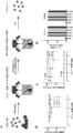
単球前駆細胞を産生するための方法が、本明細書において提供される。本発明よりも以前は、いくつかの技術的な問題により、創薬における単球およびマクロファージの使用は限定されていた。プロジェクトが納期に間に合うのを保証するために、細胞数、拡張性、再現性、および表現型関連性など因子は、必須である。本発明者らは、公開されているプロトコール(van Wilgenburg et al.2013)を修正し、分化時間を減少させながら、収率および再現性を増加させることができた。好ましい実施形態において、胚葉体(EB)は、ラミニンでコーティングされた細胞培養支持体に播種した人工多能性幹細胞(iPSC)から生成される。これらのEBは、初期の胚形成に類似し、3つの生殖系列の層(原始線条)の形成を開始する。EBは、次いで、細胞を、BMP4を含む規定培地と接触させることによって事前分化され、細胞の運命決定を中胚葉系統へと誘導する。形成され事前分化された後、EBは、播種され、骨髄系統に沿ってさらに分化して、造血工場を形成し、これが、単球前駆体を産生し上清中に放出する(図2)。造血工場は、100日間を上回って維持することができ、単球前駆体を、培養上清から採取することができる(最大で1週間に2回)。採取した後、これらの前駆体は、1週間以内に非分極化マクロファージに分化し得るか、または炎症促進性もしくは抗炎症性のいずれかのサブタイプを促進する特定のサイトカインの添加によってさらに分極化されてもよい。造血工場の拡張性を、10から1000cm2の培養面積に増加させることによって、本発明は、創薬および開発プロジェクトに関連する作業ならびに中規模サイズの薬物スクリーニングプログラムの必要性に合った細胞採取および取り扱い時間を達成している。別の態様において、ミクログリア様細胞の生成のための新規な共培養設定を、確立した。 A method for producing monocyte progenitor cells is provided herein. Prior to the present invention, several technical issues limited the use of monocytes and macrophages in drug discovery. Factors such as cell number, scalability, reproducibility, and phenotypic relevance are essential to ensure that projects meet deadlines. The inventors have modified a published protocol (van Wilgenburg et al. 2013) to increase yield and reproducibility while decreasing differentiation time. In a preferred embodiment, embryoid bodies (EBs) are generated from induced pluripotent stem cells (iPSCs) seeded on laminin-coated cell culture supports. These EBs initiate the formation of the three germline layers (primitive streak), resembling early embryogenesis. The EBs are then pre-differentiated by contacting the cells with a defined medium containing BMP4 to induce cell fate commitment toward the mesoderm lineage. After being formed and pre-differentiated, EBs are seeded and further differentiated along myeloid lineages to form hematopoietic factories that produce and release monocyte precursors into the supernatant (Figure 2). Hematopoietic factories can be maintained for more than 100 days and monocyte precursors can be harvested from the culture supernatant (up to twice a week). Once harvested, these precursors can be differentiated into non-polarized macrophages within a week or may be further polarized by the addition of specific cytokines that promote either pro- or anti-inflammatory subtypes. By increasing the scalability of the hematopoietic factories from 10 to 1000 cm2 of culture area, the present invention achieves cell harvesting and handling times that are in line with the needs of work related to drug discovery and development projects as well as medium-sized drug screening programs. In another aspect, a novel co-culture setup for the generation of microglia-like cells was established.
単球前駆細胞の生成
多能性幹細胞は、自己複製の特徴を有し、成体哺乳動物身体のすべての主要な細胞型に分化することができる。多能性幹細胞は、標準化された細胞培養条件下において、多量に産生することができる。したがって、好ましい実施形態において、単球前駆細胞は、多能性幹細胞から生成される、すなわち、分化する。一実施形態において、単球前駆細胞は、胚性幹細胞から生成される、すなわち、分化する。好ましい実施形態において、単球前駆細胞は、人工多能性幹細胞(iPSC)から生成、すなわち、分化する。一実施形態において、iPSCは、再プログラミングされた体細胞から生成される。体細胞のiPSCへの再プログラミングは、iPSC特性の維持に関与する特定の遺伝子を導入することによって達成することができる。体細胞のiPSCへの再プログラミングに好適な遺伝子としては、Oct4、Sox2、Klf4、およびC-Myc、ならびにこれらの組合せが挙げられるが、これらに限定されない。一実施形態において、再プログラミングのための遺伝子は、Oct4、Sox2、Klf4、およびC-Mycである。
Generation of Monocyte Progenitor Cells Pluripotent stem cells have the characteristic of self-renewal and can differentiate into all major cell types of the adult mammalian body. Pluripotent stem cells can be produced in large quantities under standardized cell culture conditions. Thus, in a preferred embodiment, monocyte progenitor cells are generated, i.e., differentiated, from pluripotent stem cells. In one embodiment, monocyte progenitor cells are generated, i.e., differentiated, from embryonic stem cells. In a preferred embodiment, monocyte progenitor cells are generated, i.e., differentiated, from induced pluripotent stem cells (iPSCs). In one embodiment, iPSCs are generated from reprogrammed somatic cells. Reprogramming somatic cells into iPSCs can be achieved by introducing specific genes involved in maintaining iPSC properties. Suitable genes for reprogramming somatic cells into iPSCs include, but are not limited to, Oct4, Sox2, Klf4, and C-Myc, and combinations thereof. In one embodiment, the genes for reprogramming are Oct4, Sox2, Klf4, and C-Myc.
内臓、皮膚、骨、血液、および結合組織は、すべて、体細胞から作製されている。iPSCを生成するために使用される体細胞としては、線維芽細胞、脂肪細胞、およびケラチノサイトが挙げられるがこれらに限定されず、皮膚生検から得ることができる。他の好適な体細胞は、白血球、血液試料もしくは上皮細胞から得られる赤芽球、または血液もしくは尿試料から得られる他の細胞であり、当該技術分野において公知であり本明細書に記載される方法によって、iPSCに再プログラミングされる。体細胞は、健常な個体から、または罹患した個体から得ることができる。一実施形態において、体細胞は、疾患を患う対象(例えば、ヒト対象)に由来する。一実施形態において、疾患は、慢性炎症(例えば、炎症性腸疾患)、原発性もしくは後天性免疫不全(例えば、裸リンパ球症候群)、または神経変性疾患(例えば、多発性硬化症、アルツハイマー病、もしくはパーキンソン病)のいずれかと関連している。本明細書に記載される再プログラミングのための遺伝子は、当該技術分野において公知の方法、再プログラミングベクターを介した細胞への送達または小分子を介した前記遺伝子の活性化のいずれかによって、体細胞に導入される。再プログラミングのための方法は、とりわけ、レトロウイルス、レンチウイルス、アデノウイルス、プラスミドおよびトランスポゾン、マイクロRNA、小分子、改変されたRNA、メッセンジャーRNA、ならびに組換えタンパク質を含む。一実施形態において、レンチウイルスが、本明細書に記載される遺伝子の送達に使用される。別の実施形態において、Oct4、Sox2、Klf4、およびC-Mycは、センダイウイルス粒子を使用して体細胞に送達される。加えて、体細胞を、少なくとも1つの小分子の存在下において培養してもよい。一実施形態において、この小分子は、プロテインキナーゼのRho関連コイルドコイル形成タンパク質セリン/スレオニンキナーゼ(ROCK)ファミリーの阻害剤を含む。ROCK阻害剤の非限定的な例は、ファスジル(1-(5-イソキノリンスルホニル)ホモピペラジン)、チアゾビビン(N-ベンジル-2-(ピリミジン-4-イルアミノ)チアゾール-4-カルボキサミド)、およびY-27632((+)-(R)-トランス-4-(1-アミノエチル)-N-(4-ピリジル)シクロ-ヘキサンカルボキサミドジヒドロクロリド)を含む。 Internal organs, skin, bone, blood, and connective tissues are all made from somatic cells. Somatic cells used to generate iPSCs include, but are not limited to, fibroblasts, adipocytes, and keratinocytes, and can be obtained from skin biopsies. Other suitable somatic cells are white blood cells, erythroblasts obtained from blood samples or epithelial cells, or other cells obtained from blood or urine samples, and are reprogrammed into iPSCs by methods known in the art and described herein. Somatic cells can be obtained from healthy or diseased individuals. In one embodiment, the somatic cells are derived from a subject (e.g., a human subject) suffering from a disease. In one embodiment, the disease is associated with either chronic inflammation (e.g., inflammatory bowel disease), primary or acquired immune deficiency (e.g., bare lymphocyte syndrome), or neurodegenerative disease (e.g., multiple sclerosis, Alzheimer's disease, or Parkinson's disease). The genes for reprogramming described herein are introduced into somatic cells by methods known in the art, either delivery to the cells via a reprogramming vector or activation of said genes via a small molecule. Methods for reprogramming include, among others, retroviruses, lentiviruses, adenoviruses, plasmids and transposons, microRNAs, small molecules, modified RNAs, messenger RNAs, and recombinant proteins. In one embodiment, lentiviruses are used to deliver the genes described herein. In another embodiment, Oct4, Sox2, Klf4, and c-Myc are delivered to somatic cells using Sendai virus particles. In addition, somatic cells may be cultured in the presence of at least one small molecule. In one embodiment, the small molecule comprises an inhibitor of the Rho-associated coiled-coil-forming protein serine/threonine kinase (ROCK) family of protein kinases. Non-limiting examples of ROCK inhibitors include fasudil (1-(5-isoquinolinesulfonyl)homopiperazine), thiazovivin (N-benzyl-2-(pyrimidin-4-ylamino)thiazole-4-carboxamide), and Y-27632 ((+)-(R)-trans-4-(1-aminoethyl)-N-(4-pyridyl)cyclo-hexanecarboxamide dihydrochloride).
多能性幹細胞の所定の単一層を提供することが、得られる培養物の再現性および効率性に好ましい。本発明者らは、驚くべきことに、幹細胞維持培養においてラミニンでコーティングされた基質を使用することによる置換が、造血工場の分化時間を減少させ、細胞培養物のスループットを増加させたことを見出した。一実施形態において、多能性幹細胞の単一層は、細胞を、単一細胞に酵素的に解離させ、それらを接着性基質、例えば、ラミニン基質でコーティングされた細胞培養物格納容器(例えば、フラスコ)に播種することによって、産生され得る。好ましい実施形態において、接着性基質(コーティング)は、ラミニンである。一実施形態において、ラミニンは、ラミニンサブユニットアルファ-4を含む。一実施形態において、ラミニンは、ラミニンサブユニットアルファ-5を含む。一実施形態において、ラミニンは、ラミニンサブユニットベータ-1を含む。一実施形態において、ラミニンは、ラミニンサブユニットベータ-2を含む。一実施形態において、ラミニンは、ラミニンサブユニットガンマ-1を含む。一実施形態において、ラミニンは、ラミニンサブユニットアルファ-4、ベータ-1、およびガンマ-1(ラミニン-411)を含む。一実施形態において、ラミニンは、ラミニンサブユニットアルファ-5、ベータ-1、およびガンマ-1(ラミニン-511)を含む。好ましい実施形態において、ラミニンは、ラミニンサブユニットアルファ-5、ベータ-2、およびガンマ-1(ラミニン-521、例えば、BioLamina rhLaminin-521)を含む。 Providing a predetermined monolayer of pluripotent stem cells is favorable for the reproducibility and efficiency of the resulting culture. The inventors have surprisingly found that substitution of a substrate coated with laminin in stem cell maintenance cultures reduced the differentiation time of hematopoietic factories and increased the throughput of the cell culture. In one embodiment, a monolayer of pluripotent stem cells can be produced by enzymatically dissociating the cells into single cells and seeding them onto a cell culture containment vessel (e.g., a flask) coated with an adhesive substrate, e.g., a laminin substrate. In a preferred embodiment, the adhesive substrate (coating) is laminin. In one embodiment, the laminin comprises laminin subunit alpha-4. In one embodiment, the laminin comprises laminin subunit alpha-5. In one embodiment, the laminin comprises laminin subunit beta-1. In one embodiment, the laminin comprises laminin subunit beta-2. In one embodiment, the laminin comprises laminin subunit gamma-1. In one embodiment, the laminin comprises laminin subunits alpha-4, beta-1, and gamma-1 (laminin-411). In one embodiment, the laminin comprises laminin subunits alpha-5, beta-1, and gamma-1 (laminin-511). In a preferred embodiment, the laminin comprises laminin subunits alpha-5, beta-2, and gamma-1 (laminin-521, e.g., BioLamina rhLaminin-521).
単一細胞への解離に好適な酵素の例としては、Accutase(Invitrogen)、トリプシン(Invitrogen)、TrypLe Express(Invitrogen)が挙げられる。一実施形態において、1cm2当たり20,000~60,000個の細胞が、接着性基質に播種される。本明細書において使用される培地は、多能性幹細胞が単一細胞として単一層で付着および増殖するのを促進する、多能性培地である。一実施形態において、多能性培地は、プロテインキナーゼのRho関連コイルドコイル形成タンパク質セリン/スレオニンキナーゼ(ROCK)ファミリーの小分子阻害剤(本明細書においてROCKキナーゼ阻害剤と称される)を補充した、無血清培地である。 Examples of enzymes suitable for dissociation into single cells include Accutase (Invitrogen), trypsin (Invitrogen), TrypLe Express (Invitrogen). In one embodiment, 20,000-60,000 cells per cm2 are seeded onto the adherent substrate. The medium used herein is a pluripotency medium that promotes pluripotent stem cells to attach and grow in a monolayer as single cells. In one embodiment, the pluripotency medium is a serum-free medium supplemented with a small molecule inhibitor of the Rho-associated coiled-coil-forming protein serine/threonine kinase (ROCK) family of protein kinases (referred to herein as a ROCK kinase inhibitor).
したがって、一実施形態において、本明細書に記載される方法は、ラミニン基質に、多能性培地中の多能性幹細胞の単一層を提供することを含み、ここで、この多能性培地は、ROCKキナーゼ阻害剤を補充した無血清培地である。 Thus, in one embodiment, the method described herein involves providing a monolayer of pluripotent stem cells in pluripotency medium on a laminin substrate, where the pluripotency medium is serum-free medium supplemented with a ROCK kinase inhibitor.
多能性幹細胞の基質への付着に好適な無血清培地の例としては、Stem Cell TechnologiesからのmTeSR1もしくはTeSR2、ReproCELLからのPrimate ES/iPS細胞培地、MiliporeからのPluriSTEM、Milenyi BiotecからのStemMACS iPS-Brew、およびInvitrogenからのStemPro hESC SFM、LonzaからのX-VIVOがある。本明細書において有用なROCKキナーゼ阻害剤の例は、ファスジル(1-(5-イソキノリンスルホニル)ホモピペラジン)、チアゾビビン(N-ベンジル-2-(ピリジン-4-イルアミノ)チアゾール-4-カルボキサミド)、およびY27632((+)-(R)-トランス-4-(1-アミノエチル)-N-(4-ピリジル)シクロ-ヘキサンカルボキサミドジヒドロクロリド、例えば、Tocris bioscienceのカタログ番号:1254)がある。一実施形態において、多能性培地は、約2~20μMのY27632、好ましくは約5~10μMのY27632を補充した無血清培地である。別の実施形態において、多能性培地は、約2~20μMのファスジルを補充した無血清培地である。別の実施形態において、多能性培地は、約0.2~10μMのチアゾビビンを補充した無血清培地である。 Examples of serum-free media suitable for attachment of pluripotent stem cells to a substrate include mTeSR1 or TeSR2 from Stem Cell Technologies, Primate ES/iPS Cell Medium from ReproCELL, PluriSTEM from Millipore, StemMACS iPS-Brew from Milenyi Biotec, and StemPro hESC SFM from Invitrogen, and X-VIVO from Lonza. Examples of ROCK kinase inhibitors useful herein are fasudil (1-(5-isoquinolinesulfonyl)homopiperazine), thiazovivin (N-benzyl-2-(pyridin-4-ylamino)thiazole-4-carboxamide), and Y27632 ((+)-(R)-trans-4-(1-aminoethyl)-N-(4-pyridyl)cyclo-hexanecarboxamide dihydrochloride, e.g., Tocris bioscience catalog number: 1254). In one embodiment, the pluripotency medium is serum-free medium supplemented with about 2-20 μM Y27632, preferably about 5-10 μM Y27632. In another embodiment, the pluripotency medium is serum-free medium supplemented with about 2-20 μM fasudil. In another embodiment, the pluripotency medium is a serum-free medium supplemented with about 0.2-10 μM thiazovivin.
一実施形態において、本明細書に記載される方法は、ラミニン基質に多能性培地中の多能性幹細胞の単一層を提供すること、およびこの単一層を多能性培地において、少なくとも1日間(24時間)増殖させることを含む。別の実施形態において、本明細書に記載される方法は、多能性培地中の多能性幹細胞の単一層を提供すること、およびこの単一層を多能性培地において、18時間~30時間、好ましくは23~25時間増殖させることを含む。さらなる実施形態において、本明細書に記載される方法は、ラミニン基質に多能性培地中の多能性幹細胞の単一層を提供すること、およびこの単一層を多能性培地において、少なくとも2日間、3日間、4日間、5日間、6日間、7日間、8日間、9日間、10日間、または10日間を上回って増殖させることを含む。 In one embodiment, the method described herein comprises providing a monolayer of pluripotent stem cells in pluripotency medium on a laminin substrate and growing the monolayer in pluripotency medium for at least 1 day (24 hours). In another embodiment, the method described herein comprises providing a monolayer of pluripotent stem cells in pluripotency medium and growing the monolayer in pluripotency medium for 18 to 30 hours, preferably 23 to 25 hours. In a further embodiment, the method described herein comprises providing a monolayer of pluripotent stem cells in pluripotency medium on a laminin substrate and growing the monolayer in pluripotency medium for at least 2 days, 3 days, 4 days, 5 days, 6 days, 7 days, 8 days, 9 days, 10 days, or more than 10 days.
別の実施形態において、本明細書に記載される方法は、ラミニン基質に、mTesR1培地である多能性培地中の多能性幹細胞の単一層を提供すること、およびこの単一層を多能性培地において、少なくとも1日間(24時間)増殖させることを含む。別の実施形態において、本明細書に記載される方法は、ラミニン基質に、mTesR1である多能性培地中の多能性幹細胞の単一層を提供すること、およびこの単一層を多能性培地において、18時間~30時間、好ましくは23~25時間増殖させることを含む。 In another embodiment, the method described herein includes providing a monolayer of pluripotent stem cells in a pluripotency medium that is mTesR1 medium on a laminin substrate, and growing the monolayer in the pluripotency medium for at least 1 day (24 hours). In another embodiment, the method described herein includes providing a monolayer of pluripotent stem cells in a pluripotency medium that is mTesR1 on a laminin substrate, and growing the monolayer in the pluripotency medium for 18 hours to 30 hours, preferably 23 hours to 25 hours.
次の工程b)において、多能性幹細胞は、採取され、懸濁培養物に移される。一実施形態において、多能性幹細胞を、中胚葉誘導培地と接触させる。一実施形態において、中胚葉誘導培地は、組換え骨形成タンパク質-4(BMP4)を含む。一実施形態において、中胚葉誘導培地は、約10~100ng/mlのBMP4(例えば、hBMP4)、好ましくは、約50ng/mlのBMP4を補充した無血清培地である。 In the next step b), the pluripotent stem cells are harvested and transferred to a suspension culture. In one embodiment, the pluripotent stem cells are contacted with a mesoderm induction medium. In one embodiment, the mesoderm induction medium comprises recombinant bone morphogenetic protein-4 (BMP4). In one embodiment, the mesoderm induction medium is a serum-free medium supplemented with about 10-100 ng/ml of BMP4 (e.g., hBMP4), preferably about 50 ng/ml of BMP4.
さらなる実施形態において、中胚葉誘導培地は、血管内皮増殖因子(VEGF)をさらに含む。一実施形態において、中胚葉誘導培地は、約10~100ng/mlのVEGF(例えば、hVEGF)、好ましくは、約50ng/mlのVEGFを補充した無血清培地である。 In a further embodiment, the mesoderm induction medium further comprises vascular endothelial growth factor (VEGF). In one embodiment, the mesoderm induction medium is a serum-free medium supplemented with about 10-100 ng/ml VEGF (e.g., hVEGF), preferably about 50 ng/ml VEGF.
さらなる実施形態において、中胚葉誘導培地は、幹細胞因子(SCF)をさらに含む。一実施形態において、中胚葉誘導培地は、約5~50ng/mlのSCF(例えば、hSCF)、好ましくは、約20ng/mlのSCFを補充した無血清培地である。 In a further embodiment, the mesoderm induction medium further comprises stem cell factor (SCF). In one embodiment, the mesoderm induction medium is a serum-free medium supplemented with about 5-50 ng/ml SCF (e.g., hSCF), preferably about 20 ng/ml SCF.
好ましい実施形態において、中胚葉誘導培地は、BMP4、VEGF、およびSCF、具体的には、約10~100ng/mlのBMP4、約10~100ng/mlのVEGF、および約5~50ng/mlのSCFを含む。好ましい実施形態において、中胚葉誘導培地は、約50ng/mlのBMP4、約50ng/mlのVEGF、および約20ng/mlのSCFを含む。 In a preferred embodiment, the mesoderm induction medium contains BMP4, VEGF, and SCF, specifically, about 10-100 ng/ml BMP4, about 10-100 ng/ml VEGF, and about 5-50 ng/ml SCF. In a preferred embodiment, the mesoderm induction medium contains about 50 ng/ml BMP4, about 50 ng/ml VEGF, and about 20 ng/ml SCF.
一実施形態において、多能性幹細胞を、少なくとも約1日間(24時間)、中胚葉誘導培地と接触させる。さらなる実施形態において、多能性幹細胞を、約2日間、3日間、4日間、5日間、6日間、7日間、8日間、9日間、10日間、または約10日間を上回って、中胚葉誘導培地と接触させる。一実施形態において、多能性幹細胞を、約24時間~約72時間、好ましくは、約36~約60時間、中胚葉誘導培地と接触させる。 In one embodiment, the pluripotent stem cells are contacted with the mesoderm induction medium for at least about 1 day (24 hours). In a further embodiment, the pluripotent stem cells are contacted with the mesoderm induction medium for about 2 days, 3 days, 4 days, 5 days, 6 days, 7 days, 8 days, 9 days, 10 days, or more than about 10 days. In one embodiment, the pluripotent stem cells are contacted with the mesoderm induction medium for about 24 hours to about 72 hours, preferably about 36 hours to about 60 hours.
一実施形態において、細胞は、工程c)において、中胚葉誘導後の細胞の付着に好適な細胞培養支持体に播種される。好ましい実施形態において、細胞は、基底膜生体材料、例えば、マトリゲル、Cultrex BME、Geltrex Matrixなどでコーティングされた細胞培養支持体に播種される。一実施形態において、基底膜生体材料は、ラミニン、IV型コラーゲン、ヘパリン硫酸プロテオグリカン、およびエンタクチン/ナイドジェン-1,2を含む。好ましい実施形態において、細胞は、マトリゲルでコーティングされた細胞培養支持体に播種される。 In one embodiment, the cells are seeded in step c) on a cell culture support suitable for attachment of the cells after mesoderm induction. In a preferred embodiment, the cells are seeded on a cell culture support coated with a basement membrane biomaterial, such as Matrigel, Cultrex BME, Geltrex Matrix, etc. In one embodiment, the basement membrane biomaterial comprises laminin, type IV collagen, heparin sulfate proteoglycan, and entactin/nidogen-1,2. In a preferred embodiment, the cells are seeded on a cell culture support coated with Matrigel.
一実施形態において、細胞は、工程c)において、大規模細胞培養容器に播種される。特定の実施形態において、106個を上回る、107個を上回る、108個を上回る、109個を上回る、1010個を上回る、1011個を上回る、1012個を上回る細胞が、個々の大規模細胞培養物格納容器に播種される。一実施形態において、大規模細胞培養物は、1つの単一細胞培養物格納容器を含む。別の実施形態において、大規模細胞培養物は、複数の細胞培養物格納容器のアセンブリを含む。さらなる実施形態において、大規模細胞培養物(格納容器)は、少なくとも100cm2、500cm2、1,000cm2、2,000cm2、5,000cm2、10,000cm2の細胞培養面積を含む。一実施形態において、大規模細胞培養物(系)には、少なくとも106個、107個、108個、109個、1010個、1011個、1012個の細胞が接種される。 In one embodiment, cells are seeded in the large scale cell culture vessel in step c). In a particular embodiment, more than 106 , more than 107 , more than 108 , more than 109 , more than 1010 , more than 1011 , more than 1012 cells are seeded in each large scale cell culture containment vessel. In one embodiment, the large scale cell culture comprises one single cell culture containment vessel. In another embodiment, the large scale cell culture comprises an assembly of multiple cell culture containment vessels. In further embodiments, the large scale cell culture (containment vessel) comprises a cell culture area of at least 100 cm2 , 500 cm2 , 1,000 cm2 , 2,000 cm2 , 5,000 cm2 , 10,000 cm2 . In one embodiment, large scale cell cultures (lines) are inoculated with at least 10 6 , 10 7 , 10 8 , 10 9 , 10 10 , 10 11 , 10 12 cells.
次の工程において、大規模細胞培養物中の細胞を、骨髄系統に沿ってさらに分化させる。一実施形態において、播種した細胞を、工程c)において、骨髄系成熟培地と接触させる。好適な骨髄系成熟培地は、当該技術分野において公知であり、本明細書にも記載されている。一実施形態において、骨髄系成熟培地は、インターロイキン3(IL-3)を含む。一実施形態において、骨髄系成熟培地は、約1~50ng/mlのIL-3(例えば、hIL-3)、好ましくは、約25ng/mlのIL-3を補充した無血清培地である。一実施形態において、細胞を、約4日間(約96時間)、骨髄系成熟培地と接触させる。さらなる実施形態において、細胞を、約2日間、3日間、4日間、5日間、6日間、7日間、8日間、9日間、10日間、または約10日間を上回って、骨髄系成熟培地と接触させる。一実施形態において、細胞を、約72時間~約120時間、好ましくは、約84~約108時間、骨髄系成熟培地と接触させる。骨髄系成熟の工程中に、大規模細胞培養物は、単球前駆細胞の産生を開始する。単球前駆細胞は、骨髄系成熟後に、細胞培養物の上清を回収することによって、接着性細胞培養物から採取することができる。一実施形態において、本発明による工程c)の大規模細胞培養物は、約10日間を上回って、15日間を上回って、20日間を上回って、25日間を上回って、30日間を上回って、40日間を上回って、50日間を上回って、60日間を上回って、70日間を上回って、80日間を上回って、90日間を上回って、または100日間を上回って、単球前駆細胞を産生することができる。一実施形態において、工程c)の大規模培養物は、1週間につき、細胞培養面積1cm2当たり、少なくとも約100,000個の単球前駆細胞を産生することができる。 In the next step, the cells in the large scale cell culture are further differentiated along a myeloid lineage. In one embodiment, the plated cells are contacted with a myeloid maturation medium in step c). Suitable myeloid maturation medium are known in the art and described herein. In one embodiment, the myeloid maturation medium comprises interleukin 3 (IL-3). In one embodiment, the myeloid maturation medium is a serum-free medium supplemented with about 1-50 ng/ml IL-3 (e.g., hIL-3), preferably about 25 ng/ml IL-3. In one embodiment, the cells are contacted with the myeloid maturation medium for about 4 days (about 96 hours). In a further embodiment, the cells are contacted with the myeloid maturation medium for about 2 days, 3 days, 4 days, 5 days, 6 days, 7 days, 8 days, 9 days, 10 days, or more than about 10 days. In one embodiment, the cells are contacted with the myeloid maturation medium for about 72 hours to about 120 hours, preferably about 84 hours to about 108 hours. During the step of myeloid maturation, the large-scale cell culture starts to produce monocyte precursor cells. The monocyte precursor cells can be harvested from the adherent cell culture by harvesting the cell culture supernatant after myeloid maturation. In one embodiment, the large-scale cell culture of step c) according to the invention can produce monocyte precursor cells for more than about 10 days, more than 15 days, more than 20 days, more than 25 days, more than 30 days, more than 40 days, more than 50 days, more than 60 days, more than 70 days, more than 80 days, more than 90 days, or more than 100 days. In one embodiment, the large-scale culture of step c) can produce at least about 100,000 monocyte precursor cells per cm2 of cell culture area per week.
単球前駆細胞からマクロファージへの分化
単球前駆細胞は、当該技術分野において公知であり本明細書にも記載される方法によって、マクロファージに分化させることができる。一実施形態において、単球前駆細胞を、マクロファージ分化培地と接触させる。一実施形態において、細胞を、約1~10日間、4~8日間、2日間、3日間、4日間、5日間、6日間、7日間、8日間、9日間、または約10日間を上回って、マクロファージ分化培地と接触させる。一実施形態において、マクロファージ分化培地は、マクロファージコロニー刺激因子(M-CSF)を含む。一実施形態において、マクロファージ分化培地は、10~200ng/mlのM-CSF(例えば、hM-CSF)、好ましくは、100ng/mlのM-CSFを補充した無血清培地である。好ましい実施形態において、細胞を、約6日間、マクロファージ分化培地と接触させる。一実施形態において、細胞をマクロファージ分化培地と接触させる前またはそれと同時に、細胞を、コーティングされていない組織培養支持体に播種する。一実施形態において、マクロファージを、コーティングされていない組織培養支持体に再播種する。一実施形態において、マクロファージを、ハイスループットプレート形式で再播種する。一実施形態において、マクロファージを、24ウェルプレート形式、96ウェルプレート形式、または384ウェルプレート形式で再播種する。
Differentiation of Monocyte Progenitor Cells to Macrophages Monocyte progenitor cells can be differentiated into macrophages by methods known in the art and described herein. In one embodiment, monocyte progenitor cells are contacted with macrophage differentiation medium. In one embodiment, the cells are contacted with macrophage differentiation medium for about 1-10 days, 4-8 days, 2 days, 3 days, 4 days, 5 days, 6 days, 7 days, 8 days, 9 days, or more than about 10 days. In one embodiment, the macrophage differentiation medium includes macrophage colony stimulating factor (M-CSF). In one embodiment, the macrophage differentiation medium is a serum-free medium supplemented with 10-200 ng/ml M-CSF (e.g., hM-CSF), preferably 100 ng/ml M-CSF. In a preferred embodiment, the cells are contacted with macrophage differentiation medium for about 6 days. In one embodiment, the cells are seeded onto an uncoated tissue culture support prior to or simultaneously with contacting the cells with macrophage differentiation medium. In one embodiment, the macrophages are replated onto an uncoated tissue culture support. In one embodiment, the macrophages are replated in a high throughput plate format. In one embodiment, the macrophages are replated in a 24-well plate format, a 96-well plate format, or a 384-well plate format.
単球前駆細胞からミクログリアへの分化
単球前駆細胞は、当該技術分野において公知であり本明細書にも記載される方法によって、ミクログリアに分化させることができる。一実施形態において、単球前駆細胞を、ニューロンと接触させる。一実施形態において、ニューロンは、国際公開第2017081250号に記載される方法を使用して生成される。いくつかの実施形態において、ニューロンを、(少なくとも)約1週間、2週間、3週間、4週間、5週間、6週間、7週間、8週間、9週間、10週間、分化させる。好ましい実施形態において、ニューロンを、約2~5週間分化させる。いくつかの実施形態において、細胞を、約2日間、3日間、4日間、5日間、6日間、7日間、8日間、9日間、または約10日間を上回って、ニューロンと接触させる。いくつかの実施形態において、細胞を、約5~20日間または約10~18日間、ニューロンと接触させる。一実施形態において、細胞を、共培養分化培地において、ニューロンと共培養する。一実施形態において、共培養分化培地は、顆粒球マクロファージコロニー刺激因子(GM-CSF)および/またはインターロイキン34(IL-34)を含む。一実施形態において、共培養分化培地は、10~200ng/mlのGM-CSF(例えば、hGM-CSF)、好ましくは、100ng/mlのGM-CSFを補充した無血清培地である。一実施形態において、共培養分化培地は、1~500ng/mlのIL-34(例えば、hIL-34)、好ましくは、100ng/mlのIL-34を補充した無血清培地である。好ましい実施形態において、細胞を、10~200ng/mlのGM-CSF(例えば、hGM-CSF)および1~500ngのIL-34、好ましくは、100ng/mlのGM-CSFおよび100ng/mlのIL-34を補充した無血清培地において、約14日間、ニューロンおよび共培養分化培地と接触させる。
Differentiation of Monocyte Progenitor Cells to Microglia Monocyte progenitor cells can be differentiated into microglia by methods known in the art and described herein. In one embodiment, the monocyte progenitor cells are contacted with neurons. In one embodiment, the neurons are generated using the methods described in WO2017081250. In some embodiments, the neurons are differentiated for (at least) about 1 week, 2 weeks, 3 weeks, 4 weeks, 5 weeks, 6 weeks, 7 weeks, 8 weeks, 9 weeks, 10 weeks. In preferred embodiments, the neurons are differentiated for about 2-5 weeks. In some embodiments, the cells are contacted with the neurons for about 2 days, 3 days, 4 days, 5 days, 6 days, 7 days, 8 days, 9 days, or more than about 10 days. In some embodiments, the cells are contacted with the neurons for about 5-20 days or about 10-18 days. In one embodiment, the cells are co-cultured with the neurons in co-culture differentiation medium. In one embodiment, the co-culture differentiation medium comprises granulocyte macrophage colony stimulating factor (GM-CSF) and/or interleukin 34 (IL-34). In one embodiment, the co-culture differentiation medium is a serum-free medium supplemented with 10-200 ng/ml GM-CSF (e.g., hGM-CSF), preferably 100 ng/ml GM-CSF. In one embodiment, the co-culture differentiation medium is a serum-free medium supplemented with 1-500 ng/ml IL-34 (e.g., hIL-34), preferably 100 ng/ml IL-34. In a preferred embodiment, the cells are contacted with the neuronal and co-culture differentiation medium for about 14 days in serum-free medium supplemented with 10-200 ng/ml GM-CSF (e.g., hGM-CSF) and 1-500 ng IL-34, preferably 100 ng/ml GM-CSF and 100 ng/ml IL-34.
例示的な実施形態:
1. 単球前駆細胞を産生するための方法であって、
a)ラミニンでコーティングされた細胞培養支持体に、多能性培地中の多能性幹細胞を播種する工程と、
b)多能性幹細胞を採取し、多能性幹細胞を懸濁培養物中の中胚葉誘導培地と接触させる工程と、
c)細胞を、細胞の付着に好適な細胞培養支持体に播種する工程と、
d)懸濁液から単球前駆細胞を採取する工程と
を含む、方法。
2. 工程a)における細胞を、少なくとも約1日間、2日間、3日間、4日間、5日間、6日間、7日間、8日間、9日間、または10日間、具体的には、少なくとも約1日間、ラミニンでコーティングされた細胞培養支持体上で培養する、実施形態1に記載の方法。
3. 工程a)におけるラミニンが、ラミニンサブユニットアルファ-5を含む、特に、工程a)におけるラミニンが、ラミニンサブユニットアルファ-5、ベータ-2、およびガンマ1を含む、実施形態1または2に記載の方法。
4. 中胚葉誘導培地が、組換え骨形成タンパク質-4(BMP4)を含む化学的規定培地である、実施形態1から3のいずれか1つに記載の方法。
5. 培地が、約10~100ng/mlのBMP4、好ましくは、約50ng/mlのBMP4を含む、実施形態4に記載の方法。
6. 中胚葉誘導培地が、血管内皮増殖因子(VEGF)をさらに含む、実施形態4または5に記載の方法。
7. 中胚葉誘導培地が、約10~100ng/mlのVEGF、好ましくは、約50ng/mlのVEGFを含む、実施形態6に記載の方法。
8. 中胚葉誘導培地が、幹細胞因子(SCF)をさらに含む、実施形態4から7のいずれか1つに記載の方法。
9. 中胚葉誘導培地が、約5~50ng/mlのSCF、好ましくは、約20ng/mlのSCFを含む、実施形態8に記載の方法。
10. 細胞を、約1~10日間、2~6日間、2日間、3日間、4日間、5日間、6日間、7日間、8日間、9日間、中胚葉誘導培地と接触させる、実施形態1から9のいずれか1つに記載の方法。
11. 細胞を、約4日間、中胚葉誘導培地と接触させる、実施形態1から10のいずれか1つに記載の方法。
12. 工程b)における細胞が、胚葉体(EB)を形成する、実施形態1から11のいずれか1つに記載の方法。
13. 工程c)における細胞培養支持体が、基底膜生体材料でコーティングされている、実施形態1から12のいずれか1つに記載の方法。
14. 基底膜生体材料が、ラミニン、IV型コラーゲン、ヘパリン硫酸プロテオグリカン、およびエンタクチン/ナイドジェン-1,2を含む、実施形態13に記載の方法。
15. 工程c)における細胞を、骨髄系成熟培地と接触させる、実施形態1から14のいずれか1つに記載の方法。
16. 骨髄系成熟培地が、マクロファージコロニー刺激因子(M-CSF)を含む、実施形態1から15のいずれか1つに記載の方法。
17. 骨髄系成熟培地が、約20~200ng/mlのM-CSF、好ましくは、約100ng/mlのM-CSFを含む、実施形態16に記載の方法。
18. 骨髄系成熟培地が、IL-3をさらに含む、実施形態15から17のいずれか1つに記載の方法。
19. 培地が、約1~50ng/mlのIL-3、好ましくは、約25ng/mlのIL-3を含む、実施形態18に記載の方法。
20. 細胞を、約1~10日間、2~6日間、2日間、3日間、4日間、5日間、6日間、7日間、8日間、9日間、骨髄系成熟培地と接触させる、実施形態15から19のいずれか1つに記載の方法。
21. 細胞を、約4日間、骨髄系成熟培地と接触させる、実施形態15から20のいずれか1つに記載の方法。
22. 工程d)において、単球前駆細胞を、細胞培養物の上清を回収することによって、採取する、実施形態1から21のいずれか1つに記載の方法。
23. 工程d)において、単球前駆細胞を、細胞培養物の上清を回収することによって、バッチで採取する、実施形態1から22のいずれか1つに記載の方法。
24. 単球前駆細胞を規則的な間隔で、具体的には、毎日、1日おきに、3日ごとに、4日ごとに、5日ごとに、または6日ごとに、バッチで採取する、実施形態1から23のいずれか1つに記載の方法。
25. 単球前駆細胞を継続的に採取する、実施形態1から24のいずれか1つに記載の方法。
26. 工程d)において、単球前駆細胞を、細胞培養物から上清を除去し、必要に応じて、除去した上清を新しい培地と置き換えることによって、継続的に採取する、実施形態1から25のいずれか1つに記載の方法。
27. e)採取した単球前駆細胞をマクロファージに分化させる工程をさらに含む、実施形態1から26のいずれか1つに記載の方法。
28. 工程e)における細胞を、マクロファージ分化培地と接触させる、実施形態27に記載の方法。
29. マクロファージ分化培地が、マクロファージコロニー刺激因子(M-CSF)を含む、実施形態27に記載の方法。
30. マクロファージ分化培地が、約10~200ng/mlのM-CSF、好ましくは、約100ng/mlのM-CSFを含む、実施形態28または29に記載の方法。
31. 細胞を、約1~10日間、4~8日間、2日間、3日間、4日間、5日間、6日間、7日間、8日間、9日間、マクロファージ分化培地と接触させる、実施形態28から30のいずれか1つに記載の方法。
32. 細胞を、約6日間、マクロファージ分化培地と接触させる、実施形態28から31のいずれか1つに記載の方法。
33. 工程e)における細胞を、コーティングされていない組織培養物支持体に播種する、実施形態28から32のいずれか1つに記載の方法。
34. マクロファージを、コーティングされていない組織培養物支持体に再播種する、実施形態28から33のいずれか1つに記載の方法。
35. マクロファージを、24ウェルプレート形式、96ウェルプレート形式、または384ウェルプレート形式で再播種する、実施形態28から34のいずれか1つに記載の方法。
36. e)単球前駆細胞をミクログリアに分化させる工程をさらに含む、実施形態1から26のいずれか1つに記載の方法。
36. 工程e)における単球前駆体を神経細胞と共培養する、実施形態35に記載の方法。
37. 工程e)における細胞を共培養分化培地と接触させる、実施形態35または36に記載の方法。
38. 共培養分化培地が、顆粒球マクロファージコロニー刺激因子(GM-CSF)および/またはインターロイキン34(IL-34)を含む、実施形態37に記載の方法。
39. 共培養分化培地が、約10~200ng/mlのGM-CSF、好ましくは、約100ng/mlのGM-CSFを含む、実施形態38に記載の方法。
40. 共培養分化培地培地が、約1~500ng/mlのIL-34(例えば、hIL-34)、好ましくは、約100ng/mlのIL-34を含む、実施形態38または39に記載の方法。
41. 細胞を、約1~28日間、7~21日間、8日間、9日間、10日間、11日間、12日間、13日間、14日間、15日間、共培養分化培地と接触させる、実施形態38から40のいずれか1つに記載の方法。
42. 細胞を、約14日間、共培養分化培地と接触させる、実施形態38から41のいずれか1つに記載の方法。
43. 神経細胞が、多能性幹細胞に由来する、実施形態36から42のいずれか1つに記載の方法。
44. 神経細胞が、国際公開第2017/081250号に記載される均一に分配された分化したNCの標準化細胞培養物を産生するための方法に従って産生される、実施形態36から43のいずれか1つに記載の方法。
45. 多能性幹細胞が、哺乳動物細胞、特に、ヒト細胞である、実施形態1から44のいずれか1つに記載の方法。
46. 多能性幹細胞が、胚性幹細胞(ESC)である、実施形態1から45のいずれか1つに記載の方法。
47. 多能性幹細胞が、人工多能性幹細胞(iPSC)である、実施形態1から45のいずれか1つに記載の方法。
48. 単球前駆細胞を産生するための接着性大規模細胞培養物であって、1週間につき、細胞培養面積1cm2当たり少なくとも約100,000個の単球前駆細胞を産生することができる、接着性大規模細胞培養物。
49. 請求項1から26のいずれか一項に記載の方法の工程a)からc)によって産生される、実施形態48に記載の接着性大規模細胞培養物。
49. 本明細書に記載される発明。
Exemplary embodiments:
1. A method for producing monocyte progenitor cells, comprising:
a) seeding pluripotent stem cells in a pluripotency medium onto a cell culture support coated with laminin;
b) harvesting pluripotent stem cells and contacting the pluripotent stem cells with a mesoderm induction medium in a suspension culture;
c) seeding the cells on a cell culture support suitable for cell attachment;
and d) harvesting the monocyte precursor cells from the suspension.
2. The method according to
3. The method according to
4. The method according to any one of
5. The method of
6. The method of
7. The method of
8. The method of any one of
9. The method of
10. The method of any one of
11. The method of any one of
12. The method according to any one of the preceding embodiments, wherein the cells in step b) form embryoid bodies (EBs).
13. The method according to any one of the preceding embodiments, wherein the cell culture support in step c) is coated with a basement membrane biomaterial.
14. The method of embodiment 13, wherein the basement membrane biomaterial comprises laminin, type IV collagen, heparin sulfate proteoglycan, and entactin/nidogen-1,2.
15. The method according to any one of the preceding embodiments, wherein the cells in step c) are contacted with a myeloid maturation medium.
16. The method of any one of
17. The method of embodiment 16, wherein the myeloid maturation medium comprises about 20-200 ng/ml M-CSF, preferably about 100 ng/ml M-CSF.
18. The method of any one of
19. The method of embodiment 18, wherein the medium comprises about 1-50 ng/ml of IL-3, preferably about 25 ng/ml of IL-3.
20. The method of any one of
21. The method of any one of
22. The method according to any one of the preceding embodiments, wherein in step d), the monocyte precursor cells are harvested by collecting the supernatant of the cell culture.
23. The method according to any one of the preceding embodiments, wherein in step d), the monocyte precursor cells are harvested in batches by harvesting the supernatant of the cell culture.
24. The method according to any one of the preceding embodiments, wherein the monocyte precursor cells are harvested in batches at regular intervals, specifically every day, every other day, every third day, every fourth day, every fifth day, or every sixth day.
25. The method of any one of
26. The method according to any one of the preceding claims, wherein in step d), the monocyte precursor cells are continuously harvested by removing the supernatant from the cell culture and, if necessary, replacing the removed supernatant with fresh culture medium.
27. The method of any one of
28. The method of embodiment 27, wherein the cells in step e) are contacted with a macrophage differentiation medium.
29. The method of embodiment 27, wherein the macrophage differentiation medium comprises macrophage colony-stimulating factor (M-CSF).
30. The method of embodiment 28 or 29, wherein the macrophage differentiation medium comprises about 10-200 ng/ml M-CSF, preferably about 100 ng/ml M-CSF.
31. The method of any one of embodiments 28 to 30, wherein the cells are contacted with the macrophage differentiation medium for about 1-10 days, 4-8 days, 2 days, 3 days, 4 days, 5 days, 6 days, 7 days, 8 days, 9 days.
32. The method of any one of embodiments 28 to 31, wherein the cells are contacted with the macrophage differentiation medium for about 6 days.
33. The method according to any one of embodiments 28 to 32, wherein the cells in step e) are seeded on an uncoated tissue culture support.
34. The method of any one of embodiments 28 to 33, wherein the macrophages are replated onto an uncoated tissue culture support.
35. The method of any one of embodiments 28 to 34, wherein the macrophages are replated in a 24-well plate format, a 96-well plate format, or a 384-well plate format.
36. The method of any one of
36. The method of embodiment 35, wherein the monocyte precursors in step e) are co-cultured with neuronal cells.
37. The method according to embodiment 35 or 36, wherein the cells in step e) are contacted with a co-culture differentiation medium.
38. The method of embodiment 37, wherein the co-culture differentiation medium comprises granulocyte-macrophage colony-stimulating factor (GM-CSF) and/or interleukin 34 (IL-34).
39. The method of embodiment 38, wherein the co-culture differentiation medium comprises about 10-200 ng/ml of GM-CSF, preferably about 100 ng/ml of GM-CSF.
40. The method of embodiment 38 or 39, wherein the co-culture differentiation medium comprises about 1-500 ng/ml of IL-34 (eg, hIL-34), preferably about 100 ng/ml of IL-34.
41. The method of any one of embodiments 38 to 40, wherein the cells are contacted with the co-culture differentiation medium for about 1 to 28 days, 7 to 21 days, 8 days, 9 days, 10 days, 11 days, 12 days, 13 days, 14 days, 15 days.
42. The method of any one of embodiments 38 to 41, wherein the cells are contacted with the co-culture differentiation medium for about 14 days.
43. The method of any one of embodiments 36 to 42, wherein the neural cells are derived from pluripotent stem cells.
44. The method according to any one of embodiments 36 to 43, wherein the neural cells are produced according to the method for producing standardized cell cultures of uniformly distributed differentiated NCs described in WO 2017/081250.
45. The method of any one of
46. The method of any one of
47. The method of any one of
48. An adherent large-scale cell culture for producing monocyte precursor cells, the adherent large-scale cell culture being capable of producing at least about 100,000 monocyte precursor cells per cm2 of cell culture area per week.
49. An adherent large-scale cell culture according to embodiment 48, produced by steps a) to c) of the method according to any one of
49. The invention described herein.
以下は、本発明の組成物および方法の非限定的な例である。上記に提供されている一般的な説明を踏まえ、様々な他の実施形態を実施することができることが理解される。 The following are non-limiting examples of compositions and methods of the present invention. It is understood that various other embodiments can be practiced in light of the general description provided above.
材料および方法
ヒト人工多能性幹細胞からマクロファージを生成するために、本発明者らは、公開されているプロトコール(van Wilgenburg et al.2013)を採用した。これは、図3に示される複数工程のプロトコールをもたらした。このプロトコールは、5つの工程で構成されている:iPSC維持(工程1)、EB形成(工程2)、EBの播種(工程3)、マクロファージ分化(工程4)、およびマクロファージ分極化(工程5)。
Materials and Methods To generate macrophages from human induced pluripotent stem cells, we adapted a published protocol (van Wilgenburg et al. 2013). This resulted in a multi-step protocol shown in Figure 3. The protocol is composed of five steps: iPSC maintenance (step 1), EB formation (step 2), EB seeding (step 3), macrophage differentiation (step 4), and macrophage polarization (step 5).
フィーダーフリー条件でのiPSC維持
培養皿(Corning)を、使用前に少なくとも2時間、カルシウムおよびマグネシウムを含有するPBS中12.5ug/mlのrhラミニン-521(BioLamina)でコーティングした。hiPS細胞を、5% CO2で37℃において、mTesR1培地(StemCell Technologies)に播種し、培養し、培地を毎日交換した。細胞を、90%の培養密度で継代させた。そうして、培地を除去し、細胞を、PBSで1回洗浄し、37℃で2~5分間、アキュターゼで剥離した。遠心分離によってアキュターゼを除去した後、細胞を、維持または分化の開始のいずれかに使用した。
iPSC maintenance in feeder-free conditions Culture dishes (Corning) were coated with 12.5ug/ml rh-laminin-521 (BioLamina) in PBS containing calcium and magnesium for at least 2 hours prior to use. hiPS cells were seeded and cultured in mTesR1 medium (StemCell Technologies) at 37°C with 5 % CO2, with medium changed daily. Cells were passaged at 90% confluency. The medium was then removed and cells were washed once with PBS and detached with Accutase for 2-5 minutes at 37°C. After removing Accutase by centrifugation, cells were used either for maintenance or to initiate differentiation.
EBの形成および中胚葉の誘導
均質なEBを得るために、iPS細胞を、Aggrewell 800(StemCell Technologies)プレートに播種した。そうして、10μMのROCK阻害剤(Y27632、Callbiochem)を補充し、4×10⁶個のiPS単一細胞を含有する、2mlのmTesR1を、それぞれのAggrewellに添加し、100gで3分間遠心分離して、iPS細胞がaggrewellのマイクロウェルに均一かつ高速で分配されるのを確実にした。翌日、mTeSR1培地の75%(それぞれのウェルにおいて、2mlのうちの1mlを2回交換する)を、50ng/mlのhBMP4、50ng/mlのhVEGF、および20ng/mlのhSCFを補充した新しいmTeSR1培地と交換することによって、中胚葉誘導を開始した。さらなる分化のために、これを、続いて2日間繰り返した。
To obtain homogenous EBs, iPS cells were seeded in Aggrewell 800 (StemCell Technologies) plates. Then, 2 ml of mTesR1 supplemented with 10 μM ROCK inhibitor (Y27632, Callbiochem) and containing 4×10 6 iPS single cells was added to each Aggrewell and centrifuged at 100 g for 3 minutes to ensure that the iPS cells were distributed uniformly and rapidly into the microwells of the Aggrewell. The next day, mesoderm induction was initiated by replacing 75% of the mTeSR1 medium (2 changes of 1 ml of 2 ml in each well) with fresh mTeSR1 medium supplemented with 50 ng/ml hBMP4, 50 ng/ml hVEGF, and 20 ng/ml hSCF, which was repeated for the next 2 days for further differentiation.
EBの播種および骨髄系統に沿った成熟の継続
分化の4日目に、aggrewellをPBSですすぐことにより、EBを緩徐に取り除くことによって、EBを採取した。EBを、40μmのストレーナーで回収し、2mMのGlutamax、1%のペニシリン/ストレプトマイシン、50ug/mlのメルカプトエタノール、M-CSF(20~200ng/ml)、およびIL3(1~50ng/ml)を補充したXVIVO15培地(Lonza)からなる工場培地に移した。EBを、室温において1時間、低温DMEM F12 1:1 1×Glutamax Gibco 31331-028)中に希釈した増殖因子を低減させたマトリゲル(354230 Corning)で事前コーティングされた所望される表面積(2~2000cm2)の細胞培養容器に、0.8~1.5個のEB/cm2の密度で播種した。EBの接着を可能にするために、EBを、緩やかな動作によって均一に分配させ、培養容器を、即座に37℃で5% CO2におき、分化の最初の1週間はいずれのさらなる干渉は行わなかった。次の2週間の分化では、開始体積の50%の新しい工場培地を、1週間に1回添加した。分化の3週間目から、上清中の(CD14+)単球前駆体の産生および放出が検出可能となるまで、培地の半分の交換を行った。この時点以降、新しい工場培地との完全な培地交換を、1週間に2回行った。
Plating of EBs and continuing maturation along the myeloid lineage On
単球の採取
単球を、遠心分離(4分間、300g)によって、上清から回収し、細胞を、再懸濁させ、計数し、フローサイトメトリーによるマーカー発現の品質コントロール(CD68、Ki67、CD11b、およびCD14)を、1週間に1回行った。単球前駆体を、分化培地に移し、マクロファージに分化させたか、またはニューロンとの共培養においてミクログリアに分化させた。
Monocyte harvesting Monocytes were collected from the supernatant by centrifugation (4 min, 300 g), cells were resuspended, counted and quality controls for marker expression by flow cytometry (CD68, Ki67, CD11b and CD14) were performed once a week. Monocyte precursors were transferred to differentiation medium and differentiated into macrophages or into microglia in co-culture with neurons.
マクロファージの分化
適用要件によると、マクロファージは、必要とされるプレート形式で直接分化させたか、またはupcell(商標)プレートで6日間、事前分化させ、次いで、製造業者のプロトコールに従って、アッセイ開始の1日前に、最終的なプレート形式に再播種したかのいずれかであった。分化のために、細胞を、XVIVO 15(2mMのGlutamax、1%のPenstrep、および10~200ng/mlのM-CSFを補充)またはRPMI1640(1%のPenstrepおよび10~200ng/mlのM-CSFもしくは1~10%のウシ胎児血清を補充)のいずれかにおいて培養した。培地を、播種の3日後に交換し、細胞を、7日間分化させた。
Macrophage Differentiation According to application requirements, macrophages were either directly differentiated in the required plate format or pre-differentiated in upcell™ plates for 6 days and then re-seeded in the
マクロファージの分極化
マクロファージの炎症促進性(M1)または制御性表現型(M2)への分極化のために、細胞を、それぞれ、2mMのGlutamax、1%のPenstrep、5~100ng/mlのGM-CSF、および1~100ng/mlのINFyを補充した(M1)、または2mMのGlutamax、1%のPenstrep、5~100ng/mlのM-CSF、および1~100ng/mlのIL-4を補充した(M2)、XIVIVO15培地において、所望される分極化期間、培養した。
Macrophage Polarization For polarization of macrophages towards a pro-inflammatory (M1) or regulatory phenotype (M2), cells were cultured in XIVIVO15 medium supplemented with 2 mM Glutamax, 1% Penstrep, 5-100 ng/ml GM-CSF, and 1-100 ng/ml INFy (M1), or 2 mM Glutamax, 1% Penstrep, 5-100 ng/ml M-CSF, and 1-100 ng/ml IL-4 (M2), respectively, for the desired polarization period.
ニューロン共培養物におけるミクログリア様細胞の生成
単球からミクログリア様細胞への分化のために、単球を、事前分化させたニューロンに播種し、分析の前に2週間共培養した。
Generation of Microglia-Like Cells in Neuronal Co-Cultures For differentiation of monocytes into microglia-like cells, monocytes were seeded onto pre-differentiated neurons and co-cultured for 2 weeks before analysis.
ニューロンの生成
ニューロンを、国際公開第2017081250号に記載されるように分化させ、多量の原液を、21日目に凍結させた。共培養の開始の2週間前に、ニューロンを解凍し、BDNF、GDNF、cAMP、アスコルビン酸、および10μMのROCK阻害剤(Y27632、Callbiochem)を含有するN2/B27培地において、1cm2当たり50~200000個の細胞の密度で、5ug/mlの組換えヒトラミニン-521(BNioLamina)で事前コーティングされた細胞培養容器に播種した。培地を、3日ごとに交換した(ニューロン成熟のさらなる過程についてはROCK阻害剤なし)。
Generation of neurons Neurons were differentiated as described in WO2017081250 and bulk stocks were frozen on day 21. Two weeks prior to the initiation of co-culture, neurons were thawed and seeded onto cell culture vessels pre-coated with 5ug/ml recombinant human laminin-521 (BNioLamina) at a density of 50-200000 cells per cm2 in N2/B27 medium containing BDNF, GDNF, cAMP, ascorbic acid and 10μM ROCK inhibitor (Y27632, Callbiochem). Medium was changed every 3 days (without ROCK inhibitor for further process of neuronal maturation).
共培養
新しく採取した単球前駆体を、N2培地(Advanced DMEM F-12、N2補充物質、Glutamax、50μMのメルカプトエタノール、1%のP/S、および1~100ng/mlのGM-CSF、ならびに1~500ngのIL-34からなる)において、成熟ニューロンの上に播種した。ミクログリア細胞を、14日間、1週間に2回の培地交換で、共培養物中で成熟させた。
Freshly harvested monocyte precursors were seeded on top of mature neurons in N2 medium (consisting of Advanced DMEM F-12, N2 supplement, Glutamax, 50 μM mercaptoethanol, 1% P/S, and 1-100 ng/ml GM-CSF and 1-500 ng IL-34). Microglial cells were allowed to mature in the co-culture for 14 days with two medium changes per week.
単球の回収および懸濁培養物における中間体の保管
新しく採取した単球前駆体を、回収し、2mMのGlutamax、1%のペニシリン/ストレプトマイシン、50ug/mlのメルカプトエタノール、M-CSF(20~200ng/ml)、およびIL3(1~50ng/ml)を補充したXVIVO15培地(Lonza)において、数週間にわたって、「スピナー」と称される懸濁培養において培養した。細胞数を50~200万個/mlに調節し、培地交換を1週間に2回行い、細胞を再懸濁させ、計数し、品質コントロールを行った(CD68、Ki67)。
Monocyte harvesting and intermediate storage in suspension cultures Freshly harvested monocyte precursors were harvested and cultured in suspension cultures, termed "spinners", in XVIVO15 medium (Lonza) supplemented with 2 mM Glutamax, 1% penicillin/streptomycin, 50 ug/ml mercaptoethanol, M-CSF (20-200 ng/ml), and IL3 (1-50 ng/ml) for several weeks. Cell numbers were adjusted to 0.5-2 million cells/ml, medium changes were performed twice a week, and cells were resuspended, counted, and quality controls were performed (CD68, Ki67).
実施例1
改変された幹細胞の維持により、造血工場の分化が促進され、単球の収率が増加する
人工多能性幹細胞を、上述のように、フィーダーフリー条件下において培養し、造血工場に分化させた。幹細胞維持培養物におけるラミニン-521コーティング基質によるマトリゲルの置換は、造血工場の分化時間を減少させた。ラミニン-521で培養したiPSCに由来する造血工場は、分化の21日目に単球前駆体の産生を開始したが、一方で、分化の34日目まで、マトリゲルで培養したiPSCに由来する造血工場によって上清中に放出される単球前駆体はなかった(図4)。マクロファージの早期放出と一致して、単球のマーカー遺伝子発現もまた、分化プロセスの早期に増加し、週ごとの採取収率は、ラミニン-521で培養したiPSCに由来する造血工場において、有意に高かった(図5~9)。この観察により、効率的な分化プロセスのためには、iPSC維持条件が高度に関連することが指摘される。
Example 1
Modified stem cell maintenance promotes differentiation of hematopoietic factories and increases monocyte yield Induced pluripotent stem cells were cultured under feeder-free conditions and differentiated into hematopoietic factories as described above. Replacement of Matrigel with laminin-521 coated substrates in stem cell maintenance cultures decreased the differentiation time of hematopoietic factories. Hematopoietic factories derived from iPSCs cultured in laminin-521 started producing monocyte precursors at day 21 of differentiation, whereas no monocyte precursors were released into the supernatant by hematopoietic factories derived from iPSCs cultured in Matrigel until day 34 of differentiation (Figure 4). Consistent with the early release of macrophages, monocyte marker gene expression was also increased early in the differentiation process and weekly harvest yields were significantly higher in hematopoietic factories derived from iPSCs cultured in laminin-521 (Figures 5-9). This observation points out that iPSC maintenance conditions are highly relevant for an efficient differentiation process.
実施例2
iPSCに由来する単球前駆体は、培養した初代ヒトマクロファージと同等のマーカーパターンを有するマクロファージに分化する。
iPSCに由来するマクロファージを、初代マクロファージと比較するために、iPS細胞に由来する単球前駆体、およびLONZAから入手したCD14陽性血中単球を、上述のようにマクロファージに分化させた。開始集団(単球/図10)およびマクロファージ(図11)におけるマーカー遺伝子の発現を、CD14、CD11b、CD68、およびKi67について、フローサイトメーターによって評価した。iPS細胞に由来する単球は、より高いレベルのCD14およびより低いCD11b発現を有したが、全体的に同等のマーカー発現パターンを有した(図10)。両方の源から分化したマクロファージは、同様に類似のマーカーパターンを呈し(図11)、iPSCに由来する単球前駆体およびマクロファージが、骨髄生物学のin vitroモデルの有効な代替源であることが示された。
Example 2
Monocyte precursors derived from iPSCs differentiate into macrophages with marker patterns comparable to those of cultured primary human macrophages.
To compare iPSC-derived macrophages with primary macrophages, monocyte precursors derived from iPSCs and CD14-positive blood monocytes obtained from LONZA were differentiated into macrophages as described above. Expression of marker genes in the starting population (monocytes/FIG. 10) and macrophages (FIG. 11) was assessed by flow cytometry for CD14, CD11b, CD68, and Ki67. Monocytes derived from iPSCs had higher levels of CD14 and lower CD11b expression, but had an overall comparable marker expression pattern (FIG. 10). Macrophages differentiated from both sources also exhibited similar marker patterns (FIG. 11), indicating that iPSC-derived monocyte precursors and macrophages are a valid alternative source for an in vitro model of bone marrow biology.
実施例3
造血工場の培養条件の強化により、EBの接着および造血工場の安定性が向上する
異なるドナーに由来する3つのiPSC株由来のEBを、上述のように生成し、増殖因子を低減したマトリゲルで事前コーティングされた培養容器または未処置の培養容器のいずれかに播種し、接着性および培養安定性を、分化期間にわたって視覚的にモニタリングした(図12および13)。すべてのドナーに由来するEBは、増殖因子を低減したマトリゲルでコーティングされたプレートに、より良好に接着し、より多くのEBからの細胞増殖物が、コーティングプレート上で観察された。このプロトコールの変更により、培養物の安定性が増加し、それによって、長期培養成功率が増加する。
Example 3
Enhancing the culture conditions of hematopoietic factories improves EB attachment and hematopoietic factory stability EBs from three iPSC lines derived from different donors were generated as described above and seeded onto either growth factor reduced Matrigel pre-coated or untreated culture vessels, and attachment and culture stability were visually monitored over the differentiation period (Figures 12 and 13). EBs from all donors attached better to plates coated with growth factor reduced Matrigel, and more cell outgrowth from EBs was observed on the coated plates. This protocol modification increases the stability of the cultures, thereby increasing the long-term culture success rate.
実施例4
新しい培養プロトコールにより、異なるiPS細胞株からの機能性マクロファージの生成が可能となる。
単球前駆体およびマクロファージを、上述のように、3つの異なるiPS細胞株から導出した。マクロファージの機能性を評価するために、これらのマクロファージの食作用能力を、これらをpHrodo緑色標識ザイモサンとともに120分間インキュベートし、続いてフローサイトメトリー分析によって緑色の細胞を検出することによって、試験した(図14)。120分間のインキュベーション後、細胞のうちの約60%が、緑色の蛍光によって測定されるように、ザイモサン粒子を取り込んでいた。3つの異なるiPSCドナー間で観察された違いは、わずかしかなく(図14)、これにより、分化プロトコールの堅牢性が強調される。
Example 4
The new culture protocol allows the generation of functional macrophages from different iPS cell lines.
Monocyte precursors and macrophages were derived from three different iPS cell lines as described above. To assess the functionality of the macrophages, their phagocytic capacity was tested by incubating them with pHrodo green-labeled zymosan for 120 min followed by detection of green cells by flow cytometry analysis (Figure 14). After 120 min of incubation, approximately 60% of the cells had ingested zymosan particles as measured by green fluorescence. Only minor differences were observed between the three different iPSC donors (Figure 14), highlighting the robustness of the differentiation protocol.
実施例5
ニューロンとの共培養物におけるミクログリアの分化
上述のようにiPS細胞株から導出した単球前駆体は、それらをミクログリア様細胞に分化させるために、ヒトiPSCに由来するニューロンと共培養することができる(Haenseler et al.2017a)(概要、図15)。本明細書では、本発明者らは、公開されているプロトコールを短縮し、単球前駆体をニューロンに播種する際、分化の3週間目で再解凍した(国際公開第2017081250号)。この凍結されたニューロン原液の使用により、実験的共培養設計において、高い柔軟性およびスループットが可能となる。安定したGFPの発現を有するiPS細胞を使用することによって、GFP陽性単球前駆体およびミクログリア様細胞を生成することができ、これにより、生細胞のイメージングおよび共培養物中のミクログリアの検出が容易となる(図15および16)。興味深いことに、LPS刺激を受けると、共培養物中のミクログリアは、単一培養のマクロファージおよびニューロン単一培養物(図17)と比較して、サイトカイン放出パターンの違いを示し、神経炎症モデルとしての使用の可能性を示した。サイトカイン放出における変化とは対照的に、ハイコンテントイメージング設定において、pHrodo赤色ザイモサンをGFP陽性マクロファージおよびミクログリアと組み合わせて使用することによる食作用測定は、マクロファージおよびミクログリアによるザイモサン粒子の同様の取り込みを示した(図18)。ハイコンテントイメージングを利用し、アッセイを384ウェルに小型化することにより、この設定を、マクロファージおよびミクログリアにおける食作用のモジュレーターをスクリーニングするために使用することができ(図19)、これは、本明細書において、サイトカラシンDによる食作用の用量依存的阻害、および血清インキュベーションによる用量依存性の刺激により示される。
Example 5
Microglial Differentiation in Co-Cultures with Neurons Monocyte precursors derived from iPS cell lines as described above can be co-cultured with neurons derived from human iPSCs to differentiate them into microglia-like cells (Haenseler et al. 2017a) (overview, Fig. 15). Herein, we shortened the published protocol and re-thawed the monocyte precursors at the third week of differentiation when seeding them onto neurons (WO2017081250). The use of this frozen neuronal stock allows high flexibility and throughput in experimental co-culture design. By using iPS cells with stable expression of GFP, GFP-positive monocyte precursors and microglia-like cells can be generated, which facilitates live cell imaging and detection of microglia in co-cultures (Figs. 15 and 16). Interestingly, upon LPS stimulation, microglia in co-cultures showed differences in cytokine release patterns compared to monocultured macrophages and neuronal monocultures (Figure 17), indicating their potential use as a neuroinflammation model. In contrast to the changes in cytokine release, phagocytosis measurements using pHrodo Red zymosan in combination with GFP-positive macrophages and microglia in a high content imaging setup showed similar uptake of zymosan particles by macrophages and microglia (Figure 18). By utilizing high content imaging and miniaturizing the assay to 384 wells, this setup can be used to screen modulators of phagocytosis in macrophages and microglia (Figure 19), as shown here by dose-dependent inhibition of phagocytosis by cytochalasin D and dose-dependent stimulation by serum incubation.
実施例6
大規模スクリーニングキャンペーンのための懸濁培養物中の単球前駆体の回収
単球前駆体を、造血工場から採取し、懸濁培養物中に数週間回収した(図20A)。単球前駆体の生存率、ならびにマーカー発現は、懸濁培養物中で少なくとも6週間、一定なままであった(図20BおよびC)。単球を、異なる時点で懸濁培養物から取り出し、マクロファージに分化させると、得られたマクロファージのマーカー発現は、採取後に直接導出された細胞と比較して、懸濁培養物から導出された細胞間で違いを示さなかった(図20D)。単球前駆体の大規模な均一集団の生成が可能であることは、スクリーニング用途に非常に好適であり、したがって、そのような懸濁培養物で保管されている細胞は、直接的に分化させたマクロファージと同等の機能特徴を有するマクロファージを発生させるはずである。マクロファージの機能性を評価するために、懸濁培養物に由来するマクロファージ(「スピナー」)および新しい採取物に由来するマクロファージ(「採取物」)の食作用能力を、これらをpHrodo赤色標識ザイモサンとともに120分間インキュベートして、続いてハイコンテントに基づく分析によって食作用性細胞を検出することによって、試験した(図21A)。2つの条件の間で違いは観察されなかった(図21A)。マクロファージの第2の機能特性は、走化性物質に向かって遊走する能力であり、本発明者らは、IncuCyte transwellアッセイ(Essen Bioscience)および走化性物質C5aを使用することによって、2つのマクロファージ集団について、これを評価した。この設定においても、懸濁培養物に由来する細胞(「スピナー」)は、新しく採取した単球前駆体から分化させた細胞(「採取物」)と比較して、それらの遊走挙動に有意な差を示さなかった(図21B)。同等の機能特性およびマーカー発現により、大規模機能性および表現型アッセイに関して、懸濁培養物に由来する細胞の表現型および有用性が確認された。
Example 6
Recovery of monocyte precursors in suspension culture for large-scale screening campaigns Monocyte precursors were harvested from hematopoietic factories and harvested in suspension culture for several weeks (Figure 20A). The viability of monocyte precursors, as well as marker expression, remained constant in suspension culture for at least 6 weeks (Figures 20B and C). When monocytes were removed from suspension culture at different time points and differentiated into macrophages, the marker expression of the resulting macrophages showed no difference between cells derived from suspension culture compared to cells derived directly after harvest (Figure 20D). The ability to generate large homogenous populations of monocyte precursors is highly suitable for screening applications, and therefore cells stored in such suspension culture should give rise to macrophages with functional characteristics comparable to directly differentiated macrophages. To assess macrophage functionality, the phagocytic capacity of macrophages derived from suspension cultures ("spinners") and from fresh harvests ("harvests") was tested by incubating them with pHrodo red-labeled zymosan for 120 min followed by detection of phagocytic cells by high-content-based analysis (Figure 21A). No differences were observed between the two conditions (Figure 21A). A second functional property of macrophages is their ability to migrate towards a chemotactic agent, which we assessed for the two macrophage populations by using the IncuCyte transwell assay (Essen Bioscience) and the chemotactic agent C5a. Even in this setting, cells derived from suspension cultures ("spinners") showed no significant differences in their migratory behavior compared to cells differentiated from freshly harvested monocyte precursors ("harvests") (Figure 21B). Equivalent functional properties and marker expression confirmed the phenotype and utility of cells derived from suspension cultures for large-scale functional and phenotypic assays.
前述の発明は、理解の明確性のために、例示および実施例を用いていくらか詳細に記載されているが、説明および実施例は、本発明の範囲を制限すると解釈されるものではない。本明細書において引用されるすべての特許および科学文献の開示は、参照によりそれらの全体が明示的に組み込まれる。 The foregoing invention has been described in some detail by way of illustrations and examples for clarity of understanding, but the descriptions and examples are not to be construed as limiting the scope of the invention. The disclosures of all patent and scientific literature cited herein are expressly incorporated by reference in their entirety.
Claims (12)
a)ラミニンでコーティングされた細胞培養支持体に、多能性培地中の多能性幹細胞を播種する工程と、
b)多能性幹細胞を採取し、多能性幹細胞を懸濁培養物中の中胚葉誘導培地と接触させる工程であって、該細胞が胚葉体(EB)を形成する、前記工程と、
c)該EBを、基底膜生体材料でコーティングされている、EBの付着に好適な細胞培養支持体に播種する工程と、
d)細胞培養上清から単球前駆細胞を採取する工程と
を含む、方法。 1. A method for producing monocyte progenitor cells, comprising:
a) seeding pluripotent stem cells in a pluripotency medium onto a cell culture support coated with laminin;
b) harvesting pluripotent stem cells and contacting the pluripotent stem cells with a mesoderm induction medium in a suspension culture, wherein the cells form embryoid bodies (EBs);
c) seeding the EBs onto a cell culture support suitable for attachment of EBs, the support being coated with a basement membrane biomaterial;
d) harvesting monocyte precursor cells from the cell culture supernatant.
をさらに含む、請求項1から9のいずれか一項に記載の方法。 The method according to any one of claims 1 to 9 , further comprising the step of e) differentiating the collected monocyte precursor cells into macrophages.
をさらに含む、請求項1から9のいずれか一項に記載の方法。 The method according to any one of claims 1 to 9 , further comprising the step of e) differentiating the collected monocyte precursor cells into microglia.
Applications Claiming Priority (3)
| Application Number | Priority Date | Filing Date | Title |
|---|---|---|---|
| EP19176852.2 | 2019-05-28 | ||
| EP19176852 | 2019-05-28 | ||
| PCT/EP2020/064481 WO2020239714A1 (en) | 2019-05-28 | 2020-05-26 | Method to generate monocytic progenitor cells |
Publications (2)
| Publication Number | Publication Date |
|---|---|
| JP2022534249A JP2022534249A (en) | 2022-07-28 |
| JP7701879B2 true JP7701879B2 (en) | 2025-07-02 |
Family
ID=66668751
Family Applications (1)
| Application Number | Title | Priority Date | Filing Date |
|---|---|---|---|
| JP2021570293A Active JP7701879B2 (en) | 2019-05-28 | 2020-05-26 | Methods for generating monocyte progenitor cells |
Country Status (5)
| Country | Link |
|---|---|
| US (1) | US20220298490A1 (en) |
| EP (1) | EP3976765A1 (en) |
| JP (1) | JP7701879B2 (en) |
| CN (1) | CN113874494A (en) |
| WO (1) | WO2020239714A1 (en) |
Families Citing this family (4)
| Publication number | Priority date | Publication date | Assignee | Title |
|---|---|---|---|---|
| JP2025531152A (en) | 2022-09-13 | 2025-09-19 | エフ. ホフマン-ラ ロシュ アーゲー | A novel phagocytosis assay combining a synthetic cell death switch and a phagocytosis reporter system |
| CN116769710A (en) * | 2023-01-28 | 2023-09-19 | 深圳市寰宇生物科技有限公司 | Macrophage differentiated from human induced pluripotent stem cells, and preparation method and application thereof |
| EP4695381A1 (en) * | 2023-04-13 | 2026-02-18 | Board of Regents, The University of Texas System | Patient specific induced pluripotent stem cell derived macrophages for cell based therapy |
| CN116543838B (en) * | 2023-07-05 | 2023-09-05 | 苏州凌点生物技术有限公司 | Data analysis method for biological gene selection expression probability |
Citations (10)
| Publication number | Priority date | Publication date | Assignee | Title |
|---|---|---|---|---|
| WO2010125110A1 (en) | 2009-04-28 | 2010-11-04 | Life & Brain Gmbh | Method for obtaining human microglial precursor cells from pluripotent stem cells |
| JP2011515110A (en) | 2008-03-27 | 2011-05-19 | ジェロン・コーポレーション | Differentiation of primate pluripotent stem cells into hematopoietic lineage cells |
| WO2011090684A2 (en) | 2009-12-29 | 2011-07-28 | Cornell University | Methods for developing endothelial cells from pluripotent cells and endothelial cells derived |
| WO2011125860A1 (en) | 2010-03-31 | 2011-10-13 | オリエンタル酵母工業株式会社 | Method for culturing cells in system containing laminin-5 |
| JP2014023457A (en) | 2012-07-25 | 2014-02-06 | St Marianna Univ School Of Medicine | Nerve cell sheet and method for producing the same |
| JP2014533289A (en) | 2011-11-14 | 2014-12-11 | アドバンスド セル テクノロジー、インコーポレイテッド | Human RPE cell medicine and use thereof |
| WO2016014418A1 (en) | 2014-07-21 | 2016-01-28 | The Board Of Trustees Of The Leland Stanford Junior University | Macrophage or monocyte enhanced wound healing |
| WO2018038242A1 (en) | 2016-08-25 | 2018-03-01 | 国立大学法人京都大学 | Method for culturing pluripotent stem cell on specific laminin |
| WO2018067826A1 (en) | 2016-10-05 | 2018-04-12 | Cellular Dynamics International, Inc. | Generating mature lineages from induced pluripotent stem cells with mecp2 disruption |
| US20180179494A1 (en) | 2015-06-24 | 2018-06-28 | Whitehead Institute For Biomedical Research | Culture medium for generating microglia from pluripotent stem cells and related methods |
Family Cites Families (1)
| Publication number | Priority date | Publication date | Assignee | Title |
|---|---|---|---|---|
| HUE067905T2 (en) | 2015-11-12 | 2024-11-28 | Hoffmann La Roche | Oligonucleotides for inducing paternal ube3a expression |
-
2020
- 2020-05-26 JP JP2021570293A patent/JP7701879B2/en active Active
- 2020-05-26 CN CN202080038620.8A patent/CN113874494A/en active Pending
- 2020-05-26 WO PCT/EP2020/064481 patent/WO2020239714A1/en not_active Ceased
- 2020-05-26 EP EP20727643.7A patent/EP3976765A1/en active Pending
-
2021
- 2021-11-05 US US17/520,267 patent/US20220298490A1/en active Pending
Patent Citations (11)
| Publication number | Priority date | Publication date | Assignee | Title |
|---|---|---|---|---|
| JP2011515110A (en) | 2008-03-27 | 2011-05-19 | ジェロン・コーポレーション | Differentiation of primate pluripotent stem cells into hematopoietic lineage cells |
| WO2010125110A1 (en) | 2009-04-28 | 2010-11-04 | Life & Brain Gmbh | Method for obtaining human microglial precursor cells from pluripotent stem cells |
| WO2011090684A2 (en) | 2009-12-29 | 2011-07-28 | Cornell University | Methods for developing endothelial cells from pluripotent cells and endothelial cells derived |
| US20120301443A1 (en) | 2009-12-29 | 2012-11-29 | Cornell University | Methods for developing endothelial cells from pluripotent cells and endothelial cells derived |
| WO2011125860A1 (en) | 2010-03-31 | 2011-10-13 | オリエンタル酵母工業株式会社 | Method for culturing cells in system containing laminin-5 |
| JP2014533289A (en) | 2011-11-14 | 2014-12-11 | アドバンスド セル テクノロジー、インコーポレイテッド | Human RPE cell medicine and use thereof |
| JP2014023457A (en) | 2012-07-25 | 2014-02-06 | St Marianna Univ School Of Medicine | Nerve cell sheet and method for producing the same |
| WO2016014418A1 (en) | 2014-07-21 | 2016-01-28 | The Board Of Trustees Of The Leland Stanford Junior University | Macrophage or monocyte enhanced wound healing |
| US20180179494A1 (en) | 2015-06-24 | 2018-06-28 | Whitehead Institute For Biomedical Research | Culture medium for generating microglia from pluripotent stem cells and related methods |
| WO2018038242A1 (en) | 2016-08-25 | 2018-03-01 | 国立大学法人京都大学 | Method for culturing pluripotent stem cell on specific laminin |
| WO2018067826A1 (en) | 2016-10-05 | 2018-04-12 | Cellular Dynamics International, Inc. | Generating mature lineages from induced pluripotent stem cells with mecp2 disruption |
Non-Patent Citations (5)
| Title |
|---|
| Nat. Med.,2016年,Vol.22, N0.11,pp.1358-1367 |
| Philos. Trans. R. Soc. Lond. B. Biol. Sci.,2018年,Vol.373, No.1750,20170219 |
| PLOS ONE,2013年,Vol.8,e71098 |
| Stem Cell Res. Ther.,2013年,Vol.4, No.3,71 |
| Stem Cells Int.,2019年01月22日,Vol.2019,9704945 |
Also Published As
| Publication number | Publication date |
|---|---|
| JP2022534249A (en) | 2022-07-28 |
| EP3976765A1 (en) | 2022-04-06 |
| US20220298490A1 (en) | 2022-09-22 |
| WO2020239714A1 (en) | 2020-12-03 |
| CN113874494A (en) | 2021-12-31 |
Similar Documents
| Publication | Publication Date | Title |
|---|---|---|
| JP7701879B2 (en) | Methods for generating monocyte progenitor cells | |
| KR102625865B1 (en) | Methods for differentiating pluripotent cells | |
| JP6185907B2 (en) | Prestimulation of pluripotent stem cells for neural differentiation | |
| EP2383333B1 (en) | Method for producing polarized retinal progenitor cells from pluripotent stem cells and their differentiation into retinal pigment epithelium cells | |
| CA2811732C (en) | Cell culture substrate, and cell culturing method using the substrate and method for inducing differentiation of pluripotent stem cells using the substrate | |
| WO2017025061A1 (en) | Induced extended pluripotent stem cells, methods of making and using | |
| Zeng et al. | Collagen/β1 integrin interaction is required for embryoid body formation during cardiogenesis from murine induced pluripotent stem cells | |
| US20150147301A1 (en) | Methods and compositions of producing patient-specific multipotent neuronal stem cells | |
| Peh et al. | CD133 expression by neural progenitors derived from human embryonic stem cells and its use for their prospective isolation | |
| US20150250824A1 (en) | Methods and compositions for expansion of stem cells and other cells | |
| CN107109361A (en) | Method for preparing cerebellar progenitor tissue | |
| WO2011132799A1 (en) | Method for inducing differentiation of pluripotent stem cells into skeletal muscle or skeletal muscle progenitor cells | |
| WO2004007700A1 (en) | Process for producing nerve cells | |
| WO2013063305A2 (en) | Directed cardiomyocyte differentiation of stem cells | |
| US11001803B2 (en) | Method for generating neuronal and muscular cells | |
| CN102884177B (en) | CD49F promoting proliferation, multipotency and reprogramming of adult stem cells through PI3K/AKT/GSK3 pathway | |
| HK40060360A (en) | Method to generate monocytic progenitor cells | |
| EP4114921A1 (en) | Methods for preparing keratinocytes | |
| US20240084265A1 (en) | Methods to convert somatic human cell to a totipotency-like state with engineered sox17 | |
| WO2025029744A2 (en) | Synthetic hyaluronic acid-dopamine coatings | |
| TW201816114A (en) | Method for producing peripheral nerve cells | |
| HK40031891A (en) | Method for manufacturing telencephalon or progenitor tissue thereof | |
| Dawson | Cardiac Tissue Engineering | |
| Fagundez et al. | Mouse Embryonic Stem Cells Basics from a Fertilized Zygote to These Promising Pluripotent Stem Cells |
Legal Events
| Date | Code | Title | Description |
|---|---|---|---|
| A621 | Written request for application examination |
Free format text: JAPANESE INTERMEDIATE CODE: A621 Effective date: 20230316 |
|
| A131 | Notification of reasons for refusal |
Free format text: JAPANESE INTERMEDIATE CODE: A131 Effective date: 20240205 |
|
| RD04 | Notification of resignation of power of attorney |
Free format text: JAPANESE INTERMEDIATE CODE: A7424 Effective date: 20240307 |
|
| A601 | Written request for extension of time |
Free format text: JAPANESE INTERMEDIATE CODE: A601 Effective date: 20240425 |
|
| A521 | Request for written amendment filed |
Free format text: JAPANESE INTERMEDIATE CODE: A523 Effective date: 20240802 |
|
| A131 | Notification of reasons for refusal |
Free format text: JAPANESE INTERMEDIATE CODE: A131 Effective date: 20241202 |
|
| A601 | Written request for extension of time |
Free format text: JAPANESE INTERMEDIATE CODE: A601 Effective date: 20250225 |
|
| A521 | Request for written amendment filed |
Free format text: JAPANESE INTERMEDIATE CODE: A523 Effective date: 20250520 |
|
| TRDD | Decision of grant or rejection written | ||
| A01 | Written decision to grant a patent or to grant a registration (utility model) |
Free format text: JAPANESE INTERMEDIATE CODE: A01 Effective date: 20250526 |
|
| A61 | First payment of annual fees (during grant procedure) |
Free format text: JAPANESE INTERMEDIATE CODE: A61 Effective date: 20250620 |
|
| R150 | Certificate of patent or registration of utility model |
Ref document number: 7701879 Country of ref document: JP Free format text: JAPANESE INTERMEDIATE CODE: R150 |