KR20170077117A - Retinal ganglion cells and progenitors thereof - Google Patents
Retinal ganglion cells and progenitors thereof Download PDFInfo
- Publication number
- KR20170077117A KR20170077117A KR1020177008900A KR20177008900A KR20170077117A KR 20170077117 A KR20170077117 A KR 20170077117A KR 1020177008900 A KR1020177008900 A KR 1020177008900A KR 20177008900 A KR20177008900 A KR 20177008900A KR 20170077117 A KR20170077117 A KR 20170077117A
- Authority
- KR
- South Korea
- Prior art keywords
- cells
- precursor cells
- cell
- precursor
- brn3a
- Prior art date
- Legal status (The legal status is an assumption and is not a legal conclusion. Google has not performed a legal analysis and makes no representation as to the accuracy of the status listed.)
- Ceased
Links
Images


Classifications
-
- A—HUMAN NECESSITIES
- A61—MEDICAL OR VETERINARY SCIENCE; HYGIENE
- A61K—PREPARATIONS FOR MEDICAL, DENTAL OR TOILETRY PURPOSES
- A61K35/00—Medicinal preparations containing materials or reaction products thereof with undetermined constitution
- A61K35/12—Materials from mammals; Compositions comprising non-specified tissues or cells; Compositions comprising non-embryonic stem cells; Genetically modified cells
- A61K35/30—Nerves; Brain; Eyes; Corneal cells; Cerebrospinal fluid; Neuronal stem cells; Neuronal precursor cells; Glial cells; Oligodendrocytes; Schwann cells; Astroglia; Astrocytes; Choroid plexus; Spinal cord tissue
-
- A—HUMAN NECESSITIES
- A61—MEDICAL OR VETERINARY SCIENCE; HYGIENE
- A61P—SPECIFIC THERAPEUTIC ACTIVITY OF CHEMICAL COMPOUNDS OR MEDICINAL PREPARATIONS
- A61P27/00—Drugs for disorders of the senses
- A61P27/02—Ophthalmic agents
-
- A—HUMAN NECESSITIES
- A61—MEDICAL OR VETERINARY SCIENCE; HYGIENE
- A61P—SPECIFIC THERAPEUTIC ACTIVITY OF CHEMICAL COMPOUNDS OR MEDICINAL PREPARATIONS
- A61P27/00—Drugs for disorders of the senses
- A61P27/02—Ophthalmic agents
- A61P27/06—Antiglaucoma agents or miotics
-
- C—CHEMISTRY; METALLURGY
- C12—BIOCHEMISTRY; BEER; SPIRITS; WINE; VINEGAR; MICROBIOLOGY; ENZYMOLOGY; MUTATION OR GENETIC ENGINEERING
- C12N—MICROORGANISMS OR ENZYMES; COMPOSITIONS THEREOF; PROPAGATING, PRESERVING, OR MAINTAINING MICROORGANISMS; MUTATION OR GENETIC ENGINEERING; CULTURE MEDIA
- C12N5/00—Undifferentiated human, animal or plant cells, e.g. cell lines; Tissues; Cultivation or maintenance thereof; Culture media therefor
- C12N5/0018—Culture media for cell or tissue culture
-
- C—CHEMISTRY; METALLURGY
- C12—BIOCHEMISTRY; BEER; SPIRITS; WINE; VINEGAR; MICROBIOLOGY; ENZYMOLOGY; MUTATION OR GENETIC ENGINEERING
- C12N—MICROORGANISMS OR ENZYMES; COMPOSITIONS THEREOF; PROPAGATING, PRESERVING, OR MAINTAINING MICROORGANISMS; MUTATION OR GENETIC ENGINEERING; CULTURE MEDIA
- C12N5/00—Undifferentiated human, animal or plant cells, e.g. cell lines; Tissues; Cultivation or maintenance thereof; Culture media therefor
- C12N5/06—Animal cells or tissues; Human cells or tissues
- C12N5/0602—Vertebrate cells
- C12N5/0618—Cells of the nervous system
- C12N5/0621—Eye cells, e.g. cornea, iris pigmented cells
-
- C—CHEMISTRY; METALLURGY
- C12—BIOCHEMISTRY; BEER; SPIRITS; WINE; VINEGAR; MICROBIOLOGY; ENZYMOLOGY; MUTATION OR GENETIC ENGINEERING
- C12N—MICROORGANISMS OR ENZYMES; COMPOSITIONS THEREOF; PROPAGATING, PRESERVING, OR MAINTAINING MICROORGANISMS; MUTATION OR GENETIC ENGINEERING; CULTURE MEDIA
- C12N2501/00—Active agents used in cell culture processes, e.g. differentation
- C12N2501/01—Modulators of cAMP or cGMP, e.g. non-hydrolysable analogs, phosphodiesterase inhibitors, cholera toxin
-
- C—CHEMISTRY; METALLURGY
- C12—BIOCHEMISTRY; BEER; SPIRITS; WINE; VINEGAR; MICROBIOLOGY; ENZYMOLOGY; MUTATION OR GENETIC ENGINEERING
- C12N—MICROORGANISMS OR ENZYMES; COMPOSITIONS THEREOF; PROPAGATING, PRESERVING, OR MAINTAINING MICROORGANISMS; MUTATION OR GENETIC ENGINEERING; CULTURE MEDIA
- C12N2501/00—Active agents used in cell culture processes, e.g. differentation
- C12N2501/10—Growth factors
- C12N2501/13—Nerve growth factor [NGF]; Brain-derived neurotrophic factor [BDNF]; Cilliary neurotrophic factor [CNTF]; Glial-derived neurotrophic factor [GDNF]; Neurotrophins [NT]; Neuregulins
-
- C—CHEMISTRY; METALLURGY
- C12—BIOCHEMISTRY; BEER; SPIRITS; WINE; VINEGAR; MICROBIOLOGY; ENZYMOLOGY; MUTATION OR GENETIC ENGINEERING
- C12N—MICROORGANISMS OR ENZYMES; COMPOSITIONS THEREOF; PROPAGATING, PRESERVING, OR MAINTAINING MICROORGANISMS; MUTATION OR GENETIC ENGINEERING; CULTURE MEDIA
- C12N2501/00—Active agents used in cell culture processes, e.g. differentation
- C12N2501/10—Growth factors
- C12N2501/155—Bone morphogenic proteins [BMP]; Osteogenins; Osteogenic factor; Bone inducing factor
-
- C—CHEMISTRY; METALLURGY
- C12—BIOCHEMISTRY; BEER; SPIRITS; WINE; VINEGAR; MICROBIOLOGY; ENZYMOLOGY; MUTATION OR GENETIC ENGINEERING
- C12N—MICROORGANISMS OR ENZYMES; COMPOSITIONS THEREOF; PROPAGATING, PRESERVING, OR MAINTAINING MICROORGANISMS; MUTATION OR GENETIC ENGINEERING; CULTURE MEDIA
- C12N2501/00—Active agents used in cell culture processes, e.g. differentation
- C12N2501/30—Hormones
- C12N2501/33—Insulin
-
- C—CHEMISTRY; METALLURGY
- C12—BIOCHEMISTRY; BEER; SPIRITS; WINE; VINEGAR; MICROBIOLOGY; ENZYMOLOGY; MUTATION OR GENETIC ENGINEERING
- C12N—MICROORGANISMS OR ENZYMES; COMPOSITIONS THEREOF; PROPAGATING, PRESERVING, OR MAINTAINING MICROORGANISMS; MUTATION OR GENETIC ENGINEERING; CULTURE MEDIA
- C12N2501/00—Active agents used in cell culture processes, e.g. differentation
- C12N2501/30—Hormones
- C12N2501/38—Hormones with nuclear receptors
- C12N2501/385—Hormones with nuclear receptors of the family of the retinoic acid recptor, e.g. RAR, RXR; Peroxisome proliferator-activated receptor [PPAR]
-
- C—CHEMISTRY; METALLURGY
- C12—BIOCHEMISTRY; BEER; SPIRITS; WINE; VINEGAR; MICROBIOLOGY; ENZYMOLOGY; MUTATION OR GENETIC ENGINEERING
- C12N—MICROORGANISMS OR ENZYMES; COMPOSITIONS THEREOF; PROPAGATING, PRESERVING, OR MAINTAINING MICROORGANISMS; MUTATION OR GENETIC ENGINEERING; CULTURE MEDIA
- C12N2501/00—Active agents used in cell culture processes, e.g. differentation
- C12N2501/40—Regulators of development
- C12N2501/42—Notch; Delta; Jagged; Serrate
-
- C—CHEMISTRY; METALLURGY
- C12—BIOCHEMISTRY; BEER; SPIRITS; WINE; VINEGAR; MICROBIOLOGY; ENZYMOLOGY; MUTATION OR GENETIC ENGINEERING
- C12N—MICROORGANISMS OR ENZYMES; COMPOSITIONS THEREOF; PROPAGATING, PRESERVING, OR MAINTAINING MICROORGANISMS; MUTATION OR GENETIC ENGINEERING; CULTURE MEDIA
- C12N2506/00—Differentiation of animal cells from one lineage to another; Differentiation of pluripotent cells
- C12N2506/02—Differentiation of animal cells from one lineage to another; Differentiation of pluripotent cells from embryonic cells
-
- C—CHEMISTRY; METALLURGY
- C12—BIOCHEMISTRY; BEER; SPIRITS; WINE; VINEGAR; MICROBIOLOGY; ENZYMOLOGY; MUTATION OR GENETIC ENGINEERING
- C12N—MICROORGANISMS OR ENZYMES; COMPOSITIONS THEREOF; PROPAGATING, PRESERVING, OR MAINTAINING MICROORGANISMS; MUTATION OR GENETIC ENGINEERING; CULTURE MEDIA
- C12N5/00—Undifferentiated human, animal or plant cells, e.g. cell lines; Tissues; Cultivation or maintenance thereof; Culture media therefor
Landscapes
- Health & Medical Sciences (AREA)
- Engineering & Computer Science (AREA)
- Life Sciences & Earth Sciences (AREA)
- Biomedical Technology (AREA)
- Chemical & Material Sciences (AREA)
- Biotechnology (AREA)
- Zoology (AREA)
- Organic Chemistry (AREA)
- Bioinformatics & Cheminformatics (AREA)
- Cell Biology (AREA)
- General Health & Medical Sciences (AREA)
- Wood Science & Technology (AREA)
- Genetics & Genomics (AREA)
- Ophthalmology & Optometry (AREA)
- Neurosurgery (AREA)
- Developmental Biology & Embryology (AREA)
- Neurology (AREA)
- Pharmacology & Pharmacy (AREA)
- Animal Behavior & Ethology (AREA)
- Medicinal Chemistry (AREA)
- Public Health (AREA)
- Veterinary Medicine (AREA)
- General Engineering & Computer Science (AREA)
- Microbiology (AREA)
- Biochemistry (AREA)
- Immunology (AREA)
- Virology (AREA)
- Epidemiology (AREA)
- Chemical Kinetics & Catalysis (AREA)
- General Chemical & Material Sciences (AREA)
- Nuclear Medicine, Radiotherapy & Molecular Imaging (AREA)
- Medicines Containing Material From Animals Or Micro-Organisms (AREA)
- Micro-Organisms Or Cultivation Processes Thereof (AREA)
Abstract
임의적으로, 피더세포 무함유 조건하에서, 및 추가로 임의적으로, 제노 프리 조건하에서 만능 줄기 세포로부터 망막 신경절(RG) 전구체 세포 및 성숙한 RG 세포를 제조하는 방법을 제공한다. RG 전구체 세포 및 성숙한 RG 세포의 조성물 뿐만 아니라, 그의 치료학적 용도를 비롯한, 그의 사용 방법도 추가로 제공한다. 예시적인 방법은 RG 전구체 세포 및 성숙한 RG 세포의 실질적으로 순수한 집단 및 배양물을 제조할 수 있다. Optionally, a method is provided for producing retinal ganglion (RG) precursor cells and mature RG cells from pluripotent stem cells under feeder cell free conditions, and optionally further under genoporous conditions. RG precursor cells and mature RG cells, as well as their therapeutic uses, as well as methods for their use. Exemplary methods can produce substantially pure populations and cultures of RG precursor cells and mature RG cells.
Description
관련 출원Related application
본 출원은 2014년 9월 5일 출원된 미국 가출원 시리얼 번호 62/046,165(발명의 명칭: "RETINAL GANGLION CELLS AND PROGENITORS THEREOF")의 이점을 주장하고, 상기 출원은 그 내용 전문이 본원에서 참조로 포함된다. This application claims the benefit of U.S. Provisional Serial No. 62 / 046,165, entitled "RETINAL GANGLION CELLS AND PROGENITORS THEREOF" filed on September 5, 2014, which application is incorporated herein by reference in its entirety do.
본 발명의 배경BACKGROUND OF THE INVENTION
망막 질환은 대개 유사분열 이후 뉴런 세포 손실에 기인하여 실명을 유발하게 된다. 망막 질환 중에는 녹내장, 색소성 망막염, 간체 또는 추체 이영양증, 망막 변성, 당뇨병성 망막증, 황반 변성, 레베르 선천성 흑암시(Leber congenital amaurosis) 및 스타르카르트병(Stargardt disease)이 있다. 녹내장은 특히 망막 신경절(RG) 세포의 손상 또는 변성을 포함한다. Retinal disease usually causes blindness due to neuronal cell loss after mitosis. Retinal diseases include glaucoma, retinitis pigmentosa, diabetic retinopathy, retinal degeneration, macular degeneration, Leber congenital amaurosis, and Stargardt disease. Glaucoma involves in particular damage or degeneration of retinal ganglion (RG) cells.
망막 신경절(RG) 세포의 잠재적인 대체 공급원은 줄기 세포, 예컨대, 만능 줄기 세포이다. 초기 연구를 통해 마우스 배아 줄기 세포로부터 망막 전구 세포의 시험관내 생성(문헌 [Ikeda et al. PNAS 102(32):11331-11336, 2005]), 생후 1일된 마우스 망막으로부터의 망막 전구체 세포 생성(문헌 [Klassen et al. Invest. Ophthal. Vis. Sci. 45(11):4167-4175, 2004]), 망막 변성을 앓는 RCS 래트 모델에서의 골수 중간엽 줄기 세포의 이식 문헌 [Inoue et al. Exp. Eye Res. 8(2):234-241, 2007]), 망막 전구체 세포 생산을 위한 인간 섬유아세포로부터의 유도성 만능 줄기 세포(iPS) 분화(문헌 [Lamba et al. PLoS ONE 5(1):e8763.doi:10.1371/journal.pone.0008763]), 및 H1 인간 배아 줄기 세포주로부터의, 아마크린 세포, 광수용기, 양극 세포 및 수평 세포를 비롯한, 망막 전구체 세포 생산(문헌 [Lamba et al. Proc. Natl. Acad. Sci. 10(34):12769-12774, 2006])이 보고되었다.A potential alternative source of retinal ganglion (RG) cells is stem cells, such as pluripotent stem cells. Early studies have shown that in vitro production of retinal progenitor cells from mouse embryonic stem cells (Ikeda et al. PNAS 102 (32): 11331-11336, 2005)), retinal precursor cell production from the first day old mouse retina [Inoue et al., Transplantation of Bone Marrow Mesenchymal Stem Cells in RCS Rat Model with Retinal Degeneration]. [Inoue et < RTI ID = 0.0 > al. Exp. Eye Res. 8 (2): 234-241, 2007), induced pluripotent stem cell (iPS) differentiation from human fibroblasts for retinal precursor cell production (Lamba et al. PLoS ONE 5 (1): e8763.doi : 10.1371 / journal.pone.0008763]), and retinal precursor cell production from H1 human embryonic stem cell lines, including amaranthin cells, photoreceptors, bipolar cells and horizontal cells (Lamba et al., Proc. Natl. Acad. Sci. 10 (34): 12769-12774, 2006).
이러한 후반 연구에서는 미성숙한 및 성숙한 망막 세포가 상이한 세포 유형으로 이루어진 혼합된 집단으로서 생성되었고, 추정 망막 신경절 세포는 상기 집단 중 적은 비율을 차지하였다. 또한, 신경미세섬유, Tuj1 및 HuC/D를 비롯한, 특정 세포 유형을 확인하는 데 사용되는 마커는 망막 신경절(RG) 세포 및/또는 그의 전구세포에는 특이적이지 않다. 이들 연구 중 하나를 통해 Brn3 발현에 기반한 세포를 확인하였고, 소수의 세포가 마커를 발현하였다는 것을 발견하게 되었다. 따라서, 상기 접근법들 중 어느 것도 균질한, 또는 거의 균질한, 망막 신경절(RG) 세포 집단, 또는 RG 전구체 세포 집단을 생산하지 못했다. 상기 접근법들 중 어느 것도 예를 들어, 시험관내 방법, 예컨대, 스크리닝 검정법에서, 또는 생체내 방법에서 유용한, 충분한 개수의 RG 세포 또는 RG 전구체 세포를 생산하지 못했다. In this latter study, immature and mature retinal cells were generated as a mixed population of different cell types, and presumptive retinal ganglion cells accounted for a smaller proportion of the population. In addition, markers used to identify specific cell types, including neurofibrillary tangles, Tujl and HuC / D, are not specific for retinal ganglion (RG) cells and / or their precursor cells. One of these studies identified cells based on Brn3 expression and found that a few cells expressed markers. Thus, none of these approaches produced homogeneous, or nearly homogeneous, retinal ganglion (RG) cell populations, or RG precursor cell populations. None of these approaches have produced sufficient numbers of RG or RG precursor cells, for example, in vitro methods, such as screening assays, or in vivo methods.
RG 세포의 단리 기점이 될 수 있는 공여자 유래 조직(예컨대, 사체, 태아 조직, 및 살아있는 동물)의 공급은 제한되어 있다. 1차 RG 세포를 배양하고자 하는 시도를 통해 최대 약 2주 동안 지속된 배양물을 얻었고, 그후 성숙한 RG 세포는 생존불가능하였다. 1차 RG 전구체 세포를 단리시키는 것은 보고된 바 없다. The supply of donor-derived tissues (for example, carcasses, fetal tissues, and living animals), which can be a starting point of isolation of RG cells, is limited. An attempt to cultivate primary RG cells resulted in cultures that lasted up to about two weeks, after which adult RG cells were unable to survive. No isolation of primary RG precursor cells has been reported.
본 발명의 요약SUMMARY OF THE INVENTION
만능 줄기 세포를 시험관내에서 무한대로 증식 및 확장시켜 인간 요법을 위한 비공여자 유래 세포의 잠재적으로 무진장한 공급원을 제공할 수 있다. 만능 줄기 세포의 성숙한 RG 세포(균질한, 또는 거의 균질한 성숙한 RG 세포 집단 포함) 및/또는 조기 및/또는 후기 RG 전구체 세포(균질한, 또는 거의 균질한 RG 전구체 세포 집단 포함)로의 분화는 이식 및 망막 질환 치료, 또는 시험관내 용도, 예컨대, 스크리닝 검정법을 위한 비공여자 유래 세포를 충분히 공급할 수 있다. 고도로 정제된 성숙한 RG 세포 집단은 앞서 수득된 바 없으며, 충분한 개수 또는 순도의 RG 전구체 세포 집단 또한 앞서 수득된 적 없다. 본 발명은 적어도 선행 기술의 방법 및 조성물이 가지는 이러한 한계를 극복한다. Multiply and expand pluripotent stem cells in vitro to provide a potentially limitless source of non-femoral stem cells for human therapy. Differentiation of pluripotent stem cells into mature RG cells (including homogeneous or nearly homogeneous adult RG cell populations) and / or early and / or late RG precursor cells (including homogeneous or nearly homogeneous RG precursor cell populations) And retinal disease treatments, or for in vitro use, e. G., Screening assays. A highly refined mature RG cell population has not been previously obtained and a population of RG precursor cells of sufficient number or purity has not been previously obtained. The present invention overcomes these limitations of at least the prior art methods and compositions.
본 발명은 그 중에서도 특히 RG 전구체 세포 및/또는 성숙한 RG 세포를 포함하는 조성물 또는 제제 뿐만 아니라, 상기 세포 및 상기 제제를 제조하는 방법을 제공한다. 놀랍게도, 본 발명의 RG 전구체 세포 및 성숙한 RG 세포는 1차 신경절 세포에 비하여 더욱 우수한 통합 및 이동 특성, 및 장기화된 생존 기간을 보이는 것으로 나타났다. The present invention provides, inter alia, compositions or formulations comprising RG precursor cells and / or mature RG cells, as well as methods of producing such cells and the agents. Surprisingly, the RG precursor cells and mature RG cells of the present invention showed better integration and migration characteristics and prolonged survival time than primary ganglion cells.
특정 실시양태에서, 본 발명은 복수 개의 RG 전구체 세포, 및 RG 전구체 세포의 생존능을 유지시키는 데 적합한 배지를 포함하는, 망막 신경절(RG) 전구체 세포의 실질적으로 순수한 제제를 제공한다. 상기 배지는 글루코스, 인슐린, cAMP 수준을 증가시키는 인자, 예컨대, 포스콜린, 및 신경 영양 인자, 예컨대, 섬모 신경 영양 인자(CNTF) 및 뇌 유래 신경 영양 인자(BDNF)를 포함할 수 있다. In certain embodiments, the invention provides a substantially pure formulation of retinal ganglion (RG) precursor cells, comprising a plurality of RG precursor cells and a medium suitable for maintaining the viability of the RG precursor cells. The medium can include glucose, insulin, factors that increase cAMP levels, such as forskolin, and neurotrophic factors such as ciliary neurotrophic factor (CNTF) and brain derived neurotrophic factor (BDNF).
특정 실시양태에서, 본 발명은 50% 이상의 RG 전구체 세포를 함유하는 복수 개의 세포, 및 RG 전구체 세포의 생존능을 유지시키는 데 적합한 배지를 포함하는, RG 전구체 세포의 제제를 제공한다. In certain embodiments, the invention provides a formulation of RG precursor cells comprising a plurality of cells containing at least 50% RG precursor cells, and a medium suitable for maintaining the viability of the RG precursor cells.
특정 실시양태에서, 본 발명은 RG 전구체 세포가 아닌 세포, 예컨대, 만능 줄기 세포, 안구 영역 전구체 세포, 광수용기 전구체 세포, 성숙한 광수용기, 및/또는 아마크린 세포는 실질적으로 함유하지 않는, 복수 개의 RG 전구체 세포를 포함하는 RG 전구체 세포의 제제를 제공한다. 일부 실시양태에서, 제제는 RG 전구체 세포가 아닌 세포를 10% 미만으로, 및 더욱더 바람직하게는, 상기 세포를 5%, 2%, 1%, 0.1% 미만으로, 또는 심지어 0.01% 미만으로 포함한다. 일부 실시양태에서, 제제는 RG 세포(즉, 성숙한 RG 세포)를 실질적으로 함유하지 않을 수 있다. 상기 제제 중 임의의 것은 RG 전구체 세포의 생존능을 유지시키는 데 적합한 배지를 추가로 포함할 수 있다. In certain embodiments, the invention provides a method of treating a subject suffering from a disease or condition selected from the group consisting of a plurality of non-RG precursor cells, substantially free of cells, such as pluripotent stem cells, ocular region precursor cells, photoreceptor precursor cells, mature photoreceptors, and / RG < / RTI > precursor cells. In some embodiments, the agent comprises less than 10% of cells that are not RG precursor cells, and still more preferably less than 5%, 2%, 1%, 0.1%, or even 0.01% of such cells . In some embodiments, the agent may be substantially free of RG cells (i. E., Mature RG cells). Any of the formulations may further comprise a medium suitable for maintaining the viability of the RG precursor cells.
일부 실시양태에서, 제제는 성숙한 RG 세포 및 RG 전구체의 혼합물을 포함할 수 있지만, 다른 세포 유형, 예컨대, 만능 줄기 세포, 안구 영역 전구체 세포, 광수용기 전구체 세포, 성숙한 광수용기, 및/또는 아마크린 세포는 포함하지 않을 수 있다. In some embodiments, the agent may comprise a mixture of mature RG cells and RG precursors, but may also contain other cell types, such as pluripotent stem cells, ocular region precursor cells, photoreceptor precursor cells, mature photoreceptors, and / Cells may not be included.
특정 실시양태에서, 본 발명은 복수 개의 RG 전구체 세포; 및 포유동물 환자 내로의 이식을 위해 RG 전구체 세포의 생존능을 유지시키기 위한 약학적으로 허용되는 담체를 포함하는, 포유동물 환자에서의 사용에 적합한 RG 전구체 세포의 약학 제제를 제공한다. In certain embodiments, the invention provides a method of treating cancer comprising: a plurality of RG precursor cells; And a pharmaceutically acceptable carrier for maintaining the viability of the RG precursor cells for transplantation into a mammalian patient. The present invention also provides pharmaceutical formulations of RG precursor cells suitable for use in mammalian patients.
특정 실시양태에서, 본 발명은 109개 이상의 RG 전구체 세포, 및 RG 전구체 세포에 맞고, 해동 후 상기 세포의 생존능을 유시시킬 수 있는 냉동보존 시스템을 포함하는, 극저온 세포 제제를 제공한다. In certain embodiments, the present invention provides cryogenic cell preparations comprising 10 9 or more RG precursor cells, and a cryopreservation system capable of confining the viability of said cells after thawing against the RG precursor cells.
RG 전구체 세포를 포함하는 제제의 일부 실시양태에서, 제제 중 70% 이상의 세포는 면역세포화학적으로 Math5(+)이고, 더욱더 바람직하게, 제제 중 80%, 90%, 95% 또는 98% 이상의 세포는 면역세포화학적으로 Math5(+)이다. 일부 실시양태에서, 제제 중 세포는 또한 Brn3a(+)이다. 일부 실시양태에서, 제제 중 세포는 또한 Brn3b(+)이다. 일부 실시양태에서, 제제 중 세포는 또한 Isl1(+)이다. 일부 실시양태에서, 제제 중 세포는 또한 Brn3a(+) 및 Brn3b(+)이다. 일부 실시양태에서, 제제 중 세포는 또한 Brn3a(+), Brn3b(+) 및 Isl1(+)이다. In some embodiments of the agent comprising RG precursor cells, at least 70% of the cells in the formulation are Math5 (+) immunocytochemically and even more preferably 80%, 90%, 95%, or 98% Immunocytochemistry is Math5 (+). In some embodiments, the cells in the formulation are also Brn3a (+). In some embodiments, the cells in the formulation are also Brn3b (+). In some embodiments, the cells in the formulation are also Isl1 (+). In some embodiments, the cells in the formulation are also Brn3a (+) and Brn3b (+). In some embodiments, the cells in the formulation are also Brn3a (+), Brn3b (+), and Isl1 (+).
RG 전구체 세포를 포함하는 일부 제제에서, 제제 중 70% 이상의 세포는 면역세포화학적으로 Math5(+) 및 Brn3a(+)(즉, Math5 및 Brn3a에 대해 양성)이고, 더욱더 바람직하게, 제제 중 80%, 90%, 95% 또는 98% 이상의 세포는 면역세포화학적으로 Math5(+) 및 Brn3a(+)이다. 일부 실시양태에서, 제제 중 세포는 또한 Brn3b(+)이다. 일부 실시양태에서, 제제 중 50%, 60%, 70%, 80%, 90%, 95% 또는 98% 이상의 세포는 면역세포화학적으로 Math5(+), Brn3a(+) 및 Brn3b(+)이다. 일부 실시양태에서, 제제 중 세포는 또한 Isl1(+)이다. 일부 실시양태에서, 제제 중 50%, 60%, 70%, 80%, 90%, 95% 또는 98% 이상의 세포는 면역세포화학적으로 Math5(+), Brn3a(+), Brn3b(+) 및 Isl(+)이다. 일부 실시양태에서, 30%, 20%, 10%, 5%, 1% 미만의 세포가 Thy1을 발현하거나, 또는 어떤 세포도 Thy1을 발현한다. RG 전구체 세포 또한 Tuj1을 발현할 수 있다. In some formulations comprising RG precursor cells, more than 70% of the cells in the formulation are immunocytochemically positive for Math5 (+) and Brn3a (+) (i.e., positive for Math5 and Brn3a) , 90%, 95%, or 98% of the cells are Math5 (+) and Brn3a (+) immunocytochemically. In some embodiments, the cells in the formulation are also Brn3b (+). In some embodiments, 50%, 60%, 70%, 80%, 90%, 95% or 98% or more of the cells in the formulation are Math5 (+), Brn3a (+) and Brn3b (+) immunocytochemically. In some embodiments, the cells in the formulation are also Isl1 (+). In some embodiments, 50%, 60%, 70%, 80%, 90%, 95% or 98% or more of the cells in the preparation are immunocytochemically labeled with Math5 (+), Brn3a (+), Brn3b (+). In some embodiments, less than 30%, 20%, 10%, 5%, 1% cells express Thy1, or some cells express Thy1. RG precursor cells can also express Tujl.
RG 전구체 세포를 포함하는 일부 제제에서, 제제 중 50%, 60% 또는 70% 이상의 세포는 면역세포화학적으로 Brn3a(+) 및/또는 신경미세섬유(+)이고, 더욱더 바람직하게, 제제 중 80%, 90%, 95% 또는 98% 이상의 세포는 면역세포화학적으로 Brn3a(+) 및/또는 신경미세섬유(+)이다. 일부 실시양태에서, 제제 중 50%, 60% 또는 70% 이상의 세포는 면역세포화학적으로 Brn3a(+) 및 신경미세섬유(+)이고, 더욱더 바람직하게, 제제 중 80%, 90%, 95% 또는 98% 이상의 세포는 면역세포화학적으로 Brn3a(+) 및 신경미세섬유(+)이다. 일부 실시양태에서, 제제 중 세포는 또한 Thy1(+)이다. 일부 실시양태에서, 제제 중 50%, 60% 또는 70% 이상의 세포는 면역세포화학적으로 Brn3a(+), 신경미세섬유(+) 및 Thy1(+)이다. RG 전구체 세는 또한 Tuj1을 발현할 수 있다. In some formulations comprising RG precursor cells, 50%, 60% or 70% or more of the cells in the preparation are immunocytochemically Brn3a (+) and / or neuronal microfibrils (+) and even more preferably 80% , 90%, 95% or 98% or more of the cells are immunocytochemically Brn3a (+) and / or neuronal microfibrils (+). In some embodiments, 50%, 60% or 70% or more of the cells in the formulation are Brn3a (+) and neurofibrillary (+) immunocytochemically and even more preferably 80%, 90%, 95% More than 98% of the cells are immunocytochemically Brn3a (+) and nerve microfibrils (+). In some embodiments, the cells in the formulation are also Thy1 (+). In some embodiments, 50%, 60% or 70% or more of the cells in the formulation are Brn3a (+), neuronal microfibrils (+) and Thy1 (+) immunocytochemically. The RG precursor taxane can also express Tujl.
RG 전구체 세포는 증식성 세포이다. 일부 실시양태에서, RG 전구체 세포의 제제 중 70%, 80%, 90%, 95% 또는 98% 이상의 세포는 증식성 세포이다.RG precursor cells are proliferative cells. In some embodiments, 70%, 80%, 90%, 95%, or 98% or more of the cells in the formulation of RG precursor cells are proliferating cells.
특정 실시양태에서, RG 전구체 세포가 HLA-유전자형적으로 동일한 것이고, 바람직하게, 게놈적으로 동일한 것이다.In certain embodiments, the RG precursor cells are HLA-genotypically identical and preferably genomically identical.
특정 실시양태에서, RG 전구체 세포의 평균 말단 제한 절편 길이(TRF)는 7 kb, 7.5 kb, 8 kb, 8.5 kb, 9 kb, 9.5 kb, 10 kb, 10.5 kb, 11 kb, 11.5 kb 또는 심지어 12 kb보다 더 길다. In certain embodiments, the average terminal restriction fragment length (TRF) of RG precursor cells is 7 kb, 7.5 kb, 8 kb, 8.5 kb, 9 kb, 9.5 kb, 10 kb, 10.5 kb, 11 kb, 11.5 kb, or even 12 kb.
특정 실시양태에서, RG 전구체 세포는 인간 환자에게 투여하는 데 적합한 것이다. In certain embodiments, the RG precursor cells are suitable for administration to human patients.
특정 실시양태에서, RG 전구체 세포는 인간외 수의학적 환자에게 투여하는 데 적합한 것이다. In certain embodiments, the RG precursor cells are suitable for administration to an extra-human veterinary patient.
상기 제제의 바람직한 실시양태에서, RG 전구체 세포는 바람직하게, 배아 줄기 세포 및 유도성 만능 줄기 세포로 이루어진 군으로부터 선택되는, 포유동물 만능 줄기 세포, 특히, 인간 만능 줄기 세포로부터 유래된 것이다. In a preferred embodiment of the formulation, the RG precursor cells are preferably derived from mammalian pluripotent stem cells, particularly human pluripotent stem cells, selected from the group consisting of embryonic stem cells and inducible pluripotent stem cells.
특정 실시양태에서, RG 전구체 세포는 공통 만능 줄기 세포 공급원으로부터 분화된 것이다. In certain embodiments, the RG precursor cells are differentiated from a common universal stem cell source.
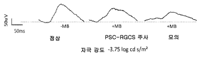
특정 실시양태에서, RG 전구체 세포는 안내압이 높은 녹내장 마우스 또는 래드 모델 시스템의, 또는 시신경(ON) 압박 손상 마우스 모델 시스템의 유리체 또는 망막하 공간 내로 이식될 수 있고, 신경절 세포층으로 이동할 것이며, 모델 시스템 중 임의의 것에서 패턴 ERG(망막전위도검사) 반응 및 시력을 개선시킬 것이다. 특정 실시양태에서, RG 세포 및 RG 전구체 세포는 시신경을 재생시킬 수 있다. In certain embodiments, the RG precursor cells can be implanted into the vitreous or subretinal space of a high-glaucomatous glaucoma or lad model system, or an optic nerve (ON) compromised mouse model system, and will migrate to the ganglion cell layer, It will improve pattern ERG (retinal positivity) response and visual acuity in any of the systems. In certain embodiments, RG cells and RG precursor cells are capable of regenerating the optic nerve.
특정 실시양태에서, RG 전구체 세포 및 RG 세포는 하나 이상의 신경보호 인자를 분비하고, 이로써, 신경보호를 효과를 일으킨다. 상기 신경보호는 시신경 손상 또는 녹내장을 앓는 동물 모델을 사용하여 측정될 수 있다. In certain embodiments, the RG precursor cells and RG cells secrete one or more neuroprotective factors, thereby effecting neuroprotection. The neuroprotection can be measured using an animal model with optic nerve damage or glaucoma.
특정 실시양태에서, RG 전구체 세포의 생존능을 유지시키는 데 적합한 배지는 배양 배지, 냉동보존제, 및 인간 환자에서 주사하기 적합한 생체적합성 주사용 배지로 이루어진 군으로부터 선택된다. In certain embodiments, the medium suitable for maintaining the viability of the RG precursor cells is selected from the group consisting of a culture medium, a cryopreservative, and a biocompatible injection medium suitable for injection in a human patient.
특정 실시양태에서, RG 전구체 세포 제제는 발열원 및 마이코젠 무함유이다In certain embodiments, RG precursor cell preparations are pyrogen-free and mycogen-free
본 발명의 또 다른 측면은, 만능 줄기 세포 유래 RG 세포를 포함하는, 포유동물 환자에서 사용하기에 적합한, RG 세포의 약학 제제로서, 여기서, 제제 중의 세포는 신경미세섬유 및/또는 Brn3a에 대하여 면역세포화학적으로 양성이고, 따라서, 일부 경우에서, 신경미세섬유(+) Brn3a(+)일 수 있는 것인, RG 세포의 약학 제제를 제공한다. RG 세포는 Tuj1(+)일 수 있고, 이는 또한 임의적으로, Thy1(+)일 수 있다. 약학 제제는 포유동물 환자 내로의 이식을 위해 RG 세포의 생존능을 유지시키기 위한 약학적으로 허용되는 담체를 추가로 포함할 수 있다.Another aspect of the present invention is a pharmaceutical preparation of RG cells suitable for use in a mammalian patient, comprising an allogenic stem cell-derived RG cell, wherein the cells in the formulation are immunized against neuronal microfibrils and / or Brn3a (+) Brn3a (+), which is cytochemically positive and, thus, in some cases, neurofibrillary (+) Brn3a (+). The RG cell may be Tuj1 (+), which may also be, optionally, Thy1 (+). The pharmaceutical preparations may further comprise a pharmaceutically acceptable carrier for sustaining the viability of the RG cells for transplantation into a mammalian subject.
본 발명의 또 다른 측면은, 만능 줄기 세포 유래 RG 세포, 및 포유동물 환자 내로의 이식을 위해 RG 세포의 생존능을 유지시키기 위한 약학적으로 허용되는 담체를 포함하는, 포유동물 환자에서 사용하기에 적합한, RG 세포의 약학 제제로서, 여기서, 제제 중의 세포는 면역세포화학적으로 신경미세섬유(+) Brn3a(+) 및 Tuj1(+), 및 임의적으로, Thy1(+)인 것인, RG 세포의 약학 제제를 제공한다. Another aspect of the present invention relates to a pharmaceutical composition comprising a compound of formula I, which is suitable for use in a mammalian patient, comprising a universal stem cell RG cell and a pharmaceutically acceptable carrier for maintaining the viability of RG cells for transplantation into a mammalian patient , A pharmaceutical preparation of RG cells wherein the cells in the formulation are immunocytochemically neuronal microfibrils (+) Brn3a (+) and Tuj1 (+), and optionally, Thy1 The formulation is provided.
본 발명의 또 다른 측면은, 만능 줄기 세포 유래 RG 세포, 및 포유동물 환자 내로의 이식을 위해 RG 세포의 생존능을 유지시키기 위한 약학적으로 허용되는 담체를 포함하는, 포유동물 환자에서 사용하기에 적합한, RG 세포의 약학 제제로서, 여기서, 70%, 80%, 90%, 95% 또는 심지어 98% 초과의 세포는 면역세포화학적으로 신경미세섬유(+) 및 Brn3a(+)인 것인, RG 세포의 약학 제제를 제공한다. 일부 경우에서, 제제 중 50%, 60% 또는 70% 초과의 세포는 면역세포화학적으로 Thy1(+)이다. 일부 실시양태에서, 70%, 80%, 90%, 95% 또는 심지어 98% 이상의 세포는 면역세포화학적으로 Tuj1(+)이다. Another aspect of the present invention relates to a pharmaceutical composition comprising a compound of formula I, which is suitable for use in a mammalian patient, comprising a universal stem cell RG cell and a pharmaceutically acceptable carrier for maintaining the viability of RG cells for transplantation into a mammalian patient Wherein the RG cell is a pharmaceutical preparation wherein the cells are 70%, 80%, 90%, 95%, or even more than 98% immunocytochemically neuronal microfibrils (+) and Brn3a (+ ≪ / RTI > In some cases, 50%, 60% or more than 70% of the cells in the formulation are Thy1 (+) immunocytochemically. In some embodiments, 70%, 80%, 90%, 95%, or even 98% or more cells are Tuj1 (+) immunocytochemically.
일부 실시양태에서, RG 세포의 약학 제제 중의 세포는 유사분열상 불활성이고(또는 유사분열 이후의 것이고), 이는 상기 세포 비증식성 세포라는 것을 의미한다. 이는 당업계에 공지되는 증식 검정법, 예컨대, 삼중수소 티미딘 흡수 검정법 등을 사용하여 측정될 수 있다. 일부 실시양태에서, 제제 중 50%, 60%, 70%, 80%, 90%, 95%, 또는 98% 초과의 세포는 유사분열상 불활성이다. In some embodiments, the cells in the pharmaceutical preparation of RG cells are in mitotic phase inactivation (or after mitosis), which means that the cells are non-proliferating cells. This can be measured using a propagation assay method known in the art, for example, a tritium thymidine absorption assay. In some embodiments, 50%, 60%, 70%, 80%, 90%, 95%, or more than 98% of the cells in the formulation are in mitotic phase inactivity.
일부 실시양태에서, RG 세포의 약학 제제 중의 세포는 RG 전구체 부류보다 더 많은 개수의 수상 돌기 및/또는 더욱 복잡한 수상 돌기를 포함한다. 일부 실시양태에서, 제제 중 50%, 60%, 70%, 80%, 90%, 95%, 또는 98% 초과의 세포는 (전구체 부류와 비교하였을 때) 더 많은 개수의 수상 돌기 및/또는 더욱 복잡한 수상 돌기를 포함한다. 따라서, 성숙한 RG 세포는 RG 전구체 세포와 비교하였을 때, 증가된 개수의 1차 수상 돌기 및/또는 2차, 3차 및 더 높은 수준에서 증가된 수상 돌기 분기, 및/또는 증가된 수상 돌기 총 길이를 나타낸다. 예를 들어, RG 세포는 RG 전구체 세포보다 세포 1개 기준으로 2-5배 더 많은 수상 돌기를 가질 수 있다(예컨대, RG 세포는 세포 1개당 4-5개의 수상 돌기를 가질 수 있고, RG 전구체 세포는 세포 1개당 1-2개의 수상 돌기를 가질 수 있다).In some embodiments, the cells in the pharmaceutical preparation of the RG cell comprise a greater number of dendrites and / or more complex dendrites than the RG precursor class. In some embodiments, cells of greater than 50%, 60%, 70%, 80%, 90%, 95%, or 98% (as compared to the precursor class) of the formulation exhibit a greater number of dendrites and / It includes complex dendrites. Thus, mature RG cells have an increased number of primary dendrites and / or increased dendritic branching at secondary, tertiary and higher levels, and / or increased dendritic lengths when compared to RG precursor cells . For example, RG cells may have 2-5-fold more dendritic cells per cell than RG precursor cells (e.g., RG cells may have 4-5 dendrites per cell, and RG precursor Cells may have 1-2 dendrites per cell).
본 발명의 추가의 또 다른 측면은 망막 색소 상피 세포, 광수용기 전구체 세포, 및/또는 광수용기 세포와 함께 RG 전구체 세포 및/또는 RG 세포; 및 포유동물 환자 내로의 이식을 위해 RG 전구체 세포 및/또는 RG 세포의 생존능을 유지시키기 위한 약학적으로 허용되는 담체를 포함하는 약학 제제를 제공한다. 제제는 또한 양극 세포 및/또는 아마크린 세포를 포함할 수 있다. 세포의 제제는 (함께 혼합되거나, 또는 공동으로 전달하고자 하는 개별 용량의 세포를 포함하는 키트 형태의) 세포 현탁액으로서, 3차원 구조로서, 예를 들어,(임의적으로, 생체적합성 매트릭스 또는 고체 지지체 상에 배치된) 시트 또는 단층 또는 다층 세포 이식편으로서 제공될 수 있다. 다층 세포 이식편의 경우, RPE 세포는 단층, 바람직하게, 분극된 단층으로서 제공될 수 있다. 따라서, 일부 실시양태에서, RG 세포 및/또는 RG 전구체 세포는 RPE 세포, 광수용기 세포 또는 광수용기 전구체 세포를 비롯한, 다른 신경망막 감각 세포와 함께 사용된다. Yet another aspect of the present invention is the use of RG precursor cells and / or RG cells together with retinal pigment epithelial cells, photoreceptor precursor cells, and / or photoreceptor cells; And a pharmaceutically acceptable carrier for maintaining the viability of RG precursor cells and / or RG cells for transplantation into a mammalian patient. The agent may also include bipolar cells and / or amaranthin cells. The preparation of a cell can be a cell suspension (in the form of a kit containing individual doses of cells to be mixed together or co-delivered), as a three-dimensional structure, for example (optionally, a biocompatible matrix or solid support phase Or as a single layer or multilayer cell implant). In the case of multilayered cell grafts, the RPE cells may be provided as a single layer, preferably as a polarized single layer. Thus, in some embodiments, RG cells and / or RG precursor cells are used with other neural retinal sensory cells, including RPE cells, photoreceptor cells, or photoreceptor precursor cells.
본 발명의 추가의 또 다른 측면은 본원에 기술된 상기 약학 제제, 예컨대, RG 전구체 세포 또는 RG 세포의 제제, 또는 그 둘 모두를 투여하는 단계를 포함하는, 환자에서 망막 신경절 세포의 손실 또는 기능 장애에 의해 유발되는 질환 및 장애를 치료하는 방법을 제공한다. 제제는 국소적으로 예컨대, 환자 안구의 망막하 공간 내로, 환자의 유리체 내로 주사할 수 있거나, 또는 전신으로 전달할 수 있거나, 또는 세포가 존속할 수 있는 다른 체강 내로 전달할 수 있다. Still another aspect of the invention is a method of treating a patient suffering from a loss or dysfunction of retinal ganglion cells in a patient, comprising administering the pharmaceutical agent as described herein, such as a preparation of RG precursor cells or RG cells, Lt; RTI ID = 0.0 > and / or < / RTI > The formulation can be injected topically, e.g., into the subretinal space of the patient ' s eye, into the vitreous of the patient, or can be delivered systemically, or into another body cavity where the cells can survive.
망막 신경절 세포의 손실에 의해 유발되는 질환 또는 장애로는 녹내장, 시신경 손상, 허혈성 시신경병증, 시신경염, 당뇨병성 망막증, 유전성 신경절 변성, 및 유전성 망막 이영양증을 포함한다. Diseases or disorders caused by loss of retinal ganglion cells include glaucoma, optic nerve damage, ischemic optic neuropathy, optic neuritis, diabetic retinopathy, hereditary ganglion degeneration, and hereditary retinal dystrophy.
따라서, 피험체에서 망막 신경절 세포의 손실 또는 기능 장애에 의해 유발되는 질환 또는 장애 치료용 의약 제조에서의, 본원에 기술되고/거나, 본원에 기술된 방법을 사용하여 제조된 RG 전구체 세포 집단 중 임의의 것의 용도 또한 제공한다. 피험체에서 망막 신경절 세포의 손실 또는 기능 장애에 의해 유발되는 질환 또는 장애 치료 방법에서 사용하기 위한, 본원에 기술되고/거나, 본원에 기술된 방법을 사용하여 제조된 RG 전구체 세포 집단 중 임의의 것의 용도 또한 제공한다. Thus, in the manufacture of a medicament for the treatment of a disease or disorder caused by the loss or dysfunction of retinal ganglion cells in a subject, any of the RG precursor cell populations prepared using the methods described herein and / It also serves the purpose of. Any of the RG precursor cell populations prepared using the methods described herein and / or described herein for use in a method of treating a disease or disorder caused by a loss or dysfunction of retinal ganglion cells in a subject Uses are also provided.
유사하게, 피험체에서 망막 신경절 세포의 손실 또는 기능 장애에 의해 유발되는 질환 또는 장애 치료용 의약 제조에서의, 본원에 기술되고/거나, 본원에 기술된 방법을 사용하여 제조된 RG 세포 집단 중 임의의 것의 용도 또한 제공한다. 피험체에서 망막 신경절 세포의 손실 또는 기능 장애에 의해 유발되는 질환 또는 장애 치료 방법에서 사용하기 위한, 본원에 기술되고/거나, 본원에 기술된 방법을 사용하여 제조된 RG 세포 집단 중 임의의 것의 용도 또한 제공한다. Similarly, any of the RG cell populations produced in the manufacture of a medicament for the treatment of a disease or disorder caused by the loss or dysfunction of retinal ganglion cells in a subject, using the methods described herein and / or described herein It also serves the purpose of. The use of any of the RG cell populations described herein and / or prepared using the methods described herein for use in a method of treating a disease or disorder caused by a loss or dysfunction of retinal ganglion cells in a subject It also provides.
특정 실시양태에서, 본 발명은 In certain embodiments,
(a) 안구 영역 전구체 세포를, 바람직하게, 세포 클러스터로서, 및 바람직하게, 저부착 또는 비부착 조건하에서 상기 세포 클러스터가 RG 전구체 세포를 포함하는 개별 세포 구체를 형성하는 데 충분한 기간 동안 신경절 세포 배지 중에서 배양하는 단계; 및 (a) the ocular region precursor cells are cultured in a ganglion cell culture medium, preferably for a period of time sufficient to allow the cell clusters to form individual cell spheres comprising RG precursor cells, ; And
(b) 배양물 중 세포 대다수가 Brn3a(+) 및/또는 신경미세섬유(+) (및 바람직하게, Brn3a(+) 및 신경미세섬유(+)) 및 Tuj1(+), 및 임의적으로, 또한 Thy1(+)인 것을 특징으로 하는 RG 세포가 될 때까지, 세포 구체를 부착 조건하에 망막 신경절 세포 분화 배지 중에서, 바람직하게, 매트릭스(예컨대, 생체물질 스캐폴드) 상에서 배양하는 단계를 포함하는, RG 세포를 제조하는 방법을 제공한다. (b) the majority of the cells in the culture are Brn3a (+) and / or neuronal microfibrils (+) (and preferably Brn3a + and neuronal microfibrils +) and Tuj1 The method comprising culturing the cell sphere on a matrix (e.g., a biocide scaffold), preferably in a retinal ganglion cell differentiation medium under adherent conditions, until the RG cell becomes a Thy1 (+). Lt; / RTI > cells.
특정 실시양태에서, 본 발명은 안구 영역 전구체 세포를, 바람직하게, 세포 클러스터로서, 및 바람직하게, 저부착 또는 비부착 조건하에서 상기 세포 클러스터가 RG 전구체 세포를 형성하는 데 충분한 기간 동안 신경절 세포 배지 중에서 배양하는 단계를 포함하는 방법을 제공한다. 배양물 중 세포 대다수가 Brn3a(+), 신경미세섬유(+) 및 Tuj1(+), 및 임의적으로, Thy1(+)인 것을 특징으로 하는 RG 세포가 될 때까지, RG 전구체 세포를 임의적으로 부착 조건하에 망막 신경절 세포 분화 배지 중에서, 바람직하게, 매트릭스(예컨대, 생체물질 스캐폴드) 상에서 배양할 수 있다. In certain embodiments, the invention encompasses the use of an ocular zone precursor cell, preferably as a cell cluster, and preferably under low adherence or nonadherence conditions, for a period of time sufficient to allow the cell cluster to form RG precursor cells And culturing the cells. Until RG cells are characterized by the majority of cells in the culture being Brn3a (+), neuronal microfibrils (+) and Tuj1 (+), and optionally Thy1 (+), RG precursor cells are arbitrarily attached , Preferably in a matrix (e. G., A biomaterial scaffold) in a retinal ganglion cell differentiation medium.
일부 실시양태에서, 안구 영역 전구체 세포는 Pax6(+) 및 Rx1(+) 및 Oct4(-) 및 Nanog(-)인 것을 특징으로, 예컨대, 면역세포화학상 특징으로 하고, 더욱더 바람직하게, 이는 또한 Six3(+), Six6(+), Lhx2(+), Tbx3(+), Sox2(+), Otx2(+) 및 네스틴(+)인 것을 특징으로 하고, 예컨대, 이는 면역염색 및/또는 유세포 분석법 또는 세포에서 마커 발현의 특징을 규명하는 데 사용되는 다른 표준 검정법에 의해 측정될 수 있다. In some embodiments, the ocular region precursor cells are characterized by Pax6 (+) and Rx1 (+) and Oct4 (-) and Nanog (-), (+), Six6 +, Lhx2 +, Tbx3 +, Sox2 +, Otx2 + and nestin. For example, it can be detected by immunohistochemistry and / Or other standard assays used to characterize marker expression in a cell.
일부 실시양태에서, RG 전구체 세포는 Math5(+), 또는 Math5(+) 및 Brn3a(+), 또는 Math5(+), Brn3a(+) 및 Brn3b(+), 또는 Math5(+), Brn3a(+), Brn3b(+) 및 Isl1(+), 또는 Brn3a(+) 및 신경미세섬유(+), 또는 Brn3a(+), 신경미세섬유(+) 및 Thy1(+)인 것을 특징으로 하고, 이는 예컨대, 면역염색 및/또는 유세포 분석법 또는 세포에서 마커 발현의 특징을 규명하는 데 사용되는 다른 표준 검정법에 의해 측정될 수 있다. In some embodiments, the RG precursor cells are Math5 (+), or Math5 (+) and Brn3a (+), or Math5 (+), Brn3a (+) and Brn3b ), Brn3b (+) and Isl1 (+), or Brn3a (+) and neuronal microfibrils (+) or Brn3a (+), neuronal microfibrils (+) and Thy1 , Immunostaining and / or flow cytometry, or other standard assays used to characterize marker expression in a cell.
특정 실시양태에서, 부착 조건은, 단지, 예시를 위해, 폴리에스테르, 폴리프로필렌, 폴리알킬렌, 폴리플루오로클로로에틸렌, 폴리비닐 클로라이드, 폴리비닐 플루오라이드 수지, 폴리스티렌, 폴리술폰, 폴리우레탄, 폴리에틸렌 테레프탈레이트, 셀룰로스, 유리 섬유, 세라믹 입자, 생체물질 스캐폴드, 폴리-L-락트산, 덱스트란, 불활성 금속 섬유, 실리카, 나트론 유리, 보로실리케이트 유리, 키토산, 또는 수세미 중 하나 이상의 것을 포함할 수 있는, 부착성 물질을 포함하는, 세포가 부착할 수 있는 표면을 가지는 배양 시스템을 포함한다. 일부 실시양태에서, 부착성 물질은 정전기적으로 하전된 것이다. 특정 실시양태에서, 생체물질 스캐폴드는 세포외 매트릭스, 예컨대, 콜라겐(예컨대, 콜라겐 IV형 또는 I형), 804G-유도된 매트릭스, 피브로넥틴, 비트로넥틴, 콘드로넥틴, 라미닌 또는 마트리겔(Matrigel)™이다. 다른 실시양태에서, 생체물질은 젤라틴, 알기네이트, 폴리글리콜리드, 피브린, 또는 자가 조립 펩티드이다.In certain embodiments, the attachment conditions include, but are not limited to, for example, polyester, polypropylene, polyalkylene, polyfluorochloroethylene, polyvinyl chloride, polyvinyl fluoride resin, polystyrene, polysulfone, polyurethane, polyethylene May include one or more of terephthalate, cellulose, glass fiber, ceramic particles, biomaterial scaffold, poly-L-lactic acid, dextran, inert metal fibers, silica, natron glass, borosilicate glass, chitosan, Which comprises an adherent material and has a surface to which the cell can adhere. In some embodiments, the adherent material is electrostatically charged. In certain embodiments, the biomaterial scaffold is an extracellular matrix such as collagen (e.g., collagen IV or I), 804G-derived matrix, fibronectin, bitronectin, chondronectin, laminin or Matrigel . In another embodiment, the biomaterial is gelatin, alginate, polyglycolide, fibrin, or self-assembling peptide.
특정 실시양태에서, RG 전구체 세포, 및 그 결과로서의 RG 세포는 만능 줄기 세포, 예컨대, 배아 줄기 세포 또는 유도성 만능 줄기 세포로부터 유래된 것이다. In certain embodiments, RG precursor cells, and consequently RG cells, are derived from pluripotent stem cells, such as embryonic stem cells or inducible pluripotent stem cells.
바람직한 실시양태에서, 만능 줄기 세포를 실질적으로 함유하지 않는, 생성된, RG 전구체 세포의 제제를 제공하며, 즉, 제제 중 10% 미만의 세포는 만능 줄기 세포이고, 더욱더 바람직하게, 제제 중 5%, 2%, 1%, 0.1% 미만 또는 심지어 0.01% 미만의 세포는 만능 줄기 세포이다.In a preferred embodiment, there is provided a preparation of RG precursor cells that is substantially free of pluripotent stem cells, i.e. less than 10% of the cells in the formulation are pluripotent stem cells and even more preferably 5% , 2%, 1%, less than 0.1% or even less than 0.01% of cells are pluripotent stem cells.
바람직한 실시양태에서, 안구 영역 전구체 세포를 실질적으로 함유하지 않는, 생성된, RG 전구체 세포의 제제를 제공하며, 즉, 제제 중 10% 미만, 및 더욱더 바람직하게, 5%, 2%, 1%, 0.1% 미만 또는 심지어 0.01% 미만의 세포는 안구 영역 전구체 세포이다.In a preferred embodiment, a preparation of RG precursor cells is provided that is substantially free of ocular region precursor cells, i.e. less than 10%, and even more preferably 5%, 2%, 1% Cells that are less than 0.1% or even less than 0.01% are eye area precursor cells.
바람직한 실시양태에서, 생성된, RG 전구체 세포의 제제의 세포 성분은 다른 세포 유형 대비 50% 이상으로 순수하고(즉, 제제 중 50% 이상의 세포는 RG 전구체 세포이다), 바람직하게, 제제 중 75% 이상, 85% 이상, 95% 이상, 99% 이상 또는 약 100%의 세포는 RG 전구체이다.In a preferred embodiment, the resulting cellular component of the preparation of RG precursor cells is more than 50% pure (i.e., more than 50% of the cells are RG precursor cells) relative to other cell types, preferably 75% , At least 85%, at least 95%, at least 99%, or at least about 100% of the cells are RG precursors.
특정 실시양태에서, 본 방법은 RG 전구체 세포 또는 RG 세포를 냉동보존하는 추가 단계를 포함한다. 세포는 바람직하게는 냉동된 세포의 해동물(및 해동된 세포 그 자체)에 맞는 냉동보존제 중에서 냉동되고, 임의적으로, 세포를 세척하여 냉동보존제를 제거한 후, RG 전구체 세포 또는 RG 세포는 (예컨대, 배양 효율 기준으로) 25% 이상의 세포 생존능, 및 더욱 바람직하게, 50%, 60%, 70%, 80% 이상 또는 심지어 90% 이상의 세포 생존능을 유지한다. In certain embodiments, the method comprises an additional step of cryopreserving RG precursor cells or RG cells. The cells are preferably frozen in a cryopreservative compatible with the thawed (and thawed) cells of the frozen cells and, optionally, after washing the cells to remove the cryopreservation agent, the RG precursor cells or RG cells (e. G. Cell viability of greater than or equal to 25%, and more preferably, greater than or equal to 50%, 60%, 70%, 80%, or even 90% of cell viability.
RG 전구체 세포 또는 RG 세포는 냉동보존될 수 있다. 일부 실시양태에서, RG 전구체 세포는 구체로서 냉동보존된다. RG precursor cells or RG cells can be cryopreserved. In some embodiments, the RG precursor cells are cryopreserved as spheres.
특정 실시양태에서, 신경절 세포 배지는 기본 배지, 예컨대, 뉴로베이살(Neurobasal)™ 배지(라이프 테크놀러지스(Life Technologies))(또는 유사 성분을 포함하는 다른 배지), D-글루코스, 항생제, 예컨대, 페니실린, 및 스트렙토마이신, 글루타맥스™(GlutaMAX), N2 보충제(인비트로겐(Invitrogen)), B27 보충제(이 또한 인비트로겐으로부터 입수), 포스콜린, BDNF 및 CNTF를 포함할 수 있다. B27 보충제는 그 중에서도 특히, SOD, 카탈라제 및 다른 항산화제 (GSH), 및 독특한 지방산, 예컨대, 리놀레산, 리놀렌산, 리포산을 함유한다. N2 보충제는 예를 들어, 하기 칵테일: 트랜스페린(10 g/L), 인슐린(500 mg/L), 프로게스테론(0.63 mg/L), 푸트레신(1611 mg/L) 및 셀레나이트(0.52 mg/L)로 대체될 수 있다. In certain embodiments, the ganglion cell culture medium comprises a basal medium such as Neurobasal (TM) media (Life Technologies) (or other media containing similar components), D-glucose, antibiotics such as penicillin , And streptomycin, GlutaMAX, N2 supplement (Invitrogen), B27 supplement (also available from Invitrogen), phoscholine, BDNF and CNTF. B27 supplements include, inter alia, SOD, catalase and other antioxidants (GSH), and unique fatty acids such as linoleic acid, linolenic acid, and lipoic acid. N2 supplementation can be achieved, for example, with the following cocktails: transferrin (10 g / L), insulin (500 mg / L), progesterone (0.63 mg / L), putrescine (1611 mg / L).
상기 측면 및 실시양태의 특정 실시양태에서, RG 전구체 세포는 만능 줄기 세포 공급원으로부터, 예컨대, Oct4, 알칼리성 포스파타제, Sox2, SSEA-3, SSEA-4, TRA-1-60, 및 TRA-1-80을 발현하는 만능 줄기 세포(예컨대, 제한하는 것은 아니지만, 배아 줄기(ES: embryonic stem) 세포주 또는 유도성 만능 줄기(iPS) 세포주)로부터, 및 더욱더 바람직하게, 공통 만능 줄기 세포 공급원으로부터 분화된 것이다. In certain aspects of the above aspects and embodiments, the RG precursor cells are from a source of pluripotent stem cells such as Oct4, alkaline phosphatase, Sox2, SSEA-3, SSEA-4, TRA- 1-60, From an embryonic stem (ES) cell line or an inducible pluripotent stem (iPS) cell line, and even more preferably from a common pluripotent stem cell source expressing the pluripotent stem cells.
상기 측면 및 실시양태의 특정 실시양태에서, RG 전구체 세포의 평균 말단 제한 절편 길이(TRF)는 7 kb, 7.5 kb, 8 kb, 8.5 kb, 9 kb, 9.5 kb, 10 kb, 10.5 kb, 11 kb, 11.5 kb 또는 심지어 12 kb보다 길다. In certain embodiments of the foregoing aspects and embodiments, the average terminal restriction fragment length (TRF) of RG precursor cells is 7 kb, 7.5 kb, 8 kb, 8.5 kb, 9 kb, 9.5 kb, 10 kb, 10.5 kb, 11 kb , 11.5 kb, or even 12 kb.
상기 측면 및 실시양태의 특정 실시양태에서, 제제는 인간 환자에게 투여하는 데 적합하고, 더욱 바람직하게, 발열원 무함유이고/거나, 인간외 동물 생성물을 함유하지 않는다. In certain aspects of the above aspects and embodiments, the agent is suitable for administration to a human patient, and more preferably, it is pyrogen-free and / or does not contain an extraneous animal product.
상기 측면 및 실시양태의 특정 실시양태에서, 제제는 인간외 수의학적 포유동물, 예컨대, 제한하는 것은 아니지만, 개, 고양이, 또는 말에게 투여하는 데 적합하다. In certain aspects of the above aspects and embodiments, the agent is suitable for administration to an extra-human veterinary mammal such as, but not limited to, a dog, a cat, or a horse.
한 측면에서, 본 개시내용은 만능 줄기 세포를 (i) 망막 유도(RI) 배지, (ii) 신경 분화(ND) 배지, 및 (iii) 신경절 세포(GC) 배지 중에서 순차적으로 배양하는 단계를 포함하는, RG 전구체 세포를 제조하는 방법을 제공한다. In one aspect, this disclosure includes sequential culturing of pluripotent stem cells in (i) retinal induction (RI) medium, (ii) neural differentiation (ND) medium, and (iii) ganglion cell (GC) Lt; RTI ID = 0.0 > RG < / RTI > precursor cells.
만능 줄기 세포는 인간 세포일 수 있다. 일부 실시양태에서, 만능 줄기 세포는 인간 ES 세포 또는 인간 iPS 세포일 수 있다. 만능 줄기 세포는 비분화된 상태로 피더세포 무함유 및/또는 제노 프리 조건하에서 임의적으로, 매트릭스 존재하에서 배양될 수 있다. 만능 줄기 세포는 비분화된 상태로 기판, 예컨대, 제한하는 것은 아니지만, 마트리겔™을 포함하는 기판 상에서, 및 임의적으로, mTESR1 배지 중에서 배양될 수 있다. The pluripotent stem cells can be human cells. In some embodiments, the pluripotent stem cells may be human ES cells or human iPS cells. The pluripotent stem cells may be cultured in the presence of a matrix in the undifferentiated state, optionally under feeder cell free and / or genotype free conditions. The pluripotent stem cells can be cultured in a non-differentiated state on a substrate, such as, but not limited to, a substrate comprising Matrigel (TM), and optionally, mTESR1 medium.
분화는 만능 줄기 세포를 망막 유도 배지 중에서 배양함으로써 시작된다. 망막 유도 배지는 기본 배지, 예컨대, DMEM/F12, DMEM/고 글루코스, 또는 DMEM/넉 아웃, 또는 DMEM/F12의 성분을 포함하는 배지, 또는 DMEM/고 글루코스의 성분을 포함하는 배지, 또는 DMEM/넉 아웃의 성분을 포함하는 배지를 포함할 수 있다. Differentiation begins by culturing pluripotent stem cells in retinal induction medium. The retinal induction medium may be a medium containing components of a base medium, such as DMEM / F12, DMEM / high glucose, or DMEM / knockout, or a component of DMEM / F12, or a medium containing components of DMEM / high glucose, Lt; RTI ID = 0.0 > knockout. ≪ / RTI >
망막 유도 배지는 D-글루코스를 포함할 수 있다. The retinal induction medium may comprise D-glucose.
신경 분화 배지는 D-글루코스를 포함할 수 있다. D-글루코스는 0-10 mg/ml, 2.5-7.5 mg/ml, 3-6 mg/ml, 4-5 mg/ml, 또는 약 4.5 mg/ml인 농도로 존재할 수 있다. The neural differentiation medium may comprise D-glucose. D-glucose may be present at a concentration of 0-10 mg / ml, 2.5-7.5 mg / ml, 3-6 mg / ml, 4-5 mg / ml, or about 4.5 mg / ml.
망막 유도 배지는 하나 이상의 항생제를 포함할 수 있다. 항생제는 페니실린, 및 스트렙토마이신 중 어느 하나, 또는 그 둘 모두를, 임의적으로, 약 0-100 유니트/ml 또는 약 100 유니트/ml 농도의 페니실린, 및 임의적으로, 약 0-100 ㎍/ml 또는 약 100 ㎍/ml 농도의 스트렙토마이신을 포함할 수 있다. The retinal induction medium may comprise one or more antibiotics. The antibiotic may be selected from the group consisting of penicillin, and streptomycin, or both, optionally penicillin at a concentration of about 0-100 units / ml or about 100 units / ml, and optionally about 0-100 占 퐂 / ml or about 100 [mu] g / ml of streptomycin.
망막 유도 배지는 하나 이상의 혈청 보충제를 포함할 수 있다. 망막 유도 배지는 N2 보충제를 포함할 수 있다. N2 보충제는 약 0.1 내지 5% (부피/부피, 또는 v/v) 또는 약 0.1 내지 2% (v/v) 또는 약 1% (v/v)인 농도로 존재할 수 있다. The retinal induction medium may comprise one or more serum supplements. Retinal induction medium can include N2 supplement. N2 supplement may be present at a concentration of about 0.1 to 5% (volume / volume, or v / v) or about 0.1 to 2% (v / v) or about 1% (v / v).
망막 유도 배지는 B-27 보충제를 포함할 수 있다. B-27 보충제는 약 0.05-2.0% (v/v)또는 약 0.2% (v/v)인 농도로 존재할 수 있다.The retinal induction medium can include a B-27 supplement. B-27 supplements may be present at a concentration of about 0.05-2.0% (v / v) or about 0.2% (v / v).
망막 유도 배지는 비필수 아미노산 또는 최소 필수 배지(MEM) 비필수 아미노산을 포함할 수 있다. 비필수 아미노산 또는 MEM 비필수 아미노산은 스톡으로부터 1X 농도로 존재할 수 있으며, 여기서, 스톡은 전형적으로 100X인 것으로 간주된다. (스톡 농도의 1X인) 글리신의 최종 농도는 약 0.1 mM이고, 따라서, 스톡은 10 mM 글리신이다. 스톡은 전형적으로 L-알라닌, L-아스파라긴, L-아스파르트산, L-글루탐산, L-프롤린, 및 L-세린 또한 포함한다. Retinal induction medium may include non-essential amino acids or minimal essential medium (MEM) non-essential amino acids. Non-essential amino acids or MEM non-essential amino acids may be present in a 1X concentration from the stock, where the stock is typically considered to be 100X. The final concentration of glycine (1X of stock concentration) is about 0.1 mM, thus the stock is 10 mM glycine. The stock typically also includes L-alanine, L-asparagine, L-aspartic acid, L-glutamic acid, L-proline, and L-serine.
망막 유도 배지는 인슐린을 포함할 수 있다. 인슐린은 인간 인슐린일 수 있다. 인슐린은 약 5-50 ㎍/ml 또는 약 20 ㎍/ml인 농도로 존재할 수 있다. The retinal induction medium may comprise insulin. Insulin can be human insulin. Insulin may be present at a concentration of about 5-50 [mu] g / ml or about 20 [mu] g / ml.
망막 유도 배지는 BMP 신호전달 억제제를 포함할 수 있다. BMP 신호전달 억제제는 노긴(Noggin) 폴리펩티드, 도소모르핀, LDN-193189, 및 그의 임의 조합으로 이루어진 군으로부터 선택될 수 있다. The retinal induction medium may comprise a BMP signal transduction inhibitor. The BMP signal transduction inhibitor may be selected from the group consisting of Noggin polypeptides, docomorphin, LDN-193189, and any combination thereof.
망막 유도 배지는 노긴 폴리펩티드를 포함할 수 있다. 노긴 폴리펩티드는 약 5-100 ng/ml 또는 약 10-100 ng/ml 또는 약 10-500 ng/ml 또는 약 50 ng/ml인 농도로 존재할 수 있다. 일부 실시양태에서, 노긴 폴리펩티드는 약 10-500 ng/ml로 존재한다. The retinal induction medium may comprise a donin polypeptide. The committed polypeptide may be present at a concentration of about 5-100 ng / ml, or about 10-100 ng / ml, or about 10-500 ng / ml, or about 50 ng / ml. In some embodiments, the agonist polypeptide is present at about 10-500 ng / ml.
망막 유도 배지는 전체적으로 또는 부분적으로 매일 새 망막 유도 배지로 대체될 수 있다. 일부 경우에서, 세포는 2 x 10⁴/㎠ 내지 6 x 10⁴/㎠ 범위의 밀도로 성장된다. Retinal induction medium can be replaced entirely or partially with new retinal induction medium each day. In some cases, the cells are grown at a density ranging from 2 x 10 4 / cm 2 to 6 x 10 4 / cm 2.
망막 유도 배지 중에서 배양하는 것은 약 4-6일, 또는 약 4일, 약 5일, 또는 약 6일 동안 이루어질 수 있다. Culturing in retinal induction medium can be about 4-6 days, or about 4 days, about 5 days, or about 6 days.
신경 분화 배지는 기본 배지, 예컨대, 뉴로베이살™ 배지(라이프 테크놀러지스), 또는 뉴로베이살™ 배지의 성분을 포함하는 기본 배지를 포함할 수 있다.The neural differentiation medium may comprise a basal medium, for example, NeuroBaseal ™ medium (Life Technologies), or a basic medium comprising components of NeuroBaseal ™ medium.
신경 분화 배지는 D-글루코스를 포함할 수 있다. D-글루코스는 0-10 mg/ml, 2.5-7.5 mg/ml, 3-6 mg/ml, 4-5 mg/ml, 또는 약 4.5 mg/ml인 농도로 존재할 수 있다. The neural differentiation medium may comprise D-glucose. D-glucose may be present at a concentration of 0-10 mg / ml, 2.5-7.5 mg / ml, 3-6 mg / ml, 4-5 mg / ml, or about 4.5 mg / ml.
신경 분화 배지는 하나 이상의 항생제를 포함할 수 있다. 항생제는 페니실린, 및 스트렙토마이신 중 어느 하나, 또는 그 둘 모두를, 임의적으로, 약 0-100 유니트/ml 또는 약 100 유니트/ml 농도의 페니실린, 및 임의적으로, 약 0-100 ㎍/ml 또는 약 100 ㎍/ml 농도의 스트렙토마이신을 포함할 수 있다. The neural differentiation medium may comprise one or more antibiotics. The antibiotic may be selected from the group consisting of penicillin, and streptomycin, or both, optionally penicillin at a concentration of about 0-100 units / ml or about 100 units / ml, and optionally about 0-100 占 퐂 / ml or about 100 [mu] g / ml of streptomycin.
신경 분화 배지는 하나 이상의 혈청 보충제를 포함할 수 있다. 신경 분화 배지는 N2 보충제를 포함할 수 있다. N2 보충제는 약 0.1 내지 5% (v/v) 또는 0.1 내지 2% (v/v) 또는 약 1% (v/v)인 농도로 존재할 수 있다. The neural differentiation medium may comprise one or more serum supplements. The neural differentiation medium may comprise an N2 supplement. The N2 supplement may be present at a concentration of about 0.1 to 5% (v / v) or 0.1 to 2% (v / v) or about 1% (v / v).
신경 분화 배지는 B-27 보충제(라이프 테크놀러지스)를 포함할 수 있다. B-27 보충제는 약 0.05 내지 5.0% (v/v), 약 0.5 내지 2.0% (v/v) 또는 약 2% (v/v)인 농도로 존재할 수 있다. Neurogenic media may include B-27 supplements (Life Technologies). The B-27 supplement may be present at a concentration of about 0.05 to 5.0% (v / v), about 0.5 to 2.0% (v / v), or about 2% (v / v).
신경 분화 배지는 비필수 아미노산 또는 MEM 비필수 아미노산 또는 글루타민 또는 글루타맥스™를 포함할 수 있다. 비필수 아미노산 또는 MEM 비필수 아미노산은 스톡 농도의 약 1X로 존재할 수 있으며, 여기서, 스톡은 약 200 mM이다 (그리고, 100X로 간주된다). 글루타맥스™는 (200 mM인 100X 스톡 용액을 사용하여) 2 mM 또는 1X 스톡 희석액으로 존재할 수 있다. The neural differentiation medium can include non-essential amino acids or MEM non-essential amino acids or glutamine or Glutamax ™. Non-essential amino acids or MEM non-essential amino acids may be present at about 1X of the stock concentration, where the stock is about 200 mM (and is considered to be 100X). Glutamax ™ may be present as a 2 mM or 1X stock dilution (using a 200X stock solution of 100X).
신경 분화 배지는 BMP 신호전달 억제제를 포함할 수 있다. BMP 신호전달 억제제는 노긴 폴리펩티드, 도소모르핀, LDN-193189, 및 그의 임의 조합으로 이루어진 군으로부터 선택될 수 있다. The neural differentiation medium may comprise a BMP signal transduction inhibitor. The BMP signal transduction inhibitor may be selected from the group consisting of angin polypeptide, docomorphine, LDN-193189, and any combination thereof.
신경 분화 배양 배지는 노긴 폴리펩티드를 포함할 수 있다. 노긴 폴리펩티드는 약 10 내지 1000 ng/ml, 또는 약 10 내지 500 ng/ml, 또는 10 내지 100 ng/ml, 또는 약 50 ng/ml인 농도로 존재할 수 있다. The neural differentiation culture medium may comprise a noggin polypeptide. The committed polypeptide may be present at a concentration of about 10 to 1000 ng / ml, or about 10 to 500 ng / ml, or 10 to 100 ng / ml, or about 50 ng / ml.
신경 분화 배지의 존재하에서 배양하는 것은 약 8-12일, 또는 약 9-10일, 또는 약 9일 동안 이루어질 수 있다. 신경 분화 배지는 전체적으로 또는 부분적으로 매일, 또는 매 2일마다, 또는 매 3일마다 교체될 수 있다. Culturing in the presence of the neural differentiation medium may be about 8-12 days, or about 9-10 days, or about 9 days. The neural differentiation medium may be replaced wholly or partially daily, every 2 days, or every 3 days.
신경절 세포 배지는 기본 배지, 예컨대, 뉴로베이살™ 배지(라이프 테크놀러지스) 또는 뉴로베이살™ 배지의 성분을 포함하는 배지를 포함할 수 있다. The ganglion cell culture medium may comprise a basal medium, for example, a medium comprising components of NeuroBaseal ™ (Life Technologies) or NeuroBaseal ™.
신경절 세포 배지는 D-글루코스를 포함할 수 있다. 신경 분화 배지는 D-글루코스를 포함할 수 있다. D-글루코스는 0-10 mg/ml, 2.5-7.5 mg/ml, 3-6 mg/ml, 4-5 mg/ml, 또는 약 4.5 mg/ml인 농도로 존재할 수 있다. Ganglion cell culture medium may include D-glucose. The neural differentiation medium may comprise D-glucose. D-glucose may be present at a concentration of 0-10 mg / ml, 2.5-7.5 mg / ml, 3-6 mg / ml, 4-5 mg / ml, or about 4.5 mg / ml.
신경절 세포 배지는 하나 이상의 항생제를 포함할 수 있다. 항생제는 페니실린, 및 스트렙토마이신 중 어느 하나, 또는 그 둘 모두를, 임의적으로, 0-100 유니트/ml 또는 약 100 유니트/ml 농도의 페니실린, 및 임의적으로, 0-100 ㎍/ml 또는 약 100 ㎍/ml의 스트렙토마이신을 포함할 수 있다. The ganglion cell culture medium may comprise one or more antibiotics. The antibiotic may be selected from the group consisting of penicillin and streptomycin, or both, optionally penicillin at a concentration of 0-100 units / ml or about 100 units / ml, and optionally 0-100 占 퐂 / ml or about 100 占 퐂 / ml < / RTI > streptomycin.
신경절 세포 배지는 하나 이상의 혈청 보충제를 포함할 수 있다. 신경절 세포 배지는 N2 보충제를 포함할 수 있다. N2 보충제는 약 0.1 내지 5% (v/v) 또는 0.1 내지 2% (v/v) 또는 약 1% (v/v)인 농도로 존재할 수 있다. The ganglion cell culture medium may comprise one or more serum supplements. The ganglion cell culture medium may include N2 supplement. The N2 supplement may be present at a concentration of about 0.1 to 5% (v / v) or 0.1 to 2% (v / v) or about 1% (v / v).
신경절 세포 배지는 B-27 보충제(포뮬러 번호 080085-SA)(라이프 테크놀러지스)를 포함할 수 있다. B-27 보충제(포뮬러 번호 080085-SA)는 약 0.05 내지 5.0% (v/v), 약 1.5 내지 2.5% (v/v) 또는 약 2% (v/v)인 농도로 존재할 수 있다. Ganglion cell culture medium may include a B-27 supplement (Formula No. 080085-SA) (Life Technologies). The B-27 supplement (formula number 080085-SA) may be present at a concentration of about 0.05 to 5.0% (v / v), about 1.5 to 2.5% (v / v) or about 2% (v / v).
신경절 세포 배지는 글루타맥스™를 포함할 수 있다. 글루타맥스™는 (200 mM인 100X 스톡 용액을 사용하여) 1X 스톡 희석액으로 존재할 수 있다. Ganglion cell media may include glutamax ™. Glutamax ™ may be present as a 1X stock dilution (using a 200X stock solution of 100X).
신경절 세포 배지는 포스콜린, BDNF 및 CNTF 중 하나, 둘, 또는 그들 모두를 포함할 수 있다. 포스콜린은 1-20 μM 또는 1-10 μM 또는 약 5 μM인 농도로 존재할 수 있다. BDNF는 1-200 ng/ml 또는 5-50 ng/ml 또는 5-25 ng/ml 또는 약 10 ng/ml인 농도로 존재할 수 있다. CNTF는 1-200 ng/ml 또는 5-50 ng/ml 또는 5-25 ng/ml 또는 약 10 ng/ml인 농도로 존재할 수 있다. BDNF는 인간 BDNF일 수 있다. CTNF는 인간 CTNF일 수 있다. Ganglion cell media can include one, two, or all of the following: phoscholine, BDNF, and CNTF. Phosphocholine may be present at a concentration of 1-20 [mu] M or 1-10 [mu] M or about 5 [mu] M. BDNF may be present at a concentration of 1-200 ng / ml, or 5-50 ng / ml, or 5-25 ng / ml, or about 10 ng / ml. CNTF may be present at a concentration of 1-200 ng / ml, or 5-50 ng / ml, or 5-25 ng / ml, or about 10 ng / ml. BDNF may be human BDNF. CTNF may be human CTNF.
신경절 세포 배지의 존재하에서 배양하는 것은 약 40-50일, 또는 약 45일 동안 이루어질 수 있다. 신경절 세포 배지는 전체적으로 또는 부분적으로, 매일, 또는 매 2일마다, 또는 매 3일마다, 또는 덜 빈번하게 교체될 수 있다. The incubation in the presence of the ganglion cell medium can be for about 40-50 days, or about 45 days. Ganglion cell media may be replaced wholly or partially, daily, every 2 days, every 3 days, or less frequently.
본 방법에 의해 제조된 RG 전구체는 배양물 중 50% 이상, 75% 이상, 85% 이상, 95% 이상, 99% 이상 또는 약 100%의 세포를 포함할 수 있고, 임의적으로, 상기 배양물은 1주, 2주, 또는, 3주, 4주 또는 더 긴 시간을 비롯한, 2주 초과 동안 증식될 수 있다. The RG precursor produced by the method may comprise at least 50%, at least 75%, at least 85%, at least 95%, at least 99%, or at least about 100% of the cells in the culture, May be proliferated for more than two weeks, including one week, two weeks, or three weeks, four weeks, or longer.
RG 전구체 세포는 마커: Math5, Brn3a, Brn3b, Isl1, 신경미세섬유, 및 Thy1 중 하나 이상의 것을 발현할 수 있고, 따라서, Math5(+), Brn3a(+), Brn3b(+), Isl1(+), 신경미세섬유(+), 및/또는 Thy1(+)인 것을 특징으로 할 수 있다. RG 전구체 세포는 마커: Math5, Brn3a, Brn3b, 및 Isl1 중 하나 이상의 것을 발현할 수 있고, 따라서, Math5(+), Brn3a(+), Brn3b(+), 및/또는 Isl1(+)인 것을 특징으로 할 수 있다. RG 전구체 세포는 마커: Brn3a, 신경미세섬유 및 Thy1 중 하나 이상의 것을 발현할 수 있고, 따라서, Brn3a(+), 신경미세섬유(+), 및/또는 Thy1(+)인 것을 특징으로 할 수 있다. RG 전구체는 Pax6 및/또는 Rx1을 발현하지 않을 수 있고, 따라서, Pax6(-) 및/또는 Rx1(-)인 것을 특징으로 할 수 있다. 특정 RG 전구체 세포는 Math5 및/또는 Brn3b를 발현하지 않을 수 있고, 따라서, Math5(-) 및/또는 Brn3b(-)인 것을 특징으로 할 수 있다. RG precursor cells can express one or more of the following markers: Math5, Brn3a, Brn3b, Isl1, nerve microfibrils, and Thy1 and thus Math5 (+), Brn3a (+), Brn3b (+), Isl1 , Neuronal fine fibers (+), and / or Thy1 (+). RG precursor cells can express one or more of the following markers: Math5, Brn3a, Brn3b, and Isl1 and are therefore characterized by Math5 (+), Brn3a (+), Brn3b (+), and / . RG precursor cells can express one or more of the following markers: Brn3a, neuronal fine fibers, and Thy1, and can therefore be characterized as being Brn3a (+), neuronal microfibrils (+), and / or Thy1 . The RG precursor may not express Pax6 and / or Rx1 and may therefore be characterized as being Pax6 (-) and / or Rx1 (-). Certain RG precursor cells may not express Math5 and / or Brn3b and thus may be characterized as being Math5 (-) and / or Brn3b (-).
RG 전구체 세포는 Math5(+), 또는 Math5(+), Brn3a(+), 또는 Math5(+), Brn3a(+), Brn3b(+), 또는 Math5(+), Brn3a(+), Brn3b(+), Isl1(+), 또는 Brn3a(+), 또는 신경미세섬유(+), 또는 Brn3a(+), 신경미세섬유(+), 또는 Brn3a(+), 신경미세섬유(+), Thy1(+)인 것을 특징으로 할 수 있다. RG 전구체는 Pax6 및/또는 Rx1을 발현하지 않을 수 있고, 따라서, Pax6(-) 및/또는 Rx1(-)인 것을 특징으로 할 수 있다. RG 전구체 세포는 Tuj1(+)일 수 있다. RG 전구체 세포는 증식성일 수 있다. RG precursor cells are Math5 (+) or Math5 (+), Brn3a (+), or Math5 (+), Brn3a (+), Brn3b ), Isl1 (+), or Brn3a (+), or neuronal microfibers (+) or Brn3a (+), neuronal microfibers (+) or Brn3a ). ≪ / RTI > The RG precursor may not express Pax6 and / or Rx1 and may therefore be characterized as being Pax6 (-) and / or Rx1 (-). RG precursor cells may be Tuj1 (+). RG precursor cells may be proliferative.
RG 전구체 세포는 냉동보존될 수 있다. RG 전구체 세포는 인간 세포일 수 있다.RG precursor cells can be cryopreserved. RG precursor cells can be human cells.
본 방법은 망막 신경절 세포(RGC) 분화 배지의 존재하에서의 후속 배양 단계를 추가로 포함할 수 있다. 상기 단계는 중간 냉동보존 단계 없이 이루어질 수 있거나, 또는 RG 전구체 세포의 냉동보존 및 해동 이후에 이루어질 수 있다. The method may further comprise a subsequent incubation step in the presence of retinal ganglion cell (RGC) differentiation medium. This step can be done without intermediate cryopreservation step, or after cryopreservation and thawing of RG precursor cells.
RGC 분화 배지는 기본 배지, 예컨대, 뉴로베이살™ 배지(라이프 테크놀러지스) 또는 뉴로베이살™ 배지의 성분을 포함하는 배지를 포함할 수 있다.The RGC differentiation medium may comprise a basal medium, for example a medium comprising components of NeuroBaseal ™ media (Life Technologies) or NeuroBaseal ™ media.
RGC 분화 배지는 D-글루코스를 포함할 수 있다. 신경 분화 배지는 D-글루코스를 포함할 수 있다. D-글루코스는 0-10 mg/ml, 2.5-7.5 mg/ml, 3-6 mg/ml, 4-5 mg/ml, 또는 약 4.5 mg/ml인 농도로 존재할 수 있다. The RGC differentiation medium may comprise D-glucose. The neural differentiation medium may comprise D-glucose. D-glucose may be present at a concentration of 0-10 mg / ml, 2.5-7.5 mg / ml, 3-6 mg / ml, 4-5 mg / ml, or about 4.5 mg / ml.
RGC 분화 배지는 하나 이상의 항생제를 포함할 수 있다. 항생제는 페니실린, 및 스트렙토마이신 중 어느 하나, 또는 그 둘 모두를, 임의적으로, 0-100 유니트/ml 또는 약 100 유니트/ml 농도의 페니실린, 및 임의적으로, 0-100 ㎍/ml 또는 약 100 ㎍/ml의 스트렙토마이신을 포함할 수 있다. The RGC differentiation medium may comprise one or more antibiotics. The antibiotic may be selected from the group consisting of penicillin and streptomycin, or both, optionally penicillin at a concentration of 0-100 units / ml or about 100 units / ml, and optionally 0-100 占 퐂 / ml or about 100 占 퐂 / ml < / RTI > streptomycin.
RGC 분화 배지는 하나 이상의 혈청 보충제를 포함할 수 있다. RGC 분화 배지는 B-27 보충제(포뮬러 080085-SA)를 포함할 수 있다. B-27 보충제는 약 0.05 내지 5.0% (v/v), 약 1.5 내지 2.5% (v/v) 또는 약 2% (v/v)인 농도로 존재할 수 있다. The RGC differentiation medium may comprise one or more serum supplements. The RGC differentiation medium may include a B-27 supplement (formula 080085-SA). The B-27 supplement may be present at a concentration of about 0.05 to 5.0% (v / v), about 1.5 to 2.5% (v / v), or about 2% (v / v).
RGC 분화 배지는 글루타맥스™를 포함할 수 있다. 글루타맥스™는 1X 스톡 희석액으로 존재할 수 있다. The RGC differentiation medium may include glutamax ™. Glutamax ™ may be present as a 1X stock dilution.
RGC 분화 배지는 레티노산을 포함할 수 있다. 레티노산은 약 0.5-20 μM, 약 0.5-10 μM, 약 1-5 μM, 또는 약 2 μM인 농도로 존재할 수 있다. The RGC differentiation medium may comprise retinoic acid. The retinoic acid may be present at a concentration of about 0.5-20 μM, about 0.5-10 μM, about 1-5 μM, or about 2 μM.
RGC 분화 배지는 포스콜린, BDNF, CNTF, cAMP, 및 DAPT 모두를 비롯한, 그들 중 하나 이상의 것을 포함할 수 있다. RGC 분화 배지는 BDNF 및 CNTF 중 하나, 또는 그 둘 모두를 포함할 수 있다. RGC 분화 배지는 포스콜린, cAMP, 및 DAPT 또는 다른 노치(Notch) 경로 억제제 또는 노치 억제제 (예컨대, 노치 차단 항체 또는 항체 단편, 노치 음성 조절 영역 항체 또는 항체 단편, 알파-세크레타제 억제제, 감마-세크레타제 억제제, 스테이플드 펩티드, 소분자 차단제 및 siRNA, shRNA 및 miRNA) 모두를 비롯한, 그들 중 하나 이상의 것을 포함할 수 있다. 포스콜린은 1-20 μM 또는 1-10 μM 또는 약 5 μM인 농도로 존재할 수 있다. BDNF는 1-200 ng/ml 또는 5-50 ng/ml 또는 5-25 ng/ml 또는 약 10 ng/ml인 농도로 존재할 수 있다. CNTF는 1-200 ng/ml 또는 5-50 ng/ml 또는 5-25 ng/ml 또는 약 10 ng/ml인 농도로 존재할 수 있다. BDNF는 인간 BDNF일 수 있다. CTNF는 인간 CTNF일 수 있다. cAMP는 1-500 ng/ml 또는 10-250 ng/ml 또는 5-150 ng/ml 또는 약 100 ng/ml인 농도로 존재할 수 있다. APT는 1-100 μM 또는 1-50 μM 또는 1-20 μM, 또는 약 10 μM인 농도로 존재할 수 있다. The RGC differentiation medium may include one or more of them, including both forcoline, BDNF, CNTF, cAMP, and DAPT. The RGC differentiation medium may comprise one or both of BDNF and CNTF. The RGC differentiation medium may be selected from the group consisting of a phosholin, cAMP, and DAPT or other Notch pathway inhibitor or a Notch Inhibitor (such as a Notch Blocking Antibody or Antibody Fragment, a Notch Negative Regulation Domain Antibody or Antibody Fragment, Secretase inhibitors, stapled peptides, small molecule blockers and siRNAs, shRNAs and miRNAs), all of which may include one or more of these. Phosphocholine may be present at a concentration of 1-20 [mu] M or 1-10 [mu] M or about 5 [mu] M. BDNF may be present at a concentration of 1-200 ng / ml, or 5-50 ng / ml, or 5-25 ng / ml, or about 10 ng / ml. CNTF may be present at a concentration of 1-200 ng / ml, or 5-50 ng / ml, or 5-25 ng / ml, or about 10 ng / ml. BDNF may be human BDNF. CTNF may be human CTNF. The cAMP may be present at a concentration of 1-500 ng / ml or 10-250 ng / ml or 5-150 ng / ml or about 100 ng / ml. APT may be present at a concentration of 1-100 [mu] M or 1-50 [mu] M, 1-20 [mu] M, or about 10 [mu] M.
RGC 분화 배지는 인슐린을 포함할 수 있다. 인슐린은 인간 인슐린일 수 있다. 인슐린은 약 5-50 ㎍/ml 또는 약 20 ㎍/ml인 농도로 존재할 수 있다. The RGC differentiation medium may comprise insulin. Insulin can be human insulin. Insulin may be present at a concentration of about 5-50 [mu] g / ml or about 20 [mu] g / ml.
RGC 분화 배지의 존재하에서 배양하는 것은 약 1-4주 또는 약 2-4주 또는 약 2-3주 또는 약 2주 동안 이루어질 수 있다. RGC 분화 배지는 전체적으로 또는 부분적으로, 매일, 또는 매 2일마다, 또는 매 3일마다, 또는 덜 빈번하게 교체될 수 있다. Culturing in the presence of RGC differentiation medium may be about 1-4 weeks or about 2-4 weeks or about 2-3 weeks or about 2 weeks. The RGC differentiation medium may be replaced wholly or partially, daily, every 2 days, every 3 days, or less frequently.
본 방법에 의해 제조된 RG 세포는 배양물 중 50% 이상, 75% 이상, 85% 이상, 95% 이상, 99% 또는 약 100%의 세포를 포함할 수 있고, 임의적으로, 상기 배양물은 1주, 2주, 또는, 3주, 4주 또는 더 긴 시간을 비롯한, 2주 초과 동안 증식될 수 있다. The RG cells produced by the method may comprise at least 50%, at least 75%, at least 85%, at least 95%, at least 99%, or at least about 100% of the cells in the culture, Week, two weeks, or three weeks, four weeks, or longer.
RG 세포는 마커: Brn3a, 신경미세섬유, Tuj1 및 Thy1 중 하나 이상의 것을 발현할 수 있고, 따라서, Brn3a(+), 신경미세섬유(+), Tuj1(+), 및/또는 Thy1(+)인 것을 특징으로 할 수 있다. RG 세포는 Brn3a(+), 신경미세섬유(+), 또는 Brn3a(+), 신경미세섬유(+), Thy1(+), 또는 Brn3a(+), 신경미세섬유(+), Thy1(+), 또는 Brn3a(+), 신경미세섬유(+), Tuj1(+), 또는 Brn3a(+), 신경미세섬유(+), Tuj1(+), Thy1(+)인 것을 특징으로 할 수 있다. RG 세포는 유사분열 이후의 것일 수 있고, RG 전구체 세포보다 더 많은 개수, 더 긴 및/또는 더욱 복잡한 수상 돌기를 포함할 수 있다. RG cells can express one or more of the following markers: Brn3a, neuronal microfibrils, Tuj1 and Thy1 and thus can express Brn3a (+), neurofilament (+), Tuj1 (+), and / or Thy1 . ≪ / RTI > RG cells express Brn3a (+), neuronal microfibrils (+), or Brn3a (+), neuronal microfibers (+), Thy1 (+), or Brn3a , Or Brn3a (+), nerve microfibers (+), Tuj1 (+) or Brn3a (+), nerve microfibers (+), Tuj1 (+) and Thy1 (+). RG cells may be after mitosis and may contain more, more, and / or more complex water dendrites than RG precursor cells.
RG 세포는 냉동보존될 수 있다. RG 세포는 인간 세포일 수 있다.RG cells can be cryopreserved. RG cells can be human cells.
또 다른 측면에서, 본 개시내용은 신경절 세포 배지 중에서 안구 영역 전구체 세포를 배양하는 단계를 포함하는, RG 전구체 세포를 제조하는 방법을 제공한다. In another aspect, the disclosure provides a method of producing RG precursor cells, comprising culturing ocular area precursor cells in a ganglion cell culture medium.
안구 영역 전구체 세포는, 그 중 50% 이상, 75% 이상, 85% 이상, 95% 이상, 99% 또는 약 100%가 안구 영역 전구체 세포인, 세포 집단으로서 제공될 수 있다. 안구 영역 전구체 세포는 Pax6(+) 및 Rx1(+) 및 임의적으로, 또는 Six3(+), Six6(+), Lhx2(+), 및/또는 Tbx3(+)일 수 있다. 안구 영역 전구체 세포는 또한 Sox2(+)일 수 있다. 안구 영역 전구체 세포는 네스틴(Nestin)(+)일 수 있다. 안구 영역 전구체 세포는 또한 Otx2(+)일 수 있다. The ocular region precursor cells may be provided as a population of cells in which at least 50%, at least 75%, at least 85%, at least 95%, at least 99%, or at least about 100% are ocular region precursor cells. The ocular region precursor cells may be Pax6 (+) and Rx1 (+) and optionally, or Six3 (+), Six6 (+), Lhx2 (+), and / or Tbx3 (+). The ocular region precursor cells may also be Sox2 (+). The ocular region precursor cells may be nestin (+). The ocular region precursor cells may also be Otx2 (+).
안구 영역 전구체는 만능 줄기 세포의 시험관내 분화에 의해 수득될 수 있다. 일부 실시양태에서, 만능 줄기 세포는 ES 세포 또는 iPS 세포일 수 있다. 만능 줄기 세포는 피더세포 무함유 및/또는 제노 프리 조건하에서 배양될 수 있다. The ocular region precursor can be obtained by in vitro differentiation of pluripotent stem cells. In some embodiments, the pluripotent stem cells may be ES cells or iPS cells. The pluripotent stem cells can be cultured under feeder cell free and / or genotype free conditions.
안구 영역 전구체 세포의 배양은 약 5-45일, 또는 약 5-20일, 또는 약 35-45일 동안 이루어질 수 있다. 신경절 세포 배지, 및 신경절 세포 배지에서의 배양 파라미터는 상기 기술된 바와 같을 수 있다. The incubation of the ocular region precursor cells can be for about 5-45 days, or about 5-20 days, or about 35-45 days. Ganglion cell culture medium, and ganglion cell culture medium may be as described above.
본 방법에 의해 제조된 RG 전구체 세포는 배양물 중 50% 이상, 75% 이상, 85% 이상, 95% 이상, 99% 이상 또는 약 100%의 세포를 포함할 수 있고, 임의적으로, (RG 전구체 세포를 상기 비율(%)로 포함하는) 상기 배양물은 1주, 2주, 또는, 3주, 4주 또는 더 긴 시간을 비롯한, 2주 초과 동안 증식될 수 있다. The RG precursor cells produced by the present method can comprise cells in the culture of at least 50%, at least 75%, at least 85%, at least 95%, at least 99%, or at least about 100% The culture containing the cells in the above ratio (%) can be proliferated for more than two weeks, including one week, two weeks, or three weeks, four weeks or longer.
RG 전구체 세포의 표현형은 상기 기술된 바와 같다. 따라서, 한 예로서, RG 전구체 세포는 마커: Math5, Brn3a, Brn3b, Isl1, 신경미세섬유, 및 Thy1 중 하나 이상의 것을 발현할 수 있고, 따라서, Math5(+), Brn3a(+), Brn3b(+), Isl1(+), 신경미세섬유(+), 및/또는 Thy1(+)인 것을 특징으로 할 수 있다. RG 전구체 세포는 마커: Math5, Brn3a, Brn3b, 및 Isl1 중 하나 이상의 것을 발현할 수 있고, 따라서, Math5(+), Brn3a(+), Brn3b(+), 및/또는 Isl1(+)인 것을 특징으로 할 수 있다. RG 전구체 세포는 마커: Brn3a, 신경미세섬유, 및 Thy1 중 하나 이상의 것을 발현할 수 있고, 따라서, Brn3a(+), 신경미세섬유(+), 및/또는 Thy1(+)인 것을 특징으로 할 수 있다. RG 전구체는 Pax6 또는 Rx1을 발현하지 않을 수 있고, 따라서, Pax6(-) 및/또는 Rx1(-)인 것을 특징으로 할 수 있다. RG 전구체 세포는 증식성일 수 있다. 다른 표현형은 상기 기술된 바와 같다. The phenotype of the RG precursor cells is as described above. Thus, as an example, RG precursor cells can express one or more of the following markers: Math5, Brn3a, Brn3b, Isl1, neuronal microfibrils, and Thy1 and thus Math5 (+), Brn3a (+), Brn3b ), Isl1 (+), nerve microfibers (+), and / or Thy1 (+). RG precursor cells can express one or more of the following markers: Math5, Brn3a, Brn3b, and Isl1 and are therefore characterized by Math5 (+), Brn3a (+), Brn3b (+), and / . RG precursor cells can express one or more of the following markers: Brn3a, neuronal microfibrils, and Thy1 and can therefore be characterized as Brn3a (+), neuronal microfibrils (+), and / or Thy1 have. The RG precursor may not express Pax6 or Rx1 and therefore may be characterized as being Pax6 (-) and / or Rx1 (-). RG precursor cells may be proliferative. Other phenotypes are as described above.
RG 전구체 세포는 냉동보존될 수 있다. RG 전구체 세포는 인간 세포일 수 있다.RG precursor cells can be cryopreserved. RG precursor cells can be human cells.
또 다른 측면에서, 본 개시내용은 안구 영역 전구체 세포를 신경절 세포 배지 및 망막 신경절 세포(RGC) 분화 배지 중에서 순차적으로 배양하는 단계를 포함하는, RG 세포를 제조하는 방법을 제공한다. In another aspect, the disclosure provides a method of preparing RG cells, comprising sequentially culturing the ocular area precursor cells in a ganglion cell culture medium and a retinal ganglion cell (RGC) differentiation medium.
안구 영역 전구체 세포는, 그 중 50% 이상, 75% 이상, 85% 이상, 95% 이상, 99% 또는 약 100%가 안구 영역 전구체 세포인, 세포 집단으로서 제공될 수 있다. 안구 영역 전구체 세포는 Pax6(+) 및 Rx1(+) 및 임의적으로, 또한 Six3(+), Six6(+), Lhx2(+), 및/또는 Tbx3(+)일 수 있다. 안구 영역 전구체 세포는 또한 Sox2(+)일 수 있다. 안구 영역 전구체 세포는 네스틴(+)일 수 있다. 안구 영역 전구체 세포는 또한 Otx2(+)일 수 있다. The ocular region precursor cells may be provided as a population of cells in which at least 50%, at least 75%, at least 85%, at least 95%, at least 99%, or at least about 100% are ocular region precursor cells. The ocular region precursor cells may be Pax6 (+) and Rx1 (+) and optionally also Six3 (+), Six6 (+), Lhx2 (+), and / or Tbx3 (+). The ocular region precursor cells may also be Sox2 (+). The ocular region precursor cells may be nestin (+). The ocular region precursor cells may also be Otx2 (+).
안구 영역 전구체는 만능 줄기 세포의 시험관내 분화에 의해 수득될 수 있다. 일부 실시양태에서, 만능 줄기 세포는 ES 세포 또는 iPS 세포일 수 있다. 만능 줄기 세포는 피더세포 무함유 및/또는 제노 프리 조건하에서 배양될 수 있다. The ocular region precursor can be obtained by in vitro differentiation of pluripotent stem cells. In some embodiments, the pluripotent stem cells may be ES cells or iPS cells. The pluripotent stem cells can be cultured under feeder cell free and / or genotype free conditions.
신경절 세포 배지, 및 신경절 세포 배지에서의 배양 파라미터는 상기 기술된 바와 같을 수 있다. RGC 분화 배지, 및 RGC 분화 배지에서의 배양 파라미터는 상기 기술된 바와 같을 수 있다. Ganglion cell culture medium, and ganglion cell culture medium may be as described above. RGC differentiation medium, and RGC differentiation medium may be as described above.
본 방법에 의해 제조된 RG 세포는 배양물 중 50% 이상, 75% 이상, 85% 이상, 95% 이상, 99% 또는 약 100%의 세포를 포함할 수 있고, 임의적으로, (RG 세포를 상기 비율(%)로 포함하는) 상기 배양물은 1주, 2주, 또는, 3주, 4주 또는 더 긴 시간을 비롯한, 2주 초과 동안 증식될 수 있다. The RG cells produced by the present method may comprise cells in the culture at least 50%, at least 75%, at least 85%, at least 95%, at least 99%, or at least about 100% The culture may be grown for more than two weeks, including 1, 2, or 3 weeks, 4 weeks, or longer.
RG 세포의 표현형은 상기 기술된 바와 같다. 따라서, 한 예로서, RG 세포는 마커: 마커: Brn3a, 신경미세섬유, Tuj1, 및 Thy1 중 하나 이상의 것을 발현할 수 있고, 따라서, Brn3a(+), 신경미세섬유(+), Tuj1(+), 및/또는 Thy1(+)인 것을 특징으로 할 수 있다. RG 세포는 유사분열 이후의 것일 수 있다. RG 세포는 유사분열 이후의 것일 수 있고, RG 전구체 세포보다 더 복잡한 수상 돌기, 더 긴 수상 돌기, 및/또는 더 많은 수상 돌기를 가질 수 있다. 다른 표현형은 상기 기술되어 있다. The phenotype of RG cells is as described above. Thus, by way of example, RG cells can express one or more of the following: marker: marker: Brn3a, neuronal microfilament, Tuj1, and Thy1 and thus Brn3a (+), neurofilament (+), Tuj1 , And / or Thy1 (+). RG cells may be after mitosis. RG cells may be after mitosis and may have more complex water dendrites, longer water dendrites, and / or more water dendrites than RG precursor cells. Other phenotypes have been described above.
RG 세포는 냉동보존될 수 있다. RG 세포는 인간 세포일 수 있다.RG cells can be cryopreserved. RG cells can be human cells.
또 다른 측면에서, 본 개시내용은 RG 전구체 세포를 망막 신경절 세포(RGC) 분화 배지 중에서 배양하는 단계를 포함하는, RG 세포를 제조하는 방법을 제공한다. In another aspect, the disclosure provides a method of producing RG cells, comprising culturing RG precursor cells in a retinal ganglion cell (RGC) differentiation medium.
RG 전구체 세포는, 그 중 50% 이상, 75% 이상, 85% 이상, 95% 이상, 99% 또는 약 100%가 RG 전구체 세포인, 세포 집단으로서 제공될 수 있다. RG 전구체 세포는 마커: Math5, Brn3a, Brn3b, Isl1, 신경미세섬유, 및 Thy1 중 하나 이상의 것을 발현할 수 있고, 따라서, Math5(+), Brn3a(+), Brn3b(+), Isl1(+), 신경미세섬유(+), 및/또는 Thy1(+)인 것을 특징으로 할 수 있다. RG 전구체 세포는 마커: Math5, Brn3a, Brn3b, 및 Isl1 중 하나 이상의 것을 발현할 수 있고, 따라서, Math5(+), Brn3a(+), Brn3b(+), 및/또는 Isl1(+)인 것을 특징으로 할 수 있다. RG 전구체 세포는 마커: Brn3a, 신경미세섬유, 및 Thy1 중 하나 이상의 것을 발현할 수 있고, 따라서, Brn3a(+), 신경미세섬유(+), 및/또는 Thy1(+)인 것을 특징으로 할 수 있다. RG 전구체는 Pax6 또는 Rx1 발현하지 않을 수 있고, Pax6(-) 및/또는 Rx1(-)인 것을 특징으로 할 수 있다. RG 전구체는 만능 줄기 세포의 시험관내 분화에 의해 수득될 수 있다. 일부 실시양태에서, 만능 줄기 세포는 ES 세포 또는 iPS 세포일 수 있다. 만능 줄기 세포는 피더세포 무함유 및/또는 제노 프리 조건하에서 배양될 수 있다. RG precursor cells may be provided as a population of cells in which at least 50%, at least 75%, at least 85%, at least 95%, 99% or about 100% of the RG precursor cells are RG precursor cells. RG precursor cells can express one or more of the following markers: Math5, Brn3a, Brn3b, Isl1, nerve microfibrils, and Thy1 and thus Math5 (+), Brn3a (+), Brn3b (+), Isl1 , Neuronal fine fibers (+), and / or Thy1 (+). RG precursor cells can express one or more of the following markers: Math5, Brn3a, Brn3b, and Isl1 and are therefore characterized by Math5 (+), Brn3a (+), Brn3b (+), and / . RG precursor cells can express one or more of the following markers: Brn3a, neuronal microfibrils, and Thy1 and can therefore be characterized as Brn3a (+), neuronal microfibrils (+), and / or Thy1 have. The RG precursor may not express Pax6 or Rx1, and may be characterized by being Pax6 (-) and / or Rx1 (-). The RG precursor can be obtained by in vitro differentiation of pluripotent stem cells. In some embodiments, the pluripotent stem cells may be ES cells or iPS cells. The pluripotent stem cells can be cultured under feeder cell free and / or genotype free conditions.
RGC 분화 배지 및 RGC 분화 배지에서의 배양 파라미터는 상기 기술된 바와 같을 수 있다. The culture parameters in RGC differentiation medium and RGC differentiation medium may be as described above.
본 방법에 의해 제조된 RG 세포는 배양물 중 50% 이상, 75% 이상, 85% 이상, 95% 이상, 99% 이상 또는 약 100%의 세포를 포함할 수 있고, 임의적으로, (RG 세포를 상기 비율(%)로 포함하는) 상기 배양물은 1주, 2주, 또는, 3주, 4주 또는 더 긴 시간을 비롯한, 2주 초과 동안 증식될 수 있다. The RG cells produced by the present method may comprise cells in the culture at least 50%, at least 75%, at least 85%, at least 95%, at least 99%, or at least about 100% The culture may be grown for more than 2 weeks, including 1, 2, or 3 weeks, 4 weeks or longer.
RG 세포는 마커: Brn3a, 신경미세섬유, Tuj1, 및 Thy1 중 하나 이상의 것을 발현할 수 있고, 따라서, Brn3a(+), 신경미세섬유(+), Tuj1(+), 및 Thy1(+)인 것을 특징으로 할 수 있다. RG cells can express one or more of the following markers: Brn3a, neuronal microfibrils, Tuj1, and Thy1 and thus can express Brn3a (+), neurofilament (+), Tuj1 (+), and Thy1 .
RG 세포는 냉동보존될 수 있다. RG 세포는 인간 세포일 수 있다.RG cells can be cryopreserved. RG cells can be human cells.
또 다른 측면에서, 본 개시내용은 본원에 기술된 바와 같은, 예컨대, 이전 단락에서 기술된 바와 같은 방법을 사용하여 제조된 RG 전구체 세포를 포함하는 조성물을 제공한다. In another aspect, the disclosure provides compositions comprising RG precursor cells as described herein, for example, using methods such as those described in the previous paragraph.
또 다른 측면에서, 본 개시내용은 임의적으로, 인간 세포인 것인, RG 전구체 세포를 포함하는 조성물을 제공한다. In yet another aspect, the disclosure provides a composition comprising RG precursor cells, optionally, human cells.
RG 전구체 세포는 조성물 중 50% 이상, 75% 이상, 85% 이상, 95% 이상, 99% 또는 약 100%의 세포를 포함할 수 있다. The RG precursor cells may comprise at least 50%, at least 75%, at least 85%, at least 95%, at least 99%, or at least about 100% of the cells in the composition.
RG 전구체 세포는 마커: Math5, Brn3a, Brn3b, Isl1, 신경미세섬유, 및 Thy1 중 하나 이상의 것을 발현할 수 있고, 따라서, Math5(+), Brn3a(+), Brn3b(+), Isl1(+), 신경미세섬유(+), 및/또는 Thy1(+)인 것을 특징으로 할 수 있다. RG 전구체 세포는 Math5, Brn3a, Brn3b, 및 Isl1을 발현할 수 있고, 따라서, Math5(+), Brn3a(+), Brn3b(+), 및 Isl1(+)인 것을 특징으로 할 수 있다. RG 전구체 세포는 Brn3a, 신경미세섬유, 및 Thy1을 발현할 수 있고, 따라서, Brn3a(+), 신경미세섬유(+), 및 Thy1(+)인 것을 특징으로 할 수 있다. RG 전구체 세포에 대한 다른 표현형은 상기 기술된 바와 같다. RG precursor cells can express one or more of the following markers: Math5, Brn3a, Brn3b, Isl1, nerve microfibrils, and Thy1 and thus Math5 (+), Brn3a (+), Brn3b (+), Isl1 , Neuronal fine fibers (+), and / or Thy1 (+). RG precursor cells are capable of expressing Math5, Brn3a, Brn3b, and Isl1 and are therefore characterized by being Math5 (+), Brn3a (+), Brn3b (+), and Isl1 (+). RG precursor cells are capable of expressing Brn3a, neuronal microfibrils, and Thy1, and thus may be characterized as Brn3a (+), nerve microfibrils (+), and Thy1 (+). Other phenotypes for RG precursor cells are as described above.
RG 전구체 세포는 냉동보존될 수 있다. RG 전구체 세포는 인간 세포일 수 있다.RG precursor cells can be cryopreserved. RG precursor cells can be human cells.
또 다른 측면에서, 본 개시내용은 치료를 필요로 하는 개체에게 RG 전구체 세포를 포함하는 조성물(예컨대, 본원에 기술된 바와 같은 조성물 또는 본원에 기술된 바와 같은 방법을 사용하여 제조된 조성물)을 투여하는 단계를 포함하는, 치료를 필요로 하는 개체를 치료하는 방법을 제공한다. In another aspect, the disclosure provides a method of treating a subject in need of treatment comprising administering to the subject a composition comprising RG precursor cells (e.g., a composition as described herein or a composition prepared using a method as described herein) Comprising administering a therapeutically effective amount of a compound of the invention to a subject in need thereof.
조성물은 안구, 유리체, 망막하 공간, 또는 정맥내로 투여될 수 있다. RG 전구체 세포는 인간 세포일 수 있다.The composition may be administered into the eye, the vitreous, the subretinal space, or intravenously. RG precursor cells can be human cells.
또 다른 측면에서, 본 개시내용은 본원에, 예컨대, 이전 단락에 기술된 바와 같은 방법에 따라 제조된 RG 세포를 포함하는 조성물을 제공한다. In another aspect, the disclosure provides herein a composition comprising RG cells, for example, prepared according to the methods described in the previous paragraph.
본 방법에 의해 제조된 RG 세포는 조성물 중 50% 이상, 75% 이상, 85% 이상, 95% 이상, 99% 또는 약 100%의 세포를 포함할 수 있고, 임의적으로, (RG 세포를 상기 비율(%)로 포함하는) 상기 조성물은 1주, 2주, 또는, 3주, 4주 또는 더 긴 시간을 비롯한, 2주 초과 동안 증식될 수 있다. The RG cells produced by the present method may comprise cells at least 50%, at least 75%, at least 85%, at least 95%, at least 99%, or at least about 100% (%)) Can be propagated for more than two weeks, including one week, two weeks, or three weeks, four weeks, or longer.
RG 세포는 마커: Brn3a, 신경미세섬유, Tuj1, 및 Thy1 중 하나 이상의 것을 발현할 수 있고, 따라서, Brn3a(+), 신경미세섬유(+), Tuj1(+), 및/또는 Thy1(+)인 것을 특징으로 할 수 있다. RG cells can express one or more of the following markers: Brn3a, neuronal fine fibers, Tuj1, and Thy1, and thus can express Brn3a (+), neurofilament (+), Tuj1 (+), and / .
RG 세포는 냉동보존될 수 있다. RG 세포는 인간 세포일 수 있다.RG cells can be cryopreserved. RG cells can be human cells.
또 다른 측면에서, 본 개시내용은 치료를 필요로 하는 개체에게 RG 세포를 포함하는 조성물, 예컨대, 본원에, 예컨대, 이전 단락에 기술된 바와 같은 조성물 또는 본원에, 예컨대, 이전 단락에 기술된 바와 같은 방법에 의해 제조된 조성물을 투여하는 단계를 포함하는, 치료를 필요로 하는 개체를 치료하는 방법을 제공한다. In another aspect, the disclosure provides a composition comprising RG cells to a subject in need of treatment, such as a composition as described herein, for example, in the previous paragraph, or a composition as described herein, for example, There is provided a method of treating an individual in need of treatment comprising administering a composition produced by the same method.
조성물은 안구, 유리체, 망막하 공간, 또는 정맥내로 투여될 수 있다. The composition may be administered into the eye, the vitreous, the subretinal space, or intravenously.
또 다른 실시양태에서, 본 발명은 마우스 섬유아세포 피더세포 플랫폼상에서 성장시키지 않은 세포로부터 기원된, 인간 기원의 세포, 바람직하게는 비공여자 유래 RG 전구체 세포 또는 RG 세포의 실질적으로 순수한 제제에 관한 것이다. 예를 들어, 제제는 85%-95% 순수한 것일 수 있다. 한 실시양태에서, 본 발명은 마우스 섬유아세포 피더세포 플랫폼으로부터 유래된 세포에 대한 필요성이 생략된 인간 기원의 RG 전구체 세포 또는 RG 세포의 실질적으로 순수한 제제를 제조하는 방법에 관한 것이다. 피더세포 시스템을 본 발명의 방법으로 교체하면, 예컨대, 75% 내지 100% 또는 85% 내지 95%로 더욱 균질한 RG 전구체 세포 또는 RG 세포를 얻을 수 있다. 피더세포 무함유 줄기 세포의 분화는 또한 외인성 유도 인자의 도입 없이도 일어날 수 있으며, 이것이 선행 기술에 비하여 더 나은 실질적으로 개선된 점이다. 그러나, 비록 분화가 필수적인 것은 아니지만, 노긴 폴리펩티드의 임의적 첨가는 줄기 세포의 분화를 가속화시킬 수 있다. In another embodiment, the invention relates to a substantially pure formulation of cells of human origin, preferably non-fowl-derived RG precursor cells or RG cells, originating from cells that have not grown on a mouse fibroblast feeder cell platform. For example, the formulation may be 85% -95% pure. In one embodiment, the present invention relates to a method for preparing a substantially pure formulation of RG precursor cells or RG cells of human origin omitting the need for cells derived from a mouse fibroblast feeder cell platform. By replacing the feeder cell system with the method of the present invention, more homogeneous RG precursor cells or RG cells can be obtained, for example, from 75% to 100% or 85% to 95%. Differentiation of feeder cell-free stem cells can also occur without the introduction of an exogenous inducer, which is a substantially better improvement over the prior art. However, although the differentiation is not essential, the random addition of the gene may accelerate differentiation of stem cells.
또 다른 실시양태에서, 분화 방법은 피더세포 무함유 및 제노 프리이다. 따라서, 일부 실시양태에서, 인간 세포는 인간외 종으로부터 유래된 임의의 배지 성분 부재하에서 배양된다. In another embodiment, the differentiation method is feeder cell-free and genopre. Thus, in some embodiments, the human cells are cultured in the absence of any medium components derived from an external species.
도 1a 및 b. 인간 ES 세포 시험관내 분화 동안의 유전자 발현. (도 1a) 시험관내 세포 분화 9일째, 11일째 및 13일째의 안구 영역 전구체 마커, Pax6 및 Rx1의 유세포 분석법 분석. 도 1b. ES 세포(각 쌍의 첫번째 막대) 및 안구 영역 전구체 (각 쌍의 두번째 막대)에서의 ES 세포 만능 마커의 정량적 RT-PCR 분석.
도 2a 및 b. 유세포 분석법 분석은 Pax6 및 Rx1 (도 2a), 및 Otx2, Sox2 및 네스틴 (도 2b)의 발현을 나타내었다.
도 3a 및 b. 망막 신경절 세포(RGC) 분화에 필수적인 전사 인자의 발현. (도 3a) 배양 35일째 ES 세포(각 막대 쌍의 첫번째 막대) 및 망막 신경절 세포(RGC) 전구체(각 막대 쌍의 두번째 막대)에서의 Math5, Brn3a, Brn3b 및 Isl-1의 발현에 대한 실시간 RT-PCR 분석. (도 3b) 면역염색 및 유세포 분석법에 의한 Math5, Pax6 및 Brn3a 발현 정량화.
도 4. ES 세포(각 트리플릿에서 첫번째 막대), 조기 망막 신경절 세포(RGC) 전구체(각 트리플릿에서 두번째 또는 중간 막대), 및 후기 망막 신경절 세포(RGC) 전구체(각 트리플릿에서 세번째 또는 마지막 막대)에서의 Math5, Brn3a 및 Brn3b의 발현에 대한 실시간 RT-PCR 분석.
도 5. ES 세포 유래의 성숙한 망막 신경절 세포(RGC)에서의 Thy1의 발현. 좌측 패널, Thy1의 면역염색; 우측 패널, 유세포 분석법에 의한 Thy1 발현의 정량화.
도 6a 및 b. 분화된 인간 ES 세포 유래 망막 신경절 세포. (도 6a) ES 세포 유래 망막 신경절 세포의 형태. (도 6b) 세포는 Brn3a, 신경미세섬유 및 Tuj1을 발현하였다.
도 7. 레이저 광응고술을 포함하는 녹내장 래트 모델에서의 동물 연구에 관한 개략도. 눈에 마이크로비드 주사를 포함하는 또 다른 동물 녹내장 모델은 하기에 기술되어 있다.
도 8. 대조군 래트(고 IOP + PBS), 세포 처리된 래트(고 IOP + 세포) 및 정상 래트에서의 안압(IOP) 상승 후 5주째의 망막 신경절 세포(RGC) 생존 정량화.
도 9. 만능 줄기 세포에서부터 망막 신경절 전구체 세포 및 망막 신경절 세포까지로의 분화에 관한 타임라인.
도 10. 마이크로비드(MB) 마우스 녹내장 모델에서 RGC 전구체가 안압에 미치는 효과. MB 처리는 한쪽 눈에 주사(2 ㎕의 15 ㎛ 마이크로비드, 5x10⁶/㎕)하는 것으로 이루어졌다. 반대쪽 눈을 내부 대조군으로서 사용하였다. 세포 처리는 2 ㎕의 RGCP(300,000개의 세포)의 유리체 주사에 의해 이루어졌다. 유리체 주사에 의해 2 ㎕의 배지를 받은 마우스는 대조군으로서 사용되었다. MB를 0일째 주사하고, 7일째 RGCP 또는 비히클 주사를 수행하고, 28일째 또는 56일째(4 및 8주째) 마우스를 희생시켰다. RGC, RGC 및 망막 기능(pSTR 및 ERG, 문헌 [Fortune et al. IVOS, 2004, 45: 1854-1862]; [Smith et al. Doc Ophthalmol, 2014, 128:155-168]), 및 시각적 행동(시운동성)을 평가하였다. 실험군 사이에 IOP에 있어서 어떤 통계학상 유의적인 차이도 관찰되지 않았으며, 이는 이식된 RGCP가 IOP에 영향을 주지 않으며, 오히려 또 다른 기전을 통해 작용한다는 것을 시사하는 것이다.
도 11. 이식된 망막 신경절 세포 전구체(RGCP)의 통합. 이식된 RGCP는 RGC 마커 Tuj-1을 발현하였고(좌측 하단), MB 처리된 마우스의 망막으로 통합되었고, 추가 분화시, 연장된 긴 신경돌기를 형성하였다(시신경 헤드 방향으로, 하단 중간).
도 12. 이식된 망막 신경절 세포 전구체(RGCP)의 숙주 망막 신경절 층으로의 통합.
도 13a 및 b. MB 처리된 마우스에서의 RGC 손실 정량화. 도 13a는 녹내장 마우스에서 ESC-RGCP 이식이 RGCP 주사 후 4주째(좌측) 및 8주째(우측)의 숙주 RGC의 생존을 지원한다는 것을 보여주는 것이다(검은색 막대). 흰색 막대는 PBS 주사를 맞은 마우스를 나타낸다. *p<0.05, **p<0.01. 도 13b는 망막 신경절 층의 샘플링이 이루어진 영역을 보여주는 것이다. 타원형으로 표시된 부분은 RGCP 이식체의 면적을 나타낸다. 유의적으로, 숙주 망막 신경절 층을 주사 부위로부터 멀리 떨어져 있는 영역에서 샘플링하였고, 이 경우에도 역시 RGCP 주사의 결과로서 RGC 손실 감소가 관찰되었다. 이는 측분비 기전이 내인성, 천연 망막 신경절 층의 생존을 촉진시킬 수 있는 이식된 세포의 능력에 관여할 수 있다는 것을 제안한다.
도 14a-d. 녹내장 마우스에서 RGCP 이식이 pSTR 진폭을 증진시킨다. pSTR은 RGC 기능의 척도이다. 도 14a는 정상 마우스(MB 처리되지 않은 것), 모의 처리된 마우스(MB 및 비히클 주사), 및 ESC-RGC 처리된 마우스(MB 및 PSC-RGCP 주사)에 대한 pSTR 기록의 대표적인 파형을 보여주는 것이다. 도 14b는 주사 후 4주째의 정상(정상), RGCP 이식 (MB+RGCP) 또는 비히클 주사(MB+비히클) 녹내장 마우스로부터의 pSTR 진폭의 정량화를 보여주는 것이다. PBS 주사를 맞은 녹내장 눈은 정상 기준선으로부터 유의적으로 감소된 pSTR 진폭을 보인 반면, RGCP 주사를 맞은 것은 기준선과 비교하였을 때, pSTR 진폭에 있어 어떤 유의적인 감소도 보이지 않았다. 도 14c는 상기 3개 실험군(정상, MB+비히클, 및 MB+RGCP)의 상대적인 pSTR 진폭을 보여주는 것으로, 이는 상대적인 진폭에 있어서 모의 처리된 마우스와 RGCP 처리된 마우스 사이의 차이는 유의적이라는 것을 나타내는 것이다. 도 14d는 OKR을 이용한, 시각적 대비 감도 정량화를 보여주는 것으로, 이는 주사 후 8주째 비히클 주사 눈과 비교하였을 때, RGCP 이식된 눈에서는 시각적 대비 감도가 유의적으로 증진되었음을 나타낸다. *p<0.05, **p<0.01; NS(non-significant): 비유의적인 차이.
도 15. 시신경 압박 모델 개략도.
도 16a-j. 시신경이 손상된 마우스에서의 RGCP 재증식, 분화, 및 시냅스 형성. 도 16a-c는 시신경 손상 후 3주째 마우스로부터 채취된 Tuj1 면역표지된 망막 플랫 마운트에서의 생착된 RGCP(GFP+)의 표면형광 영상이다. 도 16a는 GFP 염색을 보여주는 것이고, 도 16b는 Tuj1 염색을 보여주는 것이고, 도 16c는 GFP 및 Tuj1 염색을 보여주는 것이다. 거의 모든 GFP+ 세포가 Tuj1에 대하여 양성으로 면역표지되었고, 다수는 긴 신경돌기를 가진다. 도 16d-e는 hESC 유래 RGCP 이식을 받은 마우스의 Tuj1 면역표지된 망막 플랫 마운트를 기록한, 3D 광 시트 현미경의 비디오로 촬영된 영상이다. GFP+ RGCP 유래 세포는 RGC 마커(Tuj1)와 함께 공동 국재화되었고(도 16d, DAPI로 대조염색), 신경절 세포층(GCL)으로 재증식되었다(도 16e). INL: 내부 핵 층. 도 16f-j는 시냅스 후 마커 PSD95(도 16i) 및/또는 시냅스 전 마커, 마커, 시냅토태그민(Synt)(도 16f, GFP, Synt 및 DAPI에 대하여 염색)에 대하여 면역표지된 망막 절편(도 16f, h-j), 또는 플랫 마운트(도 16g)로부터 촬영된 대표적인 표면형광 공초점 영상이다. 화살표 표시는 양성으로 면역표지된 세포 지점의 영역을 가리키는 것이다. 도 16j의 오버레이 영상은 GFP+ 세포 상의 시냅스 전 및 시냅스 후 마커의 공동 국재화를 보여주는 것으로, 이는 생착된 세포에 의해 기능적 시냅스 형성을 제안하는 것이다.
표 1. DMEM/F12 배지, 뉴로베이살™ 배지(라이프 테크놀러지스), N2 보충제, 및 B27 보충제의 성분. 1a and b. Gene expression during human ES cell in vitro differentiation. (FIG. 1A) Flow cytometry analysis of the ocular region precursor markers, Pax6 and Rx1 on
2a and b. Flow cytometric analysis showed expression of Pax6 and Rx1 (FIG. 2a), and Otx2, Sox2 and nestin (FIG. 2b).
3a and b. Expression of transcription factors essential for retinal ganglion cell (RGC) differentiation. (Fig. 3a) Real-time RT for the expression of Math5, Brn3a, Brn3b and Isl-1 in ES cells (first bar of each bar pair) and retinal ganglion cell (RGC) precursor (second bar of each bar pair) -PCR analysis. (Fig. 3b) Quantification of expression of Math5, Pax6 and Brn3a by immunostaining and flow cytometry.
Figure 4 shows that in ES cells (the first rod in each triplet), the early retinal ganglion cell (RGC) precursor (second or middle rod in each triplet), and the late retinal ganglion cell (RGC) precursor (the third or last rod in each triplet) Real-time RT-PCR analysis of expression of Math5, Brn3a and Brn3b.
Figure 5. Expression of Thy1 in mature retinal ganglion cells (RGC) derived from ES cells. Immunostaining of the left panel, Thy1; Right panel, quantification of Thy1 expression by flow cytometry.
6a and b. Retinal ganglion cells derived from differentiated human ES cells. (Fig. 6a) Morphology of retinal ganglion cells derived from ES cells. (Fig. 6b). The cells expressed Brn3a, nerve microfibrils and Tujl.
Figure 7. Schematic diagram of animal studies in a glaucoma rat model including laser photocoagulation. Another animal glaucoma model involving micro-bead injection in the eye is described below.
Figure 8. Quantification of retinal ganglion cell (RGC) survival at 5 weeks after elevated intraocular pressure (IOP) in control rats (high IOP + PBS), cell treated rats (high IOP + cells) and normal rats.
Figure 9. Timeline for differentiation from pluripotent stem cells to retinal ganglion precursor cells and retinal ganglion cells.
Figure 10. Effect of RGC precursor on intraocular pressure in a microbial (MB) mouse glaucoma model. MB treatment was done by injection (in 2 ㎕ 15 ㎛ microbeads, 5x10 6 / ㎕) in one eye. The opposite eye was used as an internal control. Cell treatment was achieved by the injection of 2 쨉 l of RGCP (300,000 cells). Mice receiving 2 占 퐇 of the medium by the vitrification were used as a control. MB was scored on
Figure 11. Integration of transplanted retinal ganglion cell precursor (RGCP). The transplanted RGCP expressed the RGC marker Tuj-1 (lower left), integrated into the retina of MB-treated mice, and upon further differentiation formed elongated long neurites (towards the optic nerve head, lower middle).
Figure 12. Integration of transplanted retinal ganglion cell precursor (RGCP) into the host retinal ganglion layer.
Figures 13a and b. Quantification of RGC loss in MB treated mice. Figure 13a shows that ESC-RGCP transplantation in glaucoma mice supports the survival of host RGCs at 4 weeks (left) and 8 weeks (right) after RGCP injection (black bars). The white bars represent mice that received PBS injections. * p < 0.05, ** p < 0.01. FIG. 13B shows a region where sampling of the retinal ganglion layer is made. The ellipses indicate the area of the RGCP implant. Significantly, the host retinal ganglion layer was sampled in an area far from the injection site, and in this case also a reduction in RGC loss was observed as a result of RGCP injection. Suggesting that the dendritic mechanism may be involved in the ability of the transplanted cells to promote the survival of the endogenous, native retinal ganglion layer.
14a-d. RGCP transplantation in glaucoma mice promotes pSTR amplitudes. pSTR is a measure of the RGC function. 14A shows a representative waveform of a pSTR record for a normal mouse (not MB processed), a simulated mouse (MB and vehicle scan), and an ESC-RGC treated mouse (MB and PSC-RGCP scan). FIG. 14B shows quantification of pSTR amplitudes from normal (normal), RGCP transplantation (MB + RGCP) or vehicle injection (MB + vehicle) glaucoma mice at 4 weeks after injection. Glaucoma eyes with PBS injections showed significantly reduced pSTR amplitudes from the normal baseline, whereas RGCP injections did not show any significant decrease in pSTR amplitude when compared to baseline. Figure 14c shows the relative pSTR amplitudes of the three experimental groups (normal, MB + vehicle, and MB + RGCP) indicating that the difference between the simulated and RGCP treated mice is significant for relative amplitudes . Figure 14d shows visual contrast sensitivity quantification using OKR, indicating that visual contrast sensitivity was significantly enhanced in RGCP-transplanted eyes when compared to vehicle-injected eyes at 8 weeks post-injection. * p < 0.05, ** p <0.01; NS (non-significant): a non-significant difference.
Figure 15. Schematic diagram of optic nerve compression model.
16a-j. RGCP repopulation, differentiation, and synapse formation in optic nerve injured mice. 16a-c are surface fluorescence images of engrafted RGCP (GFP +) in a Tujl immunoreactive retinal flat mount harvested from mice at 3 weeks after optic nerve injury. Figure 16a shows GFP staining, Figure 16b shows Tujl staining and Figure 16c shows GFP and Tujl staining. Nearly all GFP + cells were immunologically positive for Tuj1, and many had long neurites. 16D-e are video images of a 3D optical sheet microscope recording Tuj1 immunoreactive retinal flat mounts of mice that received hESC-derived RGCP transplantation. GFP + RGCP-derived cells were co-localized with the RGC marker (Tuj1) (Fig. 16d, contrast dyeing with DAPI) and repopulated with ganglion cell layer (GCL) (Fig. 16e). INL: inner nuclear layer. Figure 16f-j shows immunoreacted retinal slices (Figure 16b) for post-synaptic marker PSD95 (Figure 16i) and / or for pre-synaptic markers, markers, synaptic tagging Synt (Figure 16f, staining for GFP, Synt and DAPI) 16f, hj), or a flat mount (Fig. 16g). The arrow indicates the area of the positively immunologically labeled cell point. The overlay image of Figure 16j shows co-localization of synaptic and post-synaptic markers on GFP + cells suggesting functional synapse formation by engrafted cells.
Table 1. Ingredients of DMEM / F12 medium, NeuroBaseal ™ medium (Life Technologies), N2 supplement, and B27 supplement.
본 발명의 상세한 설명DETAILED DESCRIPTION OF THE INVENTION
본 발명은 망막 신경절(RG) 세포 및 RG 전구체 세포를 생성하는 방법을 제공한다. 본 방법은 만능 줄기 세포 및 안구 영역(EF) 전구체를 비롯한, 더 이른 시점의 전구체로부터의 시험관내 분화를 포함한다. 본원에서 제공하는 본 방법은 출발 물질로서 상기 전구체 집단 중 임의의 것(줄기 세포 포함)을 사용할 수 있다. The present invention provides methods for producing retinal ganglion (RG) cells and RG precursor cells. The method involves in vitro differentiation from earlier precursors, including pluripotent stem cell and eye area (EF) precursors. The present methods provided herein can use any of the above group of precursors (including stem cells) as a starting material.
본 발명은 시험관내에서 1차 안구 영역(EF) 전구체(즉, 피험체로부터 수득된 EF 전구체 세포)로부터 RG 세포 및 RG 전구체 세포를 생성하는 것도 추가로 고려한다. The present invention further contemplates the production of RG and RG precursor cells from a primary eye area (EF) precursor (i. E. EF precursor cells obtained from the subject) in vitro.
생체내 또는 시험관내에서의 망막 신경 발생은 다수의 발생 단계를 거쳐 이루어지며, 이들은 각각 (예컨대, 마커 발현 프로파일에 의해) 표현형에 의해, 및/또는 기능적으로 정의될 수 있다. 시험관내에서 만능 줄기 세포는 EF 전구체로 분화되고, 이어서, 조기 RG 전구체 세포 및 후기 RG 전구체 세포를 포함할 수 있는 RG 전구체 세포로 분화되고, 최종적으로는 성숙한 RG 세포(본원에서 RG 세포로 지칭됨)로 분화된다. RG 전구체 세포 내에서, 조기 RG 전구체 세포는 후기 RG 전구체 세포로 분화될 수 있다. 후기 RG 전구체 세포는 성숙한 RG 세포로 분화될 수 있다. 조기 RG 전구체 세포는 성숙한 RG 세포로 직접 분화될 수 있다. 본원에서 제공하는 방법은 RG 세포 및 RG 전구체 세포를 효율적으로 생성한다. Retinal nerve development in vivo or in vitro is accomplished through a number of developmental stages, each of which can be defined by phenotype (e. G., By a marker expression profile) and / or functionally. In vitro, pluripotent stem cells differentiate into EF precursors and then differentiate into RG precursor cells, which may include early RG precursor cells and late RG precursor cells, and eventually mature RG cells (referred to herein as RG cells) ). Within RG precursor cells, early RG precursor cells can be differentiated into late RG precursor cells. Later RG precursor cells can be differentiated into mature RG cells. Early RG precursor cells can be directly differentiated into mature RG cells. The methods provided herein efficiently produce RG cells and RG precursor cells.
만능 줄기 세포에서 RG 전구체 세포 및 RG 세포로의 발생에 관한 전반적인 발생 경로는 도 9에 개략적으로 도시되어 있다. 각 발생 단계에 대한 마커 발현 프로파일 또한 제시되어 있다. The overall pathway of development from pluripotent stem cells to RG precursor cells and RG cells is schematically illustrated in Fig. Marker expression profiles for each developmental stage are also provided.
본원에서 사용되는 바, 전구체 세포란, 유사분열은 유지하면서, 더 많은 전구체 세포를 생산할 수 있거나, 또는 최종 세포 계통으로 분화될 수 있는 세포를 의미한다. 전구체 및 전구라는 용어는 상호교환적으로 사용된다. 이들 각 단계의 세포는 본원에서 보다 상세하게 논의될 것이다. As used herein, a precursor cell refers to a cell capable of producing more precursor cells, or capable of differentiating into a final cell line, while maintaining mitosis. The terms precursor and bulb are used interchangeably. Cells of each of these steps will be discussed in greater detail herein.
본원에서 제공하는 RG 전구체 및 RG 세포는 다양한 생체내 및 시험관내 방법에서 사용될 수 있다. 예를 들어, RG 전구체 세포는 생체내에서 녹내장, 예컨대, 개방각 녹내장, 녹내장, 정상 안압 녹내장, 선천성 녹내장, 색소 녹내장, 속발성 녹내장, 가박탈성 녹내장, 외상 녹내장, 신생혈관 녹내장, 및 홍채 각막 내피 증후군 뿐만 아니라, 시신경 손상, 임의적으로, 자가면역 질환, 외상, 바이러스 감염, 및 허혈성 재관류 손상, 임의적으로, 뇌졸중과 관련된 허혈성 재관류 손상과 관련된 시신경 손상, 당뇨병성 망막증, 또는 눈으로의 혈액 공급을 손상시키는 다른 병태를 포함하나, 이에 제한되지 않는, 망막 병태를 치료하는 데 사용될 수 있다. The RG precursors and RG cells provided herein can be used in a variety of in vivo and in vitro methods. For example, RG precursor cells may be used in vivo to treat glaucoma such as open angle glaucoma, glaucoma, normal tension glaucoma, congenital glaucoma, pigment glaucoma, secondary glaucoma, impaired glaucoma, trauma glaucoma, neovascular glaucoma, Damage to optic nerve damage, diabetic retinopathy, or blood supply to the eye, as well as optic nerve damage, optionally, autoimmune disease, trauma, viral infection, and ischemic reperfusion injury, optionally, ischemic reperfusion injury associated with stroke Including, but not limited to, other conditions that can be used to treat a retinal disease.
본 발명은 본원에 기술된 방법에 의해 수득된 RG 전구체 세포 및 RG 세포를 추가로 제공한다. RG 전구체 세포 및 RG 세포는 만능 줄기 세포 또는 그의 분화된 자손, 예컨대, 안구 영역 전구체 세포의 시험관내 분화에 의해 수득된다. 안구 영역 전구체 세포 그 자체는 만능 줄기 세포의 시험관내 분화로부터 수득될 수 있거나, 또는 피험체로부터 수득된 1차 안구 영역 전구체일 수 있다. The present invention further provides RG precursor cells and RG cells obtained by the methods described herein. RG precursor cells and RG cells are obtained by in vitro differentiation of pluripotent stem cells or their differentiated progeny, such as ocular region precursor cells. The ocular region precursor cells themselves may be obtained from in vitro differentiation of pluripotent stem cells or may be primary ocular region precursors obtained from the subject.
본 발명은 1차 공급원으로부터 획득한 것이 아니거나, 또는 획득할 수 없는 RG 전구체 세포 집단 및 RG 세포 집단을 제공한다. 상기 집단은 그의 세포 내용물에 있어서 균질성이거나, 또는 거의 균질성일 수 있다. 예를 들어, 상기 집단 중 50% 이상, 60% 이상, 70% 이상, 80% 이상, 90% 이상, 95% 이상, 99%, 또는 약 100%의 세포는 RG 전구체 세포일 수 있다. 또 다른 예로서, 상기 집단 중 50% 이상, 60% 이상, 70% 이상, 80% 이상, 90% 이상, 95% 이상, 99%, 또는 약 100%의 세포는 RG 세포일 수 있다. 상기 집단 중 상기 세포는 단일 일배체형일 수 있다. 예를 들어, 이는 HLA 매칭될 수 있다. 상기 집단 중 상기 세포는 유전적으로 동일할 수 있다. 본 발명은 HLA 유형의 RG 전구체 세포 은행을 추가로 제공한다. 상기 세포는 다양한 인간 피험체에서 사용될 수 있다. The present invention provides RG precursor cell populations and RG cell populations that are not obtainable or obtainable from a primary source. The population may be homogeneous, or nearly homogeneous, in its cell contents. For example, cells that are 50% or more, 60% or more, 70% or more, 80% or more, 90% or more, 95% or more, 99% or about 100% of the population may be RG precursor cells. As another example, cells of more than 50%, 60%, 70%, 80%, 90%, 95%, 99%, or about 100% of the population may be RG cells. The cell in the population may be a single haplotype. For example, it can be HLA-matched. The cells in the population may be genetically identical. The present invention further provides a bank of RG precursor cells of the HLA type. The cells can be used in a variety of human subjects.
추가로, 본원에서 제공하는 방법은 다르게는 1차 공급원으로서는 가능한 것이 아닌, 다수의 RG 전구체 세포 및 RG 세포를 생성하는 데 사용될 수 있다. 예를 들어, 본 방법은 출발 만능 세포 100만 개당 108개의 RG 전구체 세포 또는 108개의 RG 세포를 생산할 수 있다. In addition, the methods provided herein can be used to generate multiple RG precursor cells and RG cells, which are otherwise not possible as a primary source. For example, the method can be produced from all-around 1 million cells per 10 8 cells or
분화된(Differentiated ( 비줄기Rain ) 세포 유형) Cell type
안구 영역 전구체 세포( EF 전구체 세포). EF 전구체 세포는 최소 Pax6(+) 및 Rx1(+) 세포로 정의된다. 이는 또한 Six3(+), Six6(+), Lhx2(+), 및 Tbx3(+)일 수 있다. 상기 마커들 모두 전사 인자이다. EF 전구체 세포는 또한 네스틴(+) 및/또는 Sox2(+), 및/또는 Otx2(+)일 수 있다. EF 전구체 세포는 Oct4 및 Nanog를 발현하지 않는다(즉, 이는 Oct4(-) 및 Nanog(-)이다). EF 전구체 세포는 만능 줄기 세포의 시험관내 분화를 통해 형성될 수 있다. 상기 분화는 망막 유도(RI) 배지의 존재하에서 발생할 수 있고, 외인성 인자, 예컨대, 노긴 폴리펩티드 또는 노긴 유사 화합물의 존재 또는 부재하에서 발생할 수 있다. Precursor cells in the eye area ( EF precursor cells). EF precursor cells are defined as minimal Pax6 (+) and Rx1 (+) cells. It may also be Six3 (+), Six6 (+), Lhx2 (+), and Tbx3 (+). All of the markers are transcription factors. EF precursor cells may also be nestin (+) and / or Sox2 (+), and / or Otx2 (+). EF precursor cells do not express Oct4 and Nanog (i.e., it is Oct4 (-) and Nanog (-)). EF precursor cells can be formed through in vitro differentiation of pluripotent stem cells. The differentiation can occur in the presence of retinal induction (RI) medium and can occur in the presence or absence of an exogenous factor, such as a noggin polypeptide or a noggin-like compound.
망막 신경절( RG ) 전구체 세포. 본 발명의 분화 방법을 통해 RG 발생은 이전에 가능했던 것보다 한층 더 깊이 분석될 수 있으며, 이로써, 2개의 RG 전구체 단계가 밝혀졌다. EF 전구체 세포로부터 발생하는 제1 단계는 조기 RG 전구체 세포 단계이다. 상기 세포는 Math5, Brn3a, Brn3b, 및 Isl1 중 하나 이상 또는 그들 모두의 발현(즉, Math5(+) 및/또는 Brn3a(+) 및/또는 Brn3b(+) 및/또는 Isl1(+))에 의해 및 Pax6 및 Rx1의 비발현 또는 검출불가능한 발현(즉, Pax6(-) 및 Rx1(-))에 의해 정의된다. 상기 RG 전구체는 Math5(+), 또는 Math5(+), Brn3a(+), 또는 Math5(+), Brn3a(+), Brn3b(+), 또는 Math5(+), Brn3a(+), Brn3b(+), Isl1(+)일 수 있다. 상기 조기 RG 전구체 세포는 후기 RG 전구체 세포로 추가로 분화된다. 상기 세포는 Brn3a, 신경미세섬유, 및 Thy1 중 하나 이상 또는 그들 모두의 발현(즉, Brn3a(+) 및/또는 신경미세섬유(+) 및/또는 Thy1(+))에 의해 및 Math5, Brn3b, 및 Isl1의 비발현 또는 검출불가능한 발현(즉, 즉, Math5(-), Brn3b(-) 및 Isl1(-))에 의해 정의된다. 상기 RG 전구체는 Brn3a(+), 신경미세섬유(+) 및 Thy1(+)일 수 있다. 이어서, 상기 후기 RG 전구체 세포는 RG 세포로 분화된다. 조기 RG 전구체 세포는 또한, 예를 들어, RGC 분화 배지 중에서 배양되었을 때에는 RG 세포로 직접 분화될 수 있다. RG 전구체 세포는 증식성이다. Retinal ganglion ( RG ) precursor cells. Through the differentiation method of the present invention, RG generation can be further analyzed in depth than previously possible, thereby revealing two RG precursor steps. The first step that results from EF precursor cells is the early RG precursor cell stage. The cells are expressed by expression of one or more of Math5, Brn3a, Brn3b, and Isl1 (i.e., Math5 (+) and / or Brn3a (+) and / or Brn3b (+) and / or Isl1 And Pax6 and Rx1 (i.e., Pax6 (-) and Rx1 (-)). (+), Brn3a (+), Brn3a (+), Brn3a (+), Brn3b (+) or Math5 (+ ), And Isl1 (+). The early RG precursor cells are further differentiated into late RG precursor cells. The cells are expressed by expression (i.e., Brn3a (+) and / or neuronal microfibrils (+) and / or Thy1 (+)) of one or more of Brn3a, neuronal microfibrils, and Thy1 and Math5, Brn3b, And Isl1 (i.e., Math5 (-), Brn3b (-) and Isl1 (-)). The RG precursor may be Brn3a (+), neuronal microfibrils (+) and Thy1 (+). The latter RG precursor cells are then differentiated into RG cells. Early RG precursor cells can also be directly differentiated into RG cells when cultured, for example, in RGC differentiation medium. RG precursor cells are proliferative.
망막 신경절( RG ) 세포. RG 세포는 최소 신경미세섬유, Tuj1, Thy1 및 Brn3a 중 하나 이상 또는 그들 모두의 발현(즉, 즉, 신경미세섬유(+) 및/또는 Tuj1(+) 및/또는 Thy1(+) 및/또는 Brn3a(+))에 의해 정의된다. RG 세포는 Brn3a(+), 신경미세섬유(+), Tuj1(+) 및 임의적으로, Thy1(+)일 수 있다. RG 세포는 유사분열 이후의 것이다. RG 세포는 RG 전구체 세포보다 더 많은 수상 돌기, 더욱 복잡한 수상 돌기, 및 더욱 긴 수상 돌기를 가질 수 있다. Retinal ganglion ( RG ) cells. RG cells express expression of one or more of the minimal nerve fibers, Tuj1, Thy1 and Brn3a, or both (i. E., Neural microfibrils (+) and / or Tuj1 (+) and / or Thy1 (+) and / or Brn3a (+)). RG cells may be Brn3a (+), nerve microfibrils (+), Tuj1 (+), and optionally, Thy1 (+). RG cells are those after mitosis. RG cells may have more dendrites, more complex dendrites, and longer dendrites than RG precursor cells.
배양 방법Culture method
만능 줄기 세포 유지 배양. 만능 줄기 세포는 피더세포 무함유 시스템, 임의적으로, 매트릭스 상에서 유지될 수 있고, 따라서, 증식될 수 있다. 적합한 매트릭스로는 라미닌, 피브로넥틴, 비트로넥틴, 프로테오글리칸, 엔탁틴, 콜라겐, 콜라겐 I, 콜라겐 IV, 콜라겐 VIII, 헤파란 술페이트, 마트리겔™(엔겔브레쓰-홀름-스웜(EHS: Engelbreth-Holm-Swarm) 마우스 육종 세포로부터의 가용성 제제), 셀스타트(Cellstart)(인간 기저막 추출물), 또는 그의 임의 조합을 포함하거나, 또는 그로 이루어진 것을 포함하나, 이에 제한되지 않는다. 일부 실시양태에서, 매트릭스는 마트리겔™을 포함하거나, 그로 이루어지거나, 또는 본질적으로 그로 이루어진다. 마트리겔™은 상업적 공급원, 예컨대, BD 바이오사이언시스(BD Biosciences)로부터 이용가능하다. 재조합 인간 라미닌을 비롯한 라미닌은 마트리겔™에 대한 대체물로서 사용될 수 있다. Maintenance of pluripotent stem cells . The pluripotent stem cells can be maintained on a feeder cell-free system, optionally, on a matrix, and thus can be proliferated. Suitable matrices include laminin, fibronectin, vitronectin, proteoglycans, enstatin, collagen, collagen I, collagen IV, collagen VIII, heparan sulfate, EHS (Engelbreth-Holm- Including, but not limited to, soluble agents (e.g., soluble agents from Swarm) mouse sarcoma cells, Cellstart (human basement membrane extract), or any combination thereof. In some embodiments, the matrix comprises, consists of, or essentially consists of Matrigel. Martrigel ™ is available from commercial sources such as BD Biosciences. Laminin, including recombinant human laminin, can be used as a replacement for Matrigel ™.
예시적인 방법에서, 만능 줄기 세포를 그의 비분화된 상태로 마트리겔™ 또는 라미닌 상에서 피더세포 무함유 조건하에서 유지시킨다. 전형적으로는 전면생장률이 약 80-90%일 때, 세포를 계대접종하거나, 또는 냉동시킨다. 효소적 수단(예컨대, 디스파제) 또는 비효소적 수단(예컨대, PBS 기반 해리 완충제, 인비트로겐)을 사용하여 계대접종을 수행할 수 있다. 일부 실시양태에서, 만능 줄기 세포를 제노 프리 조건하에서 배양할 수 있고, 일부 경우에서, 혈청 보충제, 예컨대, 제한하는 것은 아니지만, N2 혈청 보충제의 존재하에서 배양하는 것을 포함할 수 있다. In an exemplary method, the pluripotent stem cells are maintained in their non-differentiated state under feeder cell-free conditions on Matrigel or laminin. Typically, the cells are inoculated or frozen when the overall growth rate is about 80-90%. Transfection can be performed using enzymatic means (e.g., dispase) or non-enzymatic means (e.g., PBS-based dissociation buffer, Invitrogen). In some embodiments, the pluripotent stem cells may be cultured under genoporous conditions, and in some cases may include culturing in the presence of serum supplements, such as, but not limited to, N2 serum supplements.
만능 줄기 세포 분화 배양. 예를 들어, 배양체(EB)에서 발생할 수 있는 바, 만능 줄기 세포의 RG 세포 또는 RG 전구체 세포로의 분화가 비신경절 세포 계통으로의 분화를 포함하지 않도록 의도하면서, 만능 줄기 세포를 직접 분화시킬 수 있다. 따라서, 본원에서 제공하는 방법은 EB 형성을 필요로 하지 않고, 바람직하게는 그를 회피한다. 이것이 선행 기술 방법에 대해 개선된 점이다. Allogeneic stem cell differentiation culture. For example, it is possible to directly differentiate pluripotent stem cells while intending that the differentiation of pluripotent stem cells into RG cells or RG precursor cells, which may occur in cultures (EBs), does not involve differentiation into non-ganglion cell lines have. Thus, the methods provided herein do not require, and preferably avoid, EB formation. This is an improvement over the prior art method.
RG 전구체 세포 또는 RG 세포는 신경구 형성을 포함하는 현탁 배양에 의해 생성될 수 있거나, 또는 매트릭스, 예컨대, 제한하는 것은 아니지만, 마트리겔™ 또는 라미닌에 부착될 수 있다. RG precursor cells or RG cells may be produced by suspension culture involving neural tube formation or may be attached to a matrix, such as, but not limited to, Matrigel or laminin.
하기는 만능 줄기 세포의 RG 전구체 세포 및 RG 세포로의 분화를 위한 대한 예시적인 프로세스이다. The following is an exemplary process for the differentiation of pluripotent stem cells into RG precursor cells and RG cells.
망막 유도(RI) 배지 및 신경 분화(ND) 배지 중에서 만능 줄기 세포를 배양함으로써 만능 줄기 세포를 먼저 EF 전구체 세포로 분화시킨다. Pluripotent stem cells are first differentiated into EF precursor cells by culturing pluripotent stem cells in retinal induction (RI) medium and neurogenic (ND) medium.
0일째 전면생장률이 약 15-20%인 만능 줄기 세포에 RI 배지를 첨가한다. RI 배지는 기본 배지, 예컨대, DMEM/F12 또는 DMEM/F12의 성분, D-글루코스, 항생제, 예컨대, 페니실린 및 스트렙토마이신, 하나 이상의 혈청 보충제, 예컨대, N2 보충제 및 B27 보충제, 비필수 아미노산, 인슐린, 및 임의적으로, 외인성 분화 인자, 예컨대, 노긴 폴리펩티드 또는 노긴 유사 인자(하기 기술)를 포함하는 배지를 포함한다. On
DMEM/F12의 성분, N2 보충제, 및 B27 보충제는 하기 표 1에 제공되어 있다.The components of DMEM / F12, N2 supplements, and B27 supplements are provided in Table 1 below.
RI 배지 중에서의 배양은 약 5일 동안 이루어질 수 있다. 완전한 배지 교체는 1-4일째 수행된다. Culture in RI medium can be done for about 5 days. Complete media change is performed on days 1-4.
약 4일 경과 후(예컨대, 4, 5 또는 6일째 당일 또는 대략 그 시점에), 배지를 신경 분화(ND) 배지로 교체한다. ND 배지는 기본 배지, 예컨대, 뉴로베이살™ 배지(라이프 테크놀러지스), 또는 뉴로베이살™ 배지의 성분, D-글루코스, 항생제, 예컨대, 페니실린 및 스트렙토마이신, 글루타맥스™, 하나 이상의 혈청 보충제, 예컨대, N2 보충제 및 B27 보충제, 비필수 아미노산, 및 임의적으로, 외인성 분화 인자, 예컨대, 노긴 폴리펩티드 또는 노긴 유사 인자(하기 기술)를 포함하는 배지를 포함한다. After about 4 days (e. G., On the 4th, 5th, or 6th day, or about the same time), the medium is replaced with neurogenic (ND) medium. The ND medium may be supplemented with a basic medium such as the NeuroBaseal ™ medium (Life Technologies) or the components of NeuroBaseal ™ medium, D-glucose, antibiotics such as penicillin and streptomycin, Glutamax ™, one or more serum supplements, For example, a medium comprising an N2 supplement and a B27 supplement, an nonessential amino acid, and optionally an exogenous differentiation factor such as a Noggin polypeptide or a Noggin like factor (described below).
뉴로베이살™ 배지(라이프 테크놀러지스) 성분은 하기 표 1에 제공되어 있다.The components of the NeuroBaseal ™ medium (Life Technologies) are provided in Table 1 below.
글루타맥스™는 L-글루타민의 안정화된 형태인, L-알라닐-L-글루타민이다. 이는 배양물 중에서 덜 분해되고, 이로써, 장기간 배양에서 발생할 수 있는 암모니아 증가(ammonia build-up)는 감소된다. Glutamax ™ is L-alanyl-L-glutamine, a stabilized form of L-glutamine. Which is less degraded in the culture, thereby reducing the ammonia build-up that can occur in long-term cultures.
세포를 ND 배지 중에 추가로 약 8일(6-13일째) 동안 배양한다. 배지는 정기적으로 교체한다(예컨대, 대략 매 2일마다). 상기 배양 기간 동안 세포는 콜로니로서 성장하고, 여기서, 가장자리에 있는 세포는 평평하고, 커지게 되는 반면, 더욱 중앙부에 위치하는 세포는 더욱 작아지고, 조밀한 세포 클러스터를 형성하게 된다. The cells are further cultured in ND medium for about 8 days (6-13 days). The medium is replaced regularly (e.g., approximately every 2 days). During the incubation period, the cells grow as colonies, where the cells at the edge are flattened and larger, while the cells located further in the middle become smaller and form dense cell clusters.
약 10-11일째, 세포는 Pax6을 발현하기 시작한 후, 12일째 Rx1 발현을 시작한다(도 1a). RT-PCR 분석 결과, Oct4 및 Nanog 발현 손실이 나타났다(도 1b). 13일째, 정량화된 유세포 분석법에 따르면, 90% 초과의 세포가 Pax6 및 Rx1을 공동 발현하고(도 2a), 99% 초과의 세포가 네스틴, Otx2 및 Sox2를 발현한다(도 2b). 이러한 결과는 만능 줄기 세포가 EF 전구체 세포로 분화되었다는 것을 나타내는 것이다. About 10-11 days, the cells begin to express Pax6 and then begin to express Rx1 at day 12 (Fig. 1a). RT-PCR analysis revealed loss of expression of Oct4 and Nanog (Fig. 1b). On
약 14일 경과 후(예컨대, 11, 12, 13, 14, 또는 15일째 당일 또는 대략 그 시점에), 배지를 신경절 세포(GC) 배지로 교체한다. GC 배지는 기본 배지, 예컨대, 뉴로베이살™ 배지(라이프 테크놀러지스) 또는 뉴로베이살™ 배지의 성분, D-글루코스, 하나 이상의 항생제, 예컨대, 페니실린 및 스트렙토마이신, 글루타맥스™, 하나 이상의 혈청 보충제, 예컨대, N2 보충제 및 B27 보충제(포뮬러 080085-SA), 및 포스콜린, BDNF 및 CNTF 모두를 포함하는 것 중 하나 이상의 것을 포함하는 배지를 포함한다. 세포는 약 20일째까지 GC 배지 중에서 계속 배양된다. The medium is replaced with ganglion cell (GC) medium after about 14 days (e. G., 11, 12, 13, 14, or 15th day or approximately at that time). The GC medium may be supplemented with a standard medium such as the components of NeuroBaseal ™ (Life Technologies) or NeuroBasis ™ media, D-glucose, one or more antibiotics such as penicillin and streptomycin, glutamax ™, one or more serum supplements , Such as N2 supplement and B27 supplement (Formula 080085-SA), and a medium comprising at least one of phosholin, BDNF and CNTF. The cells are continuously cultured in GC medium until about 20 days.
약 20일째, 세포 스크레이퍼를 사용하여 성장 표면으로부터 세포를 들어 올리고, GC 배지 중에서 기계적으로 클러스터로 단편화시킨다. 세포 클러스터를 100 mm 초저 부착 배양 디쉬로 옮긴다. 세포 클러스터는 현탁 배양물 중에서 둥근 형태가 되고, 개별 구체를 형성한다. GC 배지를 매 3일마다 교체한다. 약 35일째, 실시간 RT-PCR 결과, Math5, Brn3a, Brn3b 및 Isl1 발현이 강력하게 상향조절된 것으로 나타났다(도 3a). 유세포 분석법 결과, >99% 세포가 전사 인자 Math5 및 Brn3a를 발현한 것으로 나타났다. 약 94% Math5(+) 세포는 Pax6에 대해 음성으로 염색되었다(도 3b). 상기 결과는 조기 RG 전구체 세포가 존재함을 나타낸다. About 20 days, cells are lifted from the growth surface using a cell scraper and mechanically clustered in a GC medium. Transfer the cell clusters to a 100 mm ultra low adhesion culture dish. The cell clusters become round in the suspension culture and form individual spheres. Replace the GC medium every 3 days. At approximately day 35, real-time RT-PCR showed strong upregulation of Math5, Brn3a, Brn3b and Isl1 expression (Fig. 3a). Flow cytometry showed> 99% cells expressed transcription factors Math5 and Brn3a. Approximately 94% Math5 (+) cells were stained negative for Pax6 (Figure 3b). These results indicate the presence of early RG precursor cells.
약 36-60일째 사이에, 신경구를 GC 배지 중에서 배양하고, 여기서, 배지를 약 매 3-4일마다 교체한다. Between about 36-60 days, neurons are cultured in GC medium, where the medium is replaced approximately every 3-4 days.
약 60일째, 실시간 RT-PCR 결과, Brn3a의 발현이 계속되는 것으로 나타났다. Math5 및 Brn3b의 발현 수준은 현저히 하향조절되었다(도 4). 유세포 분석법 결과, 95.3%의 세포가 Brn3a 및 신경미세섬유를 공동 발현한 것으로 나타났다(데이터 나타내지 않음). 이러한 결과는 후기 RG 전구체 세포가 존재함을 나타낸다. At about 60 days, real-time RT-PCR showed that Brn3a expression continued. Expression levels of Math5 and Brn3b were significantly downregulated (Figure 4). Flow cytometry showed that 95.3% of cells co-expressed Brn3a and neurofibrillary tangles (data not shown). These results indicate the presence of late RG precursor cells.
RG 세포를 형성하는 RG 전구체 세포의 분화. 어큐타제(Accutase)® 세포 탈착 용액을 사용하여 RG 전구체 세포를 단일 세포로 해리시킨다. 세포를 약 10-40/㎠의 밀도로 망막 신경절 세포(RGC) 분화 배지 중 폴리-D-리신/라미닌 코팅된 플레이트 상에 플레이팅한다. RCG 분화 배지는 기본 배지, 예컨대, 뉴로베이살™ 배지(라이프 테크놀러지스) 또는 뉴로베이살™ 배지의 성분, D-글루코스, 항생제, 예컨대, 페니실린 및 스트렙토마이신, 글루타맥스™, 하나 이상의 혈청 보충제, 예컨대, B27 보충제(포뮬러 080085-SA), 인슐린, BDNF 및 CNTF 중 하나 또는 그 둘 모두, 및 포스콜린, cAMP 및 DAPT 모두를 포함하는 것 중 하나 이상의 것, 또는 다른 노치 억제제를 포함하는 배지를 포함한다. 뉴로베이살™ 배지의 성분은 하기 표 1에 제공되어 있다. 배지를 약 매 2일마다 교체하였다. Differentiation of RG precursor cells forming RG cells . Accutase® cell desorption solution is used to dissociate RG precursor cells into single cells. Cells are plated on poly-D-lysine / laminin coated plates in retinal ganglion cell (RGC) differentiation medium at a density of about 10-40 / cm2. The RCG differentiation medium can be supplemented with a base medium such as the components of NeuroBaseal ™ (Life Technologies) or NeuroBasis ™ media, D-glucose, antibiotics such as penicillin and streptomycin, Glutamax ™, one or more serum supplements, Include, for example, a medium containing one or more of B27 supplement (Formula 080085-SA), insulin, BDNF and CNTF, or both, and one or more of including forcoline, cAMP and DAPT, or other notch inhibitor do. The components of NeuroBaseal ™ medium are provided in Table 1 below. The medium was replaced approximately every 2 days.
2주 경과 후, 약 72%의 세포가 Thy1에 대하여 양성으로 염색되었다(도 5). 세포는 수상 돌기 및 축삭돌기를 가졌다(도 6a). 면역염색 결과, Brn3a, 신경미세섬유 및 Tuj1의 발현이 나타났다(도 6b). 상기 결과는 RG 세포가 존재함을 나타낸다. After 2 weeks, about 72% of the cells were stained positive for Thy1 (Figure 5). The cells had dendrites and axons (Fig. 6A). Immunostaining revealed expression of Brn3a, nerve microfilaments and Tuj1 (Fig. 6b). The results indicate that RG cells are present.
RG 전구체 및 RG 세포 냉동보존. 조기 RG 전구체 세포, 후기 RG 전구체 세포, 및 RG 세포는 신경구로서 동물 무함유 냉동보존 완충제, 예컨대, 크라이오스토르(Cryostor) CS10 중에서 냉동/냉동보존될 수 있다. RG precursor and RG cell cryopreservation. Early RG precursor cells, late RG precursor cells, and RG cells can be cryopreserved / cryopreserved in animal free cryopreservation buffers such as Cryostor CS10 as neurons.
세포 배양 배지Cell culture medium
본 발명의 실시양태에서, 세포를 다양한 세포 배양 배지 중에서 증식시키거나, 또는 분화시킨다. 상기 배지는 하기에서 더욱 상세하게 기술된다. In an embodiment of the invention, the cells are grown or differentiated in various cell culture media. The medium is described in more detail below.
망막 유도(RI) 배지. 망막 유도(RI) 배지는 만능 줄기 세포를 EF 전구체 세포로 분화시키는 데 사용된다. 망막 유도(RI) 배지는 기본 배지, 예컨대, DMEM/F12, 또는 DMEM/F12의 성분(하기 표 1에 제시), D-글루코스(임의적으로, 0-10 mg/ml, 2.5-7.5 mg/ml, 3-6 mg/ml, 4-5 mg/ml, 또는 약 4.5 mg/ml), 하나 이상의 항생제, 예컨대, 페니실린(임의적으로, 0-100 유니트/ml 또는 약 100 유니트/ml) 및 스트렙토마이신(임의적으로, 0-100 ㎍/ml 또는 약 100 ㎍/ml), 하나 이상의 혈청 보충제, 예컨대, N2 보충제(임의적으로, 약 0.1 내지 5% (v/v) 또는 약 0.1 내지 2% (v/v) 또는 약 1% (v/v)) 및 B27 보충제(임의적으로, 약 0.05-2.0% (v/v) 또는 약 0.2% (v/v)), MEM 비필수 아미노산 용액(임의적으로, 스톡으로부터 제조된 약 1X 농도, 대략 0.1 mM 글리신), 및 임의적으로, 인슐린(임의적으로, 5-50 ㎍/ml 또는 약 20 ㎍/ml)을 포함하는 배지를 포함한다. RI 배지는 약 5-100 ng/ml 또는 약 10-100 ng/ml 또는 약 10-500 ng/ml 또는 약 50 ng/ml 농도의 노긴 폴리펩티드를 포함할 수 있다. 망막 유도 배지는 임의적으로, 노긴 폴리펩티드, 도소모르핀, LDN-193189, 및 그의 임의 조합으로 이루어진 군으로부터 선택되는 BMP 신호전달 억제제를 포함할 수 있다. RI 배지는 인슐린, 노긴 폴리펩티드, 또는 인슐린 및 노긴 폴리펩티드를 포함할 수 있다. Retinal Induction (RI) Medium. Retinal Induction (RI) medium is used to differentiate pluripotent stem cells into EF precursor cells. Retinal induction (RI) medium was prepared by adding the components of the basal medium such as DMEM / F12 or DMEM / F12 (shown in Table 1 below), D-glucose (optionally 0-10 mg / ml, 2.5-7.5 mg / ml (Optionally, 0-100 units / ml or about 100 units / ml), and streptomycin (e.g., 3-6 mg / ml, 4-5 mg / ml, or about 4.5 mg / ml), one or more antibiotics such as penicillin (Optionally, about 0.1 to 5% (v / v) or about 0.1 to 2% (v / v)) of one or more serum supplements, such as N2 supplementation (optionally, 0-100 g / ml or about 100 g / v / v) or about 1% (v / v)) and B27 supplement (optionally about 0.05-2.0% (v / v) or about 0.2% (v / v) About 0.1 mM glycine), and optionally, insulin (optionally, 5-50 [mu] g / ml or about 20 [mu] g / ml). The RI medium may comprise about 5-100 ng / ml, or about 10-100 ng / ml, or about 10-500 ng / ml, or about 50 ng / ml, of the agonist polypeptide. The retinal induction medium may optionally comprise a BMP signal transduction inhibitor selected from the group consisting of angigen polypeptide, docomorphin, LDN-193189, and any combination thereof. The RI medium may comprise insulin, a nongen polypeptide, or an insulin and a nongen polypeptide.
노긴 폴리펩티드가 존재할 때, (EF 전사 인자 발현 증가로 명시되는 바와 같이) EF 전구체 세포로의 분화 증가가 관찰되기는 하지만, 노긴 폴리펩티드가 반드시 필요한 것은 아니다. 노긴 폴리펩티드는 SB431542를 포함하나, 이에 제한되지 않는, 유사한 활성을 가지는 다른 모이어티로 치환될 수 있다. While there is an increase in differentiation into EF precursor cells (as indicated by increased EF transcription factor expression) in the presence of agonist polypeptides, the Noggin polypeptide is not necessarily required. The committed polypeptide may be substituted with other moieties having similar activity, including but not limited to SB431542.
노긴 폴리펩티드는 보고에 따르면, 고친화도로 BMP2, BMP4, 및 BMP7에 결합하여 TGFβ 패밀리 활성을 차단하는 것인, 분비된 BMP 억제제이다. SB431542는 보고에 따르면, ACTRIB, TGFβR1, 및 ACTRIC 수용체의 인산화를 차단함으로써 TGFβ/액티빈(Activin)/노달(Nodal)을 억제시키는 소분자이다. SB431542는 내인성 액티빈 및 BMP 신호를 차단함으로써 액티빈- 및 Nanog-매개 만는성 네트워크를 탈안정화시킬 뿐만 아니라, BMP 유도성 영양포, 중배엽, 및 내배엽 세포 운명을 억제시키는 것으로 여겨진다. Noggin polypeptides are reportedly secreted BMP inhibitors that bind to BMP2, BMP4, and BMP7 at high affinity and block TGF [beta] family activity. SB431542 is reported to be a small molecule that inhibits ACTRIB, TGFβR1, and TGFβ / Activin / Nodal by blocking phosphorylation of ACTRIC receptors. It is believed that SB431542 inhibits BMP-induced nutrient, mesoderm, and endodermal cell destruction as well as destabilizing the actibin- and Nanog-mediated ganglion network by blocking endogenous actinib and BMP signals.
예컨대, 개시된 방법 맥락에서 사용되는 바, 상기 언급된 활성 중 하나 이상을 가지는 작용제가 노긴 폴리펩티드 및 SB431542 중 하나, 또는 그 둘 모두의 기능을 대체할 수 있거나, 또는 그를 증강시킬 수 있을 것으로 기대된다. 예를 들어, 노긴 폴리펩티드 및/또는 소분자 SB4312542는 하기의 3가지 표적 영역: 1) 리간드의 수용체에의 결합 방해; 2) 수용체(예컨대, 도소모르핀)의 활성화 차단, 및 3) SMAD 세포내 단백질/전사 인자 억제 중 임의의 것 또는 그들 모두에 영향을 주는 하나 이상의 억제제에 의해 대체되거나, 또는 증강될 수 있다. 잠재적으로 적합한 예시적인 인자로는 천연의 분비된 BMP 억제제 코르딘(Chordin)(BMP4 차단) 및 폴리스타틴(Follistatin)(액티빈 차단) 뿐만 아니라, 그의 유사체 또는 모방체를 포함한다. 노긴 폴리펩티드의 효과를 모방할 수 있는 추가의 예시적인 인자는 BMP2, BMP4, 및/또는 BMP7을 격리시키는 우성 음성 수용체 또는 차단 항체의 사용을 포함한다. 추가로, 수용체 인산화를 차단하는 것과 관련하여, 도소모르핀(또는 화합물 C)은 줄기 세포에 대하여 유사한 영향을 미치는 것으로 보고되었다. 가용성 억제제, 예컨대, SIS3 (6,7-디메톡시-2-((2E)-3-(1-메틸-2-페닐-1H-피롤로[2,3-b]피리딘-3-일-프롭-2-에노일))-1,2,3,4-테트라하이드로이소퀴놀린, Smad의 특이적인 억제제3(Specific Inhibitor of Smad3), SIS3), 억제제 SMAD(예컨대, SMAD6, SMAD7, SMAD10) 중 하나 이상의 것의 과다발현 또는 수용체 SMAD(SMAD1, SMAD2, SMAD3, SMAD5, SMAD8/9) 중 하나에 대한 RNAi를 사용하여 SMAD 단백질을 억제시키는 것 또한 영향을 받을 수 있다. 신경 전구체를 생성하는 데 적합한 것으로 예상되는 인자의 또 다른 조합은 백혈병 억제 인자(LIF), GSK3 억제제(CHIR 99021), 화합물 E(γ 세크레타제 억제제 XXI) 및 신경능선 줄기 세포를 생성하는 데 효과적인 것으로 이미 밝혀져 있는 TGFβ 억제제 SB431542(문헌 [Li et al., Proc Natl Acad Sci U S A. 2011 May 17;108(20):8299-304])의 칵테일을 포함한다. 추가의 예시적인 인자는 SB431542의 유도체, 예컨대, 하나 이상의 첨가된 또는 상이한 치환기, 유사한 작용기 등을 포함하고, 하나 이상의 SMAD 단백질에 대해 유사한 억제 효과를 발휘하는 분자를 포함할 수 있다. 적합한 인자 또는 인자의 조합은 만능 세포를 상기 인자(들)와 접촉시키고, 예를 들어, EF 전구체 세포의 표현형, 예컨대, 특징적인 유전자 발현(본원에 기술된 마커의 발현, EF 전구체 세포 프로모터에 커플링된 리포터 유전자의 발현 등), 본원에 개시된 세포 유형, 예컨대, 망막 신경 전구체 세포, RG 전구체 세포 또는 RG 세포를 형성할 수 있는 능력 채용에 대하여 모니터링함으로써 확인할 수 있다. For example, as used in the context of the disclosed method, it is anticipated that an agent having one or more of the above-mentioned activities will be able to replace or enhance the function of one or both of the nuclear polypeptide and SB431542. For example, the Noggin polypeptide and / or small molecule SB4312542 can be used in three target regions: 1) inhibit the binding of the ligand to the receptor; 2) blocking the activation of receptors (e.g., docomorphin), and 3) inhibiting protein / transcription factor in SMAD cells, or both. Potentially suitable exemplary factors include the naturally occurring secreted BMP inhibitor Chordin (BMP4 blocking) and Follistatin (blocking actibin), as well as analogs or mimetics thereof. Additional exemplary factors that can mimic the effects of the agonist polypeptides include the use of dominant negative receptors or blocking antibodies that sequester BMP2, BMP4, and / or BMP7. In addition, with respect to blocking receptor phosphorylation, dosomorphin (or compound C) has been reported to have a similar effect on stem cells. Solubility inhibitors such as SIS3 (6,7-dimethoxy-2- ((2E) -3- (1 -methyl- 2-enoyl)) - 1,2,3,4-tetrahydroisoquinoline, specific inhibitors of the Smad 3 (S pecific I nhibitor of S mad 3), SIS3), SMAD inhibitors (e.g., SMAD6, SMAD7, Suppression of the SMAD protein using RNAi against either overexpression of one or more of the SMADs (SMAD1, SMAD10) or the receptor SMAD (SMAD1, SMAD2, SMAD3, SMAD5, SMAD8 / 9) may also be affected. Another combination of factors that are expected to be suitable for generating neural precursors are leukemia inhibitory factor (LIF), GSK3 inhibitor (CHIR 99021), compound E (gamma secretase inhibitor XXI) (Li et al., Proc Natl Acad Sci USA A. May 17; 108 (20): 8299-304). Additional exemplary factors may include molecules that include derivatives of SB431542, such as one or more added or different substituents, similar functionalities, and which exhibit similar inhibitory effects on one or more of the SMAD proteins. A suitable factor or combination of factors may be determined by contacting the universal cell with the factor (s) and determining the phenotype of the EF precursor cell, e. G., Characteristic gene expression (expression of the marker described herein, EF precursor cell promoter Expression of a linked reporter gene, etc.), cell types described herein, such as the ability to form retinal neuronal precursor cells, RG precursor cells or RG cells.
바람직하게, 세포를 신경 분화 배지를 사용하여 배양하기 이전에 망막 유도 배지로 처리하거나, 또는 상기 배지 중에서 배양한다. Preferably, the cells are treated with the retinal induction medium prior to culturing with the neural differentiation medium, or cultured in the medium.
신경 분화(ND) 배지. 신경 분화(ND) 배지는 만능 줄기 세포를 안구 영역 (EF) 전구체 세포로 분화시키는 데 사용된다. ND 배지는 기본 배지, 예컨대, 뉴로베이살™ 배지(라이프 테크놀러지스), 또는 하기 표 1에 제공되는 뉴로베이살™ 배지의 성분, D-글루코스(임의적으로, 0-10 mg/ml, 2.5-7.5 mg/ml, 3-6 mg/ml, 4-5 mg/ml, 또는 약 4.5 mg/ml), 하나 이상의 항생제, 예컨대, 페니실린(임의적으로, 0-100 유니트/ml 또는 약 100 유니트/ml) 및 스트렙토마이신(임의적으로, 0-100 ㎍/ml 또는 약 100 ㎍/ml), 글루타맥스™(임의적으로, 1X, 대략 2 mM), 하나 이상의 혈청 보충제, 예컨대, N2 보충제(임의적으로, 0.1-5% v/v or 0.1-2% v/v, 1% v/v 포함) 및 B27 보충제(임의적으로, 0.05-2% v/v, 2% v/v 포함), MEM 비필수 아미노산 용액(임의적으로, 스톡 용액으로부터 1X 농도, 또는 대략 0.1 mM 최종 농도), 및 임의적으로, 노긴 폴리펩티드(임의적으로, 10-500 ng/ml, 50 ng/ml 포함)을 포함하는 배지를 포함한다. Neurogenesis (ND) medium. Neurogenic (ND) medium is used to differentiate pluripotent stem cells into precursor cells of the eye area (EF). The ND medium is prepared by mixing the components of a basic medium such as NeuroBaseal ™ medium (Life Technologies) or NeuroBaseal ™ medium provided in Table 1 below, D-glucose (optionally, 0-10 mg / ml, 2.5-7.5 (optionally, 0-100 units / ml or about 100 units / ml), one or more antibiotics such as, for example, 1 mg / ml, 3-6 mg / ml, 4-5 mg / (Optionally, 1X, approximately 2 mM), one or more serum supplements such as N2 supplement (optionally 0.1 < RTI ID = 0.0 > (Optionally including 0.05-2% v / v, 2% v / v), MEM non-essential amino acid solution (including 5% v / v or 0.1-2% v / v, (Optionally, 1X concentration from the stock solution, or about 0.1 mM final concentration), and optionally, a donin polypeptide (optionally including 10-500 ng / ml, 50 ng / ml).
신경절 세포( GC ) 배지. 신경절 세포(GC) 배지는 만능 줄기 세포를 망막 신경절(RG) 전구체 세포로 분화시키는 데, 및 더욱 구체적으로, 안구 영역 전구체 세포를 망막 신경절(RG) 전구체 세포로 분화시키는 데 사용된다. GC 배지는 기본 배지, 예컨대, 뉴로베이살™ 배지(라이프 테크놀러지스), 또는 하기 표 1에 제공되는 뉴로베이살™ 배지의 성분, D-글루코스(임의적으로, 0-10 mg/ml, 2.5-7.5 mg/ml, 3-6 mg/ml, 4-5 mg/ml, 또는 약 4.5 mg/ml), 하나 이상의 항생제, 예컨대, 페니실린(임의적으로, 0-100 유니트/ml 또는 약 100 유니트/ml) 및 스트렙토마이신(임의적으로, 0-100 ㎍/ml 또는 약 100 ㎍/ml), 글루타맥스™(임의적으로, 1X, 또는 대략 2 mM), 하나 이상의 혈청 보충제, 예컨대, N2 보충제(임의적으로, 0.1-5% v/v 또는 0.1-2% v/v, 1% v/v 포함) 및 B27 보충제(포뮬러 080085-SA)(임의적으로, 0.5-2% v/v, 2% v/v 포함), 및 포스콜린 중 하나 이상의 것(임의적으로, 1-20 μM, 및 5 μM 포함), BDNF(임의적으로, 1-200 ng/ml, 및 10 ng/ml 포함), 및 CNTF(임의적으로, 1-200 ng/ml, 10 ng/ml 포함)을 포함하는 배지를 포함한다. Ganglion cell ( GC ) medium. Ganglion cell (GC) medium is used to differentiate pluripotent stem cells into retinal ganglion (RG) precursor cells, and more specifically, eye area precursor cells into retinal ganglion (RG) precursor cells. The GC medium may be supplemented with either the basal medium, such as NeuroBasis ™ media (Life Technologies) or the components of NeuroBasis ™ media provided in Table 1 below, D-glucose (optionally 0-10 mg / ml, 2.5-7.5 (optionally, 0-100 units / ml or about 100 units / ml), one or more antibiotics such as, for example, 1 mg / ml, 3-6 mg / ml, 4-5 mg / (Optionally, 1X, or about 2 mM), one or more serum supplements, such as N2 supplement (optionally, one or more < RTI ID = 0.0 & (Optionally including 0.5-2% v / v, 2% v / v) and B27 supplement (formula 080085-SA) ), And a phosholin (optionally including 1-20 μM and 5 μM), BDNF (optionally including 1-200 ng / ml and 10 ng / ml), and CNTF (optionally, 1-200 ng / ml, 10 ng / ml).
망막 신경절 세포( RGC ) 분화 배지. RGC 분화 배지는 만능 줄기 세포를 망막 신경절(RG) 세포로 분화시키는 데, 및 더욱 구체적으로, RG 전구체 세포를 RG 세포로 분화시키는 데 사용된다. RGC 분화 배지는 기본 배지, 예컨대, 뉴로베이살™ 배지(라이프 테크놀러지스), 또는 하기 표 1에 제공되는 뉴로베이살™ 배지의 성분, D-글루코스(임의적으로, 0-10 mg/ml, 2.5-7.5 mg/ml, 3-6 mg/ml, 4-5 mg/ml, 또는 약 4.5 mg/ml), 하나 이상의 항생제, 예컨대, 페니실린(임의적으로, 0-100 유니트/ml 또는 약 100 유니트/ml) 및 스트렙토마이신(임의적으로, 0-100 ㎍/ml 또는 약 100 ㎍/ml), 하나 이상의 혈청 보충제, 예컨대, B-27 보충제(포뮬러 080085-SA)(임의적으로, 약 0.05 내지 5.0% (v/v), 약 1.5 내지 2.5% (v/v) 또는 약 2% (v/v)), 글루타맥스™(임의적으로, 1X, 또는 약 2 mM), 및 포스콜린 모두를 포함하는 하나 이상의 것(임의적으로, 1-20 μM, 및 5 μM 포함), BDNF(임의적으로, 1-200 ng/ml 또는 5-50 ng/ml 또는 5-25 ng/ml 또는 약 10 ng/ml), CNTF(임의적으로, 1-200 ng/ml 또는 5-50 ng/ml 또는 5-25 ng/ml 또는 약 10 ng/ml), cAMP(임의적으로, 1-500 ng/ml 또는 10-250 ng/ml 또는 5-150 ng/ml 또는 약 100 ng/ml), DAPT(임의적으로, 1-100 μM 또는 1-20 μM), 및 임의적으로, 레티노산(임의적으로, 0.5-20 μM 또는 1-5 μM)을 포함하는 배지를 포함한다. BDNF는 인간 BDNF일 수 있다. CTNF는 인간 CTNF일 수 있다. RGC 분화 배지는 BDNF 및 CNTF 중 하나 또는 그 둘 모두를 포함할 수 있다. RGC 분화 배지는 포스콜린, cAMP, 및 DAPT 또는 다른 노치 억제제 모두를 포함하는 하나 이상의 것을 포함할 수 있다. RGC 분화 배지는 인간 인슐린을 비롯한, 인슐린(임의적으로, 약 5-50 ㎍/ml 또는 약 20 ㎍/ml)을 포함할 수 있다. Retinal ganglion cell ( RGC ) differentiation medium. RGC differentiation medium is used to differentiate pluripotent stem cells into retinal ganglion (RG) cells and, more specifically, to differentiate RG precursor cells into RG cells. RGC differentiation medium can be prepared by mixing the components of the basal medium, such as NeuroBasis ™ media (Life Technologies), or the NeuroBasis ™ media provided in Table 1 below, D-glucose (optionally, 0-10 mg / (Optionally, 0-100 units / ml or about 100 units / ml), one or more antibiotics such as penicillin ), And streptomycin (optionally 0-100 占 퐂 / ml or about 100 占 퐂 / ml), one or more serum supplements such as B-27 supplements (Formula 080085-SA) / RTI > (v / v), about 1.5 to 2.5% (v / v) or about 2% (v / v)), glutamax (optionally 1X or about 2 mM) BDNF (optionally, 1-200 ng / ml or 5-50 ng / ml or 5-25 ng / ml or about 10 ng / ml), CNTF (optionally 1-20 μM and 5 μM) (Optionally, 1-200 ng / ml or 5-50 ng / ml or 5-25 ng / ml or about 10 ng / ml) , cAMP (optionally 1-500 ng / ml or 10-250 ng / ml or 5-150 ng / ml or about 100 ng / ml), DAPT (optionally 1-100 uM or 1-20 uM) And optionally a medium comprising retinoic acid (optionally, 0.5-20 [mu] M or 1-5 [mu] M). BDNF may be human BDNF. CTNF may be human CTNF. The RGC differentiation medium may comprise one or both of BDNF and CNTF. The RGC differentiation medium can include one or more of including a phosholin, cAMP, and DAPT or other notch inhibitor. RGC differentiation medium may comprise insulin (optionally about 5-50 [mu] g / ml or about 20 [mu] g / ml), including human insulin.
DMEM/F12, 뉴로베이살™ 배지(라이프 테크놀러지스), N2 혈청 보충제, 및 B27 혈청 보충제 및 B27 혈청 보충제(포뮬러 080085-SA) 성분은 하기 표 1에 제공되어 있다. 본 발명은 상기 특정 배지 및 보충제, 또는 상기 성분을 포함하거나, 그로 이루어지거나, 또는 본질적으로 그로 이루어진 배지 또는 보충제의 용도를 고려한다는 것을 이해하여야 한다. The components of DMEM / F12, Neurobasal ™ medium (Life Technologies), N2 serum supplement, and B27 serum supplement and B27 serum supplement (formula 080085-SA) are provided in Table 1 below. It is to be understood that the present invention contemplates the use of such specific media and supplements, or the use of media or supplements comprising, consisting of, or consisting essentially of, the said ingredients.
세포 제제 및 그의 순도Cell preparation and its purity
본원에서 사용되는 바, 제제라는 용어, 특히, 세포 제제에 관한 상기 용어는 집단 및 조성물이라는 용어와 함께 상호교환적으로 사용된다. 간략하게 하기 위해, 측면 및 실시양태는 제제라는 용어로 기술될 수 있지만, 상기 기술은 집단 및 조성물에 동일하게 적용된다는 것을 이해하여야 한다. 제제는 다른 비세포 치환체를 포함할 수 있다는 것을 이해하여야 한다. As used herein, the term formulation, especially the term for cell preparations, is used interchangeably with the terms group and composition. For simplicity, aspects and embodiments can be described in terms of formulation, but it should be understood that the techniques apply equally to groups and compositions. It is to be understood that formulations may include other non-cellular substituents.
본 개시내용 전역에 걸쳐 사용되는 바, 세포 제제의 순도 수준은 제제 중 세포의 총 개수 대비 원하는 세포 유형의 하나 이상의 마커(본 출원에서 확인된 상기 마커 또는 당업계에 공지된 다른 것 포함)를 발현하는 제제 중 세포의 비율로서 정량화될 수 있다. 순도는 관심의 대상이 되는 하나 이상의 마커를 발현하는 세포 및/또는 그를 발현하지 않는 세포를 검출하고, 개수를 확인함으로써, 및 임의적으로 세포 총 개수를 확인함으로써 측정될 수 있다. 세포 개수를 확인하는 데 사용될 수 있는 예시적인 방법으로는 제한 없이, 혈광 활성화 세포 분류법(FACS), 면역조직화학법, 계내 하이브리드화, 및 당업계에 공지된 다른 적합한 방법을 포함한다. 전형적으로, 상기 분석은 제제 중 생존불가능한 세포는 배제한다. As used throughout this disclosure, the purity level of a cell preparation expresses one or more markers of the desired cell type relative to the total number of cells in the preparation, including the markers identified in the present application or others known in the art Lt; RTI ID = 0.0 > cell / cell < / RTI > Purity can be determined by detecting cells expressing one or more markers of interest and / or cells that do not express them, identifying the number, and optionally identifying cell counts. Exemplary methods that can be used to identify cell numbers include, without limitation, platelet activated cell sorting (FACS), immunohistochemistry, in situ hybridization, and other suitable methods known in the art. Typically, the assay excludes cells that are not viable in the formulation.
본원에서 사용되는 바, 세포 대다수라는 것은 50% 이상인 것을 의미하며, 실시양태에 따라, 60% 이상, 70% 이상, 80% 이상, 90% 이상, 95%, 또는 약 100%의 세포인 것을 포함할 수 있다. As used herein, the term " majority of cells " means at least 50%, and according to an embodiment, at least 60%, 70%, 80%, 90%, 95%, or about 100% can do.
본 개시내용은 전구체 세포, 예컨대, 제한하는 것은 아니지만, 만능 줄기 세포를 직접 분화시킬 수 있는 개시된 본 방법의 능력에 기초하여 다양한 세포 집단의 실질적으로 순수한(또는 균질한) 제제를 제공한다. 본원에서 사용되는 바, 직접 분화란, 부분적으로는 전구체 세포에 제공되는 인자 또는 다른 자극제에 기인하여 전구체 세포 집단이 원하는 계통으로 또는 그러한 방향으로 분화됨에 따라, 이로써, 다른 원치않는, 그리고, 이로써, 잠재적으로 오염성을 띠는 계통으로 분화되지 못하게 막는 것을 의도한다. 본원에서 제공하는 방법은 배양체(EB)를 생성하지 않고, 예를 들어, 만능 줄기 세포의 안구 영역 전구체로의 분화를 구동시킨다. 하기 기술되는 바와 같이, EB는 배아 줄기(ES) 세포를 포함하나, 이에 제한되지 않는 만능 줄기 세포의 분화 동안 형성될 수 있고, 전형적으로 중배엽, 외배엽 및 내배엽 계통의 전구체를 비롯한, 세포를 함유하는 3차원 세포 클러스터이다. EB의 3차원적 성질은 본원에 기술된 비EB 기반 방법에서 생성된 것과는 상이한 세포-세포 상호작용 및 상이한 세포-세포 신호전달을 비롯한, 상이한 환경을 생성시킬 수 있다. 추가로, EB 내의 세포는 외인적으로 첨가된 작용제, 예컨대, 주변 배지에 존재하는 분화 인자를 유사한 용량으로 전부 받는 것은 아니며, 이로써, EB의 발생 동안 다양한 분화 이벤트 및 결정이 이루어질 수 있다. The present disclosure provides substantially pure (or homogeneous) formulations of various cell populations based on the ability of the disclosed method to directly differentiate precursor cells, such as, but not limited to, pluripotent stem cells. As used herein, direct differentiation refers to the fact that as a population of precursor cells is differentiated into or into a desired lineage due to factors or other stimulants that are provided to the precursor cells in part, It is intended to prevent it from being differentiated into potentially contaminated systems. The methods provided herein drive the differentiation of pluripotent stem cells into an ocular region precursor without producing a culture (EB), for example. As described below, EBs can be formed during the differentiation of pluripotent stem cells, including, but not limited to, embryonic stem (ES) cells, and typically comprise cells, including mesoderm, ectoderm and endoderm lineage precursors It is a three dimensional cell cluster. The three-dimensional nature of EB can create different environments, including different cell-cell interactions and different cell-cell signaling than those produced in the non-EB-based methods described herein. In addition, the cells in the EB are not exclusively receiving exogenously added agents, such as differentiation factors present in the surrounding medium, at a similar dose, whereby a variety of differentiation events and determinations can be made during the development of the EB.
대조적으로, 전구체 세포를 배양하는 본 발명의 배양 방법은 EB 형성을 필요로 하지 않고, 바람직하게는 EB 형성을 회피한다. 대신, 본 방법은 상기 배지 중 인자를 포함하여, 주변 배지와의 균등한 접촉을 세포에 제공하는 조건에서 세포를 배양한다. 세포는 예를 들어, 배양 표면에 부착된 단층 또는 거의 단층으로 성장될 수 있다. In contrast, the culture method of the present invention for culturing precursor cells does not require EB formation, and preferably avoids EB formation. Instead, the method cultures cells under conditions that provide for equal contact of the cells with the surrounding medium, including factors in the medium. The cells can be grown, for example, as monolayers or nearly monolayers attached to the culture surface.
대략적으로 동일한 정도로 그의 주변 배지, 및 따라서, 상기 배지 중의 인자와 접촉할 수 있는 전구체 세포 모두 또는 그들 대다수의 능력을 통해 상기 전구체 세포는 유사한 시점에, 및 유사한 정도로 분화된다. 전구체 세포 집단의 분화 타임라인이 상기와 같이 유사하다는 것은 상기 세포가 동시에 발생된다는 것을 나타낸다. 일부 경우에서 또한 세포는 세포 주기가 동시에 발생되는 것일 수 있다. 상기와 같은 동시 발생성을 통해 그의 세포 구성이 균질성 또는 거의 균질성인 세포 집단이 생성된다. 한 예로서, 본원에 기술된 방법은 50% 이상, 60% 이상, 70% 이상, 80% 이상, 90% 이상, 95%, 또는 약 100%의 세포가 관심의 대상이 되는 특정 세포인 것인 세포 집단을 생산할 수 있다. 관심의 대상이 되는 세포는 예를 들어, 세포내 또는 세포외 마커 발현에 의해 표현형에 의해 정의될 수 있다. 관심의 대상이 되는 세포는 안구 영역 전구체 세포, (조기 또는 후기 여부와 상관 없이) RG 전구체 세포, 또는 RG 세포일 수 있다. .The precursor cells are differentiated at similar times, and to a similar degree, through their peripheral media, and thus the ability of all or a majority of the precursor cells capable of contacting the factors in the medium to approximately the same degree. The similarity of the differentiation timelines of the precursor cell population indicates that the cells are generated at the same time. In some cases, the cells may also be those in which the cell cycle is coincident. Such co-occurrence results in a population of cells whose cell organization is homogeneous or nearly homogeneous. By way of example, the methods described herein may be those in which cells of greater than or equal to 50%, greater than 60%, greater than 70%, greater than 80%, greater than 90%, greater than 95%, or about 100% Cell population can be produced. Cells of interest can be defined, for example, by phenotypes by intracellular or extracellular marker expression. Cells of interest may be ocular precursor cells, RG precursor cells (whether early or late), or RG cells. .
본원에서 사용되는 바, "세포의 실질적으로 순수한 제제"란, 세포가 60% 이상 순수한 세포 제제를 의미한다. 따라서, 제제 중 60% 이상의 세포가 특정 세포 유형의 하나 이상의 마커(본 출원에서 확인된 마커 또는 당업계에 공지된 다른 것 포함)를 발현한다. 세포는 65% 이상 순수 또는 70% 이상 순수한 것일 수 있다. 세포는 75%, 80%, 85% 또는 90% 이상 순수한 것을 비롯하여, 약 70% 초과로 순수한 것일 수 있다. As used herein, "substantially pure formulation of cells" means a cell preparation in which the cells are at least 60% pure. Thus, over 60% of the cells in the formulation express one or more markers of a particular cell type (including markers identified in this application or others known in the art). Cells can be more than 65% pure or 70% pure. Cells may be more than about 70% pure, including 75%, 80%, 85%, or 90% pure or more.
따라서, 예를 들어, "RG 세포의 실질적으로 순수한 제제"란, 제제 중 60% 이상의 세포가 RG 세포의 하나 이상의 마커(본 출원에서 확인된 마커 또는 당업계에 공지된 다른 것 포함)를 발현하는 세포 제제를 의미한다. 제제 중 70% 이상의 세포가 RG 세포의 하나 이상의 마커를 발현할 수 있고, 이 경우, 제제는 70% 순수한 것이다. 제제 중 85% 이상의 세포가 RG 세포의 하나 이상의 마커를 발현할 수 있고, 이 경우, 제제는 85% 순수한 것이다. 제제 중 95% 이상의 세포가 RG 세포의 하나 이상의 마커를 발현할 수 있고, 이 경우, 제제는 95% 순수한 것이다. 일부 실시양태에서, 제제는 약 85% 내지 95% 순수한 것일 수 있다. Thus, for example, "substantially pure formulation of RG cells" means that at least 60% of the cells in the formulation express one or more markers of RG cells (including markers identified in this application or others known in the art) Means a cell preparation. More than 70% of the cells in the formulation can express one or more of the RG cell markers, in which case the formulation is 70% pure. More than 85% of the cells in the formulation can express one or more of the RG cell markers, in which case the formulation is 85% pure. More than 95% of the cells in the formulation can express one or more of the RG cell markers, in which case the formulation is 95% pure. In some embodiments, the formulation may be about 85% to 95% pure.
또 다른 예로서, "RG 전구체 세포의 실질적으로 순수한 제제"란, 제제 중 60% 이상의 세포가 RG 전구체 세포의 하나 이상의 마커(본 출원에서 확인된 마커 또는 당업계에 공지된 다른 것 포함)를 발현하는 세포 제제를 의미한다. 제제 중 70% 이상의 세포가 RG 전구체 세포의 하나 이상의 마커를 발현할 수 있고, 이 경우, 제제는 70% 순수한 것이다. 제제 중 85% 이상의 세포가 RG 전구체 세포의 하나 이상의 마커를 발현할 수 있고, 이 경우, 제제는 85% 순수한 것이다. 제제 중 95% 이상의 세포가 RG 전구체 세포의 하나 이상의 마커를 발현할 수 있고, 이 경우, 제제는 95% 순수한 것이다. 일부 실시양태에서, 제제는 약 85% 내지 95% 순수한 것일 수 있다. As another example, "substantially pure formulation of RG precursor cells" means that at least 60% of the cells in the formulation express one or more markers of the RG precursor cells, including those identified in this application or others known in the art ≪ / RTI > More than 70% of the cells in the formulation can express one or more markers of the RG precursor cells, in which case the formulation is 70% pure. More than 85% of the cells in the formulation can express one or more markers of the RG precursor cells, in which case the formulation is 85% pure. More than 95% of the cells in the formulation can express one or more markers of the RG precursor cells, in which case the formulation is 95% pure. In some embodiments, the formulation may be about 85% to 95% pure.
본 개시내용의 특정 실시양태에서, RG 전구체 세포 및 RG 세포를 포함하나, 이에 제한되지 않는 세포의 평균 말단 제한 절편 길이(TRF)는 7 kb, 7.5 kb, 8 kb, 8.5 kb, 9 kb, 9.5 kb, 10 kb, 10.5 kb, 11 kb, 11.5 kb 또는 심지어 12 kb보다 더 길다. 평균 말단 제한 절편 길이(TRF)를 측정하는 방법은 당업계에 공지되어 있다. 예를 들어, 문헌 [Kimura et al. Nat. Protocol, 5(9):1596-1607, 2010]을 참조할 수 있다. In certain embodiments of the disclosure, the average terminal restriction fragment length (TRF) of cells including, but not limited to, RG precursor cells and RG cells is 7 kb, 7.5 kb, 8 kb, 8.5 kb, 9 kb, 9.5 kb, 10 kb, 10.5 kb, 11 kb, 11.5 kb, or even 12 kb. Methods for measuring the average terminal restriction fragment length (TRF) are known in the art. See, for example, Kimura et al. Nat. Protocol, 5 (9): 1596-1607, 2010].
발생 단계의 함수로서의 세포 Cells as a function of development stage 마커Marker 발현 Expression
마커, 예컨대, 제한하는 것은 아니지만, Pax6, Rx1, Six3, Six6, Lhx2, Tbx3, Sox2, Chx10, Math5, Brn3(Brn3a 및 Brn3b 포함), Isl1, 신경미세섬유, Tuj1, Thy1, 네스틴, Otx2, Nanog, Oct4의 발현은 단백질 및/또는 mRNA로 평가될 수 있다(문헌 [Fischer AJ, Reh TA, Dev Neurosci. 2001:23(4-5):268-76]; [Baumer et al., Development. 2003 Jul;130(13):2903-15]; [Swaroop et al., Nat Rev Neurosci. 2010 Aug;11(8):563-76]; [Agathocleous and Harris, Annu. Rev. Cell Dev. Biol. 2009. 25:45-69]; [Beby F, Lamonerie T, Exp. Eye Res. 2013; 111, 9-16](상기 문헌들은 각각 그 전문이 본원에서 참조로 포함된다)). Markers such as but not limited to Pax6, Rx1, Six3, Six6, Lhx2, Tbx3, Sox2, Chx10, Math5, Brn3 (including Brn3a and Brn3b), Isl1, neurofilament, Tuj1, Thy1, nestin, Otx2, Expression of Nanog, Oct4 can be assessed as protein and / or mRNA (Fischer AJ, Reh TA, Dev Neurosci. 2001: 23 (4-5): 268-76); Baumer et al., Development. [Agathocleous and Harris, Annu. Rev. Cell Dev. Biol., 2002 Jul; 13 (13): 2903-15]; [Swaroop et al., Nat Rev Neurosci. 2009. 25: 45-69], [Beby F, Lamonerie T, Exp. Eye Res., 2013, 111, 9-16], each of which is hereby incorporated by reference in its entirety).
마커는, 예컨대, 문맥상 달리 명시되는 경우를 제외하면, 일반적으로는 인간 마커이다. 세포 마커는, 기술이 당업계의 숙련가에게 널리 공지되어 있는 종래의 면역세포화학법 또는 종래의 PCR 방법을 사용하여 확인될 수 있다.A marker is, for example, a human marker in general, unless the context otherwise requires. Cell markers can be identified using conventional immunocytochemistry or conventional PCR methods in which the techniques are well known to those skilled in the art.
만능 Universal 줄기 세포Stem Cells
본원에서 사용되는 바, "만능 줄기 세포"라는 용어는 (a) 면역결핍(SCID) 마우스에 이식되었을 때, 기형종을 유도할 수 있고; (b) 모든 3종의 배엽층의 세포 유형(예컨대, 외배엽, 중배엽, 및 내배엽 세포 유형)으로 분화될 수 있고; 및 (c) 만능성의 하나 이상의 마커(예컨대, Oct 4, 알칼리성 포스파타제, SSEA-3 표면 항원, SSEA-4 표면 항원, nanog, TRA-1-60, TRA-1-81, SOX2, REX1 등)를 발현하는 세포이다. 특정 실시양태에서, 만능 줄기 세포는 OCT-4, 알칼리성 포스파타제, SSEA-3, SSEA-4, TRA-1-60, 및 TRA-1-81로 이루어진 군으로부터 선택되는 하나 이상의 마커를 발현한다. As used herein, the term "allogeneic stem cells" refers to (a) when implanted in an immunodeficient (SCID) mouse, can induce teratomas; (b) can be differentiated into cell types of all three types of germ layers (e. g., ectoderm, mesoderm, and endoderm cell types); And (c) one or more markers of universality (e.g., Oct 4, alkaline phosphatase, SSEA-3 surface antigen, SSEA-4 surface antigen, nanog, TRA-1-60, TRA-1-81, SOX2, REX1, etc.) Lt; / RTI > In certain embodiments, the universal stem cell expresses one or more markers selected from the group consisting of OCT-4, alkaline phosphatase, SSEA-3, SSEA-4, TRA-1-60, and TRA-1-81.
만능 줄기 세포는 배아 줄기(ES) 세포, 배아 유래 줄기 세포, 배아 생식(EG) 세포, 배아 암종(EC) 세포, 유도성 만능 줄기(iPS) 세포, 및 자극 유발성의 만능성 획득(STAP) 세포를 포함한다. 예시적인 만능 줄기 세포는 당업계에 공지된 임의의 다양한 방법을 사용하여 생성될 수 있다. 당업계에서 이해되는 바와 같이, 배양체는 그 중에서도 특히 ES 세포 또는 EG 세포로부터 형성될 수 있다. 이 또한 당업계에서 이해되는 바와 같이, ES 세포는 배아 파괴 없이 생성될 수 있다. 예를 들어, ES 세포는 배아가 아닌 단성생식형 난자(난세포)로부터 생성될 수 있고, ES 세포는 의도적으로 생존불가능하도록(예컨대, 이식이 불가능하도록) 조작된 것을 비롯한, 생존불가능한 배아로부터 생성될 수 있고, ES 세포는 생존불가능한 배아를 비롯한, 한 배아의 단일 난할구로부터 생성될 수 있다. The pluripotent stem cells are pluripotent stem cells (ES cells), embryonic stem cells, embryonic reproductive (EG) cells, embryonal carcinoma (EC) cells, inductive pluripotent stem cells (iPS) . Exemplary pluripotent stem cells can be produced using any of a variety of methods known in the art. As is understood in the art, cultures can be formed from ES cells or EG cells, among others. As also also understood in the art, ES cells can be produced without embryonic destruction. For example, ES cells may be generated from uninoculated embryos (oocytes) rather than embryos, and ES cells may be generated from non-viable embryos, including those that have been engineered to be intentionally unviable (e.g., not transplantable) And ES cells can be generated from a single intestinal epithelium of an embryo, including non-viable embryos.
만능 줄기 세포는 수명, 효능, 귀소를 증가시키도록, 및/또는 동종면역 반응을 방해하거나, 감소시키도록, 및/또는 원하는 인자를 만능 줄기 세포의 분화된 자손으로 전달하도록, 및/또는 만능 줄기 세포의 분화된 자손에서 원하는 인자를 발현시키도록 변형(유전적으로 변형 포함)될 수 있다. The pluripotent stem cells can be used to increase lifespan, efficacy, progeny, and / or to prevent or reduce allogeneic immune responses and / or to deliver the desired factors to the differentiated offspring of pluripotent stem cells, and / (Genetically modified) to express the desired factor in the differentiated offspring of the cell.
만능 줄기 세포는 임의 종으로부터의 것일 수 있다. ES 세포 및 iPS 세포를 비롯한, 각종 만능 줄기 세포 유형은 마우스, 인간 및 다른 종으로부터 생성될 수 있다. 따라서, 본 방법에서 사용되는 만능 줄기 세포는 제한 없이, 인간, 인간외 영장류, 설치류(마우스, 래트), 유제류(소, 양 등), 개(애완견 야생견), 고양이(애완용 고양이 및 야생 고양이, 예컨대, 사자, 호랑이, 치타), 토끼, 햄스터, 염소, 코끼리, 판다(자이언트 판다 포함), 돼지, 라쿤, 말, 얼룩말, 수중 포유동물(돌고래, 고래 등)을 포함하는 임의의 종으로부터 수득될 수 있다. 특정 실시양태에서, 종은 멸종위기 종이다. 특정 실시양태에서, 종은 현재 멸종된 종이다.The pluripotent stem cells may be from any species. Various pluripotent stem cell types, including ES cells and iPS cells, can be generated from mice, humans, and other species. Thus, the pluripotent stem cells used in the present methods are, without limitation, human, non-human primates, rodents (mice, rats), milk (cattle, sheep, etc.), dogs (wild dogs), cats (pet cats and wild cats, (Dolphins, whales, and the like) that are derived from mammals such as rabbits, horses, goats, elephants, pandas (including giant pandas), pigs, raccoon, horses, zebras, . In certain embodiments, the species is an endangered species. In certain embodiments, the species is a species that is currently extinct.
배아 줄기( ES ) 세포. 배아 줄기 세포는 그의 공급원 또는 그를 생산하는 데 사용되는 특정 방법과 상관 없이, (i) 모든 3종의 배엽층의 세포 유형으로 분화될 수 있고, (ii) 적어도 Oct 4 및 알칼리성 포스파타제를 발현하고, (iii) 면역결핍(SCID) 동물로 이식되었을 때, 기형종을 생성할 수 있는 세포이다. 상기 용어는 세포주로서 연속적으로 계대접종된 것을 비롯한, 인간 배반포 또는 상실배의 내부 세포괴(ICM: inner cell mass)로부터 유래된 세포; 정자 및 난세포의 인공적인 융합, 정자와의 난세포의 수정, 핵 이식, 예컨대, 체세포 핵 이식(SCNT: somatic cell nuclear transfer), 처녀 생식, 동정 생식에 의해, 또는 염색질의 재프로그래밍화 및 이어서, 재프로그래밍된 염색질의 세포 내로의 도입을 통해 생산된, 접합체, 난할구(예컨대, 분할 단계 또는 상실배 단계 배아의 것), 또는 배반포 단계 포유동물 배아로부터 유래된 세포를 포함한다. Embryonic stem ( ES ) cells. Regardless of the source of the embryonic stem cell or the particular method used to produce it, (i) it can be differentiated into cell types of all three types of embryonic layers, (ii) expresses at least Oct 4 and alkaline phosphatase, (iii) Immunodeficiency (SCID) is a cell capable of producing teratoma when transplanted into an animal. The term refers to a cell derived from an inner cell mass (ICM) of a human blastocyst or stomach, including those serially inoculated as a cell line; Or by reprogramming the chromatin, followed by artificial fusion of sperm and oocytes, modification of oocytes with sperm, nuclear transfer, such as somatic cell nuclear transfer (SCNT), virgin reproduction, (For example, in a split or stomach-laden stage embryo), or cells derived from a blastocyst stage mammalian embryo, produced through the introduction of a programmed chromatin into a cell.
ES 세포라는 용어는 세포가 배아의 파괴를 통해 생성되었다는 것을 의미하는 것으로 추정되지 않고, 그러한 것으로 추정되지 않아야 한다. 대조적으로, 배아, 예컨대, 인간 배아를 파괴하지 않고 ES 세포를 생성하는 데 다양한 방법이 이용가능하며, 사용될 수 있다. 한 예로서, ES 세포는 이식 전 유전자 진단(PGD)을 위한 난할구의 추출과 유사한 방식으로, 배아로부터 유래된 단일 난할구로부터 생성될 수 있다. 상기 세포주의 예로는 NED1, NED2, NED3, NED4, NED5, 및 NED7을 포함한다. 사용될 수 있는 예시적인 인간 배아 줄기 세포주는 MA09 세포이다. MA09 세포의 단리 및 제조는 문헌 [Klimanskaya, et al. (2006) "Human Embryonic Stem Cell lines Derived from Single Blastomeres." Nature 444: 481-485, and Chung et al. (2008) "Human Embryonic Stem Cell Lines Generated without Embryo Destruction, Cell Stem Cell, 2(2):113-117]에 앞서 기술되어 있다. 상기 세포주들은 모두 배아 파괴 없이 생성되었다. The term ES cell should not be presumed to imply that the cell was produced through destruction of the embryo, and should not be presumed to be such. In contrast, a variety of methods are available and can be used to generate ES cells without destroying embryos, e. G., Human embryos. As an example, ES cells can be generated from a single oocyte derived from an embryo in a manner similar to the extraction of a precursor for pre-implantation gene diagnosis (PGD). Examples of such cell lines include NED1, NED2, NED3, NED4, NED5, and NED7. An exemplary human embryonic stem cell line that may be used is MA09 cells. Isolation and preparation of MA09 cells is described in Klimanskaya, et al. (2006) "Human Embryonic Stem Cell lines Derived from Single Blastomeres." Nature 444: 481-485, and Chung et al. (2008) "Human Embryonic Stem Cell Lines Generated without Embryo Destruction, Cell Stem Cell, 2 (2): 113-117].
사용될 수 있는 추가의 ES 세포로는 제한 없이, 인간 ES 세포, 예컨대, MA01, MA09, ACT-4, MA142, MA143, MA144, MA145, MA146, MA126(NED1), MA127, (NED2), MA128, (NED3), MA129, (NED4), NED7, No. 3, H1, H7, H9, H14 및 ACT30 ES 세포를 포함한다. Additional ES cells that may be used include, but are not limited to, human ES cells such as MA01, MA09, ACT4, MA142, MA143, MA144, MA145, MA146, MA126 (NED1), MA127, (NED2), MA128 NED3), MA129, (NED4), NED7, < / RTI > 3, H1, H7, H9, H14 and ACT30 ES cells.
NIH 인간 배아 줄기 세포 등록 센터(NIH Human Embryonic Stem Cell Registry) 또한 참조할 수 있다. 본 개시내용에 따라 사용되는 인간 ES 세포는 당업계에 공지된 GMP 표준에 따라 유래되고, 유지될 수 있다. The NIH Human Embryonic Stem Cell Registry may also be referred to. Human ES cells used in accordance with the disclosure may be derived and maintained in accordance with GMP standards known in the art.
본원에서 언급된 바와 같이, ES 세포는 마우스, 인간외 영장류의 다수의 종, 및 인간을 비롯한, 다양한 종으로부터 유래되고, 배아 유래 줄기 세포는 다수의 추가 종으로부터 생성되었다. As mentioned herein, ES cells are derived from a variety of species including mice, a large number of species of non-human primates, and humans, and embryo-derived stem cells have been generated from a number of additional species.
유도성 만능 줄기( iPS ) 세포. 유도성 만능 줄기 세포(iPS 세포)는 인자(본원에서 재프로그래밍 인자로 지칭됨)의 조합을 발현함으로써 체세포를 재프로그래밍하여 생성된 세포이다. Induced pluripotent stem ( iPS ) cells. Induced pluripotent stem cells (iPS cells) are cells produced by reprogramming somatic cells by expressing a combination of factors (referred to herein as reprogramming factors).
iPS 세포는 출발점으로서, 실제로 임의의 발생 단계의 임의의 체세포를 사용하여 생성될 수 있다. 예를 들어, 세포는 배아, 태아, 신생아, 청소년, 또는 성인 공여자로부터 유래될 수 있다. 일부 경우에서, 세포는 신생아, 청소년, 또는 성인 공여자로부터 유래되거나, 또는 양수로부터 수득되고, 따라서, 배아 또는 태아의 파괴가 필요하지 않다. 사용될 수 있는 예시적인 체세포로는 섬유아세포, 예컨대, 피부 샘플 또는 생검에 의해 수득된 피부 섬유아세포, 윤활 조직 유래 활막 세포, 포피 세포, 뺨 세포, 또는 폐 섬유아세포를 포함한다. 특정 실시양태에서, 체세포 섬유아세포가 아니다. iPS cells can be generated as a starting point, in fact, using any somatic cell of any developmental stage. For example, a cell can be derived from an embryo, a fetus, a newborn, a juvenile, or an adult donor. In some cases, the cells are derived from a neonate, a juvenile, or an adult donor, or are obtained from amniotic fluid, and thus do not require destruction of the embryo or embryo. Exemplary somatic cells that may be used include fibroblasts, such as skin fibroblasts obtained from skin samples or biopsies, synovial cells derived from lubrication tissues, epithelial cells, cheek cells, or lung fibroblasts. In certain embodiments, it is not a somatic fibroblast.
특정 실시양태에서, 1개 이상, 2개 이상, 또는 3개 이상, 또는 4개 이상의 재프로그래밍 인자가 체세포에서 발현되고, 이로써, 체세포는 성공적으로 재프로그래밍된다. 재프로그래밍 인자는, 비록 하기와 같이 제한되는 것은 아니지만, Oct 4(이는 종종 Oct 3/4로도 지칭됨), SOX2, c-Myc, Klf4, Nanog 및 Lin28로부터 선택될 수 있다. 일부 실시양태에서, 재프로그래밍 인자는 Oct 4(이는 종종 Oct 3/4로도 지칭됨), SOX2, c-Myc 및 Klf4의 조합, 또는 Oct 4, SOX2, Nanog 및 Lin28의 조합을 포함한다. In certain embodiments, one or more, two or more, or three or more, or four or more reprogramming factors are expressed in somatic cells, whereby the somatic cells are successfully reprogrammed. The reprogramming factor may be selected from Oct 4 (sometimes also referred to as
재프로그래밍 인자는 제한 없이, 통합 벡터, 비통합 벡터(예컨대, 에피솜 플라스미드, 예컨대, 문헌 [Yu et al., Science. 2009 May 8;324(5928):797-801]에 기술되어 있는 것), 화학적 수단, 단백질 형질도입, 전기천공, 미세주입, 양이온성 양친매성 물질, 지질 이중층과의 융합, 계면활성제 투과화 등 ) 또는 그의 조합을 비롯한, 다양한 방법들 중 임의의 것을 사용하여 체세포 내로 도입될 수 있다. 추가로, 재프로그래밍 인자의 발현은 체세포와, 재프로그래밍 인자의 발현을 유도하는 1종 이상의 작용제, 예컨대, 소형 유기 분자 작용제를 접촉시킴으로써 유도될 수 있다. Reprogramming factors include, but are not limited to, integrative vectors, non-integrative vectors (such as those described in Episomal plasmids, e.g., Yu et al., Science. 2009 May 8: 324 (5928): 797-801) , By introducing into somatic cells using any of a variety of methods including, but not limited to, chemical means, protein transduction, electroporation, microinjection, cationic amphipathic materials, fusion with lipid bilayer, . In addition, the expression of the reprogramming factor can be induced by contacting somatic cells with one or more agents, such as small organic molecule agents, that induce the expression of reprogramming factors.
통합 바이러스 벡터를 사용하여 Oct3/4, SOX2, c myc 및 Klf4를 발현시키는 것은 마우스 체세포를 재프로그래밍시키는 데 충분한 것으로 밝혀졌다. 통합 바이러스 벡터를 사용하여 Oct3/4, SOX2, NANOG 및 Lin28을 발현시키는 것은 인간 체세포를 재프로그래밍시키는 데 충분한 것으로 밝혀졌다. Expression of Oct3 / 4, SOX2, c myc and Klf4 using integrated viral vectors was found to be sufficient to reprogram mouse somatic cells. Expression of Oct3 / 4, SOX2, NANOG and Lin28 using integrated viral vectors was found to be sufficient to reprogram human somatic cells.
iPS 세포는 세포 은행으로부터 수득될 수 있다. iPS 세포는 특정 환자, 또는 조직이 매칭되는 RG 세포 또는 RG 전구체 세포를 생성하기 위한 목적과 매칭되는 공여자로부터의 물질을 이용하여 특별히 생성될 수 있다. iPS 세포는 의도하는 수혜자에서 실질적으로 면역원성을 띠지 않는 세포로부터 생산될 수 있고, 예컨대, 자가 유래 세포로부터 또는 의도하는 수용자에 조직적합성인 세포로부터 생산될 수 있다. iPS cells can be obtained from cell banks. iPS cells may be specifically generated using a material from a donor that matches a particular patient, or a purpose to produce RG cells or RG precursor cells with which the tissue is matched. iPS cells can be produced from cells that are not substantially immunogenic in the intended recipient and can be produced, for example, from autologous cells or from cells that are histocompatible to the intended recipient.
자극 유발성의 만능성 획득( STAP ) 세포. 자극 유발성의 만능성 획득(STAP) 세포는 준치사 자극, 예컨대, 낮은 pH 노출을 이용하여 체세포를 재프로그래밍함으로써 생산된 만능 줄기 세포이다. 재프로그래밍은 체세포의 유전자 조작 또는 핵 이식을 필요로 하지 않는다. 문헌 [Obokata et al., Nature, 505:676-680, 2014]를 참조할 수 있다. Stimulation-provoking universal acquisition ( STAP ) cells. Stimulatory priming (STAP) cells are pluripotent stem cells produced by reprogramming somatic cells using sub-lethal stimuli, such as low pH exposure. Reprogramming does not require somatic cell manipulation or nuclear transfer. See Obokata et al., Nature, 505: 676-680, 2014.
성인 줄기 세포 . "성인 줄기 세포"란, 성인 조직으로부터 단리된 다능성 세포를 의미하며, 제대혈 줄기 세포 및 지방 줄기 세포를 포함할 수 있고, 인간 기원의 것이다. Adult stem cells . "Adult stem cells" means pluripotent cells isolated from adult tissues, and may include cord blood stem cells and adipose stem cells, and are of human origin.
본 발명의 방법은 EB 분화 단계를 사용하지 않는다는 점이 중요하다(즉, 분화는 EB 형성 없이 만능 줄기 세포로부터 망막 신경절 세포로의 분화로 이루어진다). 본원에서 논의되는 바와 같이, 상기 분화 과정에 대하여 예상 밖의 이점이 존재한다. It is important that the method of the present invention does not use the EB differentiation step (i.e., differentiation consists of differentiation from pluripotent stem cells into retinal ganglion cells without EB formation). As discussed herein, there is an unexpected benefit to the differentiation process.
"배양체"는 비부착 조건하에, 예컨대, 저부착성 기판 상에서 또는 "현적"으로 만능 줄기 세포를 배양함으로써 형성될 수 있는, 만능 줄기 세포(예컨대, iPSC 또는 ESC)를 비롯한, 세포 클럼프 또는 클러스터를 의미한다. 상기 배양에서, 만능 세포는 배양체로 명명되는 세포 클럼프 또는 클러스터를 형성할 수 있다. 문헌 [Itskovitz-Eldor et al., Mol Med. 2000 Feb;6(2):88-95](상기 문헌은 그 전문이 본원에서 참조로 포함된다)를 참조할 수 있다. 전형적으로, 배양체는 초기에 만능 세포의 고형 클럼프 또는 클러스터로서 형성되고, 시간이 경과함에 따라 배양체 중 일부는 유체 충전된 체강을 포함하게 되고, 전자는 상기 문헌에서 "단순" EB로 지칭되고, 후자는 "낭성" 배양체로 지칭된다. "Cultured" means a cell clump or cluster, including pluripotent stem cells (e.g., iPSC or ESC), which can be formed by culturing pluripotent stem cells on unattached conditions, it means. In such cultures, pluripotent cells can form clumps or clusters of cells called cultures. Itskovitz-Eldor et al., Mol. Med. 2000 Feb; 6 (2): 88-95), which is incorporated herein by reference in its entirety. Typically, the cultures are initially formed as solid clumps or clusters of pluripotent cells, and over time some of the cultures will contain fluid-filled body fluids, the former being referred to in the literature as "simple" EB, Quot; cystic "cultures.
적용 및 용도Application and Usage
스크리닝 검정법 Screening assay method
본 발명은 안구 영역 전구체 세포의 망막 신경절 계통으로의 분화를 조정하는 각종 작용제를 스크리닝하는 방법을 제공한다. 이는 또한 배양에서 안구 영역 전구체 세포로부터 생성될 수 있는 RG 전구체 세포를 지지하고/거나, 또는 구제하는 치료제를 발현하는 데에도 사용될 수 있다. The present invention provides a method of screening various agents that modulate the differentiation of ocular region precursor cells into the retinal ganglion lineage. It can also be used to express therapeutic agents that support and / or ameliorate RG precursor cells that may be produced from eye area precursor cells in culture.
본 발명은 RG 전구체 세포의 분화를 조정하는 각종 작용제를 스크리닝하는 방법을 제공한다. 이는 또한 배양에서 RG 전구체 세포로부터 생성되는 성숙한 RG 세포를 지지하고/거나, 또는 구제하는 치료제를 발현하는 데에도 사용될 수 있다. The present invention provides a method for screening various agents that modulate the differentiation of RG precursor cells. It can also be used to express therapeutic agents that support and / or ameliorate mature RG cells produced from RG precursor cells in culture.
본 발명의 목적을 위해, "작용제"는 제한하는 것은 아니지만, 생물학적 또는 화학적 화합물, 예컨대, 단순 또는 복합 유기 또는 무기 분자, 펩티드, 단백질(예컨대, 항체), 폴리뉴클레오티드(예컨대, 안티센스, RNA 간섭(RNAi, shRNA, 마이크로RNA)), 또는 리보자임을 포함하는 것으로 의도된다. 예를 들어, 중합체, 예컨대, 폴리펩티드, 및 폴리뉴클레오티드, 및 합성 유기 화합물과 같은, 방대한 양의 수많은 화합물이 다양한 코어 구조에 기초하여 합성될 수 있고, 이들 또한 "작용제"라는 용어에 포함된다. 추가로, 각종 천연 공급원, 예컨대, 식물 또는 동물 추출물 등이 스크리닝용 화합물을 제공할 수 있다. 비록 항상 명확하게 언급되는 것은 아니지만, 작용제는 단독으로 사용되거나, 또는 본 발명의 스크린에 의해 확인된 작용제와 동일하거나, 또는 상이한 생물학적 활성을 가지는 또 다른 작용제와 함께 조합되어 사용된다는 것을 이해하여야 한다. For purposes of the present invention, "agents" include, but are not limited to, biological or chemical compounds such as simple or complex organic or inorganic molecules, peptides, proteins (e.g., antibodies), polynucleotides (e.g., antisense, RNA interference RNAi, shRNA, microRNA)), or ribozyme. A vast amount of a large number of compounds, such as, for example, polymers, such as polypeptides and polynucleotides, and synthetic organic compounds, can be synthesized based on various core structures, and these are also encompassed by the term "agonist ". In addition, various natural sources such as plant or animal extracts can provide compounds for screening. Although not always explicitly mentioned, it is to be understood that the agents may be used alone or in combination with another agent having the same or different biological activity as the agent identified by the screen of the present invention.
시험관내에서 스크리닝 방법을 실시하기 위해, 단리된 세포 집단은 본원에 기술된 바와 같이 수득될 수 있다. 작용제가 DNA 또는 RNA 이외의 조성물, 예컨대, 상기 기술된 바와 같은 소분자일 때, 작용제는 세포에 직접 첨가될 수 있거나, 또는 첨가를 위해 배양 배지에 첨가될 수 있다. 당업자에게 자명한 바와 같이, 실험적으로 결정될 수 있는 "유효"량이 첨가되어야 한다. 작용제가 폴리뉴클레오티드일 때, 이는 유전자 총 또는 전기천공의 사용에 의해 직접 첨가될 수 있다. 대안적으로, 유전자 전달 비히클 또는 상기 기술된 다른 방법을 사용하여 세포 내로 삽입될 수 있다. 양성 및 음성 대조군은 약물 또는 다른 작용제의 알려진 활성을 확인하기 위해 검정될 수 있다.To perform the screening method in vitro, isolated cell populations can be obtained as described herein. When the agent is a composition other than DNA or RNA, such as a small molecule as described above, the agent may be added directly to the cell, or may be added to the culture medium for addition. As will be apparent to those skilled in the art, an "effective" amount that can be determined experimentally must be added. When the agent is a polynucleotide, it can be added directly by use of a gene gun or electroporation. Alternatively, it can be inserted into a cell using a gene delivery vehicle or other methods as described above. Positive and negative controls may be assayed to ascertain the known activity of the drug or other agonist.
약물 스크리닝Drug Screening
본 발명의 또 다른 측면은 IOP 상승, 글루타메이트 독성, 또는 천연 망막 신경절 세포 및 그의 전구체의 기능 손실 또는 세포 사멸을 유발할 수 있는 다른 환경 또는 유전적 상태의 영향으로부터 신경절을 보호할 수 있는 약물을 확인하는 검정법의 개발 및 용도에 관한 것이다. 보호는 예를 들어, 상기의 각종 영향에 대한 신경절의 감수성을 감소시킴으로써 달성될 수 있다. 다른 실시양태에서, 본 검정법은 천연 망막 신경절 세포 및/또는 그의 전구체의 증식을 촉진시키고/거나, 천연 망막 신경절 전구체의 성숙한 망막 신경절 세포로의 분화/성숙화를 유도할 수 있는 작용제를 확인하는 데 사용될 수 있다. 상기 검정법에서 시험될 수 있는 작용제로는 소분자(예컨대, 분자량이 10,000 원자 질량 단위(amu) 미만인 유기 화합물), 단백질 및 펩티드, 핵산 등을 포함하나, 이에 한정되지 않는다. 시험하고자 작용제는 예를 들어, 박테리아, 효모 또는 다른 유기체(예컨대, 천연 생성물)에 의해 생산될 수 있거나, 화학적으로 생산될 수 있거나(예컨대, 펩티도미메틱스를 비롯한, 소분자), 또는 재조합적으로 생산될 수 있다. Another aspect of the invention is to identify drugs capable of protecting the ganglia from the effects of IOP elevation, glutamate toxicity, or other environmental or genetic conditions that may result in the loss of function or the apoptosis of native retinal ganglion cells and their precursors Development and use of the assay method. Protection can be achieved, for example, by reducing the susceptibility of the ganglion to the various effects described above. In another embodiment, the assay can be used to identify agents capable of promoting proliferation of native retinal ganglion cells and / or their precursors and / or inducing differentiation / maturation of native retinal ganglion precursors into mature retinal ganglion cells . Agents that can be tested in the assay include, but are not limited to, small molecules (e.g., organic compounds having molecular weights below 10,000 atomic mass units (amu)), proteins and peptides, nucleic acids, and the like. The agent to be tested may be produced, for example, by bacteria, yeast or other organisms (e. G. Natural products), chemically produced (e. G., Small molecules including peptidomimetics), or recombinantly Can be produced.
예를 들어, 본 검정법은 배양물 중 망막 신경절 세포(예컨대, 본원에서 제공하는 방법을 사용하여 만능 줄기 세포, 안구 영역 전구체, 또는 RG 전구체 세포의 분화에 의해 유도될 수 있는 것)를 글루타메이트 유도 독성으로부터 보호할 수 있는 화합물의 능력을 평가할 수 있다. 다양한 검정 포맷이 충분할 것이며, 본원에 서 명확하게 기술되지 않았음에도 불구하고, 이러한 것도 본 개시내용에 비추어 당업계의 숙련가는 이해할 수 있을 것이다. 화합물 및 천연 추출물의 라이브러리를 시험하는 다수의 약물 스크리닝 프로그램에서, 주어진 시간 동안 조사되는 화합물 개수를 최대화시키기 위해서는 고처리량 검정법이 바람직할 수 있다. 예를 들어, 아폽토시스 마커의 상대적인 변화를 검출함으로써 시험 작용제의 보호 활성을 검출 및 정량화를 수행할 수 있으며 - 보호성 작용제는 예컨대, 글루타메이트 수준 상승과 같은 신경독성 상태하에서 비처리된 대조군 세포와 비교하였을 때, 아폽토시스 마커의 출현율/그의 발현 수준을 감소시켜야 한다. 작용제의 효능은 다양한 농도의 시험 화합물을 사용하여 수득된 데이터로부터 용량 반응 곡선을 작성함으로써 평가될 수 있다. 비교를 위한 기준선을 제공하기 위해 대조군 검정법 또한 수행될 수 있다. 대조군 검정법에서는 시험 화합물의 부재하에서 아폽토시스 정도를 정량화할 수 있다. 일반적으로, 반응물이 혼합될 수 있는 순서는 달라질 수 있고, 이는 동시에 혼합될 수 있다는 것을 이해할 것이다. For example, the assay can be used to determine whether glutamate-induced toxicity (such as that induced by differentiation of pluripotent stem cells, ocular region precursors, or RG precursor cells, using methods provided herein) ≪ / RTI > can be assessed. Various assay formats will suffice and, although not explicitly described herein, will be apparent to those skilled in the art in light of this disclosure. In a number of drug screening programs that test libraries of compounds and natural extracts, high throughput assays may be desirable to maximize the number of compounds being irradiated for a given period of time. For example, the protective activity of the test agent can be detected and quantified by detecting the relative changes in the apoptosis marker - the protective agent is compared to the untreated control cells under a neurotoxic condition, such as an increase in glutamate level , The rate of expression of the apoptosis marker / its expression level must be reduced. The efficacy of an agonist can be evaluated by preparing a dose response curve from the data obtained using test compounds at various concentrations. A control test may also be performed to provide a baseline for comparison. In the control test, the degree of apoptosis can be quantified in the absence of the test compound. In general, it will be appreciated that the order in which the reactants can be mixed can vary and can be mixed simultaneously.
감각신경Sensory nerve 망막 구조 Retinal structure
RG 전구체 세포, 및 임의적으로, 그로부터 분화된 RG 세포는 망막 색소 상피(RPE) 세포, 광수용기 세포 및 RG 전구체 또는 RG 세포 그 자체를 포함하는 감각신경 망막 구조를 생성하는 데 사용될 수 있다. 예를 들어, 본 발명은 RG 세포 또는 RG 전구체 세포, RPE 세포, 및 광수용기 세포(또는 광수용기 전구체 세포)로 구성된 다층 세포 구조의 생성을 고려한다. 이러한 구조는 약물 스크리닝을 위해, 질환에 대한 모델로서, 또는 약학 제제에서 사용될 수 있다. 후자의 경우, 약학 제제는 "패치"로처 이식될 수 있는 생체적합성 고체 지지체 또는 매트릭스(바람직하게, 생체재흡수성 매트릭스 또는 지지체) 상에 배치될 수 있는, RPE-광수용기-RG 이식편일 수 있다. RG precursor cells, and optionally, RG cells differentiated therefrom, can be used to generate sensory neuroretinal structures comprising retinal pigment epithelium (RPE) cells, photoreceptor cells and RG precursors or RG cells themselves. For example, the present invention contemplates the production of multi-layered cell structures composed of RG cells or RG precursor cells, RPE cells, and photoreceptor cells (or photoreceptor precursor cells). Such structures can be used for drug screening, as a model for disease, or in pharmaceutical preparations. In the latter case, the pharmaceutical preparation may be an RPE-photoreceptor-RG graft, which may be placed on a biocompatible solid support or matrix (preferably a bioresorbable matrix or support) that can be "patched "
RG 세포 또는 RG 전구체 세포는 망막 신경 전구체(예컨대, Chx10(+) 세포)로부터 광수용기와 함께 생성될 수 있고, 상기 세포는 RPE 세포 층(예컨대, 단층)과 함께 이식될 수 있다는 것이 추가로 고려된다. It is further contemplated that RG cells or RG precursor cells may be generated with a photoreceptor from a retinal nerve precursor (e.g., Chx10 (+) cells) and that the cells can be implanted with an RPE cell layer (e.g., a monolayer) do.
추가로 예시하기 위해, 세포에 대한 생체적합성 지지체는 망막 전구체 세포에 대한 생체분해성 폴리에스테르 필름 지지체일 수 있다. 생체분해성 폴리에스테르는 망막 전구체 세포의 증식 및 분화를 지지하는 기판 또는 스캐폴드로서 사용하는 데 적합한 임의의 생체분해성 폴리에스테르일 수 있다. 폴리에스테르는 박막, 바람직하게는 미세구조의 막을 형성할 수 있어야 하고, 조직 또는 세포 이식을 위해 사용되는 경우, 생체분해성이어야 한다. 본 발명에서 사용하는 데 적합한 생체분해성 폴리에스테르로는 폴리락트산(PLA), 폴리락티드, 폴리하이드록시알카노에이트, 동종중합체와 공중합체 둘 모두, 예컨대, 폴리하이드록시부티레이트(PHB), 폴리하이드록시부티레이트 코-하이드록시발레레이트(PHBV), 폴리하이드록시부티레이트 코-하이드록시헥사노에이트(PHBHx), 폴리하이드록시부티레이트 코-하이드록시옥토노에이트(PHBO) 및 폴리하이드록시부티레이트 코-하이드록시옥타데카노에이트(PHBOd), 폴리카프로락톤(PCL), 폴리에스테르아미드(PEA), 지방족 코폴리에스테르, 예컨대 폴리부티렌 숙시네이트(PBS) 및 폴리부티렌 숙시네이트/아디페이트(PBSA), 방향족 코폴리에스테르를 포함한다. 고 분자량 및 저 분자량 폴리에스테르 둘 모두, 치환 및 비치환 폴리에스테르, 블록, 분지형 또는 랜덤, 및 폴리에스테르 혼합물 및 블렌드가 사용될 수 있다. 바람직하게, 생체분해성 폴리에스테르는 폴리카프로락톤(PCL)이다. To further illustrate, a biocompatible support for a cell may be a biodegradable polyester film support for retinal precursor cells. The biodegradable polyester may be any biodegradable polyester suitable for use as a substrate or scaffold to support the proliferation and differentiation of retinal precursor cells. The polyester should be capable of forming a thin film, preferably a microstructural film, and, when used for tissue or cell transplantation, be biodegradable. Suitable biodegradable polyesters for use in the present invention include polylactic acid (PLA), polylactide, polyhydroxyalkanoate, both homopolymers and copolymers such as polyhydroxybutyrate (PHB), polyhydroxybutyrate (PHBV), polyhydroxybutyrate co-hydroxyhexanoate (PHBHx), polyhydroxybutyrate co-hydroxyoctanoate (PHBO), and polyhydroxybutyrate co-hydroxy (PBS), polybutylene succinate / adipate (PBSA), aromatic polyesters such as octadecanoate (PHBOd), polycaprolactone (PCL), polyester amide (PEA), aliphatic copolyesters such as polybutylene succinate And copolyester. Both high and low molecular weight polyesters, substituted and unsubstituted polyesters, blocks, branched or random, and polyester blends and blends may be used. Preferably, the biodegradable polyester is polycaprolactone (PCL).
특정 실시양태에서, 생체적합성 지지체는 폴리(p-크실릴렌) 중합체, 예컨대, 파릴렌 N, 파릴렌 D, 파릴렌-C, 파릴렌 AF-4, 파릴렌 SF, 파릴렌 HT, 파릴렌 VT-4 및 파릴렌 CF, 및 가장 바람직하게, 파릴렌-C이다.In certain embodiments, the biocompatible support is a poly (p-xylylene) polymer such as parylene N, parylene D, parylene-C, parylene AF-4, parylene SF, parylene HT, parylene VT-4 and parylene-CF, and most preferably parylene-C.
중합체 지지체는 전형적으로 공지된 기술을 사용하여 박막으로 형성될 수 있다. 막 두께는 유리하게는 약 1 ㎛ 내지 약 50 ㎛, 바람직하게는, 약 5 ㎛ 두께이다. 필름 표면은 부드러울 수 있거나, 또는 필름 표면은 부분적으로 또는 완전하게 미세구조일 수 있다. 적합한 표면 구조는 예를 들어, 미세그루브 또는 미세포스트를 포함한다. 필름을 절단 및 성형하여 이식에 적합한 형상을 형성할 수 있다.The polymeric support may typically be formed into a thin film using known techniques. The film thickness is advantageously about 1 탆 to about 50 탆, preferably about 5 탆 thick. The film surface may be smooth, or the film surface may be partially or completely microstructured. Suitable surface structures include, for example, fine grooves or fine posts. The film can be cut and formed to form a shape suitable for implantation.
RPE, 광수용기 세포 또는 광수용기 전구체 세포, 및 RG 전구체 세포 및/또는 RG 세포는 함께 또는 순차적으로(예컨대, RPE 층이 형성된 후, 광수용기 세포 또는 광수용기 전구체 세포) 필름 상에 직접 플레이팅되어 생체적합성 스캐폴드를 형성할 수 있다. 대안적으로, 중합체 필름은 적합한 코팅 물질, 예컨대, 폴리-D-리신, 폴리-L-리신, 피브로넥틴, 라미닌, 콜라겐 I, 콜라겐 IV, 비트로넥틴 및 마트리겔™로 코팅될 수 있다. 세포는 임의의 바람직한 밀도로 플레이팅될 수 있지만, RPE 세포의 단일 층(RPE 단층)이 바람직하다.RPE, photoreceptor cell or photoreceptor precursor cells, and RG precursor cells and / or RG cells are plated directly onto the film either together or sequentially (e.g., after the RPE layer is formed, photoreceptor cells or photoreceptor precursor cells) To form a biocompatible scaffold. Alternatively, the polymer film may be coated with a suitable coating material such as poly-D-lysine, poly-L-lysine, fibronectin, laminin, collagen I, collagen IV, vitronectin and Matrigel. Cells can be plated at any desired density, but a single layer of RPE cells (RPE monolayers) is preferred.
치료학적 용도Therapeutic use
한 예시적인 실시양태에서, 세포는 그를 필요로 하는 래트, 예컨대, 안내압이 높은 래트, 또는 다른 질환을 앓는 동물 모델 내로 이식될 수 있고, 시기능에 미친 발생 효과는 시운동 반응 검사, ERG(망막전위도검사), 발광 역치 기록법 및/또는 시중심 혈류량 검정법에 의해 검출될 수 있다 . In one exemplary embodiment, the cells may be implanted into rats in need thereof, such as rats with a higher pres- sureting pressure, or with an animal model with other diseases, and the visual effect of the development may be determined by a test of progressive response, ERG Sphygmomanometer), photoluminescence threshold recording method and / or a CPV test method.
본원에서 사용되는 바, 질환의 "징후"란, 광범위하게는 질환의 주관적인 적응증인 증상과 달리, 질환의 객관적인 적응증인, 환자 검사시에 발견될 수 있는, 질환을 나타내는 임의의 이상을 의미한다. As used herein, "symptom" of a disease means any abnormality that indicates a disease that can be found at the time of a patient examination, which is an objective indication of the disease, unlike symptoms, which are broadly subjective indications of the disease.
본원에서 사용되는 바, 질환의 "증상"이란, 광범위하게는 환자가 경험하고, 질환을 나타내는, 구조, 기능, 또는 감각에 있어 정상으로부터의 이탈 또는 임의의 병적인 현상을 의미한다. As used herein, the term "symptom" of a disease refers broadly to a condition experienced by a patient and indicative of a disease, a deviation from normal or a diseased phenomenon in structure, function, or sensation.
본원에서 사용되는 바, "요법," 치료학적," "치료하는," "치료하다," 또는 "치료"란, 광범위하게는 질환을 치료하거고/거나, 질환의 발생 또는 그의 임상적 증상을 중지 또는 감소시키고/거나, 질환을 완화시켜 질환 또는 그의 임상적 증상을 퇴행시키는 것을 의미한다. 요법은 예방, 방지, 치료, 치유, 교정, 감소, 완화, 및/또는 질환, 질환의 징후 및/또는 증상의 경감 제공을 포함한다. 요법은 질환 징후 및/또는 증상이 진행되는 환자에서 징후 및/또는 증상의 완화를 포함한다. 요법은 또한 "예방" 및 "방지"를 포함한다. 예방은 환자에서 질환의 치료 이후에 발생되는 질환의 방지 또는 환자에서 질환의 발병률 또는 중증도의 감소를 포함한다. 요법의 목적으로 "감소된"이라는 용어는 광범위하게 징후 및/또는 증상의 임상적으로 유의적인 감소를 의미한다. 요법은 재발 또는 빈발한 징후 및/또는 증상의 치료를 포함한다. 요법은 언제든지 징후 및/또는 증상의 출현의 방지하는 것 뿐만 아니라, 현존 징후 및/또는 증상을 감소시키고/거나, 현존 징후 및/또는 증상을 제거하는 것을 포함하나, 이에 제한되지 않는다. 요법은 만성 질환("유지") 및 급성 질환을 치료하는 것을 포함한다. 예를 들어, 치료는 징후 및/또는 증상의 재발 또는 빈발을 치료 또는 방지하는 것을 포함한다. As used herein, the terms "therapy," "therapeutic," "treating," "treating," or "treatment" refer broadly to treatment of a disease, and / Treatment, healing, correcting, reducing, alleviating, and / or alleviating a disease, a symptom of a disease and / or a condition, and / Or symptom relief of the symptoms and / or symptoms in a patient in which the symptoms and / or symptoms of the disease are progressing. The therapy also includes "prevention" and "prevention. &Quot; Prevention or reduction of the incidence or severity of a disease in a patient. For purposes of therapy, the term "reduced" broadly refers to the clinically significant reduction of symptoms and / or symptoms . The regimen may include, but is not limited to, preventing the appearance of signs and / or symptoms at any time, as well as reducing existing signs and / or symptoms and / or preventing existing signs and / ("Maintenance") and treatment of an acute disease. For example, treatment may include treatment of a recurrence or frequent symptom and / or symptom Or < / RTI >
일부 실시양태에서, 치료는 안정적이거나, 또는 개선된 암소시 역치 반응, 안정적이거나, 또는 개선된 시운동성 반사(OKR), 안정적이거나, 또는 개선된 시각 유발 전위(VEP), 및/또는 안정적이거나, 또는 개선된 시력(예컨대, ERG로 측정)으로 나타날 수 있다. 안정적이거나, 또는 개선된 수준은 치료 이전의 수준에 대해 상대적인, 또는 치료되지 않은 대조군, 예컨대, 반대쪽 눈에 대해 상대적인 것이다. 일부 경우에서, 상기 기능 또는 반응들 중 하나 이상은 치료되지 않을 경우, 시간이 경과함에 따라 악화될 것이며, 본 발명의 이식된 세포는 그러한 악화를 지연시키거나, 감소시키거나, 또는 제한한다는 것을 이해하여야 한다. 따라서, 일부 경우에서, 기능 똔느 반응은 대조군과 비교하였을 때, 안정적으로 유지됨과 동시에, 다른 경우에서는 개선될 수 있으며, 상기 두 상황 모두 피험체에게 유익한 것으로 간주되고, 이로써, 이는 치료인 것으로 간주된다. In some embodiments, the treatment is stable or includes an improved covalent threshold response, stable or improved conditioned progressive reflection (OKR), stable or improved visual evoked potential (VEP), and / Or improved visual acuity (e.g., measured by ERG). The stable or improved level is relative to the pre-treatment level, or relative to the untreated control, e.g., the opposite eye. In some cases, it is understood that one or more of the above functions or reactions will deteriorate over time if not treated, and that the transplanted cells of the invention delay, reduce, or limit such deterioration shall. Thus, in some cases, the functional response may be maintained stable, as well as being improved in other cases, when compared to the control, and both of these situations are considered beneficial to the subject, thereby being considered therapeutic .
상기 및 다른 실시양태에서, 피험체는 그의 망막 신경절 세포층에 근본적으로 유익한 이득을 경험할 수 있다. 예를 들어, 천연 RG 세포층은 대조군과 비교하였을 때, 생존이 개선될 수 있거나, 또는 세포 손실이 감소될 수 있다. 추가의 다른 실시양태에서, 피험체는 시신경에서 축삭돌기 형성 증가를 경험할 수 있다. In these and other embodiments, the subject may experience essentially beneficial benefits to his retinal ganglion cell layer. For example, a native RG cell layer can improve survival or reduce cell loss when compared to a control. In another further embodiment, the subject may experience increased axonal formation in the optic nerve.
본 발명은 또한 환자에게 본 발명의 RG 전구체 세포, 또는 그로부터 유래된 RG 세포 또는 그의 조합을 포함하는 약학 제제를 투여하는 단계를 포함하는, RG 세포 대체 또는 수복 치료를 필요로 하는 환자에서 RG 세포를 대체 또는 수복시키는 방법을 제공한다. 본원에 기술된 바와 같이, 약학 제제는 시험관내에서 이식가능한 조직으로 형성된 세포의 현탁액 또는 세포일 수 있다. 많은 경우에서, 세포는 안구내 주사(유리체 주사 및 망막 주사 포함)에 의해 투여될 것이다. 따라서, 세포는 이환된 또는 변성된 망막하 공간으로 안구내로 투여될 수 있다. 그러나, RG 전구체 세포 신경보호 효과를 가지고 있는 바, 상기 세포는 국소적으로, 그러나 망막 외부에(예컨대, 유리체)에 또는 데포에 의해 또는 신체의 다른 부분로의 전신 전달에 의해 투여될 수 있다. The present invention also provides a method of treating RG cells in a patient in need of RG cell replacement or restoration treatment, comprising administering to the patient a pharmaceutical preparation comprising the RG precursor cells of the invention, or RG cells derived therefrom, or a combination thereof. Replacement or restoration. As described herein, the pharmaceutical preparation may be a suspension or a cell of a cell formed with an implantable tissue in vitro. In many cases, the cells will be administered by intraocular injection (including vitrectomized and retinal injections). Thus, the cells may be administered into the affected eye into the affected or modified subretinal space. However, having an RG precursor cell neuron protective effect, the cells can be administered locally, but outside the retina (e.g., the vitreous body) or by depot or by systemic delivery to another part of the body.
본 발명의 약학 제제는 망막 변성 관련 질환을 비롯한, 시각계 악화를 초래하는, 광범위한 질환 및 장애에서 사용될 수 있다. 그러한 질환 및 장애는 노화에 의해 유발될 수 있으며, 따라서, 악화의 실질적인 원인인 것으로 확인될 수 있는 손상 또는 질환은 없는 것으로 보여진다. 당업자는 상기 질환 상태를 진단하고/거나, 상기 손상의 공지된 징후를 검사하는 확립된 방법에 대해 이해할 것이다. 추가로, 문헌은 동물의 시각계 측면에서 연령 관련 쇠퇴 또는 악화에 대한 정보로 충만하다. "망막 변성-관련 질환"이라는 용어는 선천적 또는 생후 망막 변성 또는 이상으로부터 초래되는 임의의 질환을 의미하는 것으로 의도된다. 망막 변성-관련 질환의 예로는 망막 이형성증, 노화 황반 변성, 당뇨병성 망막증, 색소성 망막염, 선천성 망막 이영양증, 레베르 선천성 흑암시, 망막 박리, 녹내장, 시각 신경병증, 및 외상을 포함한다. The pharmaceutical preparations of the present invention can be used in a wide variety of diseases and disorders, including diseases related to retinal degeneration, leading to visual deterioration. Such diseases and disorders can be caused by aging and, therefore, no damage or disease can be identified that is the actual cause of worsening. Those skilled in the art will understand how to diagnose the disease condition and / or established methods for examining known signs of such damage. In addition, the literature is full of information on age-related decline or deterioration in the animal's visual system. The term "retinal degeneration-related disease" is intended to mean any disease resulting from congenital or postnatal retinal degeneration or an abnormality. Examples of retinal degenerative-related diseases include retinal dysplasia, senile macular degeneration, diabetic retinopathy, pigmented retinitis, congenital retinal dystrophy, Leber congenital darkness, retinal detachment, glaucoma, optic neuropathy, and trauma.
추가로 또는 대안적으로, 시계 성분, 예컨대, 감각신경 망막의 악화는 손상, 예를 들어, 시계 그 자체(예컨대, 눈), 머리 또는 뇌, 또는 보다 일반적으로 신체에 대한 외상에 의해 유발될 수 있다. 상기와 같은 특정 손상은 연령 관련 손상, 즉, 노화에 따른 그의 가능성 또는 빈도 증가로 알려져 있다. 상기 손상의 예로는 망막 열공, 황반 천공, 망막 전막, 및 망막 박리를 포함하고, 이들은 각각 임의 연령대의 동물에서 일어날 수 있지만, 다르게는, 건강한 노령 동물을 비롯한, 노령 동물에서 더욱 큰 가능성을 가지고 일어날 수 있거나, 또는 더 높은 빈도로 일어날 수 있다. 다른 경우에서, 시게 성분 악화는 허혈성 및/또는 재관류 손상에 기인하여 발생할 수 있다. Additionally or alternatively, deterioration of the clock components, such as the sensory retina, can be induced by damage, e.g., by trauma to the body itself (e.g., the eye), the head or brain, or more generally the body have. Such specific impairments are known to be age-related impairments, i. E. Increased likelihood or frequency with aging. Examples of such injuries include retinal tears, macular holes, retinal detachment, and retinal detachment, each of which can occur in any age range of animals, but which, in turn, can occur with greater likelihood in older animals, including healthy older animals Or may occur at a higher frequency. In other cases, exacerbation of the visceral component may occur due to ischemic and / or reperfusion injury.
시계 또는 그의 성분의 악화는 또한 질환에 의해 유발될 수 있다. 질환 중에서도 시계에 영향을 미치는 다양한 연령 관련 질환이 포함된다. 이러한 질환은 젊은층보다는 보다 늙은 동물에서 더욱 큰 가능성으로 및/또는 더욱 큰 빈도로 발생한다. 예를 들어, 감각신경 망막층을 비롯한, 시계에 영향을 미치고, 그의 악화를 유발할 수 있는 질환의 예로는 다양한 형태의 망막염, 시신경염, 황반 변성, 증식성 또는 비증식성 당뇨병성 망막증, 당뇨성 황반 부종, 진행성 망막 위축증, 진행성 망막 변성, 급성 망막 변성, 면역 매개 망막병증, 망막 이형성증, 맥락망막염, 망막 허혈, 망막 출혈(전망막, 망막내 및/또는 망막하), 고혈압성 망막병증, 망막 염증, 망막 부종, 망막아세포종, 또는 색소성 망막염을 포함한다. Worsening of the clock or its components may also be caused by the disease. Among the diseases, various age-related diseases affecting the clock are included. These diseases occur with greater likelihood and / or greater frequency in older animals than in younger individuals. Examples of diseases that affect the clock, including the sensory retina, that can cause deterioration include various forms of retinitis, optic neuritis, macular degeneration, proliferative or non-proliferative diabetic retinopathy, diabetic macular edema Retinal ischemia, retinal hemorrhage (prospecting membrane, intraretinal and / or subretinal), hypertensive retinopathy, retinal inflammation, retinal detachment, retinal detachment, Retinal edema, retinoblastoma, or pigmented retinitis.
상기 질환 중 일부는 특정 동물, 예컨대, 반려 동물, 예컨대, 개 및/또는 고양이에 특이적인 경향이 있다. 질환 중 일부는 총칭하여 열거하였으며, 즉, 많은 유형의 망막염, 또는 망막 출혈이 존재할 수 있고; 따라서, 질환 중 일부는 하나의 특정 기병성인자에 의해 유발되지 않지만, 질환의 유형 또는 그 결과를 더욱 잘 기술한다. 시계의 하나 이상의 성분의 쇠퇴 또는 악화를 유발할 수 있는 많은 질환은 동물의 시계에 대해 원발성 및 속발성 또는 그보다 더 먼 이후에 영향을 미치는 효과를 가질 수 있다.Some of these diseases tend to be specific to certain animals, such as companion animals such as dogs and / or cats. Some of the diseases are listed generically, that is, there may be many types of retinitis, or retinal hemorrhage; Thus, some of the diseases are not caused by one particular avian influenza, but better describe the type or outcome of the disease. Many diseases that can cause the deterioration or aggravation of one or more components of the watch can have effects that affect the primary and secondary or more distantly later on the animal's watch.
유리하게, 본 발명의 약학 제제는 허혈 재관류 손상, 망막 혈관 질환, (자가면역, 외상, 및/또는 바이러스 기반인지에 상관 없이) 시신경 손상, (유형에 상관 없이) 녹내장, (저산소 및 혈관신생 기반인지에 상관 없이) 당뇨병성 망막증, 유전성 신경절 변성, 및 유전성 시각 신경병증(예컨대, 레베르 유전성 시각 신경병증(LHON: Leber's hereditary optic neuropathy) 또는 레베르 시신경 위축증)을 포함하나, 이에 제한되지 않는, 다수의 병태를 치료 또는 호전시키는 데 사용될 수 있다. Advantageously, the pharmaceutical preparations of the invention are useful for the treatment of ischemic reperfusion injury, retinal vascular disease, optic nerve damage (regardless of whether it is autoimmune, traumatic, and / or viral based), glaucoma (regardless of type) Including, but not limited to, diabetic retinopathy, hereditary ganglia degeneration, and hereditary optic neuropathy (e.g., Leber's hereditary optic neuropathy (LHON) or Leber's optic neuropathy) Can be used to treat or ameliorate a number of conditions.
한 측면에서, 세포는 치료 또는 완화를 필요로 하는 환자에서 상기 병태 중 임의의 것의 증상을 치료 또는 완화시킬 수 있다. 세포는 자가 유래이거나, 환자에 동종이계일 수 있다. 추가의 측면에서, 본 발명의 세포는 다른 치료와 함께 조합하여 투여될 수 있다. In one aspect, the cell can treat or ameliorate the symptoms of any of the above conditions in a patient in need of treatment or alleviation. Cells can be autologous or homologous to the patient. In a further aspect, the cells of the invention can be administered in combination with other treatments.
본 발명의 RG 전구체 세포는 퇴해성 질환 치료에서 사용될 수 있는 것으로 보여진다. 세포는 세포를 의도하는 안구 부위, 예컨대, 망막의 신경절 세포층에 이식하거나, 또는 그 부위로 이동할 수 있도록 기능이 결핍된 부위를 재구성 또는 재생시킬 수 있도록 허용하는 방식으로 투여된다. It is believed that the RG precursor cells of the invention can be used in the treatment of sedative diseases. The cell is administered in a manner that allows the cell to be transplanted or intended to reconstitute or regenerate a functionally deficient site so that it can be implanted in an intended ocular region, such as the retinal ganglion cell layer.
유전적으로 조작된 RG 전구체 세포 또는 RG 세포는 또한 변성 부위로 유전자 생성물을 표적화하는 데 사용될 수 있고, 예를 들어, 분비된 생물학적으로 활성인 단백질, 펩티드 또는 핵산(예컨대, RNA 간섭 분자 또는 압타머) 등을 코딩하는 하나 이상의 유전자로 조작될 수 있다. 이러한 유전자 생성물로는 천연 퇴행 뉴런을 구제하기 위한 생존 촉진 인자, 부위 특이적 뉴런으로 이식된 세포의 생존 및 분화를 촉진하거나 또는 기능적 회복을 허용하는 신경전달물질(들)을 전달하도록 자가분비 방식으로 작용하는 인자를 포함할 수 있다. 예를 들어, 하나 이상의 생물학적으로 활성인 분자(들)로는 항혈관신생 항체 및 분자, 항혈관신생 항체 스캐폴드, 가용성 수용체, 면역 경로 분자를 표적화 및 억제 또는 조정하는 작용제, 성장 인자 억제제, 성장 인자, 신경 영양 인자, 혈관신생 인자, 신경전달물질, 호르몬, 효소, 항염증성 인자, 또는 다른 치료학적 단백질을 포함할 수 있다. 다양한 실시양태에서, 상기 분자로는 C3a 억제제, C3b 억제제, 면역 경로 분자를 표적화 및 억제 또는 조정하는 다른 작용제, 뇌 유래 신경 영양 인자(BDNF), NT-4, 섬모 신경 영양 인자(CNTF), 엑소킨(Axokine), 염기성 섬유아세포 성장 인자(bFGF), 인슐린 유사 성장 인자 I(IGF I), 인슐린 유사 성장 인자 II(IGF II), 산성 섬유아세포 성장 인자(aFGF), 섬유아세포 성장 인자(aFGF) 표피 성장 인자(EGF), 형질전환 성장 인자 알파(TGF-알파), 형질전환 성장 인자 베타(TGF-베타), 신경 성장 인자(NGF), 혈소판 유래 성장 인자(PDGF), 신경교 유래 신경 영양 인자(GDNF), 미드킨(Midkine), 트리오포틴, 액티빈, 티로트로핀 방출 호르몬, 인터루킨, 골 형태형성 단백질, 대식세포 염증성 단백질, 헤파린 술페이트, 앰피레귤린, 레티노산, 종양 괴사 인자 알파, 섬유아세포 성장 인자 수용체, 표피 성장 인자 수용체(EGFR), PEDF, LEDGF, NTN, 뉴블라스틴, 뉴로트로핀, 림포카인, VEGF 억제제, PDGF 억제제, 태반 성장 인자(PIGF) 억제제, Tie2, CD55, C59, VEGF 및 PDGF에 동시에 결합하는 이중특이성 분자, 및 잠재적 표적 조직에 치료학상 유용한 영향을 미칠 것으로 예상되는 다른 작용제를 포함할 수 있지만, 이에 제한되지 않는다. 특정의 바람직한 실시양태에서, 재조합적으로 조작된 RG 전구체 세포 또는 RG 세포는, 망막 변성 모델에서 세포 사멸 과정을 유의적으로 저속화시키는 것으로 밝혀져 있는 인자인, 하나 이상의 재조합 성장 인자 및 뉴로트로핀, 예컨대, FGF2, NGF, 섬모 신경 영양 인자(CNTF), 및 뇌 유래 신경 영양 인자(BDNF)를 발현하도록 조작된다. 한 성장 인자 또는 성장 인자의 조합을 합성하도록 조작된 RG 전구체 및/또는 RG 세포를 사용하는 요법은 신경 보호약물의 지속적인 전달을 보장할 수 있을 뿐만 아니라, 손상된 망막을 재구성할 수도 있다. Genetically engineered RG precursor cells or RG cells can also be used to target gene products to denaturation sites and can be used, for example, to express secreted biologically active proteins, peptides or nucleic acids (e.g., RNA interference molecules or abatamers) ≪ / RTI > and the like. Such gene products include, but are not limited to, survival-promoting factors for relieving natural degenerative neurons, autocrine mechanisms for promoting survival and differentiation of cells transplanted with site-specific neurons, or neurotransmitter (s) Lt; / RTI > For example, one or more biologically active molecule (s) may include, but are not limited to, antiangiogenic antibodies and molecules, antiangiogenic antibody scaffolds, soluble receptors, agents that target and inhibit or modulate immune pathway molecules, , Neurotrophic factors, angiogenic factors, neurotransmitters, hormones, enzymes, anti-inflammatory factors, or other therapeutic proteins. In various embodiments, the molecules include but are not limited to C3a inhibitors, C3b inhibitors, other agents that target and inhibit or modulate immune pathway molecules, brain derived neurotrophic factor (BDNF), NT-4, ciliary neurotrophic factor (CNTF) (AFGF), fibroblast growth factor (aFGF), insulin-like growth factor I (IGF), insulin-like growth factor II (IGF II) (EGF), transforming growth factor alpha (TGF-alpha), transforming growth factor beta (TGF-beta), nerve growth factor (NGF), platelet derived growth factor (PDGF) GDNF), Midkine, triopotin, actibin, thyrotropin releasing hormone, interleukin, bone morphogenetic protein, macrophage inflammatory protein, heparin sulfate, ampipuregulin, retinoic acid, tumor necrosis factor alpha, fiber Cell growth factor receptor, epidermal (PGF) inhibitor, Tie2, CD55, C59, VEGF and PDGF at the same time, as well as the ability of the receptor to bind to the receptor (EGFR), PEDF, LEDGF, NTN, neublastin, neurotrophin, lymphokine, VEGF inhibitor, PDGF inhibitor, , And other agents that are expected to have a therapeutically beneficial effect on the potential target tissue. In certain preferred embodiments, recombinantly engineered RG precursor cells or RG cells are selected from the group consisting of one or more recombinant growth factors and neurotrophins, such as neurotrophins, e. G. , FGF2, NGF, ciliary neurotrophic factor (CNTF), and brain-derived neurotrophic factor (BDNF). Remedies using RG precursors and / or RG cells engineered to synthesize a combination of growth factors or growth factors not only can ensure sustained delivery of the neuroprotective drug, but may also reconstitute the damaged retina.
특정 실시양태에서, RG 전구체 또는 RG 세포를 조작하는 데 사용되는 유전자 구성체는 신경절 세포 사멸 및/또는 녹내장, 예컨대, 안압(IOP) 상승, 글루타메이트 수준 상승 등을 촉진시키는 병태에 감수성인 전사 및/또는 번역 제어 요소(예컨대, 인핸서 또는 프로모터)를 포함할 수 있다. IOP 상승에 감수성인, 예시적인 프로모터는 매트릭스 Gla 단백질(MGP\) 유전자로부터의 프로모터이다(예를 들어, 문헌 [Gonzalez et al., Investigative Ophthalmology & Visual Science, May 2004, 45(5): 1389](상기 문헌은 그 전문이 본원에서 참조로 포함된다) 참조). 선택적 표적화는 또한, 발현이 특이적으로 TM의 가장 바깥쪽 전방 및 후방 영역으로 지시되는 것인, 키티나제 3 유사 1("Ch3L1": chitinase 3-like 1) 유전자의 5' 프로모터 영역을 사용하여 달성되었다(문헌 [Liton et al., Invest Ophthalmol Vis Sci. 2005, 46:183](상기 문헌은 그 전문이 본원에서 참조로 포함된다)). 추가로, 섬유주대 세포 선택성 프로모터에 대한 추가의 대안적 구조를 제공하는, 섬유주대에 관한 유전자 프로파일링 연구가 다수 공개되어 있다(문헌 [[Gonzalez et al., Invest Ophthalmol Vis Sci. 2000, 41:3678-3693]; [Wirtz et al., Invest Ophthalmol Vis Sci. 2002, 43:3698]; [Tomarev et al., Invest Ophthalmol Vis Sci. 2003, 44:2588-2596]; [Liton et al., Invest Ophthalmol Vis Sci. 2005, 46:183]; [Liton et al., Mol Vis. 2006, 12:774]; [Fan et al., Invest Ophthalmol Vis Sci. 2008, 49:1886]; [Fuchshofer et al., Exp Eye Res. 2009, 88:1020]; [Paylakhi et al., Mol Vis. 2012, 18:241]; 및 [Liu et al., Invest Ophthalmol Vis Sci. 2013, 54:6382](상기 문헌은 각각 그 전문이 본원에서 참조로 포함된다)).In certain embodiments, the genetic construct used to engineer RG precursors or RG cells is a transcriptionally sensitive transcription and / or ganglion cell death and / or a condition susceptible to glaucoma, such as elevation of IOP, elevation of glutamate levels, Translation control elements (e.g., enhancers or promoters). Exemplary promoters that are sensitive to IOP elevation are promoters from the matrix Gla protein (MGP) gene (see, for example, Gonzalez et al., Investigative Ophthalmology & Visual Science, May 2004, 45 (5): 1389) (Cited above, which is incorporated herein by reference in its entirety). Selective targeting is also performed using the 5 'promoter region of the chitinase 3-like 1 gene, in which the expression is specifically directed to the outermost anterior and posterior regions of the TM (Liton et al., Invest Ophthalmol Vis Sci. 2005, 46: 183), which is hereby incorporated by reference in its entirety). In addition, a number of gene profiling studies on fiber strands, which provide a further alternative structure to the fiber strand cell selectivity promoter, have been published (Gonzalez et al., Invest Ophthalmol Vis Sci. 2000, 41: Invest Ophthalmol Vis Sci. 2003, 44: 2588-2596]; [Liton et al., Invest., Vol. 3678-3693]; [Wirtz et al., Invest Ophthalmol Vis Sci. Et al., Mol. Vis. 2006, 12: 774); [Fan et al., Invest Ophthalmol Vis Sci. 2008, 49: 1886]; [Fuchshofer et al. , Liu et al., Invest Ophthalmol Vis Sci. 2013, 54: 6382], which is incorporated herein by reference in its entirety), [Exp Eye Res. 2009, 88: 1020]; [Paylakhi et al., Mol Vis. Each of which is incorporated herein by reference).
본 발명의 방법에서, 이식되는 세포는 인간 투여를 위해 충분히 멸균성인 조건하에서 제조된 등장성 부형제를 포함하는 임의의 생리적으로 허용되는 부형제 중에서 수혜자에게로 전달된다. 의약 제제의 일반 원리에 대해, 독자는 문헌 [Cell Therapy: Stem Cell Transplantation, Gene Therapy, and Cellular Immunotherapy, by G. Morstyn & W. Sheridan eds, Cambridge University Press, 1996]을 참조한다. 세포 부형제 및 조성물의 임의의 수반 성분에 관한 선택은 투여에 사용되는 장치 및 경로에 따라 적합화될 것이다. 세포는 주사, 카테터 등에 의해 도입될 수 있다. 세포는 액체 질소 온도에서 냉동되고, 장기간 동안 보관될 수 있고, 해동시 사용될 수 있다. 냉동되는 경우, 세포는 10% DMSO, 50% FCS, 40% RPMI 1640 배지 중에서 보관될 수 있다. 바람직한 실시양태에서, 세포는 동물 무함유 냉동보존 완충제, 예컨대, 크라이오스토르 CS10에서 보관될 것이다. In the methods of the present invention, the transplanted cells are delivered to the recipient in any physiologically acceptable excipient, including isotonic excipients manufactured under conditions that are sufficiently sterile for human administration. For general principles of pharmaceutical formulations, the reader should refer to Cell Therapy: Stem Cell Transplantation, Gene Therapy, and Cellular Immunotherapy, by G. Morstyn and W. Sheridan eds, Cambridge University Press, 1996. The selection of cell excipients and any of the attendant components of the composition will be tailored according to the device and route used for administration. Cells may be introduced by injection, catheter, or the like. Cells can be frozen at liquid nitrogen temperature, stored for long periods, and used for thawing. When frozen, the cells can be stored in 10% DMSO, 50% FCS, 40% RPMI 1640 medium. In a preferred embodiment, the cells will be stored in an animal-free cryopreservation buffer, e.g., cryostor CS10.
본 발명의 약학 제제는 임의적으로 바람직한 목적을 위해 작성된 설명서와 함께 적합한 용기에 포장된다. 상기 제제는 RG 전구체 또는 RG 세포와의 조합에 적합한 형태로, 망막 분화 및/또는 영양성 인자의 칵테일을 포함할 수 있다. 상기 조성물은 동물에 전달하는 데 적절한 적합한 완충제 및/또는 부형제를 추가로 포함할 수 있다. 상기 조성물은 생착시키고자 하는 세포를 추가로 포함할 수 있다. The pharmaceutical preparations of the invention are optionally packaged in suitable containers with instructions made for the desired purposes. The agent may comprise a retinal differentiation and / or a cocktail of nutritional factors, in a form suitable for combination with RG precursor or RG cells. The composition may further comprise suitable buffers and / or excipients suitable for delivery to the animal. The composition may further comprise cells to be engrafted.
약학 제제Pharmaceutical preparation
RG 전구체 세포 또는 RG 세포는 약학적으로 허용되는 담체와 함께 제제화될 수 있다. 예를 들어, RG 전구체 세포 또는 RG 세포는 단독으로 또는 약학 제제의 성분으로서 투여될 수 있다. 대상 화합물은 의약에서 사용하기 위한 임의의 편리한 방식으로 투여하기 위해 제제화될 수 있다. 투여에 적합한 약학 제제는 항산화제, 완충제, 살균제, 용질 또는 현탁화제 또는 증점화제를 함유할 수 있는, 하나 이상의 약학적으로 허용되는 멸균 등장성 수성 또는 비수성 용액(예컨대, 평형 염 용액(BSS)), 분산액, 현탁액 또는 에멀젼, 또는 사용 직전 멸균 주사액 또는 분산액으로 재구성될 수 있는 멸균 분제와 함께 조합하여, RG 전구체 세포 또는 RG 세포를 포함할 수 있다. 예시적인 약학 제제는 알콘® BSS 플러스®(ALCON® BSS PLUS®)(물 중 각 1 mL 당 염화나트륨 7.14 mg, 염화칼륨 0.38 mg, 염화칼슘 2수화물 0.154 mg, 염화마그네슘 6수화물 0.2 mg, 이염기성 인산나트륨 0.42 mg, 중탄산나트륨 2.1 mg, 덱스트로스 0.92 mg, 글루타티온 디술피드(산화된 글루타티온) 0.184 mg, 염산 및/또는 수산화나트륨(pH를 대략 7.4로 조정하기 위해)를 함유하는 평형 염 용액)와 함께 조합하여 RG 전구체 세포 또는 RG 세포를 포함한다. The RG precursor cells or RG cells may be formulated together with a pharmaceutically acceptable carrier. For example, RG precursor cells or RG cells may be administered alone or as a component of a pharmaceutical preparation. The subject compound may be formulated for administration in any convenient manner for use in medicine. Pharmaceutical formulations suitable for administration include one or more pharmaceutically acceptable sterile isotonic aqueous or nonaqueous solutions, such as a balanced salt solution (BSS), which may contain an antioxidant, a buffering agent, a bactericide, a solvate or a suspending or thickening agent, ), Dispersions, suspensions or emulsions, or sterile powders that can be reconstituted into a sterile injectable solution or dispersion just prior to use, as well as RG precursor cells or RG cells. An exemplary pharmaceutical formulation is ALCON® BSS PLUS® (7.14 mg of sodium chloride per 1 mL of water, 0.38 mg of potassium chloride, 0.154 mg of calcium chloride dihydrate, 0.2 mg of magnesium chloride hexahydrate, 0.42 mg of dibasic sodium phosphate 0.1 mg of glutathione disulfide (oxidized glutathione), and a balanced salt solution containing hydrochloric acid and / or sodium hydroxide (to adjust the pH to about 7.4), in an amount of 0.1 mg / mg, sodium bicarbonate 2.1 mg, dextrose 0.92 mg, RG precursor cells or RG cells.
투여시, 본 개시내용에서 사용하기 위한 약학 제제는 발열원 무함유, 생리학적으로 허용되는 형태일 수 있다. Upon administration, the pharmaceutical preparations for use in the present disclosure may be in a pyrogen-free, physiologically acceptable form.
RG 전구체 세포 또는 RG 세포를 포함하는 제제는 현탁액, 겔, 콜로이드, 슬러리, 또는 혼합물로 이식될 수 있다. 추가로, 제제는 바람직하게는 망막 또는 맥락막 손상 부위로 전달하기 위해 캡슐화되거나, 또는 유리체액 내로 점성 형태로 주사될 수 있다. 또한, 주사시, 냉동보존된 RG 전구체 세포 또는 RG 세포는 망막하 주사에 의한 투여에 바람직한 오스몰 농도 및 농도를 달성하기 위해 평형 염 용액으로 재현탁될 수 있다. 제제는 투여된 세포의 부착 및/또는 생존을 촉진시킬 수 있는, 질환에 완전하게 소실되지 않은 주변 황반 영역에 투여될 수 있다. 예를 들어, 녹내장 동물 모델인 경우를 비롯한, 일부 경우에서, RG 전구체 및/또는 RG 세포는 유리체내로 투여된다. Agents comprising RG precursor cells or RG cells can be implanted in a suspension, gel, colloid, slurry, or mixture. In addition, the agent may be encapsulated, preferably for delivery to a retinal or choroidal lesion site, or may be injected into a viscous form into a glass body fluid. Also, at the time of injection, cryopreserved RG precursor cells or RG cells can be resuspended in a balanced salt solution to achieve a desired osmolality and concentration for administration by subretinal injection. The agent may be administered to a peripheral macular area that is not completely lost to the disease, which can promote adherence and / or survival of the administered cells. For example, in some cases, including in the case of glaucoma animal models, the RG precursor and / or RG cells are administered in the vitreous.
RG 전구체 세포 및/또는 RG 세포는 본원에 기술된 바와 같이 냉동(냉동보존)될 수 있다. 해동시, 상기 세포의 생존도는 20% 이상, 30% 이상, 40% 이상, 50% 이상, 60% 이상, 70% 이상, 80% 이상, 90% 이상, 95% 이상 또는 약 100%일 수 있다(예컨대, 해동 후 수거된 세포 중 20% 이상, 30% 이상, 40% 이상, 50% 이상, 60% 이상, 70% 이상, 80% 이상, 90% 이상, 95% 이상 또는 약 100%의 세포가 생존가능한 것이거나, 또는 처음 냉동된 세포 개수의 20% 이상, 30% 이상, 40% 이상, 50% 이상, 60% 이상, 70% 이상, 80% 이상, 90% 이상, 95% 이상 또는 약 100%가 해동 후 생존가능한 상태로 수거된다).RG precursor cells and / or RG cells may be frozen (cryopreserved) as described herein. When thawed, the viability of the cells may be 20% or more, 30% or more, 40% or more, 50% or more, 60% or more, 70% or more, 80% or more, 90% or more, 95% or more or 100% (For example, 20% or more, 30% or more, 40% or more, 50% or more, 60% or more, 70% or more, 80% or more, 90% or more, 95% Or more of the number of cells that are initially frozen or more than 20%, 30%, 40%, 50%, 60%, 70%, 80%, 90%, 95% About 100% are collected in a viable state after thawing).
RG 전구체 세포 또는 RG 세포는 안내 주사용으로 적합한 약학적으로 허용되는 안과용 비히클 또는 제제 중에서 제제화되고, 전달될 수 있다. 일부 경우에서, 제제는 세포가 이환된 눈 부위, 예를 들어, 전안방, 후안방, 유리체, 수양액, 유리체액, 각막, 홍채/모양체, 수정체, 맥락막, 망막, 공막, 맥락막상 공간, 결막, 결막하 공간, 공막외 공간, 각막내 공간, 각막외 공간, 초자체 절제, 외과적으로 유도된 무혈관 영역 또는 황반을 투과할 수 있도록 허용하는 충분한 시간 동안 안구 표면과 접촉한 상태로 그대로 유지되도록 디자인된다. RG precursor cells or RG cells can be formulated and delivered in a pharmaceutically acceptable ophthalmic vehicle or formulation suitable for intravenous injection. In some cases, the agent is administered to a subject in need of treatment, wherein the cell is a subject having an affected eye site, such as anterior, posterior, vitreous, liquid, vitreous humor, cornea, iris / ciliary body, lens, choroid, retina, Designed to remain in contact with the ocular surface for a sufficient time to permit penetration of the subconjunctival space, extraocular space, corneal space, extra corneal space, vitrectomy, surgically induced atherosclerotic zone, or macula do.
RG 전구체 세포 또는 RG 세포는 세포 시트에 함유될 수 있다. 예를 들어, RG 전구체 세포 또는 RG 세포를 포함하는 세포 시트는 무손상 세포 시트가 박리될 수 있는 기판, 예컨대, 열반응성 중합체, 예컨대, 열반응성 폴리(N-이소프로필아크릴아미드)(PNIPAAm)-이식된 표면 상에서 RG 전구체 세포 또는 RG 세포를 배양함으로써 제조될 수 있고, 그 위에 세포가 부착되고, 배양 온도에서 증식된 후, 이어서, 온도가 변화되었을 때, 표면 특징이 변화되어 배양된 세포 시트가 박리된다(예를 들어, 하한 임계 용액 온도(LCST) 아래로 냉각시킴으로써 이루어짐. 예를 들어, 문헌 [da Silva et al., Trends Biotechnol. 2007 Dec;25(12):577-83]; [Hsiue et al., Transplantation 2006 Feb 15;81(3):473-6]; [Ide, T. et al. (2006); Biomaterials 27, 607-614, Sumide, T. et al. (2005), FASEB J. 20, 392-394]; [Nishida, K. et al. (2004), Transplantation 77, 379-385]; 및 [Nishida, K. et al. (2004), N. Engl. J. Med. 351, 1187-1196](상기 문헌은 각각 그 전문이 본원에서 참조로 포함된다)). 세포 시트는 이식에 적합한 기판, 예컨대, 시트가 숙주 유기체 내로 이식되었을 때, 생체내에서 용해될 수 있는 기판에 부착될 수 있고, 예컨대, 이식에 적합한 기판 상에서 세포를 배양함으로써, 또는 이식에 적합한 기판 상에서 또 다른 기판(예컨대, 열반응성 중합체)으로부터 세포를 박리시킴으로써 제조될 수 있다. 이식에 적합한 예시적인 기판은 젤라틴(문헌 [Hsiue et al., 상기 문헌 동일) 참조)을 포함할 수 있다. 이식에 적합한 대안적인 기판은 피브린계 매트릭스 등을 포함한다. 세포 시트는 망막 변성 질환의 예방용 또는 치료용 의약의 제조에서 사용될 수 있다. RG precursor cells or RG cells may be contained in a cell sheet. For example, a cell sheet comprising an RG precursor cell or RG cell may be a substrate on which the intact cell sheet can be detached, such as a thermally reactive polymer, such as a thermally reactive poly (N-isopropylacrylamide) (PNIPAAm) Can be prepared by culturing RG precursor cells or RG cells on an implanted surface, onto which the cells are attached, propagated at the culture temperature, and then, when the temperature is changed, (See, for example, da Silva et al., Trends Biotechnol. 2007 Dec; 25 (12): 577-83); [Hsiue (2006); Biomaterials 27, 607-614, Sumide, T. et al. (2005), FASEB Nishida, K. et al. (2004) Transplantation 77, 379-385; and Nishida, K. et al. (2004), N. Engl. 351, 1187-1196] (Each of which is hereby incorporated by reference in its entirety). The cell sheet can be affixed to a substrate suitable for implantation, e. G., When the sheet has been implanted into the host organism, to a substrate that is capable of dissolution in vivo, for example, by culturing cells on a substrate suitable for implantation, (E.g., a thermally reactive polymer) on another substrate. Exemplary substrates suitable for implantation may include gelatin (see Hsiue et al., Supra). Alternative substrates suitable for implantation include fibrin-based matrices and the like. The cell sheet may be used in the manufacture of a medicament for the prevention or treatment of retinal degenerative diseases.
세포 시트는 예를 들어, RG 전구체 세포 또는 RG 세포의 시트를 이식하는 중심와하 막절제술을 비롯한, 수술 또는 다른 침습선 요법과 함께 그를 필요로 하는 눈으로 도입될 수 있다. 따라서, 세포 시트는 중심와하 막절제술과 함께 이식용 의약 제조에서 사용될 수 있다. The cell sheet can be introduced into the eye that requires it, for example, with surgery or other invasive pre-operative therapy, including a central subpleural resection that implants a sheet of RG precursor cells or RG cells. Thus, the cell sheet can be used in the manufacture of transplantable medicaments with central submandibular resection.
본원에 기술된 방법에 따라 투여되는 제제의 부피는 인자, 예컨대, 투여 모드, RG 전구체 세포 또는 RG 세포 개수, 환자 연령 및 체중, 및 치료되는 질환 유형 및 중증도에 의존할 수 있다. 제제를 국소적으로, 예를 들어, 유리체내 주사에 의해 눈으로 투여할 때, 최소 부피로 전달할 수 있도록 제제는 충분히 농축되어야 한다. 예를 들어, 주사에 의해 투여될 때, RG 전구체 세포 또는 RG 세포로 이루어진 약학 제제의 부피는 약 1, 1.5, 2, 2.5, 3, 4, 또는 5 ml, 또는 약 1-2 ml일 수 있다. 예를 들어, 주사에 의해 투여될 때, RG 전구체 세포 또는 RG 세포로 이루어진 약학 제제의 부피는 약 1, 2, 3, 4, 5, 6, 7, 8, 9, 10, 11, 12, 13, 14, 15, 16, 17, 18, 19, 20, 21, 22, 23, 24, 25, 26, 27, 28, 29, 30, 31, 32, 33, 34, 35, 36, 37, 38, 39, 40, 41, 42, 43, 44, 45, 46, 47, 48, 49, 50, 51, 52, 53, 54, 55, 56, 57, 58, 59, 60, 61, 62, 63, 64, 65, 66, 67, 68, 69, 70, 71, 72, 73, 74, 75, 76, 77, 78, 79, 80, 81, 82, 83, 84, 85, 86, 87, 88, 89, 90, 91, 92, 93, 94, 95, 96, 97, 98, 99, 100, 101, 102, 103, 104, 105, 106, 107, 108, 109, 100, 111, 112, 113, 114, 115, 116, 117, 118, 119, 120, 121, 122, 123, 124, 125, 126, 127, 128, 129, 130, 131, 132, 133, 134, 135, 136, 137, 138, 139, 140, 141, 142, 143, 144, 145, 146, 147, 148, 149, 150, 151, 152, 153, 154, 155, 156, 157, 158, 159, 160, 161, 162, 163, 164, 165, 166, 167, 168, 169, 170, 171, 172, 173, 174, 175, 176, 177, 178, 179, 180, 181, 182, 183, 184, 185, 186, 187, 188, 189, 190, 191, 192, 193, 194, 195, 196, 197, 198, 199, 또는 200 ㎕(마이크로리터) 이상일 수 있다. 예를 들어, 본 개시내용의 제제의 부피는 약 10-50, 20-50, 25-50, 또는 1-200 ㎕일 수 있다. 본 개시내용의 제제의 부피는 약 10, 20, 30, 40, 50, 100, 110, 120, 130, 140, 150, 160, 170, 180, 190, 또는 200 ㎕ 또는 그 초과일 수 있다. The volume of the agent administered according to the methods described herein may depend on factors such as the mode of administration, the number of RG precursor cells or RG cells, the age and weight of the patient, and the type and severity of the disease being treated. When the formulation is administered topically, for example, by intravitreal injection, the formulation should be sufficiently concentrated so that it can be delivered in a minimum volume. For example, when administered by injection, the volume of the pharmaceutical preparation consisting of RG precursor cells or RG cells may be about 1, 1.5, 2, 2.5, 3, 4, or 5 ml, or about 1-2 ml . For example, when administered by injection, the volume of the pharmaceutical preparation consisting of RG precursor cells or RG cells is about 1, 2, 3, 4, 5, 6, 7, 8, 9, 10, 11, 12, 13 , 14,15,16,17,18,19,20,21,22,23,24,25,26,27,28,29,30,31,32,33,34,35,36,37,38 , 39, 40, 41, 42, 43, 44, 45, 46, 47, 48, 49, 50, 51, 52, 53, 54, 55, 56, 57, 58, 59, 60, , 64, 65, 66, 67, 68, 69, 70, 71, 72, 73, 74, 75, 76, 77, 78, 79, 80, 81, 82, 83, 84, 85, , 89, 90, 91, 92, 93, 94, 95, 96, 97, 98, 99, 100, 101, 102, 103, 104, 105, 106, 107, 108, 109, 118, 119, 120, 121, 122, 123, 124, 125, 126, 127, 128, 129, 130, 131, 132, 133, 134, 135, 136, 137, 138 , 139, 140, 141, 142, 143, 144, 145, 146, 147, 148, 149, 150, 151, 152, 153, 154, 155, 156, 157, 158, 159, 160, 161, 162, 163 , 164, 165, 166, 167, 168, 169, 170, 171, 172, 173, 174, 175, 176, 177, 178, 179, 180, 181, 182, 183, 184, 185, 186, 187, 188 , 189, 190, 191, 192, 193, 194, 195, 19 6, 197, 198, 199, or 200 μl (microliter) or more. For example, the volume of the formulation of the present disclosure can be about 10-50, 20-50, 25-50, or 1-200 [mu] l. The volume of the formulation of the present disclosure can be about 10, 20, 30, 40, 50, 100, 110, 120, 130, 140, 150, 160, 170, 180, 190,
주사를 위한 농도는 본원에 기술된 인자에 의존하여, 효과적이며, 비독성인 임의 수준일 수 있다. 환자 치료용의, RG 전구체 세포 또는 RG 세포로 이루어진 약학 제제는 약 103, 104, 105, 106, 107, 108, 109, 또는 1010개 이상의 RG 전구체 세포 또는 RG 세포/ml를 비롯한, 약 103개 이상의 세포/ml인 용량으로 제제화될 수 있다. 약학 제제는 약 1x10³, 2x10³, 3x10³, 4x10³, 5x10³, 6x10³, 7x10³, 8x10³, 9x10³, 1x10⁴, 2x10⁴, 3x10⁴, 4x10⁴, 5x10⁴, 6x10⁴, 7x10⁴, 8x10⁴, 또는 9x10⁴개 이상의 RG 전구체 세포 또는 RG 세포/㎕를 포함할 수 있다. 제제는 50 ㎕당 100,000개의 RG 전구체 세포 또는 RG 세포, 또는 90 ㎕당 180,000개의 RG 전구체 세포 또는 RG 세포를 비롯한, 2,000개 이상의 RG 전구체 세포 또는 RG 세포/㎕를 포함할 수 있다. The concentration for injection may be any level that is effective, non-toxic, depending on the factors described herein. A pharmaceutical preparation consisting of RG precursor cells or RG cells for patient treatment comprises about 10 3 , 10 4 , 10 5 , 10 6 , 107, 10 8 , 10 9 , or 10 10 or more RG precursor cells or RG cells / ml , ≪ / RTI > at least about 10 3 cells / ml. The pharmaceutical preparations are about 1x10 3, 2x10 3, 3x10 3 ,
RG 전구체 세포 또는 RG 세포로 이루어진 약학 제제는 약 1,000; 2,000; 3,000; 4,000; 5,000; 6,000; 7,000; 8,000; 또는 9,000개 이상의 RG 전구체 세포 또는 RG 세포를 포함할 수 있다. RG 전구체 세포 또는 RG 세포로 이루어진 약학 제제는 약 1x10⁴, 2x10⁴, 3x10⁴, 4x10⁴, 5x10⁴, 6x10⁴, 7x10⁴, 8x10⁴, 9x10⁴, 1x10⁵, 2x10⁵, 3x10⁵, 4x10⁵, 5x10⁵, 6x10⁵, 7x10⁵, 8x10⁵, 9x10⁵, 1x10⁶, 2x10⁶, 3x10⁶, 4x10⁶, 5x10⁶, 6x10⁶, 7x10⁶, 8x10⁶, 9x10⁶, 1x10⁷, 2x10⁷, 3x10⁷, 4x10⁷, 5x10⁷, 6x10⁷, 7x10⁷, 8x10⁷, 9x10⁷, 1x10⁸, 2x10⁸, 3x10⁸, 4x10⁸, 5x10⁸, 6x10⁸, 7x10⁸, 8x10⁸, 9x10⁸, 1x10⁹, 2x10⁹, 3x10⁹, 4x10⁹, 5x10⁹, 6x10⁹, 7x10⁹, 8x10⁹, 9x10⁹, 1x10¹⁰, 2x10¹⁰, 3x10¹⁰, 4x10¹⁰, 5x10¹⁰, 6x10¹⁰, 7x10¹⁰, 8x10¹⁰, 또는 9x10¹⁰개 이상의 RG 전구체 세포 또는 RG 세포를 포함할 수 있다. RG 전구체 세포 또는 RG 세포로 이루어진 약학 제제는 약 1x10²-1x10³, 1x10²-1x10⁴, 1x10⁴-1x10⁵, 또는 1x10³-1x10⁶개 이상의 RG 전구체 세포 또는 RG 세포를 포함할 수 있다. RG 전구체 세포 또는 RG 세포로 이루어진 약학 제제는 약 10,000, 20,000, 25,000, 50,000, 75,000, 100,000, 125,000, 150,000, 175,000, 180,000, 185,000, 190,000, 또는 200,000개 이상의 RG 전구체 세포 또는 RG 세포를 포함할 수 있다. 예를 들어, 전구체 세포 또는 RG 세포로 이루어진 약학 제제는 약 50-200 ㎕ 이상의 부피로 약 20,000-200,000개 이상의 RG 전구체 세포 또는 RG 세포를 포함할 수 있다. 추가로, RG 전구체 세포 또는 RG 세포로 이루어진 약학 제제는 150 ㎕의 부피로 약 50,000개의 RG 전구체 세포 또는 RG 세포, 150 ㎕의 부피로 약 200,000개의 RG 전구체 세포 또는 RG 세포, 또는 약 150 ㎕ 이상의 부피로 약 180,000개 이상의 RG 전구체 세포 또는 RG 세포를 포함할 수 있다. A pharmaceutical preparation consisting of RG precursor cells or RG cells is about 1,000; 2,000; 3,000; 4,000; 5,000; 6,000; 7,000; 8,000; Or more than 9,000 RG precursor cells or RG cells. The pharmaceutical preparations consisting of RG precursor cells or RG cells are about 1x10 4, 2x10 4, 3x10 4 , 4x10 4, 5x10 4, 6x10 4, 7x10 4, 8x10 4, 9x10 4, 1x10 5, 2x10 5, 3x10 5, 4x10 5 , 5x10 5, 6x10 5, 7x10 5, 8x10 5, 9x10 5,
상기 약학 제제 및 조성물에서, RG 전구체 세포 또는 RG 세포의 개수수 또는 RG 전구체 세포 또는 RG 세포의 농도는 생존가능한 세포를 계수하고, 비생존 생존불가능한 세포는 배제함으로써 결정될 수 있다. 예를 들어, 생존불가능한 RG 전구체 세포 또는 RG 세포는 생염료(예컨대, 트리판 블루) 배제 실패에 의해, 또는 기능성 검정법(예컨대, 배양 기판에 부착할 수 있는 능력 등)를 이용하여 검출될 수 있다. 추가로, RG 전구체 세포 또는 RG 세포의 개수 또는 RG 전구체 세포 또는 RG 세포의 농도는 1하나 이상의 RG 전구체 세포 또는 RG 세포 마커를 발현하는 세포를 계수하고/거나, RG 전구체 세포 또는 RG 세포 이외의 세포 유형을 나타내는 하나 이상의 마커를 발현하는 세포를 배제함으로써 결정될 수 있다. In such pharmaceutical preparations and compositions, the number of RG precursor cells or RG cells or the concentration of RG precursor cells or RG cells can be determined by counting viable cells and excluding non-viable, non-viable cells. For example, non-viable RG precursor cells or RG cells can be detected by failure to exclude the biotin (e.g., trypan blue) or by using functional assays (e.g., ability to attach to a culture substrate, etc.) . Furthermore, the number of RG precursor cells or RG cells or the concentration of RG precursor cells or RG cells can be determined by counting cells expressing at least one RG precursor cell or RG cell marker and / or measuring the number of RG precursor cells or RG cells other than RG precursor cells or RG cells By excluding cells expressing one or more markers indicative of the type.
망막 변성을 치료하는 방법은 면역억제제의 투여를 추가로 포함할 수 있다. 사용될 수 있는 면역억제제로는 항림프구 글로불린(ALG: anti-lymphocyte globulin) 다중클론 항체, 항흉선세포 글로불린(ATG: anti-thymocyte globulin) 다중클론 항체, 아자티오프린, 바실릭시맙(BASILIXIMAB)®(항IL-2Rα 수용체 항체), 사이클로스포린(사이클로스포린 A), 다클리주맙(DACLIZUMAB)®(항IL-2Rα 수용체 항체), 에버롤리무스, 마이코페놀산, 리툭시맙(RITUXIMAB)®(항CD20 항체), 시롤리무스, 및 타클로리무스를 포함하나, 이에 제한되지 않는다. 면역역제제는 약 1, 2, 4, 5, 6, 7, 8, 9, 또는 10 mg/kg 이상으로 투약될 수 있다. 면역억제제가 사용될 때, 이는 전신으로 또는 국소적으로 투여될 수 있고, 이들은 RG 전구체 세포 또는 RG 세포의 투여 이전에, 그와 동시에 또는 그 이후에 투여될 수 있다. 면역억제 요법은 RG 전구체 세포 또는 RG 세포 투여 후 수주, 수개월, 수년 동안, 또는 무한적으로 지속될 수 있다. 예를 들어, 환자는 RG 전구체 세포 또는 RG 세포 투여 후 6주 동안 5 mg/kg 사이클로스포린을 투여받을 수 있다. Methods of treating retinal degeneration may additionally include administration of an immunosuppressive agent. Immunosuppressants that may be used include anti-lymphocyte globulin (ALG) polyclonal antibodies, anti-thymocyte globulin (ATG) polyclonal antibodies, azathioprine, BASILIXIMAB® (Anti-IL-2Rα receptor antibody), cyclosporine (cyclosporine A), DACLIZUMAB® (anti-IL-2Rα receptor antibody), everolimus, mycophenolic acid, rituximab ), Sylolimus, and tacrolimus. Immune reversal agents can be administered at about 1, 2, 4, 5, 6, 7, 8, 9, or 10 mg / kg or more. When an immunosuppressive agent is used, it can be administered systemically or topically, and these can be administered before, concurrently with, or after the administration of the RG precursor cells or RG cells. Immunosuppressive therapy may last for weeks, months, years, or indefinitely after administration of RG precursor cells or RG cells. For example, a patient may receive 5 mg / kg cyclosporine for 6 weeks after RG precursor cells or RG cells administration.
망막 변성을 치료하는 방법은 RG 전구체 세포 또는 RG 세포를 단일 용량으로 투여하는 것을 포함할 수 있다. 대안적으로, 본원에 기술된 치료 방법은 RG 전구체 세포 또는 RG 세포를 일부 기간 동안 다회에 걸쳐 투여하는 요법 과정을 포함할 수 있다. 예시적인 치료 과정은 매주, 격주, 매월, 분기별, 반년마다 또는 매년 치료를 포함할 수 있다. 대안적으로, 치료는 단계적으로 진행함으로써, 다회분 용량을 초기에 투여하고(예컨대, 첫주 동안 매일 투약), 이어서, 더 적은 및 덜 빈번한 투약이 요구된다. Methods of treating retinal degeneration can include administering a single dose of RG precursor cells or RG cells. Alternatively, the therapeutic methods described herein may include a therapy procedure in which RG precursor cells or RG cells are administered over multiple periods over a period of time. Exemplary healing procedures may include weekly, biweekly, monthly, quarterly, semi-annual, or annual therapy. Alternatively, the treatment may be progressed step by step, wherein multiple ash doses are initially administered (e.g., daily dosing for the first week), followed by less and less frequent dosing.
안구내 주사를 비롯한, 주사에 의해 투여되는 경우, RG 전구체 세포 또는 RG 세포는 환자 생애 동안 주기적으로 1회 이상 전달될 수 있다. 예를 들어, RG 전구체 세포 또는 RG 세포는 1년에 1회, 6-12개월마다 1회, 3-6개월마다 1회, 1-3개월마다 1회, 또는 1-4주마다 1회 전달될 수 있다. 대안적으로, 특정 병태 또는 장애에 대해서는 더욱 빈번한 투여가 바람직할 수 있다. 임플란트 또는 장치에 의해 투여되는 경우, RG 전구체 세포 또는 RG 세포는 특정 환자 및 치료하려는 장애 및 병태에 대한 필요에 따라, 환자 생애 동안 1회, 또는 1회 이상 주기적으로 투여될 수 있다. 유사하게는, 시간 경과에 따라 변화되는 치료 요법이 고려된다. 예를 들어, 개시 시점에는 더욱 빈번하기 치료가 요구될 수 있다(예컨대, 매일 또는 맺 치료). 시간이 경과함에 따라, 환자의 병태가 호전되면, 치료는 덜 빈번하게 요구되거나, 또는 심지어는 추가 치료가 요구되지 않을 수도 있다. When administered by injection, including intra-ocular injection, the RG precursor cells or RG cells may be delivered at least one time periodically during the life of the patient. For example, RG precursor cells or RG cells are delivered once a year, once every 6-12 months, once every 3-6 months, once every 1-3 months, or once every 1-4 weeks . Alternatively, more frequent administration may be desirable for certain conditions or disorders. When administered by an implant or device, the RG precursor cells or RG cells may be administered once, or more than once, periodically during the life of the patient, depending on the particular patient and the need for the disorder and condition to be treated. Similarly, therapies that vary over time are contemplated. For example, more frequent treatments may be required at the onset (e. G., Daily or concurrent treatment). As the time elapses, if the condition of the patient improves, treatment may be required less frequently, or even additional treatment may not be required.
본원에 기술된 방법은 피험체에서 망막 전위도 반응, 시운동 시력 역치, 또는 발광 역치를 측정하여 치료 또는 예방의 효율을 모니터링하는 단계를 추가로 포함할 수 있다. 본 방법은 또한 눈에서 세포의 이동 또는 통합 또는 생존, 또는 세포의 면역원성을 모니터링함으로써 치료 또는 예방의 효능을 모니터링하는 단계 또한 포함할 수 있다. The method described herein may further comprise the step of monitoring the efficiency of treatment or prevention by measuring the retinal potential response, the synovial visual acuity threshold, or the emission threshold in the subject. The method may also include monitoring the efficacy of the treatment or prevention by monitoring cell migration or integration or survival in the eye, or immunogenicity of the cells.
RG 전구체 세포 또는 RG 세포는 망막 변성을 치료하기 위한 의약 제조에서 사용될 수 있다. 본 개시내용 또한 실명의 치료에서의, RG 전구체 세포 또는 RG 세포를 포함하는 제제의 용도를 포함한다. 예를 들어, 인간 RG 전구체 세포 또는 RG 세포를 포함하는 제제는 망막 손상 및 실명을 일으키는 다수의 시력 변경 질병, 예컨대, 예컨대, 허혈 재관류 손상, 망막 혈관 질환, (자가면역, 외상, 및/또는 바이러스 기반인지에 상관 없이) 시신경 손상, (유형에 상관 없이) 녹내장, (저산소 및 혈관신생 기반인지에 상관 없이) 당뇨병성 망막증, 및 유전성 시각 신경병증(예컨대, 레베르 유전성 시각 신경병증(LHON) 또는 레베르 시신경 위축증)과 관련된 망막 변성을 치료하는 데 사용될 수 있다. 일부 실시양태에서, 인간 RG 전구체 세포 또는 RG 세포를 포함하는 제제는 단독으로 사용될 수 있거나, 또는 황반 변성(연령 관련 황반 변성, 예컨대, 습식 연령 관련 황반 변성 및 건식 연령 관련 황반 변성), 색소성 망막염, 및 스타르카르트병(황반 안저), 야맹증 및 색맹을 치료하는 다른 치료제(예컨대, 다른 세포 요법, 예컨대, RPE 세포 및/또는 광수용기 세포 집단)와 함께 사용될 수 있다. 본 제제는 약 5,000-500,000개 이상의 RG 전구체 세포 또는 RG 세포(예컨대, 100,000개의 RG 전구체 세포 또는 RG 세포)를 포함할 수 있다. RG precursor cells or RG cells can be used in the manufacture of medicaments for treating retinal degeneration. This disclosure also covers the use of agents, including RG precursor cells or RG cells, in the treatment of blindness. For example, agents comprising human RG precursor cells or RG cells can be used to treat a number of vision-altering diseases that cause retinal damage and blindness, such as ischemia reperfusion injury, retinal vascular disease (autoimmune, trauma, and / Glaucoma, diabetic retinopathy (regardless of hypoxia and angiogenesis-based), and hereditary optic neuropathy (e.g., Leber's hereditary optic neuropathy (LHON) or ≪ / RTI > Leber's optic atrophy). In some embodiments, the agent comprising human RG precursor cells or RG cells may be used alone or in combination with one or more agents selected from the group consisting of macular degeneration (age-related macular degeneration such as wet age-related macular degeneration and dry age-related macular degeneration) , And other therapeutic agents (e.g., other cell therapies, such as RPE cells and / or photoreceptor cell populations) to treat Starr's disease (macular eye fundus), night blindness and color blindness. The agent may comprise from about 5,000 to 500,000 RG precursor cells or RG cells (e.g., 100,000 RG precursor cells or RG cells).
RG 전구체 세포 및 RG 세포 중 어느 하나, 또는 그 둘 모두가 사용될 수 있다. 인간 세포는 인간 환자에서 뿐만 아니라, 동물 모델 또는 동물 환자에서도 사용될 수 있다. 예를 들어, 인간 세포는 망막 변성을 앓는 마우스, 래트, 고양이, 개, 또는 인간외 영장류 모델에서 시험될 수 있다. 추가로, 인간 세포는 예컨대, 수의학적 의약에서와 같이, 그를 필요로 하는 동물을 치료하는 데 치료학적으로 사용될 수 있다. Either RG precursor cells or RG cells, or both, may be used. Human cells can be used not only in human patients, but also animal models or animal patients. For example, human cells can be tested in mouse, rat, cat, dog, or non-human primate models with retinal degeneration. In addition, human cells can be used therapeutically to treat animals in need thereof, such as in veterinary medicine.
본원에서 정의되는 바와 같이, 단수 형태는 예시적인 목적으로 제공되지만, 이는 또한 어구의 복수형에도 적용될 수 있다. As defined herein, the singular forms are provided for illustrative purposes, but they may also be applied to plural forms of the phrases.
번호로 표기된 본 발명의 다양한 실시양태를 하기에서 언급된다.Various embodiments of the invention, numbered below, are mentioned below.
1. 복수 개의 RG 전구체 세포, 및 1. a plurality of RG precursor cells, and
RG 전구체 세포의 생존능을 유지시키는 데 적합한 배지를 포함하는, RG 전구체 세포의 실질적으로 순수한 제제로서, A substantially pure formulation of RG precursor cells, comprising a medium suitable for maintaining the viability of the RG precursor cells,
여기서, 상기 제제 중 90% 초과의 세포가 면역세포화학적으로 Math5(+), 및 임의적으로, Math5(+) 및 Brn3a(+)인 것인, RG 전구체 세포의 실질적으로 순수한 제제.Wherein substantially more than 90% of the cells of the formulation are immunocytochemically Math5 (+), and optionally Math5 (+) and Brn3a (+).
2. 실시양태 1에 있어서, 제제 중 99% 초과의 세포가 면역세포화학적으로 Math5(+), 및 임의적으로, Math5(+) 및 Brn3a(+)인 것인, RG 전구체 세포의 실질적으로 순수한 제제.2. A substantially pure formulation of RG precursor cells according to
3. 실시양태 1 또는 2에 있어서, 제제 중 99% 초과의 세포가 면역세포화학적으로 Pax6(-) 및 Rx1(-)인 것인, RG 전구체 세포의 실질적으로 순수한 제제.3. A substantially pure formulation of RG precursor cells according to
4. 실시양태 1, 2, 또는 3에 있어서, 제제 중 세포가 면역세포화학적으로 Brn3b(+) 및/또는 Isl1(+)인 것인, RG 전구체 세포의 실질적으로 순수한 제제.4. A substantially pure formulation of RG precursor cells according to
5. 복수 개의 RG 전구체 세포, 및5. a plurality of RG precursor cells, and
RG 전구체 세포의 생존능을 유지시키는 데 적합한 배지를 포함하는, RG 전구체 세포의 실질적으로 순수한 제제로서, A substantially pure formulation of RG precursor cells, comprising a medium suitable for maintaining the viability of the RG precursor cells,
여기서, 상기 제제 중 90% 초과의 세포가 면역세포화학적으로 Brn3a(+) 및 신경미세섬유(+)인 것인, RG 전구체 세포의 실질적으로 순수한 제제.Wherein substantially more than 90% of the cells in the formulation are immunocytochemically Brn3a (+) and neurofibrillary (+).
6. 실시양태 5에 있어서, 제제 중 세포가 Thy1(+)인 것인, RG 전구체 세포의 실질적으로 순수한 제제.6. The substantially pure formulation of RG precursor cells of embodiment 5, wherein the cells in the formulation are Thy1 (+).
7. 50% 이상의 RG 전구체 세포를 함유하는 복수 개의 세포, 및7. a plurality of cells containing at least 50% RG precursor cells, and
RG 전구체 세포의 생존능을 유지시키는 데 적합한 배지를 포함하는, RG 전구체 세포의 제제로서, A preparation of RG precursor cells, comprising a medium suitable for maintaining the viability of the RG precursor cells,
여기서, RG 전구체 세포는 면역세포화학적으로 Math5(+), Brn3a(+), Brn3b(+), 및 임의적으로, Isl1(+)인 것인, RG 전구체 세포의 제제. Wherein the RG precursor cells are immunocytochemically Math5 (+), Brn3a (+), Brn3b (+), and optionally, Isl1 (+).
8. 50% 이상의 RG 전구체 세포를 함유하는 복수 개의 세포, 및8. a plurality of cells containing at least 50% RG precursor cells, and
RG 전구체 세포의 생존능을 유지시키는 데 적합한 배지를 포함하는, RG 전구체 세포의 제제로서, A preparation of RG precursor cells, comprising a medium suitable for maintaining the viability of the RG precursor cells,
여기서, RG 전구체 세포는 면역세포화학적으로 Brn3a(+), 신경미세섬유(+), 및 임의적으로, Thy1(+)인 것인, RG 전구체 세포의 제제. Wherein the RG precursor cells are immunocytochemically Brn3a (+), nerve microfibrils (+), and optionally, Thy1 (+).
9. 만능 줄기 세포, 성숙한 광수용기, 및/또는 아마크린 세포를 실질적으로 함유하지 않는, 복수 개의 RG 전구체 세포; 및9. A kit comprising: a plurality of RG precursor cells substantially free of pluripotent stem cells, mature photoreceptors, and / or amaranth cells; And
RG 전구체 세포의 생존능을 유지시키는 데 적합한 배지를 포함하는, RG 전구체 세포의 제제로서, A preparation of RG precursor cells, comprising a medium suitable for maintaining the viability of the RG precursor cells,
여기서, RG 전구체 세포는 면역세포화학적으로 Math5(+), Brn3a(+), Brn3b(+), 및 임의적으로, Isl1(+)인 것인, RG 전구체 세포의 제제. Wherein the RG precursor cells are immunocytochemically Math5 (+), Brn3a (+), Brn3b (+), and optionally, Isl1 (+).
10. 만능 줄기 세포, 성숙한 광수용기, 및/또는 아마크린 세포를 실질적으로 함유하지 않는, 복수 개의 RG 전구체 세포; 및10. A plurality of RG precursor cells substantially free of pluripotent stem cells, mature photoreceptor vessels, and / or amaranth cells; And
RG 전구체 세포의 생존능을 유지시키는 데 적합한 배지를 포함하는, RG 전구체 세포의 제제로서, A preparation of RG precursor cells, comprising a medium suitable for maintaining the viability of the RG precursor cells,
여기서, RG 전구체 세포는 면역세포화학적으로 Brn3a(+), 신경미세섬유(+), 및 임의적으로, Thy1(+)인 것인, RG 전구체 세포의 제제.Wherein the RG precursor cells are immunocytochemically Brn3a (+), nerve microfibrils (+), and optionally, Thy1 (+).
11. (a) 제제 중 90% 초과의 세포가 면역세포화학적으로 Math5(+), 및 임의적으로, Math5(+) 및 Brn3a(+)인 것인, 복수 개의 RG 전구체 세포; 및 11. A method according to any of the preceding claims, wherein (a) a plurality of RG precursor cells are cells wherein more than 90% of the cells are immunocytochemically Math5 (+), and optionally Math5 (+) and Brn3a (+); And
(b) 포유동물 환자 내로의 이식을 위해 RG 전구체 세포의 생존능을 유지시키기 위한 약학적으로 허용되는 담체를 포함하는, 포유동물 환자에서의 사용에 적합한 RG 전구체 세포의 약학 제제.(b) a pharmaceutical acceptable carrier for sustaining the viability of the RG precursor cells for transplantation into a mammalian patient.
12. 실시양태 11에 있어서, 제제 중 99% 초과의 세포가 면역세포화학적으로 Math5(+), 및 임의적으로, Math5(+) 및 Brn3a(+)인 것인, 약학 제제. 12. The pharmaceutical formulation of embodiment 11 wherein greater than 99% of the cells in the formulation are immunocytochemically Math5 (+), and optionally Math5 (+) and Brn3a (+).
13. 실시양태 11 또는 12에 있어서, 제제 중 99% 초과의 세포가 면역세포화학적으로 Pax6(-) 및 Rx1(-)인 것인, 약학 제제. 13. The pharmaceutical preparation according to
14. 실시양태 11, 12, 또는 13에 있어서, 제제 중 세포가 면역세포화학적으로 Brn3b(+) 및/또는 Isl1(+)인 것인, 약학 제제. 14. The pharmaceutical preparation according to
15. (a) 제제 중 90% 초과의 세포가 면역세포화학적으로 Brn3a(+) 및 신경미세섬유(+)인 것인, 복수 개의 RG 전구체 세포; 및 15. (a) a plurality of RG precursor cells wherein more than 90% of the cells of the preparation are immunocytochemically Brn3a (+) and neural microfibrils (+); And
(b) 포유동물 환자 내로의 이식을 위해 RG 전구체 세포의 생존능을 유지시키기 위한 약학적으로 허용되는 담체를 포함하는, 포유동물 환자에서의 사용에 적합한 RG 전구체 세포의 약학 제제.(b) a pharmaceutical acceptable carrier for sustaining the viability of the RG precursor cells for transplantation into a mammalian patient.
16. 실시양태 15에 있어서, 제제 중 세포가 Thy1(+)인 것인, 약학 제제. 16. The pharmaceutical preparation according to embodiment 15, wherein the cells in the formulation are Thy1 (+).
17. 109개 이상의 RG 전구체 세포를 포함하는 극저온 세포 제제로서, 17. A cryogenic cell preparation comprising 10 < RTI ID = 0.0 > 9 < / RTI > or more RG precursor cells,
(a) 제제 중 90% 초과의 세포가 면역세포화학적으로 Math5(+), 및 임의적으로, Math5(+) 및 Brn3a(+)인 것인, 복수 개의 RG 전구체 세포; 및 (a) a plurality of RG precursor cells wherein more than 90% of the cells in the preparation are immunocytochemically Math5 (+), and optionally, Math5 (+) and Brn3a (+); And
(b) 해동시 RG 전구체 세포의 생존능에 맞는 냉동보존 시스템을 포함하는 것인, 극저온 세포 제제. (b) a cryopreservation system that is compatible with the viability of the RG precursor cells upon thawing.
18. 실시양태 17에 있어서, 제제 중 99% 초과의 세포가 면역세포화학적으로 Math5(+), 및 임의적으로, Math5(+) 및 Brn3a(+)인 것인, 극저온 세포 제제. 18. The preparation of cryo-cellular agent according to embodiment 17, wherein greater than 99% of the cells in the formulation are immunocytochemically Math5 (+), and optionally Math5 (+) and Brn3a (+).
19. 실시양태 17 또는 18에 있어서, 제제 중 99% 초과의 세포가 면역세포화학적으로 Pax6(-) 및 Rx1(-)인 것인, 극저온 세포 제제. 19. The preparation according to
20. 실시양태 17, 18 또는 19에 있어서, 제제 중 세포가 면역세포화학적으로 Brn3b(+) 및/또는 Isl1(+)인 것인, 극저온 세포 제제. 20. The cryogenic cell preparation of
21. 109개 이상의 RG 전구체 세포를 포함하는 극저온 세포 제제로서, 21. A cryogenic cell preparation comprising 10 < RTI ID = 0.0 > 9 < / RTI > or more RG precursor cells,
(a) 제제 중 90% 초과의 세포가 면역세포화학적으로 Brn3a(+) 및 신경미세섬유(+)인 것인, 복수 개의 RG 전구체 세포, 및 (a) a plurality of RG precursor cells wherein more than 90% of the cells of the preparation are immunocytochemically Brn3a (+) and neural microfibrils (+), and
(b) 해동시 RG 전구체 세포의 생존능에 맞는 냉동보존 시스템을 포함하는 것인, 극저온 세포 제제. (b) a cryopreservation system that is compatible with the viability of the RG precursor cells upon thawing.
22. 실시양태 21에 있어서, 제제 중 세포가 면역세포화학적으로 Thy1(+)인 것인, 극저온 세포 제제.22. The cryogenic cell preparation of embodiment 21 wherein the cells in the preparation are immunocytochemically Thy1 (+).
23. 실시양태 1 내지 22 중 어느 한 실시양태에서, RG 전구체 세포가 만능 줄기 세포로부터 유래된 것인 제제.23. The formulation of any one of embodiments 1-22, wherein the RG precursor cells are derived from pluripotent stem cells.
24. 실시양태 23에 있어서, 만능 줄기 세포가 인간 배아 줄기 세포 및 유도성 만능 줄기 세포로 이루어진 군으로부터 선택되는 것인 제제. 24. The formulation according to embodiment 23, wherein the pluripotent stem cells are selected from the group consisting of human embryonic stem cells and inducible pluripotent stem cells.
25. 실시양태 1 내지 24 중 어느 한 실시양태에서, RG 전구체 세포가 인간 세포인 것인 제제.25. The formulation of any one of embodiments 1-24, wherein the RG precursor cells are human cells.
26. 실시양태 1 내지 25 중 어느 한 실시양태에서, RG 전구체 세포가 HLA-유전자형적으로 동일한 것인 제제.26. The formulation of any one of embodiments 1-25, wherein the RG precursor cells are HLA-genotypically identical.
27. 실시양태 1 내지 26 중 어느 한 실시양태에서, RG 전구체 세포가 게놈적으로 동일한 것인 제제.27. The formulation of any one of embodiments 1-26 wherein the RG precursor cells are genomically identical.
28. 실시양태 1 내지 27 중 어느 한 실시양태에서, RG 전구체 세포의 평균 말단 제한 절편 길이(TRF)가 8 kb보다 더 긴 것인 제제. 28. The formulation according to any one of embodiments 1-27, wherein the average terminal restriction fragment length (TRF) of the RG precursor cells is greater than 8 kb.
29. 실시양태 1 내지 28중 어느 한 실시양태에서, RG 전구체 세포가 증가된 세포 생존 및 신경보호 활성을 가지는 것인 제제. 29. The formulation according to any one of embodiments 1-28, wherein the RG precursor cells have increased cell survival and neuroprotective activity.
30. 실시양태 11 내지 16 중 어느 한 실시양태에서, 인간 환자에게 투여하기에 적합한 것인 제제. 30. The formulation of any one of embodiments 11-16, wherein the formulation is suitable for administration to a human patient.
31. 실시양태 11 내지 16 중 어느 한 실시양태에서, 인간외 수의학적 환자에게 투여하기에 적합한 것인 제제. 31. The formulation of any one of embodiments 11-16, wherein the formulation is suitable for administration to an external human veterinary patient.
32. 실시양태 23 또는 24에 있어서, RG 전구체 세포가 공통 만능 줄기 세포 공급원으로부터 유래되는 것인 제제. 32. The formulation of
33. 실시양태 1 내지 10 중 어느 한 실시양태에서, RG 전구체 세포의 생존능을 유지시키는 데 적합한 배지가 배양 배지, 냉동보존제, 및 인간 환자에서 주사하기 적합한 생체적합성 주사용 배지로 이루어진 군으로부터 선택되는 것인 제제. 33. The method according to any one of
34. 실시양태 1 내지 33 중 어느 한 실시양태에서, RG 전구체 세포가 시신경 압박 모델 마우스의 유리체 공간 내로 이식되었을 때, 마우스에서 외부 유핵 층으로 이동하고, 패턴 ERG 반응을 개선시키고/거나, 시신경 재생을 증진시키는 것인 제제. 34. The method of any of embodiments 1-33, wherein RG precursor cells are implanted into the vitreous space of an optic nerve compression model mouse, move from the mouse to the outer nucleus layer, improve the pattern ERG response, and / ≪ / RTI >
35. 실시양태 11 내지 16 중 어느 한 실시양태에서, 제제가 발열원 및 마이코젠 무함유인 것인 제제.35. The formulation of any one of embodiments 11-16, wherein the formulation is a pyrogenic and mycogen-free formulation.
36. 실시양태 1 내지 35 중 어느 한 실시양태에서, RG 전구체 세포가 하나 이상의 신경보호 인자를 분비하는 것인 제제.36. The formulation of any one of
37. (a) 60% 초과의 세포가 면역세포화학적으로 Brn3a(+), 신경미세섬유(+), 및 Tuj1(+)인 것인, 만능 줄기 세포 유래 RG 세포; 및 37. (a) an allogeneic stem cell-derived RG cell wherein more than 60% of the cells are immunocytochemically Brn3a (+), nerve microfibrils (+), and Tuj1 (+); And
(b) 줄기 세포 유래 RG 세포의 생존능을 유지시키는 데 적합한 배지를 포함하는, 만능 줄기 세포 유래 RG 세포의 실질적으로 순수한 제제.(b) a substantially pure preparation of pluripotent stem cell-derived RG cells, comprising a medium suitable for maintaining the viability of stem cell-derived RG cells.
38. 실시양태 37에 있어서, 제제 중 세포가 Thy1(+)인 것인, 실질적으로 순수한 제제. 38. The substantially pure formulation of embodiment 37, wherein the cells in the formulation are Thy1 (+).
39. 실시양태 37 또는 38에 있어서, 약 70% 초과의 세포가 면역세포화학적으로 Brn3a(+), 신경미세섬유(+), 및 Tuj1(+)인 것인, 실질적으로 순수한 제제. 39. The substantially pure formulation of embodiment 37 or 38 wherein more than about 70% of the cells are immunocytochemically Brn3a (+), neuronal microfibrils (+), and Tuj1 (+).
40. (a) 제제 중 60% 초과의 세포가 면역세포화학적으로 Brn3a(+), 신경미세섬유(+), 및 Tuj1(+)인 것인, 만능 줄기 세포 유래 RG 세포; 및 40. An allogeneic stem cell-derived RG cell wherein (a) more than 60% of the cells in the preparation are immunocytochemically Brn3a (+), neuronal microfibrils (+), and Tuj1 (+); And
(b) 포유동물 환자 내로의 이식을 위해 RG 세포의 생존능을 유지시키기 위한 약학적으로 허용되는 담체를 포함하는, 포유동물 환자에서의 사용에 적합한 RG 세포의 약학 제제.(b) a pharmaceutical acceptable carrier for maintaining the viability of RG cells for transplantation into a mammalian patient.
41. 실시양태 40에 있어서, 제제 중 세포가 Thy1(+)인 것인, 약학 제제.41. The pharmaceutical preparation according to
42. 실시양태 40에 있어서, 약 70%의 세포가 면역세포화학적으로 Brn3a(+), 신경미세섬유(+), 및 Tuj1(+)인 것인, 약학 제제.42. The pharmaceutical preparation according to
43. 환자에게 실시양태 11 내지 16 중 어느 한 실시양태의 RG 전구체 세포의 약학 제제, 또는 실시양태 40의 RG 세포의 약학 제제, 또는 그 둘 모두를 유효량으로 투여하는 단계를 포함하는, 환자에서 RG 세포 손실에 의해 유발되는 질환 또는 장애를 치료하는 방법.43. A method comprising administering to a patient an effective amount of a pharmaceutical preparation of RG precursor cells of any of embodiments 11 to 16, or a pharmaceutical preparation of the RG cell of
44. 실시양태 43에 있어서, 세포의 제제를 환자의 망막하 공간 내로 주사하는 것인 방법. 44. The method of embodiment 43 wherein the cell preparation is injected into a subretinal space of a patient.
45. 실시양태 43에 있어서, 세포의 제제를 유리체내로 주사하는 것인 방법. 45. The method of embodiment 43 wherein the cell preparation is injected into the vitreous.
46. 안구 영역 전구체 세포를, 바람직하게, 세포 클러스터로서, 및 바람직하게, 저부착 또는 비부착 조건하에서 상기 세포 클러스터가 Math5(+), Brn3a(+), Brn3b(+), 및 임의적으로, Isl1(+) 또는 Brn3a(+), 신경미세섬유(+) 및 임의적으로, Thy1(+)인 것을 특징으로 하는 RG 전구체 세포를 포함하는 개별 세포 구체를 형성하는 데 충분한 기간 동안 신경절 세포 배지 중에서 배양하는 단계로서, 여기서, 안구 영역 전구체 세포는 면역염색 및/또는 유세포 분석법에 의해 측정된 바, Pax6(+) 및 Rx1(+) 및 Oct4(-) 및 Nanog(-)인 것을 특징으로 하고, 바람직하게, 또한 Six3(+), Six6(+), Lhx2(+), Tbx3(+), Sox2(+), Otx2(+) 및 네스틴(+)인 것을 특징으로 하는 것인 단계를 포함하는, RG 전구체 세포를 제조하는 방법. 46. An eye area precursor cell, preferably as a cell cluster, and preferably under low attachment or nonadhesion conditions, wherein said cell cluster is selected from the group consisting of Math5 (+), Brn3a (+), Brn3b Cultured in ganglion cell culture medium for a period of time sufficient to form individual cell bodies comprising RG precursor cells, characterized in that they are positive (+) or Brn3a (+), neurofibrillary (+) and optionally Thy1 (+), Oct4 (-) and Nanog (-), as measured by immunostaining and / or flow cytometry, and are preferably characterized in that the ocular area precursor cells are Pax6 (+) and Rx1 (+), Six (+), Six6 (+), Lhx2 (+), Tbx3 (+), Sox2 Lt; / RTI > cells.
47. 실시양태 46에 있어서, 안구 영역 전구체 세포가 만능 줄기 세포가 유래된 것인 방법. 47. The method of embodiment 46, wherein the ocular area precursor cells are derived from pluripotent stem cells.
48. 실시양태 47에 있어서, 만능 줄기 세포가 배아 줄기 세포 또는 유도성 만능 줄기 세포, 임의적으로, 인간 배아 줄기 세포 또는 인간 유도성 만능 줄기 세포인 것인 방법. 48. The method of embodiment 47, wherein the pluripotent stem cells are embryonic stem cells or inducible pluripotent stem cells, optionally human embryonic stem cells or human induced pluripotent stem cells.
49. 실시양태 46 또는 47에 있어서, RG 전구체 세포가 만능 줄기 세포를 실질적으로 함유하지 않는 상태로 제공되는 것인 방법. 49. The method of embodiment 46 or 47 wherein RG precursor cells are provided in a substantially free form of pluripotent stem cells.
50. 실시양태 46 내지 49 중 어느 한 실시양태에서, RG 전구체 세포가 다른 세포 유형 대비 50% 이상으로 순수하고, 5% 이상, 85% 이상, 95% 이상, 99% 이상 또는 약 100% 순수한 것인 방법. 50. The method according to any one of embodiments 46 to 49, wherein the RG precursor cells are at least 50% pure, at least 5%, at least 85%, at least 95%, at least 99%, or at least about 100% pure / RTI >
51. 실시양태 46 내지 50 중 어느 한 실시양태에서, RG 전구체 세포를 냉동보존하는 단계를 추가로 포함하는 것인 방법. 51. The method of any one of embodiments 46-50, further comprising cryopreserving the RG precursor cells.
52. (a) 피더세포 무함유 시스템 중에서 만능 줄기 세포를 배양하여 하나 이상의 안구 영역 전구체 세포를 제조하는 단계; (A) culturing pluripotent stem cells in a feeder cell-free system to produce at least one ocular area precursor cell;
(b) 상기 하나 이상의 안구 영역 전구체 세포를 배양하여 Math5(+), Brn3a(+), Brn3b(+), 및 임의적으로, Isl1(+) 또는 Brn3a(+), 신경미세섬유(+) 및 임의적으로, Thy1(+)인 RG 전구체 세포를 제조하는 단계를 포함하는, 만능 줄기 세포 유래 RG 전구체 세포의 실질적으로 순수한 배양물을 제조하는 방법. (+), Brn3b (+), and optionally, Isl1 (+) or Brn3a (+), neuronal microfibrils (+), and optionally To produce a substantially pure culture of pluripotent stem cell-derived RG precursor cells, comprising the step of producing RG precursor cells that are Thy1 (+).
53. 신경절 세포 배지 중에서 안구 영역(EF) 전구체 세포를 배양하는 단계를 포함하는, RG 전구체 세포를 제조하는 방법. 53. A method of producing RG precursor cells, comprising culturing ocular region (EF) precursor cells in a ganglion cell culture medium.
54. 실시양태 53에 있어서, EF 전구체 세포가 Pax6(+) 및 Rx1(+)인 것인 방법. 54. The method of embodiment 53, wherein the EF precursor cells are Pax6 (+) and Rx1 (+).
55. 실시양태 53 또는 54에 있어서, EF 전구체 세포가 Six3(+), Six6(+), Lhx(+), Tbs(+)인 것인 방법. 55. The method of embodiment 53 or 54 wherein the EF precursor cells are Six3 (+), Six6 (+), Lhx (+), Tbs (+).
56. 실시양태 53 내지 55 중 어느 한 실시양태에 있어서, EF 전구체 세포가 Sox2(+)인 것인 방법. 56. The method according to any one of embodiments 53-55, wherein the EF precursor cells are Sox2 (+).
57. 실시양태 53 내지 56 중 어느 한 실시양태에 있어서, EF 전구체 세포가 50% 이상, 60% 이상, 70% 이상, 80% 이상, 90% 이상, 95% 이상, 또는 약 100% EF 전구체 세포인 세포 집단으로서 제공되는 것인 방법. 57. The method of any one of embodiments 53-56, wherein the EF precursor cells comprise at least 50%, at least 60%, at least 70%, at least 80%, at least 90%, at least 95%, or at least about 100% EF precursor cells Lt; RTI ID = 0.0 > cell population. ≪ / RTI >
58. 실시양태 53 내지 57 중 어느 한 실시양태에 있어서, 신경절 세포 배지가 기본 배지, 예컨대, 뉴로베이살™ 배지를 포함하는 것인 방법.58. The method according to any one of embodiments 53-57, wherein the ganglion cell culture medium comprises a basal medium, e.g., NeuroBaseal ™.
59. 실시양태 53 내지 58 중 어느 한 실시양태에 있어서, 신경절 세포 배지가 BDNF를 포함하는 것인 방법.59. The method according to any one of embodiments 53-58, wherein the ganglion cell culture medium comprises BDNF.
60. 실시양태 59에 있어서, BDNF가 인간의 것인 방법.60. The method of embodiment 59, wherein BDNF is human.
61. 실시양태 59 또는 60에 있어서, BDNF가 약 1-200 ng/ml, 5-50 ng/ml 또는 약 5-25 ng/ml 또는 약 10 ng/ml인 농도로 존재하는 것인 방법. 61. The method of
62. 실시양태 53 내지 61 중 어느 한 실시양태에 있어서, 신경절 세포 배지가 CNTF를 포함하는 것인 방법.62. The method according to any one of embodiments 53-61, wherein the ganglion cell culture medium comprises CNTF.
63. 실시양태 62에 있어서, 상기 CNTF가 인간의 것인 방법.63. The method of embodiment 62 wherein said CNTF is human.
64. 실시양태 63에 있어서, 상기 CNTF가 약 1 - 200 ng/ml, 5-50 ng/ml 또는 약 5-25 ng/ml 또는 약 10 ng/ml인 농도로 존재하는 것인 방법. 64. The method of embodiment 63 wherein said CNTF is present at a concentration of about 1-200 ng / ml, 5-50 ng / ml, or about 5-25 ng / ml or about 10 ng / ml.
65. 실시양태 53 내지 64 중 어느 한 실시양태에 있어서, 신경절 세포 배지가 포스콜린을 포함하는 것인 방법.65. The method according to any one of embodiments 53-64, wherein the ganglion cell culture medium comprises a phocolin.
66. 실시양태 65에 있어서, 상기 포스콜린이 약 1-20 μM 또는 약 1-10 μM 또는 약 5 μM인 농도로 존재하는 것인 방법. 66. The method of embodiment 65 wherein said phosholin is present at a concentration of about 1-20 [mu] M or about 1-10 [mu] M or about 5 [mu] M.
67. 실시양태 53 내지 66 중 어느 한 실시양태에 있어서, 신경절 세포 배지가 약 0-10 mg/ml D-글루코스, 약 2.5-7.5 mg/ml D-글루코스, 약 3-6 mg/ml D-글루코스, 약 4-5 mg/ml D-글루코스, 또는 약 4.5 mg/ml D-글루코스를 포함하는 것인 방법. 67. The method of any one of embodiments 53-66 wherein the ganglion cell culture medium comprises about 0-10 mg / ml D-glucose, about 2.5-7.5 mg / ml D-glucose, about 3-6 mg / ml D- Glucose, about 4-5 mg / ml D-glucose, or about 4.5 mg / ml D-glucose.
68. 실시양태 53 내지 67 중 어느 한 실시양태에 있어서, 신경절 세포 배지가 하나 이상의 항생제를 포함하는 것인 방법. 68. The method according to any one of embodiments 53 to 67, wherein the ganglion cell culture medium comprises at least one antibiotic.
69. 실시양태 68에 있어서, 하나 이상의 항생제가 임의적으로, 약 0-100 유니트/ml의 페니실린 및 약 0-100 ㎍/ml의 스트렙토마이신인 농도의, 페니실린 및 스트렙토마이신으로 이루어진 군으로부터 선택되는 것인 방법. 69. The method of embodiment 68 wherein at least one antibiotic is selected from the group consisting of penicillin and streptomycin, optionally at a concentration of about 0-100 units / ml of penicillin and about 0-100 占 퐂 / ml of streptomycin / RTI >
70. 실시양태 53 내지 69 중 어느 한 실시양태에 있어서, 신경절 세포 배지가 N2 보충제를 포함하는 것인 방법. 70. The method according to any one of embodiments 53-69, wherein the ganglion cell culture medium comprises an N2 supplement.
71. 실시양태 70에 있어서, N2 보충제가 약 0.1 내지 5% 또는 약 1%인 농도로 존재하는 것인 방법.71. The method of
72. 실시양태 53 내지 71 중 어느 한 실시양태에 있어서, 신경절 세포 배지가 B-27 보충제(포뮬러 080085-SA)를 포함하는 것인 방법. 72. The method of any one of embodiments 53-71, wherein the ganglion cell culture medium comprises a B-27 supplement (formula 080085-SA).
73. 실시양태 72에 있어서, B-27 보충제(포뮬러 080085-SA)가 약 0.05-5.0% 또는 약 1.5-2.5% 또는 약 2.0%인 농도로 존재하는 것인 방법.73. The method of embodiment 72, wherein the B-27 supplement (formula 080085-SA) is present at a concentration of about 0.05-5.0%, or about 1.5-2.5% or about 2.0%.
74. 실시양태 53 내지 73 중 어느 한 실시양태에 있어서, 신경절 세포 배지가 글루타민 또는 글루타맥스™를 를 포함하는 것인 방법. 74. The method according to any one of embodiments 53 to 73, wherein the ganglion cell culture medium comprises glutamine or glutamaxes.
75. 실시양태 53 내지 74 중 어느 한 실시양태에 있어서, 안구 영역 전구체 세포가 만능 줄기 세포로부터 유래된 것인 방법. 75. The method according to any one of embodiments 53-74, wherein the ocular area precursor cells are derived from pluripotent stem cells.
76. 실시양태 75에 있어서, 만능 줄기 세포가 인간 ES 세포 또는 인간 iPS 세포인 것인 방법. 76. The method of embodiment 75 wherein the pluripotent stem cells are human ES cells or human iPS cells.
77. 실시양태 75 또는 76에 있어서, 만능 줄기 세포를 피더세포 무함유 및/또는 제노 프리 조건하에서 배양하는 것인 방법. 77. The method of embodiment 75 or 76 wherein the pluripotent stem cells are cultured under feeder cell free and / or genotype free conditions.
78. 실시양태 53 내지 77 중 어느 한 실시양태에 있어서, 배양이 약 5-45일 동안 이루어지는 것인 방법.78. The method of any one of embodiments 53-77 wherein the incubation is for about 5-45 days.
79. 실시양태 78에 있어서, 배양이 약 5-20일 동안 이루어지는 것인 방법.79. The method of embodiment 78 wherein the incubation is for about 5-20 days.
80. 실시양태 78에 있어서, 배양이 약 35-45일 동안 이루어지는 것인 방법. 80. The method of embodiment 78 wherein the incubation is for about 35-45 days.
81. 실시양태 79 또는 80에 있어서, 세포를 냉동보존하는 단계를 추가로 포함하는 것인 방법. 81. The method of
82. 실시양태 53 내지 80 중 어느 한 실시양태에 있어서, 세포를 망막 신경절 세포 분화 배지 중에서 배양함으로써 망막 신경절(RG) 세포를 제조하는 단계를 추가로 포함하는 것인 방법. 82. The method of any one of embodiments 53-80, further comprising the step of culturing the cells in a retinal ganglion cell differentiation medium to produce retinal ganglion (RG) cells.
83. 실시양태 82에 있어서, 망막 신경절 세포 분화 배지가 뉴로베이살™ 배지를 포함하는 것인 방법. 83. The method of embodiment 82, wherein the retinal ganglion cell differentiation medium comprises NeuroBaseal ™ media.
84. 실시양태 82 또는 83에 있어서, 망막 신경절 세포 분화 배지가 약 0-10 mg/ml D-글루코스, 약 2.5-7.5 mg/ml D-글루코스, 약 3-6 mg/ml D-글루코스, 약 4-5 mg/ml D-글루코스, 또는 약 4.5 mg/ml D-글루코스를 포함하는 것인 방법.84. The method of embodiment 82 or 83 wherein the retinal ganglion cell differentiation medium comprises about 0-10 mg / ml D-glucose, about 2.5-7.5 mg / ml D-glucose, about 3-6 mg / ml D- 4-5 mg / ml D-glucose, or about 4.5 mg / ml D-glucose.
85. 실시양태 82 내지 84 중 어느 한 실시양태에 있어서, 망막 신경절 세포 분화 배지가 하나 이상의 항생제를 포함하는 것인 방법.85. The method according to any one of embodiments 82-84, wherein the retinal ganglion cell differentiation medium comprises one or more antibiotics.
86. 실시양태 85에 있어서, 항생제가 임의적으로, 약 0-100 유니트/ml의 페니실린 및 약 0-100 ㎍/ml의 스트렙토마이신인 농도의, 페니실린 및 스트렙토마이신으로 이루어진 군으로부터 선택되는 것인 방법. 86. The method of embodiment 85 wherein the antibiotic is optionally selected from the group consisting of penicillin and streptomycin at a concentration of about 0-100 units / ml of penicillin and about 0-100 占 퐂 / ml of streptomycin .
87. 실시양태 82 내지 86 중 어느 한 실시양태에 있어서, 망막 신경절 세포 분화 배지가 인슐린, BDNF 및 CNTF 중 하나 이상 또는 그들 모두를 포함하는 것인 방법.87. The method according to any one of embodiments 82-86, wherein the retinal ganglion cell differentiation medium comprises at least one or both of insulin, BDNF and CNTF.
88. 실시양태 87에 있어서, 망막 신경절 세포 분화 배지가 인슐린을 포함하는 것인 방법.88. The method of embodiment 87, wherein the retinal ganglion cell differentiation medium comprises insulin.
89. 실시양태 88에 있어서, 인슐린이 인간 인슐린인 것인 방법.89. The method of embodiment 88 wherein the insulin is human insulin.
90. 실시양태 88 또는 89에 있어서, 인슐린이 약 5-50 ㎍/ml 또는 약 20 ㎍/ml인 농도로 존재하는 것인 방법. 90. The method of embodiment 88 or 89, wherein the insulin is present at a concentration of about 5-50 占 퐂 / ml or about 20 占 퐂 / ml.
91. 실시양태 87 내지 90 중 어느 한 실시양태에 있어서, 망막 신경절 세포 분화 배지가 BDNF를 포함하는 것인 방법.91. The method according to any one of embodiments 87-90, wherein the retinal ganglion cell differentiation medium comprises BDNF.
92. 실시양태 91에 있어서, BDNF가 인간의 것인 방법.92. The method of embodiment 91, wherein BDNF is human.
93. 실시양태 91 또는 92에 있어서, BDNF가 약 1-200 ng/ml, 5-50 ng/ml 또는 약 5-25 ng/ml 또는 약 10 ng/ml인 농도로 존재하는 것인 방법. 93. The method of embodiment 91 or 92 wherein the BDNF is present at a concentration of about 1-200 ng / ml, 5-50 ng / ml, or about 5-25 ng / ml or about 10 ng / ml.
94. 실시양태 87 내지 93 중 어느 한 실시양태에 있어서, 망막 신경절 세포 분화 배지가 CNTF를 포함하는 것인 방법.94. The method according to any one of embodiments 87-93, wherein the retinal ganglion cell differentiation medium comprises CNTF.
95. 실시양태 94에 있어서, CNTF가 인간의 것인 방법.95. The method of embodiment 94, wherein CNTF is human.
96. 실시양태 93 또는 94에 있어서, CNTF가 약 1 - 200 ng/ml, 5-50 ng/ml 또는 약 5-25 ng/ml 또는 약 10 ng/ml인 농도로 존재하는 것인 방법. 96. The method of embodiment 93 or 94 wherein the CNTF is present at a concentration of about 1 - 200 ng / ml, 5-50 ng / ml or about 5-25 ng / ml or about 10 ng / ml.
97. 실시양태 82 내지 96 중 어느 한 실시양태에 있어서, 망막 신경절 세포 분화 배지가 포스콜린, cAMP 및 DAPT 중 하나 이상 또는 그들 모두를 포함하는 것인 방법.97. The method according to any one of embodiments 82-96, wherein the retinal ganglion cell differentiation medium comprises one or more of phosholin, cAMP, and DAPT, or both.
98. 실시양태 97에 있어서, 망막 신경절 세포 분화 배지가 포스콜린을 포함하는 것인 방법.98. The method of embodiment 97, wherein the retinal ganglion cell differentiation medium comprises a phocolin.
99. 실시양태 98에 있어서, 상기 포스콜린이 약 1-20 μM 또는 약 1-10 μM 또는 약 5 μM인 농도로 존재하는 것인 방법. 99. The method of embodiment 98, wherein said phosholin is present at a concentration of about 1-20 [mu] M or about 1-10 [mu] M or about 5 [mu] M.
100. 실시양태 97에 있어서, 망막 신경절 세포 분화 배지가 cAMP를 포함하는 것인 방법.100. The method of embodiment 97, wherein the retinal ganglion cell differentiation medium comprises cAMP.
101. 실시양태 100에 있어서, cAMP가 약 5-200 ng/ml인 농도로 존재하는 것인 방법. 101. The method of
102. 실시양태 97에 있어서, 망막 신경절 세포 분화 배지가 DAPT를 포함하는 것인 방법.102. The method of embodiment 97, wherein the retinal ganglion cell differentiation medium comprises DAPT.
103. 실시양태 102에 있어서, DAPT가 약 1-50 μM인 농도로 존재하는 것인 방법. 103. The method of
104. 실시양태 82 내지 103 중 어느 한 실시양태에 있어서, 망막 신경절 세포 분화 배지가 B-27 보충제(포뮬러 080085-SA)를 포함하는 것인 방법.104. The method of any one of embodiments 82-103, wherein the retinal ganglion cell differentiation medium comprises a B-27 supplement (formula 080085-SA).
105. 실시양태 104에 있어서, B-27 보충제(포뮬러 080085-SA)가 약 0.05-5.0%, 약 0.05-2.5% 또는 약 2%인 농도로 존재하는 것인 방법. 105. The method of
106. 실시양태 82 내지 105 중 어느 한 실시양태에 있어서, 망막 신경절 분화 배지가 글루타민 또는 글루타맥스™를 포함하는 것인 방법.106. The method according to any one of embodiments 82-105, wherein the retinal ganglia differentiation medium comprises glutamine or glutamax.
107. 실시양태 82 내지 106 중 어느 한 실시양태에 있어서, 망막 신경절 분화 배지가 레티노산을 포함하는 것인 방법.107. The method according to any one of embodiments 82-106, wherein the retinal ganglion differentiation medium comprises retinoic acid.
108. 실시양태 107에 있어서, 레티노산이 약 1-50 mM, 약 5-40 mM, 약 10-30 mM, 또는 약 20 mM인 농도로 존재하는 것인 방법. 108. The method of embodiment 107, wherein the retinoic acid is present at a concentration of about 1-50 mM, about 5-40 mM, about 10-30 mM, or about 20 mM.
109. 실시양태 82 내지 106 중 어느 한 실시양태에 있어서, 세포를 망막 신경절 세포 분화 배지 중에서 약 7-60일 또는 약 14-35일 또는 약 24일 동안 배양하는 것인 방법. 109. The method of any one of embodiments 82-106, wherein the cells are cultured in retinal ganglion cell differentiation medium for about 7-60 days or about 14-35 days or about 24 days.
110. 실시양태 53 내지 80 중 어느 한 실시양태에 있어서, 망막 신경절 전구체 세포가 배양물 중 50% 이상, 75% 이상, 85% 이상, 95% 이상, 99% 또는 약 100%의 세포를 포함하는 것인 방법. 110. The method of any one of embodiments 53-80, wherein the retinal ganglione precursor cells comprise at least 50%, at least 75%, at least 85%, at least 95%, at least 99%, or at least about 100% How it is.
111. 실시양태 110에 있어서, 망막 신경절 전구체 세포가 마커: Math5, Brn3a, Brn3b 및 Isl1 중 하나 이상 또는 그들 모두를 발현하는 것인 방법.111. The method of embodiment 110, wherein the retinal ganglione precursor cells express one or more of the markers: Math5, Brn3a, Brn3b and Isl1 or both.
112. 실시양태 111에 있어서, 90%, 94%, 96%, 98% 또는 99% 이상의 망막 신경절 전구체 세포가 Math5 및 임의적으로, Brn3a를 발현하는 것인 방법. 112. The method of Embodiment 111 wherein 90%, 94%, 96%, 98% or 99% of the retinal ganglion precursor cells express Math5 and optionally, Brn3a.
113. 실시양태 111 또는 112에 있어서, 망막 신경절 전구체 세포가 Pax6(-) 및 Rx1(-)인 것인 방법.113. The method of embodiment 111 or 112 wherein the retinal ganglione precursor cells are Pax6 (-) and Rx1 (-).
114. 실시양태 113에 있어서, 90%, 92% 또는 94% 이상의 망막 신경절 전구체 세포가 Pax6(-) 및 Rx1(-)인 것인 방법.114. The method of embodiment 113 wherein 90%, 92%, or 94% or more of retinal ganglion precursor cells are Pax6 (-) and Rx1 (-).
115. 실시양태 52 내지 80 중 어느 한 실시양태에 있어서, 망막 신경절 전구체 세포가 Brn3a, 신경미세섬유 및 Thy1 중 하나 이상 또는 그들 모두를 발현하는 것인 방법.115. The method according to any one of embodiments 52-80, wherein the retinal ganglione precursor cells express one or more of Brn3a, neuronal microfibrils and Thy1, or both.
116. 실시양태 115에 있어서, 90%, 92%, 또는 94% 이상의 상기 망막 신경절 전구체 세포가 Brn3a 및 신경미세섬유를 발현하는 것인 방법. 116. The method of embodiment 115 wherein 90%, 92%, or 94% or more of the retinal ganglion precursor cells express Brn3a and neural microfibers.
117. 실시양태 115 또는 116에 있어서, 망막 신경절 전구체 세포가 Math5(-) 및/또는 Brn3b(-)인 것인 방법.117. The method of embodiment 115 or 116 wherein the retinal ganglione precursor cells are Math5 (-) and / or Brn3b (-).
118. 실시양태 52 내지 80 중 어느 한 실시양태에 있어서, 망막 신경절 전구체 세포를 망막 신경절 세포로 분화시키는 단계를 추가로 포함하는 것인 방법. 118. The method of any one of embodiments 52-80, further comprising the step of differentiating the retinal ganglion precursor cells into retinal ganglion cells.
119. 실시양태 52 내지 118 중 어느 한 실시양태의 방법에 따라 제조된 망막 신경절 전구체 세포 또는 망막 신경절 세포를 포함하는 조성물.119. A composition comprising retinal ganglion precursor cells or retinal ganglion cells prepared according to the methods of any of embodiments 52 to 118.
120. 망막 신경절 전구체 세포 또는 망막 신경절 세포를 포함하는 조성물.120. A composition comprising a retinal ganglion precursor cell or retinal ganglion cell.
121. 실시양태 119 또는 120에 있어서, 상기 망막 신경절 전구체 세포가 상기 조성물 중 50% 이상, 70% 이상, 75% 이상, 85% 이상, 95% 이상, 99% 이상 또는 약 100%의 세포를 포함하는 것인 조성물.121. The method of
122. 실시양태 119 또는 120에 있어서, 상기 망막 신경절 세포가 상기 조성물 중 50% 이상, 60% 이상 또는 70% 이상의 세포를 포함하는 것인 조성물.122. The composition of
123. 실시양태 119 내지 122 중 어느 한 실시양태에 있어서, 망막 신경절 전구체 세포가 마커: Math5, Brn3a, Brn3b 및 Isl1 중 하나 이상 또는 그들 모두를 발현하는 것인 조성물.123. The composition of any one of embodiments 119-122, wherein the retinal ganglione precursor cells express one or more of the markers: Math5, Brn3a, Brn3b and Isl1, or both.
124. 실시양태 123에 있어서, 90%, 94%, 96%, 98% 또는 99% 이상의 망막 신경절 전구체가 Math5 및 임의적으로, Brn3a를 발현하는 것인 조성물.124. The composition of Embodiment 123 wherein 90%, 94%, 96%, 98% or 99% of the retinal ganglion precursors express Math5 and optionally, Brn3a.
125. 실시양태 123 또는 124에 있어서, 망막 신경절 전구체 세포가 Pax6(-) 및 Rx1(-)인 것인 조성물.125. The method of embodiment 123 or 124 wherein the retinal ganglione precursor cells are Pax6 (-) and Rx1 (-).
126. 실시양태 125에 있어서, 90%, 92% 또는 94% 이상의 망막 신경절 전구체 세포가 Pax6(-) 및 Rx1(-)인 것인 조성물.126. The composition of embodiment 125, wherein the retinal ganglion precursor cells are 90%, 92%, or 94% or more of Pax6 (-) and Rx1 (-).
127. 실시양태 119 내지 122 중 어느 한 실시양태에 있어서, 망막 신경절 전구체 세포가 Brn3a, 신경미세섬유 및 Thy1 중 하나 이상 또는 그들 모두를 발현하는 것인 조성물.127. The composition of any one of embodiments 119-122, wherein the retinal ganglione precursor cells express one or more of Brn3a, neural microfilament, and Thy1, or both.
128. 실시양태 127에 있어서, 90%, 92%, 또는 95% 이상의 망막 신경절 전구체 세포가 Brn3a 및 신경미세섬유를 발현하는 것인 조성물.128. The composition of embodiment 127, wherein at least 90%, 92%, or 95% of the retinal ganglion precursor cells express Brn3a and neural microfibers.
129. 실시양태 127 또는 128에 있어서, 망막 신경절 전구체 세포가 Math5(-) 및 Brn3b(-)인 것인 조성물.129. The composition of embodiment 127 or 128 wherein the retinal ganglione precursor cells are Math5 (-) and Brn3b (-).
130. 실시양태 119 내지 122 중 어느 한 실시양태에 있어서, 망막 신경절 세포가 Brn3a, 신경미세섬유 및 Tuj1 중 하나 이상 또는 그들 모두를 발현하는 것인 조성물.130. The composition of any one of embodiments 119-122, wherein the retinal ganglion cells express one or more of Brn3a, neuronal microfibrils and Tuj1 or both.
131. 실시양태 119 내지 129 중 어느 한 실시양태에 있어서, 망막 신경절 전구체 세포가 냉동보존되는 것인 조성물.131. The composition of any one of embodiments 119-129, wherein the retinal ganglione precursor cells are cryopreserved.
132. 실시양태 131에 있어서, 해동 후, 약 80-90%의 망막 신경절 전구체 세포가 회복되는 것인 조성물. 132. The composition of embodiment 131, wherein after thawing, about 80-90% of retinal ganglion precursor cells are recovered.
133. 치료를 필요로 하는 개체에게 실시양태 119 내지 120 중 어느 한 실시양태의 조성물, 또는 해동 후, 실시양태 131 또는 132의 조성물을 투여하는 단계를 포함하는 것인, 치료를 필요로 하는 개체를 치료하는 방법. 133. A method of treating a subject in need of treatment, comprising administering to a subject in need of treatment a composition of any one of embodiments 119 to 120, or after thawing, the composition of embodiment 131 or 132. How to cure.
134. 실시양태 133에 있어서, 조성물이 눈, 망막하 공간 또는 유리체내로 투여되는 것인 방법. 134. The method of embodiment 133, wherein the composition is administered in the eye, subretinal space, or a vitreous body.
135. 실시양태 52 내지 80, 및 실시양태 82 내지 117 중 어느 한 실시양태에 있어서, 안구 영역 전구체 세포를 신경절 세포 배지 중에서 배양하는 단계가 (i) 안구 영역 전구체 세포를 단편화하여 세포 클러스터를 수득하는 단계, 및 (ii) 세포 클러스터를 저부착 조건하에서 플레이팅하여 구체를 형성하는 단계를 포함하는 것인 방법. 135. The method of any one of embodiments 52-80, and embodiments 82-117, wherein the step of culturing the ocular area precursor cells in a ganglion cell culture medium comprises (i) fragmenting the ocular area precursor cells to obtain a cell cluster And (ii) plating the cell clusters under low attachment conditions to form spheres.
136. 실시양태 135에 있어서, 단계 (i)이 세포를 저부착 플레이트 상에서 배양하는 것을 포함하는 것인 방법. 136. The method of embodiment 135, wherein step (i) comprises culturing the cells on a low adhesion plate.
137. 실시양태 135 또는 136에 있어서, 단편화가 기계적으로 또는 효소적으로 파괴시킴으로써 수행되는 것인 방법. 137. The method of embodiment 135 or 136 wherein the fragmentation is performed by mechanically or enzymatically disrupting.
138. 실시양태 135 내지 137 중 어느 한 실시양태에 있어서, 세포를 망막 신경절 세포 분화 배지 중에서 배양하는 단계가 세포를 단일 세포로 해리시키고, 구체를 마트리겔™, 라미닌 또는 콜라겐 상에 플레이팅시키는 것을 포함하는 것인 방법. 138. The method according to any one of embodiments 135-137, wherein the step of culturing the cells in a retinal ganglion cell differentiation medium comprises dissociating the cells into single cells and plating the spheres on Matrigel, laminin or collagen How to include.
139. 실시양태 138에 있어서, 해리된 세포를 라미닌 상에 플레이팅시키는 것을 포함하는 것인 방법. 139. The method of embodiment 138 comprising plating dissociated cells on laminin.
140. 실시양태 138에 있어서, 해리된 세포를 폴리-D-리신/라미닌 코팅된 플레이트 상에 플레이팅시키는 것을 추가로 포함하는 것인 방법. 140. The method of embodiment 138 further comprising plating the dissociated cells onto a poly-D-lysine / laminin coated plate.
141. 실시양태 139 또는 140에 있어서, 해리된 세포가 10-40 세포/㎠의 밀도로 플레이팅되는 것인 방법. 141. The method of embodiments 139 or 140 wherein the dissociated cells are plated at a density of 10-40 cells / cm2.
하기는 본 발명을 예시하는 실시예이며, 이는 본 발명의 범주를 제한하는 것으로 간주되지 않아야 한다. The following are illustrative examples of the invention and should not be construed as limiting the scope of the invention.
실시예Example
실시예Example 1. 만능1. All-around 줄기 세포로부터의Stem cell RR GG 전구체 세포 및 Precursor cells and RGRG 세포 제조 Cell Manufacturing
1. 인간 배아 줄기 세포를 마트리겔™(BD 바이오사이언시스) 상의 mTESR1 배지(스템 셀 테크놀러지(Stem Cell Technology)) 중 피더세포 무함유 조건하에서 배양한다. 전면생장률이 80-90%가 되었을 때, 세포를 계대접종하거나, 냉동시켜야 한다. 줄기 세포의 계대접종은 효소적(예컨대, 디스파제) 또는 비효소적(예컨대, PBS 기반 세포 해리, 인비트로겐) 기술을 사용하여 수행한다. 1. Human embryonic stem cells are cultured under feeder cell-free conditions in mTESR1 medium (Stem Cell Technology) on Matrigel ™ (BD Biosciences). When the overall growth rate reaches 80-90%, the cells should be inoculated or frozen. Stem cell inoculation is carried out using enzymatic (e.g., dispase) or non-enzymatic (e.g., PBS-based cell dissociation, Invitrogen) techniques.
2. 직접적인 분화 방법은 망막 뉴런/전구체의 생성을 위해 사용된다. 배아체 단계는 없다.2. Direct differentiation is used for the production of retinal neurons / precursors. There is no embryo stage.
3. 0일째: 15-20%의 전면생장률로 세포 분화가 유도된다. 배지를 망막 유도(RI) 배지: 4.5 mg/ml D-글루코스, 100 유니트/ml의 페니실린, 100 ㎍/ml의 스트렙토마이신, 1% N2 보충제(인비트로겐)(0.1-2%), 0.2% B27 보충제(0.05-2%), 1X MEM 비필수 아미노산 용액, 50 ng/ml 노긴(10-500 ng/ml), 및 20 ㎍/ml 인간 인슐린(5-50 ㎍/ml)으로 보충된, DMEM/F12로 교체한다.On
4. 1일째-4일째: 완전한 배지 교체를 매일 수행한다.4.
5. 5일째: 5일째 세포 배양물의 전면생장률이 80-90%가 된다. 배지를 신경 분화(ND) 배지: 4.5 mg/ml D-글루코스, 100 유니트/ml의 페니실린, 100 ㎍/ml의 스트렙토마이신, 1X 글루타맥스™, 1% N2 보충제(인비트로겐) (0.1-2%), 2% B27 보충제(0.5-2%), 1X MEM 비필수 아미노산 용액, 및 50 ng/ml 노긴(10-500 ㎍/ml)으로 보충된, 뉴로베이살™ 배지(라이프 테크놀러지스)로 교체한다. 5. Day 5: On day 5, the overall growth rate of the cell culture is 80-90%. The medium was replaced with neurogenic (ND) medium: 4.5 mg / ml D-glucose, 100 units / ml penicillin, 100 ug / ml streptomycin, 1X Glutamax ™, 1% N2 supplement (Invitrogen) (Life Technologies) supplemented with 2% B27 supplement (0.5-2%), 1X MEM non-essential amino acid solution, and 50 ng / ml Noggin (10-500 ug / ml) do.
6. 6일째-13일째: 세포를 ND 배지 중에 유지시킨다. 매 2일마다 배지를 교체한다. 세포 콜로니는 ND 배지 중에서 계속 성장한다. 가장자리에 있는 세포는 평평하고, 커지게 되는 반면, 중앙부에 위치하는 세포는 더욱 작아지고, 조밀한 세포 클러스터를 형성하게 된다. 10-11일째, 세포는 PAX6을 발현하기 시작하고, 12일째 Rx1 발현을 개시한다(도 1a). RT-PCR 분석 결과, Oct4 및 Nanog 발현 손실이 나타났다(도 1b). 13일째, 정량화된 유세포 분석법에 따르면, 90% 초과의 세포가 Pax6 및 Rx1을 공동 발현한다(도 2a). 99% 초과의 세포가 Otx2, 네스틴 및 Sox2를 발현한다(도 2b). 이러한 결과는 상기 세포가 안구 영역 전구체 세포가 되었다는 것을 나타내는 것이다. 6.
7. 14일째-19일째: 15일째, 배지를 4.5 mg/ml D-글루코스, 100 유니트/ml의 페니실린, 100 ㎍/ml의 스트렙토마이신, 1X 글루타맥스™, 1% N2 보충제(0.1-2%), 2% B27 보충제(포뮬러 번호 080085-SA)(0.5-2%), 5 μM 포스콜린(1-20 μM), 10 ng/ml BDNF(1-200 ng/ml), 및 10 ng/ml CNTF (1-200 ng/ml)로 보충된 신경 기본 배지로 교체한다. 7. Days 14-19: On day 15, the medium was replaced with 4.5 mg / ml D-glucose, 100 units / ml penicillin, 100 ug / ml streptomycin, 1X glutamax ™, 1% N2 supplement Ml), 10 ng / ml BDNF (1-200 ng / ml), and 10 ng / ml of a 2% B27 supplement (formula number 080085-SA) ml CNTF (1-200 ng / ml).
8. 20일째-35일째: 20일째, 성장 표면으로부터 세포를 들어 올리고, GC 배지 중에서 기계적으로 클러스터로 단편화시킨다. 세포 클러스터를 100 mm 초저 부착 배양 디쉬로 옮긴다. 세포 클러스터는 현탁 배양물 중에서 둥근 형태가 되고, 개별 구체를 형성한다. 배지를 매 3일마다 교체한다. 35일째, 실시간 RT-PCR 결과, Math5, Brn3a, Brn3b 및 Isl1 발현이 강력하게 상향조절된 것으로 나타났다(도 3a). 유세포 분석법 결과, >99% 세포가 전사 인자 Math5 및 Brn3a를 발현한 것으로 나타났다. 약 94% Math5 양성 세포는 Pax6에 대해 음성으로 염색되었다(도 3b). 8. 20th day -35th day: 20th day, cells are lifted from the growth surface and mechanically clustered in GC medium. Transfer the cell clusters to a 100 mm ultra low adhesion culture dish. The cell clusters become round in the suspension culture and form individual spheres. Replace the media every 3 days. On day 35, RT-PCR showed strong upregulation of Math5, Brn3a, Brn3b and Isl1 expression (Fig. 3a). Flow cytometry showed> 99% cells expressed transcription factors Math5 and Brn3a. Approximately 94% Math5 positive cells were stained negative for Pax6 (Figure 3b).
9. 36일째-0일째: 신경구를 GC 배지 중에서 유지시키고, 배지를 약 매 3-4일마다 교체한다. 60일째, 실시간 RT-PCR 결과, Brn3a의 발현이 계속되는 것으로 나타났다. Math5 및 Brn3b의 발현 수준은 현저히 하향조절되었다(도 4). 유세포 분석법 결과, 95.3%의 세포가 Brn3a 및 신경미세섬유를 공동 발현한 것으로 나타났다.9. Days 36 and 0: Neurons are maintained in GC medium and medium is replaced approximately every 3-4 days. At 60 days, real-time RT-PCR showed that Brn3a expression continued. Expression levels of Math5 and Brn3b were significantly downregulated (Figure 4). Flow cytometry showed that 95.3% of the cells co-expressed Brn3a and nerve microfibrils.
10. RG 전구체 세포의 RG 세포로의 분화: 10. Differentiation of RG precursor cells into RG cells:
a. 어큐타제® 세포 해리 완충제를 사용하여 세포를 단일 세포로 해리시킨다. 세포를 RGC 분화 배지: 4.5 mg/ml D-글루코스, 100 유니트/ml의 페니실린, 100 ㎍/ml의 스트렙토마이신, 1X 글루타맥스™, 2% B27 보충제(포뮬러 번호 080085-SA, GC 배지와 동일), 5 μM 포스콜린, 10 ng/ml BDNF, 10 ng/ml CNTF, 100 ng/ml cAMP, 1-50 μM DAPT, 2 μM 레티노산 및 20 ㎍/ml 인간 인슐린으로 보충된 신경 기본 배지 중에서 10-40/㎠의 밀도로 폴리-D-리신/라미닌 코팅된 플레이트 상에 플레이팅한다. 배지 중 절반을 매 2일마다 교체하였다. a. Use eokyu hydratase ® cell dissociation buffer, thereby dissociating cells of a single cell. Cells were treated with RGC differentiation medium: 4.5 mg / ml D-glucose, 100 units / ml penicillin, 100 ug / ml streptomycin, 1X Glutamax ™, 2% B27 supplement (Formula No. 080085-SA, ), 10 μg / ml BDNF, 10 ng / ml CNTF, 100 ng / ml cAMP, 1-50 μM DAPT, 2 μM retinoic acid and 20 μg / ml human insulin Plate on a poly-D-lysine / laminin coated plate at a density of -40 / cm2. Half of the medium was replaced every 2 days.
b. 2주 경과 후, 약 72%의 세포가 Thy1에 대하여 양성으로 염색되었다(도 5). 세포는 수상 돌기 및 축삭돌기를 가졌다(도 6a). 면역염색 결과, Brn3a, 신경미세섬유 및 Tuj1의 발현이 나타났다(도 6b). b. After 2 weeks, about 72% of the cells were stained positive for Thy1 (Figure 5). The cells had dendrites and axons (Fig. 6A). Immunostaining revealed expression of Brn3a, nerve microfilaments and Tuj1 (Fig. 6b).
실시예Example 2. (레이저 2. (Laser 광응고술을Photocoagulation 사용하여 이루어진) 실험상의 녹내장 모델에서의 Lt; RTI ID = 0.0 > glaucoma < / RTI > RGRG 전구체 세포 Precursor cells
도 7에 개략적으로 나타낸 바와 같이, 실험상의 녹내장 동물 모델을 사용하여 생체내에서의 RG 전구체 세포의 효과를 연구하였다. 상기 모델 시스템에서, 3개월된 브라운 노르웨이(Brown Norway) 수컷 래트에서 섬유주 레이저 광응고술에 의해 만성적인 안압(IOP) 상승을 유도하였다. 다음날, 5 ㎕의 PBS 중에 현탁된 2x10⁵개의 RG(조기) 전구체 세포를 유리체강 내로 주사하였다. 대조군 안구에는 PBS를 주사하였다. 모든 래트는 (처리 2주 전부터 처리 후 5주째까지) 주 2회에 걸쳐 토노랩(TonoLab)에 의해 암기 및 명기 IOP 측정을 받았다. 처리 1주 전부터 처리 후 5주째까지 물 중 사이클로스포린 A를 투여하였다. 처리 후 5주째, Rbpms 면역조직화학법을 사용하여 망막 신경절(RG) 세포의 국소 해부학적 정량화를 수행하였다. 대조군 눈에서의 평균 세포 손실은 27.3% RG 세포인 반면, 세포 처리군에서는 평균 13.5%의, 유의적으로 더 적은 RG 세포 사멸이 이루어졌다(도 8). 이러한 결과는 녹내장 래트 모델에서의 RG 전구체 세포 처리에 의한 RG 세포 사멸 구제를 제안한다. As shown schematically in Figure 7, the effect of RG precursor cells in vivo was studied using an experimental glaucoma animal model. In this model system, chronic eye pressure (IOP) elevation was induced by trabecular laser photocoagulation in three-month-old Brown Norway male rats. The following day, 2x10 < 5 > RG (early) precursor cells suspended in 5 [mu] l PBS were injected into the vitreous cavity. Control eyes were injected with PBS. All rats (2 weeks before treatment to 5 weeks after treatment) received memorization and nominal IOP measurements by TonoLab twice a week. Cyclosporine A was administered in water from 1 week before treatment to 5 weeks after treatment. At 5 weeks after treatment, Rbpms immunohistochemistry was used to perform local anatomic quantification of retinal ganglion (RG) cells. Mean cell loss in the control eyes was 27.3% RG cells, whereas in the cell treated group an average of 13.5%, significantly less RG cell death was achieved (FIG. 8). These results suggest RG apoptotic remission by RG precursor cell treatment in glaucoma rat model.
실시예Example 3: 인간 3: human RGRG 전구체 세포 및 Precursor cells and RGRG 세포의 냉동보존 Cryopreservation of cells
조기 RG 전구체 세포(실시예 1에서 단계 8) 및 후기 RG 전구체 세포(실시예 2에서 단계 9)를 신경구로서 동물 무함유 냉동보존 완충제, 예컨대, 크라이오스토르 CS10, 또는 또 다른 냉동보존 완충제, 예컨대, 90% FBS 및 10% DMSO 중에서 냉동시킬 수 있다. RG 세포 또한 상기 완충제 중에서 냉동보존시킬 수 있다. Early RG precursor cells (
실시예Example 4: 실험상의 녹내장 모델( 4: Experimental glaucoma model ( 마이크로비드Microbead (MB) 주사)에서의 (MB) scan) hESChESC 및/또는 iPSC로부터 유래된 RGCP의 이식 And / or transplantation of RGCPs derived from iPSCs
hESChESC 및 And iPSC로부터의from iPSC RGCP의RGCP 생성 produce
hESC 세포주 GFP-H1(WA01 하에 NIH 등록, 생체내에서 용이한 확인을 위해 GFP 발현), 및 iPSC 세포주 HA1(게놈 통합 없이 mRNA에 의해 생성된 것31)을 앞서 보고된 바와 같이 유지시킨다.31 본원에 기술된 바와 같이 통상적으로, 및 계속해서 수행되는 프로세스에서, 상기 세포를 먼저 EFP로 분화시킨 후, 이어서, 마트리겔 상에 EFP를 리플레이팅하고, 본원에 기술된 바와 같은 조건하에서 배양하여 상이한 발생 단계에서 RGCP를 생성한다. 분화 프로세스 동안, EFP 유전자(Pax6, Rx1, 네스틴 and Sox2)의 발현을 모니터링한다. 생체내 이식 이전에, 후기 단계의 RGCP(신경미세섬유+ Brn3a+ Math5-)를 수집하고, 어큐타제를 사용하여 단일 세포 현탁액으로 해리시킨다. hESC cell line GFP-H1 (NIH registered under WA01, GFP expression for ease of identification in vivo), and the iPSC cell line HA1 (produced by mRNA without genomic integration 31 ) as previously reported. 31 In a process typically and subsequently carried out as described herein, the cells are first differentiated into EFPs, followed by refolding the EFPs on the matrigel and culturing under the conditions as described herein to produce different Generate RGCP in the generation step. During the differentiation process, the expression of the EFP genes (Pax6, Rx1, nestin and Sox2) is monitored. Prior to in vivo transplantation, late phase RGCPs (neurofilaments + Brn3a + Math5-) are collected and dissociated into single cell suspensions using an accelerator.
녹내장 마우스로의 Glaucoma mouse RGCPRGCP 이식 transplantation
성체 C57BL/6J 마우스는 (대조군으로서) PBS를, 또는 고안압증(녹내장)를 유도하기 위해 앞서 기술된 바와 같은 마이크로비드의 전안방 주사를 맞았다.29 생착된 RGCP의 생존이 녹내장 질환 프로세스에 의해 영향을 받지 여부에 관하여 다루기 위해, IOP가 정상인, PBS 주사를 맞은 대조군을 수혜자 마우스 군에 첨가하였다. Adult C57BL / 6J mice were anteriorly injected with a microbead as described above to induce PBS (as control) or ocular hypertension (glaucoma). 29 To address the question of whether the survival of engrafted RGCPs is affected by the glaucoma disease process, a control group with normal IOP, PBS injection, was added to the recipient mouse group.
PBS 또는 마이크로비드 주사 후 7일이 경과하였을 때, GFP-hESC 또는 HA1-iPSC 유래 RGCP(100,000개의 세포/㎕) 또는 비히클 대조군 용액을 정상 IOP (PBS 주사를 맞은) 및 녹내장(마이크로비드 주사를 맞은) 마우스, 둘 모두에 유리체내로 주사하였다. 질환 증상의 소견을 보이는 환자의 임상 소견에 맞게 잘 조정하기 위해 이식을 위해 상기 시점을 선택하였다. 다른 보고와 일치하여,9 예비 연구에서는 이식된 세포가 면역 특권 부위인 것으로 간주되는 마우스 눈에서 면역 억제인자의 부재하에서 이식 후 최대 8주까지 생존하는 것으로 관찰되었다(도 13, 14, 및 16).When 7 days have elapsed after PBS or microbead injection, RGCP (100,000 cells / μl) or vehicle control solution from GFP-hESC or HA1-iPSC was diluted in normal IOP (with PBS injection) and glaucoma ) Mice were injected into the vitreous body. The timing was chosen for transplantation to better accommodate the clinical manifestations of patients with symptomatic disease. Consistent with other reports, 9 preliminary studies have shown that grafted cells survive for up to 8 weeks post-graft in the absence of immunosuppressive agents in mouse eyes considered to be immunocompetent sites (Figures 13, 14, and 16) .
망막에서의 RGC의 전기적 활성을 모니터링하는 pSTR을 4 및 8주째되는 시점에 측정하였으며, 여기서, 첫번째 측정은 기준선 값을 얻기 위해 마이크로비드 주사 전 1-2일째에 수행하였다. 4 및 8주째 pSTR 검사를 사용하여 대조군 및 녹내장 마우스를 분석하였다. A pSTR monitoring the electrical activity of RGC in the retina was measured at 4 and 8 weeks, where the first measurement was performed 1-2 days before microbead injection to obtain baseline values. Control and glaucoma mice were analyzed using the pSTR test at 4 and 8 weeks.
이식된 RGCP의 치료학적 효능을 평가하는 데 사용된 또 다른 검사는 시운동성 반사(OKR)이다. OKR은 눈 및 머리 움직임에 따른 드리프팅 스트라이프 패턴을 추적함으로써 시력 및 대비 감도를 측정한다. 녹내장 마우스는 OKR 검정에서 시력 손상 없이 현저한 시각적 대비 감도 감소를 보인다. 이러한 관찰 결과는 인간 환자에서 관찰되는 것과 일치한다.37 ,38 기준선 OKR은 마이크로비드 주사 전 1-2일째에 기록하였고, 시기능 및 망막 형태 변화를 계속 평가하기 위해 OKR을 PBS 또는 마이크로비드 주사 후 8주째에 평가하였다. Another test used to evaluate the therapeutic efficacy of transplanted RGCPs is citrullus reflex (OKR). OKR measures vision and contrast sensitivity by tracking the drifting stripe pattern with eye and head movement. Glaucoma mice show significant visual contrast sensitivity reduction without visual loss in OKR test. These observations are consistent with those observed in human patients. 37 , 38 Baseline OKR was recorded 1-2 days before microbead injection, and OKR was evaluated at 8 weeks after PBS or microbead injection to continue assessing changes in visual function and retinal morphology.
이식된 세포 생존을 유지시키는 데 면역 억제가 필요한지 여부를 시험하기 위해, 면역 억제인자 사이클로스포린-A로 처리한 마우스를 연구한다. 상기 마우스 군은 이식 전 1일째를 시작으로 하여 희생될 때까지 식수 중 사이클로스포린-A로 매일 처리하였다. To test whether immunosuppression is required to maintain transplanted cell survival, mice treated with the immunosuppressive factor cyclosporin-A are studied. The mice were treated daily with cyclosporin-A in drinking water until sacrifice, starting on
이식된 RGCP의 치료학적 효능을 평가하는 데 사용하고자 하는 또 다른 검사는 시각 유발 전위(VEP)이다. VEP는 시각적 자극에 대한 반응으로 피질에 의해 생성되는 총 전기 신호를 측정한다. 녹내장 마우스는 VEP에서 연장된 N1 잠복기 및 감소된 P1-N1 진폭을 보인다. 이러한 관찰 결과는 인간 환자에서 관찰되는 것과 일치한다.37,38 기준선 VEP는 마이크로비드 주사 전 1-2일째에 기록한다. Another test we would like to use to assess the therapeutic efficacy of transplanted RGCPs is the visual evoked potential (VEP). VEP measures the total electrical signal generated by the cortex in response to visual stimuli. Glaucoma mice show a prolonged N1 latency in VEP and reduced P1-N1 amplitude. These observations are consistent with those observed in human patients. 37,38 Baseline VEP should be recorded on day 1-2 before microbead injection.
모든 실험군에서, 시기능 및 망막 형태 변화를 계속 평가하기 위해 VEP 및 SD-OCT를 PBS 또는 마이크로비드 주사 후 4, 8, 및 12주째에 평가한다. PBS 또는 마이크로비드 주사 후 12주째까지 pSTR 및 OKR을 분석한다. PBS 또는 마이크로비드 주사 후 4, 8, 및 12주째에, 마지막 VEP, OKR, pSTR, 및 SD-OCT 평가 후, 마우스를 12주째에 희생시킨다. 망막 및 시신경 절편을 앞서 기술된 바와 같이 제조한다.29 망막 절편을 RGC 특이 마커, 예컨대, Tuj1 또는 Brn3b에 대한 항체로 면역표지하고, 핵 마커 DAPI로 대조염색하여 숙주 RGC 형태학적 성질 및 망막 층 구조를 밝혀낸다. 살아있는 동물에서 SD-OCT에 의해 종양 형성을 모니터링할 뿐만 아니라, 망막 절편에서도 검사한다. 시신경 절편에서 기술된 바와 같이29 축삭돌기 계수를 수행하여 RGCP 이식 후 축삭돌기 변성/보호를 측정한다. In all study groups, VEP and SD-OCT were evaluated at 4, 8, and 12 weeks after PBS or microbead injection to continue assessing visual function and retinal morphology changes. Analyze pSTR and OKR for up to 12 weeks after PBS or microbead injection. Mice are sacrificed at 12 weeks after the last VEP, OKR, pSTR, and SD-OCT assessments at 4, 8, and 12 weeks after PBS or microbead injection. Retina and optic nerve section are prepared as described above. 29 retinal sections are immuno-labeled with antibodies against RGC-specific markers, such as Tuj1 or Brn3b, and counterstained with nuclear marker DAPI to reveal morphological properties and retinal layer structure of the host RGC. In addition to monitoring tumor formation by SD-OCT in live animals, they are also examined in retinal sections. Measure axon metamorphosis / protection after RGCP transplantation by performing 29 axon projection coefficients as described in optic nerve section.
IOP가 상승된 마우스 및 상승되지 않은 마우스(PBS 주사 대 마이크로비드 주사) 상기 후기 시점에 이식된 세포의 생존을 평가하고, 또한 면역 억제를 받은, 또는 그렇지 않은 마우스 군 사이를 비교한다. IOP elevated and non-elevated mice (PBS injected versus microbead injections) are evaluated for survival of the transplanted cells at the later time points and also compared between groups of mice with or without immunosuppression.
추가로, 망막 형태학적 성질을 모니터링하는 데, 잠재적인 종양 형성 뿐만 아니라, 신경절 세포층(GCL) 및 신경 섬유층(NFL) 두께 측정을 포함하는, 임상적으로 적용가능한 비침습성 영상화 기술인 스펙트럼 도메인 광 간섭성 단층촬영기술(SD-OCT)이 사용된다. 다른 연구에서는 녹내장 마우스에서 (GCL 및 NFL을 포함하는) 신경절 세포 복합체(GCC) 층의 얇아지는 것을 정량화하는 데 SD-OCT가 성공적으로 적용되었다고 보고되었고30; 이 관찰 결과는 녹내장 환자에서 관찰되는 것과 일치한다. In addition, monitoring of retinal morphological properties may be performed using spectral domain optical coherence, a clinically applicable non-invasive imaging technique, including ganglion cell layer (GCL) and nerve fiber layer (NFL) The tomographic technique (SD-OCT) is used. In another study of glaucoma it has been reported in mice (GCL and that includes the NFL) to quantify the ganglion cell complex (GCC) thinning of the layer SD-OCT has been successfully applied to 30; These observations are consistent with those observed in patients with glaucoma.
통계학적 분석Statistical analysis
데이터는 평균 ± 표준 편차로 제시되어 있고, 윈도우스용 그래프패드 프리즘 5.01(GraphPad Prism 5.01 for Windows)(그래프패드 소프트웨어 인크.(GraphPad Software Inc.))을 이용하여 분석된다. 통계학적 분석에 쌍별 ANOVA 및 만-위트니(Mann-Whitney)가 사용된다. 차이는 p < 0.05로 유의적인 것으로 간주되어야 한다. Data are presented as means ± standard deviation and analyzed using GraphPad Prism 5.01 for Windows (GraphPad Software Inc.) for Windows. Pairwise ANOVA and Mann-Whitney are used for statistical analysis. The difference should be considered significant at p <0.05.
결과result
이전 실시예에서 입증된 바와 같이, 본 개시내용에 따라, 인간 PSC를 강력하게 분화시켜 고도로 균질성인(>95%) RGCP 집단을 생성할 수 있는 신규한 방법이 개발되었다. 또한, 상기 RGCP가 녹내장 마우스 내로 이식되었을 때, 면역조직화학법, pSTR 및 OKR에 의해 측정된 바와 같이, RGC 생존(도 13a) 및 기능(도 14a-d)은 유의적으로 개선된 것이 관찰되었다. 상기 데이터는 이식된 RGCP가 녹내장에서 시력을 보존시킬 수 있다는 것을 시사하는 것이다. As evidenced in the previous examples, in accordance with the present disclosure, novel methods have been developed that can strongly differentiate human PSCs to produce highly homogeneous (> 95%) RGCP populations. In addition, when the RGCP was transplanted into a glaucoma mouse, RGC survival (Fig. 13a) and function (Fig. 14a-d) were observed to be significantly improved as measured by immunohistochemistry, pSTR and OKR . The data suggest that implanted RGCP can preserve vision in glaucoma.
실시예Example 5: 5: RGCPRGCP 이식 후 After implantation RGCRGC 유사 세포의 특징 규명 Characterization of pseudocytes
실시예 5는 (1) 녹내장 마우스 망막 내로의 이식 후, RGCP의 RGC 유사 세포로의 분화율(%)을 조사하고, (2) 면역조직화학법 및 RNA 서열분석(RNA-seq)을 이용하는, 인간 ESC 및 iPSC 유래 RGC 유사 세포의 형태학적 및 분자적 특성을 주사하고, 및 (3) 상기 세포의 유전자 프로파일을 분석한다. Example 5 examines (1) the rate of differentiation of RGCP into RGC-like cells after transplantation into the glaucoma mouse retina, (2) immunohistochemistry and RNA sequence analysis (RNA-seq) Injecting morphological and molecular characteristics of RGC-like cells derived from human ESC and iPSC, and (3) analyzing the gene profile of the cells.
녹내장 마우스로의 Glaucoma mouse RGCPRGCP 이식 및 Transplantation and RGCRGC 유사 세포로의 As a pseudo-cell RGCPRGCP 분화율( Differentiation rate ( %% ) 정량화) Quantification
실시예 1 및 4에 기술된 바와 같이 GFP+ hESC 또는 iPSC로부터 RGCP를 생성한다. 성체 C57BL/6J 마우스는 상기 녹내장 모델에서의 고안압증 유도를 위해 전안방에 마이크로비드 주사를 맞는다.29 마이크로비드 주사 후 7일째, 사이클로스포린-A로 매일 처리하거나, 또는 처리하지 않으면서, GFP-hESC 또는 HA1-iPSC 유래 RGCP(100,000개의 세포/㎕)를 녹내장 마우스에 유리체내로 주사한다. 예비 분석에서는 사이클로스포린-A 부재하에서 RGCP 생존 및 분화에 있어서 어떤 이상도 검출되지 않았지만, 세포의 표현형 및 트랜스크립톰 비교는 면역 억제인자가 생착된 RGCP에 미치는 효과에 관한 추가 정보를 제공한다. RGCP 분화를 정량적으로 평가하기 위해, 이식 후 3주째 망막 플랫 마운트를 수집하고, RGC 특이 마커, 예컨대, Brn3b 및 Tuj1에 대해 면역표지한다. 생착된 RGCP는 GFP 발현 또는 인간 특이적 미토콘드리아 항체에 의해 확인된다. 생착된 세포(GFP+) 및 RGC 유사 세포(GFP+/Brn3b+)를 각각 계수하고, RGC 유사 세포(GFP+/Brn3b+)가 된 생착된 RGCP의 비율(%)을 계산한다. 기술된 바와 같이28 ,39, 다른 망막 세포 마커(예컨대, 아마크린 세포, 양극 세포, 광수용기, 및 뮬러(Muller) 세포)에 대한 면역검출 또한 수행하여 생착된 RGCP가 다른 망막 세포 유형으로 발생하였는지 여부를 측정할 수 있다. 시냅스 전 및 시냅스 후 마커, 예컨대, 시냅토태그민, PSD-95, GluR2/3의 면역표지 또한 수행할 수 있고, 시냅스 전 마커(예컨대, 시냅토태그민) 및 시냅스 후 마커(예컨대, PSC95)의 공동 국재화는 기능성 시냅스가 형성되었음을 제안하는 것이다.40 , 41 RGCP is generated from GFP + hESC or iPSC as described in Examples 1 and 4. Adult C57BL / 6J mice are injected with microbeads in the anterior chamber for induction of ocular hypertension in the glaucoma model. On day 7 after 29 microbead injection, GG-hESC or HA1-iPSC-derived RGCP (100,000 cells /)) are injected into the glaucoma mouse in the vitreous without or with daily treatment with cyclosporin-A. In the preliminary analysis, no anomalies were detected in RGCP survival and differentiation in the absence of cyclosporin-A, but cell phenotype and transcriptom compare provide additional information on the effect of immunosuppressive factors on RGCP induced. To quantitatively assess RGCP differentiation, retinal flat mounts were collected at 3 weeks post-transplantation and immunostained for RGC specific markers such as Brn3b and Tujl. The engrafted RGCP is identified by GFP expression or human specific mitochondrial antibodies. (GFP +) and RGC-like cells (GFP + / Brn3b +) are counted, and the ratio (%) of the engrafted RGCP as the RGC-like cell (GFP + / Brn3b +) is calculated. Immuno detection of 28 , 39 , and other retinal cell markers (such as amaranthin cells, bipolar cells, photoreceptor vessels, and Muller cells) 28 , 39 , as described , was also performed to determine if the engrafted RGCP occurred in another retinal cell type Can be measured. (E. G., Synaptophagogens) and post-synaptic markers (e. G., PSC95), which may also perform immunostaining for pre- and post-synaptic markers such as synaptotagmin, PSD-95, GluR2 / , Suggesting that functional synapses have been formed. 40 , 41
RGCRGC 분화의 Eruptive 게놈와이드Genome wide 사인의 확인 Identification of sign
생착된 RGCP를 FACS를 사용하여 그의 GFP 발현에 기초하여 (세포 분화를 허용하기 위해) 이식 후 3주째 마우스 망막으로부터 단리시킨다. 전체 RNA를 단리된 GFP+ RGCP로부터, 그뿐만 아니라, 배양된 EFP(Pax6+, Rx1+, 네스틴+ 및 Sox2+ 발현에 의해 확인) 및 RGCP (신경미세섬유+/Brn3a+/Math5-)로부터 추출한다. 4개의 주사맞은 망막으로부터 쉽게 단리시킬 수 있는, ~40,000개의 생착된 세포가 상기 분석에 필요한 양의 RNA를 제공하는 데 충분하다. 간략하면, 일루미나(Illumina) Truseq 키트 v2를 사용하여 RNA-Seq를 위한 라이브러리를 생성하고, 일루미나 HiSeq로 서열분석하였다. 서열을 인간 트랜스크립톰에 대해 정렬하여 정렬된 BAM 파일을 작성하고, 커프링크스(Cufflinks) 알고리즘을 이용하여 공지된 유전자 및 전사체를 정량화한다. 유전자 주해와 함께, 유전자 및 공지 전사체의 차별적인 발현을 측정한다. GSEA 알고리즘을 사용하는 유전자 집단 농축도 분석(Gene Set Enrichment Analysis) 및 인제뉴이티 경로 분석(Ingenuity Pathway Analysis)을 수행한다. 관심의 대상이 되는 후보 전사체를 qPCR에 의해 확인한다. 생착된 RGCP, 및 ESC 및 iPSC 유래 배양물로부터 직접 채취된 EFP 및 RGCP로부터 얻은 데이터를 비교한다. 세포가 EFP에서 RGCP로 분화됨에 따라, 및 또한 이식된 RGCP의 분화에 의해 생체내에서 생산된 RGC 유사 세포에서, RGC 계통 아이덴티티에 상응하는 유전자 발현(예컨대, POU, Islet1, Islet2, Thy1.1)42을 모니터링한다. 예컨대, 광수용기 특이 마커와 같은, 다른 망막 세포 유전자의 발현 또한 RGCP 및 RGC 유사 세포에서 모니터링한다. The engrafted RGCP is isolated from the mouse retina at 3 weeks post-transplantation (to allow cell differentiation) based on its GFP expression using FACS. Total RNA is extracted from isolated GFP + RGCP as well as from cultured EFPs (identified by Pax6 +, Rx1 +, nestin + and Sox2 + expression) and RGCP (neurofilament + / Brn3a + / Math5-). ~ 40,000 engrafted cells, which can be easily isolated from the retina with four injections, are sufficient to provide the required amount of RNA for this assay. Briefly, a library for RNA-Seq was generated using Illumina Truseq kit v2 and sequenced with an illuminator HiSeq. The sequences are aligned against the human transcriptome to generate an aligned BAM file and the known genes and transcripts are quantified using the Cufflinks algorithm. With gene annotation, differential expression of genes and known transcripts is measured. Gene Set Enrichment Analysis and Ingenuity Pathway Analysis using the GSEA algorithm are performed. Candidate transcripts of interest are identified by qPCR. The data obtained from EGFP and RGCP directly harvested from cultured RGCP and ESC and iPSC derived cultures are compared. (E.g., POU, Islet1, Islet2, Thy1.1) corresponding to the RGC lineage identity in the RGC-like cells produced in vivo by the differentiation of the cells into RGCP in the EFP, and also by the differentiation of the transplanted RGCP, 42 < / RTI > For example, expression of other retinal cell genes, such as photoreceptor specific markers, is also monitored in RGCP and RGC-like cells.
통계학적 분석Statistical analysis
데이터는 평균 ± 표준 편차로 제시되어 있고, 윈도우스용 그래프패드 프리즘 5.01(그래프패드 소프트웨어 인크.)을 이용하여 분석된다. 통계학적 분석에 쌍별 ANOVA 및 만-위트니가 사용된다. 차이는 p < 0.05로 유의적인 것으로 간주되어야 한다. The data are presented as means ± standard deviation and analyzed using Graph Pad Prism 5.01 for Windows (GraphPad Software Inc.). Pairwise ANOVA and Bay-Whitney are used for statistical analysis. The difference should be considered significant at p <0.05.
실시예Example 6: 6: RGCPRGCP 이식 transplantation
실시예 6는 RGCP의 이식이 시력 상실을 역전시키는지 또는 이식된 세포의 성숙한 RGC로의 분화에 의한 기능, 및 뇌로 연결되는 시신경으로의 긴 축삭돌기 연장을 회복시키는지 여부를 조사한다. Example 6 examines whether transplantation of RGCP reverses vision loss or functions by differentiation of transplanted cells into mature RGC and restores long axonal extension to the optic nerve connected to the brain.
시신경 압박 손상된 마우스로의 Optic nerve compression with damaged mouse RGCPRGCP 이식 transplantation
실시예 1 및 4에 기술된 바와 같이 GFP+ hESC 또는 iPSC로부터 RGCP를 생성하였다. GFP+ hESC-RGCP는 그의 GFP 발현에 기인하여 이식 후, hESC-RGCP의 운명을 추적하는 데 더 쉽기 때문에 GFP+ hESC-RGCP를 사용하였다. 성체 C57BL/6J 마우스는 앞서 기술된 바와 같이 시신경 압박 손상되었고, 이로써, 모든 시신경 축삭돌기는 완전히 손상된다.22 손상 후 7일째, GFP+ hESC-RGCP(100,000개의 세포/㎕) 또는 비히클 대조군 용액을 상기 마우스 유리체내에 주사하였다. 손상 후 8일째, 마우스를 희생시켰다. RGCP was generated from GFP + hESC or iPSC as described in Examples 1 and 4. GFP + hESC-RGCP was used because it is easier to track the fate of hESC-RGCP after transplantation due to its GFP expression. Adult C57BL / 6J mice were compromised with optic nerve damage as previously described, thereby causing complete damage to all optic nerve axons. On day 7 after 22 injury, GFP + hESC-RGCP (100,000 cells /)) or vehicle control solution was injected into the mouse glass body. Eight days after injury, mice were sacrificed.
RGC 생존, 분화, 신경 성장의 정량화를 위해 망막 플랫 마운트 및 종축 시신경 절편을 하기와 같이 제조하였다: (1) RGC 생존 및 분화를 분석하기 위해, 망막 플랫 마운트를 Tuj1로 면역표지하여 내인성(숙주) 및 신생(GFP+) RGC를 확인하였다. 생존 숙주 RGC(오직 Tuj1+만)의 개수 및 새로 생성된 RGC(GFP+/Tuj1+)의 개수를 기록하였고; (GFP+/Tuj1+ 세포를 계수된 GFP+ 세포의 총 개수로 나누어) RGC 마커 Tuj1을 발현하는 이식된 (GFP+) 세포의 비율(%)을 계산하였다. 생착된 RGCP가 추가의 세포 유형을 발생시켰는지 여부를 측정하기 위해 다른 망막 세포 마커에 대한 면역검출 또한 수행하였다; 그리고, (2) 축삭돌기 생존 및 재성장을 평가하기 위해, 종축 시신경 절편을 신경미세섬유 단백질에 대한 1차 항체로 염색하여 내인성 축삭돌기를 나타낸 반면, 신생 축삭돌기는 GFP로 표지하여 확인하였다. To quantify RGC survival, differentiation, and nerve growth, retinal flat mounts and axonal optic nerve sections were prepared as follows: (1) To analyze RGC survival and differentiation, retinal flat mounts were immunostained with Tuj1 to produce endogenous (host) And neonatal (GFP +) RGC. The number of surviving host RGCs (Tujl + only) and the number of newly generated RGCs (GFP + / Tujl +) were recorded; (%) Of transplanted (GFP +) cells expressing the RGC marker Tujl was calculated (dividing GFP + / Tujl + cells by the total number of counted GFP + cells). Immuno detection of other retinal cell markers was also performed to determine whether the engrafted RGCP generated additional cell types; (2) In order to evaluate the survival and regrowth of axon proliferation, endothelial optic discs were stained with primary antibody against neurofilament protein to show endogenous axons, while neonatal axons were labeled with GFP.
숙주 축삭돌기는 모두 손상 후 4주째까지 압부 부위 너머까지 손상되고, 변성되고, 오직 재생 축삭돌기만이 압박 부위 뒤에서 관찰될 것으로 예상된다. 이를 위해, 종축 신경 절편에서 신생 GFP+ 축삭돌기의 개수 및 그에 의한 이동 거리를 정량화하였다. 신생 축삭돌기가 뇌 중의 중추 표적에 도달하였는지 여부를 측정하기 위해, 마우스 뇌 절편을 수집하고; 시신경의 시삭 및 중추 표적 (외측 슬상핵 및 상구 포함)에서 관찰되는 GFP+ 축삭돌기 개수를 기록한다. 또한, 뇌 절편에서 시냅스 마커, 예컨대, 시냅토피신 및 PSD95에 대한 면역표시를 수행하여 GFP+ 축삭돌기가 시냅스를 발생시키고, 시냅스 전 마커를 발현하였는지 여부를 측정하였다. All host axons are damaged and degenerated beyond the compression site until 4 weeks after injury, and only the regenerating axons are expected to be observed behind the compression site. To this end, the number of neonatal GFP + axons and the distance traveled by them were quantified in the longitudinal axon segments. To determine whether the new axons reached the central target in the brain, mouse brain slices were collected; Record the number of GFP + axon prominences observed in the optic nerve head and central target (including the outer dorsomedial nucleus and supra). Immunomodulation of synaptic markers, such as synaptophysin and PSD95, was also performed in brain slices to determine whether GFP + axons generated synapses and expressed pre-synaptic markers.
다른 마우스는 손상 후 4, 8, 및 12주째에 희생시킨다. 손상 전 1-2일째 및 희생 전에 VEP, pSTR, 및 OKR을 기록하여 RGCP 이식이 가지는 잠재적인 이점을 평가한다. SD-OCT 또한 수행하여 살아있는 마우스에서 망막 형태학적 성질을 모니터링한다. Other mice are sacrificed at 4, 8, and 12 weeks after injury. Record the VEP, pSTR, and OKR before 1-2 days and before sacrifice to assess the potential benefits of RGCP transplantation. SD-OCT is also performed to monitor the morphological properties of the retina in live mice.
통계학적 분석Statistical analysis
데이터는 평균 ± 표준 편차로 제시되어 있고, 윈도우스용 그래프패드 프리즘 5.01(그래프패드 소프트웨어 인크.)을 이용하여 분석된다. 통계학적 분석에 쌍별 ANOVA 및 만-위트니가 사용된다. 차이는 p < 0.05로 유의적인 것으로 간주되어야 한다. The data are presented as means ± standard deviation and analyzed using Graph Pad Prism 5.01 for Windows (GraphPad Software Inc.). Pairwise ANOVA and Bay-Whitney are used for statistical analysis. The difference should be considered significant at p <0.05.
결과result
시신경 손상 후 3주째, GFP+ 세포는 숙주 망막을 재증식시키고, 긴 신경돌기를 연장시켰다. 이식된 세포 중 거의 모두(>95%)가 Tuj1+ RGC로 분화되었다(도 16a-c). 공초점 현미경법 및 광 시트 현미경법, 둘 모두를 이용하였을 때, 시냅스 전 마커 및 시냅스 후 마커의 공동 국재화에 의해 입증되는 바와 같이, 생착된 RGCP가 신경절 세포층으로 통합되었고(도 16d-e), 기능성 시냅스를 발생시킨 것으로 관찰되었다(도 16f-j). 본 데이터는 hESC 유래 RGCP가 시신경이 손상된 마우스 내로의 이식 후 RGC 유사 세포로 분화될 수 있고, 숙주 망막으로 통합될 수 있으며, 기능성 시냅스를 발생시킬 수 있고, 손실된 세포를 대체할 수 있다는 것을 입증한다. At 3 weeks after optic nerve injury, GFP + cells repopulated the host retina and prolonged the long nerve processes. Almost all of the transplanted cells (> 95%) were differentiated into Tuj1 + RGC (Fig. 16a-c). When engrafted RGCP was integrated into the ganglion cell layer (Fig. 16d-e), as demonstrated by co-localization of synaptic and post-synaptic markers when both confocal microscopy and light-sheet microscopy were used, , And functional synapses (Fig. 16f-j). This data demonstrates that hESC-derived RGCP can differentiate into RGC-like cells after transplantation into the optic nerve-damaged mouse, integrate into the host retina, generate functional synapses, and replace lost cells do.
결론conclusion
순수하고, 재생가능한, 인간 망막 신경절 세포 전구체(RGCP) 집단을 연구 및 요법에 제공하는 데 있어서 그의 한계를 극복하기 위해, 인간 만능 줄기 세포(PSC)를 망막 뉴런으로 강력하게 분화시키기 위한 정의된 방법이 개발되었다. 본 방법은 고도로 균질성인(>95%) RGCP 집단을 생성하기 위해 다중의 hESC 및 iPSC 세포주(배반포 또는 단일 난할구로부터 hESC 세포주 5개; 에피솜 벡터 또는 mRNA 재프로그래밍에 의해 수득된 6 iPSC 6개)를 사용하여 수행되었다. 본 방법은 상기 모든 세포주에서 망막 분화 프로세스를 동시 발생시킬 수 있고, 이는 일관되게 PSC의 안구 영역 전구체(EFP, 도 2)16 -18, 조기 RGCP(Math5+, 도 3b 및 4)19 -21, 및 후기 RGCP(Math5-/Thy1.1+ 도 3b, 4 및 5)의 단계적인 분화를 유도할 수 있었고, 이는 시험관내 및 생체내에서 성숙한 RGC의 생성을 유도하였다(도 13a, 14a-b, 및 16a-j). In order to overcome its limitations in providing pure and reproducible populations of human retinal ganglion cell precursors (RGCP) to research and therapy, a defined method for strongly differentiating human pluripotent stem cells (PSC) into retinal neurons Was developed. The method is based on the use of multiple hESCs and iPSC cell lines (five hESC cell lines from a blastocyst or a single complex, episomal vectors or six iPSCs obtained by mRNA reprogramming) to produce a highly homogeneous (> 95% ). ≪ / RTI > The method it is possible to simultaneously generate the retina differentiation process in the all cell line, which consistently eye region of the precursor PSC (EFP, Fig. 2), 16-18, early RGCP (Math5 +, 3b and 4), 19-21, and It was possible to induce gradual differentiation of late RGCP (Math5- / Thy1.1 + Figure 3b, 4 and 5), which led to the production of mature RGCs in vitro and in vivo (Figures 13a, 14a-b, 16a-j).
상기 세포의 치료학적 잠재능을 시험하는 예비 연구에서, 녹내장이 유도된 마우스의 유리체 내에 GFP+ hESC-RGCP를 주사하였다. RGCP 이식을 받은 녹내장 마우스에서, RGC의 척도인 양성 암소시 임계 반응(pSTR: positive scotopic threshold response)의 진폭 증가(도 14a-c)과 함께, 이를 동반하여, RGC 생존이 유의적으로 개선된 것이 관찰되었다(도 13a, 하단 패널).In a preliminary study examining the therapeutic potential of these cells, GFP + hESC-RGCP was injected into the vitreous of glaucoma-induced mice. In glaucoma mice that received RGCP transplantation, there was a significant improvement in RGC survival, along with an increase in the amplitude of the positive scotopic threshold response (pSTR) (Fig. 14a-c), a measure of RGC (Fig. 13A, lower panel).
중증 시각 신경병증에서의 RGCP의 치료학적 잠재능을 추가로 확립하기 위해, 시신경 압박 손상 모델에서의 손실된 RGC를 대체할 수 있고, 신생 축삭돌기를 형성할 수 있는 상기 세포의 능력을 분석하였다. 본 결과를 통해 세포 주사 후 3주째, 이식된 세포 모두 Tuj1+ RGC로 분화되었고, 신생 신경돌기의 강력한 이동, 통합, 및 형성을 보인 것으로 나타났다(도 16a-j). To further establish the therapeutic potential of RGCP in severe optic neuropathy, the ability of the cells to replace the lost RGC in the optic nerve compression injury model and to form neonatal axons was analyzed. These results indicated that all of the transplanted cells were differentiated into Tuj1 + RGC at 3 weeks after the injection of cells and showed strong migration, integration, and formation of neuronal processes (Fig. 16a-j).
상기의 조합된 결과는 인간 PSC-RGCP가 녹내장 요법에 사용될 수 있고, 이는 또한 다른 형태의 시각 신경병증 치료에 사용될 수 있다는 것을 시사한다.This combined result suggests that human PSC-RGCP can be used in glaucoma therapy, which can also be used in the treatment of other forms of optic neuropathy.

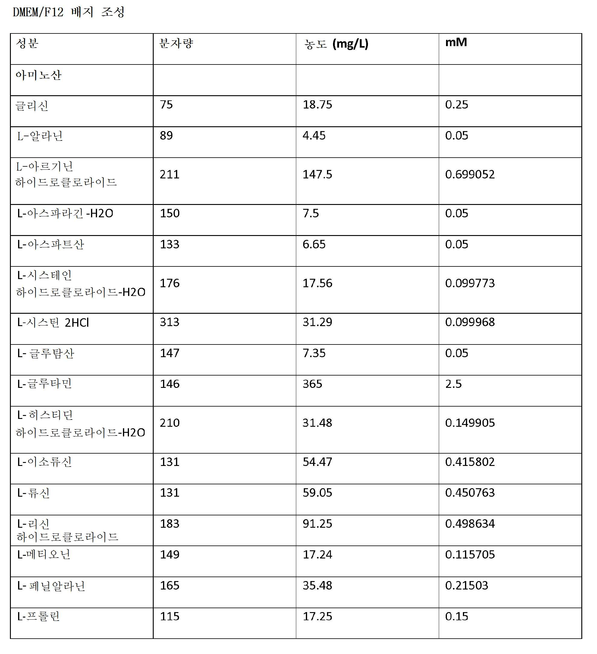
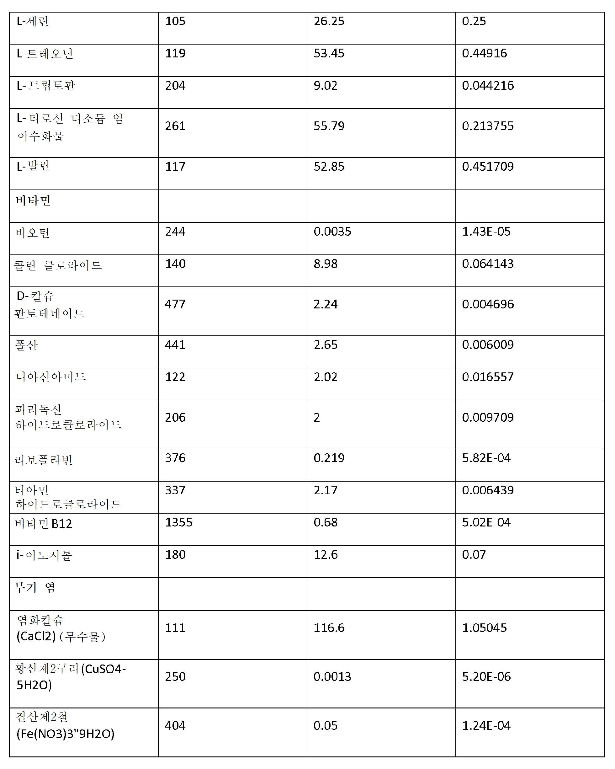

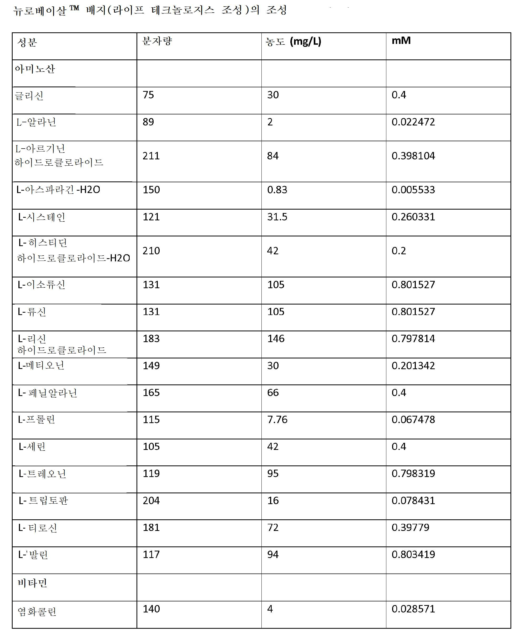

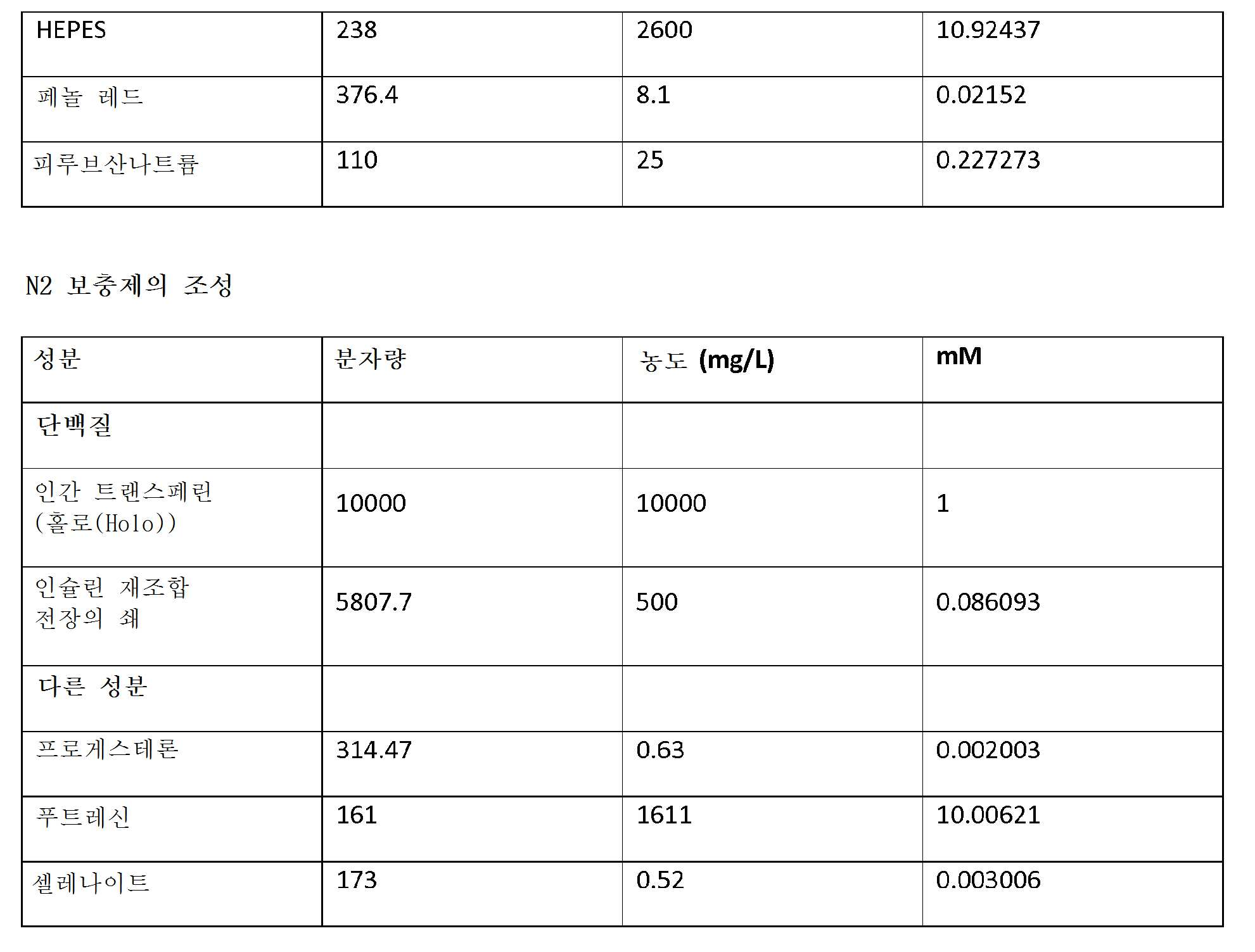
참고 문헌references


Claims (21)
RG 전구체 세포의 생존능을 유지시키는 데 적합한 배지를 포함하는, RG 전구체 세포의 실질적으로 순수한 제제로서,
여기서, 상기 제제 중 90% 초과의 세포가 면역세포화학적으로 Math5(+), 및 임의적으로, Math5(+) 및 Brn3a(+)인, RG 전구체 세포의 실질적으로 순수한 제제.A plurality of RG precursor cells, and
A substantially pure formulation of RG precursor cells, comprising a medium suitable for maintaining the viability of the RG precursor cells,
Wherein substantially more than 90% of the cells of the formulation are immunocytochemically Math5 (+), and optionally Math5 (+) and Brn3a (+).
RG 전구체 세포의 생존능을 유지시키는 데 적합한 배지를 포함하는, RG 전구체 세포의 실질적으로 순수한 제제로서,
여기서, 상기 제제 중 90% 초과의 세포가 면역세포화학적으로 Brn3a(+) 및 신경미세섬유(+)인, RG 전구체 세포의 실질적으로 순수한 제제.A plurality of RG precursor cells, and
A substantially pure formulation of RG precursor cells, comprising a medium suitable for maintaining the viability of the RG precursor cells,
Wherein substantially more than 90% of the cells of the formulation are immunocytochemically Brn3a (+) and nerve microfibrils (+), substantially pure preparations of RG precursor cells.
RG 전구체 세포의 생존능을 유지시키는 데 적합한 배지를 포함하는, RG 전구체 세포의 제제로서,
여기서, RG 전구체 세포는 면역세포화학적으로 Math5(+), Brn3a(+), Brn3b(+), 및 임의적으로, Isl1(+)인, RG 전구체 세포의 제제. A plurality of cells containing at least 50% RG precursor cells, and
A preparation of RG precursor cells, comprising a medium suitable for maintaining the viability of the RG precursor cells,
Here, the RG precursor cells are immunocytochemically labeled with Math5 (+), Brn3a (+), Brn3b (+), and optionally, Isl1 (+).
RG 전구체 세포의 생존능을 유지시키는 데 적합한 배지를 포함하는, RG 전구체 세포의 제제로서,
여기서, RG 전구체 세포는 면역세포화학적으로 Brn3a(+), 신경미세섬유(+), 및 임의적으로, Thy1(+)인, RG 전구체 세포의 제제. A plurality of cells containing at least 50% RG precursor cells, and
A preparation of RG precursor cells, comprising a medium suitable for maintaining the viability of the RG precursor cells,
Here, RG precursor cells are immunocytochemically made of Brn3a (+), neuronal microfibrils (+), and optionally, Thy1 (+), preparations of RG precursor cells.
RG 전구체 세포의 생존능을 유지시키는 데 적합한 배지를 포함하는, RG 전구체 세포의 제제로서,
여기서, RG 전구체 세포는 면역세포화학적으로 Math5(+), Brn3a(+), Brn3b(+), 및 임의적으로, Isl1(+)인, RG 전구체 세포의 제제. A plurality of RG precursor cells substantially free of pluripotent stem cells, pluripotent stem cells, adult stem cells, adult photoreceptors, and / or amaranth cells; And
A preparation of RG precursor cells, comprising a medium suitable for maintaining the viability of the RG precursor cells,
Here, the RG precursor cells are immunocytochemically labeled with Math5 (+), Brn3a (+), Brn3b (+), and optionally, Isl1 (+).
RG 전구체 세포의 생존능을 유지시키는 데 적합한 배지를 포함하는, RG 전구체 세포의 제제로서,
여기서, RG 전구체 세포는 면역세포화학적으로 Brn3a(+), 신경미세섬유(+), 및 임의적으로, Thy1(+)인, RG 전구체 세포의 제제.A plurality of RG precursor cells substantially free of pluripotent stem cells, pluripotent stem cells, adult stem cells, adult photoreceptors, and / or amaranth cells; And
A preparation of RG precursor cells, comprising a medium suitable for maintaining the viability of the RG precursor cells,
Here, RG precursor cells are immunocytochemically made of Brn3a (+), neuronal microfibrils (+), and optionally, Thy1 (+), preparations of RG precursor cells.
(b) 포유동물 환자 내로의 이식을 위해 RG 전구체 세포의 생존능을 유지시키기 위한 약학적으로 허용되는 담체
를 포함하는, 포유동물 환자에서의 사용에 적합한 RG 전구체 세포의 약학 제제.(a) a plurality of RG precursor cells wherein more than 90% of the cells in the preparation are immunocytochemically Math5 (+), and optionally, Math5 (+) and Brn3a (+); And
(b) a pharmaceutically acceptable carrier for maintaining the viability of the RG precursor cells for transplantation into a mammalian subject
Lt; RTI ID = 0.0 > RG < / RTI > precursor cells for use in a mammalian patient.
(b) 포유동물 환자 내로의 이식을 위해 RG 전구체 세포의 생존능을 유지시키기 위한 약학적으로 허용되는 담체
를 포함하는, 포유동물 환자에서의 사용에 적합한 RG 전구체 세포의 약학 제제.(a) a plurality of RG precursor cells wherein more than 90% of the cells of the preparation are immunocytochemically Brn3a (+) and neural microfibrils (+); And
(b) a pharmaceutically acceptable carrier for maintaining the viability of the RG precursor cells for transplantation into a mammalian subject
Lt; RTI ID = 0.0 > RG < / RTI > precursor cells for use in a mammalian patient.
(b) 해동시 RG 전구체 세포의 생존능에 맞는 냉동보존 시스템
을 포함하는, 109개 이상의 RG 전구체 세포를 포함하는 극저온 세포 제제. (a) a plurality of RG precursor cells wherein more than 90% of the cells in the preparation are immunocytochemically Math5 (+), and optionally, Math5 (+) and Brn3a (+); And
(b) Cryopreservation systems suitable for the viability of RG precursor cells at thawing
, Cryogenic cell preparation containing 10 9 or more RG precursor cells, including.
(b) 해동시 RG 전구체 세포의 생존능에 맞는 냉동보존 시스템
을 포함하는, 109개 이상의 RG 전구체 세포를 포함하는 극저온 세포 제제.(a) a plurality of RG precursor cells wherein more than 90% of the cells in the preparation are immunocytochemically Brn3a (+) and neuronal microfibrils (+), and
(b) Cryopreservation systems suitable for the viability of RG precursor cells at thawing
, Cryogenic cell preparation containing 10 9 or more RG precursor cells, including.
(b) 줄기 세포 유래 RG 세포의 생존능을 유지시키는 데 적합한 배지
를 포함하는, 만능 줄기 세포 유래 RG 세포의 실질적으로 순수한 제제.(a) pluripotent stem cell-derived RG cells in which more than 60% of the cells are immunocytochemically Brn3a (+), neuronal microfibrils (+), and Tuj1 (+); And
(b) a medium suitable for maintaining the viability of stem cell-derived RG cells
A substantially pure preparation of allogeneic stem cell-derived RG cells.
(b) 포유동물 환자 내로의 이식을 위해 RG 세포의 생존능을 유지시키기 위한 약학적으로 허용되는 담체
를 포함하는, 포유동물 환자에서의 사용에 적합한 RG 세포의 약학 제제.(a) pluripotent stem cell-derived RG cells in which more than 60% of the cells in the preparation are immunocytochemically Brn3a (+), nerve microfibrils (+), and Tuj1 (+); And
(b) a pharmaceutically acceptable carrier for maintaining the viability of RG cells for transplantation into a mammalian patient
Lt; RTI ID = 0.0 > RG < / RTI > cells suitable for use in a mammalian patient.
(b) 상기 하나 이상의 안구 영역 전구체 세포를 배양하여 Math5(+), Brn3a(+), Brn3b(+), 및 임의적으로, Isl1(+) 또는 Brn3a(+), 신경미세섬유(+) 및 임의적으로, Thy1(+)인 RG 전구체 세포를 제조하는 단계
를 포함하는, 만능 줄기 세포 유래 RG 전구체 세포의 실질적으로 순수한 배양물을 제조하는 방법. (a) culturing pluripotent stem cells in a feeder cell-free system to produce at least one ocular area precursor cell;
(+), Brn3b (+), and optionally, Isl1 (+) or Brn3a (+), neuronal microfibrils (+), and optionally , Producing Thy1 (+) RG precursor cells
Derived RG precursor cells. ≪ RTI ID = 0.0 > 18. < / RTI >
A method of treating a subject in need of treatment comprising administering to the subject in need of treatment a composition of claim 17 or 18, or after thawing, a composition of claim 19 or 20.
Applications Claiming Priority (3)
| Application Number | Priority Date | Filing Date | Title |
|---|---|---|---|
| US201462046165P | 2014-09-05 | 2014-09-05 | |
| US62/046,165 | 2014-09-05 | ||
| PCT/US2015/048741 WO2016037159A1 (en) | 2014-09-05 | 2015-09-05 | Retinal ganglion cells and progenitors thereof |
Publications (1)
| Publication Number | Publication Date |
|---|---|
| KR20170077117A true KR20170077117A (en) | 2017-07-05 |
Family
ID=55440432
Family Applications (1)
| Application Number | Title | Priority Date | Filing Date |
|---|---|---|---|
| KR1020177008900A Ceased KR20170077117A (en) | 2014-09-05 | 2015-09-05 | Retinal ganglion cells and progenitors thereof |
Country Status (13)
| Country | Link |
|---|---|
| US (1) | US20170274019A1 (en) |
| EP (2) | EP3922717A1 (en) |
| JP (2) | JP6778181B2 (en) |
| KR (1) | KR20170077117A (en) |
| CN (1) | CN107148276A (en) |
| AU (2) | AU2015311702A1 (en) |
| CA (1) | CA2960211A1 (en) |
| ES (1) | ES2883600T3 (en) |
| IL (3) | IL250924B (en) |
| PL (1) | PL3189135T3 (en) |
| PT (1) | PT3189135T (en) |
| SG (3) | SG10201912296RA (en) |
| WO (1) | WO2016037159A1 (en) |
Cited By (1)
| Publication number | Priority date | Publication date | Assignee | Title |
|---|---|---|---|---|
| KR20200122066A (en) | 2019-04-17 | 2020-10-27 | 경북대학교 산학협력단 | Method for Isolating of Retinal Ganglion Cell and Uses of the Cells Thereby Isolated |
Families Citing this family (12)
| Publication number | Priority date | Publication date | Assignee | Title |
|---|---|---|---|---|
| HUE044778T2 (en) * | 2011-11-14 | 2019-11-28 | Astellas Inst For Regenerative Medicine | Human RPE Cell Pharmaceutical Preparations and Applications |
| PL3869195T3 (en) | 2015-03-23 | 2024-07-22 | Astellas Institute For Regenerative Medicine | Improved assays for potency of human retinal pigment epithelium (rpe) cells and photoreceptor progenitors |
| IL299326A (en) | 2015-08-18 | 2023-02-01 | Astellas Inst For Regenerative Medicine | Clinical formulations |
| EP4467565A3 (en) | 2016-12-21 | 2025-03-12 | Amgen Inc. | Anti-tnf alpha antibody formulations |
| WO2019078781A1 (en) | 2017-10-17 | 2019-04-25 | National University Of Singapore | Systems and methods for producing photoreceptor progenitors |
| US12213996B2 (en) | 2018-02-02 | 2025-02-04 | University Of Maryland, Baltimore | Methods of treating optic nerve diseases using neural progenitor cell growth factors |
| US11621058B2 (en) * | 2018-02-08 | 2023-04-04 | Ohio State Innovation Foundation | Synthetic IHC-stained digital sides generated using artificial neural networks |
| EP3810758B1 (en) | 2018-06-20 | 2023-07-19 | BioLamina AB | Methods and compositions for producing retinal pigment epithelium cells |
| CN112251408B (en) * | 2020-09-14 | 2022-04-12 | 郑州大学 | Preparation method of retinal ganglion cells |
| CN114917367A (en) * | 2021-06-02 | 2022-08-19 | 中国科学院动物研究所 | Application of LHX2 in promoting injury repair and regeneration of central system neurons |
| CN115404219B (en) * | 2022-09-09 | 2023-10-27 | 中国医学科学院医学生物学研究所 | Separation culture method of tree shrew retinal ganglion cells |
| CN117821382A (en) * | 2023-12-29 | 2024-04-05 | 广州赛莱拉干细胞科技股份有限公司 | A serum-free culture medium and culture method for hair follicle stem cells |
Family Cites Families (5)
| Publication number | Priority date | Publication date | Assignee | Title |
|---|---|---|---|---|
| JP2010536852A (en) * | 2007-08-23 | 2010-12-02 | ザ ボード オブ トラスティーズ オブ ザ リランド スタンフォード ジュニア ユニヴァーシティ | Regulation of synapse formation |
| US20110014701A1 (en) * | 2007-12-04 | 2011-01-20 | Proteobioactives Pty Ltd. | Protection of Progenitor Cells and Regulation of Their Differentiation |
| CA2771901C (en) * | 2009-08-24 | 2020-03-10 | Wisconsin Alumni Research Foundation | Substantially pure human retinal progenitor, forebrain progenitor, and retinal pigment epithelium cell cultures and methods of making the same |
| WO2011043591A2 (en) * | 2009-10-06 | 2011-04-14 | Snu R&Db Foundation | Method for differentiation into retinal cells from stem cells |
| KR101712556B1 (en) | 2014-08-27 | 2017-03-08 | 서울대학교산학협력단 | Method for differentiation into retinal ganglion cells from stem cells |
-
2015
- 2015-09-05 US US15/508,533 patent/US20170274019A1/en active Pending
- 2015-09-05 SG SG10201912296RA patent/SG10201912296RA/en unknown
- 2015-09-05 EP EP21168050.9A patent/EP3922717A1/en not_active Withdrawn
- 2015-09-05 SG SG10201901559SA patent/SG10201901559SA/en unknown
- 2015-09-05 ES ES15838263T patent/ES2883600T3/en active Active
- 2015-09-05 WO PCT/US2015/048741 patent/WO2016037159A1/en not_active Ceased
- 2015-09-05 PL PL15838263T patent/PL3189135T3/en unknown
- 2015-09-05 CA CA2960211A patent/CA2960211A1/en not_active Abandoned
- 2015-09-05 KR KR1020177008900A patent/KR20170077117A/en not_active Ceased
- 2015-09-05 AU AU2015311702A patent/AU2015311702A1/en not_active Abandoned
- 2015-09-05 EP EP15838263.0A patent/EP3189135B1/en active Active
- 2015-09-05 CN CN201580056489.7A patent/CN107148276A/en active Pending
- 2015-09-05 SG SG11201701709YA patent/SG11201701709YA/en unknown
- 2015-09-05 JP JP2017512937A patent/JP6778181B2/en active Active
- 2015-09-05 PT PT158382630T patent/PT3189135T/en unknown
-
2017
- 2017-03-05 IL IL250924A patent/IL250924B/en active IP Right Grant
-
2020
- 2020-07-20 JP JP2020124010A patent/JP7263293B2/en active Active
- 2020-10-04 IL IL277780A patent/IL277780B/en unknown
-
2021
- 2021-08-01 IL IL285258A patent/IL285258A/en unknown
- 2021-09-28 AU AU2021240145A patent/AU2021240145A1/en not_active Abandoned
Cited By (1)
| Publication number | Priority date | Publication date | Assignee | Title |
|---|---|---|---|---|
| KR20200122066A (en) | 2019-04-17 | 2020-10-27 | 경북대학교 산학협력단 | Method for Isolating of Retinal Ganglion Cell and Uses of the Cells Thereby Isolated |
Also Published As
| Publication number | Publication date |
|---|---|
| US20170274019A1 (en) | 2017-09-28 |
| CN107148276A (en) | 2017-09-08 |
| SG10201901559SA (en) | 2019-03-28 |
| IL250924B (en) | 2020-10-29 |
| PT3189135T (en) | 2021-08-19 |
| JP2017532954A (en) | 2017-11-09 |
| PL3189135T3 (en) | 2021-12-20 |
| AU2021240145A1 (en) | 2021-10-28 |
| EP3189135A1 (en) | 2017-07-12 |
| IL285258A (en) | 2021-09-30 |
| EP3189135B1 (en) | 2021-06-02 |
| JP2020185004A (en) | 2020-11-19 |
| IL277780B (en) | 2021-08-31 |
| ES2883600T3 (en) | 2021-12-09 |
| AU2015311702A1 (en) | 2017-04-06 |
| EP3189135A4 (en) | 2018-04-25 |
| SG10201912296RA (en) | 2020-02-27 |
| WO2016037159A1 (en) | 2016-03-10 |
| JP6778181B2 (en) | 2020-10-28 |
| IL277780A (en) | 2020-11-30 |
| CA2960211A1 (en) | 2016-03-10 |
| EP3922717A1 (en) | 2021-12-15 |
| SG11201701709YA (en) | 2017-04-27 |
| JP7263293B2 (en) | 2023-04-24 |
| IL250924A0 (en) | 2017-04-30 |
Similar Documents
| Publication | Publication Date | Title |
|---|---|---|
| US12558380B2 (en) | Photoreceptors and photoreceptor progenitors produced from pluripotent stem cells | |
| JP7263293B2 (en) | Retinal ganglion cells and their precursors | |
| HK40119302A (en) | Photoreceptors and photoreceptor progenitors produced from pluripotent stem cells | |
| HK40097681A (en) | Photoreceptors and photoreceptor progenitors produced from pluripotent stem cells | |
| HK40064920A (en) | Retinal ganglion cells and progenitors thereof | |
| HK1218931B (en) | Photoreceptors and photoreceptor progenitors produced from pluripotent stem cells | |
| HK1240622B (en) | Retinal ganglion cells and progenitors thereof | |
| HK1240622A1 (en) | Retinal ganglion cells and progenitors thereof |
Legal Events
| Date | Code | Title | Description |
|---|---|---|---|
| PA0105 | International application |
Patent event date: 20170331 Patent event code: PA01051R01D Comment text: International Patent Application |
|
| PG1501 | Laying open of application | ||
| PA0201 | Request for examination |
Patent event code: PA02012R01D Patent event date: 20200902 Comment text: Request for Examination of Application |
|
| E902 | Notification of reason for refusal | ||
| PE0902 | Notice of grounds for rejection |
Comment text: Notification of reason for refusal Patent event date: 20220415 Patent event code: PE09021S01D |
|
| E601 | Decision to refuse application | ||
| PE0601 | Decision on rejection of patent |
Patent event date: 20221028 Comment text: Decision to Refuse Application Patent event code: PE06012S01D Patent event date: 20220415 Comment text: Notification of reason for refusal Patent event code: PE06011S01I |