RU2425651C2 - Method for initial stabilisation of acetabular component at hip replacement - Google Patents
Method for initial stabilisation of acetabular component at hip replacement Download PDFInfo
- Publication number
- RU2425651C2 RU2425651C2 RU2009134346/14A RU2009134346A RU2425651C2 RU 2425651 C2 RU2425651 C2 RU 2425651C2 RU 2009134346/14 A RU2009134346/14 A RU 2009134346/14A RU 2009134346 A RU2009134346 A RU 2009134346A RU 2425651 C2 RU2425651 C2 RU 2425651C2
- Authority
- RU
- Russia
- Prior art keywords
- acetabular component
- acetabulum
- wedge
- endoprosthesis
- fixation
- Prior art date
Links
- 238000000034 method Methods 0.000 title claims abstract description 15
- 238000011540 hip replacement Methods 0.000 title description 2
- 230000006641 stabilisation Effects 0.000 title 1
- 210000000588 acetabulum Anatomy 0.000 claims abstract description 12
- 210000000988 bone and bone Anatomy 0.000 claims abstract description 6
- 210000004394 hip joint Anatomy 0.000 claims description 4
- 239000003814 drug Substances 0.000 abstract description 3
- 230000000694 effects Effects 0.000 abstract description 2
- 230000000399 orthopedic effect Effects 0.000 abstract description 2
- 208000014674 injury Diseases 0.000 abstract 1
- 239000000126 substance Substances 0.000 abstract 1
- 230000008733 trauma Effects 0.000 abstract 1
- 239000012634 fragment Substances 0.000 description 6
- 210000001624 hip Anatomy 0.000 description 3
- 238000011882 arthroplasty Methods 0.000 description 2
- 238000009434 installation Methods 0.000 description 2
- 210000002239 ischium bone Anatomy 0.000 description 2
- 230000002980 postoperative effect Effects 0.000 description 2
- 210000000689 upper leg Anatomy 0.000 description 2
- 241000906034 Orthops Species 0.000 description 1
- 206010031264 Osteonecrosis Diseases 0.000 description 1
- 238000013459 approach Methods 0.000 description 1
- 210000004204 blood vessel Anatomy 0.000 description 1
- 238000011161 development Methods 0.000 description 1
- 238000003745 diagnosis Methods 0.000 description 1
- 201000010099 disease Diseases 0.000 description 1
- 208000037265 diseases, disorders, signs and symptoms Diseases 0.000 description 1
- 235000012907 honey Nutrition 0.000 description 1
- 238000007654 immersion Methods 0.000 description 1
- 238000002513 implantation Methods 0.000 description 1
- 208000030175 lameness Diseases 0.000 description 1
- 210000003141 lower extremity Anatomy 0.000 description 1
- 238000003801 milling Methods 0.000 description 1
- 210000000056 organ Anatomy 0.000 description 1
- 238000010883 osseointegration Methods 0.000 description 1
- 238000012545 processing Methods 0.000 description 1
- 238000001356 surgical procedure Methods 0.000 description 1
- 238000012360 testing method Methods 0.000 description 1
Images

Landscapes
- Prostheses (AREA)
Abstract
Description
Изобретение относится к медицине, а именно к травматологии и ортопедии и применяется для более надежной первичной фиксации ацетабулярного компонента эндопротеза тазобедренного сустава при отсутствии в нем отверстий для фиксации винтами.The invention relates to medicine, namely to traumatology and orthopedics, and is used for more reliable primary fixation of the acetabular component of the hip joint prosthesis in the absence of holes for fixing it with screws.
В последние десятилетия эндопротезирование является наиболее частым методом лечения дегенеративно-дистрофических заболеваний тазобедренного сустава, позволяющим восстановить опороспособность нижней конечности, добиться достаточной амплитуды движений, избавить пациента от боли, хромоты, возвратить его к активному образу жизни [1, 4].In recent decades, endoprosthetics has been the most frequent method of treating degenerative-dystrophic diseases of the hip joint, which allows restoring the lower limb support ability, achieving sufficient range of motion, relieving the patient of pain, lameness, and returning him to an active lifestyle [1, 4].
Несмотря на достигнутые успехи оперативного лечения, одной из проблем является достижение надежной первичной фиксации ацетабулярного компонента типа pressfit [3].Despite the successes of surgical treatment, one of the problems is to achieve reliable primary fixation of the acetabular component of the pressfit type [3].
Во время операции эндопротезирования при непрочной фиксации выбранного ацетабулярного компонента часто появляется необходимость использовать чашку эндопротеза большего диаметра, что является крайне нежелательным в связи с невозможностью повторной рестерилизации эндопротеза и требует альтернативного подхода.During an endoprosthesis replacement operation with weak fixation of the selected acetabular component, it is often necessary to use a larger diameter endoprosthesis cup, which is highly undesirable due to the impossibility of re-restoring the endoprosthesis and requires an alternative approach.
Классическим методом достижения первичной фиксации является дополнительная фиксация ацетабулярного компонента винтами. Недостатком этого метода является невозможность использования конструкций эндопротезов с большой головкой (типа Biomet-Magnum, Zimmer-LDH-Durom), т.к. на ацетабулярном компоненте этого эндопротеза отсутствуют отверстия для дополнительной фиксации винтами [3].The classic method of achieving primary fixation is the additional fixation of the acetabular component with screws. The disadvantage of this method is the inability to use large-endoprosthesis designs (such as Biomet-Magnum, Zimmer-LDH-Durom), because there are no holes on the acetabular component of this endoprosthesis for additional fixation with screws [3].
Известен способ улучшения фиксации ацетабулярного компонента за счет увеличения погружения чашки эндопротеза с вклинивания в дно вертлужной впадины остеотомированного костного фрагмента - медиализация по И.П.Соболеву [2]. Недостатками данного способа являются травматичность операции и опасность повреждения сосудов и тазовых органов.There is a method of improving the fixation of the acetabular component by increasing the immersion of the endoprosthesis cup with wedging in the acetabulum of the osteotomy bone fragment - mediation according to IP Sobolev [2]. The disadvantages of this method are the invasiveness of the operation and the risk of damage to blood vessels and pelvic organs.
Техническим результатом настоящего изобретения является достижение прочной фиксации ацетабулярного компонента типа «pressfit» без отверстий, низкая травматичность операции.The technical result of the present invention is to achieve a strong fixation of the acetabular component of the type "pressfit" without holes, low invasiveness of the operation.
Результат изобретения достигается путем внедрения клиновидного фрагмента в седалищную кость своим основанием выступающего над поверхностью обработанной вертлужной впадины.The result of the invention is achieved by introducing a wedge-shaped fragment into the sciatic bone with its base protruding above the surface of the treated acetabulum.
На чертежах изображено следующее:The drawings show the following:
фиг.1 - удаленная головка бедренной кости с клиновидным фрагментом (1);figure 1 - remote femoral head with a wedge-shaped fragment (1);
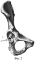
фиг.2 - схематическое изображение вертлужной впадины с расположенным клиновидным фрагментом (1) в седалищной кости.figure 2 - schematic representation of the acetabulum with a sphenoid fragment (1) located in the ischium.
Соответствие технического решения критерию «новизна» определяется отсутствием в уровне техники идентичного технического решения, позволяющего добиться заявляемого результата.The compliance of the technical solution with the criterion of "novelty" is determined by the absence in the prior art of an identical technical solution to achieve the claimed result.
Соответствие критерию «изобретательский уровень» определилось тем, что данное решение не следует явным образом из уровня техники, а также тем, что эффект, достигаемый посредством заявляемого решения, выявлен в результате целенаправленного поиска самими изобретателями. Соответствие критерию «промышленная применимость» определяется безопасностью и эффективностью проведения манипуляции по описанной методике. Эти факторы поддерживают к данному способу интерес здравоохранения, готового перейти к широкому внедрению заявляемого способа, для чего заявитель и обращается за патентной защитой новой разработки.Compliance with the criterion of "inventive step" was determined by the fact that this solution does not follow explicitly from the prior art, and also by the fact that the effect achieved by the claimed solution is revealed as a result of a targeted search by the inventors themselves. Compliance with the criterion of "industrial applicability" is determined by the safety and effectiveness of the manipulation according to the described methodology. These factors support the interest in this method of health care, ready to move on to the widespread implementation of the proposed method, for which the applicant is applying for patent protection of a new development.
Такой способ дополнительной фиксации ацетабулярного компонента решил несколько задач.This method of additional fixation of the acetabular component has solved several problems.
1. Позволяет достичь прочную первичную фиксацию ацетабулярного компонента pressfit без отверстий, что создает условия для вторичной фиксации за счет остеоинтеграции в структурированную поверхность эндопротеза.1. It allows to achieve strong primary fixation of the acetabular component of pressfit without holes, which creates the conditions for secondary fixation due to osseointegration into the structured surface of the endoprosthesis.
2. Снижает травматичность операции.2. Reduces the invasiveness of the operation.
3. При непрочной фиксации ацетабулярного компонента позволяет использовать чашку выбранного диаметра.3. With weak fixation of the acetabular component allows you to use a cup of the selected diameter.
Способ осуществляется следующим образом.The method is as follows.
В ходе операции эндопротезирования тазобедренного сустава после обработки вертлужной впадины фрезами до необходимого размера и выявления недостаточного качества костной ткани из удаленной головки бедренной кости выпиливается клиновидный фрагмент (фиг.1). В проекции седалищной кости со стороны впадины выполняется отверстие. В сформированное отверстие внедряется верхушка клина (фиг.2). Необходимо оставить над поверхностью обработанной вертлужной впадины 4-5 мм трасплантата. После чего производится имплантация ацетабулярного компонента, который более плотно заклинивается между стенками сформированной вертлужной впадины.During the operation of hip replacement after processing the acetabulum with milling cutters to the required size and identifying insufficient quality of bone tissue, a wedge-shaped fragment is cut out from the removed head of the femur (Fig. 1). A hole is made in the projection of the ischium from the cavity. The wedge tip is embedded in the formed hole (Fig. 2). It is necessary to leave 4-5 mm of the graft above the surface of the treated acetabulum. After which the implantation of the acetabular component is performed, which is more tightly wedged between the walls of the formed acetabulum.
Клинический примерClinical example
В 11-м отделении РНИИТО им. Вредена пациенту П. 54-х лет с диагнозом асептический некроз головки левой бедренной кости выполнена операция по предложенному способу.In the 11th branch of RNIITO them. Harmful to a patient P. 54 years old with a diagnosis of aseptic necrosis of the head of the left femur, the operation was performed according to the proposed method.
Под СМА в положении больного на правом боку доступом Хардинга вскрыт левый тазобедренный сустав. В ходе операции при установке эндопротеза отмечена недостаточная фиксация ацетабулярного компонента. Произведено моделирование костного клиновидного фрагмента из головки бедренной кости с основанием 1,5 см и высотой 3 см, его установка в ранее сформированное отверстие дна вертлужной впадины в проекции седалищной кости и установка ацетабулярного компонента. При тестовом контроле отмечена его стабильная фиксация. В отдаленном послеоперационном периоде признаков нестабильности компонентов эндопротеза не выявлено.Under the MCA in the position of the patient on the right side, Harding opened the left hip joint. During the operation, when installing the endoprosthesis, insufficient fixation of the acetabular component was noted. A bone-shaped wedge-shaped fragment from the femoral head with a base of 1.5 cm and a height of 3 cm was simulated, its installation in the previously formed opening of the bottom of the acetabulum in the projection of the sciatic bone and the installation of the acetabular component. During test control, its stable fixation was noted. In the remote postoperative period, there were no signs of instability of the components of the endoprosthesis.
ЛИТЕРАТУРАLITERATURE
1. Рожнев Е.В. Осложнения раннего послеоперационного периода первичного тотального эндопротезирования тазобедренного сустава. Автореф. дис. канд. мед. наук / Рожнев Евгений Валерьевич. - Пермь. -2007. - 26 с.1. Rozhnev E.V. Complications of the early postoperative period of primary total hip arthroplasty. Abstract. dis. Cand. honey. Sciences / Rozhnev Evgeniy Valerevich. - Perm. 2007. - 26 p.
2. Способ эндопротезирования тазобедренного сустава. Соболев И.П., Кикачеишвили Т.Т., Безгодков Ю.А. Патент №96110428/14 от 1999 года.2. The method of hip arthroplasty. Sobolev I.P., Kikacheishvili T.T., Bezgodkov Yu.A. Patent No. 96110428/14 of 1999.
3. Тихилов P.M. Руководство по эндопротезированию тазобедренного сустава / под. ред. P.M.Тихилова, В.М.Шаповалова. - СПб.: РНИИТО им. P.P.Вредена, 2008. - 324 с., ил.3. Tikhilov P.M. Hip Endoprosthetics Guide / Under. ed. P.M. Tikhilova, V.M. Shapovalova. - SPb .: RNIITO named after P.P. Vredena, 2008 .-- 324 p., Ill.
4. Muller M.E. Total hip prostheses / M.E.Muller // Clin Orthop. - 1970. - V.72. - P.46-68.4. Muller M.E. Total hip prostheses / M.E. Muller // Clin Orthop. - 1970. - V.72. - P. 46-68.
Claims (1)
Priority Applications (1)
| Application Number | Priority Date | Filing Date | Title |
|---|---|---|---|
| RU2009134346/14A RU2425651C2 (en) | 2009-09-14 | 2009-09-14 | Method for initial stabilisation of acetabular component at hip replacement |
Applications Claiming Priority (1)
| Application Number | Priority Date | Filing Date | Title |
|---|---|---|---|
| RU2009134346/14A RU2425651C2 (en) | 2009-09-14 | 2009-09-14 | Method for initial stabilisation of acetabular component at hip replacement |
Publications (2)
| Publication Number | Publication Date |
|---|---|
| RU2009134346A RU2009134346A (en) | 2011-03-20 |
| RU2425651C2 true RU2425651C2 (en) | 2011-08-10 |
Family
ID=44053441
Family Applications (1)
| Application Number | Title | Priority Date | Filing Date |
|---|---|---|---|
| RU2009134346/14A RU2425651C2 (en) | 2009-09-14 | 2009-09-14 | Method for initial stabilisation of acetabular component at hip replacement |
Country Status (1)
| Country | Link |
|---|---|
| RU (1) | RU2425651C2 (en) |
Families Citing this family (1)
| Publication number | Priority date | Publication date | Assignee | Title |
|---|---|---|---|---|
| CN105147417A (en) * | 2015-06-24 | 2015-12-16 | 中国人民解放军第四军医大学 | Bone grafting stent for acetabular osteotomy reconstruction |
Citations (2)
| Publication number | Priority date | Publication date | Assignee | Title |
|---|---|---|---|---|
| RU2135110C1 (en) * | 1996-05-29 | 1999-08-27 | Соболев Игорь Петрович | Method of endoprosthetics of hip joint |
| RU2309688C1 (en) * | 2006-03-15 | 2007-11-10 | Федеральное государственное учреждение "Российский научно-исследовательский институт травматологии и ортопедии им. Р.Р. Вредена Федерального агентства по здравоохранению и социальному развитию" (ФГУ "РНИИТО им. Р.Р. Вредена Росздрава") | Method for osseous plasty of the defects of median wall of cotyloid cavity at revision arthroplastycaused by instability of cotyloid component of total hip joint endoprosthesis |
-
2009
- 2009-09-14 RU RU2009134346/14A patent/RU2425651C2/en not_active IP Right Cessation
Patent Citations (2)
| Publication number | Priority date | Publication date | Assignee | Title |
|---|---|---|---|---|
| RU2135110C1 (en) * | 1996-05-29 | 1999-08-27 | Соболев Игорь Петрович | Method of endoprosthetics of hip joint |
| RU2309688C1 (en) * | 2006-03-15 | 2007-11-10 | Федеральное государственное учреждение "Российский научно-исследовательский институт травматологии и ортопедии им. Р.Р. Вредена Федерального агентства по здравоохранению и социальному развитию" (ФГУ "РНИИТО им. Р.Р. Вредена Росздрава") | Method for osseous plasty of the defects of median wall of cotyloid cavity at revision arthroplastycaused by instability of cotyloid component of total hip joint endoprosthesis |
Non-Patent Citations (1)
| Title |
|---|
| ТИХИЛОВ P.M. и др. Основы эндопротезирования тазобедренного сустава. - СПб.: НПО «Профессионал», 2008, с.221-223. BARRACK RL "Uncemented total hip arthroplasty with superior acetabular deficiency. Femoral head autograft technique and early clinical results" J Arthroplasty. 1990 Jun; 5(2): 159-67(Abstract). * |
Also Published As
| Publication number | Publication date |
|---|---|
| RU2009134346A (en) | 2011-03-20 |
Similar Documents
| Publication | Publication Date | Title |
|---|---|---|
| JP6529717B2 (en) | System and method for implanting a second glenoid prosthesis | |
| US10149763B2 (en) | Multipurpose void filling prosthesis | |
| Hummel | Hip Replacement | |
| WO2007112123A2 (en) | Apparatus and method for the treatment of periprosthetic fractures | |
| EP3023067B1 (en) | Strut plate and cabling system | |
| US9486320B2 (en) | Subchondral treatment of osteoarthritis in joints | |
| AU2016353265B2 (en) | Joint implants and methods | |
| RU2360627C1 (en) | Method of prosthetics of hip joint at fractures and posttraumatic defects of acetabular cavity | |
| RU2425651C2 (en) | Method for initial stabilisation of acetabular component at hip replacement | |
| RU2475202C1 (en) | Method of hip replacement in cotyloid bone defect | |
| RU2456949C1 (en) | Method of plasty of acetabulum roof in case of its defects and displasias with structural autotransplant | |
| RU2083172C1 (en) | Method for prosthetics of thigh bone capitulum and endoprosthesis itself | |
| Arif et al. | Revision of total hip arthroplasty using an anterior cortical window, extensive strut allografts, and an impaction graft: follow-up study | |
| RU2355339C2 (en) | Cotyloid cavity autoosteoplasty technique in inspective hip joint replacement | |
| US20060155381A1 (en) | Orthopedic system for total hip replacement surgery | |
| RU68278U1 (en) | GUIDELINES FOR PROCESSING THE FEMAL CHANNEL WHEN REVISING HIP JOINT PROSTHETIS | |
| RU2451493C1 (en) | Hip replacement technique required in cotyloid fractures and posttraumatic defects | |
| RU2400170C1 (en) | Method of surgical prevention of osteoporosis-related proximal femoral fractures by hip replacement | |
| RU2204350C1 (en) | Method for two-stage endoprosthetics of hip joint | |
| RU2637105C1 (en) | Method for plasty of defects of acetabulum antero- and posterosuperior edge with structural autobone in hip joint endoprosthesis | |
| RU2758556C1 (en) | Method for modeling a bone tissue defect for studying osseointegration of osteoplastic material and regeneration of cancellous bone tissue in an experiment on rabbits | |
| RU2309688C1 (en) | Method for osseous plasty of the defects of median wall of cotyloid cavity at revision arthroplastycaused by instability of cotyloid component of total hip joint endoprosthesis | |
| RU2773382C1 (en) | Method for removing stable cementless femoral components of a hip joint endoprosthesis | |
| RU2475197C1 (en) | Method of revision plastic hip replacement in unstable endoprosthesis components inserted in wing of ilium in congenital high hip dislocation | |
| RU2518141C1 (en) | Method for hip replacement in congenital hip dislocation |
Legal Events
| Date | Code | Title | Description |
|---|---|---|---|
| MM4A | The patent is invalid due to non-payment of fees |
Effective date: 20110915 |