US4149081A - Removal of spectral artifacts and utilization of spectral effects in computerized tomography - Google Patents
Removal of spectral artifacts and utilization of spectral effects in computerized tomography Download PDFInfo
- Publication number
- US4149081A US4149081A US05/745,709 US74570976A US4149081A US 4149081 A US4149081 A US 4149081A US 74570976 A US74570976 A US 74570976A US 4149081 A US4149081 A US 4149081A
- Authority
- US
- United States
- Prior art keywords
- sub
- radiation
- detector
- measurements
- source
- Prior art date
- Legal status (The legal status is an assumption and is not a legal conclusion. Google has not performed a legal analysis and makes no representation as to the accuracy of the status listed.)
- Expired - Lifetime
Links
Images


Classifications
-
- A—HUMAN NECESSITIES
- A61—MEDICAL OR VETERINARY SCIENCE; HYGIENE
- A61B—DIAGNOSIS; SURGERY; IDENTIFICATION
- A61B6/00—Apparatus or devices for radiation diagnosis; Apparatus or devices for radiation diagnosis combined with radiation therapy equipment
- A61B6/02—Arrangements for diagnosis sequentially in different planes; Stereoscopic radiation diagnosis
- A61B6/025—Tomosynthesis
-
- A—HUMAN NECESSITIES
- A61—MEDICAL OR VETERINARY SCIENCE; HYGIENE
- A61B—DIAGNOSIS; SURGERY; IDENTIFICATION
- A61B6/00—Apparatus or devices for radiation diagnosis; Apparatus or devices for radiation diagnosis combined with radiation therapy equipment
- A61B6/02—Arrangements for diagnosis sequentially in different planes; Stereoscopic radiation diagnosis
- A61B6/03—Computed tomography [CT]
- A61B6/032—Transmission computed tomography [CT]
-
- A—HUMAN NECESSITIES
- A61—MEDICAL OR VETERINARY SCIENCE; HYGIENE
- A61B—DIAGNOSIS; SURGERY; IDENTIFICATION
- A61B6/00—Apparatus or devices for radiation diagnosis; Apparatus or devices for radiation diagnosis combined with radiation therapy equipment
- A61B6/40—Arrangements for generating radiation specially adapted for radiation diagnosis
- A61B6/4035—Arrangements for generating radiation specially adapted for radiation diagnosis the source being combined with a filter or grating
-
- A—HUMAN NECESSITIES
- A61—MEDICAL OR VETERINARY SCIENCE; HYGIENE
- A61B—DIAGNOSIS; SURGERY; IDENTIFICATION
- A61B6/00—Apparatus or devices for radiation diagnosis; Apparatus or devices for radiation diagnosis combined with radiation therapy equipment
- A61B6/42—Arrangements for detecting radiation specially adapted for radiation diagnosis
- A61B6/4208—Arrangements for detecting radiation specially adapted for radiation diagnosis characterised by using a particular type of detector
- A61B6/4241—Arrangements for detecting radiation specially adapted for radiation diagnosis characterised by using a particular type of detector using energy resolving detectors, e.g. photon counting
-
- A—HUMAN NECESSITIES
- A61—MEDICAL OR VETERINARY SCIENCE; HYGIENE
- A61B—DIAGNOSIS; SURGERY; IDENTIFICATION
- A61B6/00—Apparatus or devices for radiation diagnosis; Apparatus or devices for radiation diagnosis combined with radiation therapy equipment
- A61B6/48—Diagnostic techniques
- A61B6/482—Diagnostic techniques involving multiple energy imaging
-
- A—HUMAN NECESSITIES
- A61—MEDICAL OR VETERINARY SCIENCE; HYGIENE
- A61B—DIAGNOSIS; SURGERY; IDENTIFICATION
- A61B6/00—Apparatus or devices for radiation diagnosis; Apparatus or devices for radiation diagnosis combined with radiation therapy equipment
- A61B6/58—Testing, adjusting or calibrating thereof
- A61B6/582—Calibration
- A61B6/583—Calibration using calibration phantoms
-
- G—PHYSICS
- G06—COMPUTING OR CALCULATING; COUNTING
- G06T—IMAGE DATA PROCESSING OR GENERATION, IN GENERAL
- G06T12/00—Tomographic reconstruction from projections
- G06T12/10—Image preprocessing, e.g. calibration, positioning of sources or scatter correction
-
- Y—GENERAL TAGGING OF NEW TECHNOLOGICAL DEVELOPMENTS; GENERAL TAGGING OF CROSS-SECTIONAL TECHNOLOGIES SPANNING OVER SEVERAL SECTIONS OF THE IPC; TECHNICAL SUBJECTS COVERED BY FORMER USPC CROSS-REFERENCE ART COLLECTIONS [XRACs] AND DIGESTS
- Y02—TECHNOLOGIES OR APPLICATIONS FOR MITIGATION OR ADAPTATION AGAINST CLIMATE CHANGE
- Y02A—TECHNOLOGIES FOR ADAPTATION TO CLIMATE CHANGE
- Y02A90/00—Technologies having an indirect contribution to adaptation to climate change
- Y02A90/10—Information and communication technologies [ICT] supporting adaptation to climate change, e.g. for weather forecasting or climate simulation
Definitions
- This invention relates to an apparatus and method for reconstructing two-dimensional pictures of object slices within a human or other body by means of passing penetrating radiation, typically X-radiation or gamma radiation, through the plane of the body from a plurality of angles.
- penetrating radiation typically X-radiation or gamma radiation
- the field is commonly known as computerized tomography or transverse axial tomography.
- the present invention is concerned with apparatus and methods for constructing two dimensional pictures of an object slice by means of detection of penetrating radiation not attenuated by the object slice during an analysis of said object slice in computerized tomography.
- a computer program takes as its input empirical values of the various attenuation cross-sections present, and by means of the least squares technique, gives parameters which enable one to set up analytical expressions for the three various attenuation cross-sections present in any object under study.
- a technique is disclosed by means of which the three analytical expressions are combined with expressions for detector efficiency and input spectra to represent a set of two equations. It is then shown that if one takes two sets of measurements, these two sets of equations can be solved, yielding output pictures of atomic charge distribution and electron density distribution, or any combination thereof, for either a thick or thin beam case of the object under study.
- These two sets of measurements may be taken by means of using different peak energy settings of the X-ray source, using detectors with different efficiencies or energy response ranges, by using an input source filter, or by any combination of the above.
- a technique is disclosed for optimizing the thickness of such a source filter.
- a similar technique can be used to optimize source spectra, detector sensitivity, or combinations thereof.
- the machine is also calibrated empirically during the solution of the sets of equations.
- the techniques of the invention are independent of the reconstruction algorithm used, and independent of the source-detector geometry used.
- a statistical error analysis is then provided which shows the statistical error present in the output pictorial representations as a function of statistical errors in the two sets of measurements.
- the techniques of the invention may be collectively termed “spectrally independent transverse axial tomography” (SITAT).
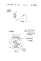
- FIG. 1 is a graph of number of photons vs. energy in a typical X-ray beam useful for medical diagnostic purposes in computerized tomography.
- FIG. 2a is a partial diagrammatic plane view and a partial block diagram of a tomographic instrument employing the present invention.
- FIG. 2b is a diagrammatic plane view of a tomographic instrument employing the present invention with a test object situated therein.
- FIG. 2c is an illustration of the output of a tomographic instrument employing the present invention wherein two separate output facsimiles are displayed.
- FIG. 3 is a block diagram of the method of the present invention.
- the reconstruction algorithm Given the line integral of the attenuation along lines in all directions drawn through all points in a plane, the reconstruction algorithm calculates the X-ray attenuation density distribution at all points within the plane.
- One formulation of the solution to this problem is described in U.S. patent application Ser. No. 643,896 to Pavkovich, filed Dec. 23, 1975, and in U.S. patent application Ser. No. 643,894 to Pavkovich and Nunan, filed Dec. 23, 1975, and assigned to the same assignee as the present invention.
- the function H is known at some particular energy E.
- the function H can be determined from Beer's Law since in this case the line integral H is simply given by the logarithm of the transmission of the beam through the object under study along lines passing through each point in the plane at all angles. The problem is more difficult if the X-ray beam used for the measurement is not monochromatic.
- a superior technique to the solution of this problem, described here, takes advantage of known functional dependence of X-ray scattering attenuation cross-sections on the incident X-ray energy.
- the elemental X-ray cross-sections for attenuation of X-rays from an X-ray beam are the result of Compton scattering, Rayleigh scattering, and photo-electric absorption.
- the functional dependencies of these cross sections are known and for our purposes depend only on the energy of the incident photon and the elemental charge. Therefore, given a known X-ray beam spectrum, detector efficiency curve, and the density and chemical composition of an object, the transmission ratio I/I o given by equations 3 and 4 cam be theoretically calculated.
- ⁇ c is the attenuation cross-section due to Compton scatter
- ⁇ a is the attenuation cross-section due to photoelectric absorption
- ⁇ R is the total attenuation cross-section.
- the functions q a and q c are functions of the position R in the object, and are the attenuations due to photoelectric absorption and Compton scatter, respectively. They depend on the effective Z of the material through which the X-rays are passing, and also upon its electron density at each point R. These functions are given to first order by the following expressions.
- ⁇ (R) is the number of atoms per unit volume at the position (R).
- I 2 /I 02 is the ratio of the detected intensity after passing through the sample to the intensity with no sample in the beam using the spectrum ⁇ 2 .
- a different X-ray spectrum ⁇ 2 either by (1) changing the peak energy setting of the X-ray power supply, by (2) inserting a suitable filter into the X-ray beam, or by (3) changing the efficiency ⁇ (E) of the detector or using detectors sensitive over different energy ranges, such as a high energy range and a low energy range.
- the latter is preferable because it is the only approach in which all measurements for the spectrally independent reconstruction are taken at once.
- FIG. 2a is a partial diagrammatic plane view and a partial block diagram of a tomographic instrument employing the present invention.
- Source 20 and detector array 30 are fixedly mounted on gantry 10 which rotates circularly in a plane containing the planar cross-section of object slice 35 which it is desired to depict. For example, a physician may wish to view a facsimile of tumor 36 within a human body.
- Motor 70 drives drive gear 80 which energizes gantry 10 by means of teeth 85 and 86.
- Rollers 90 keep gantry 10 aligned.
- Source 20 is a source of penetrating radiation such as X-radiation or gamma radiation.
- Collimator 40 which is comprised of a heavy substance such as lead, serves to collimate the radiation beam towards the object slice and detector array, and serves to protect the operator and patient from spurious radiation.
- FIG. 2a shows the beam shaped into a thin fan shape, which is a preferred shape for human diagnosis.
- the beam may be arbitrarily thin (up to a few millimeters thick) and fan across an angle from arbitrary smallness up to 180°, preferably about 35°. It is helpful if the distribution of photons is substantially uniform across the thickness and breadth of the beam.
- detectors 30, may be a continuous arcuately shaped detector, or a set of discrete detectors, typically numbering about 300.
- the detectors may be scintillators/photomultipliers, solid state devices, or chambers filled with a gas such as xenon or krypton which is ionized by the incoming radiation producing signals which are then counted.
- signals corresponding to non-attenuated radiation are fed into computer 100, which is preferably analog when detector 30 is continuous and digiral when detector array 30 comprises a discrete set of detectors.
- collimators 50 which are comprised of a heavy material such as lead, provide shielding between adjacent detectors, thus preventing cross-talk and other interference.
- computer 100 Upon receiving the measured data from detector(s) 30, computer 100 processes the data using the programs described herein as well as conventional reconstruction programs, and produces output data representative of two separate representations of object slice 35, each of which is free from spectral artifacts, as described herein. These output data are then transmitted to display device 110 where they are portrayed, either sequentially or simultaneously.
- Display 110 is any device capable of graphically portraying three-dimensional information, e.g., an electrostatic printer programmed to show contours, or a cathode ray tube 120 where differences in amplitude (here attenuation densities) are shown as different shades of grey or different colors.
- FIG. 2a shows the case where the two output pictures are displayed sequentially on CRT 120. In each case, FIG. 135, corresponding to object slice 35, and reconstructed tumor 136, corresponding to tumor 36, are clearly shown. The significance of the two pictures is described elsewhere in this specification.
- the first embodiment entails changing the source energy distribution so that two successive scans of the object are taken at two different energy spectra. This may be accomplished by means of, for example, dial 60, for changing the peak energy setting, and hence the energy distribution itself, by means of changing the voltage in the source power supply. Dial 60 may have a plurality of settings; in fact, where it is continuous, it may have an arbitrarily large number of settings.
- the second embodiment entails the adjustment of the energy spectrum between two separate scans by means of a filter 55 which is affixed between source 20 and object slice 35.
- FIG. 2a shows filter 55 fixedly mounted on collimator 40.
- Filter 55 is comprised of a material, such as Lucite (TM), copper, or tungsten, which allows some of the radiation to pass through while attenuating some of the radiation.
- TM Lucite
- tungsten tungsten
- FIG. 2a shows each detector 30 broken into a low energy sub-detector 31 and a high energy sub-detector 32, each of which is tuned to be responsive above or below a predetermined cutoff energy. Data from each set of sub-detectors is simultaneously fed into computer 100 which then processes the data into two output pictures as described herein.
- FIG. 2c illustrates these two output pictures.
- DISPLAY 1 and DISPLAY 2 each display object 35, as 37 and 38, respectively. These two displays may be displayed either simultaneously or sequentially upon output display device 110 as described herein. The various organs or sub-objects within object 35 are portrayed separately by the two displays as 47 and 48, respectively.
- FIG. 2b illustrates the apparatus of FIG. 2a but with a test object 33 having known attenuation characteristics inserted in lieu of object 35.
- the test object may be, e.g., a cross-section of an aluminum cylinder.
- Equation 11 and 12 by definition the G a and G c depend only on the energy of the incident photons and that h a and h c depend only on the line integrals of the functions g, which in turn depend only on the spatial distribution of the electron density and effective Z along the path of the line integral.
- section VII we discuss the use of the waterbag technique and compare the results of that technique with results which can be obtained from the above technique.
- Equations 15 and 16 now form only a special case in which we have F 1 and F 2 given by the right hand side of these two equations. Note that for image presentation purposes it is not necessary to define both F 1 and F 2 . Only one of these functions need be defined if only one image is desired. As will be discussed later, it may be possible to find by judicious choice some functional form of F 1 which minimizes the effect of statistical and other errors and maximum display of particular physiological properties of interest.
- this effective density represents the average X-ray absorption density which would be measured using an X-ray beam with an energy spectrum given by ⁇ (E) in passing through an infinitely thin distance of material whose properties are represented by q a and q c .
- Another represention of the effective density, the thick sample case, can be defined by the equation ##EQU4##
- the exponent accounts for the spectrum modification of the beam upon passage through some finite thickness of T of some material.
- the quantities h a and h c represent line integrals over this fixed distance.
- Equation 21 therefore represents the average X-ray attenuation density which would be observed using an X-ray beam with an incident spectrum ⁇ (E) passing through a finite thickness T of some material represented by the exponentials in the equation.
- T the thickness of the material and its composition must be defined. It can be chosen equal to some nominal appropriate value (e.g., the average diameter of the human head and a tissue equivalent material like water).
- equations 22 to 25 can be considered as another set of alternative definitions of F 1 and F 2 . Also if ⁇ (E) and ⁇ f (E) are replaced by ⁇ (E) ⁇ (E)' and ⁇ f (E) ⁇ (E) respectively in equations 20 and 21 we have still another set of definitions of effective attenuation density which may have utility.
- T 1 n ,p and T 2 n ,p represent the measured transmissions for the nth measurement of the Pth projection, and h is determined using equations 26 and 27.
- equation 28 we have temporarily dropped the subscripts a or c from the variables g and h.
- F(q a , q c ) where q a and q c depend on the measured T 1 and T 2 through equations 26 and 27.
- Equation 29 can be written in the form ##EQU7## where the values of 2h/2T, T, and ⁇ T are for the ray n,P.
- the other partial derivatives can be obtained by applying either or both of the following operations to equation 38: (i) interchanging the subscripts 1 and 2, (ii) interchanging the subscripts a and c.
- Equation 37 In our derivation of eqn. 36 we essentially assumed that the right hand side of equation 37 is constant independent of h a and h c . This is not generally true. However, it is true for some special cases. For example, if ⁇ 1 and ⁇ 2 are Dirac delta functions at energy E 1 and E 2 then t cancels from the numerator and denominator of equation 37. In section V we describe numerical calculations with some typical spectra for ⁇ 1 and ⁇ 2 over a variety of materials and values for h a and h c as given in equation 10. In these calculations we observe only slow variation of the various T ⁇ h/ ⁇ T functions.
- section VI we describe experimental calibration techniques for use in the application of the spectrally independent transverse axial tomography analysis method. To apply this calibration technique an accurate analytical representation of the measured X-ray cross-sections is required.
- Program 1 is a FORTRAN program which was written and used to obtain an accurate least square parameterization of X-ray cross/sections/tabulated in NBS Bulletin 29 ("Photon Cross Sections, Attenuation Coefficients, and Energy Absorption Coefficients from 10 keV to 100 GeV," U.S. Department of Commerce, National Bureau of Standards, August, 1969.)
- G a (E), G c (E), f a (Z) and f c (Z) are given by the following expressions:
- the FORTRAN variables (equations 41, 42, 43, 44, and 46) for these functions which have been used in the FORTRAN program.
- the arrays ⁇ a (n), ⁇ ' a (n), ⁇ R (n) and ⁇ ' R (n) are parameters determined by the least-square program.
- the parameters Z o and E o are fixed input parameters. For example, the total cross-section SIGTH is calculated using these expressions in the program.
- ⁇ a is the photoelectric cross-section
- ⁇ c is the Compton cross-section
- ⁇ R is the Rayleigh scattering cross-section.
- the first step in the procedure is to determine a set of parameters which best fit the absorption cross-section using the functional form given in equation 48.
- the expression for the Compton scattering cross-section given in equation 49 is in fact an exact analytical expression which fits this portion of the X-ray cross-section without further fitting.
- the next step in the program is, therefore, to fit the Rayleigh scattering cross-section using the expression given in equation 50 where we hold the parameters ⁇ a (n), which were determined in the fit of the absorption cross-section ⁇ a , constant.
- the program In addition to calculating the results related to statistical error in the SITAT technique, the program also conveniently calculates certain computer generated data suitable for test of the calibration procedure relating to the solutions of equations 11 and 12 for h a and h c and the related experimental calibration techniques for obtaining these solutions from data obtained in an actual instrument.
- a description of the formulation used in the program to calculate these computer generated data will be presented in this section; however, the detailed discussion of their significance and use for testing the calibration procedure is presented in section VI along with the description of the procedure itself.
- equation 36 Of central significance in using this equation to calculate the error are the partial derivatives of the form T ⁇ h/ ⁇ T which are obtained using equation 37. An examination of this equation shows that these partial derivatives are calculated from integrals over the spectra ⁇ 1 and ⁇ 2 of the following form.
- equation 53 A comparison of the right hand side of equation 53 and equation 36 will show that given the numerical values of the variables represented by equation 53 the only pieces of information required to calculate the statistical error in the function F (q a , q c ) are the partial derivatives F with respect to qa and q c and the relative error in the measured transmissions.
- the calculations in Program 2 are based on the assumption of uniform density samples of material. The results are calculated for various atomic numbers and various lengths L. We also assume a detector efficiency equal to unity. The cross-sections used in the program are based upon the least-square fit which was discussed in section IV. In the program numerical integrations were performed over three incident X-ray spectra. These spectra correspond to experimentally observed spectral distributions from X-ray tubes operated at 70, 100, and 130 kV DC potential. The results are then presented in terms of the spectrum pairs: (70, 100), (70, 130), and (100, 130). The option for calculating a filtered spectrum as a result of passage through an X-ray filter of some type is also incorporated in the program.
- equations 15 and 16 These are labeled by the parameters ERZSZ and ERRSR, respectively, in the program.
- equation 57, 15 in 36 we obtain the following expression for the relative error in atomic number: ##EQU21## where the subscripts refer to spectra ⁇ 1 and ⁇ 2 .
- equation 57, 16 in 36 we obtain the following expression for the relative error in electron density: ##EQU22##
- the program calculates the error EREFON in the effective density as given in the presentation frame represented by equation 23.
- EREFDN is the name of the FORTRAN variable.
- Equation 10 the values of h a and h c can be calculated for samples of materials of known atomic composition (for example, particular elements or compounds), known density, and given length.
- q a and q c are given by the product of the electron density and f a and f c respectively, as defined in equation 39. Therefore, through the selection of a series of materials of known uniform chemical composition (for example, beryllium, carbon, water, aluminum, and titanium) and using samples of a variety of lengths, it is possible to obtain samples with varying values of h a and h c over the entire range of interest for these parameters.
- equations 72 and 73 Given these relationships and measured values T 1 and T 2 one can use equations 72 and 73 to calculate x and y, then one can evaluate S a and S c through the use of equations 74 and 75 and in turn obtain h a and h c from equations 70 and 71.
- equations 74 and 75 For practical spectra ⁇ 1 and ⁇ 2 and detector efficiency ⁇ , the functions represented by equations 74 and 75 are relatively slowly varying and can be well approximated by polynomial expansion.
- this probram Given a set of calibration data this probram can be used to calculate the coefficients A aij and A cij in these equations. This completes the description of the empirical calibration procedure which has been developed for use in the SITAT technique.
- ⁇ H and ⁇ H represent the X-ray cross section and electron density of water
- ⁇ and ⁇ represent the X-ray attenuation cross section and the electron density of the unknown object under investigation.
- equation 83 can be written in the form ##EQU32## or, neglecting second order terms,
- the effective attenuation density represents the attenuation density of the object under study, not including the water bag, averaged over the energy spectrum.
- equation 84 we obtain ##EQU33## Through the use of results that are represented by equation 88 and some straightforward calibration measurements it is possible to obtain the effective photon attenuation at all points throughout the sample under investigation. This effective photon attenuation is represented by the equation ##EQU34## This equation then represents the result which can be obtained using the water bag technique in transverse axial tomography.
- equation 90 can be written as ##EQU36##
- the very rough calculation of this difference gives a value of approximately 4 ⁇ 10 -5 for this difference when a 1% portion of a 24 cm effective water path length is assumed to be air.
- ⁇ i is the electron density of the element with charge Z i at the location R. The sum is taken over all of the elements which compose the material under study at the point R. After subscripts a and c have been added to q and f in equation 94 this equation then represents the quantities which are, in fact, measured by the transverse axial tomography technique, and which are determined by the application of a reconstruction algorithm.
- a FORTRAN program, Program 4 was developed which calculates the effective charge and average charge density for 18 representative anatomical materials.
- Table 4 was generated giving results which have been obtained from the program.
- the effective charge as defined by equation 99 is given along with the relative electron densities for each of the elements in the first 18 tables of Table 4.
- the percent differences of these effective charge values from a median effective charge (calculated excluding bone) is given.
- This table shows SITAT accuracies which will be required to distinguish the various materials. Using this chart in conjunction with the known statistical error as determined in the previous sections allows one ro select an appropriate presentation frame for distinguishing between desired anatomical organs.
- Computer 100 can be programmed to display which of a preselected list of organs can be distinguished using which presentation frames, and vice versa.
- the results can be applied for a variety of purposes. For example, they can be used to study the statistical accuracy of SITAT as presented in Section V.
- the results of Section III are applied for optimization of measurement parameters.
- the set of measurement parameters to be optimized can be aribitrarily chosen. In the example for this section we have chosen the following parameters: We assume that the two required spectra are generated by measurements with and without a filter in a source of fixed spectrum.
- the measurement parameters were optimized to produce a minimum relative error in the effective charge in the measurement, i.e., the error in Z divided by Z at an arbitrary point within the object.
- the measurement parameters to be optimized were chosen to be (1) the thickness of the filter, (2) the atomic number of the filter and (3) the relative time duration of the two measurements. This choice of the result to be optimized and the measurement parammeters chosen is reasonable; however, it is only one set out of a large number of possibilities which depend on the exact nature of the goals of the measurement and the apparatus used.
- program 5 was generated, which performs a calculation for a particular set of conditions (e.g. a phantom of diameter 16 cm, etc.)
- a particular set of conditions e.g. a phantom of diameter 16 cm, etc.
- the minimum value for the relative statistical error in the measurement of the effective charge is 0.046594. It is obtained using a filter with an atomic number of 47.5 and a thickness of 0.0475 cm. The two measurements are to be performed for times (or beam intensities) in the ratio of 0.325 to 0.675.
- T 1 is the transmission
- l is the thickness
- T H .sbsb.2 O the transmission of water bag
- T the transmission of the object under observation
- l' the total thickness
- equation 56 the required incident intensities I and I BAG are therefore ##EQU41##
- equation 108 the two is introduced in the denominator to account for the fact that two measurements are required for SITAT.
- a square root of T H .sbsb.2 O is introduced in the numerator to account for dose attenuation by water in the incident beam.
Landscapes
- Health & Medical Sciences (AREA)
- Life Sciences & Earth Sciences (AREA)
- Engineering & Computer Science (AREA)
- Medical Informatics (AREA)
- Physics & Mathematics (AREA)
- Radiology & Medical Imaging (AREA)
- Heart & Thoracic Surgery (AREA)
- High Energy & Nuclear Physics (AREA)
- Veterinary Medicine (AREA)
- Nuclear Medicine, Radiotherapy & Molecular Imaging (AREA)
- Optics & Photonics (AREA)
- Pathology (AREA)
- Public Health (AREA)
- Biomedical Technology (AREA)
- Biophysics (AREA)
- Molecular Biology (AREA)
- Surgery (AREA)
- Animal Behavior & Ethology (AREA)
- General Health & Medical Sciences (AREA)
- Theoretical Computer Science (AREA)
- General Physics & Mathematics (AREA)
- Pulmonology (AREA)
- Apparatus For Radiation Diagnosis (AREA)
- Analysing Materials By The Use Of Radiation (AREA)
- Medical Treatment And Welfare Office Work (AREA)
Priority Applications (7)
| Application Number | Priority Date | Filing Date | Title |
|---|---|---|---|
| US05/745,709 US4149081A (en) | 1976-11-29 | 1976-11-29 | Removal of spectral artifacts and utilization of spectral effects in computerized tomography |
| GB46688/77A GB1594627A (en) | 1976-11-29 | 1977-11-09 | Computerized tomography |
| NL7712938A NL7712938A (nl) | 1976-11-29 | 1977-11-23 | Tomografische inrichting voor het grafisch reproduceren van informatie uit een object. |
| CA291,712A CA1124889A (fr) | 1976-11-29 | 1977-11-24 | Enlevement des artefacts spectraux et utilisation d'effets spectraux en tomographie assistee par ordinateur |
| DE19772753004 DE2753004A1 (de) | 1976-11-29 | 1977-11-28 | Beseitigung spektraler kuenstlicher effekte und benutzung spektraler effekte bei der computerisierten tomographie |
| FR7735921A FR2373832A1 (fr) | 1976-11-29 | 1977-11-29 | Procede et appareil de tomographie par calculateur avec elimination des effets spectraux |
| JP14234177A JPS53134387A (en) | 1976-11-29 | 1977-11-29 | Deletion of false spectrum and utilization of various spectrum effects in computer tomography |
Applications Claiming Priority (1)
| Application Number | Priority Date | Filing Date | Title |
|---|---|---|---|
| US05/745,709 US4149081A (en) | 1976-11-29 | 1976-11-29 | Removal of spectral artifacts and utilization of spectral effects in computerized tomography |
Publications (1)
| Publication Number | Publication Date |
|---|---|
| US4149081A true US4149081A (en) | 1979-04-10 |
Family
ID=24997902
Family Applications (1)
| Application Number | Title | Priority Date | Filing Date |
|---|---|---|---|
| US05/745,709 Expired - Lifetime US4149081A (en) | 1976-11-29 | 1976-11-29 | Removal of spectral artifacts and utilization of spectral effects in computerized tomography |
Country Status (7)
| Country | Link |
|---|---|
| US (1) | US4149081A (fr) |
| JP (1) | JPS53134387A (fr) |
| CA (1) | CA1124889A (fr) |
| DE (1) | DE2753004A1 (fr) |
| FR (1) | FR2373832A1 (fr) |
| GB (1) | GB1594627A (fr) |
| NL (1) | NL7712938A (fr) |
Cited By (90)
| Publication number | Priority date | Publication date | Assignee | Title |
|---|---|---|---|---|
| US4222104A (en) * | 1978-11-02 | 1980-09-09 | E M I Limited | Radiography |
| US4225789A (en) * | 1977-09-14 | 1980-09-30 | U.S. Philips Corporation | Device for computer tomography |
| US4260895A (en) * | 1978-07-14 | 1981-04-07 | Siemens Aktiengesellschaft | Radiation diagnostic apparatus for generating tomographic images |
| US4272820A (en) * | 1977-12-07 | 1981-06-09 | U.S. Philips Corporation | Method of and device for reducing artefacts in computed tomography images |
| US4314337A (en) * | 1978-04-15 | 1982-02-02 | U.S. Philips Corporation | Method of and apparatus for generating improved reconstruction images in computerized tomography equipment |
| EP0049464A1 (fr) * | 1980-10-08 | 1982-04-14 | Kabushiki Kaisha Toshiba | Appareil de prise de données radiologiques d'absorption par un appareil de tomographie à ordinateur |
| US4345158A (en) * | 1979-10-29 | 1982-08-17 | Siemens Aktiengesellschaft | Tomographic apparatus for the production of transverse layer images |
| US4403289A (en) * | 1979-11-23 | 1983-09-06 | U.S. Philips Corporation | Method and device for computed tomography |
| US4590558A (en) * | 1981-12-30 | 1986-05-20 | General Electric Company | Method and apparatus for removing objects from CT images |
| US4651005A (en) * | 1983-10-12 | 1987-03-17 | Matsushita Electric Industrial Co., Ltd. | Energy separated quantum-counting radiography |
| FR2588180A1 (fr) * | 1985-10-08 | 1987-04-10 | Thomson Cgr | Appareil d'examen radiologique |
| EP0211956A4 (fr) * | 1984-04-14 | 1987-04-14 | Yokogawa Medical Syst | Unite de tomographie informatisee. |
| EP0231037A1 (fr) * | 1986-01-15 | 1987-08-05 | Koninklijke Philips Electronics N.V. | Appareil de balayage radiologique pourvu d'un dispositif d'imagerie à deux spectres d'énergie |
| FR2594321A1 (fr) * | 1986-02-17 | 1987-08-21 | Inglese Jean Marc | Dispositif d'aide a la gestion des soins pour cabinet dentaire |
| US4780897A (en) * | 1986-05-06 | 1988-10-25 | General Electric Company | Dual energy imaging with kinestatic charge detector |
| US4789930A (en) * | 1985-11-15 | 1988-12-06 | Picker International, Inc. | Energy dependent gain correction for radiation detection |
| US4821306A (en) * | 1984-06-19 | 1989-04-11 | B.V. Optische Industrie "De Oude Delft" | System for detecting two X-ray energies |
| US4839808A (en) * | 1987-05-22 | 1989-06-13 | The University Of Michigan | Correction for compton scattering by analysis of energy spectra |
| US4845731A (en) * | 1985-06-05 | 1989-07-04 | Picker International | Radiation data acquistion |
| US4887285A (en) * | 1986-03-18 | 1989-12-12 | U.S. Philips Corporation | Method and device for determining the spatial distribution of chemicals in an object |
| US4974247A (en) * | 1987-11-24 | 1990-11-27 | The Boeing Company | System for radiographically inspecting an object using backscattered radiation and related method |
| US5056146A (en) * | 1987-09-29 | 1991-10-08 | Kabushiki Kaisha Toshiba | Three-dimensional labeling apparatus for two-dimensional slice image information |
| US5081581A (en) * | 1987-05-22 | 1992-01-14 | The University Of Michigan | Correction for Compton scattering by analysis of energy spectra |
| US5115394A (en) * | 1983-11-25 | 1992-05-19 | Technicare Corporation | Dual energy computerized tomography system |
| US5222021A (en) * | 1989-07-20 | 1993-06-22 | General Electric Cgr S.A. | Method to correct the measurement of the bone density in a scanner using a calibration phantom having two inserts |
| US5265172A (en) * | 1989-10-13 | 1993-11-23 | Texas Instruments Incorporated | Method and apparatus for producing optical flow using multi-spectral images |
| WO1996007980A1 (fr) * | 1994-09-09 | 1996-03-14 | Motorola Inc. | Procede et systeme de reconnaissance de la demarcation entre les caracteres dans un texte ecrit a la main |
| US5748705A (en) * | 1993-11-22 | 1998-05-05 | Hologic Inc. | X-ray bone densitometry |
| US5778045A (en) * | 1993-11-22 | 1998-07-07 | Hologic, Inc. | Single/dual-energy x-ray densitometry scanning, including operator selected scanning sequences |
| US5796924A (en) * | 1996-03-19 | 1998-08-18 | Motorola, Inc. | Method and system for selecting pattern recognition training vectors |
| US5905809A (en) * | 1993-11-10 | 1999-05-18 | U.S. Philips Corporation | Method of and apparatus for computed tomography |
| US6115487A (en) * | 1998-01-08 | 2000-09-05 | General Electric Company | Correction algorithm for bone-induced spectral artifacts in computed tomograph imaging |
| US6217214B1 (en) | 1993-11-22 | 2001-04-17 | Hologic, Inc. | X-ray bone densitometry apparatus |
| US6459755B1 (en) * | 2002-02-26 | 2002-10-01 | Ge Medical Systems Global Technology Co. Llc | Method and apparatus for administering low dose CT scans |
| US20030076991A1 (en) * | 2001-10-22 | 2003-04-24 | Akihiko Nishide | Three-dimensional labeling apparatus and method |
| WO2003024331A3 (fr) * | 2001-09-03 | 2003-10-09 | Siemens Ag | Procede pour determiner des distributions en densite et en nombre atomique dans des procedes d'examen radiographique |
| US20040017879A1 (en) * | 2002-07-23 | 2004-01-29 | Hoffman David M. | Methods and apparatus for performing a computed tomography scan |
| US20040017888A1 (en) * | 2002-07-24 | 2004-01-29 | Seppi Edward J. | Radiation scanning of objects for contraband |
| US20040057554A1 (en) * | 2002-07-19 | 2004-03-25 | Paul Bjorkholm | Radiation sources and compact radiation scanning systems |
| US20040077849A1 (en) * | 2002-10-16 | 2004-04-22 | Orchid Chemicals & Pharmaceuticals Limited | Process for the preparation of cefadroxil |
| WO2004051311A2 (fr) | 2002-12-04 | 2004-06-17 | Varian Medical Systems Technologies, Inc. | Unites d'exploration par rayonnement comprenant une plate-forme mobile |
| US20040202360A1 (en) * | 2003-04-11 | 2004-10-14 | Besson Guy M. | Scatter rejection for composite medical imaging systems |
| US20050002484A1 (en) * | 2003-07-01 | 2005-01-06 | Xiaoye Wu | Method and apparatus for correcting bone induced spectral artifacts |
| US20050163283A1 (en) * | 2004-01-28 | 2005-07-28 | Herbert Bruder | Method for recording and evaluating image data with the aid of a tomography machine |
| US20060002509A1 (en) * | 2004-06-30 | 2006-01-05 | Claus Bernhard E H | Method and apparatus for direct reconstuction in tomosynthesis imaging |
| US20060023835A1 (en) * | 2002-12-04 | 2006-02-02 | Seppi Edward J | Radiation scanning units with reduced detector requirements |
| US20060134000A1 (en) * | 2004-12-16 | 2006-06-22 | Bjoern Heismann | Method for producing a computed tomography display of tissue structures by applying a contrast medium |
| US20060159223A1 (en) * | 2005-01-14 | 2006-07-20 | General Electric Company | Method and apparatus for correcting for beam hardening in CT images |
| US20060193441A1 (en) * | 2005-02-28 | 2006-08-31 | Cadman Patrick F | Method and apparatus for modulating a radiation beam |
| US20060269043A1 (en) * | 2003-03-14 | 2006-11-30 | Bjorn Heismann | Imaging method based on two different x-ray spectra |
| US20060285639A1 (en) * | 2005-05-10 | 2006-12-21 | Tomotherapy Incorporated | System and method of treating a patient with radiation therapy |
| US20070014471A1 (en) * | 2005-07-18 | 2007-01-18 | Sergey Simanovsky | Method of and system for splitting compound objects in multi-energy computed tomography images |
| US20070041495A1 (en) * | 2005-07-22 | 2007-02-22 | Olivera Gustavo H | Method of and system for predicting dose delivery |
| US20070041497A1 (en) * | 2005-07-22 | 2007-02-22 | Eric Schnarr | Method and system for processing data relating to a radiation therapy treatment plan |
| US20070041496A1 (en) * | 2005-07-22 | 2007-02-22 | Olivera Gustavo H | System and method of remotely analyzing operation of a radiation therapy system |
| US20070043286A1 (en) * | 2005-07-22 | 2007-02-22 | Weiguo Lu | Method and system for adapting a radiation therapy treatment plan based on a biological model |
| US20070076846A1 (en) * | 2005-07-22 | 2007-04-05 | Ruchala Kenneth J | System and method of delivering radiation therapy to a moving region of interest |
| US20070189444A1 (en) * | 2004-03-17 | 2007-08-16 | Koninklijke Philips Electronic, N.V. | Beam-hardening and attenuation correction for coherent-scatter ct |
| US20070195929A1 (en) * | 2005-07-22 | 2007-08-23 | Ruchala Kenneth J | System and method of evaluating dose delivered by a radiation therapy system |
| US20070195922A1 (en) * | 2005-07-22 | 2007-08-23 | Mackie Thomas R | System and method of monitoring the operation of a medical device |
| US20070201613A1 (en) * | 2005-07-22 | 2007-08-30 | Weiguo Lu | System and method of detecting a breathing phase of a patient receiving radiation therapy |
| US20070276215A1 (en) * | 2004-03-10 | 2007-11-29 | Koninklijke Philips Electronics N.V. | Artifact correction |
| US20080292050A1 (en) * | 2007-02-13 | 2008-11-27 | Sentinel Scanning Corporation | CT scanning and contraband detection |
| US20090041200A1 (en) * | 2005-07-23 | 2009-02-12 | Tomotherapy Incorporated | Radiation therapy imaging and delivery utilizing coordinated motion of jaws, gantry, and couch |
| US20090110145A1 (en) * | 2007-10-25 | 2009-04-30 | Tomotherapy Incorporated | Method for adapting fractionation of a radiation therapy dose |
| US7567694B2 (en) | 2005-07-22 | 2009-07-28 | Tomotherapy Incorporated | Method of placing constraints on a deformation map and system for implementing same |
| US20090226060A1 (en) * | 2008-03-04 | 2009-09-10 | Gering David T | Method and system for improved image segmentation |
| US7609809B2 (en) | 2005-07-22 | 2009-10-27 | Tomo Therapy Incorporated | System and method of generating contour structures using a dose volume histogram |
| US7643661B2 (en) | 2005-07-22 | 2010-01-05 | Tomo Therapy Incorporated | Method and system for evaluating delivered dose |
| US20100054413A1 (en) * | 2008-08-28 | 2010-03-04 | Tomotherapy Incorporated | System and method of calculating dose uncertainty |
| US20100053208A1 (en) * | 2008-08-28 | 2010-03-04 | Tomotherapy Incorporated | System and method of contouring a target area |
| US20100131885A1 (en) * | 2008-11-26 | 2010-05-27 | General Electric Company | Systems and Methods for Displaying Multi-Energy Data |
| US7773788B2 (en) | 2005-07-22 | 2010-08-10 | Tomotherapy Incorporated | Method and system for evaluating quality assurance criteria in delivery of a treatment plan |
| US20100228116A1 (en) * | 2009-03-03 | 2010-09-09 | Weiguo Lu | System and method of optimizing a heterogeneous radiation dose to be delivered to a patient |
| US20110058646A1 (en) * | 2009-06-05 | 2011-03-10 | Michel Herranz | Transportation container inspection system and method |
| US20110107515A1 (en) * | 2009-09-29 | 2011-05-12 | Brunker Bradley J | Patient support device with low attenuation properties |
| US20110112351A1 (en) * | 2005-07-22 | 2011-05-12 | Fordyce Ii Gerald D | Method and system for evaluating quality assurance criteria in delivery of a treatment plan |
| US20110122997A1 (en) * | 2009-10-30 | 2011-05-26 | Weiguo Lu | Non-voxel-based broad-beam (nvbb) algorithm for intensity modulated radiation therapy dose calculation and plan optimization |
| US20120224668A1 (en) * | 2011-03-03 | 2012-09-06 | Lothar Baetz | Method and x-ray system to create a dual energy x-ray image |
| US20120236987A1 (en) * | 2011-03-18 | 2012-09-20 | David Ruimi | Multiple energy ct scanner |
| US9223049B2 (en) | 2002-07-23 | 2015-12-29 | Rapiscan Systems, Inc. | Cargo scanning system with boom structure |
| DE102014009439A1 (de) | 2014-06-25 | 2015-12-31 | Drägerwerk AG & Co. KGaA | Vorrichtung und Verfahren zur Verarbeitung von tomografischen Daten |
| DE102014018107A1 (de) | 2014-12-09 | 2016-06-09 | Drägerwerk AG & Co. KGaA | Vorrichtung zur Verarbeitung von tomografischen Daten zur Darstellung eines Therapieverlaufs |
| US9429530B2 (en) * | 2008-02-28 | 2016-08-30 | Rapiscan Systems, Inc. | Scanning systems |
| US9443633B2 (en) | 2013-02-26 | 2016-09-13 | Accuray Incorporated | Electromagnetically actuated multi-leaf collimator |
| US9535016B2 (en) | 2013-02-28 | 2017-01-03 | William Beaumont Hospital | Compton coincident volumetric imaging |
| US9731148B2 (en) | 2005-07-23 | 2017-08-15 | Tomotherapy Incorporated | Radiation therapy imaging and delivery utilizing coordinated motion of gantry and couch |
| US9841390B2 (en) | 2012-11-13 | 2017-12-12 | Kromek Limited | Identification of materials from a hydrogen to electron ratio |
| EP3346293A1 (fr) * | 2016-12-23 | 2018-07-11 | Nuctech Company Limited | Détecteur à double énergie et système d'inspection de rayonnement |
| US10175382B2 (en) | 2012-11-13 | 2019-01-08 | Kromek Limited | Identification of materials |
Families Citing this family (3)
| Publication number | Priority date | Publication date | Assignee | Title |
|---|---|---|---|---|
| JP4206819B2 (ja) * | 2003-05-20 | 2009-01-14 | 株式会社日立製作所 | X線撮影装置 |
| US7760848B2 (en) * | 2006-09-08 | 2010-07-20 | General Electric Company | Method and system for generating a multi-spectral image of an object |
| JP5389324B2 (ja) * | 2006-12-18 | 2014-01-15 | ジーイー・メディカル・システムズ・グローバル・テクノロジー・カンパニー・エルエルシー | X線断層撮影装置 |
Citations (1)
| Publication number | Priority date | Publication date | Assignee | Title |
|---|---|---|---|---|
| US4029963A (en) * | 1976-07-30 | 1977-06-14 | The Board Of Trustees Of Leland Stanford Junior University | X-ray spectral decomposition imaging system |
Family Cites Families (2)
| Publication number | Priority date | Publication date | Assignee | Title |
|---|---|---|---|---|
| DE2339758C3 (de) * | 1973-08-06 | 1979-04-19 | Siemens Ag, 1000 Berlin Und 8000 Muenchen | Röntgendiagnostikeinrichtung zur Herstellung eines Transversal-Schichtbildes |
| US4149247A (en) * | 1975-12-23 | 1979-04-10 | Varian Associates, Inc. | Tomographic apparatus and method for reconstructing planar slices from non-absorbed and non-scattered radiation |
-
1976
- 1976-11-29 US US05/745,709 patent/US4149081A/en not_active Expired - Lifetime
-
1977
- 1977-11-09 GB GB46688/77A patent/GB1594627A/en not_active Expired
- 1977-11-23 NL NL7712938A patent/NL7712938A/xx unknown
- 1977-11-24 CA CA291,712A patent/CA1124889A/fr not_active Expired
- 1977-11-28 DE DE19772753004 patent/DE2753004A1/de not_active Withdrawn
- 1977-11-29 FR FR7735921A patent/FR2373832A1/fr active Granted
- 1977-11-29 JP JP14234177A patent/JPS53134387A/ja active Pending
Patent Citations (1)
| Publication number | Priority date | Publication date | Assignee | Title |
|---|---|---|---|---|
| US4029963A (en) * | 1976-07-30 | 1977-06-14 | The Board Of Trustees Of Leland Stanford Junior University | X-ray spectral decomposition imaging system |
Non-Patent Citations (9)
| Title |
|---|
| Alvarez et al., "Noise and Dose in Energy Dependent Computerized Tomography," Proceedings of the Society of Photo-optical Instrumentation Engineers, vol. 96, pp. 131-137, Sep. 16-19, 1976, Washington, D.C. * |
| Alvarez et al., "Non-linearities in Projection Reconstruction," Optical Society of America Annual Meeting Program, Oct. 18-22 1976, Tucson, Arizona. * |
| Alvarez et al., "Utilization of Simple Energy Spectrum Measurements in X-Ray Computerized Tomography," appearing at MB1-1 of the Digest of Technical Papers presented at the Topical Meeting on Image Processing for 2-D and 3-D Reconstruction from Projections, held at Stanford University, Palo Alto California, Aug. 4-7, 1975. * |
| Alvarez, Robert E., Extraction of Energy Dependent Information in Radiography, Stanford University Doctoral Dissertation, Jul. 1976. * |
| American Institute of Physics Handbook, Second Edition, McGraw-Hill, New York, 1963, pp. 8-81. * |
| Macovski et al., "Correction for Spectral Shift Artifacts in X-Ray Computerized Tomography," appearing at MB1-5 of the Digest of Technical Papers presented at the Topical Meeting on Image Processing for 2-D and 3-D reconstruction from Projections held at Stanford University, Palo Alto, California, Aug. 4-7, 1975. * |
| Macovski et al., "Energy Dependent Reconstruction in X-Ray Computerized Tomography," Computers in Biology and Medicine, vol. 6, No. 4, Oct. 1976, pp. 325-336. * |
| McCullough et al., "An Evaluation of the Quantitive and Radiation Features of a Scanning X-Ray Transverse Axial Tomograph: The EMI Scanner," Radiology, 111:709-716, Jun. 1974. * |
| Zaklad, Haim, "Computerized Multiple X-Rays Give a New View of the Body's Interior," Electronics, Oct. 14, 1976, pp. 89-94. * |
Cited By (149)
| Publication number | Priority date | Publication date | Assignee | Title |
|---|---|---|---|---|
| US4225789A (en) * | 1977-09-14 | 1980-09-30 | U.S. Philips Corporation | Device for computer tomography |
| US4272820A (en) * | 1977-12-07 | 1981-06-09 | U.S. Philips Corporation | Method of and device for reducing artefacts in computed tomography images |
| US4314337A (en) * | 1978-04-15 | 1982-02-02 | U.S. Philips Corporation | Method of and apparatus for generating improved reconstruction images in computerized tomography equipment |
| US4260895A (en) * | 1978-07-14 | 1981-04-07 | Siemens Aktiengesellschaft | Radiation diagnostic apparatus for generating tomographic images |
| US4222104A (en) * | 1978-11-02 | 1980-09-09 | E M I Limited | Radiography |
| US4345158A (en) * | 1979-10-29 | 1982-08-17 | Siemens Aktiengesellschaft | Tomographic apparatus for the production of transverse layer images |
| US4403289A (en) * | 1979-11-23 | 1983-09-06 | U.S. Philips Corporation | Method and device for computed tomography |
| EP0049464A1 (fr) * | 1980-10-08 | 1982-04-14 | Kabushiki Kaisha Toshiba | Appareil de prise de données radiologiques d'absorption par un appareil de tomographie à ordinateur |
| US4590558A (en) * | 1981-12-30 | 1986-05-20 | General Electric Company | Method and apparatus for removing objects from CT images |
| US4651005A (en) * | 1983-10-12 | 1987-03-17 | Matsushita Electric Industrial Co., Ltd. | Energy separated quantum-counting radiography |
| US5115394A (en) * | 1983-11-25 | 1992-05-19 | Technicare Corporation | Dual energy computerized tomography system |
| EP0211956A4 (fr) * | 1984-04-14 | 1987-04-14 | Yokogawa Medical Syst | Unite de tomographie informatisee. |
| US4821306A (en) * | 1984-06-19 | 1989-04-11 | B.V. Optische Industrie "De Oude Delft" | System for detecting two X-ray energies |
| US4845731A (en) * | 1985-06-05 | 1989-07-04 | Picker International | Radiation data acquistion |
| US4731807A (en) * | 1985-10-08 | 1988-03-15 | Thomson-Cgr | X-ray examination apparatus |
| FR2588180A1 (fr) * | 1985-10-08 | 1987-04-10 | Thomson Cgr | Appareil d'examen radiologique |
| US4789930A (en) * | 1985-11-15 | 1988-12-06 | Picker International, Inc. | Energy dependent gain correction for radiation detection |
| EP0223545A3 (fr) * | 1985-11-15 | 1990-01-24 | Picker International, Inc. | Correction de gain en fonction de l'énergie |
| EP0231037A1 (fr) * | 1986-01-15 | 1987-08-05 | Koninklijke Philips Electronics N.V. | Appareil de balayage radiologique pourvu d'un dispositif d'imagerie à deux spectres d'énergie |
| FR2594321A1 (fr) * | 1986-02-17 | 1987-08-21 | Inglese Jean Marc | Dispositif d'aide a la gestion des soins pour cabinet dentaire |
| US4887285A (en) * | 1986-03-18 | 1989-12-12 | U.S. Philips Corporation | Method and device for determining the spatial distribution of chemicals in an object |
| US4780897A (en) * | 1986-05-06 | 1988-10-25 | General Electric Company | Dual energy imaging with kinestatic charge detector |
| US4839808A (en) * | 1987-05-22 | 1989-06-13 | The University Of Michigan | Correction for compton scattering by analysis of energy spectra |
| US5081581A (en) * | 1987-05-22 | 1992-01-14 | The University Of Michigan | Correction for Compton scattering by analysis of energy spectra |
| US5056146A (en) * | 1987-09-29 | 1991-10-08 | Kabushiki Kaisha Toshiba | Three-dimensional labeling apparatus for two-dimensional slice image information |
| US4974247A (en) * | 1987-11-24 | 1990-11-27 | The Boeing Company | System for radiographically inspecting an object using backscattered radiation and related method |
| US5222021A (en) * | 1989-07-20 | 1993-06-22 | General Electric Cgr S.A. | Method to correct the measurement of the bone density in a scanner using a calibration phantom having two inserts |
| US5265172A (en) * | 1989-10-13 | 1993-11-23 | Texas Instruments Incorporated | Method and apparatus for producing optical flow using multi-spectral images |
| US5905809A (en) * | 1993-11-10 | 1999-05-18 | U.S. Philips Corporation | Method of and apparatus for computed tomography |
| US6009147A (en) * | 1993-11-22 | 1999-12-28 | Hologic, Inc. | X-ray bone densitometry |
| US5748705A (en) * | 1993-11-22 | 1998-05-05 | Hologic Inc. | X-ray bone densitometry |
| US5778045A (en) * | 1993-11-22 | 1998-07-07 | Hologic, Inc. | Single/dual-energy x-ray densitometry scanning, including operator selected scanning sequences |
| US6217214B1 (en) | 1993-11-22 | 2001-04-17 | Hologic, Inc. | X-ray bone densitometry apparatus |
| GB2307085B (en) * | 1994-09-09 | 1998-09-23 | Motorola Inc | Method and system for recognizing a boundary between characters in handwritten text |
| US5818963A (en) * | 1994-09-09 | 1998-10-06 | Murdock; Michael | Method and system for recognizing a boundary between characters in handwritten text |
| GB2307085A (en) * | 1994-09-09 | 1997-05-14 | Motorola Inc | Method and system for recognizing a boundary between characters in handwritten text |
| WO1996007980A1 (fr) * | 1994-09-09 | 1996-03-14 | Motorola Inc. | Procede et systeme de reconnaissance de la demarcation entre les caracteres dans un texte ecrit a la main |
| US5796924A (en) * | 1996-03-19 | 1998-08-18 | Motorola, Inc. | Method and system for selecting pattern recognition training vectors |
| US6115487A (en) * | 1998-01-08 | 2000-09-05 | General Electric Company | Correction algorithm for bone-induced spectral artifacts in computed tomograph imaging |
| US7158611B2 (en) * | 2001-09-03 | 2007-01-02 | Siemens Aktiengesellschaft | Method for determining density distributions and atomic number distributions during radiographic examination methods |
| US20040223585A1 (en) * | 2001-09-03 | 2004-11-11 | Bjorn Heismann | Method for determining density distributions and atomic number distributions during radiographic examination methods |
| WO2003024331A3 (fr) * | 2001-09-03 | 2003-10-09 | Siemens Ag | Procede pour determiner des distributions en densite et en nombre atomique dans des procedes d'examen radiographique |
| CN1321618C (zh) * | 2001-09-03 | 2007-06-20 | 西门子公司 | 在放射检查方法中确定密度分布和原子序数分布的方法 |
| DE10143131B4 (de) * | 2001-09-03 | 2006-03-09 | Siemens Ag | Verfahren zur Ermittlung von Dichte- und Ordnungszahlverteilungen bei radiographischen Untersuchungsverfahren |
| US7251355B2 (en) | 2001-10-22 | 2007-07-31 | Ge Medical Systems Global Technology Company, Llc | Three-dimensional labeling apparatus and method |
| US20030076991A1 (en) * | 2001-10-22 | 2003-04-24 | Akihiko Nishide | Three-dimensional labeling apparatus and method |
| US6459755B1 (en) * | 2002-02-26 | 2002-10-01 | Ge Medical Systems Global Technology Co. Llc | Method and apparatus for administering low dose CT scans |
| US20040057554A1 (en) * | 2002-07-19 | 2004-03-25 | Paul Bjorkholm | Radiation sources and compact radiation scanning systems |
| US7162005B2 (en) | 2002-07-19 | 2007-01-09 | Varian Medical Systems Technologies, Inc. | Radiation sources and compact radiation scanning systems |
| US7050529B2 (en) | 2002-07-23 | 2006-05-23 | Ge Medical Systems Global Technolgy Company, Llc | Methods and apparatus for performing a computed tomography scan |
| US20040017879A1 (en) * | 2002-07-23 | 2004-01-29 | Hoffman David M. | Methods and apparatus for performing a computed tomography scan |
| US9223049B2 (en) | 2002-07-23 | 2015-12-29 | Rapiscan Systems, Inc. | Cargo scanning system with boom structure |
| EP1384440A3 (fr) * | 2002-07-23 | 2004-11-03 | GE Medical Systems Global Technology Company LLC | Méthodes et appareil de tomographie par ordinateur |
| US20070003003A1 (en) * | 2002-07-24 | 2007-01-04 | Seppi Edward J | Radiation scanning of objects for contraband |
| US20040017888A1 (en) * | 2002-07-24 | 2004-01-29 | Seppi Edward J. | Radiation scanning of objects for contraband |
| US20080205583A1 (en) * | 2002-07-24 | 2008-08-28 | Seppi Edward J | Radiation scanning of objects for contraband |
| US7369640B2 (en) | 2002-07-24 | 2008-05-06 | Varian Medical Systems Technologies, Inc. | Radiation scanning of objects for contraband |
| US8000436B2 (en) | 2002-07-24 | 2011-08-16 | Varian Medical Systems, Inc. | Radiation scanning units including a movable platform |
| US7672422B2 (en) | 2002-07-24 | 2010-03-02 | Varian Medical Systems, Inc. | Radiation scanning of objects for contraband |
| US7103137B2 (en) | 2002-07-24 | 2006-09-05 | Varian Medical Systems Technology, Inc. | Radiation scanning of objects for contraband |
| US20040077849A1 (en) * | 2002-10-16 | 2004-04-22 | Orchid Chemicals & Pharmaceuticals Limited | Process for the preparation of cefadroxil |
| US20060023835A1 (en) * | 2002-12-04 | 2006-02-02 | Seppi Edward J | Radiation scanning units with reduced detector requirements |
| US7356115B2 (en) | 2002-12-04 | 2008-04-08 | Varian Medical Systems Technology, Inc. | Radiation scanning units including a movable platform |
| WO2004051311A2 (fr) | 2002-12-04 | 2004-06-17 | Varian Medical Systems Technologies, Inc. | Unites d'exploration par rayonnement comprenant une plate-forme mobile |
| US7672426B2 (en) | 2002-12-04 | 2010-03-02 | Varian Medical Systems, Inc. | Radiation scanning units with reduced detector requirements |
| US20060269043A1 (en) * | 2003-03-14 | 2006-11-30 | Bjorn Heismann | Imaging method based on two different x-ray spectra |
| US7319739B2 (en) * | 2003-03-14 | 2008-01-15 | Siemens Aktiengesellschaft | Imaging method based on two different x-ray spectra |
| US20040202360A1 (en) * | 2003-04-11 | 2004-10-14 | Besson Guy M. | Scatter rejection for composite medical imaging systems |
| US7352887B2 (en) * | 2003-04-11 | 2008-04-01 | Hologic, Inc. | Scatter rejection for composite medical imaging systems |
| US20050002484A1 (en) * | 2003-07-01 | 2005-01-06 | Xiaoye Wu | Method and apparatus for correcting bone induced spectral artifacts |
| US6904120B2 (en) * | 2003-07-01 | 2005-06-07 | General Electric Company | Method and apparatus for correcting bone induced spectral artifacts |
| US7457450B2 (en) * | 2004-01-28 | 2008-11-25 | Siemens Aktiengesellschaft | Method for recording and evaluating image data with the aid of a tomography machine |
| US20050163283A1 (en) * | 2004-01-28 | 2005-07-28 | Herbert Bruder | Method for recording and evaluating image data with the aid of a tomography machine |
| US7526060B2 (en) * | 2004-03-10 | 2009-04-28 | Koninklijke Philips Electronics N.V. | Artifact correction |
| US20070276215A1 (en) * | 2004-03-10 | 2007-11-29 | Koninklijke Philips Electronics N.V. | Artifact correction |
| US20070189444A1 (en) * | 2004-03-17 | 2007-08-16 | Koninklijke Philips Electronic, N.V. | Beam-hardening and attenuation correction for coherent-scatter ct |
| US7453974B2 (en) * | 2004-03-17 | 2008-11-18 | Koninklijke Philips Electronics N.V. | Beam-hardening and attenuation correction for coherent-scatter CT |
| US20060002509A1 (en) * | 2004-06-30 | 2006-01-05 | Claus Bernhard E H | Method and apparatus for direct reconstuction in tomosynthesis imaging |
| US8774355B2 (en) * | 2004-06-30 | 2014-07-08 | General Electric Company | Method and apparatus for direct reconstruction in tomosynthesis imaging |
| US7643866B2 (en) * | 2004-12-16 | 2010-01-05 | Siemens Aktiengesellschaft | Method for producing a computed tomography display of tissue structures by applying a contrast medium |
| US20060134000A1 (en) * | 2004-12-16 | 2006-06-22 | Bjoern Heismann | Method for producing a computed tomography display of tissue structures by applying a contrast medium |
| US20060159223A1 (en) * | 2005-01-14 | 2006-07-20 | General Electric Company | Method and apparatus for correcting for beam hardening in CT images |
| US7391844B2 (en) * | 2005-01-14 | 2008-06-24 | General Electric Company | Method and apparatus for correcting for beam hardening in CT images |
| US20060193441A1 (en) * | 2005-02-28 | 2006-08-31 | Cadman Patrick F | Method and apparatus for modulating a radiation beam |
| US7957507B2 (en) | 2005-02-28 | 2011-06-07 | Cadman Patrick F | Method and apparatus for modulating a radiation beam |
| US20060285639A1 (en) * | 2005-05-10 | 2006-12-21 | Tomotherapy Incorporated | System and method of treating a patient with radiation therapy |
| US8232535B2 (en) | 2005-05-10 | 2012-07-31 | Tomotherapy Incorporated | System and method of treating a patient with radiation therapy |
| US20070014471A1 (en) * | 2005-07-18 | 2007-01-18 | Sergey Simanovsky | Method of and system for splitting compound objects in multi-energy computed tomography images |
| US7539337B2 (en) * | 2005-07-18 | 2009-05-26 | Analogic Corporation | Method of and system for splitting compound objects in multi-energy computed tomography images |
| US20110112351A1 (en) * | 2005-07-22 | 2011-05-12 | Fordyce Ii Gerald D | Method and system for evaluating quality assurance criteria in delivery of a treatment plan |
| US20070195922A1 (en) * | 2005-07-22 | 2007-08-23 | Mackie Thomas R | System and method of monitoring the operation of a medical device |
| US20070076846A1 (en) * | 2005-07-22 | 2007-04-05 | Ruchala Kenneth J | System and method of delivering radiation therapy to a moving region of interest |
| US7567694B2 (en) | 2005-07-22 | 2009-07-28 | Tomotherapy Incorporated | Method of placing constraints on a deformation map and system for implementing same |
| US7574251B2 (en) | 2005-07-22 | 2009-08-11 | Tomotherapy Incorporated | Method and system for adapting a radiation therapy treatment plan based on a biological model |
| US8229068B2 (en) | 2005-07-22 | 2012-07-24 | Tomotherapy Incorporated | System and method of detecting a breathing phase of a patient receiving radiation therapy |
| US7609809B2 (en) | 2005-07-22 | 2009-10-27 | Tomo Therapy Incorporated | System and method of generating contour structures using a dose volume histogram |
| US7639854B2 (en) | 2005-07-22 | 2009-12-29 | Tomotherapy Incorporated | Method and system for processing data relating to a radiation therapy treatment plan |
| US7639853B2 (en) | 2005-07-22 | 2009-12-29 | Tomotherapy Incorporated | Method of and system for predicting dose delivery |
| US8442287B2 (en) | 2005-07-22 | 2013-05-14 | Tomotherapy Incorporated | Method and system for evaluating quality assurance criteria in delivery of a treatment plan |
| US7643661B2 (en) | 2005-07-22 | 2010-01-05 | Tomo Therapy Incorporated | Method and system for evaluating delivered dose |
| US20070041495A1 (en) * | 2005-07-22 | 2007-02-22 | Olivera Gustavo H | Method of and system for predicting dose delivery |
| US20070041497A1 (en) * | 2005-07-22 | 2007-02-22 | Eric Schnarr | Method and system for processing data relating to a radiation therapy treatment plan |
| US20070043286A1 (en) * | 2005-07-22 | 2007-02-22 | Weiguo Lu | Method and system for adapting a radiation therapy treatment plan based on a biological model |
| US20070041496A1 (en) * | 2005-07-22 | 2007-02-22 | Olivera Gustavo H | System and method of remotely analyzing operation of a radiation therapy system |
| US8767917B2 (en) | 2005-07-22 | 2014-07-01 | Tomotherapy Incorpoated | System and method of delivering radiation therapy to a moving region of interest |
| US7773788B2 (en) | 2005-07-22 | 2010-08-10 | Tomotherapy Incorporated | Method and system for evaluating quality assurance criteria in delivery of a treatment plan |
| US20070201613A1 (en) * | 2005-07-22 | 2007-08-30 | Weiguo Lu | System and method of detecting a breathing phase of a patient receiving radiation therapy |
| US7839972B2 (en) | 2005-07-22 | 2010-11-23 | Tomotherapy Incorporated | System and method of evaluating dose delivered by a radiation therapy system |
| US20070195929A1 (en) * | 2005-07-22 | 2007-08-23 | Ruchala Kenneth J | System and method of evaluating dose delivered by a radiation therapy system |
| US9731148B2 (en) | 2005-07-23 | 2017-08-15 | Tomotherapy Incorporated | Radiation therapy imaging and delivery utilizing coordinated motion of gantry and couch |
| US20090041200A1 (en) * | 2005-07-23 | 2009-02-12 | Tomotherapy Incorporated | Radiation therapy imaging and delivery utilizing coordinated motion of jaws, gantry, and couch |
| US7929664B2 (en) | 2007-02-13 | 2011-04-19 | Sentinel Scanning Corporation | CT scanning and contraband detection |
| US8254517B2 (en) | 2007-02-13 | 2012-08-28 | Sentinel Scanning Corporation | CT scanning and contraband detection |
| US20110163239A1 (en) * | 2007-02-13 | 2011-07-07 | Sentinel Scanning Corporation | CT Scanning and Contraband Detection |
| US20080292050A1 (en) * | 2007-02-13 | 2008-11-27 | Sentinel Scanning Corporation | CT scanning and contraband detection |
| US20090110145A1 (en) * | 2007-10-25 | 2009-04-30 | Tomotherapy Incorporated | Method for adapting fractionation of a radiation therapy dose |
| US8222616B2 (en) | 2007-10-25 | 2012-07-17 | Tomotherapy Incorporated | Method for adapting fractionation of a radiation therapy dose |
| US9429530B2 (en) * | 2008-02-28 | 2016-08-30 | Rapiscan Systems, Inc. | Scanning systems |
| US8577115B2 (en) | 2008-03-04 | 2013-11-05 | Tomotherapy Incorporated | Method and system for improved image segmentation |
| US20090226060A1 (en) * | 2008-03-04 | 2009-09-10 | Gering David T | Method and system for improved image segmentation |
| US20100053208A1 (en) * | 2008-08-28 | 2010-03-04 | Tomotherapy Incorporated | System and method of contouring a target area |
| US8803910B2 (en) | 2008-08-28 | 2014-08-12 | Tomotherapy Incorporated | System and method of contouring a target area |
| US20100054413A1 (en) * | 2008-08-28 | 2010-03-04 | Tomotherapy Incorporated | System and method of calculating dose uncertainty |
| US8363784B2 (en) | 2008-08-28 | 2013-01-29 | Tomotherapy Incorporated | System and method of calculating dose uncertainty |
| US8913716B2 (en) | 2008-08-28 | 2014-12-16 | Tomotherapy Incorporated | System and method of calculating dose uncertainty |
| US8115784B2 (en) * | 2008-11-26 | 2012-02-14 | General Electric Company | Systems and methods for displaying multi-energy data |
| US20100131885A1 (en) * | 2008-11-26 | 2010-05-27 | General Electric Company | Systems and Methods for Displaying Multi-Energy Data |
| US20100228116A1 (en) * | 2009-03-03 | 2010-09-09 | Weiguo Lu | System and method of optimizing a heterogeneous radiation dose to be delivered to a patient |
| US8340245B2 (en) | 2009-06-05 | 2012-12-25 | Sentinel Scanning Corporation | Transportation container inspection system and method |
| US20110058646A1 (en) * | 2009-06-05 | 2011-03-10 | Michel Herranz | Transportation container inspection system and method |
| US9061141B2 (en) | 2009-09-29 | 2015-06-23 | Tomotherapy Incorporated | Patient support device with low attenuation properties |
| US20110107515A1 (en) * | 2009-09-29 | 2011-05-12 | Brunker Bradley J | Patient support device with low attenuation properties |
| US20110122997A1 (en) * | 2009-10-30 | 2011-05-26 | Weiguo Lu | Non-voxel-based broad-beam (nvbb) algorithm for intensity modulated radiation therapy dose calculation and plan optimization |
| US8401148B2 (en) | 2009-10-30 | 2013-03-19 | Tomotherapy Incorporated | Non-voxel-based broad-beam (NVBB) algorithm for intensity modulated radiation therapy dose calculation and plan optimization |
| US8792617B2 (en) * | 2011-03-03 | 2014-07-29 | Siemens Aktiengesellschaft | Method and x-ray system to create a dual energy x-ray image |
| US20120224668A1 (en) * | 2011-03-03 | 2012-09-06 | Lothar Baetz | Method and x-ray system to create a dual energy x-ray image |
| US20120236987A1 (en) * | 2011-03-18 | 2012-09-20 | David Ruimi | Multiple energy ct scanner |
| US9841390B2 (en) | 2012-11-13 | 2017-12-12 | Kromek Limited | Identification of materials from a hydrogen to electron ratio |
| US10175382B2 (en) | 2012-11-13 | 2019-01-08 | Kromek Limited | Identification of materials |
| US9443633B2 (en) | 2013-02-26 | 2016-09-13 | Accuray Incorporated | Electromagnetically actuated multi-leaf collimator |
| US9535016B2 (en) | 2013-02-28 | 2017-01-03 | William Beaumont Hospital | Compton coincident volumetric imaging |
| US9384549B2 (en) | 2014-06-25 | 2016-07-05 | Drägerwerk AG & Co. KGaA | Device and method for processing tomographic data |
| DE102014009439A1 (de) | 2014-06-25 | 2015-12-31 | Drägerwerk AG & Co. KGaA | Vorrichtung und Verfahren zur Verarbeitung von tomografischen Daten |
| DE102014009439B4 (de) | 2014-06-25 | 2018-05-30 | Drägerwerk AG & Co. KGaA | Vorrichtung und Verfahren zur Verarbeitung von tomografischen Daten |
| DE102014018107A1 (de) | 2014-12-09 | 2016-06-09 | Drägerwerk AG & Co. KGaA | Vorrichtung zur Verarbeitung von tomografischen Daten zur Darstellung eines Therapieverlaufs |
| US10013769B2 (en) | 2014-12-09 | 2018-07-03 | Drägerwerk AG & Co. KGaA | Device for processing tomographic data for visualizing the course of a therapy |
| DE102014018107B4 (de) | 2014-12-09 | 2022-03-10 | Drägerwerk AG & Co. KGaA | Vorrichtung zur Verarbeitung von tomografischen Daten zur Darstellung eines Therapieverlaufs |
| EP3346293A1 (fr) * | 2016-12-23 | 2018-07-11 | Nuctech Company Limited | Détecteur à double énergie et système d'inspection de rayonnement |
| US10386502B2 (en) | 2016-12-23 | 2019-08-20 | Nuctech Company Limited | Dual energy detector and radiation inspection system |
Also Published As
| Publication number | Publication date |
|---|---|
| CA1124889A (fr) | 1982-06-01 |
| NL7712938A (nl) | 1978-05-31 |
| FR2373832B1 (fr) | 1983-05-27 |
| GB1594627A (en) | 1981-08-05 |
| JPS53134387A (en) | 1978-11-22 |
| FR2373832A1 (fr) | 1978-07-07 |
| DE2753004A1 (de) | 1978-06-01 |
Similar Documents
| Publication | Publication Date | Title |
|---|---|---|
| US4149081A (en) | Removal of spectral artifacts and utilization of spectral effects in computerized tomography | |
| US6904118B2 (en) | Method and apparatus for generating a density map using dual-energy CT | |
| CN101416073B (zh) | 用于重建图像的双能量衰减数据的信噪比的动态优化 | |
| US5128864A (en) | Method for computing tomographic scans | |
| US4149247A (en) | Tomographic apparatus and method for reconstructing planar slices from non-absorbed and non-scattered radiation | |
| US4365339A (en) | Tomographic apparatus and method for reconstructing planar slices from non-absorbed and non-scattered radiation | |
| Jaszczak et al. | Whole-body single-photon emission computed tomography using dual, large-field-of-view scintillation cameras | |
| JP4152649B2 (ja) | Ctスカウト画像処理のための方法及び装置 | |
| US4149248A (en) | Apparatus and method for reconstructing data | |
| EP2002397B1 (fr) | Réduction de bruit dans une imagerie par rayons x double énergie | |
| US20090080597A1 (en) | System and method for performing material decomposition using an overdetermined system of equations | |
| CN101138501A (zh) | 用于生成目标的多光谱图像的方法和系统 | |
| CN105452852A (zh) | 通过使用相衬ct的单色衰减对比图像生成 | |
| US7787669B2 (en) | Reconstruction of local patient doses in computed tomography | |
| WO2009102990A2 (fr) | Système et méthode d'imagerie quantitative d'une composition chimique pour décomposer plusieurs matières | |
| CN1880949B (zh) | 计算吸收物质加权系数和改善图像对比度-噪声比的方法 | |
| Natterer et al. | Past and future directions in x‐ray computed tomography (CT) | |
| JPH0661328B2 (ja) | 再生像の視野外の物体に対するctデータの補償方式 | |
| JPH0799540B2 (ja) | コンピユータによる写像再構成方法および装置 | |
| CN107533766A (zh) | 用于来自牙齿图像产生系统的图像数据的图像改进方法 | |
| US6904120B2 (en) | Method and apparatus for correcting bone induced spectral artifacts | |
| Kak et al. | Computerized tomography using video recorded fluoroscopic images | |
| Brateman et al. | Compton scatter axial tomography with x-rays: SCAT-CAT | |
| JP2022113115A (ja) | ビームハードニング補正方法、x線ct装置及びビームハードニング補正プログラム | |
| CN101331516B (zh) | 用于多次迭代算法的高级收敛 |