US4312357A - Transillumination diagnostic method and apparatus - Google Patents
Transillumination diagnostic method and apparatus Download PDFInfo
- Publication number
- US4312357A US4312357A US06/104,392 US10439279A US4312357A US 4312357 A US4312357 A US 4312357A US 10439279 A US10439279 A US 10439279A US 4312357 A US4312357 A US 4312357A
- Authority
- US
- United States
- Prior art keywords
- light
- tissue
- body tissue
- emitter
- instrument
- Prior art date
- Legal status (The legal status is an assumption and is not a legal conclusion. Google has not performed a legal analysis and makes no representation as to the accuracy of the status listed.)
- Ceased
Links
- 238000002405 diagnostic procedure Methods 0.000 title 1
- WFKWXMTUELFFGS-UHFFFAOYSA-N tungsten Chemical compound [W] WFKWXMTUELFFGS-UHFFFAOYSA-N 0.000 claims abstract description 6
- 229910052721 tungsten Inorganic materials 0.000 claims abstract description 6
- 239000010937 tungsten Substances 0.000 claims abstract description 6
- 238000000034 method Methods 0.000 claims description 16
- 230000036285 pathological change Effects 0.000 claims description 6
- 231100000915 pathological change Toxicity 0.000 claims description 6
- 238000011065 in-situ storage Methods 0.000 claims description 3
- 238000002329 infrared spectrum Methods 0.000 claims 4
- 239000008280 blood Substances 0.000 description 14
- 210000004369 blood Anatomy 0.000 description 14
- 210000000481 breast Anatomy 0.000 description 11
- 238000001228 spectrum Methods 0.000 description 9
- 230000005540 biological transmission Effects 0.000 description 8
- 239000004020 conductor Substances 0.000 description 7
- 230000000007 visual effect Effects 0.000 description 5
- 230000002380 cytological effect Effects 0.000 description 4
- 206010028980 Neoplasm Diseases 0.000 description 3
- 201000011510 cancer Diseases 0.000 description 3
- 238000000605 extraction Methods 0.000 description 3
- 238000005286 illumination Methods 0.000 description 3
- 239000000463 material Substances 0.000 description 3
- 238000001429 visible spectrum Methods 0.000 description 3
- 208000031513 cyst Diseases 0.000 description 2
- 239000007788 liquid Substances 0.000 description 2
- 238000009877 rendering Methods 0.000 description 2
- 230000035945 sensitivity Effects 0.000 description 2
- 238000001356 surgical procedure Methods 0.000 description 2
- 239000004925 Acrylic resin Substances 0.000 description 1
- 229920000178 Acrylic resin Polymers 0.000 description 1
- 206010006187 Breast cancer Diseases 0.000 description 1
- 208000026310 Breast neoplasm Diseases 0.000 description 1
- 238000010521 absorption reaction Methods 0.000 description 1
- NIXOWILDQLNWCW-UHFFFAOYSA-N acrylic acid group Chemical group C(C=C)(=O)O NIXOWILDQLNWCW-UHFFFAOYSA-N 0.000 description 1
- 230000003213 activating effect Effects 0.000 description 1
- 210000001367 artery Anatomy 0.000 description 1
- 230000005907 cancer growth Effects 0.000 description 1
- 238000003745 diagnosis Methods 0.000 description 1
- 238000002474 experimental method Methods 0.000 description 1
- 239000000835 fiber Substances 0.000 description 1
- 125000001475 halogen functional group Chemical group 0.000 description 1
- 238000011835 investigation Methods 0.000 description 1
- YOBAEOGBNPPUQV-UHFFFAOYSA-N iron;trihydrate Chemical compound O.O.O.[Fe].[Fe] YOBAEOGBNPPUQV-UHFFFAOYSA-N 0.000 description 1
- 230000031700 light absorption Effects 0.000 description 1
- 230000001795 light effect Effects 0.000 description 1
- 239000004033 plastic Substances 0.000 description 1
- 229920003023 plastic Polymers 0.000 description 1
- 230000005855 radiation Effects 0.000 description 1
- 230000003595 spectral effect Effects 0.000 description 1
- 230000001360 synchronised effect Effects 0.000 description 1
- 210000003462 vein Anatomy 0.000 description 1
Images

Classifications
-
- A—HUMAN NECESSITIES
- A61—MEDICAL OR VETERINARY SCIENCE; HYGIENE
- A61B—DIAGNOSIS; SURGERY; IDENTIFICATION
- A61B5/00—Measuring for diagnostic purposes; Identification of persons
- A61B5/0059—Measuring for diagnostic purposes; Identification of persons using light, e.g. diagnosis by transillumination, diascopy, fluorescence
- A61B5/0082—Measuring for diagnostic purposes; Identification of persons using light, e.g. diagnosis by transillumination, diascopy, fluorescence adapted for particular medical purposes
- A61B5/0091—Measuring for diagnostic purposes; Identification of persons using light, e.g. diagnosis by transillumination, diascopy, fluorescence adapted for particular medical purposes for mammography
-
- A—HUMAN NECESSITIES
- A61—MEDICAL OR VETERINARY SCIENCE; HYGIENE
- A61B—DIAGNOSIS; SURGERY; IDENTIFICATION
- A61B5/00—Measuring for diagnostic purposes; Identification of persons
- A61B5/43—Detecting, measuring or recording for evaluating the reproductive systems
- A61B5/4306—Detecting, measuring or recording for evaluating the reproductive systems for evaluating the female reproductive systems, e.g. gynaecological evaluations
- A61B5/4312—Breast evaluation or disorder diagnosis
Definitions
- the present invention relates to a method of in situ diagnosticating pathological changes in human tissue, and in particular in breast tissue, and a device for carrying out the method.
- the prime object of the invention is to provide a novel method of in situ diagnosticating pathological changes in body tissue.
- the novel method is mainly based on the steps of transilluminating the body tissue by means of a low-intensity light source arranged to generate light within the whole of the visible spectrum and preferably within the range of 400-1000 nm.
- the lamp has a tungsten filament and, for example, is of the type sold by Philips under the designation 12105N and having a power of about 20 watts.
- the light from this lamp is transmitted into the body tissue by means of a shielded light conductor, for example a homogenous acrylic rod or a flexible fibre optic rod.
- This light conductor is irradiated by the light source and the other end is held against one side of the body tissue.
- the non-filter light is spread uniformly through the body tissue and the resultant image can be observed from the other side of the tissue.
- the transmission of light is substantially constant within the whole of the spectral range of the tissue in question, while the veins, arteries, changes in tissue etc. which contain blood will absorb light within the blue-green part of the spectrum, the absorption being less within the range of 600-1000 nm.
- the absorption of light by the blood within the range of 550-580 nm becomes particularly noticeable.
- tissue containing much blood will be a very dark red, or almost black colour, thereby rendering it impossible to visually observe any details, since the eye is sensitive only in the range of 400-600 nm. Consequently, in order to produce a detailed image of the tissue and its changes it is necessary to select a wave-length range within the red spectrum, i.e. within a range exceeding 600 nm.
- a wave-length range within the red spectrum i.e. within a range exceeding 600 nm.
- a colour film which is also sensitive to light within the infrared range, i.e. within a range of about 650-900 nm.
- the film is held in a manner such that the plane of the film lies substantially in a plane which intersects the axial extension of the light conductor perpendicularly, i.e. lies parallel with the surface of the light conductor lying against the tissue when said surface is planar.
- the film is then exposed with a flash lamp and the high intensity of the lamp, which transmits light through the whole of the visible spectrum, exposes the film very rapidly, for example in 1/60th of a second.
- the colour film used is also sensitive to infrared light, i.e. light within the range of 650-900 nm, as is the case with a colour film sold by Kodak under the name Kodak Electrochrome Infrared, and the difference in transmission between body tissue as such and blood is sufficient to enable all details which cannot be observed by the human eye to be clearly registered on the exposed infrared-sensitive film, there is obtained a clear and succinct documentation of the appearance of the tissue, thereby often rendering it unnecessary to take surgical steps for ascertaining the changes in the tissue.
- the novel method according to the invention is carried out by means of a device comprising two light sources, of which one is used to visually observe shadowy anomalies in the image of the transilluminated tissue, and the other comprises a flash lamp for recording said image photographically on a colour film, the two light sources being arranged to illuminate, independently of one another, the input end of a light conductor for transmitting light to the body tissue to be examined.
- the output end of the light conductor is arranged to be brought into contact with the body tissue to be transilluminated, and can be aligned onto anomalies in the body tissue by means of the light source used for visual illumination, said light source being of low intensity and emitting unfiltered light through the blue to the infrared wave-length region of the spectrum.
- the photographic film is mounted in a camera and consists of a colour film which is sensitive to infrared light and which is arranged to be exposed by activating the flash lamp, which emits unfiltered light within the blue to the infrared region of the spectrum.
- FIG. 1 is a sectional view of an illuminating device constructed in accordance with the invention
- FIG. 2 shows very schematically the manner in which the illuminating device of FIG. 1 is used when taking photographic pictures
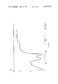
- FIG. 3 is a blood transmission curve plotted in respect of an exemplary sample of tissue.
- the reference 1 identifies a light-conducting member which in the illustrated embodiment comprises a curved and optionally flexible rod of a transluminent plastics material, such as an acrylic resin, said rod being of circular cross-section and having an end surface 7 arranged to be pressed against the region of body tissue to be examined, such as the breast 8 of a woman.
- the member 1 is covered with a thin layer of an opaque material, with the exception of its end surfaces.
- One end of the member 1 is inserted into a cylindrical housing 4 provided with a control box 3.
- the housing 4 is manufactured of an opaque material and includes two light sources 2,5, of which one is an incandescent lamp 5 arranged to be energized from a conventional, variable voltage source (not shown) arranged in the box 3.
- the other light source comprises an electronic flash source 2 arranged to be activated by means of devices (not shown) in the box 3 and which is synchronized with the shutter of a camera 9.
- the camera is located on the side of said region of tissue to be examined remote from the light source.
- the reference 6 identifies a flexible power-supply cable.
- the free end 7 of the light-conducting member 1 is held, for example, against the underside of a breast 8 to be examined.
- the incandescent lamp 5, which is of low intensity, is ignited and emits light within the whole of the visible spectrum and in all events within the blue to the infrared region of the spectrum.
- the light is diffused and spread in the body tissue, which is a requirement which must be fulfilled in order for an assessment to be made.
- the lamp is energized to such power as to obtain a transillumination of the breast.
- the light-conducting member 8 is held in a position such as to obtain the clearest visual picture of the shadow, and the camera 9 is loaded with a film which is also sensitive to light within the red spectrum, whereafter the camera is aligned onto the shadowed area at a distance of approximately 30 cm from the surface 10 of the breast, on the opposite side thereof to the side to be illuminated.
- the film is now exposed by igniting the flash device 2.
- the flash device which is highly intensive thereby to provide a short exposure of the infrared-sensitive film in the camera 9, emits light within the blue to the infrared area of the spectrum and, is completely unfiltered, similar to the light from the incandescent lamp 5, which lamp includes a tungsten filament.
- the lamp 5 may be ignited or extinguished when exposing the film 9' in the camera 9.
- the film is exposed with the camera 9 in a position such that the plane of the film 9' is substantially perpendicular to the output axis 11 of the light conductor 1.
- the developed coloured film will show the previously shadowy or black portions in colour and the normal breast tissue in a yellow colour while the fibroadenomatosis or other blood-filled anomalies are shown clearly as light-red or dark-red spots. In certain cases such spots cannot be discovered with X-rays, even though the anomaly may have been palpably detected. Cysts containing clear, uncoloured liquid will appear as a light halo or rounded change against the background of the normal, yellow breast tissue. Dark spots on the coloured photograph indicate anomalies of a serious nature.
- the incandescent lamp 5 used is preferably provided with a tungsten filament and is, for example, of the kind mentioned in the introduction.
- a needle extraction of tissue samples for cytological tests may be carried out while using the device according to the invention.
- the device enables the unhealthy change or anomaly to be localized extremely distinctly, and a sample of tissue can be extracted by comparing the actual visual picture with a previous photographic registration.
- the photographic registration will, of course, be of great importance during any subsequent surgical treatment of the patient.
- FIG. 3 illustrates in simplified form a transmission curve A for blood and a transmission curve B for tissue as such.
- the vertical axis shows the light transmission in percent of light supplied and the horizontal axis shows the wave-length of the light.
- the curve B has been plotted in respect of a very thin tissue sample and is only intended to show that the transmission through body tissue of normal type is substantial constant irrespective of the wave-length of the light. If thick tissue is transilluminated, the curve B will be moved vertically downwards.
- the invention provides for a positive analysis of suspected changes in body tissues, which can be traced by transillumination and visual observation but which can not be determined with any certainty but merely indicated that closer examination must be made.
- a colour film which is also sensitive to infrared light, and thereby enabling a region of the spectrum which cannot be observed with the naked eye to be used, there is obtained firstly a detailed image of the tissue as such and secondly a detailed image of the blood-filled portions of the tissue.
- the photograph obtained is the basis of an exact or at least relatively exact diagnosis of the unhealthy changes, thereby eliminating the necessity of X-ray radiation and the taking of tissue samples. It should be noted that all anomalies in body tissues can be detected by means of the novel method, such as liquid-filled cysts.
Landscapes
- Health & Medical Sciences (AREA)
- Life Sciences & Earth Sciences (AREA)
- General Health & Medical Sciences (AREA)
- Public Health (AREA)
- Pathology (AREA)
- Engineering & Computer Science (AREA)
- Biomedical Technology (AREA)
- Heart & Thoracic Surgery (AREA)
- Medical Informatics (AREA)
- Molecular Biology (AREA)
- Surgery (AREA)
- Veterinary Medicine (AREA)
- Biophysics (AREA)
- Physics & Mathematics (AREA)
- Animal Behavior & Ethology (AREA)
- Gynecology & Obstetrics (AREA)
- Reproductive Health (AREA)
- Investigating Or Analysing Materials By Optical Means (AREA)
- Investigating Or Analysing Biological Materials (AREA)
- Measuring And Recording Apparatus For Diagnosis (AREA)
- Radiation-Therapy Devices (AREA)
- Measurement Of The Respiration, Hearing Ability, Form, And Blood Characteristics Of Living Organisms (AREA)
- Apparatus For Radiation Diagnosis (AREA)
- Analysing Materials By The Use Of Radiation (AREA)
Applications Claiming Priority (2)
| Application Number | Priority Date | Filing Date | Title |
|---|---|---|---|
| SE7613587 | 1976-12-03 | ||
| SE7613587A SE7613587L (sv) | 1976-12-03 | 1976-12-03 | Metod for diagnostiserande av sjukliga forendringar i kvinnobrost |
Related Parent Applications (1)
| Application Number | Title | Priority Date | Filing Date |
|---|---|---|---|
| US05857696 Continuation-In-Part | 1977-12-05 |
Related Child Applications (1)
| Application Number | Title | Priority Date | Filing Date |
|---|---|---|---|
| US06/573,614 Reissue USRE32718E (en) | 1976-12-03 | 1984-01-25 | Transillumination diagnostic method and apparatus |
Publications (1)
| Publication Number | Publication Date |
|---|---|
| US4312357A true US4312357A (en) | 1982-01-26 |
Family
ID=20329651
Family Applications (2)
| Application Number | Title | Priority Date | Filing Date |
|---|---|---|---|
| US06/104,392 Ceased US4312357A (en) | 1976-12-03 | 1979-12-17 | Transillumination diagnostic method and apparatus |
| US06/573,614 Expired - Lifetime USRE32718E (en) | 1976-12-03 | 1984-01-25 | Transillumination diagnostic method and apparatus |
Family Applications After (1)
| Application Number | Title | Priority Date | Filing Date |
|---|---|---|---|
| US06/573,614 Expired - Lifetime USRE32718E (en) | 1976-12-03 | 1984-01-25 | Transillumination diagnostic method and apparatus |
Country Status (17)
| Country | Link |
|---|---|
| US (2) | US4312357A (fr) |
| JP (1) | JPS5372391A (fr) |
| AT (1) | AT359623B (fr) |
| AU (1) | AU3041777A (fr) |
| BE (1) | BE861049A (fr) |
| CA (1) | CA1113157A (fr) |
| DE (1) | DE2749191C2 (fr) |
| DK (1) | DK473877A (fr) |
| ES (1) | ES464532A1 (fr) |
| FI (1) | FI773201A7 (fr) |
| FR (1) | FR2372619A1 (fr) |
| GB (1) | GB1594824A (fr) |
| IL (1) | IL53261A (fr) |
| IT (1) | IT1086943B (fr) |
| NL (1) | NL7713444A (fr) |
| NO (1) | NO773632L (fr) |
| SE (1) | SE7613587L (fr) |
Cited By (54)
| Publication number | Priority date | Publication date | Assignee | Title |
|---|---|---|---|---|
| US4467812A (en) * | 1982-07-19 | 1984-08-28 | Spectrascan, Inc. | Transillumination apparatus |
| US4495949A (en) * | 1982-07-19 | 1985-01-29 | Spectrascan, Inc. | Transillumination method |
| US4567882A (en) * | 1982-12-06 | 1986-02-04 | Vanderbilt University | Method for locating the illuminated tip of an endotracheal tube |
| US4570638A (en) * | 1983-10-14 | 1986-02-18 | Somanetics Corporation | Method and apparatus for spectral transmissibility examination and analysis |
| US4600011A (en) * | 1982-11-03 | 1986-07-15 | The University Court Of The University Of Aberdeen | Tele-diaphanography apparatus |
| US4602642A (en) * | 1984-10-23 | 1986-07-29 | Intelligent Medical Systems, Inc. | Method and apparatus for measuring internal body temperature utilizing infrared emissions |
| US4616657A (en) * | 1982-07-19 | 1986-10-14 | The First National Bank Of Boston | Diaphanoscopy apparatus |
| US4619249A (en) * | 1985-07-24 | 1986-10-28 | Kim Landry | Transcutaneous intravenous illuminator |
| US4651743A (en) * | 1982-07-19 | 1987-03-24 | Spectrascan, Inc. | Diaphanoscopy method |
| US4662360A (en) * | 1984-10-23 | 1987-05-05 | Intelligent Medical Systems, Inc. | Disposable speculum |
| US4817623A (en) | 1983-10-14 | 1989-04-04 | Somanetics Corporation | Method and apparatus for interpreting optical response data |
| US4898172A (en) * | 1986-04-18 | 1990-02-06 | Grable Richard J | Optical light probe |
| US5079698A (en) * | 1989-05-03 | 1992-01-07 | Advanced Light Imaging Technologies Ltd. | Transillumination method apparatus for the diagnosis of breast tumors and other breast lesions by normalization of an electronic image of the breast |
| US5140989A (en) * | 1983-10-14 | 1992-08-25 | Somanetics Corporation | Examination instrument for optical-response diagnostic apparatus |
| US5349961A (en) * | 1983-10-14 | 1994-09-27 | Somanetics Corporation | Method and apparatus for in vivo optical spectroscopic examination |
| USD362910S (en) | 1994-04-21 | 1995-10-03 | Creaghan Jr Frank C | Instrument for viewing subcutaneous venous structures |
| US5699797A (en) * | 1992-10-05 | 1997-12-23 | Dynamics Imaging, Inc. | Method of investigation of microcirculation functional dynamics of physiological liquids in skin and apparatus for its realization |
| US5730133A (en) * | 1994-05-20 | 1998-03-24 | Dynamics Imaging, Inc. | Optical functional mamoscope |
| US5747789A (en) * | 1993-12-01 | 1998-05-05 | Dynamics Imaging, Inc. | Method for investigation of distribution of physiological components in human body tissues and apparatus for its realization |
| US5833367A (en) | 1996-11-12 | 1998-11-10 | Trutek, Inc. | Tympanic thermometer probe cover |
| US5865743A (en) * | 1994-02-23 | 1999-02-02 | Dynamics Imaging, Inc. | Method of living organism multimodal functional mapping |
| US5865167A (en) * | 1991-12-17 | 1999-02-02 | Dynamics Imaging, Inc. | Method of living system organism diagnostics and apparatus for its realization |
| US5967992A (en) | 1998-06-03 | 1999-10-19 | Trutex, Inc. | Radiometric temperature measurement based on empirical measurements and linear functions |
| US5980451A (en) * | 1984-10-23 | 1999-11-09 | Sherwood Services Ag | Disposable speculum with membrane bonding ring |
| US6002958A (en) * | 1992-12-24 | 1999-12-14 | Dynamics Imaging, Inc. | Method and apparatus for diagnostics of internal organs |
| US6001066A (en) | 1997-06-03 | 1999-12-14 | Trutek, Inc. | Tympanic thermometer with modular sensing probe |
| US6030117A (en) | 1996-11-12 | 2000-02-29 | Trutek, Inc. | Tympanic thermometer probe cover |
| US6048359A (en) * | 1997-08-25 | 2000-04-11 | Advanced Photodynamic Technologies, Inc. | Spatial orientation and light sources and method of using same for medical diagnosis and photodynamic therapy |
| US6123454A (en) | 1999-06-11 | 2000-09-26 | Trutek, Inc. | Tympanic thermometer disposable probe cover with further stretching prevention structure |
| US6192262B1 (en) | 1994-02-23 | 2001-02-20 | Dobi Medical Systems, Llc | Method of living organism multimodal functional mapping |
| US6923762B1 (en) * | 2001-11-01 | 2005-08-02 | Frank C. Creaghan, Jr. | Venoscope apparatus |
| US20060124862A1 (en) * | 2004-12-15 | 2006-06-15 | Rodriquez Joel J | Point of infusion lighting device |
| US20070161907A1 (en) * | 2006-01-10 | 2007-07-12 | Ron Goldman | Micro vein enhancer |
| US20080027317A1 (en) * | 2006-06-29 | 2008-01-31 | Fred Wood | Scanned laser vein contrast enhancer |
| US20080045818A1 (en) * | 2006-06-29 | 2008-02-21 | Fred Wood | Laser vein contrast enhancer |
| US20090002488A1 (en) * | 2007-06-28 | 2009-01-01 | Vincent Luciano | Automatic alignment of a contrast enhancement system |
| US20110021925A1 (en) * | 2006-06-29 | 2011-01-27 | Fred Wood | Mounted vein contrast enchancer |
| US20110112407A1 (en) * | 2006-06-29 | 2011-05-12 | Fred Wood | Multispectral detection and presentation of an object's characteristics |
| US20110125028A1 (en) * | 2009-07-22 | 2011-05-26 | Fred Wood | Vein scanner |
| US8665507B2 (en) | 2006-06-29 | 2014-03-04 | Accuvein, Inc. | Module mounting mirror endoscopy |
| US8750970B2 (en) | 2006-01-10 | 2014-06-10 | Accu Vein, Inc. | Micro vein enhancer |
| US9061109B2 (en) | 2009-07-22 | 2015-06-23 | Accuvein, Inc. | Vein scanner with user interface |
| US9072426B2 (en) | 2012-08-02 | 2015-07-07 | AccuVein, Inc | Device for detecting and illuminating vasculature using an FPGA |
| US9492117B2 (en) | 2006-01-10 | 2016-11-15 | Accuvein, Inc. | Practitioner-mounted micro vein enhancer |
| US9854977B2 (en) | 2006-01-10 | 2018-01-02 | Accuvein, Inc. | Scanned laser vein contrast enhancer using a single laser, and modulation circuitry |
| US10238294B2 (en) | 2006-06-29 | 2019-03-26 | Accuvein, Inc. | Scanned laser vein contrast enhancer using one laser |
| US10376147B2 (en) | 2012-12-05 | 2019-08-13 | AccuVeiw, Inc. | System and method for multi-color laser imaging and ablation of cancer cells using fluorescence |
| US10813588B2 (en) | 2006-01-10 | 2020-10-27 | Accuvein, Inc. | Micro vein enhancer |
| US11253198B2 (en) | 2006-01-10 | 2022-02-22 | Accuvein, Inc. | Stand-mounted scanned laser vein contrast enhancer |
| US11278240B2 (en) | 2006-01-10 | 2022-03-22 | Accuvein, Inc. | Trigger-actuated laser vein contrast enhancer |
| US12048560B2 (en) | 2006-01-10 | 2024-07-30 | Accuvein, Inc. | Vein scanner configured for single-handed lifting and use |
| US12295744B2 (en) | 2006-01-10 | 2025-05-13 | Accuvein, Inc. | Micro vein enhancer with two lasers and two optical detectors configured for removing surface topology |
| US12408865B2 (en) | 2006-01-10 | 2025-09-09 | Accuvein Inc. | Vein imaging device with differential image resolution at the center and the extremities of the vein image |
| US12471844B2 (en) | 2006-06-29 | 2025-11-18 | Accuvein, Inc. | Scanned laser vein contrast enhancer with full stopping of scanner movement during scan line reversals |
Families Citing this family (6)
| Publication number | Priority date | Publication date | Assignee | Title |
|---|---|---|---|---|
| GB2068537B (en) * | 1980-02-04 | 1984-11-14 | Energy Conversion Devices Inc | Examining biological materials |
| DE3603782A1 (de) * | 1986-02-06 | 1987-10-15 | Hauer Gerald | Verfahren zum orten einer venenstrippersonde sowie vorrichtung zur durchfuehrung des verfahrens |
| FR2618665A1 (fr) * | 1987-07-28 | 1989-02-03 | Renaud Michel | Endostroboscope pour examens naso-pharyngo-larynges |
| US20070156038A1 (en) * | 2000-01-19 | 2007-07-05 | Zeman Herbert D | Method to image the heart |
| US20120101343A1 (en) * | 2010-10-21 | 2012-04-26 | Duffy Thomas P | Medical imaging device |
| EP2829222B1 (fr) | 2013-07-24 | 2020-05-27 | Cook Medical Technologies LLC | Dispositif de localisation |
Citations (9)
| Publication number | Priority date | Publication date | Assignee | Title |
|---|---|---|---|---|
| US2902911A (en) * | 1954-08-27 | 1959-09-08 | Noyori Kimiharu | Hand-held fundus-oculi-camera |
| US3245402A (en) * | 1963-05-21 | 1966-04-12 | Barnes Eng Co | Process of diagnosis by infrared thermography |
| US3318216A (en) * | 1964-06-03 | 1967-05-09 | Edward R Hajjar | Photographic transillumination |
| US3527932A (en) * | 1967-11-16 | 1970-09-08 | James J Thomas | Transilluminating flashlight |
| US3648685A (en) * | 1969-06-25 | 1972-03-14 | James A Hepp | Photoelectric probes for determining the density of body tissue for x-ray purposes |
| US3664730A (en) * | 1970-10-15 | 1972-05-23 | Hernando Cardona | Ophthalmoscope |
| US3674008A (en) * | 1970-07-13 | 1972-07-04 | Battelle Development Corp | Quantitative pulsed transilluminator and method of operation |
| US3954329A (en) * | 1972-09-25 | 1976-05-04 | Retina Foundation | Wide-angle opthalmoscope employing transillumination |
| US4077399A (en) * | 1976-08-03 | 1978-03-07 | New Research And Development Laboratories, Inc. | Cranial transillumination device |
Family Cites Families (11)
| Publication number | Priority date | Publication date | Assignee | Title |
|---|---|---|---|---|
| BE558178A (fr) * | 1955-12-19 | |||
| US3152587A (en) * | 1960-03-31 | 1964-10-13 | Hellige & Co Gmbh F | Medical photometric apparatus |
| US3463142A (en) * | 1966-07-05 | 1969-08-26 | Trw Inc | Blood content monitor |
| US3638640A (en) * | 1967-11-01 | 1972-02-01 | Robert F Shaw | Oximeter and method for in vivo determination of oxygen saturation in blood using three or more different wavelengths |
| FR2011468A1 (fr) * | 1968-06-22 | 1970-02-27 | Tokai Rika Co Ltd | |
| US3597616A (en) * | 1969-05-07 | 1971-08-03 | Brun Sensor Systems Inc | Infrared measurement technique |
| US3704706A (en) * | 1969-10-23 | 1972-12-05 | Univ Drexel | Heart rate and respiratory monitor |
| US3867041A (en) * | 1973-12-03 | 1975-02-18 | Us Agriculture | Method for detecting bruises in fruit |
| DE2449060A1 (de) * | 1974-10-15 | 1976-04-29 | Spitalul Clinic Filantropia | Messverfahren zur bestimmung boesartiger neoplasie und anderer nichtboesartiger krankheiten |
| US3930158A (en) * | 1974-11-04 | 1975-12-30 | Sanders Associates Inc | Infrared photography |
| US3958560A (en) * | 1974-11-25 | 1976-05-25 | Wayne Front March | Non-invasive automatic glucose sensor system |
-
1976
- 1976-12-03 SE SE7613587A patent/SE7613587L/xx not_active Application Discontinuation
-
1977
- 1977-10-24 NO NO773632A patent/NO773632L/no unknown
- 1977-10-25 DK DK473877A patent/DK473877A/da unknown
- 1977-10-26 FI FI773201A patent/FI773201A7/fi not_active Application Discontinuation
- 1977-10-31 IL IL53261A patent/IL53261A/xx unknown
- 1977-11-01 CA CA289,995A patent/CA1113157A/fr not_active Expired
- 1977-11-03 DE DE2749191A patent/DE2749191C2/de not_active Expired
- 1977-11-03 IT IT29317/77A patent/IT1086943B/it active
- 1977-11-07 AU AU30417/77A patent/AU3041777A/en active Pending
- 1977-11-07 AT AT793777A patent/AT359623B/de not_active IP Right Cessation
- 1977-11-09 JP JP13454677A patent/JPS5372391A/ja active Granted
- 1977-11-10 GB GB46846/77A patent/GB1594824A/en not_active Expired
- 1977-11-18 FR FR7734783A patent/FR2372619A1/fr active Granted
- 1977-11-22 BE BE182807A patent/BE861049A/fr not_active IP Right Cessation
- 1977-11-28 ES ES464532A patent/ES464532A1/es not_active Expired
- 1977-12-05 NL NL7713444A patent/NL7713444A/xx not_active Application Discontinuation
-
1979
- 1979-12-17 US US06/104,392 patent/US4312357A/en not_active Ceased
-
1984
- 1984-01-25 US US06/573,614 patent/USRE32718E/en not_active Expired - Lifetime
Patent Citations (9)
| Publication number | Priority date | Publication date | Assignee | Title |
|---|---|---|---|---|
| US2902911A (en) * | 1954-08-27 | 1959-09-08 | Noyori Kimiharu | Hand-held fundus-oculi-camera |
| US3245402A (en) * | 1963-05-21 | 1966-04-12 | Barnes Eng Co | Process of diagnosis by infrared thermography |
| US3318216A (en) * | 1964-06-03 | 1967-05-09 | Edward R Hajjar | Photographic transillumination |
| US3527932A (en) * | 1967-11-16 | 1970-09-08 | James J Thomas | Transilluminating flashlight |
| US3648685A (en) * | 1969-06-25 | 1972-03-14 | James A Hepp | Photoelectric probes for determining the density of body tissue for x-ray purposes |
| US3674008A (en) * | 1970-07-13 | 1972-07-04 | Battelle Development Corp | Quantitative pulsed transilluminator and method of operation |
| US3664730A (en) * | 1970-10-15 | 1972-05-23 | Hernando Cardona | Ophthalmoscope |
| US3954329A (en) * | 1972-09-25 | 1976-05-04 | Retina Foundation | Wide-angle opthalmoscope employing transillumination |
| US4077399A (en) * | 1976-08-03 | 1978-03-07 | New Research And Development Laboratories, Inc. | Cranial transillumination device |
Cited By (119)
| Publication number | Priority date | Publication date | Assignee | Title |
|---|---|---|---|---|
| US4495949A (en) * | 1982-07-19 | 1985-01-29 | Spectrascan, Inc. | Transillumination method |
| US4467812A (en) * | 1982-07-19 | 1984-08-28 | Spectrascan, Inc. | Transillumination apparatus |
| US4616657A (en) * | 1982-07-19 | 1986-10-14 | The First National Bank Of Boston | Diaphanoscopy apparatus |
| US4651743A (en) * | 1982-07-19 | 1987-03-24 | Spectrascan, Inc. | Diaphanoscopy method |
| US4600011A (en) * | 1982-11-03 | 1986-07-15 | The University Court Of The University Of Aberdeen | Tele-diaphanography apparatus |
| US4567882A (en) * | 1982-12-06 | 1986-02-04 | Vanderbilt University | Method for locating the illuminated tip of an endotracheal tube |
| US4817623A (en) | 1983-10-14 | 1989-04-04 | Somanetics Corporation | Method and apparatus for interpreting optical response data |
| US4570638A (en) * | 1983-10-14 | 1986-02-18 | Somanetics Corporation | Method and apparatus for spectral transmissibility examination and analysis |
| US5349961A (en) * | 1983-10-14 | 1994-09-27 | Somanetics Corporation | Method and apparatus for in vivo optical spectroscopic examination |
| US5140989A (en) * | 1983-10-14 | 1992-08-25 | Somanetics Corporation | Examination instrument for optical-response diagnostic apparatus |
| US5980451A (en) * | 1984-10-23 | 1999-11-09 | Sherwood Services Ag | Disposable speculum with membrane bonding ring |
| US4602642A (en) * | 1984-10-23 | 1986-07-29 | Intelligent Medical Systems, Inc. | Method and apparatus for measuring internal body temperature utilizing infrared emissions |
| US4662360A (en) * | 1984-10-23 | 1987-05-05 | Intelligent Medical Systems, Inc. | Disposable speculum |
| USRE33234E (en) * | 1985-07-24 | 1990-06-19 | Kim Landry | Transcutaneous intravenous illuminator |
| US4619249A (en) * | 1985-07-24 | 1986-10-28 | Kim Landry | Transcutaneous intravenous illuminator |
| US4898172A (en) * | 1986-04-18 | 1990-02-06 | Grable Richard J | Optical light probe |
| US5079698A (en) * | 1989-05-03 | 1992-01-07 | Advanced Light Imaging Technologies Ltd. | Transillumination method apparatus for the diagnosis of breast tumors and other breast lesions by normalization of an electronic image of the breast |
| US5865167A (en) * | 1991-12-17 | 1999-02-02 | Dynamics Imaging, Inc. | Method of living system organism diagnostics and apparatus for its realization |
| US5699797A (en) * | 1992-10-05 | 1997-12-23 | Dynamics Imaging, Inc. | Method of investigation of microcirculation functional dynamics of physiological liquids in skin and apparatus for its realization |
| US6002958A (en) * | 1992-12-24 | 1999-12-14 | Dynamics Imaging, Inc. | Method and apparatus for diagnostics of internal organs |
| US5747789A (en) * | 1993-12-01 | 1998-05-05 | Dynamics Imaging, Inc. | Method for investigation of distribution of physiological components in human body tissues and apparatus for its realization |
| US6192262B1 (en) | 1994-02-23 | 2001-02-20 | Dobi Medical Systems, Llc | Method of living organism multimodal functional mapping |
| US5865743A (en) * | 1994-02-23 | 1999-02-02 | Dynamics Imaging, Inc. | Method of living organism multimodal functional mapping |
| USD362910S (en) | 1994-04-21 | 1995-10-03 | Creaghan Jr Frank C | Instrument for viewing subcutaneous venous structures |
| US5730133A (en) * | 1994-05-20 | 1998-03-24 | Dynamics Imaging, Inc. | Optical functional mamoscope |
| US5833367A (en) | 1996-11-12 | 1998-11-10 | Trutek, Inc. | Tympanic thermometer probe cover |
| US6030117A (en) | 1996-11-12 | 2000-02-29 | Trutek, Inc. | Tympanic thermometer probe cover |
| US6042266A (en) | 1996-11-12 | 2000-03-28 | Trutek, Inc. | Tympanic thermometer probe cover |
| US6001066A (en) | 1997-06-03 | 1999-12-14 | Trutek, Inc. | Tympanic thermometer with modular sensing probe |
| US6186959B1 (en) | 1997-06-03 | 2001-02-13 | Trutek, Inc. | Tympanic thermometer with modular sensing probe |
| US6048359A (en) * | 1997-08-25 | 2000-04-11 | Advanced Photodynamic Technologies, Inc. | Spatial orientation and light sources and method of using same for medical diagnosis and photodynamic therapy |
| US5967992A (en) | 1998-06-03 | 1999-10-19 | Trutex, Inc. | Radiometric temperature measurement based on empirical measurements and linear functions |
| US6123454A (en) | 1999-06-11 | 2000-09-26 | Trutek, Inc. | Tympanic thermometer disposable probe cover with further stretching prevention structure |
| US6923762B1 (en) * | 2001-11-01 | 2005-08-02 | Frank C. Creaghan, Jr. | Venoscope apparatus |
| US20060124862A1 (en) * | 2004-12-15 | 2006-06-15 | Rodriquez Joel J | Point of infusion lighting device |
| US8750970B2 (en) | 2006-01-10 | 2014-06-10 | Accu Vein, Inc. | Micro vein enhancer |
| US8818493B2 (en) | 2006-01-10 | 2014-08-26 | Accuvein, Inc. | Three-dimensional imaging of veins |
| US12446825B2 (en) | 2006-01-10 | 2025-10-21 | Accuvein, Inc. | Scanned laser vein contrast enhancer with image averaging |
| US12408865B2 (en) | 2006-01-10 | 2025-09-09 | Accuvein Inc. | Vein imaging device with differential image resolution at the center and the extremities of the vein image |
| US12295744B2 (en) | 2006-01-10 | 2025-05-13 | Accuvein, Inc. | Micro vein enhancer with two lasers and two optical detectors configured for removing surface topology |
| US12089951B2 (en) | 2006-01-10 | 2024-09-17 | AccuVeiw, Inc. | Scanned laser vein contrast enhancer with scanning correlated to target distance |
| US12048560B2 (en) | 2006-01-10 | 2024-07-30 | Accuvein, Inc. | Vein scanner configured for single-handed lifting and use |
| US20110208121A1 (en) * | 2006-01-10 | 2011-08-25 | Ron Goldman | Micro vein enhancer |
| US11642080B2 (en) | 2006-01-10 | 2023-05-09 | Accuvein, Inc. | Portable hand-held vein-image-enhancing device |
| US8478386B2 (en) | 2006-01-10 | 2013-07-02 | Accuvein Inc. | Practitioner-mounted micro vein enhancer |
| US11638558B2 (en) | 2006-01-10 | 2023-05-02 | Accuvein, Inc. | Micro vein enhancer |
| US11484260B2 (en) | 2006-01-10 | 2022-11-01 | Accuvein, Inc. | Patient-mounted micro vein enhancer |
| US11399768B2 (en) | 2006-01-10 | 2022-08-02 | Accuvein, Inc. | Scanned laser vein contrast enhancer utilizing surface topology |
| US11357449B2 (en) | 2006-01-10 | 2022-06-14 | Accuvein, Inc. | Micro vein enhancer for hands-free imaging for a venipuncture procedure |
| US8712498B2 (en) | 2006-01-10 | 2014-04-29 | Accuvein Inc. | Micro vein enhancer |
| US11278240B2 (en) | 2006-01-10 | 2022-03-22 | Accuvein, Inc. | Trigger-actuated laser vein contrast enhancer |
| US12496012B2 (en) | 2006-01-10 | 2025-12-16 | Accuvein, Inc. | Scanned laser vein contrast enhancer with eye safety protection with respect to emitted laser light |
| US20070161907A1 (en) * | 2006-01-10 | 2007-07-12 | Ron Goldman | Micro vein enhancer |
| US11253198B2 (en) | 2006-01-10 | 2022-02-22 | Accuvein, Inc. | Stand-mounted scanned laser vein contrast enhancer |
| US9042966B2 (en) | 2006-01-10 | 2015-05-26 | Accuvein, Inc. | Three dimensional imaging of veins |
| US9044207B2 (en) | 2006-01-10 | 2015-06-02 | Accuvein, Inc. | Micro vein enhancer for use with a vial holder |
| US11191482B2 (en) | 2006-01-10 | 2021-12-07 | Accuvein, Inc. | Scanned laser vein contrast enhancer imaging in an alternating frame mode |
| US11172880B2 (en) | 2006-01-10 | 2021-11-16 | Accuvein, Inc. | Vein imager with a dual buffer mode of operation |
| US9125629B2 (en) | 2006-01-10 | 2015-09-08 | Accuvein, Inc. | Vial-mounted micro vein enhancer |
| US9949688B2 (en) | 2006-01-10 | 2018-04-24 | Accuvein, Inc. | Micro vein enhancer with a dual buffer mode of operation |
| US11109806B2 (en) | 2006-01-10 | 2021-09-07 | Accuvein, Inc. | Three dimensional imaging of veins |
| US10813588B2 (en) | 2006-01-10 | 2020-10-27 | Accuvein, Inc. | Micro vein enhancer |
| US10617352B2 (en) | 2006-01-10 | 2020-04-14 | Accuvein, Inc. | Patient-mounted micro vein enhancer |
| US9492117B2 (en) | 2006-01-10 | 2016-11-15 | Accuvein, Inc. | Practitioner-mounted micro vein enhancer |
| US10500350B2 (en) | 2006-01-10 | 2019-12-10 | Accuvein, Inc. | Combination vein contrast enhancer and bar code scanning device |
| US10470706B2 (en) | 2006-01-10 | 2019-11-12 | Accuvein, Inc. | Micro vein enhancer for hands-free imaging for a venipuncture procedure |
| US10258748B2 (en) | 2006-01-10 | 2019-04-16 | Accuvein, Inc. | Vein scanner with user interface for controlling imaging parameters |
| US9788787B2 (en) | 2006-01-10 | 2017-10-17 | Accuvein, Inc. | Patient-mounted micro vein enhancer |
| US9788788B2 (en) | 2006-01-10 | 2017-10-17 | AccuVein, Inc | Three dimensional imaging of veins |
| US9854977B2 (en) | 2006-01-10 | 2018-01-02 | Accuvein, Inc. | Scanned laser vein contrast enhancer using a single laser, and modulation circuitry |
| US9186063B2 (en) | 2006-06-29 | 2015-11-17 | Accu Vein, Inc. | Scanned laser vein contrast enhancer using one laser for a detection mode and a display mode |
| US8838210B2 (en) | 2006-06-29 | 2014-09-16 | AccuView, Inc. | Scanned laser vein contrast enhancer using a single laser |
| US10238294B2 (en) | 2006-06-29 | 2019-03-26 | Accuvein, Inc. | Scanned laser vein contrast enhancer using one laser |
| US20080027317A1 (en) * | 2006-06-29 | 2008-01-31 | Fred Wood | Scanned laser vein contrast enhancer |
| US10357200B2 (en) | 2006-06-29 | 2019-07-23 | Accuvein, Inc. | Scanning laser vein contrast enhancer having releasable handle and scan head |
| US12471844B2 (en) | 2006-06-29 | 2025-11-18 | Accuvein, Inc. | Scanned laser vein contrast enhancer with full stopping of scanner movement during scan line reversals |
| US20080045818A1 (en) * | 2006-06-29 | 2008-02-21 | Fred Wood | Laser vein contrast enhancer |
| US20110021925A1 (en) * | 2006-06-29 | 2011-01-27 | Fred Wood | Mounted vein contrast enchancer |
| US12193838B2 (en) | 2006-06-29 | 2025-01-14 | Accuvein, Inc. | Scanned laser vein contrast enhancer with reduced laser intensity during scan line reversals |
| US20110112407A1 (en) * | 2006-06-29 | 2011-05-12 | Fred Wood | Multispectral detection and presentation of an object's characteristics |
| US8489178B2 (en) | 2006-06-29 | 2013-07-16 | Accuvein Inc. | Enhanced laser vein contrast enhancer with projection of analyzed vein data |
| US11523739B2 (en) | 2006-06-29 | 2022-12-13 | Accuvein, Inc. | Multispectral detection and presentation of an object's characteristics |
| US8594770B2 (en) | 2006-06-29 | 2013-11-26 | Accuvein, Inc. | Multispectral detection and presentation of an object's characteristics |
| US8665507B2 (en) | 2006-06-29 | 2014-03-04 | Accuvein, Inc. | Module mounting mirror endoscopy |
| US8706200B2 (en) | 2006-06-29 | 2014-04-22 | Accuvein, Inc. | Scanned laser vein contrast enhancer |
| US9345427B2 (en) | 2006-06-29 | 2016-05-24 | Accuvein, Inc. | Method of using a combination vein contrast enhancer and bar code scanning device |
| US11051697B2 (en) | 2006-06-29 | 2021-07-06 | Accuvein, Inc. | Multispectral detection and presentation of an object's characteristics |
| US11051755B2 (en) | 2006-06-29 | 2021-07-06 | Accuvein, Inc. | Scanned laser vein contrast enhancer using a retro collective mirror |
| US9226664B2 (en) | 2006-06-29 | 2016-01-05 | Accuvein, Inc. | Scanned laser vein contrast enhancer using a single laser |
| US9430819B2 (en) | 2007-06-28 | 2016-08-30 | Accuvein, Inc. | Automatic alignment of a contrast enhancement system |
| US11847768B2 (en) | 2007-06-28 | 2023-12-19 | Accuvein Inc. | Automatic alignment of a contrast enhancement system |
| US20090002488A1 (en) * | 2007-06-28 | 2009-01-01 | Vincent Luciano | Automatic alignment of a contrast enhancement system |
| US11132774B2 (en) | 2007-06-28 | 2021-09-28 | Accuvein, Inc. | Automatic alignment of a contrast enhancement system |
| US8730321B2 (en) | 2007-06-28 | 2014-05-20 | Accuvein, Inc. | Automatic alignment of a contrast enhancement system |
| US10713766B2 (en) | 2007-06-28 | 2020-07-14 | Accuvein, Inc. | Automatic alignment of a contrast enhancement system |
| US10096096B2 (en) | 2007-06-28 | 2018-10-09 | Accuvein, Inc. | Automatic alignment of a contrast enhancement system |
| US9760982B2 (en) | 2007-06-28 | 2017-09-12 | Accuvein, Inc. | Automatic alignment of a contrast enhancement system |
| US10580119B2 (en) | 2007-06-28 | 2020-03-03 | Accuvein, Inc. | Automatic alignment of a contrast enhancement system |
| US12318219B2 (en) | 2009-07-22 | 2025-06-03 | Accuvein Inc. | Vein scanner with limited photodiode FOV moving synchronously with scanned laser light |
| US10518046B2 (en) | 2009-07-22 | 2019-12-31 | Accuvein, Inc. | Vein scanner with user interface |
| US9789267B2 (en) | 2009-07-22 | 2017-10-17 | Accuvein, Inc. | Vein scanner with user interface |
| US8463364B2 (en) | 2009-07-22 | 2013-06-11 | Accuvein Inc. | Vein scanner |
| US12433535B2 (en) | 2009-07-22 | 2025-10-07 | Accuvein, Inc. | Method and system for optimizing a projected vein image and identifying vein locations using vein scanner |
| USD999380S1 (en) | 2009-07-22 | 2023-09-19 | Accuvein, Inc. | Vein imager and cradle in combination |
| US12426835B2 (en) | 2009-07-22 | 2025-09-30 | Accuvein, Inc. | Vein scanner with housing configured for single-handed lifting and use |
| US11826166B2 (en) | 2009-07-22 | 2023-11-28 | Accuvein, Inc. | Vein scanner with housing configured for single-handed lifting and use |
| US9061109B2 (en) | 2009-07-22 | 2015-06-23 | Accuvein, Inc. | Vein scanner with user interface |
| US20110125028A1 (en) * | 2009-07-22 | 2011-05-26 | Fred Wood | Vein scanner |
| USD999379S1 (en) | 2010-07-22 | 2023-09-19 | Accuvein, Inc. | Vein imager and cradle in combination |
| USD998152S1 (en) | 2010-07-22 | 2023-09-05 | Accuvein, Inc. | Vein imager cradle |
| US9782079B2 (en) | 2012-08-02 | 2017-10-10 | Accuvein, Inc. | Device for detecting and illuminating the vasculature using an FPGA |
| US9072426B2 (en) | 2012-08-02 | 2015-07-07 | AccuVein, Inc | Device for detecting and illuminating vasculature using an FPGA |
| US11510617B2 (en) | 2012-08-02 | 2022-11-29 | Accuvein, Inc. | Device for detecting and illuminating the vasculature using an FPGA |
| US10568518B2 (en) | 2012-08-02 | 2020-02-25 | Accuvein, Inc. | Device for detecting and illuminating the vasculature using an FPGA |
| US12558030B2 (en) | 2012-08-02 | 2026-02-24 | Accuvein Inc. | Device for detecting and illuminating the vasculature using an FPGA |
| US11439307B2 (en) | 2012-12-05 | 2022-09-13 | Accuvein, Inc. | Method for detecting fluorescence and ablating cancer cells of a target surgical area |
| US10376148B2 (en) | 2012-12-05 | 2019-08-13 | Accuvein, Inc. | System and method for laser imaging and ablation of cancer cells using fluorescence |
| US10376147B2 (en) | 2012-12-05 | 2019-08-13 | AccuVeiw, Inc. | System and method for multi-color laser imaging and ablation of cancer cells using fluorescence |
| US10517483B2 (en) | 2012-12-05 | 2019-12-31 | Accuvein, Inc. | System for detecting fluorescence and projecting a representative image |
Also Published As
| Publication number | Publication date |
|---|---|
| ATA793777A (de) | 1980-04-15 |
| FR2372619B1 (fr) | 1980-08-22 |
| BE861049A (fr) | 1978-05-22 |
| NL7713444A (nl) | 1978-06-06 |
| DE2749191C2 (de) | 1982-08-19 |
| CA1113157A (fr) | 1981-11-24 |
| NO773632L (no) | 1978-06-06 |
| USRE32718E (en) | 1988-07-26 |
| IL53261A0 (en) | 1977-12-30 |
| IL53261A (en) | 1980-11-30 |
| JPS5372391A (en) | 1978-06-27 |
| SE7613587L (sv) | 1978-06-04 |
| AU3041777A (en) | 1979-05-17 |
| FI773201A7 (fi) | 1978-06-04 |
| DE2749191A1 (de) | 1978-06-08 |
| IT1086943B (it) | 1985-05-31 |
| ES464532A1 (es) | 1978-09-01 |
| GB1594824A (en) | 1981-08-05 |
| DK473877A (da) | 1978-06-04 |
| AT359623B (de) | 1980-11-25 |
| JPS5650576B2 (fr) | 1981-11-30 |
| FR2372619A1 (fr) | 1978-06-30 |
Similar Documents
| Publication | Publication Date | Title |
|---|---|---|
| USRE32718E (en) | Transillumination diagnostic method and apparatus | |
| US6008889A (en) | Spectrometer system for diagnosis of skin disease | |
| US4852579A (en) | Photocharacterization and treatment of normal abnormal and ectopic endometrium | |
| US4600011A (en) | Tele-diaphanography apparatus | |
| US3335716A (en) | Diagnostic thermography method and means | |
| US6061591A (en) | Arrangement and method for diagnosing malignant tissue by fluorescence observation | |
| US5042494A (en) | Method and apparatus for detecting cancerous tissue using luminescence excitation spectra | |
| US4930516A (en) | Method for detecting cancerous tissue using visible native luminescence | |
| US6424858B1 (en) | Apparatus and method for viewing vasculature of a human being | |
| US4479499A (en) | Method and apparatus for detecting the presence of caries in teeth using visible light | |
| US4286602A (en) | Transillumination diagnostic system | |
| US6970729B2 (en) | Method and device for determining local distribution of a measuring parameter | |
| US6289236B1 (en) | Methods and apparatus for distinguishing inflamed and tumorous bladder tissue | |
| JP2020513966A (ja) | 多重光源を用いた皮膚状態測定装置 | |
| JPH07163551A (ja) | 組織診断器具、胸の組織内の腫瘍を特徴付ける器具及び異常組織を乳房x線造影検査法により特徴付ける方法 | |
| US6587578B2 (en) | Dynamic-functional imaging of biological objects using a non-rigid object holder | |
| NL8004712A (nl) | Werkwijze en inrichting voor het op aanwezigheid van caries onderzoeken van een gebit. | |
| JP2003079598A (ja) | 身体類型学的特性等級付けシステム | |
| DE4200741A1 (de) | Einrichtung zum erkennen von karies an zaehnen | |
| EP1254360A2 (fr) | Technique et dispositif de detection d'une anomalie dans un milieu hote par tomographie avec diffusion de signaux modules balayes en frequences | |
| EP0447708A2 (fr) | Appareil et procédé pour l'examen quantitatif de tissus humains et pour l'imagerie à haute résolution | |
| JPS60246733A (ja) | 生物組識の光学的撮影装置 | |
| EP1764031A1 (fr) | Transillumination avec de la lumière orange | |
| WO2008114065A2 (fr) | Dispositif pour détecter des processus malins dans des organismes vivants | |
| EP1369086B1 (fr) | Detecteur de carcinome mammaire |
Legal Events
| Date | Code | Title | Description |
|---|---|---|---|
| STCF | Information on status: patent grant |
Free format text: PATENTED CASE |
|
| RF | Reissue application filed |
Effective date: 19840125 |
|
| RF | Reissue application filed |
Effective date: 19840124 |