US5241049A - Neutrophil chemoattractants - Google Patents
Neutrophil chemoattractants Download PDFInfo
- Publication number
- US5241049A US5241049A US07/455,698 US45569889A US5241049A US 5241049 A US5241049 A US 5241049A US 45569889 A US45569889 A US 45569889A US 5241049 A US5241049 A US 5241049A
- Authority
- US
- United States
- Prior art keywords
- amcf
- proteins
- porcine
- activity
- pmn
- Prior art date
- Legal status (The legal status is an assumption and is not a legal conclusion. Google has not performed a legal analysis and makes no representation as to the accuracy of the status listed.)
- Expired - Fee Related
Links
Images


Classifications
-
- C—CHEMISTRY; METALLURGY
- C07—ORGANIC CHEMISTRY
- C07K—PEPTIDES
- C07K14/00—Peptides having more than 20 amino acids; Gastrins; Somatostatins; Melanotropins; Derivatives thereof
- C07K14/435—Peptides having more than 20 amino acids; Gastrins; Somatostatins; Melanotropins; Derivatives thereof from animals; from humans
- C07K14/46—Peptides having more than 20 amino acids; Gastrins; Somatostatins; Melanotropins; Derivatives thereof from animals; from humans from vertebrates
- C07K14/47—Peptides having more than 20 amino acids; Gastrins; Somatostatins; Melanotropins; Derivatives thereof from animals; from humans from vertebrates from mammals
-
- A—HUMAN NECESSITIES
- A61—MEDICAL OR VETERINARY SCIENCE; HYGIENE
- A61P—SPECIFIC THERAPEUTIC ACTIVITY OF CHEMICAL COMPOUNDS OR MEDICINAL PREPARATIONS
- A61P29/00—Non-central analgesic, antipyretic or antiinflammatory agents, e.g. antirheumatic agents; Non-steroidal antiinflammatory drugs [NSAID]
-
- C—CHEMISTRY; METALLURGY
- C07—ORGANIC CHEMISTRY
- C07K—PEPTIDES
- C07K14/00—Peptides having more than 20 amino acids; Gastrins; Somatostatins; Melanotropins; Derivatives thereof
- C07K14/435—Peptides having more than 20 amino acids; Gastrins; Somatostatins; Melanotropins; Derivatives thereof from animals; from humans
- C07K14/52—Cytokines; Lymphokines; Interferons
-
- A—HUMAN NECESSITIES
- A61—MEDICAL OR VETERINARY SCIENCE; HYGIENE
- A61K—PREPARATIONS FOR MEDICAL, DENTAL OR TOILETRY PURPOSES
- A61K38/00—Medicinal preparations containing peptides
Definitions
- the present invention relates generally to chemotactic proteins and, more specifically, to proteins chemotactic for human neutrophils, methods for producing these proteins, and therapeutic compositions containing the proteins.
- Inflammation is the reaction of living tissue to infection or injury, normally resulting in healing and the restoration of tissue structure and function. Inflammation also involves a complex set of responses which neutralize and remove pathogens and lead to the repair of the affected area. Symptoms of inflammation include pain, heat, redness, swelling, and dysfunction. Vascular dilation occurs, together with exudation of fluid and certain cellular components of blood into the surrounding tissue.
- PMN polymorphonuclear leukocytes
- Inflammation occurs in the initial stages of wound healing and is an integral part of the wound-healing process. Inflammation promotes the formation of granulation tissue. The earliest steps in this inflammatory response involve the influx of neutrophils into the wound space. Although PMN are important in phagocytizing wound debris and bacterial contaminants, their exact role in wound healing is unclear.
- Inflammation is also associated with a number of disease states.
- inflammation is important to eradication of infection and ultimate survival of the patient.
- unchecked inflammation may be detrimental, probably due to tissue injury caused by the oxidants and proteolytic enzymes released by PMN. In these latter conditions, a reduction in inflammation is beneficial to the patient.
- diseases states include arthritis and other inflammatory joint diseases, adult respiratory distress syndrome and idiopathic pulmonary fibrosis.
- Lung inflammation is characterized by the presence of PMN in the pulmonary interstitium and airspaces.
- interstitial and airspace neutrophils are a hallmark of adult respiratory distress syndrome (ARDS) (Pistorese et al., Chest 88: A86, 1985; Maunder et al., Am. Rev. Resoir. Dis. 135: A260, 1987). Severity of ARDS is proportional to the number of neutrophils in the lungs, and patients who have fewer airspace neutrophils relative to airspace macrophages have a better rate of survival (Maunder et al., Am. Rev. Respir. Dis. 139: A221, 1989).
- the present invention provides substantially pure porcine alveolar macrophage-derived chemotactic factor I (AMCF-I).
- the AMCF-I has the amino-terminal amino acid sequence Ala-Arg-Val-Ser-Ala-Glu-Leu-X-Arg-Gln-X-Ile-Asn-Thr-His-Ser-Thr-Pro-Phe-His, wherein X is Cys or another amino acid.
- AMCF-I with a specific activity of at least about 200,000 units/mg is provided.
- the present invention provides AMCF-I with a specific activity of at least 400,000 units/mg.
- the present invention provides substantially pure porcine alveolar macrophage-derived chemotactic factor II (AMCF-II).
- the AMCF-II has the amino-terminal amino acid sequence Ser-Pro-Ile-Glu-Ala-Ala-Glu-Ala-Ala-Val-Val-Arg-Glu-Leu-Arg-X-Met-X-Leu-Thr-Thr-Thr-Pro-Gly-Ile-His-Phe-Lys-Met-Ile, wherein X is Cys or another amino acid.
- AMCF-II with a specific activity of at least about 100,000 units/mg is provided.
- the present invention provides AMCF-II with a specific activity of at least 200,000 units/mg.
- the present invention provides a protein composition comprising porcine AMCF-I and AMCF-II, the composition having a specific activity of at least about 20,000 units/mg.
- the present invention further provides compositions comprising the substantially pure proteins and protein compositions described above in combination with a physiologically acceptable carrier or diluent.
- the present invention provides isolated DNA molecules encoding AMCF-I or AMCF-II.
- the present invention provides methods for preparing a chemotactic protein composition.
- the methods generally comprise the steps of (a) stimulating cells selected from the group consisting of porcine monocytes and porcine macrophages with a septic mediator such as bacterial endotoxin; (b) culturing the stimulated cells to produce a cell-conditioned medium; (c) concentrating the cell-conditioned medium; (d) chromatographically separating the concentrated medium under acidic conditions to produce a fraction having chemotactic activity for human neutrophils and a fraction substantially free of chemotactic activity for human neutrophils; and (e) recovering the fraction having chemotactic activity.
- the cells are porcine alveolar macrophages.
- the cell-conditioned medium is acidified prior to concentrating, and the concentrating step comprises cation exchange chromatography.
- the proteins are further purified by chromatographically fractionating the recovered fraction having chemotactic activity of step (e) to produce one or more chemotactically active fractions and a fraction substantially free of chemotactic activity, and recovering at least one of the chemotactically active fractions.
- the fractionating step may comprise reversed-phase high-performance liquid chromatography.
- FIG. 1 is a cation exchange HPLC purification profile of a chemotactic protein composition of the present invention.
- the elution gradient program is shown in the top panel.
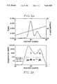
- FIGS. 2A-B illustrates the results of reversed-phase HPLC fractionation of a partially purified chemotactic protein composition.
- the top panel shows the acetonitrile elution gradient and the A 280 of the eluate.
- the bottom panel shows the chemotactic activity and gel electrophoresis patterns of columm fractions.
- FIG. 3 illustrates the final purification of AMCF-II.
- FIGS. 4A-B illustrates chemotactic dose-reponse curves of AMCF-I (A) and AMCF-II (B) for human and porcine neutrophils.
- FIG. 4C shows a comparison of the dose-response curves of AMCF-I, AMCF-II and LTB 4 for porcine neutrophils. Results are expressed as a percentage of the maximal response to the species-specific zymosan-activated serum.
- FIGS. 5A-B shows comparative micrographs of lung biopsies after instilling AMCF-I (A) or vehicle only (B).
- a control lung segment is shown in FIG. 5C.
- neutrophils arrows
- AI alveolar airspace
- FIG. 6 is a comparison of the amino-terminal amino acid sequences of AMCF-I and AMCF-II with related polypeptides.
- Lower case letters designate species: p, porcine; h, human; r, rat; b, bovine; m, mouse; c, chicken. * indicates the N-terminal alanine residue of the platelet basic protein cleavage, NAP-2.
- Specific Activity For AMCF-I and AMCF-II, specific activity is defined as units of chemotactic activity per mg of protein.
- One unit of chemotactic activity is the amount of a sample required to give at least three times the baseline (negative control) activity in a chemotaxis assay using human neutrophils as described herein.
- Substantially Pure Greater than 90% pure (on the basis of total protein) as determined by SDS-polyacrylamide gel electrophoresis, reversed-phase HPLC, or quantitative amino acid sequence analysis.
- the inventors have isolated and characterized a class of novel proteins from LPS-stimulated porcine alveolar macrophages. These proteins have potent chemotactic activity for polymorphonuclear leukocytes and, as such, are useful for enhancing inflammation. The proteins also provide useful tools for the discovery of anti-inflammatory compounds.
- the proteins of the present invention have been designated as "alveolar macrophage-derived chemotactic factors". Two of these proteins are hereinafter referred to as AMCF-I and AMCF-II. These proteins have been produced in a substantially pure form having a high specific activity as measured in a chemotaxis assay using human neutrophils. Both have a molecular weight of approximately 10 kDa as determined by polyacrylamide gel electrophoresis.
- the amino acid compositions of these proteins are presented below in Table 3. As will be appreciated by those skilled in the art, these compositions are approximate and may vary somewhat from preparation to preparation, and may also vary depending upon the source of the protein and the particular analytical methods employed.
- AMCF-I and AMCF-II are disclosed herein, including representative partial amino acid sequences, the invention also includes similar chemotactically active porcine proteins having slight variations in amino acid sequence or other properties. Such variations may arise naturally (e.g. due to genetic polymorphism) or may be produced by human intervention (e.g. by mutagenesis of cloned DNA sequences).
- the proteins of the present invention can be isolated from porcine macrophages or monocytes.
- a particularly preferred starting material is porcine alveolar macrophages, which may be obtained by lung lavage.
- the isolated cells are stimulated by culturing them according to standard cell culture methods in the presence of a mediator of sepsis. Suitable mediators include bacterial endotoxin (LPS) and the cytokines IL-1 and TNF.
- LPS bacterial endotoxin
- IL-1 and TNF cytokines
- the cell-conditioned medium is concentrated by conventional techniques, such as ultrafiltration, dialysis, salt precipitation, ion exchange chromatography, affinity chromatography or lectin adsorption.
- a particularly preferred method of concentration is cation exchange chromatography under acidic conditions.
- a particularly preferred cation exchange medium in this regard is sulfopropyl-derivatized dextran, such as SP-Sephadex C-25, available from Pharmacia, Piscataway, N.J.
- the conditioned medium is combined with the cation exchange medium in a solution having a pH of about 2.0 to 7.0, preferably about 3.5.
- the column is washed to remove unbound material, then the bound material is eluted using a high salt buffer at about neutral pH.
- a particularly preferred such buffer is 100 mM potassium phosphate buffer pH 7.0 containing 1.0 M NaCl.
- concentration Prior to concentration, it is advantageous to clarify the conditioned medium, such as by centrifugation or filtration. To minimize proteolysis, concentration is preferably carried out at low temperature, e.g. about 5° C.
- the concentrated medium is then chromatographically separated to isolate the chemotactically active proteins from other components.
- Prior to chromatography it is preferable to reduce the salt concentration of the concentrated medium, such as by dialysis against a low ionic strength, acidic buffer such as 10 mM acetic acid/Na acetate buffer, pH 3.5.
- the medium is preferably acidified prior to fractionation.
- the medium is then separated into active and inactive fractions by high performance liquid chromatography on a cation exchange resin.
- a preferred such resin is sulfopropyl-derivatized silica, such as TSK SP-5PW (Bio-Rad Laboratories, Richmond, Calif.).
- separation may be achieved by conventional liquid chromatography on a cation exchange resin.
- the column is washed with a pH gradient, and chemotactically active fractions are recovered. Prior to washing the column with the pH gradient, it is preferred to elute contaminating proteins from the column with a salt gradient.
- the resulting composition is a partially purified mixture containing AMCF-I and AMCF-II and having a specific activity typically greater than about 20,000 units/mg.
- Additional purification and separation of AMCF-I and AMCF-II is achieved by chromatographically fractionating the chemotactically active mixture.
- Preferred chromatographic methods include reversed-phase high performance liquid chromatography and molecular seive chromatograhy.
- a preferred HPLC medium in this regard is Vydac C-4 large pore silica (The Separations Group, Hesperia, Calif.). It is preferred to acidify the partially purified, active mixture prior to chromatographically fractionating it, such as by dialysis against a low pH buffer.
- a particularly preferred dialysis buffer is 10 mM acetic acid/Na acetate, pH 3.5.
- the preparation is then applied to an HPLC column, which is eluted with a gradient of an organic solvent such as acetonitrile. Column fractions are assayed for chemotactic activity, and active fractions are retained.
- AMCF-I and AMCF-II typically elute at about 35%-45% acetonitrile under these conditions.
- the purification process may be monitored throughout by assaying the chemotactic activity and protein content of the preparation.
- Chemotactic activity is preferably assayed according to standard procedures using human neutrophils.
- Protein content of the preparation may be monitored by absorption at 280 nm, polyacrylamide gel electrophoresis, quantitative analytical techniques, or other methods known in the art.
- the proteins of the present invention may also be produced by expressing cloned DNA sequences (cDNA is preferred) in recombinant cells.
- a suitable cDNA may be isolated from a porcine alveolar macrophage cDNA library. Suitable libraries may be prepared according to standard procedures. In a preferred method, a porcine alveolar macrophage cDNA expression library is screened using affinity-purified antibodies (Young and Davis, Proc. Natl. Acad. Sci. USA 80: 1194-1198, 1983; Davis et al., U.S. Pat. No. 4,788,135). Partial cDNA clones (fragments) can be extended by re-screening of the library with the cloned cDNA fragment until the full sequence is obtained.
- Additional methods of cDNA cloning are also suitable, including screening cDNA libraries with oligonucleotide probes designed on the basis of N-terminal amino acid sequences disclosed herein or the sequences of CNBr fragments from isolated proteins. Additional cloning methods are described by Maniatis et al., eds. (Molecular Cloning: A Laboratory Manual, Cold Spring Harbor Laboratory, Cold Spring Harbor, N.Y., 1982) which is incorporated herein by reference. The identities of cloned sequences are confirmed by sequencing or activity assays of expressed clones.
- a DNA sequence encoding a chemoattractant protein is inserted into a suitable expression vector.
- Expression vectors useful in this regard contain transcriptional promoter and terminator sequences operably linked to the DNA sequence to be expressed.
- expression vectors may also contain an origin of replication, enhancer sequences, and other nucleotide sequences which regulate or enhance expression levels. In many instances it is advantageous to include a bacterial origin of replication so that the vector can be replicated and manipulated using a prokaryotic host.
- Selectable markers sequences which provide for the selection and maintenance of the vector in the host cell, may also be provided in the expression vector, although in some cases a selectable marker may be introduced into the host cell on a separate vector.
- Suitable expression vectors may be derived from plasmids or viruses, or may contain elements of both. Selection of the appropriate elements and construction of vectors is within the ordinary level of skill in the art.
- Expression vectors for use in mammalian cells comprise a promoter capable of directing the transcription of a cloned gene or cDNA introduced into a mammalian cell.
- Suitable promoters include cellular promoters, such as the mouse metallothionein-1 (MT-1) promoter (Palmiter et al. Science 222: 809-814, 1983 and U.S. Pat. No. 4,579,821), and viral promoters, such as the SV40 (Subramani et al., Mol. Cell. Biol.
- polyadenylation signal located downstream of the DNA sequence insertion site.
- the polyadenylation signal may be that of the DNA sequence of interest, or may be derived from a heterologous gene.
- Expression vectors are introduced into cultured mammalian cells according to standard procedures, for example, calcium phosphate-mediated transfection (Wigler et al., Cell 14: 725, 1978; Corsaro and Pearson, Somat. Cell Genet. 7: 603, 1981; Graham and Van der Eb, Virol. 52: 456, 1973) or electroporation (Neumann et al., EMBO J. 1: 841-845, 1982).
- a small fraction of the treated cells integrate the DNA into their genomes or maintain the DNA in non-chromosomal nuclear structures.
- a selectable marker is generally introduced into the cells along with the gene of interest.
- Preferred selectable markers include genes that confer resistance to drugs such as neomycin, hygromycin, and methotrexate. Selectable markers may be introduced into the cell on a separate expression vector at the same time as the gene of interest, or they may be introduced on the same expression vector.
- the copy number of the integrated gene sequences may be increased through amplification by using certain selectable markers (e.g., a dihydrofolate reductase gene, which confers resistance to methotrexate).
- selectable marker e.g., a dihydrofolate reductase gene, which confers resistance to methotrexate.
- the selectable marker is introduced into the cells along with the gene of interest, and drug selection pressure is applied. By selecting for increased copy number of cloned sequences, expression levels may be substantially elevated.
- Cells of lower organisms may also be used within the present invention.
- a particularly preferred host is the yeast Saccharomyces cerevisiae, although other fungal cells may also be used.
- yeast Saccharomyces cerevisiae for recombinant proteins that require disulfide bonding and/or glycosylation for biological activity, or to facilitate purification of a recombinant protein, a secretory expression system is used.
- the DNA sequence encoding the protein of interest is fused, in the correct reading frame, to a sequence encoding a secretory signal peptide (a "signal sequence").
- Particularly preferred signal sequences include those encoding the pre-pro region of the MF ⁇ 1 (yeast alpha-factor) gene product (Kurjan and Herskowitz, Cell 30: 933-943, 1982; Singh, EP 123,544; and Kurjan et al., U.S. Pat. No. 4,546,082) and the secretory peptide portion of the BARI gene (MacKay et al., U.S. Pat. No. 4,613,572 and MacKay, WO 87/02670).
- the BAR1 signal sequence may be combined with the coding sequence for the third domain of the BARI gene product (MacKay et al., EP 314,096).
- Other useful signal sequences include those of the yeast PH05 (Lemontt et al., WO 86/00638) and a-factor (Brake, EP 123,289) genes.
- Suitable expression vectors include YRp7 (Struhl et al., Proc. Natl. Acad. Sci. U.S.A. 76: 1035-1039, 1979), YEp13 (Broach et al., Gene 8: 121-133, 1979), pJDB248 and pJDB219 (Beggs, ibid.), and derivatives thereof.
- Such vectors generally include a selectable marker.
- a defective selectable marker such as the leu2-d gene of Beggs (ibid.) or the POTI gene of Kawasaki and Bell (EP 171,142), is particularly preferred.
- Preferred promoters useful in yeast expression vectors include promoters from yeast glycolytic genes (Hitzeman et al., J. Biol. Chem. 255: 12073-12080, 1980; Alber and Kawasaki, J. Mol. Appl. Genet. 1: 419-434, 1982; and Kawasaki, U.S. Pat. No. 4,599,311) or alcohol dehydrogenase genes, particularly the ADH2-4c promoter (also known as "ADR3-4c;” see Russell et al., Nature 304 652-654, 1983).
- Hybrid promoters may also be utilized as disclosed by Bitter (WO 86/06077) and Rosenberg et al. (Ep 164,556). Hybrid promoters can also be constructed as disclosed in U.S. patent application Ser. No. 07/036,823 by inserting one or more copies of a yeast mating-type regulatory element into a yeast promoter.
- the transformed or transfected host cells are grown in a culture medium containing carbon and nitrogen sources and appropriate supplements. Selection of culture media appropriate for a particular cell type is within the level of ordinary skill in the art.
- the protein may be isolated from the cells or culture media as generally described above.
- the proteins of the present invention may be used within therapeutic compositions for the treatment of a broad spectrum of wounds.
- Types of wounds that may be treated with these proteins include superficial wounds and lacerations, abrasions, surgical wounds and burns.
- these proteins will be useful in any condition where the formation of granulation tissue is desired.
- such conditions include skin grafts and treatment with artificial skin.
- compositions will be formulated to provide a concentration of the active protein of between about 10 -12 M to 10 -4 M within the wound space. These compositions will generally be reapplied at one- to several-day intervals until granulation tissue formation is substantially complete.
- the exact treatment regimen will be determined by the size and nature of the wound and the overall condition of the patient. In severe cases, it may be necessary to administer the therapeutic compositions of the present invention more frequently, i.e. up to four times per day, and for longer duration.
- compositions according to the present invention comprise the proteins described herein in combination with suitable carriers, as well as adjuvants, diluents, or stabilizers.
- suitable carriers as well as adjuvants, diluents, or stabilizers.
- the proteins described herein will be used in a concentration of about 10 -8 M to 10 -5 M, although concentrations in the range of 10 -12 M to 10 -4 M may be used.
- Diluents include albumins, saline, sterile water, mannitiol, etc. Other stabilizers, antioxidants, or protease inhibitors may also be added.
- growth factors such as platelet-derived growth factor (PDGF), basic fibroblast growth factor (bFGF), epidermal growth factor (EGF), insulin-like growth factor 1 (IGF-1), etc.
- PDGF platelet-derived growth factor
- bFGF basic fibroblast growth factor
- EGF epidermal growth factor
- IGF-1 insulin-like growth factor 1
- monocyte chemoattractant a factor that influences the production of monocytes.
- the proteins may be applied to wound dressings as aqueous solutions, and the dressings may be packaged in sterile form.
- the proteins of the present invention are combined with a suitable carrier and formulated for bronchial or intravenous delivery.
- Bronchial delivery may be via nebulization or via a bronchoscope.
- Suitable carriers and diluents for bronchial delivery include sterile, pyrogen-free saline with a suitable protein carrier, such as albumin.
- Suitable diluents for intravenous administration include sterile water and sterile saline.
- the proteins are formulated to provide a dose concentration of from about 10 -12 M to 10 -4 M, more preferably 10 -8 M to 10 -5 M.
- the proteins of the present invention may also be used systemically to block localized inflammation.
- the proteins are formulated as aqueous solutions and administered intravenously as described above.
- the proteins of the present invention are useful tools for the development of anti-inflammatory agents.
- a wide variety of anti-inflammatory agents may be developed, including blocking antibodies, peptide antagonists, and small, non-peptide antagonists.
- antibodies against the proteins or their cellular receptors may be used to block their chemotactic activity.
- Analysis of the amino acid sequences and structure-function relationships of the proteins provides guidance for the synthesis of small peptides related to the active sites (receptor binding sites).
- the regions around amino acids 12-18 of AMCF-I and amino acids 19-25 of AMCF-II are useful in blocking PMN chemotaxis and thus reducing inflammation.
- the chemotaxis assay systems described hereinafter may be adapted to screen for anti-chemotactic agents. By adding a test compound to the assay, the ability of that compound to block the activity of the proteins is determined.
- Fragments of the proteins of the present invention may also be used to promote inflammation.
- the sequences of the proteins are analyzed to determine the active sites, and small peptides containing an active site sequence are prepared and assayed for chemotactic activity.
- Small peptides may also be produced by other conventional methods, such as proteolytic digestion of the intact proteins or expression of small DNA molecules in recombinant cells.
- the cuff was inflated in the trachea and sterile pyrogen free 0.9% NaCl containing 50 mM EDTA was instilled into the airways to a hydrostatic pressure of 30 cm H 2 O (approximately 2 liters).
- the cell-enriched fluid was collected by gravity drainage. This lavage procedure was repeated twice on each animal.
- An average pig lung lavage yielded 2.7 ⁇ 10 9 ⁇ 1.3 ⁇ 10 9 (mean ⁇ S.D.) viable AM, with a purity of 96% ⁇ 1.5% AM, 2% ⁇ 0.8% lymphocytes and 1% ⁇ 0.4% PMN.
- AM viability always exceeded 90% by trypan blue dye exclusion.
- the lavage fluids were spun at 200 x G for 15 minutes to pellet the cells.
- the cell pellets were washed twice with sterile pyrogen-free 0.9% NaCl, and resuspended at a final concentration of 2 ⁇ 10 6 viable AM/ml in RPMI 1640 (Gibco, Grand Island, N.Y.) containing 10 ⁇ g/ml E. coli endotoxin (026:B6, Sigma Co., St. Louis, Mo.), 100 U/ml penicillin (Flow Laboratories, McClean, Va.), 100 ⁇ g/ml streptomycin (Flow Laboratories), 2 mM L-glutamine (Flow Laboratories), and 50 ⁇ g/ml gentamicin.
- the cell suspension was plated in 150 cm 2 culture flasks at a density of 5 ⁇ 10 5 viable AM/cm 2 and incubated at 37° C. in 5% CO 2 /air. After 24 hours the conditioned media were aspirated, quantitatively cultured on blood agar plates to assess bacterial contamination, and clarified by centrifugation at 10,000 x g for 30 min. Only samples that were free of bacterial growth were used in subsequent studies. The conditioned media were stored at -70° C. for no more than 30 days.
- the AM conditioned media were tested for chemotactic activity to porcine and human PMN.
- Human PMN were obtained by antecubital venipuncture and porcine PMN were obtained by subxiphoid cardiac puncture under general anesthesia.
- PMN were isolated by the method of Ferrante and Thong (J. Immunol. Methods 36: 109-117, 1980) using Mono-Poly Resolving Media (Flow Laboratories). PMN were resuspended in RPMI-1640 containing 5% heat inactivated fetal calf serum (Hyclone Laboratories, Inc., Logan, Utah) at a concentration of 3 ⁇ 10 6 cells/ml.
- Chemotaxis was measured by the modified Boyden technique using microchemotaxis chambers (Falk et al., J. Immunol. Methods 33: 239-247, 1980) (Neuroprobe Co., Bethesda, Md.).
- PMN 60 ⁇ l containing 1.8% ⁇ 10 5 PMN
- the sample to be tested was serially diluted in phosphate buffered saline containing 0.2% bovine serum albumin, pH 7.2, and 25 ⁇ l of sample was added to each well in the bottom compartment of the chamber.
- the top and bottom compartments of the chambers were separated by nitrocellulose filters (3.0 ⁇ m pore size, Neuroprobe, Inc., Cabin John, Md.).
- PMN chemotaxis was measured as the total of cells at the end-point of migration in 10 high powered fields (x450) using a 5 ⁇ 5 mm eyepiece grid.
- the AM-conditioned media were found to contain chemotactic activity for both porcine and human PMN.
- AMCF was purified from 500 ml of LPS-stimulated AM conditioned media, generated from 1.1 ⁇ 10 9 viable AM (equivalent to that of a single pig whole-lung lavage).
- Table 1 A summary of the purification procedure showing the recovery at each step is shown in Table 1. All samples and column fractions were kept at 5° C. or frozen at -70° C. except during the two HPLC loading and elution steps, which were performed at room temperature.
- For the chemotaxis assay aliquots of crude preparations and preparations containing high salt or low pH were brought to the appropriate assay conditions of ionic strength and pH either by dilution in PBS containing 0.2% BSA, pH 7.2 or by dialyzing them against the same solution at 5° C.
- the chemotactically active fractions from the SP-Sephadex column were combined and dialyzed against 8 liters of 10 mM acetic acid/Na acetate, pH 3.5 for 12 hours.
- the dialyzed sample was then loaded onto a 75 ⁇ 7.5 cm column of TSK SP-5PW (Bio-Rad Laboratories, Richmond, Calif.) equilibrated in 10 mM acetic acid/Na acetate, pH 3.5.
- the column was eluted at a flow rate of 1.0 ml/minute using two different gradients in sequence. Over the first 20 minutes a linear salt gradient was used with a limit buffer of 10 mM acetate, containing 1 M NaCl pH 3.5.
- Chemotactically active fractions 56-64 from SP-5PW chromatography were combined, acidified by 1:2 dilution in 0.1% TFA, and loaded onto a C-4 reversed-phase HPLC column equilibrated in 0.1% trifluoroacetic acid (TFA) in 5% acetonitrile.
- TFA trifluoroacetic acid
- the column was eluted with a linear gradient over 35 minutes using a limit solution of 0.1% TFA in 80% acetonitrile.
- 0.5 ml fractions were collected at a flow rate of 1 ml/minute.
- the elution profile is shown in FIG. 2. 85% of the total protein loaded onto the column was recovered in the pass-through and gradient fractions.
- the pass-through fractions and every gradient fraction were assayed for chemotactic activity.
- Two peaks of activity eluted between 35% and 45% acetonitrile, and were designated AMCF-I and AMCF-II. They were separated by two
- Molecular weight standards included a mixture of recombinant human insulin (Humulin, Eli Lilly and Co., Indianapolis, Ind.) (6.2 kDa), albumin (67 kDa), ovalbumin (43 kDa), carbonic anhydrase (30 kDa), trypsin inhibitor (20.1 kDa), and alpha-lactalbumin (14.4 kDa)
- Molecular weights of AMCF-I and AMCF-II were estimated from a plot of Rf vs log10(molecular weight) to be 10 kDa.
- FIG. 2 shows a composite of the A 280 , chemotactic activity, and SDS-PAGE profile of material eluting between 35% and 45% aceto itrile.
- AMCF-II was further purified by rechromatography of fraction 38 (FIG. 2) on C-4 reversed-phase HPLC using a more shallow gradient and a smaller fraction volume (FIG. 3). The resulting electrophoretically pure AMCF-II (fraction 35 in FIG. 3) was used for subsequent characterization.
- the chemotactic dose-responses of AMCF-I and AMCF-II for porcine and human PMN are compared in FIGS. 4A-C.
- AMCF-I attracted both porcine and human PMN.
- the lowest concentration of AMCF-I with significant chemotactic activity was 3 ⁇ 10 -10 M for porcine PMN, and 3 ⁇ 10 -9 M for human PMN.
- the peak chemotactic activity of AMCF-I occured at 3 ⁇ 10 -8 M for porcine PMN, and at or above 1 ⁇ 10 -7 M for human PMN (FIG. 4A).
- AMCF-II attracted porcine PMN, but had very little effect on human PMN (FIG. 4B).
- the lowest concentration of AMCF-II with significant chemotactic activity was near 1 ⁇ 10 -9 M.
- AMCF-II had significant chemotactic activity for human PMN in only one of three separate experiments.
- FIG. 4C compares the dose-responses of AMCF-I, AMCF-II, and LTB4.
- the peak chemotactic activity of AMCF-I exceeded 100% of that observed with simutaneously tested zymosan-activated porcine serum.
- AMCF-II demonstrated a dose-response relationship with a slope similar to that observed with LTB4.
- a healthy 19 kg Yorkshire pig was sedated with xylazine and ketamine and anesthetized with 100 mg sodium thiopental administered intravenously.
- a fiberoptic bronchoscope was passed transorally and the tip wedged in either the lingula (control side) or the right middle lobe (experimental side).
- 10 ml of a 10 -9 M solution of AMCF-I was prepared as follows: 10 ⁇ l of fraction #34 from reversed-phase HPLC (FIG. 2) was diluted to 1.0 ml with sterile phosphate buffered saline containing 0.2% bovine serum albumin, pH 7.2. Immediately before instillation, the solution was further diluted to 10 ml in pyrogen-free 0.9% NaCl.
- a control solution without AMCF-I was prepared by diluting 10 ⁇ l of 0.1% TFA in 40% acetonitrile to 1.0 ml with sterile phosphate buffered saline containing 0.2% bovine serum albumin, pH 7.2, then further diluting the solution to 10 ml in pyrogen-free 0.9% NaCl.
- the 10 ml solutions were instilled, immediately followed by five 10 ml aliquots of air to disperse the aliquot of fluid in the lung segment. Four hours later, the pig was again sedated and anesthetized.
- Bronchoalveolar lavage was performed on each of the lung segments with the fiberoptic bronchoscope using five 30 ml aliquots of pyrogen-free 0.9% NaCl. Each lavage aliquot was collected with gentle suction. Total cell counts in the lavage fluid were determined on cytospin preparations of lavage fluids stained with Diff-Quik (American Scientific Products, McGaw Park, Ill.). The total and differential cell counts are shown in Table 2. The instillation of AMCF-I produced a 20-fold increase in total recovered cells and a 250-fold increase in total recovered PMN over control. In addition, AMCF-I also caused a 10-fold increase in AM and a 25-fold in lymphocytes.
- FIG. 5 Histopathologic sections of the lung segments are shown in FIG. 5.
- open lung biopsies of the lingula and right middle lobe were performed via a median sternotomy incision.
- Lung tissue was fixed in formalin and embedded in paraffin, and sections were stained with hematoxylin and eosin for light microscopy.
- the experimental lung segment where AMCF-I was instilled (right middle lobe) showed a marked infiltration of inflammatory cells, predominately PMN, which involved the pulmonary interstitium as well as the alveolar airspaces (FIG. 5A).
- the control lung segment (lingula) is shown for comparison and had a normal appearance (FIG. 5C).
- N-terminal sequence anaysis was determined by Edman degradation of 10 ⁇ g samples of both AMCF-I and AMCF-II with an automated gas phase peptide sequenator (model 475A Pulse Liquid Protein Sequencer; Applied Biosystems, Inc., Foster City, Calif.). Cycles with no detectable PTH derivatives were assigned to cysteine.
- the sequences of the first 20 amino acids of unmodified AMCF-I and the first 30 amino acids of unmodified AMCF-II are shown in Table 4. X indicates an amino acid tentatively identified as cysteins as described above.
Landscapes
- Health & Medical Sciences (AREA)
- Chemical & Material Sciences (AREA)
- Organic Chemistry (AREA)
- Life Sciences & Earth Sciences (AREA)
- General Health & Medical Sciences (AREA)
- Medicinal Chemistry (AREA)
- Genetics & Genomics (AREA)
- Toxicology (AREA)
- Proteomics, Peptides & Aminoacids (AREA)
- Molecular Biology (AREA)
- Biophysics (AREA)
- Zoology (AREA)
- Gastroenterology & Hepatology (AREA)
- Biochemistry (AREA)
- Animal Behavior & Ethology (AREA)
- Nuclear Medicine, Radiotherapy & Molecular Imaging (AREA)
- Public Health (AREA)
- Pain & Pain Management (AREA)
- Veterinary Medicine (AREA)
- Rheumatology (AREA)
- General Chemical & Material Sciences (AREA)
- Pharmacology & Pharmacy (AREA)
- Chemical Kinetics & Catalysis (AREA)
- Peptides Or Proteins (AREA)
- Preparation Of Compounds By Using Micro-Organisms (AREA)
- Medicines That Contain Protein Lipid Enzymes And Other Medicines (AREA)
Priority Applications (6)
| Application Number | Priority Date | Filing Date | Title |
|---|---|---|---|
| US07/455,698 US5241049A (en) | 1989-12-22 | 1989-12-22 | Neutrophil chemoattractants |
| PCT/US1990/007615 WO1991009945A1 (fr) | 1989-12-22 | 1990-12-21 | Agents chimioattractifs de neutrophiles |
| EP91901983A EP0506820A1 (fr) | 1989-12-22 | 1990-12-21 | Agents chimioattractifs de neutrophiles |
| AU71752/91A AU7175291A (en) | 1989-12-22 | 1990-12-21 | Neutrophil chemoattractants |
| JP3502920A JPH05503101A (ja) | 1989-12-22 | 1990-12-21 | 好中球化学誘引物質 |
| CA002072012A CA2072012A1 (fr) | 1989-12-22 | 1990-12-21 | Neutrophiles chimiotactiques |
Applications Claiming Priority (1)
| Application Number | Priority Date | Filing Date | Title |
|---|---|---|---|
| US07/455,698 US5241049A (en) | 1989-12-22 | 1989-12-22 | Neutrophil chemoattractants |
Publications (1)
| Publication Number | Publication Date |
|---|---|
| US5241049A true US5241049A (en) | 1993-08-31 |
Family
ID=23809912
Family Applications (1)
| Application Number | Title | Priority Date | Filing Date |
|---|---|---|---|
| US07/455,698 Expired - Fee Related US5241049A (en) | 1989-12-22 | 1989-12-22 | Neutrophil chemoattractants |
Country Status (6)
| Country | Link |
|---|---|
| US (1) | US5241049A (fr) |
| EP (1) | EP0506820A1 (fr) |
| JP (1) | JPH05503101A (fr) |
| AU (1) | AU7175291A (fr) |
| CA (1) | CA2072012A1 (fr) |
| WO (1) | WO1991009945A1 (fr) |
Cited By (13)
| Publication number | Priority date | Publication date | Assignee | Title |
|---|---|---|---|---|
| US5413778A (en) * | 1992-10-05 | 1995-05-09 | The Regents Of The University Of Michigan | Labelled monocyte chemoattractant protein material and medical uses thereof |
| US5571713A (en) * | 1992-10-22 | 1996-11-05 | The Regents Of The University Of Michigan | Therapeutic treatment for inhibiting vascular restenosis |
| US5605671A (en) * | 1992-10-05 | 1997-02-25 | The Regents Of The University Of Michigan | Radiolabeled neutrophil activating peptides for imaging |
| US5665591A (en) * | 1994-12-06 | 1997-09-09 | Trustees Of Boston University | Regulation of smooth muscle cell proliferation |
| US5688927A (en) * | 1995-06-07 | 1997-11-18 | Icos Corporation | Macrophage derived chemokine |
| US5817911A (en) * | 1995-04-07 | 1998-10-06 | Regents Of The University Of California | Transgenic mice expressing alveolar MCP-1 |
| US5932703A (en) * | 1995-06-07 | 1999-08-03 | Icos Corporation | Macrophage derived chemokine and chemokine analogs |
| US6320023B1 (en) | 1995-06-07 | 2001-11-20 | Icos Corporation | Macrophage derived chemokine |
| US6498015B1 (en) | 1995-06-07 | 2002-12-24 | Icos Corporation | Methods of identifying agents that modulate the binding between MDC and an MDC receptor |
| US6551618B2 (en) | 1994-03-15 | 2003-04-22 | University Of Birmingham | Compositions and methods for delivery of agents for neuronal regeneration and survival |
| US6737513B1 (en) | 1996-06-07 | 2004-05-18 | Icos Corporation | Macrophage derived chemokine (MDC) and chemokine analogs and assay to identify modulators of MDC activity, and therapeutic uses for same |
| US6790947B1 (en) | 1995-06-07 | 2004-09-14 | Icos Corporation | Polynucleotides encoding macrophage derived chemokine |
| US7018627B1 (en) | 1995-06-07 | 2006-03-28 | Icos Corporation | Macrophage derived chemokine (MDC), MDC analogs, MDC inhibitor substances, and uses thereof |
Citations (4)
| Publication number | Priority date | Publication date | Assignee | Title |
|---|---|---|---|---|
| WO1989004325A1 (fr) * | 1987-11-06 | 1989-05-18 | Ferring Arzneimittel Gmbh | Polypeptide activateur de neutrophiles, son procede de production et son utilisation comme agent therapeutique et de diagnostic |
| WO1989004836A1 (fr) * | 1987-11-19 | 1989-06-01 | Sandoz Ag | Facteur d'activation des neutrophiles |
| WO1989008665A1 (fr) * | 1988-03-16 | 1989-09-21 | The United States Of America, As Represented By Th | NOUVEAU FACTEUR CHIMIOTACTIQUE NEUTROPHILE, ADNc CLONE ET ANTICORPS MONOCLONAUX CONTRE CELUI-CI |
| WO1989010962A1 (fr) * | 1988-05-02 | 1989-11-16 | Dainippon Pharmaceutical Co., Ltd | Procede de production d'un polypeptide du facteur chimiotactique neutrophile humain |
-
1989
- 1989-12-22 US US07/455,698 patent/US5241049A/en not_active Expired - Fee Related
-
1990
- 1990-12-21 WO PCT/US1990/007615 patent/WO1991009945A1/fr not_active Ceased
- 1990-12-21 CA CA002072012A patent/CA2072012A1/fr not_active Abandoned
- 1990-12-21 EP EP91901983A patent/EP0506820A1/fr not_active Withdrawn
- 1990-12-21 AU AU71752/91A patent/AU7175291A/en not_active Abandoned
- 1990-12-21 JP JP3502920A patent/JPH05503101A/ja active Pending
Patent Citations (4)
| Publication number | Priority date | Publication date | Assignee | Title |
|---|---|---|---|---|
| WO1989004325A1 (fr) * | 1987-11-06 | 1989-05-18 | Ferring Arzneimittel Gmbh | Polypeptide activateur de neutrophiles, son procede de production et son utilisation comme agent therapeutique et de diagnostic |
| WO1989004836A1 (fr) * | 1987-11-19 | 1989-06-01 | Sandoz Ag | Facteur d'activation des neutrophiles |
| WO1989008665A1 (fr) * | 1988-03-16 | 1989-09-21 | The United States Of America, As Represented By Th | NOUVEAU FACTEUR CHIMIOTACTIQUE NEUTROPHILE, ADNc CLONE ET ANTICORPS MONOCLONAUX CONTRE CELUI-CI |
| WO1989010962A1 (fr) * | 1988-05-02 | 1989-11-16 | Dainippon Pharmaceutical Co., Ltd | Procede de production d'un polypeptide du facteur chimiotactique neutrophile humain |
Non-Patent Citations (16)
| Title |
|---|
| Golds et al, "Inflammatory cytokines induce synthesis and secretion of gro protein and a neutrophil chemotactic factor . . . ". Biochem. J. (1989) 259, 585-88. |
| Golds et al, Inflammatory cytokines induce synthesis and secretion of gro protein and a neutrophil chemotactic factor . . . . Biochem. J. (1989) 259, 585 88. * |
| Gregory et al, "Structure Determination of a Human Lymphocyte Derived Neutrophil Activity Peptide", Biochem. Biophys. Res. Comm. vol. 151, No. 2, 1988, pp. 883-890. |
| Gregory et al, Structure Determination of a Human Lymphocyte Derived Neutrophil Activity Peptide , Biochem. Biophys. Res. Comm. vol. 151, No. 2, 1988, pp. 883 890. * |
| Matsushima et al, "Molecular Cloning of a Human Monocyte-Derived Neutrophil Chemotactic Factor . . . " J. Exp. Med., vol. 167, Jun. 1988, pp. 1883-1893. |
| Matsushima et al, Molecular Cloning of a Human Monocyte Derived Neutrophil Chemotactic Factor . . . J. Exp. Med., vol. 167, Jun. 1988, pp. 1883 1893. * |
| Schroder et al, "Identification of Different Charged Species of a Human Monocyte Derived Neutrophil Activating peptide (MONA)", Biochem. Biophys. Res. Comm. 152:277-284, 1988. |
| Schroder et al, Identification of Different Charged Species of a Human Monocyte Derived Neutrophil Activating peptide (MONA) , Biochem. Biophys. Res. Comm. 152:277 284, 1988. * |
| Strieter et al., "Endothelial Cell Gene Expression of a Neutrophil Chemotactic Factor by TNF-a, LPS, and IL-β", Science 243:1467-1469, 1989. |
| Strieter et al., Endothelial Cell Gene Expression of a Neutrophil Chemotactic Factor by TNF a, LPS, and IL , Science 243:1467 1469, 1989. * |
| Suzuki et al, "Purification and Partial Primary Sequence of a Chemotactic Protein for Polymorphonuclear Leukocytes . . . ", J. Exp. Med. vol. 169, Jun. 1989, pp. 1895-1901. |
| Suzuki et al, Purification and Partial Primary Sequence of a Chemotactic Protein for Polymorphonuclear Leukocytes . . . , J. Exp. Med. vol. 169, Jun. 1989, pp. 1895 1901. * |
| Walz et al, "Purification and Amino Acid Sequencing of NAF . . . ", Biochem. Biophys. Res. Comm. vol. 149, No. 2, 1987, pp. 755-761. |
| Walz et al, Purification and Amino Acid Sequencing of NAF . . . , Biochem. Biophys. Res. Comm. vol. 149, No. 2, 1987, pp. 755 761. * |
| Wolpe et al, "Identification and characterization of macrophage inflammatory protein 2", Proc. Natl. Acad. Sci. USA 86:612-616, 1989. |
| Wolpe et al, Identification and characterization of macrophage inflammatory protein 2 , Proc. Natl. Acad. Sci. USA 86:612 616, 1989. * |
Cited By (14)
| Publication number | Priority date | Publication date | Assignee | Title |
|---|---|---|---|---|
| US5413778A (en) * | 1992-10-05 | 1995-05-09 | The Regents Of The University Of Michigan | Labelled monocyte chemoattractant protein material and medical uses thereof |
| US5605671A (en) * | 1992-10-05 | 1997-02-25 | The Regents Of The University Of Michigan | Radiolabeled neutrophil activating peptides for imaging |
| US5571713A (en) * | 1992-10-22 | 1996-11-05 | The Regents Of The University Of Michigan | Therapeutic treatment for inhibiting vascular restenosis |
| US6551618B2 (en) | 1994-03-15 | 2003-04-22 | University Of Birmingham | Compositions and methods for delivery of agents for neuronal regeneration and survival |
| US5665591A (en) * | 1994-12-06 | 1997-09-09 | Trustees Of Boston University | Regulation of smooth muscle cell proliferation |
| US5756673A (en) * | 1994-12-06 | 1998-05-26 | Trustees Of Boston University | Regulation of smooth muscle cell proliferation |
| US5817911A (en) * | 1995-04-07 | 1998-10-06 | Regents Of The University Of California | Transgenic mice expressing alveolar MCP-1 |
| US5932703A (en) * | 1995-06-07 | 1999-08-03 | Icos Corporation | Macrophage derived chemokine and chemokine analogs |
| US6320023B1 (en) | 1995-06-07 | 2001-11-20 | Icos Corporation | Macrophage derived chemokine |
| US6498015B1 (en) | 1995-06-07 | 2002-12-24 | Icos Corporation | Methods of identifying agents that modulate the binding between MDC and an MDC receptor |
| US5688927A (en) * | 1995-06-07 | 1997-11-18 | Icos Corporation | Macrophage derived chemokine |
| US6790947B1 (en) | 1995-06-07 | 2004-09-14 | Icos Corporation | Polynucleotides encoding macrophage derived chemokine |
| US7018627B1 (en) | 1995-06-07 | 2006-03-28 | Icos Corporation | Macrophage derived chemokine (MDC), MDC analogs, MDC inhibitor substances, and uses thereof |
| US6737513B1 (en) | 1996-06-07 | 2004-05-18 | Icos Corporation | Macrophage derived chemokine (MDC) and chemokine analogs and assay to identify modulators of MDC activity, and therapeutic uses for same |
Also Published As
| Publication number | Publication date |
|---|---|
| WO1991009945A1 (fr) | 1991-07-11 |
| EP0506820A1 (fr) | 1992-10-07 |
| CA2072012A1 (fr) | 1991-06-23 |
| AU7175291A (en) | 1991-07-24 |
| JPH05503101A (ja) | 1993-05-27 |
Similar Documents
| Publication | Publication Date | Title |
|---|---|---|
| US5241049A (en) | Neutrophil chemoattractants | |
| DE60020220T2 (de) | Plättchenadhäsion blockierendes protein | |
| EP0616615B1 (fr) | Analogues de l'interleukine-8 humaine | |
| EP0232326B1 (fr) | Inhibine et son procede de purification | |
| US5811393A (en) | Heparin binding mitogen with homology to epidermal growth factor (EGF) | |
| US7030223B2 (en) | Megakaryocyte stimulating factors | |
| JPH06505983A (ja) | 創傷治癒の促進方法および促進薬 | |
| PT948538E (pt) | Análise e separação de proteínas do factor de crescimento derivado de plaquetas | |
| JPS6322526A (ja) | 肝細胞増殖因子 | |
| JP2823690B2 (ja) | プロテアーゼ耐性pdgf及び使用法 | |
| WO1991008231A1 (fr) | FACTEUR POLYPEPTIDIQUE [Ala IL-8]77 UTILISE COMME INIHIBITEUR D'ADHESION DES LEUCOCYTES | |
| MXPA06003695A (es) | Usos terapeuticos de variantes de quimiocinas. | |
| DE68911297T2 (de) | Heparin bindende proteine, dafür kodierende dna, verfahren zu ihrer herstellung sowie sie enthaltende therapeutische präparate. | |
| DE60207043T2 (de) | Histidin-reiches glykoprotein (hrgp) zur inhibierung der angiogenese | |
| KR20040101426A (ko) | 신규한 mcp 단백질의 길항제 | |
| KR100837898B1 (ko) | 다발성 경화증의 치료에서 사용되는 케모킨 변이체 | |
| EP0550506B1 (fr) | Nouveau facteur chimiotactique | |
| CN1035257C (zh) | 修饰的血小板因子-4 | |
| Goodman et al. | Identification of two neutrophil chemotactic peptides produced by porcine alveolar macrophages | |
| DE69912988T2 (de) | Verwendung eines serinproteaseinhibitors vom kunitz-typ zur beschleunigung der schleimauflöserate | |
| AU2019366008B2 (en) | Drug for treating and/or improving septicemia associated with coagulation abnormality | |
| WO1992008473A1 (fr) | Utilisation therapeutique du facteur de croissance de fibroblaste | |
| CN101306193A (zh) | 人神经突起生长素在制备创伤愈合药物中的应用 | |
| WO2000017240A1 (fr) | Endostatine ncf pour l'inhibition de la croissance des tumeurs et de la proliferation capillaire et pour le diagnostic des maladies vasculaires et tumorales | |
| JPWO1994021809A1 (ja) | 単球又はマクロファージ遊走因子 |
Legal Events
| Date | Code | Title | Description |
|---|---|---|---|
| AS | Assignment |
Owner name: ZYMOGENETICS, INC., WASHINGTON Free format text: ASSIGNMENT OF ASSIGNORS INTEREST.;ASSIGNOR:FORSTROM, JOHN W.;REEL/FRAME:005383/0486 Effective date: 19900308 |
|
| AS | Assignment |
Owner name: VETERANS ADMINISTRATION, WASHINGTON Free format text: ASSIGNMENT OF ASSIGNORS INTEREST;ASSIGNOR:MARTIN, THOMAS R.;REEL/FRAME:006744/0387 Effective date: 19930923 Owner name: UNIVERSITY OF WASHINGTON, WASHINGTON Free format text: ASSIGNMENT OF ASSIGNORS INTEREST;ASSIGNOR:MARTIN, THOMAS R.;REEL/FRAME:006744/0387 Effective date: 19930923 |
|
| FEPP | Fee payment procedure |
Free format text: PAYOR NUMBER ASSIGNED (ORIGINAL EVENT CODE: ASPN); ENTITY STATUS OF PATENT OWNER: LARGE ENTITY |
|
| REMI | Maintenance fee reminder mailed | ||
| LAPS | Lapse for failure to pay maintenance fees | ||
| FP | Lapsed due to failure to pay maintenance fee |
Effective date: 19970903 |
|
| STCH | Information on status: patent discontinuation |
Free format text: PATENT EXPIRED DUE TO NONPAYMENT OF MAINTENANCE FEES UNDER 37 CFR 1.362 |