WO2009091200A2 - Procédé d'élimination d'artéfacts métalliques dans des images tomographiques aux rayons x - Google Patents
Procédé d'élimination d'artéfacts métalliques dans des images tomographiques aux rayons x Download PDFInfo
- Publication number
- WO2009091200A2 WO2009091200A2 PCT/KR2009/000229 KR2009000229W WO2009091200A2 WO 2009091200 A2 WO2009091200 A2 WO 2009091200A2 KR 2009000229 W KR2009000229 W KR 2009000229W WO 2009091200 A2 WO2009091200 A2 WO 2009091200A2
- Authority
- WO
- WIPO (PCT)
- Prior art keywords
- image
- data
- projection data
- initial
- ray
- Prior art date
- Legal status (The legal status is an assumption and is not a legal conclusion. Google has not performed a legal analysis and makes no representation as to the accuracy of the status listed.)
- Ceased
Links
Images
Classifications
-
- A—HUMAN NECESSITIES
- A61—MEDICAL OR VETERINARY SCIENCE; HYGIENE
- A61B—DIAGNOSIS; SURGERY; IDENTIFICATION
- A61B6/00—Apparatus or devices for radiation diagnosis; Apparatus or devices for radiation diagnosis combined with radiation therapy equipment
- A61B6/02—Arrangements for diagnosis sequentially in different planes; Stereoscopic radiation diagnosis
- A61B6/03—Computed tomography [CT]
- A61B6/032—Transmission computed tomography [CT]
-
- A—HUMAN NECESSITIES
- A61—MEDICAL OR VETERINARY SCIENCE; HYGIENE
- A61B—DIAGNOSIS; SURGERY; IDENTIFICATION
- A61B6/00—Apparatus or devices for radiation diagnosis; Apparatus or devices for radiation diagnosis combined with radiation therapy equipment
- A61B6/02—Arrangements for diagnosis sequentially in different planes; Stereoscopic radiation diagnosis
- A61B6/03—Computed tomography [CT]
-
- A—HUMAN NECESSITIES
- A61—MEDICAL OR VETERINARY SCIENCE; HYGIENE
- A61B—DIAGNOSIS; SURGERY; IDENTIFICATION
- A61B6/00—Apparatus or devices for radiation diagnosis; Apparatus or devices for radiation diagnosis combined with radiation therapy equipment
- A61B6/50—Apparatus or devices for radiation diagnosis; Apparatus or devices for radiation diagnosis combined with radiation therapy equipment specially adapted for specific body parts; specially adapted for specific clinical applications
- A61B6/51—Apparatus or devices for radiation diagnosis; Apparatus or devices for radiation diagnosis combined with radiation therapy equipment specially adapted for specific body parts; specially adapted for specific clinical applications for dentistry
-
- A—HUMAN NECESSITIES
- A61—MEDICAL OR VETERINARY SCIENCE; HYGIENE
- A61B—DIAGNOSIS; SURGERY; IDENTIFICATION
- A61B6/00—Apparatus or devices for radiation diagnosis; Apparatus or devices for radiation diagnosis combined with radiation therapy equipment
- A61B6/52—Devices using data or image processing specially adapted for radiation diagnosis
- A61B6/5258—Devices using data or image processing specially adapted for radiation diagnosis involving detection or reduction of artifacts or noise
-
- G—PHYSICS
- G06—COMPUTING OR CALCULATING; COUNTING
- G06T—IMAGE DATA PROCESSING OR GENERATION, IN GENERAL
- G06T12/00—Tomographic reconstruction from projections
- G06T12/10—Image preprocessing, e.g. calibration, positioning of sources or scatter correction
Definitions
- the present invention relates to a method of removing metal artifacts from an X-ray Citi image, and more particularly, to a method of removing metal artifacts from an X-ray C-image.
- tooth damage occurs, such as tooth decay in the patient's teeth
- the damaged tooth is filled with dental fillings or replaced by prosthetic devices.
- the metal object such as the dental filler or prosthetic device is hardly transmitted through X-rays so that the soft tissue or Create metal artifacts such as streak artifacts and star-burst artifacts in skeletal structures.
- the metal artifact affects an image of not only itself but other objects around it, thereby causing a problem of obscuring the X-ray city image.
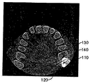
- 1 is a photograph showing an X-ray Citigraphy image in which metal artifacts exist.
- the X-ray image of FIG. 1 is an X-ray image of a dental filler or a tooth of a patient who has an artificial prosthetic device.
- the dental filler is positioned at the right molar position.
- the artificial prosthetic device shows that the streak artifact 120, which is a kind of metal artifact, is generated around the metallic object 110.
- the metallic object 110 and the neighboring molars 130 are blackened in the middle thereof, so that the black teeth 140 appear to be drilled in the molars 130.
- the above-mentioned metal artifacts may obscure the X-ray Citi image and the doctor may read the unclear X-ray Citi image, thereby causing the patient's dental condition to be diagnosed incorrectly.
- the present invention is to solve the above-mentioned disadvantages and problems of the prior art, and extracts the data corresponding to the metallic object that causes the metal artifacts from the projection data obtained from the X-ray imaging, and extracts the data X-rays to remove the metal artifacts from the reconstructed image by recovering the data lost by the POCS method and reconstructed to obtain a reconstructed image, and inserting the image of the metallic object in the region corresponding to the metallic object in the reconstructed image It is an object of the present invention to provide a method for removing metal artifacts of a Citi-photographed image.
- the object of the present invention comprises the steps of obtaining an initial projection data by positioning the object between the X-ray source and the detector; Reconstructing the initial projection data to obtain an initial reconstructed image; Extracting a high intensity region image having an intensity higher than a predetermined intensity from the initial reconstructed image; Acquiring first projection data by removing data corresponding to the high intensity region image from the initial projection data; Restoring the first projection data by a POCS method to obtain second projection data; Reconstructing the second projection data to obtain a temporary reconstruction image; And acquiring a final reconstructed image by replacing a region corresponding to the high intensity region image with the high intensity region image in the temporary reconstructed image. Is achieved.
- the object of the present invention is to restore the data removed from the first projection data by the POCS method to convert the first projection data into the second projection data is the removed data from the first projection data It is also achieved by a method for removing metal artifacts of an X-ray Citigraphic image, which further includes partially restoring by linear interpolation before restoring by the POCS method.
- the object of the present invention is a method for removing the metal artifacts of the Citi shooting image after the step of acquiring the final reconstruction image, smoothing the boundary region of the area replaced by the high-intensity region image in the final reconstruction image ( It is also achieved by a method of removing the metal artifacts of the Citi-photographed image, characterized in that it further comprises a step of smoothing).
- the object of the present invention is to extract a high-intensity region image having a higher intensity than the set intensity from the initial reconstruction image after converting the data of the initial projection data corresponding to the initial reconstruction image into hounsfield units
- a predetermined hounsfield unit higher than the hounsfield unit of bone and lower than the hounsfield unit of composite fillings is set to have a hounsfield unit higher than the set strength. It is also achieved by a method of removing metal artifacts of an X-ray Citigraphic image, which is a step of extracting a high intensity region image by extracting and obtaining data.
- the object of the present invention is that the POCS method regards the removed data as lost data, and includes a filtering process for performing low-pass filtering on the first projection data and one of first projection data after the low-pass filtering.
- the data other than the lost data is also achieved by a method of removing metal artifacts of the X-ray Citigraphic image, which is a method of restoring the removed data by repeating a restoration process of restoring the original.
- Method of removing the metal artifacts of the X-ray Citigraphy image of the present invention by providing a method for correcting the metal artifacts caused by the presence of a metal object to block X-rays, such as dental fillers or prosthetic devices in the patient's teeth
- a metal object such as dental fillers or prosthetic devices in the patient's teeth
- 1 is a photograph showing an X-ray Citigraphy image in which metal artifacts exist.
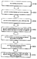
- FIG. 2 is a flowchart illustrating a method of removing metal artifacts of an X-ray Citigraphic image, according to an exemplary embodiment.
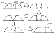
- 3 is a diagram schematically illustrating the POCS method for easy explanation.
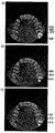
- FIG. 4 is a diagram illustrating a result of applying a method of removing metal artifacts from an X-ray Citigraphy image to an image of an actual patient's arch, according to an exemplary embodiment.
- the object is placed between an X-ray source and a detector provided in the X-ray Citigraphic apparatus, and the X-ray generated by the X-ray source is irradiated to the object and received by the detector to receive data.
- These two-dimensional images obtain projection data.
- the X-ray source and the detector acquire a plurality of projection data while scanning by rotating the circular trajectory up to 360 degrees while moving by the same angle.
- the reconstructed image of the object is obtained by reconstructing the plurality of projection data.
- the filter bag-projection algorithm is processed according to the following method.
- preprocessing and filtering are performed.
- the projection data acquired by the X-ray Citigraphy apparatus are loaded one by one.
- the Jacobian weighting process is a process for correcting a change in a value between polar coordinates and rectangular coordinates during a mathematical process.
- the projection data is filtered using a filter.
- the filtering becomes the core of the filter bag-projection algorithm technique.
- the shape of the object part derived as a result of the reconstruction appears blurred and unclear.
- each projection data is filtered so that the shape of the original object can be approximated.
- the filtering process is performed between the Jacobian weighting process and the back-projection process to be described later.
- interpolation is performed using the projection data to determine the value of each target point of the three-dimensional volume. And the value of the target point is stored.
- the reconstructed slice is then stored.
- Each of the projection data is sequentially processed through the preprocessing and filtering processing, the back-projection processing, and the reconstruction section storage, and then the reconstruction result is stored in the section.
- Such metal artifacts may reduce the sharpness of the X-ray image, which may prevent the doctor from accurately diagnosing the dental condition of the patient.
- FIG. 2 is a flowchart illustrating a method of removing metal artifacts of an X-ray Citigraphic image, according to an exemplary embodiment.
- the initial projection data and the initial reconstructed image are obtained by using the reconstructed image.
- the object is positioned between the X-ray source and the detector provided in the X-ray city photographing apparatus, the X-ray generated by the X-ray source is irradiated to the object and received by the detector to acquire data to obtain initial projection data.
- an initial reconstructed image is obtained by reconstructing the acquired initial projection data by a technique such as the filter bag-projection algorithm described above.
- the metal artifact described with reference to FIG. 1 may occur.
- the metal artifact becomes a high intensity region image having a greater intensity than other surrounding areas. .
- the amount of X-rays is attenuated by the physical characteristics of the specific material. It is expressed as an Hounsfield unit.
- the hounsfield unit calculates an attenuation coefficient of a specific material based on the attenuation coefficient of water.
- Equation 1 is a formula for obtaining Hounsfield units, and ⁇ denotes a damping coefficient of a specific material, and ⁇ w denotes a damping coefficient of water.
- the higher the hounsfield unit the higher the attenuation of the X-ray, that is, the greater the degree of blocking the X-rays.
- the soft tissue of the head will show the lowest Hounsfield unit value, the bone will have a higher unit value than the soft tissue, and dental fillers or prostheses.
- Metallic objects that block X-rays, such as devices, will exhibit much higher Hounsfield unit values than the soft tissue and bone.
- Table 1 shows representative Hounsfield unit values as a table.
- the hounsfield units At a set strength, which is higher than the hounsfield unit of the bone and lower than the hounsfield unit of the composite fillings, for example, the 2500 hounsfield unit is set as the set strength.
- the high intensity region image is extracted by extracting and obtaining the data.
- the data corresponding to the high intensity region image in the initial projection data is changed to a "null" value or a "0" value to be the first projection data. Therefore, the first projection data has data corresponding to the high intensity region image having a "null” value or a "0" value.
- the second projection data is obtained by restoring the first projection data by the Projections Onto Convex Sets (POCS) method (S500).
- POCS Projections Onto Convex Sets
- the POCS method regards data having a " null " value or a " 0 " value corresponding to the high intensity region image in the first projection data as loss data and low-pass filtering the first projection data.
- Data other than the lost data among the first projection data after the low-pass filtering are restored by restoring the original process by estimating data having the "null” or "0" value from surrounding data. This is how you do it.
- 3 is a diagram schematically illustrating the POCS method for easy explanation.
- a portion of the lost data 220 is a filtering process of performing low-pass filtering (LPF) on the first projection data 210 including the lost data 220.
- the first projection data 210 except for the lost data 200 is restored.
- the loss data 220 is repeated from the first projection data 210 adjacent to the loss data 220 by repeating the restoration process of the loss data 220 and the restoration process of the first projection data 210. Estimate and restore.
- the second projection data 230 is obtained from the first projection data 210.
- the POCS method may correctly restore the lost data 220 as the restoration process of the lost data 220 and the restoration process of the first projection data 210 are repeated more. Therefore, when the loss data 220 is always restored by the POCS method alone, it takes much time to execute.
- the method of acquiring the temporary reconstructed image by reconstructing the second projection data uses the method of obtaining the reconstructed image described above.
- a final reconstructed image is obtained by replacing the region corresponding to the high intensity region image with the high intensity region image in the temporary reconstructed image (S700).
- the temporary reconstructed image is obtained by removing data corresponding to a high intensity region image from the initial projection data to obtain the first projection data and reconstructing the removed data from the first projection data by the POCS method as second projection data.
- the high intensity region image is an image obtained by reconstruction.
- the final reconstructed image may be obtained by replacing the region corresponding to the high intensity region image with the high intensity region image in the temporary reconstructed image.
- the final reconstructed image is an image in which the image of the metallic object itself is present while the metal artifacts generated by the metallic object blocking the X-ray, such as a dental filler or a prosthetic device, are reduced.
- Such discontinuity may be a process of removing discontinuity in the boundary region by making the final reconstructed image appear unnatural.
- the final reconstructed image is naturally displayed by performing a smoothing process on the boundary region of the region replaced with the high intensity region image in the final reconstructed image.
- FIG. 4 is a diagram illustrating a result of applying a method of removing metal artifacts from an X-ray Citigraphy image to an image of an actual patient's arch, according to an exemplary embodiment.
- the reconstructed image of (a) is a reconstructed image to which the method for removing metal artifacts of the X-ray Citigraphic image according to an embodiment of the present invention is not applied, and (b) and (c)
- the reconstructed images are images to which the method for removing metal artifacts of the X-ray Citigraphic image according to an embodiment of the present invention is applied.
- the difference between the reconstructed image of (b) and (c) is different from that of the smoothing process ((b) reconstructed image) and that of the smoothing process ((c) reconstructed image).
- the metallic object 310 that blocks X-rays such as a dental filler or a prosthetic device, is displayed in white and streaks around the metallic object 310 are formed. 320) is clear. In addition, a black hole 340 appears to be formed in the molar 330 adjacent to the metallic object 310.
- the reconstructed images of (b) and (c) to which the method for removing metal artifacts of the X-ray Citigraphic image according to an embodiment of the present invention are applied not only make the metallic object 310 clearly visible but also the metallic object ( It is sharpened so that the empty space 315 can be seen inside 310.
- the empty space 315 is not visible in the reconstructed image of (a), but is shown by applying a method of removing metal artifacts of the X-ray Citigraphic image according to an embodiment of the present invention.
Landscapes
- Health & Medical Sciences (AREA)
- Life Sciences & Earth Sciences (AREA)
- Engineering & Computer Science (AREA)
- Medical Informatics (AREA)
- Physics & Mathematics (AREA)
- Radiology & Medical Imaging (AREA)
- Surgery (AREA)
- High Energy & Nuclear Physics (AREA)
- Veterinary Medicine (AREA)
- Nuclear Medicine, Radiotherapy & Molecular Imaging (AREA)
- Optics & Photonics (AREA)
- Pathology (AREA)
- Public Health (AREA)
- Biomedical Technology (AREA)
- Heart & Thoracic Surgery (AREA)
- Molecular Biology (AREA)
- Biophysics (AREA)
- Animal Behavior & Ethology (AREA)
- General Health & Medical Sciences (AREA)
- Theoretical Computer Science (AREA)
- Dentistry (AREA)
- Oral & Maxillofacial Surgery (AREA)
- General Physics & Mathematics (AREA)
- Pulmonology (AREA)
- Computer Vision & Pattern Recognition (AREA)
- Apparatus For Radiation Diagnosis (AREA)
Abstract
La présente invention concerne un procédé d'élimination d'artéfacts métalliques dans des images tomographiques aux rayons X, qui permet de corriger des artéfacts métalliques causés par des matériaux d'obturation dentaire ou des appareillages prothétiques et analogues présents dans les dents d'un patient, et révélés par les images tomographiques aux rayons X réalisées pendant l'examen de l'état dentaire du patient au cours d'un traitement dentaire.
Applications Claiming Priority (2)
| Application Number | Priority Date | Filing Date | Title |
|---|---|---|---|
| KR10-2008-0004602 | 2008-01-15 | ||
| KR1020080004602A KR100923098B1 (ko) | 2008-01-15 | 2008-01-15 | 엑스 레이 씨티 촬영 영상의 메탈 아티팩트를 제거하는방법 |
Publications (2)
| Publication Number | Publication Date |
|---|---|
| WO2009091200A2 true WO2009091200A2 (fr) | 2009-07-23 |
| WO2009091200A3 WO2009091200A3 (fr) | 2009-10-08 |
Family
ID=40885805
Family Applications (1)
| Application Number | Title | Priority Date | Filing Date |
|---|---|---|---|
| PCT/KR2009/000229 Ceased WO2009091200A2 (fr) | 2008-01-15 | 2009-01-15 | Procédé d'élimination d'artéfacts métalliques dans des images tomographiques aux rayons x |
Country Status (2)
| Country | Link |
|---|---|
| KR (1) | KR100923098B1 (fr) |
| WO (1) | WO2009091200A2 (fr) |
Cited By (3)
| Publication number | Priority date | Publication date | Assignee | Title |
|---|---|---|---|---|
| US9275454B2 (en) | 2011-10-24 | 2016-03-01 | Koninklijke Philips N.V. | Motion compensated second pass metal artifact correction for CT slice images |
| WO2019002904A1 (fr) * | 2017-06-30 | 2019-01-03 | Trophy | Procédé d'imagerie par tomodensitométrie à rayons x, système d'interface et appareil |
| CN111915696A (zh) * | 2020-08-07 | 2020-11-10 | 南京安科医疗科技有限公司 | 三维图像数据辅助的低剂量扫描数据重建方法及电子介质 |
Families Citing this family (7)
| Publication number | Priority date | Publication date | Assignee | Title |
|---|---|---|---|---|
| KR101065838B1 (ko) * | 2009-10-13 | 2011-09-19 | (주)제노레이 | 사이노그램 복원 장치 및 방법 |
| KR101275462B1 (ko) * | 2011-06-10 | 2013-06-14 | (주)제노레이 | 엑스레이 ct 영상의 하이브리드 세그멘테이션 방법 |
| KR20130098531A (ko) * | 2012-02-28 | 2013-09-05 | (주)제노레이 | 씨티의 메탈 아티팩트 감소 방법 |
| KR101571151B1 (ko) | 2014-08-14 | 2015-12-07 | 주식회사 케이유엠텍 | 컴퓨터 단층촬영 이미지의 아티팩트 제거 방법 |
| KR101530015B1 (ko) * | 2014-08-29 | 2015-06-18 | 고려대학교 산학협력단 | 컴퓨터 단층촬영 이미지의 밀도 분석 방법 |
| EP3398515B1 (fr) * | 2017-05-03 | 2020-06-24 | Siemens Healthcare GmbH | Procédé adaptatif de production de données d'image ct à artéfact réduit et unité de reconstruction d'image et produit de programme informatique associé |
| KR102423104B1 (ko) | 2017-07-21 | 2022-07-20 | 주식회사 바텍 | 듀얼 에너지 엑스선 프로젝션을 이용한 금속 이미지 구분 방법, 금속 인공음영 제거 방법 및 이를 이용한 엑스선 영상 획득 장치 |
Family Cites Families (4)
| Publication number | Priority date | Publication date | Assignee | Title |
|---|---|---|---|---|
| JPH0919533A (ja) * | 1995-07-07 | 1997-01-21 | Oki Electric Ind Co Ltd | スコアリングシステム |
| DE10231061A1 (de) * | 2002-07-10 | 2004-01-22 | Philips Intellectual Property & Standards Gmbh | Verfahren und System zur Verbesserung des Informationsgehaltes in einem Bild |
| JP4138558B2 (ja) * | 2003-04-03 | 2008-08-27 | ジーイー・メディカル・システムズ・グローバル・テクノロジー・カンパニー・エルエルシー | 画像再構成装置、画像再構成方法および放射線断層像撮影装置 |
| US7369695B2 (en) * | 2004-08-20 | 2008-05-06 | General Electric Company | Method and apparatus for metal artifact reduction in 3D X-ray image reconstruction using artifact spatial information |
-
2008
- 2008-01-15 KR KR1020080004602A patent/KR100923098B1/ko not_active Expired - Fee Related
-
2009
- 2009-01-15 WO PCT/KR2009/000229 patent/WO2009091200A2/fr not_active Ceased
Cited By (5)
| Publication number | Priority date | Publication date | Assignee | Title |
|---|---|---|---|---|
| US9275454B2 (en) | 2011-10-24 | 2016-03-01 | Koninklijke Philips N.V. | Motion compensated second pass metal artifact correction for CT slice images |
| WO2019002904A1 (fr) * | 2017-06-30 | 2019-01-03 | Trophy | Procédé d'imagerie par tomodensitométrie à rayons x, système d'interface et appareil |
| JP2020532339A (ja) * | 2017-06-30 | 2020-11-12 | トロフィー | X線ct撮像方法、インタフェースシステム、および装置 |
| JP7038149B2 (ja) | 2017-06-30 | 2022-03-17 | トロフィー エスアーエス | X線ct撮像方法、インタフェースシステム、および装置 |
| CN111915696A (zh) * | 2020-08-07 | 2020-11-10 | 南京安科医疗科技有限公司 | 三维图像数据辅助的低剂量扫描数据重建方法及电子介质 |
Also Published As
| Publication number | Publication date |
|---|---|
| WO2009091200A3 (fr) | 2009-10-08 |
| KR100923098B1 (ko) | 2009-10-22 |
| KR20090078665A (ko) | 2009-07-20 |
Similar Documents
| Publication | Publication Date | Title |
|---|---|---|
| KR100923098B1 (ko) | 엑스 레이 씨티 촬영 영상의 메탈 아티팩트를 제거하는방법 | |
| KR101018051B1 (ko) | 엑스선 촬영 영상에서 메탈 아티팩트를 감소시키는 방법 | |
| Webber et al. | X-ray image subtraction as a basis for assessment of periodontal changes. | |
| WO2013129811A1 (fr) | Procédé pour la réduction d'artefacts métalliques en tomodensitométrie (ct) | |
| US10772594B2 (en) | Methods, systems, apparatuses, and computer program products for extending the field of view of a sensor and obtaining a synthetic radiagraph | |
| Gröndahl et al. | Influence of variations in projection geometry on the detectability of periodontal bone lesions. | |
| WO2013172676A1 (fr) | Procédé et appareil pour fournir des données d'image panoramique | |
| CN113781595B (zh) | 一种口腔锥形束ct图像的金属伪影去除方法和系统 | |
| WO2022039313A1 (fr) | Procédé et dispositif de correction d'artéfact métallique dans une image tomodensitométrique | |
| CN113226184A (zh) | 在x射线牙科体积断层扫描中金属伪影减少的方法 | |
| WO2019017752A1 (fr) | Procédé de segmentation de métal utilisant une projection de rayons x à double énergie, procédé d'élimination d'artéfacts métalliques et dispositif d'acquisition d'images radiologiques l'utilisant | |
| KR20160063338A (ko) | Hdr 이미징 및 하이라이트 제거를 이용하는 구강내 이미징을 위한 방법 및 시스템 | |
| Hintze et al. | Digital subtraction radiography for assessment of simulated root resorption cavities. Performance of conventional and reverse contrast modes | |
| Nicopoulou-Karayianni et al. | Image processing for enhanced observer agreement in the evaluation of periapical bone changes | |
| EP1477922A3 (fr) | Procédé, appareil et programme de gestion d'images | |
| KR102302245B1 (ko) | 딥러닝을 이용한 파노라마 이중상 및 허상 제거를 위한 장치 및 방법 | |
| WENZEL | Effect of varying gray‐scale resolution for detectability of bone lesions in intraoral radiographs digitized for teletransmission | |
| WO2009091202A2 (fr) | Procédé de correction d'artéfacts de troncature | |
| CN113229840B (zh) | 一种口腔cbct拍摄图像运动补偿重建方法 | |
| AAGAARD et al. | Performance for obtaining maximal gain from a program for digital subtraction radiography | |
| Asadi Amiri et al. | Image quality enhancement in digital panoramic radiograph | |
| EP3706632B1 (fr) | Reconstruction de corps mobiles par tomographie assistée par ordinateur | |
| JP2002325755A (ja) | 放射線画像処理装置、画像処理システム、放射線画像処理方法、プログラムを記録したコンピュータ読出可能な記録媒体、及びプログラム | |
| CN108305252A (zh) | 用于便携式电子内镜的图像融合方法 | |
| TWI593392B (zh) | Metal Detection and Artifact Removal Methods in Computerized Tomography Images |
Legal Events
| Date | Code | Title | Description |
|---|---|---|---|
| 121 | Ep: the epo has been informed by wipo that ep was designated in this application |
Ref document number: 09701887 Country of ref document: EP Kind code of ref document: A2 |
|
| NENP | Non-entry into the national phase |
Ref country code: DE |
|
| 122 | Ep: pct application non-entry in european phase |
Ref document number: 09701887 Country of ref document: EP Kind code of ref document: A2 |