WO2009120043A2 - 중배엽 줄기세포의 증식을 촉진하기 위한 cd45+ 세포 또는 cd45+ 세포 배양액의 용도 - Google Patents
중배엽 줄기세포의 증식을 촉진하기 위한 cd45+ 세포 또는 cd45+ 세포 배양액의 용도 Download PDFInfo
- Publication number
- WO2009120043A2 WO2009120043A2 PCT/KR2009/001581 KR2009001581W WO2009120043A2 WO 2009120043 A2 WO2009120043 A2 WO 2009120043A2 KR 2009001581 W KR2009001581 W KR 2009001581W WO 2009120043 A2 WO2009120043 A2 WO 2009120043A2
- Authority
- WO
- WIPO (PCT)
- Prior art keywords
- cells
- stem cells
- cell
- mesenchymal stem
- cell culture
- Prior art date
- Legal status (The legal status is an assumption and is not a legal conclusion. Google has not performed a legal analysis and makes no representation as to the accuracy of the status listed.)
- Ceased
Links
Images


Classifications
-
- C—CHEMISTRY; METALLURGY
- C12—BIOCHEMISTRY; BEER; SPIRITS; WINE; VINEGAR; MICROBIOLOGY; ENZYMOLOGY; MUTATION OR GENETIC ENGINEERING
- C12N—MICROORGANISMS OR ENZYMES; COMPOSITIONS THEREOF; PROPAGATING, PRESERVING, OR MAINTAINING MICROORGANISMS; MUTATION OR GENETIC ENGINEERING; CULTURE MEDIA
- C12N5/00—Undifferentiated human, animal or plant cells, e.g. cell lines; Tissues; Cultivation or maintenance thereof; Culture media therefor
- C12N5/06—Animal cells or tissues; Human cells or tissues
- C12N5/0602—Vertebrate cells
- C12N5/0652—Cells of skeletal and connective tissues; Mesenchyme
- C12N5/0662—Stem cells
- C12N5/0663—Bone marrow mesenchymal stem cells (BM-MSC)
-
- C—CHEMISTRY; METALLURGY
- C12—BIOCHEMISTRY; BEER; SPIRITS; WINE; VINEGAR; MICROBIOLOGY; ENZYMOLOGY; MUTATION OR GENETIC ENGINEERING
- C12N—MICROORGANISMS OR ENZYMES; COMPOSITIONS THEREOF; PROPAGATING, PRESERVING, OR MAINTAINING MICROORGANISMS; MUTATION OR GENETIC ENGINEERING; CULTURE MEDIA
- C12N5/00—Undifferentiated human, animal or plant cells, e.g. cell lines; Tissues; Cultivation or maintenance thereof; Culture media therefor
-
- C—CHEMISTRY; METALLURGY
- C07—ORGANIC CHEMISTRY
- C07K—PEPTIDES
- C07K14/00—Peptides having more than 20 amino acids; Gastrins; Somatostatins; Melanotropins; Derivatives thereof
- C07K14/435—Peptides having more than 20 amino acids; Gastrins; Somatostatins; Melanotropins; Derivatives thereof from animals; from humans
- C07K14/705—Receptors; Cell surface antigens; Cell surface determinants
- C07K14/71—Receptors; Cell surface antigens; Cell surface determinants for growth factors; for growth regulators
-
- C—CHEMISTRY; METALLURGY
- C12—BIOCHEMISTRY; BEER; SPIRITS; WINE; VINEGAR; MICROBIOLOGY; ENZYMOLOGY; MUTATION OR GENETIC ENGINEERING
- C12N—MICROORGANISMS OR ENZYMES; COMPOSITIONS THEREOF; PROPAGATING, PRESERVING, OR MAINTAINING MICROORGANISMS; MUTATION OR GENETIC ENGINEERING; CULTURE MEDIA
- C12N5/00—Undifferentiated human, animal or plant cells, e.g. cell lines; Tissues; Cultivation or maintenance thereof; Culture media therefor
- C12N5/06—Animal cells or tissues; Human cells or tissues
-
- C—CHEMISTRY; METALLURGY
- C12—BIOCHEMISTRY; BEER; SPIRITS; WINE; VINEGAR; MICROBIOLOGY; ENZYMOLOGY; MUTATION OR GENETIC ENGINEERING
- C12N—MICROORGANISMS OR ENZYMES; COMPOSITIONS THEREOF; PROPAGATING, PRESERVING, OR MAINTAINING MICROORGANISMS; MUTATION OR GENETIC ENGINEERING; CULTURE MEDIA
- C12N5/00—Undifferentiated human, animal or plant cells, e.g. cell lines; Tissues; Cultivation or maintenance thereof; Culture media therefor
- C12N5/06—Animal cells or tissues; Human cells or tissues
- C12N5/0602—Vertebrate cells
- C12N5/0652—Cells of skeletal and connective tissues; Mesenchyme
- C12N5/0662—Stem cells
- C12N5/0665—Blood-borne mesenchymal stem cells, e.g. from umbilical cord blood
-
- C—CHEMISTRY; METALLURGY
- C12—BIOCHEMISTRY; BEER; SPIRITS; WINE; VINEGAR; MICROBIOLOGY; ENZYMOLOGY; MUTATION OR GENETIC ENGINEERING
- C12N—MICROORGANISMS OR ENZYMES; COMPOSITIONS THEREOF; PROPAGATING, PRESERVING, OR MAINTAINING MICROORGANISMS; MUTATION OR GENETIC ENGINEERING; CULTURE MEDIA
- C12N2502/00—Coculture with; Conditioned medium produced by
- C12N2502/11—Coculture with; Conditioned medium produced by blood or immune system cells
- C12N2502/1157—Monocytes, macrophages
Definitions
- the present invention provides a composition for promoting the proliferation of mesodermal stem cells, including CD45 + cells or CD45 + cell culture medium for promoting the proliferation of mesodermal stem cells, and CD45 + cells or CD45 + cell culture medium for mesodermal stem cells. It relates to a method for promoting the proliferation of mesoderm stem cells, including the addition and culturing.
- a method of injecting cells capable of self-renewal and differentiation into tissues and organs in need of regeneration is drawing attention.
- Representative examples of such cells include mesodermal stem cells.
- MSCs Mesenchymal Stem Cells
- Mesoderm stem cells are various cells and tissues that make up the human body such as osteoblasts, chondrocytes, adipocytes, neurons, epithelial cells, and muscles. Because of the differentiation, the cells are spotlighted as the most essential cells in the practical use of regenerative medicine.
- the amount of cells obtained from bone marrow, umbilical cord blood, blood, tissue, etc., which are representative tissues from which mesenchymal stem cells can be obtained is very small.
- the mesenchymal stem cells of bone marrow compared to other cells have a problem that it is difficult to secure a sufficient number of cells to be used as a cell therapy because the rate of division is slow.
- cytokines or growth factors are added during cell culture to promote cell proliferation.
- cytokines or growth factors are very expensive, and proliferation of mesoderm stem cells depending on the type or amount of cytokines or growth factors. Or may cause differentiation of mesodermal stem cells.
- cytokines or growth factors that can be used to promote proliferation of mesodermal stem cells have not yet been clearly identified.
- mesenchymal stem cells In order for mesodermal stem cells to be used as cell therapeutics, mesenchymal stem cells should be easily separated and cultured, and sufficient number of cells should be secured in a short time while maintaining the characteristics of stem cells.
- the present invention is to provide a culture method capable of efficiently propagating mesodermal stem cells in vitro.
- the present invention provides a composition for promoting the proliferation of mesodermal stem cells, including CD45 + cells or CD45 + cell culture medium for promoting the proliferation of mesodermal stem cells, and CD45 + cells or CD45 + cell culture medium for mesodermal stem cells. It provides a method for promoting the proliferation of mesoderm stem cells, including the addition and culture.
- the present invention also provides a method for culturing CD29 + cells comprising separating CD29 + cells and CD45 + cells from bone marrow, umbilical cord blood, blood, tissue or body fluid of an animal and adding a culture medium of CD45 + cells or CD45 + cells to the isolated CD29 + cells. ; And separating mononuclear cells comprising CD29 + cells and CD45 + cells from bone marrow, umbilical cord blood, blood, tissue or body fluid of an animal, culturing monocytes without isolating each cell and then separating CD29 + cells therefrom.
- a method of culturing CD29 + cells Provided is a method of culturing CD29 + cells.
- CD45 + cells or CD45 + cell culture medium When cultured by adding CD45 + cells or CD45 + cell culture medium to CD29 + cells, that is, mesodermal stem cells, colony forming ability of mesodermal stem cells is more than three times higher than that of mesoderm stem cells grown only with mesodermal stem cell growth medium.
- CD29 + cells cultured by adding CD45 + cells or CD45 + cell culture medium retain their intrinsic characteristics, such as morphological characteristics, cell markers, and multipotency as mesodermal stem cells. Therefore, CD45 + cells or CD45 + cell cultures can be used to promote the proliferation of mesodermal stem cells, and cultured by adding CD45 + cells or CD45 + cell cultures to mesodermal stem cells, the mesodermal stem cells can be used for cell therapy. You can multiply by water. Therefore, the present invention will contribute to the practical use as a cell therapy of mesoderm stem cells.
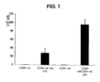
- 1 is a graph comparing the number of CD29 + cells after one week of culture in the presence or absence of CD45 + cells.
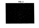
- FIG 3 shows micrographs of CD29 + cells co-cultured with CD45 + cells for 1 week.
- FIG 4 shows micrographs of CD2 + cells passaged for another week after co-culture with CD45 + cells for one week.
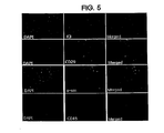
- FIG. 5 shows a picture of immunofluorescence staining of CD29 + cells with DAPI staining only nuclei of cells, CD29 + cell immunofluorescence staining of markers of mesodermal stem cells, and a combination of these pictures (X200).
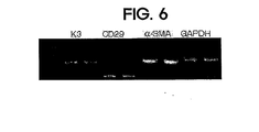
- FIG. 6 shows CD29 + cell RT-PCR photographs of markers for mesodermal stem cells.
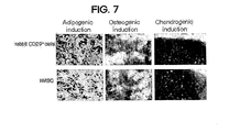
- FIG. 7 shows a photograph (X200) in which CD29 + cells co-cultured with CD45 + cells for 2 weeks were differentiated into adipocytes, osteoblasts, and chondrocytes.
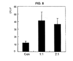
- FIG. 8 shows a graph comparing the number of colonies formed by culturing CD29 + cells under conditions treated with CD45 + cell cultures (treated only with MSCGM).
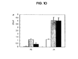
- FIG. 10 is a graph showing the number of colonies formed by passage in different culture conditions with respect to the number of cells put on a plate for the first time.
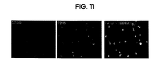
- FIG. 11 shows immunofluorescence staining photos of CD29 + cells for CD11b + and CD45 + markers, and a combination of these photos (X200).
- CD45 + cells are cells that are separated together when separating mesenchymal stem cells from tissues of origin such as bone marrow, umbilical cord blood, blood, tissues, and body fluids. They are not only necessary for obtaining mesenchymal stem cells, but also freely proliferate. It is recognized that it interferes with the skilled person is removing it.
- the culture medium obtained by culturing CD45 + cells or CD45 + cells was added to mesoderm stem cells and cultured to promote proliferation of mesoderm stem cells.
- the number of cells of mesoderm stem cells increased by an average of about 2.5 times compared with the case of culturing mesenchymal stem cells alone, and passage for one more week. In culture, the mean increase is about 3.4 times.
- coculture of CD45 + cells with mesodermal stem cells takes about 36% less than the time required for single division of mesodermal stem cells when cultured alone without CD45 + cells.
- the average number is about 42% higher.
- the mesoderm stem cells co-cultured with the CD45 + cells maintain the morphological characteristics of the mesoderm stem cells, and CD29, a-sm ( ⁇ -smooth muscle actin) and K3 (keratin 3), which are unique specific markers, It expresses CD45, a negative marker, and shows pluripotency.
- CD29 + cells can be compared with those cultured in mesodermal stem cell growth medium, which can be confirmed through the following examples.
- the culture medium of CD45 + cells or CD45 + cells may be usefully used for promoting the proliferation of mesoderm stem cells.
- the present invention relates to the use of CD45 + cells or CD45 + cell culture medium for promoting the proliferation of mesodermal stem cells, the composition for promoting the proliferation of mesodermal stem cells comprising CD45 + cells or CD45 + cell culture and CD45 + cells or CD45 + cell culture medium. It provides a method for promoting the proliferation of mesoderm stem cells comprising culturing in addition to.
- Mesodermal stem cells used in the present invention can be used regardless of where they are derived from.
- the mesoderm stem cells may be obtained from bone marrow, umbilical cord blood, blood, or tissue.
- mesoderm stem cells are bone marrow, but recently cord blood is also widely used as tissues of origin of mesoderm stem cells.
- Mesoderm stem cells can also be isolated from blood. In general, however, the amount of mesodermal stem cells is very small.
- FGF GM-CSF
- G-CSF G-CSF
- SDF-1 may also be used to promote the proliferation of mesodermal stem cells or the release of hematopoietic stem cells or endothial precursor cells in the bone marrow into the blood. And may be used to promote mesenchymal stem cell release or proliferation (Anthony D. Elias et al ,. Blood , Vol 79, No 11, 1992, pp 3036-3044; D.
- the mesoderm stem cells are mobilized or proliferated from bone marrow by one or more components selected from the group consisting of substance-P, FGF, GM-CSF, G-CSF and SDF-1.
- Mesenchymal stem cells are selected from the group consisting of substance-P, FGF, GM-CSF, G-CSF and SDF-1.
- CD29 a specific marker of mesodermal stem cells, is used to separate mesenchymal stem cells from bone marrow, umbilical cord blood, and blood. Therefore, in the present invention, CD29 + cells refers to mesoderm stem cells.
- CD45 + cells can also be obtained from bone marrow, umbilical cord blood, blood, tissues, body fluids (eg, intraperitoneal fluids) and the like.
- the CD45 + cells may be monosite or macrophage.
- the monosite or macrophages include cells derived from monosite or cells derived from macrophages.
- CD45 + cells of the present invention include cells that are CD11b +.
- the composition for promoting the proliferation of mesoderm stem cells comprising CD45 + cells or CD45 + cell culture may include a medium generally used in the culture of mesoderm stem cells.
- a medium may include, for example, Messenchymal Stem Cell Growth Medium (MSCGM), Dulbecco's Modified Eagle's Medium (DMEM), Minimum Essential Medium alpha (MEM-alpha), and the like.
- the CD45 + cell culture medium is a medium commonly used in cell culture, for example, medium such as MSCGM (Mesenchymal Stem Cell Growth Medium), DMEM (Dulbecco's Modified Eagle's Medium), MEM-alpha (Minimum Essential Medium alpha) Means a medium obtained by culturing CD45 + cells. If CD45 + cell culture medium is used for the proliferation of CD29 + cells instead of CD45 + cells, the additional process of separating CD45 + cells from CD29 + cells can be omitted.
- MSCGM Mesenchymal Stem Cell Growth Medium
- DMEM Dens Modified Eagle's Medium
- MEM-alpha Minimum Essential Medium alpha
- composition may further include a growth factor or cytokine, which may further promote the proliferation of mesoderm stem cells.
- the present invention also provides a method for culturing CD29 + cells comprising separating CD29 + cells and CD45 + cells from bone marrow, umbilical cord blood, blood, tissue or body fluid of an animal and adding a culture medium of CD45 + cells or CD45 + cells to the isolated CD29 + cells. To provide.
- the method of separating CD29 + cells and CD45 + cells from the tissue of origin is irrelevant.
- the separation of CD29 + cells and CD45 + cells may be performed by a magnetic cell sorter (MACS), but is not limited thereto.
- mononuclear cells containing CD29 + cells and CD45 + cells may be isolated from the tissue of origin and CD29 + cells and CD45 + cells may be isolated from monocytes, respectively.
- the present invention is to isolate the mononuclear cells (mononuclear cells) including CD29 + cells and CD45 + cells from bone marrow, umbilical cord blood, blood, tissue or body fluid of the animal, and culture the monocytes without isolation of each cell, and then CD29 + cells therefrom Provided is a method of culturing CD29 + cells comprising the isolation.
- CD29 + cells and CD45 + cells coexist. Therefore, instead of separating CD29 + cells from CD45 + cells, a method of culturing monocyte cell fractions containing two cells together may be used.
- the ratio of the CD29 + cells and CD45 + cells in the monocyte cell fraction may be different depending on the tissue of origin, the amount of CD45 + cells can be properly controlled to promote the proliferation of CD29 + cells without interfering with the growth of CD29 + cells. It is good to be able to play a role.
- Ficoll density gradient centrifugation is used as a method of separating monocyte cells from these tissues of origin, but is not limited thereto.
- CD29 + cells and CD45 + cells are isolated and used for co-culture, it will be easy to control the ratio of CD29 + cells and CD45 + cells at an appropriate level for proliferation of mesodermal stem cells.
- the ratio of CD29 + cell medium to CD45 + cell culture medium may be appropriately adjusted to promote the proliferation of CD29 + cells.
- This ratio may vary depending on the type of medium in which CD45 + cells are cultured, the number of CD45 + cells, or the incubation time of CD45 + cells. For example, when the number of CD45 + cells is 1 ⁇ 10 2 to 1 ⁇ 10 6 and its incubation period is 1 to 7 days, the ratio of CD29 + cell medium to CD45 + cell culture may be 1: 1 to 20: 1.
- substance-P substance-P
- FGF FGF
- GM-CSF GM-CSF
- G-CSF G-CSF
- SDF-1 SDF-1 prior to harvesting bone marrow
- umbilical cord blood blood, tissue or body fluid of an animal
- substance-P, FGF, GM-CSF, G-CSF or SDF-1 may promote release or proliferation of mesodermal stem cells from the bone marrow. Therefore, if the animal is administered with one or more components selected from the group consisting of substance-P, FGF, GM-CSF, G-CSF, and SDF-1 prior to harvesting the tissue of origin, the number of CD29 + cells in vivo should be expanded in advance. Proliferation-promoting effects due to CD45 + cells or CD45 + cell culture on mesodermal stem cells may be further maximized. Effective doses of these components capable of promoting release or proliferation of mesoderm stem cells from the bone marrow may be, but are not limited to, from 0.1 to 100 ⁇ g / kg.
- the animal to be collected from the bone marrow, cord blood, blood, tissue or body fluid may be a mammal.
- Bone marrow, umbilical cord blood, blood, tissue or bodily fluid may be of the patient's own or from others to be administered mesodermal stem cells as cell therapy when the animal is a human.
- Substance-P was injected into rabbit blood vessels at 5 nmole / kg and this was repeated once more after 24 hours.
- the mononuclear cells thus separated were washed twice with PBS.
- the isolated monocyte cell suspension was treated with 1/100 of anti-CD45 antibody (anti-CD45 antibody, Chemicon.) And then incubated for 10 minutes at 4 degrees. Centrifugation was performed to remove non-stick antibodies and the supernatant was discarded.
- Cell suspensions with anti-CD45 antibodies were treated with 20 ml of anti-mouse Ig G microbeads (Miltenyi.) And incubated for 5 minutes at 4 degrees. After centrifugation again, the supernatant was discarded and washed again with PBS.
- the cell suspension with anti-CD45 antibody-microbeads was passed through a magnetic cell sorter (MACS) column (Miltenyi.) And passed through the cells with anti-CD45 antibody-microbeads attached to the column magnets. Exit and gather. Cells with anti-CD45 antibody-microbead attached to the column were separated from the column, collected in a new tube containing PBS, centrifuged, washed with PBS, and counted.
- MCS magnetic cell sorter
- Anti-CD29 antibody (chemicon.) In 1/200 of the CD45-cell suspension and incubated for 10 minutes at 4 ° C, centrifuged to remove the non-stick antibody, anti-mouse Ig G microbeads (anti-mouse Ig G microbead, Miltenyi.) Were treated with 0.02ml and incubated for 5 minutes at 4 degrees. Passing a cell suspension with anti-CD29 antibody-microbeads through a MACS column allowed only CD29-, CD45- cells to pass through and CD29 + cells to adhere to the column. CD29 + cells attached to the column were separated from the column, collected in a new tube, and counted.
- Table 1 below shows the number of cells immediately counting CD29 + cells and CD45 + cells.
- the counted CD29 + cells and CD45 + cells were mixed 1: 1 based on the number of CD29 + cells in a 6 well plate and cultured for 1 week with MSCGM (Mesenchymal Stem Cell Growth Medium, Lonza).
- MSCGM Mesenchymal Stem Cell Growth Medium, Lonza
- the control group was cultured only CD29 + cells under the same conditions to eliminate the effects of CD45 + (Macrophage) cells. After culturing for 1 week, the number of cells was counted and subcultured in a T75 flask and incubated with MSCGM for 1 week. At week 2, cell numbers were counted to compare proliferation rates between the two groups.
- Table 2 below shows the number of CD29 + cells after one week of culture in the presence or absence of CD45 + cells.
- CD29 + cells cultured for one week in the absence of CD45 + cells increased by an average of about 18.4 times compared to the average cell count counted immediately after isolation, and CD29 + cells cultured for one week in the presence of CD45 + cells were 45.9 compared to the average cell count counted immediately after separation. Increasing fold, co-culture with CD45 + cells was found to promote, on average, about 2.5-fold the proliferation of CD29 + cells compared to the other.
- Table 3 is a table showing the number of CD29 + cells of the second week cultured for one week in the presence or absence of CD45 + cells, then passage for another week, Figure 1 averaged the number of CD29 + cells in Table 3 by group It is a graph displayed.
- the number of CD29 + cells cultured for two weeks in the absence of CD45 + cells increased 92.6 times compared to the average cell count counted immediately after isolation, and CD29 + cultured for two weeks in the presence of CD45 + cells.
- the cells increased an average of about 314.8 times compared to the average number of cells counted immediately after separation, which was found to promote 3.4-fold proliferation of CD29 + cells when co-cultured with CD45 + cells.
- CD45 + cells have a shorter survival time in vitro and fewer CD45 + cells are included in the medium at the time of passage because the CD45 + cells are less likely to fall off by trypsin during cell transfer for passage. Therefore, in the case of subculture for one week after one week of incubation, the average number of CD29 + cells was 3.4 times higher in the group co-cultured with the CD45 + cells than in the group without the CD45 + influence. Therefore, in the case of co-culture of CD45 + cells in the initial culture, the proliferation rate of CD29 + cells was much better than that of the initial culture without CD45 + even in the subsequent passage.
- Table 4 is a table showing the time required for mesoderm stem cells to divide once and the number of times of division for one week based on the data in Table 2.
- CD29 + cells co-cultured with CD45 + cells for 1 week or 2 weeks, respectively, as the mesodermal stem cells the shape of CD29 + cells was first confirmed through a stereoscopic microscope (Leica) (magnification: x 50). ).
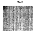
- FIG. 2 is a micrograph immediately after separation of CD29 + cells
- FIG. 3 is a picture of CD29 + cells co-cultured with CD45 + cells for 1 week, with round cells showing CD45 + cells.
- 4 is a picture of CD29 + cells passaged for one week after co-culture with CD45 + cells for one week, and no circular CD45 + cells are seen.
- CD29 + cells co-cultured with CD45 + cells exhibit a fibroblast shape, a morphological feature of mesodermal stem cells, and form mesodermal stem cells, which form colonies characteristic of stem cells. It seems to retain its characteristics as.
- CD29 + cells co-cultured with CD45 + cells exhibit specific cellular markers represented by mesodermal stem cells
- CD29, a-sm ( ⁇ -smooth muscle actin) and K3 (keratin 3) negative markers of mesodermal stem cells Expression of CD45, a negative marker, was examined.
- CD29 + cells cultured for 2 weeks were fixed at room temperature for 20 minutes with 3.7% formalin and permeabilzated for 10 minutes at room temperature with 0.3% triton x-100. After washing twice with PBS, 20% goat serum (Dako) was treated for 1 hour, followed by a blocking step, followed by anti-CD29 antibody, anti-a-sm antibody, and anti-K3 antibody at room temperature for 1 hour. Treated during.
- Dako 20% goat serum
- FIG. 5 shows immunofluorescence staining of CD29 + cells with DAPI staining only nuclei of cells, CD29 + cell immunofluorescence staining for markers of mesodermal stem cells, and a combination of these photographs.
- CD29 + cells co-cultured with CD45 + cells express CD29, a-sm, and K3, and did not express CD45, indicating that they were characterized as mesodermal stem cells.
- CD29 + cells in culture for 2 weeks were digested with trizol and RNA isolated.
- CDNA was synthesized using reverse transcriptase using mRNA in RNA as a template.
- PCR Becton Dickinson, USA
- 1 ⁇ l of the primer corresponding to the desired marker (K3, CD29, a-sm) was added and amplified 30 times (Cycle).
- Amplified gene product (K3, CD29, a-sm) was placed in 1% agarose and electrophoresed at room temperature (100 volts, 40 minutes). The gel was placed on an ultraviolet light transmitter to check the band.
- FIG. 6 shows that markers of mesodermal stem cells (K3, CD29, a-sm) are expressed at the transcription level (mRNA) as well as at the protein level in CD26 + cells. As can be seen in Figures 5 and 6, specific markers of mesodermal stem cells are expressed at both the transcriptional level and the protein level, demonstrating positive marker expression.
- GAPDH is a gene that is expressed at the same level in all cells (housekeeping gene) and was used as a criterion to show that this experiment went without problems.
- FIG. 7 shows that CD29 + cells co-cultured with CD45 + cells for 2 weeks induce differentiation into bone cells, adipocytes, and chondrocytes. Therefore, it was confirmed that the pluripotency of CD29 + mesoderm stem cells co-cultured with CD45 + cells was maintained.
- Substance-P was injected into rabbit blood vessels at 5 nmole / kg and this was repeated once more after 24 hours.
- the mononuclear cells thus separated were washed twice with PBS.
- the isolated monocyte cell suspension was treated with 1/100 of anti-CD45 antibody (anti-CD45 antibody, Chemicon.) And then incubated for 10 minutes at 4 degrees. Centrifugation was performed to remove non-stick antibodies and the supernatant was discarded.
- Cell suspensions with anti-CD45 antibodies were treated with 20 ml of anti-mouse Ig G microbeads (Miltenyi.) And incubated for 5 minutes at 4 degrees. After centrifugation again, the supernatant was discarded and washed again with PBS.
- the cell suspension with anti-CD45 antibody-microbeads was passed through a magnetic cell sorter (MACS) column (Miltenyi.) And passed through the cells with anti-CD45 antibody-microbeads attached to the column magnets. Exit and gather.
- Anti-CD45 antibody-microbead attached cells attached to the column were separated from the column, collected in a new tube containing PBS, centrifuged and washed with PBS, and then counted CD45 + cells.
- the CD45 + cells were incubated in MSCGM (Mesenchymal Stem Cell Growth Medium, Cambrex) for 2 days, and then all CD45 + cell cultures except CD45 + cells were collected.
- MSCGM Mesenchymal Stem Cell Growth Medium, Cambrex
- Anti-CD29 antibody (anti-CD29 antibody, Chemicon.) was added to the CD45-cell suspension from which the CD45 + cells were removed, incubated for 10 minutes at 4 ° C, and the non-stick antibody was removed by centrifugation.
- 0.02ml of anti-mouse Ig G microbead (anti-mouse Ig G microbead, Miltenyi.) was treated and incubated for 5 minutes at 4 degrees.
- Passing a cell suspension with anti-CD29 antibody-microbeads through a MACS column allowed only CD29-, CD45- cells to pass through and CD29 + cells to adhere to the column.
- CD29 + cells attached to the column were separated from the column, collected in a new tube, and counted (CD29 + cell preparation).
- 500 CD29 + cells were placed in a 6-well plate and cultured in MSCGM (Mesenchymal Stem Cell Growth Medium), MSCGM and CD45 + cell cultures were mixed 1: 1, and MSCGM and CD45 + cell cultures were mixed 2: 1, respectively. The number of colonies formed by incubating for 5 days was compared. The 500 CD29 + cells used at this time were cells isolated directly from blood. After 5 days of incubation the plates were stained with Trypan blue for 1 hour to count colony numbers.
- MSCGM Mesenchymal Stem Cell Growth Medium
- CD45 + cell cultures were mixed 1: 1
- MSCGM and CD45 + cell cultures were mixed 2: 1, respectively.
- the number of colonies formed by incubating for 5 days was compared.
- the 500 CD29 + cells used at this time were cells isolated directly from blood. After 5 days of incubation the plates were stained with Trypan blue for 1 hour to count colony numbers.
- the first isolated CD29 + cells were placed in 6 wells, and the CD45 + cell culture solution was treated under each condition, and the number of colonies was compared.
- the cells were cultured for the same time and subcultured. However, from Passage p1, CD45 + cell culture was no longer treated. That is, only the initial p0 state was affected by the CD45 + cell culture medium, and then continued to culture with MSCGM.
- FIG. 10 is a graph showing the percentage of colonies formed by passage culture under the above three conditions in comparison with the number of cells put on the plate first.
- P0 treated only MSCGM showed about 0.06% CFU-F
- MSCGM and CD45 + cell culture 1: 1, 2: 1, 0.16, 0.43% It showed CFU-F formation ability.
- PFU showed about 1.16% CFU-F formation ability
- P1 and CD45 + cell cultures were treated 1: 1 and 2: 1 at P0, 4.13 and 4.07%, respectively. Showed CFU-F formation ability.
- CD45 + cells isolated by the method of Example 2-1 were put in a coverslip and incubated with MSCGM. Fixed at room temperature for 20 minutes with 3.7% formalin and permeabilzation for 10 minutes at room temperature with 0.3% triton x-100. After washing twice with PBS, 20% goat serum (Dako) was treated for 1 hour followed by a blocking step followed by 40 minutes of anti-CD45 antibody at room temperature. After washing three times with PBS, the secondary antibody anti-mouse Ig G-FITC was treated for 40 minutes and then washed three times with PBS again. It is treated with anti-CD11b antibody for 40 minutes at room temperature and washed with PBS. The anti-goat-Texas red was then treated for 40 minutes.
- FIG. 11 shows immunofluorescence staining pictures of CD29 + cells for CD11b + and CD45 + markers and a combination of these pictures. Blue in the merged pictures represents cell nuclei stained with DAPI. As can be seen in FIG. 11, CD45 and CD11b are expressed at the same position, demonstrating that CD45 + cells express CD11b.
Landscapes
- Health & Medical Sciences (AREA)
- Life Sciences & Earth Sciences (AREA)
- Engineering & Computer Science (AREA)
- Chemical & Material Sciences (AREA)
- Biomedical Technology (AREA)
- Organic Chemistry (AREA)
- Genetics & Genomics (AREA)
- Zoology (AREA)
- Biotechnology (AREA)
- Bioinformatics & Cheminformatics (AREA)
- Wood Science & Technology (AREA)
- General Health & Medical Sciences (AREA)
- Cell Biology (AREA)
- Biochemistry (AREA)
- Developmental Biology & Embryology (AREA)
- General Engineering & Computer Science (AREA)
- Microbiology (AREA)
- Hematology (AREA)
- Immunology (AREA)
- Rheumatology (AREA)
- Gastroenterology & Hepatology (AREA)
- Biophysics (AREA)
- Medicinal Chemistry (AREA)
- Molecular Biology (AREA)
- Proteomics, Peptides & Aminoacids (AREA)
- Toxicology (AREA)
- Medicines Containing Material From Animals Or Micro-Organisms (AREA)
- Micro-Organisms Or Cultivation Processes Thereof (AREA)
Abstract
본 발명은 중배엽 줄기세포의 증식을 촉진하기 위한 CD45+ 세포 또는 CD45+ 세포 배양액의 용도, CD45+ 세포 또는 CD45+ 세포 배양액을 포함하는 중배엽 줄기세포의 증식 촉진용 조성물 및 CD45+ 세포 또는 CD45+ 세포 배양액을 중배엽 줄기세포에 가하여 배양하는 것을 포함하는 중배엽 줄기세포의 증식을 촉진하는 방법에 관한 것이다. 중배엽 줄기세포에 CD45+ 세포 또는 CD45+ 세포 배양액을 가하여 배양하게 되면 중배엽 줄기세포만을 단독 배양한 경우에 비해 중배엽 줄기세포의 콜로니 형성능이 3배 이상 높아진다. 또한 CD45+ 세포 또는 CD45+ 세포 배양액을 처리하여 배양한 CD29+ 세포는 중배엽 줄기세포로서의 형태적 특징, 세포 마커, 다분화능 등의 고유의 특징을 그대로 유지한다. 따라서 CD45+ 세포 또는 CD45+ 세포 배양액은 중배엽 줄기세포의 증식 촉진을 위해 이용할 수 있으며, CD45+ 세포 또는 CD45+ 세포 배양액을 중배엽 줄기세포에 가하여 배양하는 방법을 이용하면 중배엽 줄기세포를 세포치료에 이용할 수 있을 만큼 충분한 수로 증식시킬 수 있게 된다.
Description
본 발명은 중배엽 줄기세포의 증식을 촉진하기 위한 CD45+ 세포 또는 CD45+ 세포 배양액의 용도, CD45+ 세포 또는 CD45+ 세포 배양액을 포함하는 중배엽 줄기세포의 증식 촉진용 조성물 및 CD45+ 세포 또는 CD45+ 세포 배양액을 중배엽 줄기세포에 가하여 배양하는 것을 포함하는 중배엽 줄기세포의 증식을 촉진하는 방법에 관한 것이다.
각종 질환으로 인해 손상된 조직과 장기를 치료하는 기술로서, 자가복제(self-renewal) 및 분화(differentiation)가 가능한 세포를 재생이 필요한 조직 및 장기에 주입하는 방법이 주목받고 있다. 이러한 세포의 대표적인 예로 중배엽 줄기세포를 들 수 있다.
중배엽 줄기세포(Mesenchymal Stem Cell, MSC)는 보다 다양한 종류의 세포로 분화할 수 있는 다능성(multipotency)을 지닌 줄기세포이다. 중배엽 줄기세포는 뼈세포(osteoblast), 연골세포(chondrocyte), 지방세포(adipocyte), 신경세포(neuronal cell), 상피세포 (epithelial cell), 근육(muscle) 등과 같은 인체를 구성하는 각종 세포와 조직으로 분화(differentiation)할 수 있으므로, 재생의학의 실용화 측면에서 가장 핵심적인 세포로 각광받고 있다.
그러나 중배엽 줄기세포를 얻을 수 있는 대표적인 기원 조직인 골수, 제대혈, 혈액, 조직 등으로부터 수득되는 세포의 양은 아주 적은 편이다. 또한 다른 세포들에 비해 골수의 중배엽 줄기세포는 분열 속도가 느린 편이어서 세포치료제로 사용할 만큼 충분한 수의 세포를 확보하기가 어렵다는 문제점이 있다. 일반적으로 사이토카인이나 성장 인자를 세포 배양시 첨가하여 세포의 증식을 촉진하기도 하나, 사이토카인이나 성장인자는 매우 고가일 뿐만 아니라, 사이토카인 또는 성장 인자의 종류나 첨가량에 따라 오히려 중배엽 줄기 세포의 증식을 방해하거나 중배엽 줄기 세포의 분화를 야기할 수 있다. 더군다나 중배엽 줄기세포의 증식 촉진을 위해 사용할 수 있는 사이토카인 또는 성장 인자는 아직까지 확실히 규명된 것이 없다.
중배엽 줄기세포를 세포치료제로서 실용화시키기 위해서는 중배엽 줄기세포를 쉽게 분리, 배양할 수 있어야 하며, 또한 줄기세포의 특성을 유지하면서 충분한 수의 세포를 빠른 시간 내에 확보할 수 있어야 한다. 본 발명은 중배엽 줄기세포를 효율적으로 체외 증식할 수 있는 배양 방법을 제공하고자 한다.
본 발명은 중배엽 줄기세포의 증식을 촉진하기 위한 CD45+ 세포 또는 CD45+ 세포 배양액의 용도, CD45+ 세포 또는 CD45+ 세포 배양액을 포함하는 중배엽 줄기세포의 증식 촉진용 조성물 및 CD45+ 세포 또는 CD45+ 세포 배양액을 중배엽 줄기세포에 가하여 배양하는 것을 포함하는 중배엽 줄기세포의 증식을 촉진하는 방법을 제공한다.
본 발명은 또한 동물의 골수, 제대혈, 혈액, 조직 또는 체액으로부터 CD29+ 세포와 CD45+ 세포를 분리하고, 상기 분리된 CD29+ 세포에 CD45+ 세포 또는 CD45+ 세포의 배양액을 가하여 배양하는 것을 포함하는 CD29+ 세포의 배양 방법; 및 동물의 골수, 제대혈, 혈액, 조직 또는 체액으로부터 CD29+ 세포 및 CD45+ 세포를 포함하는 단핵구세포(mononuclear cell)을 분리하고, 각각의 세포의 분리 없이 단핵구세포를 배양한 후 그로부터 CD29+ 세포를 분리하는 것을 포함하는 CD29+ 세포의 배양 방법을 제공한다.
CD29+ 세포, 즉 중배엽 줄기세포에 CD45+ 세포 또는 CD45+ 세포 배양액을 가하여 배양하게 되면 중배엽 줄기세포를 중배엽줄기세포성장배지만으로 배양한 경우에 비해 중배엽 줄기세포의 콜로니 형성능이 3배 이상 높아진다. 또한 CD45+ 세포 또는 CD45+ 세포 배양액을 가하여 배양한 CD29+ 세포는 중배엽 줄기세포로서의 형태적 특징, 세포 마커, 다분화능 등의 고유의 특징을 그대로 유지한다. 따라서 CD45+ 세포 또는 CD45+ 세포 배양액을 중배엽 줄기세포의 증식 촉진을 위해 이용할 수 있으며, CD45+ 세포 또는 CD45+ 세포 배양액을 중배엽 줄기세포에 가하여 배양하는 방법을 이용하면 중배엽 줄기세포를 세포치료에 이용할 수 있을 만큼 충분한 수로 증식시킬 수 있게 된다. 따라서 본 발명은 중배엽 줄기세포의 세포치료제로서의 실용화에 기여하게 될 것이다.
도 1은 CD45+ 세포의 존재 또는 부존재 하에서의 배양 1주일 후 CD29+ 세포의 수를 비교한 그래프이다.
도 2는 CD29+ 세포를 분리한 직후의 현미경 사진을 보여준다.
도 3은 1주일간 CD45+ 세포와 공배양한 CD29+ 세포의 현미경 사진을 보여준다.
도 4는 1주일간 CD45+ 세포와 공배양한 후 다시 1주일간 계대배양한 CD2+ 세포의 현미경 사진을 보여준다.
도 5는 세포의 핵만을 염색하는 DAPI로 CD29+ 세포를 면역형광염색한 사진과 중배엽줄기세포의 대한 마커들에 대한 CD29+ 세포 면역형광염색 사진, 그리고 이들 사진을 병합한 사진(X200)을 보여준다.
도 6은 중배엽줄기세포의 대한 마커들에 대한 CD29+ 세포 RT-PCR 사진을 보여준다.
도 7은 CD45+ 세포와 2주간 공배양한 CD29+ 세포가 지방세포(adipocyte), 뼈세포(osteoblast), 연골세포(chondrocyte)로 분화된 사진(X200)을 보여준다.
도 8은 CD29+ 세포를 CD45+ 세포 배양액을 처리한 조건에서 배양하여 형성된 콜로니 수를 대조군(MSCGM만을 처리) 비교한 그래프를 보여준다.
도 9는 CD29+ 세포를 MSCGM만을 처리한 조건(대조군)과 CD45+ 세포 배양액을 혼합 처리한 조건에서 배양하여 형성된 콜로니의 사진을 보여준다.
도 10은 처음 플레이트에 넣은 세포 수에 대해 각기 다른 배양 조건에서 계대 배양하여 형성된 콜로니 수를 비교하여 이를 %로 나타낸 그래프이다.
도 11은 CD11b+ 및 CD45+ 마커들에 대한 CD29+ 세포의 면역형광염색 사진과 이들 사진을 병합한 사진(X200)을 보여준다.
CD45+ 세포는 골수, 제대혈, 혈액, 조직, 체액 등의 기원 조직으로부터 중배엽 줄기세포를 분리할 때 함께 분리되는 세포로서, 중배엽 줄기세포를 얻는 데 있어 불필요할 뿐만 아니라 분리해 낸 중배엽 줄기세포의 자유로운 증식을 방해한다고 인식되어 당업자들은 이를 제거하고 있다.
그러나 놀랍게도 CD45+ 세포 또는 CD45+ 세포를 배양하여 얻은 배양액을 중배엽 줄기세포에 가하여 배양하면 중배엽 줄기세포의 증식을 촉진할 수 있음이 본 발명을 통해 밝혀졌다.
하기 실시예를 참고하면, CD45+ 세포를 중배엽 줄기세포와 1주일간 공배양하는 경우 중배엽 줄기세포의 세포수가 중배엽 줄기세포를 단독으로 배양하는 경우에 비해 평균 약 2.5배 증가하며, 추가로 1주일 간 계대배양한 경우 평균 약 3.4배 증가한다.
또한 CD45+ 세포를 중배엽 줄기세포와 공배양하게 되면 CD45+ 세포없이 중배엽 줄기세포를 단독으로 배양할 때 중배엽 줄기세포의 1회 분열시 소요되는 시간에 비해 평균 약 36% 적게 소요되며, 1주일 동안의 분열 횟수는 평균 약 42% 정도 더 많다.
상기와 같이 CD45+ 세포와 공배양된 중배엽 줄기세포는 중배엽 줄기세포 본연의 형태적 특징을 유지하고, 고유의 특이적 마커인 CD29, a-sm(α-smooth muscle actin) 및 K3(keratin 3), 음성 마커(negative marker)인 CD45를 발현하며, 다분화능을 나타낸다.
또한, CD45+ 세포 대신 CD45+ 세포를 배양하여 얻은 배양액을 CD29+ 세포에 가하여 배양하더라도 CD29+ 세포를 중배엽줄기세포성장배지에서 배양한 것과 비교하여 CD29+ 세포의 증식을 촉진함을 하기 실시예를 통해 확인할 수 있다.
따라서 CD45+ 세포 또는 CD45+ 세포의 배양액은 중배엽 줄기세포의 증식 촉진을 위한 용도로 유용하게 사용될 수 있다.
그러므로 본 발명은 중배엽 줄기세포의 증식을 촉진하기 위한 CD45+ 세포 또는 CD45+ 세포 배양액의 용도, CD45+ 세포 또는 CD45+ 세포 배양액을 포함하는 중배엽 줄기세포의 증식 촉진용 조성물 및 CD45+ 세포 또는 CD45+ 세포 배양액을 중배엽 줄기세포에 가하여 배양하는 것을 포함하는 중배엽 줄기세포의 증식을 촉진하는 방법을 제공한다.
본 발명에서 사용되는 중배엽 줄기세포는 그것이 어디로부터 유래한 것인지 관계없이 이용될 수 있다.
한 구체예에서, 상기 중배엽 줄기세포는 골수, 제대혈, 혈액, 또는 조직으로부터 얻은 것일 수 있다.
중배엽 줄기세포의 대표적인 기원 조직은 골수이나, 최근 제대혈 또한 중배엽 줄기세포의 기원 조직으로서 많이 사용되고 있다. 중배엽 줄기세포는 또한 혈액으로부터 분리해 낼 수 있다. 그러나 일반적으로 혈액으로부터 중배엽 줄기세포를 분리해 내는 경우에는 그 양이 매우 적다.
본 발명자들의 앞선 연구에 따르면(한국등록특허 10-0593397), 물질-P를 이용하여 중배엽 줄기세포의 증식을 촉진할 수 있다. 또한, FGF, GM-CSF, G-CSF, 또는 SDF-1 또한 중배엽 줄기세포의 증식을 촉진하거나, 골수 내 조혈모세포 또는 혈관 내피 전구세포(endotherial precursor cells)의 혈중으로의 유리를 촉진하는데 이용될 수 있으므로, 이들을 이용하여 중배엽 줄기세포의 유리 또는 증식을 촉진할 수도 있다(Anthony D. Elias et al,. Blood, Vol 79, No 11, 1992, pp 3036-3044; D. Metcalf, Blood, Vol 67, No 2 (February), 1986: pp 257-267; WP Peters et al.,
Blood, Vol 81, No 7 (April 1). 1993: pp 1709-1719; Yaohong Tan et al., Cardiovasc Res. 2007 March 1; 73(4): 823 832.; Askari A. T. et al.,
Lancet. 362, 697-703(2003); Ceradini DJ. et al., Nature med. 10, 858-864 (2004)). 따라서 한 구체예에서, 상기 중배엽 줄기세포는 물질-P, FGF, GM-CSF, G-CSF 및 SDF-1로 구성된 군으로부터 선택되는 하나 이상의 성분에 의해 골수로부터 유리(mobilization) 또는 증식(proliferation)된 중배엽 줄기세포일 수 있다.
골수, 제대혈, 혈액 등으로부터 중배엽 줄기세포를 분리하기 위해서는 중배엽 줄기세포의 특이적 마커인 CD29를 이용한다. 따라서 본 발명에 있어서, CD29+ 세포는 중배엽 줄기세포를 의미한다.
CD45+ 세포 또한 골수, 제대혈, 혈액, 조직, 체액(예컨대, 복강 내 체액) 등으로부터 얻을 수 있다. 한 구체예에서, 상기 CD45+ 세포는 모노사이트(monocyte) 또는 매크로파지(macrophage)일 수 있다. 본 발명에 있어서, 상기 모노사이트 또는 매크로파지는 모노사이트 유래의 세포 또는 매크로파지 유래의 세포를 포함한다. 이에 제한되는 것은 아니나, 예를 들어, THP-1(ATCC TIB-202), KG-1(ATCC CCL-246), KG-1a(ATCC CCL-246.1), TF-1(ATCC CRL-2003), TUR(ATCC CRL-2367), Kasumi-4(ATCC CRL-2726), Kasumi-6(ATCC CRL-2775), AML-193(ATCC CRL-9589), MD(ATCC CRL-9850), SC(ATCC CRL-9855), WBC264-9C(ATCC HB-8902) 등이 포함된다. 또한, 하기 실시예에서 확인할 수 있는 바와 같이, CD45+ 세포는 CD11b+인 특성도 나타내므로, 본 발명의 CD45+ 세포는 CD11b+인 세포를 포함한다. CD45+ 세포 또는 CD45+ 세포 배양액을 포함하는 중배엽 줄기세포의 증식 촉진용 조성물은 중배엽 줄기세포의 배양시 일반적으로 사용되는 배지를 포함할 수 있다. 이에 제한되는 것은 아니나, 이러한 배지로는 예컨대, MSCGM(Mesenchymal Stem Cell Growth Medium), DMEM (Dulbecco's Modified Eagle's Medium), MEM-alpha (Minimum Essential Medium alpha) 등이 포함될 수 있다.
본 발명에 있어서, 상기 CD45+ 세포 배양액은 세포 배양시 일반적으로 사용되는 배지, 예컨대, MSCGM(Mesenchymal Stem Cell Growth Medium), DMEM (Dulbecco's Modified Eagle's Medium), MEM-alpha (Minimum Essential Medium alpha) 등의 배지에서 CD45+ 세포를 배양하여 얻은 배지를 의미한다. CD45+ 세포 대신 CD45+ 세포 배양액을 CD29+ 세포의 증식에 이용할 경우 CD29+ 세포로부터 CD45+ 세포를 분리하는 추가 과정을 생략할 수 있으므로 효율적이다.
또한 상기 조성물은 중배엽 줄기세포의 증식을 더욱 더 촉진해 줄 수 있는 성장인자(growth factor) 또는 사이토카인(cytokine)을 추가로 포함할 수 있을 것이다.
본 발명은 또한 동물의 골수, 제대혈, 혈액, 조직 또는 체액으로부터 CD29+ 세포와 CD45+ 세포를 분리하고, 상기 분리된 CD29+ 세포에 CD45+ 세포 또는 CD45+ 세포의 배양액을 가하여 배양하는 것을 포함하는 CD29+ 세포의 배양 방법을 제공한다.
상기 방법에 있어서, CD29+ 세포와 CD45+ 세포를 기원 조직으로부터 분리해 내는 방법은 어떠한 방법이던지 관계없다. 예컨대, CD29+ 세포 및 CD45+ 세포의 분리는 MACS (magnetic cell sorter)에 의해 수행될 수 있으나 이에 제한되는 것은 아니다. 다르게는, 기원 조직으로부터 CD29+ 세포와 CD45+ 세포를 포함하는 단핵구세포(mononuclear cells)을 분리하고 단핵구세포로부터 CD29+ 세포와 CD45+ 세포를 각각 분리해 낼 수도 있다.
또한 본 발명은 동물의 골수, 제대혈, 혈액, 조직 또는 체액으로부터 CD29+ 세포 및 CD45+ 세포를 포함하는 단핵구세포(mononuclear cell)을 분리하고, 각각의 세포의 분리 없이 단핵구세포를 배양한 후 그로부터 CD29+ 세포를 분리하는 것을 포함하는 CD29+ 세포의 배양 방법을 제공한다.
상기 기원 조직으로부터 단핵구세포를 분리해 내는 경우 CD29+ 세포와 CD45+ 세포가 공존한다. 따라서 CD29+ 세포와 CD45+ 세포를 따로 분리해 낼 것이 아니라 두 가지 세포를 함께 포함하고 있는 단핵구 세포 분획을 그대로 배양하는 방법을 이용할 수 있을 것이다.
다만, 기원 조직에 따라 이러한 단핵구 세포 분획 중의 CD29+ 세포와 CD45+ 세포의 존재 비율은 각각 다를 수 있으므로 CD45+ 세포의 양을 적절히 조절하여 CD29+ 세포의 성장을 방해하지 않으면서 CD29+ 세포의 증식을 촉진할 수 있는 역할을 할 수 있도록 하는 것이 좋다.
일반적으로 이들 기원 조직으로부터 단핵구 세포를 분리해 내는 방법으로는 피콜 밀도 구배 원심분리(Ficoll density gradient centrifugation)가 이용되고 있으나, 이에 제한되는 것은 아니다.
CD29+ 세포와 CD45+ 세포를 각각 분리하여 공배양에 이용할 경우 중배엽 줄기세포의 증식에 적당한 수준의 CD29+ 세포와 CD45+ 세포의 비율을 조절하기 용이할 것이다. 이에 제한되는 것은 아니나 CD29+ 세포의 성장을 방해하지 않으면서 CD29+ 세포의 증식을 촉진할 수 있는 CD45+ 세포의 비율은 CD29+세포: CD45+ 세포 = 1: 0.5 ~ 1: 30일 수 있다.
한편, CD45+ 세포 배양액을 CD29+ 세포의 배양에 이용할 경우, CD29+ 세포 배지 대 CD45+ 세포 배양액의 사용 비율은 CD29+ 세포의 증식을 촉진할 수 있도록 적절히 조절될 수 있다. 이러한 비율은 CD45+ 세포를 배양한 배지의 종류, CD45+ 세포의 수, 또는 CD45+ 세포의 배양시간 등에 따라 달라질 수 있을 것이다. 예를 들어, CD45+ 세포의 수가 1x10² 내지 1x 10⁶개이고 이의 배양기간이 1일 내지 7일일 때 CD29+ 세포 배지 대 CD45+ 세포 배양액의 사용 비율은 1:1 내지 20:1일 수 있다.
본 발명의 CD29+ 세포의 배양 방법에 있어서, 동물의 골수, 제대혈, 혈액, 조직 또는 체액을 채취하기 전에 물질-P, FGF, GM-CSF, G-CSF 및 SDF-1로 구성된 군으로부터 선택되는 하나 이상의 성분을 투여할 수 있다.
앞서 언급한 바와 같이 선행 연구들을 통해 물질-P, FGF, GM-CSF, G-CSF 또는 SDF-1는 중배엽 줄기세포의 골수로부터의 유리 또는 증식을 촉진할 수 있음이 밝혀졌다. 따라서 기원 조직의 채취 전에 물질-P, FGF, GM-CSF, G-CSF 및 SDF-1로 구성된 군으로부터 선택되는 하나 이상의 성분을 동물에 투여하여 생체 내에서 CD29+ 세포의 수를 미리 확장시켜 놓는다면 중배엽 줄기세포에 대한 CD45+ 세포 또는 CD45+ 세포 배양액으로 인한 증식 촉진 효과를 더욱 극대화시킬 수 있을 것이다. 중배엽 줄기세포의 골수로부터의 유리 또는 증식을 촉진할 수 있는 이들 성분의 유효 용량은, 이에 제한되는 것은 아니나, 0.1 내지 100 ㎍/㎏일 수 있다.
상기 방법에 있어서, 골수, 제대혈, 혈액, 조직 또는 체액의 채취 대상인 동물은 포유동물일 수 있다. 상기 동물이 인간일 경우 골수, 제대혈, 혈액, 조직 또는 체액은 중배엽 줄기세포를 세포치료제로서 투여하게 될 환자 자신의 것이나 타인 유래의 것일 수 있다.
본 발명의 이점 및 특징, 그리고 그것들을 달성하는 방법은 상세하게 후술되어 있는 실시예들을 참조하면 명확해질 것이다. 그러나 본 발명은 이하에서 개시되는 실시예들에 한정되는 것이 아니라 서로 다른 다양한 형태로 구현될 것이며, 단지 본 실시예들은 본 발명의 개시가 완전하도록 하고, 본 발명이 속하는 기술 분야에서 통상의 지식을 가진 자에게 발명의 범주를 완전하게 알려주기 위해 제공되는 것이며, 본 발명은 청구항의 범주에 의해 정의될 뿐이다.
실시예 1: CD29+ 세포와 CD45+ 세포의 공배양에 의한 CD29+ 세포의 증식 촉진 확인
1-1. CD29+ 세포와 CD45+ 세포의 분리
물질-P(Substance-P)를 토끼의 혈관에 5nmole/kg로 주사하고 이를 24시간 후 한번 더 반복하였다.
초기 주사 후 3일째, 토끼(n=4)의 귀 혈관에서 혈액을 10ml 채취하고 피콜 밀도 구배 원심분리(Ficoll density gradient centrifugation)에 의해 혈액 내의 단핵구 세포(mononuclear cell)만을 분리하였다.
이렇게 분리된 단핵구 세포를 PBS로 2번 세척하였다. 분리된 단핵구 세포 현탁액에 항-CD45 항체(anti-CD45 antibody, Chemicon.)를 1/100로 처리한 후 4도에서 10분간 배양(incubation)하였다. 붙지 않은 항체를 제거하기 위해 원심 분리를 하고 상등액은 버렸다. 항-CD45 항체가 붙어있는 세포 현탁액에 항-마우스 Ig G 마이크로비드(anti-mouse Ig G microbead, Miltenyi.)를 20ml 처리하고 4도에서 5분간 배양하였다. 다시 원심분리를 한 후 상등액은 버리고 PBS로 다시 한번 세척하였다. 항-CD45 항체-마이크로비드가 붙어있는 세포 현탁액을 MACS(magnetic cell sorter) 컬럼 (Miltenyi.)에 넣고 통과시키면 항-CD45 항체-마이크로비드가 붙어있는 세포는 컬럼 자석에 붙고 CD45- 세포만 컬럼 밖으로 빠져나와 모이게 된다. 컬럼에 붙어있는 항-CD45 항체-마이크로비드가 붙어 있는 세포를 컬럼에서 분리한 후 PBS를 포함하는 새 튜브에 모으고 원심 분리하여 PBS로 세척하는 단계를 거친 후, 세포를 계수하였다.
상기 CD45-세포 현탁액에 항-CD29 항체(anti-CD29 antibody, Chemicon.)를 1/200으로 넣고 4도에서 10분간 배양한 후, 원심분리를 통해 붙지 않은 항체를 제거하고, 여기에 항-마우스 Ig G 마이크로비드(anti-mouse Ig G microbead, Miltenyi.)를 0.02ml 처리하여 4도에서 5분간 배양하였다. 항-CD29 항체-마이크로비드가 붙어 있는 세포 현탁액을 MACS 컬럼에 넣고 통과시키면 CD29-, CD45-세포들만 통과하고 CD29+ 세포들은 컬럼에 붙어있게 된다. 컬럼에 붙어있는 CD29+ 세포들을 컬럼에서 분리하여 새 튜브에 모으고 세포수를 계수하였다.
하기 표 1은 CD29+ 세포와 CD45+ 세포를 분리 즉시 계수한 세포수를 나타낸다.
표 1 
1-2. CD45+ 세포와의 공배양에 따른 CD29+ 세포의 증식 촉진 확인
상기 계수된 CD29+ 세포와 CD45+ 세포를 CD29+ 세포의 수를 기준으로 1:1로 섞어서 6 웰 플레이트(6 well plate)에 넣고 MSCGM(Mesenchymal Stem Cell Growth Medium, Lonza)으로 일주일간 배양하였다. 대조군은 동일한 조건으로 CD29+ 세포만을 배양하여 CD45+(Macrophage)세포에 의한 영향을 없앴다. 1주일간 배양 후 세포수를 계수한 뒤 T75 플라스크에 계대 배양(subculture)하여 1주일동안 MSCGM으로 배양하였다. 2주째에 세포 수를 계수하여 두 그룹간의 증식률을 비교하였다.
하기 표 2는 CD45+ 세포의 존재 또는 부존재 하에서의 배양 1주일 후 CD29+ 세포의 수를 나타낸다.
표 2 
CD45+ 세포의 부존재 하에 1주일간 배양된 CD29+ 세포는 분리 직후 계수된 평균 세포 수에 비해 평균 약 18.4배 증가하였으며, CD45+ 세포의 존재 하에 1주일간 배양된 CD29+ 세포는 분리 직후 계수된 평균 세포 수에 비해 45.9배 증가하여, CD45+ 세포와 함께 공배양한 경우가 그렇지 않은 경우에 비해 CD29+ 세포의 증식을 평균 약 2.5배 촉진하는 것으로 나타났다.
하기 표 3은 CD45+ 세포의 존재 또는 부존재 하에서의 1주일 동안 배양한 후, 다시 1주일간 계대 배양한 2주째의 CD29+ 세포수를 나타낸 표이며, 도 1은 표 3의 CD29+ 세포수를 그룹별로 평균을 내어 표시한 그래프이다.
표 3 
표 3 및 도 1에서 볼 수 있는 바와 같이, CD45+ 세포의 부존재 하에 2주일간 배양된 CD29+ 세포의 수는 분리 직후 계수된 평균 세포수에 비해 92.6배 증가하였으며, CD45+ 세포의 존재 하에 2주일간 배양된 CD29+ 세포는 분리 직후 계수된 평균 세포수에 비해 평균 약 314.8 배 증가하여, CD45+ 세포와 함께 공배양한 경우가 그렇지 않은 경우에 비해 CD29+ 세포의 증식을 3.4배 촉진하는 것으로 나타났다.
CD45+ 세포는 체외에서는 생존기간이 짧고, 또한 계대배양을 위해 세포를 옮기는 과정에서 트립신에 의해 CD45+ 세포가 잘 떨어지지 않기 때문에 계대배양시 배지에 포함되는 CD45+ 세포의 수가 적어진다. 따라서 1주일간의 배양 후 다시 1주일간 계대배양을 실시한 경우에는 CD45+의 영향력이 줄어들었음에도 불구하고 여전히 CD45+ 세포를 공배양한 군이 그렇지 않은 군에 비해 CD29+ 세포의 수가 평균 3.4배 많았다. 따라서 초기 배양시 CD45+ 세포를 공배양하는 경우 계속된 계대배양시에도 CD29+ 세포의 증식률이 CD45+ 없이 초기 배양한 경우에 비해 훨씬 우수함을 알 수 있었다.
하기 표 4는 표 2의 데이터를 기초로 하여 중배엽 줄기세포가 1회 분열시 소요되는 시간 및 1주일 동안 분열하는 횟수를 나타낸 표이다.
표 4 
표 4에서 볼 수 있는 바와 같이, CD45+ 세포 없이 CD29+ 세포만을 7일간 배양할 경우 CD29+ 세포가 1회 분열하는데 소요되는 시간은 평균 1.875일인 반면 CD45+ 세포와 함께 공배양 하면 평균 1.2일이 소요되었으며, 1주일간의 분열 횟수를 보더라도 CD45+ 무첨가군은 3.73번 분열하는데 반해, CD45+ 공배양군은 5.08번으로 확연한 차이를 보였다. 즉, CD45+ 세포를 CD29+ 세포와 공배양 하게 되면 CD45+ 세포 없이 배양할 때 소요되는 시간에 비해 약 36% 적게 소요되며, 분열 횟수는 약 42% 정도 더 많은 것을 확인할 수 있었다.
1-3. CD45+ 세포와 공배양된 CD29+ 세포의 중배엽 줄기세포로서의 특성 확인
1-3-(1). 형태적 특성 확인
상기 과정을 통해 각각 1주일 또는 2주일간 CD45+ 세포와 공배양된 CD29+ 세포가 중배엽 줄기세포로서의 특성을 유지하고 있는지 확인하고자 먼저 입체현미경(Leica)을 통해 CD29+ 세포의 모양을 확인하였다(배율: x 50).
도 2은 CD29+ 세포를 분리한 직후의 현미경 사진이며, 도 3은 1주일간 CD45+ 세포와 공배양한 CD29+ 세포 사진으로서 동그란 세포는 CD45+ 세포를 나타낸다. 도 4는 1주일간 CD45+ 세포와 공배양한 후 다시 1주일간 계대배양한 CD29+ 세포의 사진으로 동그란 모양의 CD45+ 세포가 보이지 않는다. 도 3 및 도 4에서 볼 수 있는 바와 같이, CD45+ 세포와 공배양된 CD29+ 세포는 중배엽 줄기세포의 형태적 특징인 파이브로블라스트 모양을 나타내고 있으며 줄기세포의 특징인 콜로니를 형성하는 것으로 보아 중배엽 줄기세포로서의 특성을 유지하고 있는 것으로 보인다.
1-3-(2). 세포 발현 마커 확인
CD45+ 세포와 공배양된 CD29+ 세포가 중배엽 줄기세포가 나타내는 특이적 세포 마커를 나타내는지 확인하고자 중배엽 줄기세포의 특정 표식자인 CD29, a-sm(α-smooth muscle actin) 및 K3(keratin 3), 음성 마커(negative marker)인 CD45에 대한 발현 여부를 조사하였다. 2주 동안 배양된 CD29+ 세포를 3.7% 포르말린으로 20분간 실온에서 고정하고 0.3% triton x-100으로 실온에서 10분간 permeabilzation시켰다. PBS로 2번 세척 후, 20% 염소 혈청(goat serum)(Dako)을 1시간동안 처리하여 블로킹 단계를 거친 후 항-CD29 항체, 항-a-sm 항체, 항-K3 항체를 실온에서 1시간 동안 처리하였다. PBS로 3번 세척한 후, 2차 항체인 항-마우스 Ig G-FITC를 1시간 동안 처리한 후 DAPI가 들어있는 용액 (VECTORSHIELD)로 마운팅(mounting)하였다. 그 후 형광 입체현미경(Leica)을 이용하여 형광 발현 여부를 관찰하였다. 도 5는 세포의 핵만을 염색하는 DAPI로 CD29+ 세포를 면역형광염색한 사진과 중배엽줄기세포의 대한 마커들에 대한 CD29+ 세포 면역형광염색 사진, 그리고 이들 사진을 병합한 사진을 보여준다. 도 5에서 볼 수 있는 바와 같이 CD45+ 세포와 공배양된 CD29+ 세포는 CD29, a-sm 및 K3를 발현하며, CD45는 발현하지 않아, 중배엽 줄기세포로서의 특성을 나타냄을 확인하였다.
세포의 마커 확인은 RT-PCR (Reverse transcription-polymerase chain reaction; 역전사-중합효소 연쇄반응)을 이용하여 전사 (Transcription)수준에서 다시 한번 입증하였다.
2주 동안 배양안 CD29+ 세포들을 트라이졸을 이용하여 분해하고 RNA만을 분리하였다. RNA 중 mRNA를 주형으로 하여 역전사효소 (Reverse transcriptase)를 이용, cDNA를 합성하였다. 이렇게 얻어진 cDNA를 주형으로 하여 (PCR, Becton Dickinson, USA) 원하는 마커(K3, CD29, a-sm)에 해당하는 프라이머를 1㎕넣고 30 번(Cycle) 증폭시켰다. 증폭된 유전자 생성물 (K3, CD29, a-sm)을 1% 아가로즈에 넣고 실온에서 전기 영동하였다(100볼트, 40 분). 젤을 자외선 투과기에 올려두고 밴드를 확인하였다.
도 6은 중배엽 줄기세포의 마커 (K3, CD29, a-sm)가 CD26+ 세포에서 단백질 수준에서뿐만 아니라 전사 수준 (mRNA)에서도 발현 된다는 것을 보여준다. 도 5 및 6에서 볼 수 있는 것과 같이 중배엽 줄기세포의 특정 마커가 전사수준과 단백질 수준에서 모두 발현되는 바, 확실한 마커 발현이 증명되었다. GAPDH는 모든 세포에서 동일한 수준으로 발현되는 유전자(하우스키핑 ;housekeeping gene)로 이 실험이 문제없이 진행되었는지를 보여주는 기준으로 사용되었다.
1-3-(3). CD45+ 세포와 공배양된 CD29+ 세포의 다분화능 확인
2주동안 배양된 CD29+ 세포들이 중배엽 줄기세포의 특징인 다분화능을 가지고 있는지 확인하기 위해 지방세포(adipocyte), 뼈세포(osteoblast), 연골세포(chondrocyte)로의 분화실험을 실시하였다. 지방세포로의 분화를 유도하기 위해 각 플레이트 웰에 2x10⁵의 CD29+ 세포를 넣고 0.5mM 1-메틸-3-이소부틸크산틴(Sigma), 1μM 덱사메타손(Sigma), 10μg/ml 인슐린(Sigma), 및 10% FBS가 보충된 DMEM-high glucose (Gibco)에서 21일 동안 배양 한 뒤, oil red "O" 염색을 실시하였다. 뼈로의 분화를 유도하기 위해 4x10⁴ 의 세포를 6 웰에 넣고 1μM 덱사메타손(Sigma), 0.05mM 아스코르브산(Sigma), 10mM β-글리세로포스페이트(Sigma) 및 10% FBS DMEM-high glucose (Gibco)에서 21일 동안 배양한 후 Alizarin red로 염색하였다. 연골로의 분화를 위해 5 x10⁵
세포를 chondrogenic differentiation medium (Cambrex)을 넣은 15ml polystyrene tube에서 4주 동안 배양하였고 stained hematoxylin과 Safranin-O로 염색하였다. 양성 대조군으로 사람의 중배엽 줄기세포를 이용하여 동일한 실험을 진행하였다. 뼈세포, 연골세포, 지방세포로 유도된 세포들은 염색 후 입체현미경을 통해 관찰되었다(배율: x50).
도 7은 CD45+ 세포와 2주간 공배양한 CD29+ 세포를 뼈세포, 지방세포, 연골 세포로 분화를 유도한 사진으로 세가지 세포로 모두 분화한 것을 볼 수 있다. 따라서 CD45+ 세포와 공배양한 CD29+ 중배엽 줄기세포에서도 다분화능이 유지됨을 확인되었다.
실시예 2: CD45+ 세포 배양액의 첨가에 의한 CD29+ 세포의 증식 촉진 확인
2-1. CD45+ 세포 배양액의 제조 및 CD29+ 세포의 분리
물질-P(Substance-P)를 토끼의 혈관에 5nmole/kg로 주사하고 이를 24시간 후 한번 더 반복하였다.
초기 주사 후 3일째, 토끼의 귀 혈관에서 혈액을 10ml 채취하고 피콜 밀도 구배 원심분리(Ficoll density gradient centrifugation)에 의해 혈액 내의 단핵구 세포(mononuclear cell)만을 분리하였다.
이렇게 분리된 단핵구 세포를 PBS로 2번 세척하였다. 분리된 단핵구 세포 현탁액에 항-CD45 항체(anti-CD45 antibody, Chemicon.)를 1/100로 처리한 후 4도에서 10분간 배양(incubation)하였다. 붙지 않은 항체를 제거하기 위해 원심 분리를 하고 상등액은 버렸다. 항-CD45 항체가 붙어있는 세포 현탁액에 항-마우스 Ig G 마이크로비드(anti-mouse Ig G microbead, Miltenyi.)를 20ml 처리하고 4도에서 5분간 배양하였다. 다시 원심분리를 한 후 상등액은 버리고 PBS로 다시 한번 세척하였다. 항-CD45 항체-마이크로비드가 붙어있는 세포 현탁액을 MACS(magnetic cell sorter) 컬럼 (Miltenyi.)에 넣고 통과시키면 항-CD45 항체-마이크로비드가 붙어있는 세포는 컬럼 자석에 붙고 CD45- 세포만 컬럼 밖으로 빠져나와 모이게 된다. 컬럼에 붙어있는 항-CD45 항체-마이크로비드가 붙어 있는 세포를 컬럼에서 분리한 후 PBS를 포함하는 새 튜브에 모으고 원심 분리하여 PBS로 세척하는 단계를 거친 후, CD45+ 세포를 계수하였다. 상기 CD45+ 세포들을 각각 MSCGM (Mesenchymal Stem Cell Growth Medium, Cambrex)에서 2일 동안 배양한 후 CD45+ 세포를 제외한 CD45+ 세포 배양액을 모두 모았다.
CD45+ 세포가 제거된 상기 CD45-세포 현탁액에 항-CD29 항체(anti-CD29 antibody, Chemicon.)를 1/200으로 넣고 4도에서 10분간 배양한 후, 원심분리를 통해 붙지 않은 항체를 제거하고, 여기에 항-마우스 Ig G 마이크로비드(anti-mouse Ig G microbead, Miltenyi.)를 0.02ml 처리하여 4도에서 5분간 배양하였다. 항-CD29 항체-마이크로비드가 붙어 있는 세포 현탁액을 MACS 컬럼에 넣고 통과시키면 CD29-, CD45-세포들만 통과하고 CD29+ 세포들은 컬럼에 붙어있게 된다. 컬럼에 붙어있는 CD29+ 세포들을 컬럼에서 분리하여 새 튜브에 모으고 세포수를 계수하였다 (CD29+ 세포 준비).
2-2. CD45+ 세포 배양액의 처리에 의한 CD29+ 세포의 변화 확인
500개의 CD29+ 세포를 6 웰 플레이트에 넣고 MSCGM(Mesenchymal Stem Cell Growth Medium)으로 배양하는 조건, MSCGM과 CD45+ 세포 배양액을 1:1로 섞은 조건, MSCGM과 CD45+ 세포 배양액을 2:1로 섞은 조건에서 각각 5일 동안 배양하여 형성된 콜로니 수를 비교하였다. 이때 사용하는 500개의 CD29+ 세포는 혈액에서 바로 분리한 세포들이다. 5일 배양 후 플레이트를 트리판 블루 (Trypan blue)로 1시간 동안 염색하여 콜로니 수를 계수했다.
그 결과, 도 8 및 도 9에서 볼 수 있는 바와 같이, MSCGM만으로 배양한 조건에 비해 MSCGM : CD45+ 세포 배양액을 1:1, 2:1로 처리한 경우 각각 3.56배, 3.5배 높은 콜로니 형성능을 보였다.
위와 같은 방법으로, 처음 분리한 CD29+ 세포를 6 웰에 넣고 CD45+ 세포 배양액을 각각의 조건으로 처리하여 콜로니 수를 비교하였고 이 세포를 동일한 시간 동안 배양 후, 계대 배양 (subculture)하였다. 다만, Passage p1에서부터는 CD45+ 세포 배양액은 더 이상 처리하지 않았다. 즉, 초기 p0상태에서만 CD45+ 세포 배양액의 영향을 받게 하였고 그 이후에는 MSCGM으로 계속 배양하였다.
도 10은 처음 플레이트에 넣은 세포 수와 대비하여 위 세가지 조건에서 계대 배양하여 형성된 콜로니 수를 비교하여 이를 %로 나타낸 그래프이다. 도 10에서 볼 수 있는 바와 같이, P0 의 경우 MSCGM만을 처리한 경우 약 0.06%의 CFU-F를 보였으나 MSCGM과 CD45+ 세포 배양액을 1:1, 2:1로 처리한 경우, 0.16, 0.43%의 CFU-F 형성능을 보였다. 또한 P0에서 MSCGM만 처리했던 세포를 P1으로 계대 했을 경우, 약 1.16%의 CFU-F 형성능을 보였지만, P0에서 CD45+ 세포 배양액을 1:1, 2:1로 처리한 경우 P1에서 각각 4.13, 4.07%의 CFU-F 형성능을 보였다.
2-3. CD45+ 세포의 특성 확인
상기 실시예 2-1의 방법으로 분리해 낸 CD45+ 세포들을 커버슬립에 넣고 MSCGM으로 배양하였다. 3.7% 포르말린으로 20분간 실온에서 고정하고 0.3% triton x-100으로 실온에서 10분간 permeabilzation시켰다. PBS로 2번 세척 후, 20% 염소 혈청 (goat serum) (Dako)을 1시간동안 처리하여 블로킹 단계를 거친 후 항-CD45 항체 실온에서 40분 동안 처리하였다. PBS로 3번 세척한 후, 2차 항체인 항-마우스 Ig G-FITC를 40분 동안 처리한 후 다시 PBS로 3번 세척하였다. 여기에 항-CD11b 항체를 실온에서 40분 동안 처리하고 PBS로 세척한다. 그 이후 항-고우트 (goat)-Texas red를 40분 동안 처리하였다. 마지막으로 PBS로 3번 세척한 뒤, DAPI가 들어있는 용액 (VECTORSHIELD)로 마운팅(mounting)하였다. 그 후 형광 입체현미경(Leica)을 이용하여 형광 발현 여부를 관찰하였다. 도 11은 CD11b+ 및 CD45+ 마커들에 대한 CD29+ 세포의 면역형광염색 사진과 이들 사진을 병합한 사진을 보여준다. 병합된 사진에서 파란색은 DAPI로 염색된 세포 핵을 나타낸다. 도 11에서 볼 수 있는 바와 같이, CD45와 CD11b가 동일한 위치에서 발현되어 CD45+ 세포가 CD11b를 발현함을 증명한다.
Claims (16)
- CD45+ 세포 또는 CD45+ 세포 배양액을 포함하는 중배엽 줄기세포의 증식 촉진용 조성물.
- 제1항에 있어서,상기 중배엽 줄기세포는 골수, 제대혈, 혈액, 조직 또는 체액으로부터 얻은 것인 조성물.
- 제1항 또는 제2항에 있어서,상기 중배엽 줄기세포는 물질-P, FGF, GM-CSF, G-CSF 및 SDF-1로 구성된 군으로부터 선택되는 하나 이상의 성분에 의해 골수로부터 유리(mobilization) 또는 증식(proliferation)된 중배엽 줄기세포인 조성물.
- 제1항 또는 제2항에 있어서,상기 CD45+ 세포는 모노사이트(monocyte) 또는 매크로파지(macrophage)인 조성물.
- 제1항 또는 제2항에 있어서,상기 조성물은 성장인자(growth factor) 또는 사이토카인(cytokine)을 추가로 포함하는 것인 조성물.
- CD45+ 세포 또는 CD45+ 세포 배양액을 중배엽 줄기세포에 가하여 배양하는 것을 포함하는 중배엽 줄기세포의 증식을 촉진하는 방법.
- 제6항에 있어서,상기 중배엽 줄기세포는 골수, 제대혈, 혈액, 조직 또는 체액으로부터 얻은 것인 방법.
- 제6항 또는 제7항에 있어서,상기 중배엽 줄기세포는 물질-P, FGF, GM-CSF, G-CSF 및 SDF-1로 구성된 군으로부터 선택되는 하나 이상의 성분에 의해 골수로부터 유리(mobilization) 또는 증식(proliferation)된 중배엽 줄기세포인 방법.
- 제6항 또는 제7항에 있어서,상기 CD45+ 세포는 모노사이트(monocyte) 또는 매크로파지(macrophage)인 방법.
- 제6항 또는 제7항에 있어서,상기 조성물은 성장인자 또는 사이토카인을 추가로 포함하는 것인 방법.
- 중배엽 줄기세포의 증식 촉진을 위한 CD45+ 세포 또는 CD45+ 세포 배양액의 용도.
- 제11항에 있어서,상기 중배엽 줄기세포는 골수, 제대혈, 혈액, 조직 또는 체액으로부터 얻은 것인 용도.
- 제11항 또는 제12항에 있어서,상기 중배엽 줄기세포는 물질-P, FGF, GM-CSF, G-CSF 및 SDF-1로 구성된 군으로부터 선택되는 하나 이상의 성분에 의해 골수로부터 유리(mobilization) 또는 증식(proliferation)된 중배엽 줄기세포인 용도.
- 제11항 또는 제12항에 있어서,상기 CD45+ 세포는 모노사이트(monocyte) 또는 매크로파지(macrophage)인 용도.
- 동물의 골수, 제대혈, 혈액, 조직 또는 체액으로부터 CD29+ 세포와 CD45+ 세포를 분리하고,상기 분리된 CD29+ 세포에 CD45+ 세포 또는 CD45+ 세포의 배양액을 가하여 배양하는 것을 포함하는CD29+ 세포의 배양 방법.
- 동물의 골수, 제대혈, 혈액, 조직 또는 체액으로부터 CD29+ 세포 및 CD45+ 세포를 포함하는 단핵구세포(mononuclear cell)을 분리하고,각각의 세포의 분리 없이 단핵구세포를 배양한 후 그로부터 CD29+ 세포를 분리하는 것을 포함하는CD29+ 세포의 배양 방법.
Applications Claiming Priority (2)
| Application Number | Priority Date | Filing Date | Title |
|---|---|---|---|
| KR10-2008-0029244 | 2008-03-28 | ||
| KR20080029244 | 2008-03-28 |
Publications (2)
| Publication Number | Publication Date |
|---|---|
| WO2009120043A2 true WO2009120043A2 (ko) | 2009-10-01 |
| WO2009120043A3 WO2009120043A3 (ko) | 2009-12-30 |
Family
ID=41114475
Family Applications (1)
| Application Number | Title | Priority Date | Filing Date |
|---|---|---|---|
| PCT/KR2009/001581 Ceased WO2009120043A2 (ko) | 2008-03-28 | 2009-03-27 | 중배엽 줄기세포의 증식을 촉진하기 위한 cd45+ 세포 또는 cd45+ 세포 배양액의 용도 |
Country Status (2)
| Country | Link |
|---|---|
| KR (1) | KR101077042B1 (ko) |
| WO (1) | WO2009120043A2 (ko) |
Families Citing this family (1)
| Publication number | Priority date | Publication date | Assignee | Title |
|---|---|---|---|---|
| KR101373100B1 (ko) | 2013-05-03 | 2014-03-13 | 재단법인 통합의료진흥원 | 당귀 추출물을 포함하는 골수유래 줄기세포 증식 촉진용 조성물 |
Family Cites Families (6)
| Publication number | Priority date | Publication date | Assignee | Title |
|---|---|---|---|---|
| KR100560340B1 (ko) * | 2003-11-11 | 2006-03-14 | 한훈 | 제대혈로부터 중간엽 줄기세포의 분리 및 배양 방법 |
| EP1718733A4 (en) * | 2004-02-06 | 2007-02-07 | Theradigm Inc | COMPOSITIONS AND METHODS FOR NEURAL STEM CELL CULTURE WITH STROMAL BONE MARROW CELLS |
| WO2005113749A2 (en) * | 2004-05-14 | 2005-12-01 | Becton, Dickinson And Company | Stem cell populations and methods of use |
| KR20060058476A (ko) * | 2004-11-25 | 2006-05-30 | 고려대학교 산학협력단 | 인간 골수기질세포를 이용한 인간 배아체로부터조혈모세포로의 분화 촉진 방법 |
| US20080199849A1 (en) * | 2005-09-02 | 2008-08-21 | Agency For Science, Technology And Research | Method of Deriving Progenitor Cell Line |
| KR20060016133A (ko) * | 2006-02-02 | 2006-02-21 | 김병수 | 인간 배아줄기세포 및 배아체로부터 조혈모세포로의 분화를위한 인간 태반기질세포 생산 및 인간 배아줄기세포 및배아체로부터 조혈모세포로의 분화배양 기술 |
-
2009
- 2009-03-27 WO PCT/KR2009/001581 patent/WO2009120043A2/ko not_active Ceased
- 2009-03-27 KR KR1020090026488A patent/KR101077042B1/ko active Active
Also Published As
| Publication number | Publication date |
|---|---|
| WO2009120043A3 (ko) | 2009-12-30 |
| KR101077042B1 (ko) | 2011-10-26 |
| KR20090103828A (ko) | 2009-10-01 |
Similar Documents
| Publication | Publication Date | Title |
|---|---|---|
| Wang et al. | The immunomodulatory activity of human umbilical cord blood‐derived mesenchymal stem cells in vitro | |
| WO2018182356A1 (ko) | 중간엽줄기세포 유래 고순도, 고농도 엑소좀을 포함하는 배양액 및 이의 제조방법 | |
| Song et al. | Potential differentiation of human mesenchymal stem cell transplanted in rat corpus cavernosum toward endothelial or smooth muscle cells | |
| Heidari et al. | Comparison of proliferative and multilineage differentiation potential of sheep mesenchymal stem cells derived from bone marrow, liver, and adipose tissue | |
| US20100105132A1 (en) | Human Mesenchymal stem cells and preparation thereof | |
| CN1878860B (zh) | 分离和培养来自脐带血的间充质干细胞的方法 | |
| WO2012008813A2 (ko) | 양막유래 중간엽 줄기세포 배양을 위한 배지조성물 및 이를 이용한 양막유래 중간엽 줄기세포의 배양방법 | |
| Wei et al. | Human skeletal muscle-derived stem cells retain stem cell properties after expansion in myosphere culture | |
| WO2013081436A1 (ko) | 줄기세포를 젊게 만들기 위한 배지 조성물 | |
| CN102559586A (zh) | 一种人羊膜间充质干细胞的分离纯化及鉴定方法 | |
| Song et al. | Variations of clonal marrow stem cell lines established from human bone marrow in surface epitopes, differentiation potential, gene expression, and cytokine secretion | |
| CN111826348A (zh) | 一种人诱导性多能干细胞来源的间充质干细胞体外高效制备方法和应用 | |
| Rajput et al. | Expansion of human umbilical cord derived mesenchymal stem cells in regenerative medicine | |
| WO2014114023A1 (zh) | 一种能够大规模并快速高纯度地诱导间充质干细胞转决定成造血干细胞的方法 | |
| WO2016068596A1 (ko) | 줄기세포 배양을 위한 배지조성물 | |
| Chen et al. | The biological characteristics of sheep umbilical cord mesenchymal stem cells | |
| EP3384011B1 (en) | Methods for differentiating cells into hepatic stellate cells | |
| Yoon et al. | Bone marrow-derived side population cells are capable of functional cardiomyogenic differentiation | |
| CN102140440A (zh) | 用骨髓组织块分离及培养禽类骨髓间充质干细胞的方法 | |
| KR20160108190A (ko) | 증식력 및 분화능이 개선된 간엽줄기세포를 포함하는 폐질환 예방 또는 치료용 약학적 조성물 | |
| CN111235100A (zh) | 一种人脐带血间充质干细胞的培养方法 | |
| WO2019198962A1 (ko) | 소변유래줄기세포로부터 제작된 유도만능줄기세포의 증식 및 분화 촉진 방법 | |
| WO2020004903A1 (ko) | 엑소좀 기반의 심근세포 교차분화 유도방법 | |
| Jeon et al. | Characterization of mouse clonal mesenchymal stem cell lines established by subfractionation culturing method | |
| KR101077042B1 (ko) | 중배엽 줄기세포의 증식을 촉진하기 위한 cd45+ 세포 또는 cd45+ 세포 배양액의 용도 |
Legal Events
| Date | Code | Title | Description |
|---|---|---|---|
| 121 | Ep: the epo has been informed by wipo that ep was designated in this application |
Ref document number: 09723740 Country of ref document: EP Kind code of ref document: A2 |
|
| NENP | Non-entry into the national phase |
Ref country code: DE |
|
| 122 | Ep: pct application non-entry in european phase |
Ref document number: 09723740 Country of ref document: EP Kind code of ref document: A2 |