WO2010010862A1 - Procédé de construction de nanostructure de prn artificielle en utilisant un motif d'interaction complexe arn-protéine - Google Patents
Procédé de construction de nanostructure de prn artificielle en utilisant un motif d'interaction complexe arn-protéine Download PDFInfo
- Publication number
- WO2010010862A1 WO2010010862A1 PCT/JP2009/063023 JP2009063023W WO2010010862A1 WO 2010010862 A1 WO2010010862 A1 WO 2010010862A1 JP 2009063023 W JP2009063023 W JP 2009063023W WO 2010010862 A1 WO2010010862 A1 WO 2010010862A1
- Authority
- WO
- WIPO (PCT)
- Prior art keywords
- rna
- protein
- molecule
- protein complex
- base sequence
- Prior art date
- Legal status (The legal status is an assumption and is not a legal conclusion. Google has not performed a legal analysis and makes no representation as to the accuracy of the status listed.)
- Ceased
Links
Images









Classifications
-
- C—CHEMISTRY; METALLURGY
- C07—ORGANIC CHEMISTRY
- C07H—SUGARS; DERIVATIVES THEREOF; NUCLEOSIDES; NUCLEOTIDES; NUCLEIC ACIDS
- C07H21/00—Compounds containing two or more mononucleotide units having separate phosphate or polyphosphate groups linked by saccharide radicals of nucleoside groups, e.g. nucleic acids
- C07H21/02—Compounds containing two or more mononucleotide units having separate phosphate or polyphosphate groups linked by saccharide radicals of nucleoside groups, e.g. nucleic acids with ribosyl as saccharide radical
Definitions
- the present invention relates to a method for constructing an artificial RNP nanostructure using an RNA-protein complex interaction motif, and to induce a specific structural change of an RNA molecule using an RNA-protein complex interaction motif with a protein molecule. And a RNA molecule for use in this method.
- nanoscale structures using biomolecules are being constructed, and in the field of protein engineering, new creation of enzymes by molecular design is being attempted.
- the “material” used in these attempts is a nucleic acid or protein.
- Non-Patent Document 1 Prior art of nanoscale structure construction using biomolecules is known. In this technique, formation of a DNA polyhedron combining three types of DNA strands has been reported (see Non-Patent Document 1). However, DNA has a simple structure and the design of the structure is easy, but there is a problem that there are few variations as a part for creating the structure.
- RNA has a variety of structures and functions as compared to DNA, and the structure of the molecule itself is relatively simple. Therefore, RNA is more suitable for designing and synthesizing structures. However, it cannot be said that variations as a part for creating a structure are sufficient.
- Protein is also considered as a biomolecular material, but there is a problem that it is difficult to design a molecule because the protein has a complicated structure.
- An object of the present invention is to provide a method for designing and constructing an artificial RNA-protein nanostructure and inducing a structural change of an RNA molecule with a protein.
- a method for inducing a specific structural change of an RNA molecule with a protein molecule, which comprises a base sequence derived from an RNA-protein complex interaction motif is a method characterized in that a protein molecule comprising an amino acid sequence that specifically binds to a base sequence derived from the RNA-protein complex interaction motif of the RNA molecule is added to a natural RNA molecule.
- the amino acid sequence that specifically binds to the base sequence derived from the RNA-protein complex interaction motif of the RNA molecule refers to the protein derived from the RNA-protein complex interaction motif introduced into the RNA molecule.
- the amino acid sequence which comprises comprises the amino acid sequence which introduce
- the slight mutation refers to a mutation that does not affect the RNA-protein complex interaction.
- the present invention is a non-natural RNA molecule comprising a base sequence derived from an RNA-protein complex interaction motif, which is an RNA molecule that specifically changes its structure.
- the scaffold base sequence refers to a base sequence other than a base sequence derived from an RNA-protein complex interaction motif that causes a specific structural change.
- kits for inducing a specific structural change of an RNA molecule with a protein molecule wherein the RNA molecule specifically changes its structure as described above,
- a protein comprising a non-natural RNA molecule comprising a base sequence derived from an RNA-protein complex interaction motif and an amino acid sequence that specifically binds to the base sequence derived from the RNA-protein complex interaction motif of the RNA molecule A molecule.
- the kit further includes an RNA molecule that competes with the formation of the RNA-protein complex.
- the competing RNA has a base sequence derived from an RNA-protein complex interaction motif.
- RNA-protein complex which is an RNA molecule having a specific structure change as described above, which is derived from an RNA-protein complex interaction motif.
- a non-natural RNA molecule comprising a base sequence, an amino acid sequence that specifically binds to the base sequence derived from the RNA-protein complex interaction motif of the RNA molecule, and a protein molecule comprising a functional protein; including.
- the functional protein is preferably one or more selected from a marker protein, a membrane-permeable protein, a protein specific to a tumor, a protein having a therapeutic effect, and an antibody that recognizes a specific cell surface.
- RNA-protein complex nanostructure which comprises a non-naturally occurring sequence comprising a base sequence derived from an RNA-protein complex interaction motif.
- a protein molecule comprising an amino acid sequence that specifically binds to a base sequence derived from an RNA-protein complex interaction motif of the RNA molecule and optionally a metal ion are added to the RNA molecule. To do.
- RNA molecules that can be induced by proteins and structures composed of the RNA molecules can be constructed.
- an RNA molecule that can be induced by a protein and a functional molecule that is a complex of the protein can be provided. Any of these methods or molecules are useful as materials for artificially constructing or reconfiguring biomolecules, multifunctional nanostructures, or genetic circuits.
- FIG. 1 is a diagram schematically showing a method for inducing a structural change of an RNA molecule 1 with a protein molecule 2 according to the first embodiment.
- FIG. 2 is a ribbon diagram showing a composite formed by L7Ae and Box C / D.
- FIG. 3 (a) is a diagram schematically showing the state of an unfixed Box C / D arrangement, and (b) is a Box C / D fixed to a structure bent at 60 ° C. by L7Ae. It is a figure which shows the state of an arrangement
- FIG. 4 is an enlarged schematic view of a bent portion of the Box C / D arrangement.
- FIG. 3 is a diagram schematically showing a method for inducing a structural change of an RNA molecule 1 with a protein molecule 2 according to the first embodiment.
- FIG. 2 is a ribbon diagram showing a composite formed by L7Ae and Box C / D.
- (a) is a diagram schematically showing the state of an
- FIG. 5 is a schematic diagram showing a regular octahedron structure formed by inducing RNA molecule 1 with protein molecule 2.
- FIG. 6 is a diagram showing an RNA-protein complex in which functional protein molecules are arranged in RNA molecules.
- FIG. 7 is a diagram schematically illustrating a method for arranging a plurality of different functional protein molecules on an RNA molecule according to the fourth embodiment.
- FIG. 8 is a diagram showing the secondary structure of an RNA molecule depicted using Discovery Studio 2.0.
- FIG. 9 is a diagram showing a primary sequence of an RNA molecule designed by a computer molecule in Example 1. (a) shows a primary sequence of Long chain, and (b) shows a primary sequence of Short chain.
- FIG. 10 is a schematic diagram showing a regular octahedron structure formed by inducing RNA molecule 1 with protein molecule 2.
- FIG. 6 is a diagram showing an RNA-protein complex in which functional protein molecules are arranged in RNA molecules.
- FIG. 10 is a diagram showing the secondary structure of an RNA molecule designed by a computer molecule in Example 1.
- FIG. 11 is a diagram showing the results of confirming the formation of the RNP structure by gel shift assay.
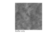
- FIG. 12 is a photomicrograph showing the results of observing a solution containing only the buffer using an atomic force microscope.
- FIG. 13 is a photomicrograph showing the result of observing a solution containing only RNA molecules using an atomic force microscope, and it can be seen that the RNA molecules exist in a circular shape.
- FIG. 14 is a photomicrograph showing the result of observing a solution containing only L7Ae using an atomic force microscope, and L7Ae can be observed in the form of dots.
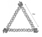
- FIG. 15 is a photomicrograph showing the result of observing a solution containing an RNA molecule and L7Ae using an atomic force microscope, and it can be seen that a triangular structure is formed by the RNA molecule and L7Ae.
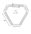
- FIG. 16 is a diagram of a large L7-3kaku drawn using Discovery Studio 2.0, which is a triangle in which three L7Ae-BoxC / Ds are linked by 48 base pair RNA.
- FIG. 17 is an L7-3kis ribbon diagram drawn using Discovery Studio 2.0, where one side of the triangle is 10 nm.
- FIG. 18 is a diagram showing a hypothetical scheme of triangle formation by the L7Ae-BoxC / D motif and the Kissing-loop motif.
- FIG. 16 is a diagram of a large L7-3kaku drawn using Discovery Studio 2.0, which is a triangle in which three L7Ae-BoxC / Ds are linked by 48 base pair RNA.
- FIG. 17 is an L7-3kis ribbon diagram drawn using
- FIG. 19 is a diagram of Delivery-3kaku drawn using Discovery Studio 2.0, where a fluorescent protein GFP is bound to one corner and a polyarginine motif is bound to two corners.
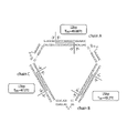
- FIG. 20 is a diagram showing secondary structures of RNA molecules constituting divided Delivery-3kaku designed by computer molecules in Example 7.
- RNA can form various three-dimensional structures.
- RNA has an enzyme function, and the correlation between the function and structure has been elucidated in detail through the analysis of the three-dimensional structure.
- RNA made of four basic units (bases) is formed by a simple construction principle. Therefore, RNA can be widely used for the design and construction of molecules having a highly three-dimensional structure as a nanoblock.
- bases basic units
- RNA-protein complex RNA-protein complex
- the present inventors considered using an RNA-protein interaction motif (RNP) as a material for designing and constructing a functional molecule such as a nanoscale structure or enzyme, and an artificial material using this material.
- RNP RNA-protein interaction motif
- the present invention has been completed by constructing specific molecules and designing and creating nanostructures.
- RNPs are structurally diverse compared to nucleic acids, and it is considered that complex structures that cannot be created only with nucleic acids can be designed and constructed relatively easily by designing RNA that is easy to design as a basic skeleton.
- the present invention is a method for inducing a structural change of an RNA molecule with a protein, wherein the RNA molecule having a base sequence derived from an RNA-protein complex interaction motif is specific for the base sequence.
- FIG. 1 The scheme of the method according to the present embodiment is schematically shown in FIG. According to the scheme shown in FIG. 1, in the absence of a protein, by adding protein 2 to RNA molecule 1 that exists in a circular shape, a triangular RNA molecule in which one protein 2 is bound to one corner is formed. Can be induced specifically.
- FIG. 1 is schematic and this invention is not limited to the induction
- the structure of an RNA molecule in which structural changes are induced by the present invention, a preparation method thereof, and a method for inducing structural changes of RNA molecules with proteins will be described in detail.
- RNA molecule having base sequence derived from RNA-protein complex interaction motif The RNA molecule whose structural change is induced by a protein used in the method according to the present embodiment is an RNA molecule having a base sequence derived from an RNA-protein complex interaction motif. More specifically, the RNA molecule in which the structural change is induced according to the present embodiment is a non-sequence comprising a base sequence derived from an RNA-protein complex interaction motif and a scaffold base sequence that is another base sequence. It is a natural molecule. Each of the scaffold base sequence and the RNA-protein interaction motif can be extracted from the sequence of a naturally occurring molecule.
- the base sequence derived from the RNA-protein complex interaction motif functions as a part that causes a structural change in the RNA molecule upon binding to the protein.
- the scaffold base sequence is a part that acts as a skeleton of the nanostructure in the RNA molecule and does not cause a structural change.
- the RNA molecule according to the present embodiment only needs to include a base sequence derived from an RNA-protein complex interaction motif, but preferably does not include a portion such as an RNA base sequence that binds nonspecifically to a protein.
- RNA-protein complex interaction motif is the base known as the sequence on the RNA side of the interaction motif between RNA and protein in a natural and known RNA-protein complex.
- a sequence and a base sequence that is a sequence on the RNA side in an artificial RNA-protein complex interaction motif obtained by an in vitro selection method (in vitro selection method) are included.
- An RNA-protein complex is an association of protein and RNA that has been confirmed in large numbers in a living body, and is a 3D object having a complicated structure.
- a base sequence derived from a natural RNA-protein complex interaction motif is usually composed of about 5 to 30 bases, and non-covalently, that is, by hydrogen bonding, with a specific amino acid sequence of a specific protein. It is known to form specific bonds.
- the nucleotide sequences derived from such natural RNA-protein complex interaction motifs are shown in Tables 1 and 2 below, and a database available on the website: http: // gibk26. bse. kyutech. ac. jp / jouhou / image / dna-protein / rna / rna. From html, a motif that produces the desired structural change can be selected.
- RNA-protein interaction motif preferably used in this embodiment is a motif whose X-ray crystal structure analysis or NMR structural analysis has already been performed, or a three-dimensional structure estimated from the three-dimensional structure of a homologous protein that has been subjected to structural analysis. It is a possible motif. Furthermore, it is desirable that the protein is a motif that specifically recognizes the secondary structure and base sequence of RNA.
- the base sequence derived from an artificial RNA-protein complex interaction motif is a base sequence on the RNA side of an RNA-protein interaction motif in an artificially designed RNA-protein complex.
- Such a base sequence is usually composed of about 5 to 30 bases, and forms a specific bond with a specific amino acid sequence of a specific protein non-covalently, that is, by hydrogen bonding. design.
- the base sequences listed in Table 3 below are also known, and these can also be used as base sequences derived from the RNA-protein complex interaction motif of the present invention.
- An artificial RNA-protein complex can be prepared by using a molecular design method, an in vitro evolution method, or a combination of both.
- aptamers and ribozymes can be obtained by repeating functional reactions such as selecting functional RNA from a molecular library with various sequence diversity and amplifying and transcribing the gene (DNA). . Therefore, an RNA-protein interaction motif adapted to RNP having a target functional structure in advance in molecular design can be extracted from natural RNP molecules or artificially created by in vitro evolution.
- the base sequence derived from the RNA-protein complex interaction motif preferably has a dissociation constant Kd of the RNA-protein complex from which the base sequence is derived from about 0.1 nM to about 1 ⁇ M. . This is because the RNA-protein interaction has a high affinity, and the state can be maintained even after the structural change is induced.
- L7Ae derived from ultra-high heat sulfate-reducing archaea, which is known to be involved in RNA modification such as RNA methylation or pseudouridine formation (SEQ ID NO: 1) ) (Moore T et al., Structure Vol. 12, pp.
- nucleotide sequences derived from these RNA-protein complex interaction motifs are determined, amino acid sequences derived from the same RNA-protein complex interaction motif are simultaneously determined, and these are specific under physiological conditions. And non-covalent binding occurs.
- RNA-protein complex interaction motif is known to change the RNA structure before and after complex formation between RNA and protein.
- a complex of 5′GGGCGUGAUGCGAAAGCUGACCCC3 ′ (SEQ ID NO: 4) (hereinafter referred to as BoxC / D), which is the base sequence to which L7Ae binds, explain.
- BoxC / D 5′GGGCGUGAUGCGAAAGCUGACCCC3 ′
- L7Ae and BoxC / D are known to form a complex as shown in FIG. 2 (Moore T et al., Structure Vol. 12, pp. 807-818 (2004)).
- L7Ae is known to specifically bind to a Box C / D sequence in which the complementary sequence portion forms a double strand.
- the Box C / D sequence in the form of a double strand is a molecule having a flexible, unfixed structure before L7Ae binds.
- Such a state of the Box C / D arrangement is schematically shown in FIG.
- the BoxC / D sequence and L7Ae interact to form a complex.
- this Box C / D arrangement is fixed to a structure bent at 60 ° C.
- the BoxC / D array and L7Ae in such a state are schematically shown in FIG.
- the structure bent at 60 ° C. is formed around the base U of the sixth strand site from the 5 ′ side in the sequence.
- the schematic diagram which expanded the bending part is shown in FIG. This is a specific structural change known for the RNA-protein complex interaction motif between the Box C / D sequence and L7Ae.
- RNA-protein complex interaction motif between the Box C / D sequence and L7Ae is an example, and the other RNA-protein complex interaction motifs listed in the above table may each have specific structural changes.
- Bacillus subtilis -derived S15 (RNA: GGGCGGCCUUCGGGCUAGACGGUGGGAGAGGCUUCGGCUGGUCCACCCGUGACGCUC (SEQ ID NO: 5)) (Protein: EmuPiaishikeiiikeikyukeibuiaikyuiefueiaruefuPijiditijiesutiibuikyubuieieruerutieruaruaienuarueruesuieichierukeibuieichikeikeidieichieichiesueichiarujierueruemuemubuijikyuRRRLLRYLQREDPERYRALIEKLGI (SEQ ID NO: 6)), an angle of
- RNA GGCAGAGCUCUCGGGACAUUGCACCUGCC (CC: SEQ ID NO: 9)
- protein AVPETRPNHTIYINNLNEKIKKKDHALKSFLDQKLDHALKGSQQLDDIFSSRKMRGDQ Such as FV (SEQ ID NO: 10)) is known.
- one RNA molecule may be arranged with a plurality of identical nucleotide sequences derived from the RNA-protein complex interaction motif.
- base sequences derived from different types of RNA-protein complex interaction motifs may be arranged in one RNA molecule. By arranging base sequences derived from different RNA-protein complex interaction motifs in one RNA molecule, multiple types of structural changes can be introduced into one RNA molecule, and multiple types of structural changes can be made. It is possible to guide.
- the base sequence other than the base sequence derived from the RNA-protein complex interaction motif that constitutes the RNA molecule according to the present embodiment is a portion that does not cause a structural change.
- it can also be said to be a part that serves as a scaffold for arranging a part that causes a structural change.
- the base sequence that does not cause such a structural change may be an artificial base sequence or a naturally-derived base sequence.
- Preferred sequences that do not cause structural changes include, for example, sequences that do not have a complicated three-dimensional structure, double strands formed by Watson-Crick base pairing, tRNA, 3-way junction, kissing-loop, and loop-receptor.
- RNA-RNA interaction motifs include, but are not limited to.
- the length, arrangement position, etc. of the base sequence that does not cause a structural change can be appropriately determined in relation to the base sequence derived from the RNA-protein complex interaction motif.
- the manufacturing method of the RNA molecule used for this embodiment including determination of length, arrangement
- the protein molecule that induces structural changes used in the method according to the present embodiment has an amino acid sequence derived from the same RNA-protein complex interaction motif as the base sequence derived from the RNA-protein complex interaction motif of the RNA molecule.
- RNA-protein complex interaction motif specifically interacts with the base sequence derived from the RNA-protein complex interaction motif of the RNA molecule in which the structural change is induced. . That is, the RNA molecule and the protein molecule that induces a structural change are selected so as to form an RNA-protein complex.
- RNA-protein complex interaction motif when selecting a base sequence derived from an RNA-protein complex interaction motif and an amino acid sequence derived from an RNA-protein complex interaction motif, a known RNA-protein complex interaction is selected.
- a mutation may be introduced into the motif-derived sequence.
- Those skilled in the art can appropriately determine and introduce mutations in the RNA-protein complex interaction motif that do not change the interaction characteristics. For example, a mutation of about 1 to 5 bases may be introduced only into the base sequence derived from the RNA-protein complex interaction motif, and 1 to 5 only into the amino acid sequence derived from the RNA-protein complex interaction motif. Mutations as large as peptides may be introduced, and mutations may be introduced into both. The number of such mutations varies depending on the RNA-protein complex interaction motif to be used, and is not limited to these values.
- the protein molecule may be a fusion protein molecule containing a functional protein.
- functional proteins include, but are not limited to, fluorescent proteins having a marker function, apoptosis-inducing proteins having a therapeutic effect, and the like.
- Such a protein molecule can be appropriately produced by those skilled in the art using a vector or the like based on the known DNA sequence information of the desired protein.
- RNA molecule from which the structural change is induced is a single RNA molecule with a plurality of different types of RNA-protein complex interaction motifs, the corresponding sequence
- protein molecules having amino acid sequences derived from different types of RNA-protein complex interaction motifs are required.
- Induction of structural changes in RNA molecules can be performed in a liquid. More specifically, it can be carried out as long as it is a liquid under physiological conditions of 4 to 80 ° C. and pH 3.5 to 10.5 under atmospheric pressure.
- the method according to the present embodiment can be performed by mixing the protein molecule with a liquid under physiological conditions containing the RNA molecule.
- the mixing amount can be determined by the number of base sequences derived from interacting RNA-protein complex interaction motifs on the RNA molecule, the molar ratio during interaction, and the like. Note that the molar ratio at the time of interaction of the RNA-protein complex is specific to each RNA-protein complex interaction motif.
- RNA molecules [Induction of structural changes in RNA molecules]
- a circular double-stranded RNA molecule having three BoxC / D sequences at regular intervals can be designed as an RNA molecule.
- the L7Ae molecule is added in a molar amount of 3 times or more of the RNA molecule.
- three L7Ae molecules bind to the double-stranded RNA molecule, and a structure having three 60-degree angles at equal intervals, that is, an equilateral triangle, is induced.
- RNA molecules having three BoxC / D sequences at regular intervals and an L7Ae dimer in which two L7Ae molecules as proteins are fused can be designed as RNA molecules.
- the RNA molecule in the absence of the L7Ae dimer, the RNA molecule is present in the fluid under physiological conditions as a circular RNA double-stranded molecule.
- the L7Ae dimer is added in a molar amount so as to be 6 or more L7Ae dimers with respect to 4 molecules of double-stranded RNA molecules.
- six L7Ae dimers bind to four RNA molecules, and a regular octahedral structure is induced.
- FIG. 5 A schematic diagram in this case is shown in FIG. In FIG. 5, it is RNA molecules that constitute each side, and an L7Ae dimer is located at the apex. The formation of such an octahedron is performed spontaneously only by mixing the RNA molecule and the L7Ae dimer.
- square RNP can be designed.
- An RNA motif that binds to protein L1 is inserted into the vertices of the four sides of double-stranded RNA. In the presence of a protein ratio, it is expected to have a circular structure, but it can be expected that a square RNP molecule can be formed by binding the L1 protein to each vertex.
- a specific structural change of a specific RNA molecule can be induced in the protein by designing the specific non-natural RNA molecule or protein molecule. Control of unnatural RNA molecular structure using such RNA-protein complex interaction motifs has not been reported so far, and it is used as a material for artificially constructing or reconfiguring biomolecules and genetic circuits. Promising as a tool for
- RNA molecular material a method for producing an RNA molecular material
- the above-described method for producing an RNA molecule having a specific structural change the step of selecting an RNA-protein complex interaction motif whose desired structural change is known, and the RNA molecule Determining a base sequence derived from the RNA-protein complex interaction motif and a scaffold base sequence by using a computer molecular modeling method, and determining the above And a step of producing RNA based on the base sequence obtained by the step.
- the RNA molecule produced in this embodiment is an RNA molecule having the characteristics described in the first embodiment and whose structural change is induced by a protein molecule.
- an RNA-protein complex interaction motif capable of realizing the structural change to be introduced is selected from the aforementioned table or database.
- structural changes of RNA molecules that can be introduced with the RNA-protein complex interaction motif include 60 degree bend, 90 degree bend, change from double stranded RNA to single stranded RNA, single stranded RNA Examples include, but are not limited to, changes to double-stranded RNA.
- a plurality of structural changes can be realized for one RNA molecule, and in this case, a plurality of different RNA-protein complex interaction motifs can be selected in combination.
- RNA-protein complex interaction motifs can be introduced.
- the step of determining the base sequence of the RNA molecular material can be performed by a computer molecular modeling method.
- Commercially available molecular modeling software can be used for this.
- computer molecular modeling first, a three-dimensional structure of a desired RNA-protein complex interaction motif is obtained. Then, one or a plurality of RNA-protein complex interaction motif-derived base sequences and RNA-protein complex interaction so that the RNA-protein complex after introduction of structural change forms a desired structure.
- the base sequence other than the base sequence derived from the motif is determined. Base sequences other than the base sequence derived from the RNA-protein complex interaction motif serve as a scaffold for locating the base sequence derived from the RNA-protein complex interaction motif.
- a sequence known not to form such a secondary structure or the like can be selected so as not to form a structure or a tertiary structure. Thereafter, the secondary structure that can be taken by the RNA having the designed sequence is predicted, and it is confirmed that the designed secondary structure is the most stable structure and does not take any other stable secondary structure.
- the secondary structure prediction program mfold http://mfold.bioinfo.rpi.edu/cgi-bin/rna-form1.cgi) of the nucleic acid used on the web can be suitably used.
- an RNA molecule is produced from the obtained base sequence by a known method. If a predetermined RNA sequence is given, it is common practice for those skilled in the art to produce RNA molecules based on this sequence. For example, template DNA is synthesized by PCR and transcription using RNA synthase is performed. RNA molecules can be produced by the reaction. In addition, RNA having a short chain length of about 30 bases can be directly synthesized by chemical synthesis. RNA molecules can be produced by such a method.
- RNA molecule that induces a structural change thus produced can be used in the first embodiment, and can be used as a molecular material together with a protein molecule that induces a structural change as a kit described later. it can.
- an RNA molecule that induces a structural change is designed and manufactured by combining a database of RNA-protein complex interaction motifs, a computer molecular modeling method, and a genetic engineering method. Can do. According to such a method, it is possible to design a molecule with high accuracy, and thus it is possible to easily and accurately design an RNA molecule in which a desired structural change is induced. Moreover, such RNA molecules can actually be produced in large quantities by genetic engineering methods. The method according to the second embodiment is very useful in producing a desired RNA molecule.
- the present invention provides a kit for inducing a structural change of an RNA molecule with a protein molecule, wherein the RNA molecule specifically changes in structure as described above, and the RNA-protein that the RNA molecule has And a protein molecule comprising an amino acid sequence that specifically binds to a base sequence derived from a complex interaction motif.
- the RNA molecule that specifically changes its structure and the protein molecule containing the amino acid sequence derived from the RNA-protein complex interaction motif may be any of those described in the first embodiment.
- the kit according to the present embodiment may include an RNA molecule that competes for RNA-protein complex formation as an optional component.
- a specific RNA base sequence competing for RNA-protein complex formation can have a base sequence derived from an RNA-protein complex interaction motif.
- the RNA molecule specifically changing in structure and the protein molecule containing the amino acid sequence derived from the RNA-protein complex interaction motif included in the kit form a complex when mixed under predetermined conditions.
- This bond is a stable and specific non-covalent bond by hydrogen bonding.
- binding can be easily suppressed and formation of the RNP structure can be controlled.
- the kit according to the third embodiment of the present invention can induce specific structural changes of RNA molecules under physiological conditions.
- RNA molecules that compete for RNA-protein complex formation which is an optional component, enables reversible structural changes in the RNP nanostructure.
- the functional RNA-protein complex described above is derived from an RNA molecule that specifically changes its structure, and an RNA-protein complex interaction motif possessed by the RNA molecule.
- a protein molecule comprising an amino acid sequence that specifically binds to a base sequence and a functional protein.
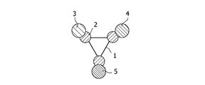
- FIG. 6 schematically shows a functional RNA-protein complex according to the fourth embodiment.
- the complex shown in the figure has three vertices of a triangle composed of RNA molecules 1 each having a base sequence derived from an RNA-protein complex interaction motif in which the structural changes described in the first embodiment are induced on each side.
- a fusion protein molecule comprising the amino acid sequence portion 2 that specifically binds to the base sequence and functional protein molecules 3, 4, and 5 is bound. All three functional protein molecules 3, 4 and 5 are different.
- the functional protein refers to a protein having a predetermined function, for example, fluorescent proteins such as GFP and YFP, proteins having a membrane permeation function such as polyarginine motif, and tissue-specific functions.
- fluorescent proteins such as GFP and YFP
- proteins having a membrane permeation function such as polyarginine motif
- tissue-specific functions include, but are not limited to, antibodies that specifically bind to the surface of cancer cells, such as proteins that bind to expressed membrane proteins, apoptosis-inducing proteins, and proteins that have a therapeutic effect on specific diseases. .
- Such a complex is an RNA molecule designed as described in the first embodiment, and an amino acid sequence that specifically binds to a base sequence derived from an RNA-protein complex interaction motif introduced into the RNA molecule. It can be obtained by producing a fusion protein molecule in which any functional protein molecule is bound to the protein molecule to be contained, and mixing the RNA molecule and the fusion protein molecule. For example, when L7Ae-BoxC / D exemplified in the first embodiment is used as an RNA-protein complex interaction motif, a fusion protein molecule of an L7Ae protein and an arbitrary protein or peptide is prepared.
- RNA-protein complex having any of a plurality of functions can be easily prepared simply by mixing with the RNA strand exemplified in one embodiment.
- the number of functional protein molecules to be bound is changed by changing the molar ratio of the multiple types of functional protein molecules to be added. And can control the type.
- the complex shown in FIG. 6 can be formed by adding a functional protein molecule to a circular RNA molecule 1 having three Box C / D sequences at regular intervals. It is obtained by adding sex protein molecules 3, 4, 5 in equimolar amounts.
- FIG. 7 shows a schematic scheme for constructing a functional RNA-protein complex.
- FIG. 7 is a scheme in the case of adding three different types of functional protein molecules 2a, 2b, and 2c to RNA molecules. In this way, each time one kind of protein molecule derived from an RNA-protein complex interaction motif fused with a functional protein molecule is added, the structure of the RNA molecule is changed, and finally the functional RNA of the desired shape— Protein complexes can be created.
- RNA-protein complex for example, if a protein that binds to a membrane-permeable peptide or a tissue-specific membrane protein is used, the complex can be delivered to specific cells. Can be used. In addition, when a fluorescent protein such as GFP or YFP is used, there is an advantage that the delivery of the complex can be visualized and observed. Furthermore, it can be used as a therapeutic agent for delivering an apoptosis-inducing protein to cancer cells. Like the complex shown in FIG. 6, a complex in which a plurality of proteins having different functions are combined has all these advantages.
- RNA and protein are bound by specific and non-covalent bonds, they can be made into a complex with a flexible binding mode as compared with conventional covalent bonds. Therefore, any protein having an RNA binding motif can be freely exchanged (installed), and modular engineering of a functional RNA-protein complex becomes possible.
- RNA molecules designed by this method can form a stable double strand and protect the hydroxyl group of the terminal nucleotide from RNase present in the cell, as shown in the examples described later and FIG. become. For this reason, it can be expected to exist stably in the cell.
- the present invention provides a method for producing an RNA-protein complex nanostructure according to the fifth embodiment, wherein an RNA molecule having a base sequence derived from an RNA-protein complex interaction motif is added to the RNA molecule.
- a protein molecule containing an amino acid sequence that specifically binds to a base sequence derived from an RNA-protein complex interaction motif and, if necessary, a metal ion such as magnesium or calcium are added.
- RNA-protein complex nanostructure a specific structural change of an RNA molecule is induced by a protein molecule to create an RNA-protein complex nanostructure.
- a desired structure can be obtained by designing an RNA molecule, and specific structures include, but are not limited to, the equilateral triangle, regular octahedron, and square exemplified in the first embodiment.
- Another example is a method for preparing an RNA-protein complex nanostructure by adding an RNA molecule, a protein molecule, and a metal ion.
- This method is preferably used when a Kissing-loop sequence is introduced as a scaffold base sequence into an RNA molecule.
- a structural change is introduced into one RNA molecule by a protein molecule to form one RNA-protein complex.
- metal ions By adding metal ions, one RNA-protein complex interacts with another RNA-protein complex.
- the metal ion can link RNA molecules non-covalently.
- an RNA-protein complex nanostructure in which a plurality of RNA molecules are bound can be produced.
- Such a substance that binds a plurality of molecules depends on the introduced motif or the like, but also includes an RNA loop receptor motif.
- the protein molecule can be a fusion protein in which a functional protein is fused.
- the fifth embodiment of the present invention it is possible not only to introduce a structural change into an RNA molecule to form a complex, but also to provide a method for creating a nanostructure by combining several complexes. it can. According to such a method, it is possible to create a larger number of desired nanostructures, and it can be said that it is easier to apply as a tool for artificially constructing biomolecules and genetic circuits.
- Example A method of creating a nanostructure using RNP is shown. This technique is a technique that makes it easier to create nanoscale structures and functional molecules than when using only proteins or RNA alone. Specific experimental examples are given below.
- Fig. 8 (a) is a view of L7-3 kaku ⁇ ⁇ depicted using Discovery Studio 2.0 from the top
- Fig. 8 (b) is a triangular side view of Fig. 8 (a) rotated 90 degrees.
- FIG. The RNA consists of 114 nt long chain (Fig. 9 (a)) and 150 nt short chain (Fig. 9 (b)).
- Fig. 9 (a) By complementary pairing of these RNAs, circular RNA with three BoxC / Ds A chain is formed.
- L7Ae By binding L7Ae to these BoxC / D sequences, it is expected that three positions of the circular RNA will be fixed at 60 degrees to form a triangle.
- the length of one side of the formed RNA triangle is about 10 nm (FIG. 10).
- Fulgidus was amplified using a restriction enzyme N using primers of L7Ae Fwd (5′-CTGACATATGTTACGTGAGATTTGAGGTTC-3 ′) (SEQ ID NO: 12), L7Ae Rev (5′-CTGACTCGAGTTACTTCTGGAGCCTTTTATC-3 ′) and SEQ ID NO: 13 It was prepared by integrating into a pET-28b + vector (Novagen) cut with XhoI. The expression purification method is shown below. First, E. coli BL21 (DE3) pLysS was transformed.
- the obtained colonies were inoculated into 5 mL of LB medium containing 25 ⁇ g / mL kanamycin and 100 ⁇ g / mL chloramphenicol, and cultured with shaking at 37 ° C. overnight. Subsequently, the entire culture was inoculated into 500 mL of LB medium containing 25 ⁇ g / mL kanamycin and 100 ⁇ g / mL chloramphenicol. O. D. The culture was shaken at 37 ° C. until 600 reached 0.6 to 0.7, and then 500 ⁇ L of 1M IPTG was added (final concentration 1 mM) to induce expression, followed by overnight shaking at 30 ° C.
- the cells were collected by centrifugation (4 ° C., 6000 rpm, 20 minutes), 5 mL of a sonication buffer (50 mM Na phosphate, 0.3 M NaCl, pH 8.0) was added, and sonication was performed to disrupt the cells. .
- a sonication buffer 50 mM Na phosphate, 0.3 M NaCl, pH 8.0
- the operation of applying ultrasonic waves for 15 seconds after cooling on ice was repeated 6 times. Thereafter, the impure protein was denatured at 80 ° C. for 15 minutes. Centrifugation (4 ° C., 6000 rpm, 20 minutes) was performed, the supernatant was collected, and the protein with a histidine tag was purified by a batch method using a Ni-NTA column (Qiagen).
- the protein was concentrated using Microcon YM-3 (Millipore) and replaced with a dialysis buffer (20 mM Hepes-KOH, 1.5 mM MgCl 2 , 150 mM KCl, 5% glycerol, pH 7.5).
- the protein concentration was determined by the Bradford method using a protein assay (BIO-RAD).
- RNA The template DNA of long chain is L Fwd (10 ⁇ M, 5′-CTAATACGACTCACTATAGGGCGCAAAGGCCTGTAATCGGCGGTGATGCTGCTGCTGCTGCCTGCCTGCCTGCTGCTGCCTGCCTGCCTGCTGCTGCCTGCCTGCCTGCTGCTGCCTGC 1ng / ⁇ L, 5'-GGCCTGTAATCGGCGTGATGAGCCATGCGAGGAGGAAATGAAGTCCAATGGCGTGATGAGCCTCTACGGGAAGAGC- 3 ' SEQ ID NO: 16) 1 ⁇ L, KOD + (TOYOBO ) 1 ⁇ L, 10 ⁇ KOD + buffer 5 ⁇ L, 25mM MgSO 4 1.6 ⁇ L, 2.5mM dNT s 4 [mu] L, mixed ultrapure water 34.4 ⁇ L, 94 °C 15 seconds, 55 ° C.
- PCR was performed in 15 cycles of 3 steps of 30 seconds at 68 ° C. for 60 seconds to synthesize template DNA.
- Each template DNA was confirmed to be synthesized by 4% agarose gel electrophoresis, and purified by phenol extraction, diethyl ether extraction, and ethanol precipitation.
- the purified template DNA was dissolved in 8 ⁇ L of ultrapure water and used for transcription.
- MEGAshortscript (trademark) (Ambion) was used as follows. Template DNA 8 ⁇ L, T7 10 ⁇ Reaction Buffer 2 ⁇ L, T7 ATP Solution (75 mM) 2 ⁇ L (same for CTP, GTP, UTP) and T7 Enzyme Mix 2 ⁇ L were mixed overnight at 37 ° C.
- TURBO DNase After the reaction, 1 ⁇ L of TURBO DNase was added and incubated at 37 ° C. for 1 hour to decompose the template DNA. 115 ⁇ L of ultrapure water and 15 ⁇ L ammonium acetate stop solution were added to the reaction solution, and further purified by phenol treatment, diethyl ether extraction, and ethanol precipitation. The precipitate was dissolved in 20 ⁇ L of a denaturing dye (80% formamide, 0.17% XC, 0.27% BPB) and separated by 10% polyacrylamide (1/30 bisacrylamide) denaturing gel electrophoresis.
- a denaturing dye 80% formamide, 0.17% XC, 0.27% BPB
- RNA was purified again by phenol extraction, diethyl ether extraction and ethanol precipitation, dissolved in ultrapure water and used in the subsequent experiments.
- a sample for observation with an atomic force microscope was prepared as follows. Mix 2 ⁇ 5 ⁇ binding buffer (100 mM Hepes-KOH (pH 7.5), 750 mM KCl, 7.5 mM MgCl 2 , 10 mM DTT, 15% glycerol) 2 ⁇ L and short chain and long chain (both 10 ng / ⁇ L) 2 ⁇ L each The RNA was denatured by heating at 80 ° C. for 3 minutes, and then placed on ice to rapidly cool and fold.
- RNA molecules (FIG. 13) that existed alone in an oval shape form a triangular structure when protein L7Ae and RNA molecules coexist (FIG. 15), and in the presence of protein L7Ae. Only in, a triangular structure as designed was confirmed.
- FIG. 16 is a diagram of a large L7-3 kaku depicted using Discovery Studio 2.0. In this large L7-3 kaku, the triangle was composed of RNA double strands, and one side of the triangle formed by RNA was about 17 nm and 48 bp.
- sequence of long chain is, 5'-GGACGAGCUGUACACCAUGGUGACCGCCGCCGGGCGUGAUGAGCUCCAAGGACCCCAACGAGAAGCGCGAUCACAUGAUCUACUUCGGCUUCGGCGUGAUGAGCCCUGUGCUGCUGCCCGAUAACCACUACCUGCCAUCACCCACGGCCCUGGGCGUGAUGAGCAUUCCACCCAGAGCGC-3 '(SEQ ID NO: 28, Artificial, 180nt), the sequence of short chain may, 5'-GGUGUACAGCUCGUCCGCUCUGGGUGGAAUGCUCUGACCCAGGGCCGUGGGUGAUGGCAGGUAGUGGUUAUCGGGCAGCAGCAC GGGCUCUGACCGAAGCCGAAGUAGAUCAUGUGAUCGCGCUUCUCGUUGGGGUCCUUGGAGCUCUGACCCGGCGGCGGUCACCAU-3 '
- Template DNA long chain is, L Fwd (2 ⁇ M, 5'-CTAATACGACTCACTATAGGACGAGCTGTACACCATGGTGACCGCCGCCGGGCGTGATGAGCTCCAAGGACCCCAACGAGAAGCGCGATCACATGATCTACTTCGGCTT-3 ', SEQ ID NO: 30, Artificial, 109nt) 10 ⁇ L, L Rev (2 ⁇ M, 5'-GCGCTCTGGGTGGAATGCTCATCACGCCCAGGGCCGTGGGTGATGGCAGGTAGTGGTTATCGGGCAGCAGCACAGGGCTCATCACGCCGAAGCCGAAGTAGATCATGTG-3', SEQ ID NO: 31, Artificial, 109nt ) 10 ⁇ L, Ex Taq (Takar a) 0.5 ⁇ L, 10 ⁇ Ex Taq buffer 5 ⁇ L, 2.5 mM dNTPs 4 ⁇ L, and ultrapure water 21.5 ⁇ L are mixed, and 5 cycles PCR in 3 steps of 94 ° C.
- Template DNA short chain is, S Fwd (2 ⁇ M, 5'-CTAATACGACTCACTATAGGTGTACAGCTCGTCCGCGCTCTGGGTGGAATGCTCTGACCCAGGGCCGTGGGTGATGGCAGGTAGTGGTTATCGGGCAGCAGCACA-3 '(SEQ ID NO: 32, Artificial, 105nt)) 10 ⁇ L, S Rev (2 ⁇ M, 5'-ATGGTGACCGCCGCCGGGTCAGAGCTCCAAGGACCCCAACGAGAAGCGCGATCACATGATCTACTTCGGCTTCGGTCAGAGCCCTGTGCTGCTGCCCGATAACC-3' (SEQ ID NO: 33, Artificial, 104 nt)) 10 ⁇ L, Ex Taq (Taka a) 0.5 ⁇ L, 10 ⁇ Ex Taq buffer 5 ⁇ L, 2.5 mM dNTPs 4 ⁇ L, and ultrapure water 21.5 ⁇ L are mixed, and 5-cycle PCR is performed in 3 steps of 94 ° C.
- the synthesized template DNA strand was subjected to electrophoresis on a non-denaturing 6% acrylamide gel (1/30 bisacrylamide) to cut out a band of the desired size.
- 500 ⁇ L of elution buffer (0.1% SDS, 0.3 M sodium acetate, pH 7.0) was added and incubated overnight at 37 ° C. to elute the DNA. Thereafter, phenol extraction, diethyl ether extraction and ethanol precipitation were performed, dissolved in 20 ⁇ L of ultrapure water, and 8 ⁇ L was used for the following transcription reaction.
- the transfer was performed as follows using MEGAshortscript (trademark) (Ambion). A total of 20 ⁇ L of template DNA 8 ⁇ L, T7 10 ⁇ Reaction Buffer 2 ⁇ L, T7 ATP Solution (75 mM) 2 ⁇ L (same for CTP, GTP, UTP) and T7 Enzyme Mix 2 ⁇ L were reacted at 37 ° C. overnight. After the reaction, 1 ⁇ L of TURBO DNase was added and incubated at 37 ° C. for 15 minutes to decompose the template DNA.
- a denaturing dye (80% formamide, 0.17% XC, 0.27% BPB) was added to the reaction solution and separated by 10% polyacrylamide (1/30 bisacrylamide) denaturing gel electrophoresis. A band of the desired size was cut out, 500 ⁇ L of elution buffer (0.1% SDS, 0.3 M sodium acetate, pH 7.0) was added, and the mixture was incubated overnight at 37 ° C. for elution. The eluted RNA was subjected to phenol extraction, diethyl ether extraction and ethanol precipitation, dissolved in ultrapure water, and used in the subsequent experiments.
- elution buffer (0.1% SDS, 0.3 M sodium acetate, pH 7.0
- RNA was confirmed by gel shift assay.
- the final concentrations of the two kinds of RNA were 50 fmol / ⁇ L, 20 mM Hepes-KOH, 150 mM KCl, 1.5 mM MgCl 2 , 2 mM DTT, 3% glycerol, and 100 to 1000 nM protein as follows.
- Example 4 Similar to Example 4, a sample was prepared as follows and observed with an atomic force microscope. 5 ⁇ binding buffer (100 mM Hepes-KOH (pH 7.5), 750 mM KCl, 7.5 mM MgCl 2 , 10 mM DTT, 15% glycerol) 2 ⁇ L and ultrapure water 6 ⁇ L, short chain and long chain (both 1 pmol / ⁇ L) 0 Each 5 ⁇ L was mixed and heated at 80 ° C. for 3 minutes to denature the RNA, and then placed at room temperature for 10 minutes for folding.
- 5 ⁇ binding buffer 100 mM Hepes-KOH (pH 7.5), 750 mM KCl, 7.5 mM MgCl 2 , 10 mM DTT, 15% glycerol
- FIG. 18 shows a hypothetical scheme of triangle formation by the L7Ae-BoxC / D motif and the Kissing-loop motif.
- a loop-shaped RNA having a Box C / D motif at the center and forming a double strand is considered to exist in a loop shape in the absence of L7Ae or Mg 2+ ions.
- L7Ae is added to this loop-shaped RNA, it is considered that the structure changes specifically to the bent-loop RNA exhibiting a 60-degree bent structure.
- Mg 2+ ions are added, it is considered that a triangle is formed by the Kissing-loop motif.
- FIG. 17 is a ribbon diagram of L7-3kiss drawn using Discovery Studio 2.0.
- this L7-3kiss one triangle consists of three RNA strands, and the three RNA strands have the same sequence.
- One side of the triangle formed by RNA was about 10 nm.
- the three RNA strands exhibited a bent structure of 60 degrees by L7Ae, formed homotrimers by kissing-loop interaction, and became triangular.
- RNA sequence of three identical RNAs was determined as CUACGGGAAGCGCGGCACCCCUAGAGGGCUCUGACCCCGAUGGGCACAGCGCGCAGCCCAUCCCGGGCGUGAUGAGCU (SEQ ID NO: 23).
- RNAs were prepared using Discovery Studio 2.0 (Accelrys), which is molecular modeling software, so that the three L7Ae-BoxC / D motifs are coplanar and form an equilateral triangle.
- Delivery-3 kaku was designed in which three chains of L7Ae were linked together, one of which was linked to the fluorescent protein GFP and the other was linked to the polyarginine motif.
- FIG. 19 is a diagram of Delivery-3kaku drawn using Discovery Studio 2.0. In this Delivery-3kaku, L7Ae fused with the polyarginine motif is bound to the upper left corner and upper right corner, and L7Ae fused to the fluorescent protein GFP is bound to the lower left corner.
- L7Ae fused to the fluorescent protein GFP is bound to the lower left corner.
- results of computer molecular design, arrangement of long chain is, 5'-GGCGCAAAGGCCUGUAAUCGGCGUGAUGAGCCAUGCGAGGAGGAAAUGAAGUCCAAUGGCGUGAUGAGCCUCUACGGGAAGAGCAUGCCCAUCCGGGCGUGAUGAGCGUAGCAA-3 '(SEQ ID NO: 24), the sequence of short chain may, 5'-GGCCUUUGCGCCUUGCUACGCUCUGACCCGGAUGGGCAUGCUCUUCCCGUAGAGGCUCUGACCAUUGGACUUCAUUUCCUCCUCGCAUGGCUCUGACCGAUUACA-3' (SEQ ID NO: 25), L7Ae- fluorescent protein GFP
- the peptide sequence is VPEDMQNEALSLLEKV ESGKVKKGTNETTKAVERGLAKLVYIAEDVDPPEIVAHLPLLCEEKNVPYIYVKSKNDLGRAVGIEVPCASAAIINEGELRKELGSLVEKIKGLQKPFTVSKGEELFTGVVPILVELDGDVNG
- RNA is divided into three at the sides of the triangle, and a triangle is constructed by base pairing between the sticky ends of the side.
- the three types of RNA-protein complexes are mixed to obtain Delivery-3kaku having one of three different proteins.
- Each of the three parts consists of two RNA strands, and the entire triangle consists of a total of six types of RNA strands.
- FIG. 20 is a diagram showing secondary structures of RNA molecules constituting divided Delivery-3kaku designed by computer molecules in Example 7.
- RNAs constituting the divided Delivery-3kaku are the chain A-Long sequence, 5′-GGCGCAAAGGCCUGUAAUCGGUGCGAUGAGCCCAUGCGA-3 ′ (SEQ ID NO: 34, Artificial, 38nt), and the chain A-Shor sequence, respectively.
- chain B-Long sequence is 5'-GGAGGAAAUGAGCCCAAUGGCGUGAUCCCArCUt h36, t-36 -GGCAUGCUCUUCCCGU GAGGCUCUGACCAUG-3 ′ (SEQ ID NO: 37, Artificial, 33 nt)
- chain C-Long is 5′-GGAAGAGCAUGCCCAUCCGGGUGCGGAUGAGCGGUAGCAA-3 ′ (SEQ ID NO: 38, Artificial, 38nt), Shain-Cin It was determined as GGCCUUUGCGCCUUGCUACGCUCUGACCCCGAUG-3 ′ (SEQ ID NO: 39, Artificial, 34 nt).
Landscapes
- Chemical & Material Sciences (AREA)
- Health & Medical Sciences (AREA)
- Life Sciences & Earth Sciences (AREA)
- Biochemistry (AREA)
- Molecular Biology (AREA)
- Organic Chemistry (AREA)
- Engineering & Computer Science (AREA)
- Biotechnology (AREA)
- General Health & Medical Sciences (AREA)
- Genetics & Genomics (AREA)
- Peptides Or Proteins (AREA)
Abstract
L'invention concerne un procédé de construction d'une nanostructure de PRN artificielle en utilisant un motif d'interaction complexe ARN-protéine. Elle concerne également un procédé pour induire le changement de structure d'une molécule d'ARN avec une protéine. Elle concerne spécifiquement un procédé d'induction du changement spécifique de structure d'une molécule d'ARN avec une molécule protéique. Le procédé se caractérise par l'addition d'une molécule protéique qui comprend une séquence d'acides aminés capable de se lier spécifiquement à une séquence nucléotidique dérivée d'un motif d'interaction complexe ARN-protéine dans une molécule d'ARN artificielle comprenant la séquence nucléotidique correspondant à la molécule d'ARN.
Priority Applications (1)
| Application Number | Priority Date | Filing Date | Title |
|---|---|---|---|
| JP2010521696A JPWO2010010862A1 (ja) | 2008-07-22 | 2009-07-21 | Rna−蛋白質複合体相互作用モチーフを利用して人工rnpナノ構造体を構築する方法 |
Applications Claiming Priority (2)
| Application Number | Priority Date | Filing Date | Title |
|---|---|---|---|
| JP2008188241 | 2008-07-22 | ||
| JP2008-188241 | 2008-07-22 |
Publications (1)
| Publication Number | Publication Date |
|---|---|
| WO2010010862A1 true WO2010010862A1 (fr) | 2010-01-28 |
Family
ID=41570323
Family Applications (1)
| Application Number | Title | Priority Date | Filing Date |
|---|---|---|---|
| PCT/JP2009/063023 Ceased WO2010010862A1 (fr) | 2008-07-22 | 2009-07-21 | Procédé de construction de nanostructure de prn artificielle en utilisant un motif d'interaction complexe arn-protéine |
Country Status (2)
| Country | Link |
|---|---|
| JP (1) | JPWO2010010862A1 (fr) |
| WO (1) | WO2010010862A1 (fr) |
Cited By (5)
| Publication number | Priority date | Publication date | Assignee | Title |
|---|---|---|---|---|
| WO2016006628A1 (fr) * | 2014-07-09 | 2016-01-14 | 国立大学法人京都大学 | Complexe arn-protéine, et système d'administration d'arn et de protéine à l'aide dudit complexe |
| WO2017010568A1 (fr) * | 2015-07-16 | 2017-01-19 | 国立大学法人京都大学 | Complexe arn-protéine et utilisation associée |
| CN107072183A (zh) * | 2014-07-14 | 2017-08-18 | 华盛顿州立大学 | 消除种系细胞的nanos敲除 |
| JP2020191806A (ja) * | 2019-05-27 | 2020-12-03 | 国立研究開発法人理化学研究所 | ナノデバイス、フォースセンサ、力の測定方法、および試薬キット |
| WO2024130257A3 (fr) * | 2022-12-16 | 2024-08-02 | Purdue Research Foundation | Production in vivo et ex vivo de neutrophiles car et leur utilisation pour le traitement et l'imagerie du cancer |
-
2009
- 2009-07-21 JP JP2010521696A patent/JPWO2010010862A1/ja active Pending
- 2009-07-21 WO PCT/JP2009/063023 patent/WO2010010862A1/fr not_active Ceased
Non-Patent Citations (4)
| Title |
|---|
| KUMAR P. ET AL.: "RNA no Kozo Henka ga Idenshi no Switch o ON ni suru", SANSOKEN TODAY, vol. 5, no. 5, 2005, pages 20 - 21 * |
| KUMAREVEL T. ET AL.: "Structural basis of HutP mediated anti-termination and roles of the Mg2+ ion and L-histidine ligand.", NATURE, vol. 434, no. 7030, 2005, pages 183 - 191 * |
| SURYADI J. ET AL.: "The crystal structure of the Methanocaldococcus jannaschii multifunctional L7Ae RNA-binding protein reveals an induced-fit interaction with the box C/D RNAs.", BIOCHEMISTRY, vol. 44, no. 28, 2005, pages 9657 - 9672 * |
| TOMOAKI HARA ET AL.: "RNP Motif L7Ae/Box C/D o Riyo shita Tanpakushitsu Oto Seigyo System no Kochiku", DAI 10 KAI NIPPON RNA GAKKAI NENKAI YOSHISHU, 23 July 2008 (2008-07-23), pages 141 - (P-41) * |
Cited By (10)
| Publication number | Priority date | Publication date | Assignee | Title |
|---|---|---|---|---|
| WO2016006628A1 (fr) * | 2014-07-09 | 2016-01-14 | 国立大学法人京都大学 | Complexe arn-protéine, et système d'administration d'arn et de protéine à l'aide dudit complexe |
| JPWO2016006628A1 (ja) * | 2014-07-09 | 2017-04-27 | 国立大学法人京都大学 | Rna−タンパク質複合体及びこれによるrnaおよびタンパク質のデリバリーシステム |
| JP2020022497A (ja) * | 2014-07-09 | 2020-02-13 | 国立大学法人京都大学 | Rna−タンパク質複合体及びこれによるrnaおよびタンパク質のデリバリーシステム |
| CN107072183A (zh) * | 2014-07-14 | 2017-08-18 | 华盛顿州立大学 | 消除种系细胞的nanos敲除 |
| US12102069B2 (en) | 2014-07-14 | 2024-10-01 | Washington State University | NANOS knock-out that ablates germline cells |
| WO2017010568A1 (fr) * | 2015-07-16 | 2017-01-19 | 国立大学法人京都大学 | Complexe arn-protéine et utilisation associée |
| JPWO2017010568A1 (ja) * | 2015-07-16 | 2018-04-26 | 国立大学法人京都大学 | Rna−タンパク質複合体とその使用 |
| JP2020191806A (ja) * | 2019-05-27 | 2020-12-03 | 国立研究開発法人理化学研究所 | ナノデバイス、フォースセンサ、力の測定方法、および試薬キット |
| JP7272643B2 (ja) | 2019-05-27 | 2023-05-12 | 国立研究開発法人理化学研究所 | ナノデバイス、フォースセンサ、力の測定方法、および試薬キット |
| WO2024130257A3 (fr) * | 2022-12-16 | 2024-08-02 | Purdue Research Foundation | Production in vivo et ex vivo de neutrophiles car et leur utilisation pour le traitement et l'imagerie du cancer |
Also Published As
| Publication number | Publication date |
|---|---|
| JPWO2010010862A1 (ja) | 2012-01-05 |
Similar Documents
| Publication | Publication Date | Title |
|---|---|---|
| Liu et al. | Branched kissing loops for the construction of diverse RNA homooligomeric nanostructures | |
| Ge et al. | Concept and development of framework nucleic acids | |
| US9678080B2 (en) | Bis-biotinylation tags | |
| Reiter et al. | Structure of a bacterial ribonuclease P holoenzyme in complex with tRNA | |
| JP5538691B2 (ja) | Rna−蛋白質相互作用モチーフを利用した蛋白質応答翻訳制御システム | |
| Bai et al. | Covalently-assembled single-chain protein nanostructures with ultra-high stability | |
| Estrich et al. | Engineered diblock polypeptides improve DNA and gold solubility during molecular assembly | |
| WO2010010862A1 (fr) | Procédé de construction de nanostructure de prn artificielle en utilisant un motif d'interaction complexe arn-protéine | |
| Feldheim et al. | Selection of biomolecules capable of mediating the formation of nanocrystals | |
| Lamppa et al. | Engineering Escherichia coli for soluble expression and single step purification of active human lysozyme | |
| Zakrevsky et al. | Truncated tetrahedral RNA nanostructures exhibit enhanced features for delivery of RNAi substrates | |
| Mittmann et al. | A magnetosome-based platform for flow biocatalysis | |
| EP3380612A1 (fr) | Robot moléculaire | |
| Wang et al. | Rolling circle transcription-amplified hierarchically structured organic–inorganic hybrid RNA flowers for enzyme immobilization | |
| Kim et al. | Metal-mediated protein assembly using a genetically incorporated metal-chelating amino acid | |
| Ojasalo et al. | Hybrid nanoassemblies from viruses and DNA nanostructures | |
| de Raad et al. | A solid-phase platform for combinatorial and scarless multipart gene assembly | |
| Lang et al. | Assembly Requirements for the Construction of Large-Scale Binary Protein Structures | |
| Seo et al. | Progresses in cell-free in vitro evolution | |
| JP4106441B2 (ja) | 任意のポリペプチドをコードするDNAの転写産物と翻訳産物との安定的な複合体を形成させる方法、該方法に用いる核酸構築物、該方法により形成される複合体、並びに該方法を利用した機能性タンパク質および該タンパク質をコードするmRNAまたはDNAのスクリーニング | |
| Nakata et al. | DNA binding adaptors to assemble proteins of interest on DNA scaffold | |
| Mentis et al. | DNA origami: a tool to evaluate and harness transcription factors | |
| Kwon et al. | Development of in vitro ribosome assembly system for ribosome engineering with ribosome biogenesis factors YrdC and SrmB | |
| Guimarães et al. | Characterization of the Thermal and Chemical Denaturation of the Matrix Protein from hRSV | |
| Hayes | Programming Assembly Pathways of Proteins Using DNA |
Legal Events
| Date | Code | Title | Description |
|---|---|---|---|
| 121 | Ep: the epo has been informed by wipo that ep was designated in this application |
Ref document number: 09800376 Country of ref document: EP Kind code of ref document: A1 |
|
| DPE1 | Request for preliminary examination filed after expiration of 19th month from priority date (pct application filed from 20040101) | ||
| WWE | Wipo information: entry into national phase |
Ref document number: 2010521696 Country of ref document: JP |
|
| NENP | Non-entry into the national phase |
Ref country code: DE |
|
| 122 | Ep: pct application non-entry in european phase |
Ref document number: 09800376 Country of ref document: EP Kind code of ref document: A1 |