WO2017010568A1 - Complexe arn-protéine et utilisation associée - Google Patents
Complexe arn-protéine et utilisation associée Download PDFInfo
- Publication number
- WO2017010568A1 WO2017010568A1 PCT/JP2016/071029 JP2016071029W WO2017010568A1 WO 2017010568 A1 WO2017010568 A1 WO 2017010568A1 JP 2016071029 W JP2016071029 W JP 2016071029W WO 2017010568 A1 WO2017010568 A1 WO 2017010568A1
- Authority
- WO
- WIPO (PCT)
- Prior art keywords
- rna
- protein
- base pairs
- binding motif
- l7ae
- Prior art date
- Legal status (The legal status is an assumption and is not a legal conclusion. Google has not performed a legal analysis and makes no representation as to the accuracy of the status listed.)
- Ceased
Links
Images








Classifications
-
- B—PERFORMING OPERATIONS; TRANSPORTING
- B82—NANOTECHNOLOGY
- B82Y—SPECIFIC USES OR APPLICATIONS OF NANOSTRUCTURES; MEASUREMENT OR ANALYSIS OF NANOSTRUCTURES; MANUFACTURE OR TREATMENT OF NANOSTRUCTURES
- B82Y5/00—Nanobiotechnology or nanomedicine, e.g. protein engineering or drug delivery
-
- C—CHEMISTRY; METALLURGY
- C07—ORGANIC CHEMISTRY
- C07K—PEPTIDES
- C07K14/00—Peptides having more than 20 amino acids; Gastrins; Somatostatins; Melanotropins; Derivatives thereof
-
- C—CHEMISTRY; METALLURGY
- C12—BIOCHEMISTRY; BEER; SPIRITS; WINE; VINEGAR; MICROBIOLOGY; ENZYMOLOGY; MUTATION OR GENETIC ENGINEERING
- C12N—MICROORGANISMS OR ENZYMES; COMPOSITIONS THEREOF; PROPAGATING, PRESERVING, OR MAINTAINING MICROORGANISMS; MUTATION OR GENETIC ENGINEERING; CULTURE MEDIA
- C12N15/00—Mutation or genetic engineering; DNA or RNA concerning genetic engineering, vectors, e.g. plasmids, or their isolation, preparation or purification; Use of hosts therefor
- C12N15/09—Recombinant DNA-technology
-
- C—CHEMISTRY; METALLURGY
- C12—BIOCHEMISTRY; BEER; SPIRITS; WINE; VINEGAR; MICROBIOLOGY; ENZYMOLOGY; MUTATION OR GENETIC ENGINEERING
- C12N—MICROORGANISMS OR ENZYMES; COMPOSITIONS THEREOF; PROPAGATING, PRESERVING, OR MAINTAINING MICROORGANISMS; MUTATION OR GENETIC ENGINEERING; CULTURE MEDIA
- C12N15/00—Mutation or genetic engineering; DNA or RNA concerning genetic engineering, vectors, e.g. plasmids, or their isolation, preparation or purification; Use of hosts therefor
- C12N15/09—Recombinant DNA-technology
- C12N15/11—DNA or RNA fragments; Modified forms thereof; Non-coding nucleic acids having a biological activity
- C12N15/115—Aptamers, i.e. nucleic acids binding a target molecule specifically and with high affinity without hybridising therewith ; Nucleic acids binding to non-nucleic acids, e.g. aptamers
-
- C—CHEMISTRY; METALLURGY
- C12—BIOCHEMISTRY; BEER; SPIRITS; WINE; VINEGAR; MICROBIOLOGY; ENZYMOLOGY; MUTATION OR GENETIC ENGINEERING
- C12N—MICROORGANISMS OR ENZYMES; COMPOSITIONS THEREOF; PROPAGATING, PRESERVING, OR MAINTAINING MICROORGANISMS; MUTATION OR GENETIC ENGINEERING; CULTURE MEDIA
- C12N9/00—Enzymes; Proenzymes; Compositions thereof; Processes for preparing, activating, inhibiting, separating or purifying enzymes
- C12N9/14—Hydrolases (3)
- C12N9/48—Hydrolases (3) acting on peptide bonds (3.4)
- C12N9/50—Proteinases, e.g. Endopeptidases (3.4.21-3.4.25)
- C12N9/64—Proteinases, e.g. Endopeptidases (3.4.21-3.4.25) derived from animal tissue
Definitions
- the present invention relates to an RNA-protein complex having a specific structure and a method for using the same.
- Non-Patent Document 1 DNA nanotechnology is expected as a platform for structures with controllable structures and functionality, such as molecular robots (Non-patent Document 2), nanomolecular devices (Non-patent Document 3), and nanomachines (Non-Patent Document 4).
- RNA is also useful for the construction of well-known nanostructures (Non-Patent Document 5).
- RNA nanostructures are already used as scaffolds to induce functional RNA nanostructures such as aptamers, ribozymes and RNA interference (siRNA), and functional RNA nanostructures are used for therapeutics such as drug delivery. It is proposed to be used for (Non-Patent Document 6). Since RNA can be synthesized intracellularly using DNA as a template, RNA nanotechnology has the advantage of application in vivo. The functionality and expression characteristics of RNA can allow nanostructures useful for the construction of molecular robots that function in cells. In particular, RNA-binding proteins and synthetic RNAs are expected to be used to control the functions of organisms as RNA-protein complexes (RNPs) that can control the structure and function of intracellular RNA. However, it is difficult to control the structure and function of RNA nanostructures by interacting with proteins.
- RNPs RNA-protein complexes
- RNA kink-turn (K-turn) motif and an L7Ae protein that binds to it (Patent Document 1, Non-Patent Document).
- Patent Document 1 Non-Patent Document.
- Reference 6 and Non-Patent Reference 7 An RNA nanostructure containing a K-turn motif can cause a structural change by changing the conformation in response to the binding of L7Ae (Non-patent Document 8). Therefore, it is possible to control the dynamic properties of RNA nanostructures using RNP interactions.
- An object is to provide an RNA nanomachine capable of controlling RNA structure and function.
- the present invention has been made to solve the above problems. That is, the present invention, according to one embodiment, includes a double-stranded RNA having two RNA-protein binding motif sequences, having functional molecules at both ends, and a protein that binds to the RNA-protein binding motif sequence. An RNA-protein complex is used. According to another embodiment, the present invention includes a double-stranded RNA containing two or more RNA-protein binding motif sequences, a protein in which a protein that binds to the RNA-protein binding motif sequence and an aggregated protein are fused. An RNA-protein complex is used.
- RNA-protein complex comprising the following (1) and (2); (1) a double-stranded RNA comprising two RNA-protein binding motif sequences, having functional molecules at both ends of the double-stranded RNA, and (2) the RNA-protein binding motif of (1) above A protein that binds to a sequence.
- RNA-protein binding motif sequences wherein the distance between the RNA-protein binding motif sequences is 20 to 24 base pairs, or 31 to 35 base pairs, or 42 to 46 base pairs -Protein complex.
- the RNA according to [1] wherein the distance between the RNA-protein binding motif sequences is 15 to 19 base pairs, or 26 to 30 base pairs, or 37 to 41 base pairs -Protein complex.
- RNA-protein complex according to [3], wherein the double-stranded RNA has overhangs of 6 base pairs or more that are complementary strands at both ends.
- a human cell comprising the RNA-protein complex according to any one of [1] to [5].
- RNA-protein binding motif sequences By changing the distance between the RNA-protein binding motif sequences from 15 base pairs to 19 base pairs, from 26 base pairs to 30 base pairs, or from 37 base pairs to 41 base pairs, The method according to [9], wherein the interaction is controlled to be negative by binding a protein that binds to a sequence.
- the double-stranded RNA has an overhang of 6 base pairs or more that are complementary strands at both ends.
- the functional molecule is an aptamer.
- the method according to any one of [8] to [13] wherein the double-stranded RNA and the fusion protein are bound in a human cell.
- RNA-protein complex comprising the following (1) and (2); (1) a double-stranded RNA comprising two or more RNA-protein binding motif sequences, and (2) a fusion protein of a protein that binds to the RNA-protein binding motif sequence and an aggregated protein.
- the distance between RNA-protein binding motif sequences in the double-stranded RNA is 20 to 24 base pairs, or 31 to 35 base pairs, or 42 to 46 base pairs.
- RNA-protein complex according to any one of [15] to [17], wherein the aggregated protein is caspase 8 or a fragment thereof.
- a human cell comprising the RNA-protein complex according to any one of [15] to [18].
- the method according to [20] wherein the distance between the RNA-protein binding motif sequences in the double-stranded RNA is 31 base pairs to 35 base pairs.
- a RNA comprising a double-stranded RNA having two RNA-protein binding motif sequences provided by the present invention and having functional molecules at both ends and a protein that binds to the RNA-protein binding motif sequence.
- Use of a protein complex is advantageous because the function can be controlled by the presence of the corresponding protein.
- use of an RNA-protein complex comprising a double-stranded RNA containing two or more RNA-protein binding motif sequences, a protein fused with an aggregated protein and a protein that binds to the RNA-protein binding motif sequence,
- the agglomeration can be agglomerated.
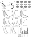
- FIG. 1a shows the protein-induced structural changes of 2Kt-33bp and 2Kt-28bp RNA nanomachines and the three-dimensional modeling results of an RNA-protein complex containing two RNA strands (blue and green) and L7Ae protein (yellow) .
- FIG. 1b shows a schematic diagram of a protein-driven RNA-nanomachine that controls the structure and function of the RNA.
- FIG. 1c shows a schematic diagram of the activation of RNA nanomachines by sensing L7Ae expressed in cells.
- FIG. 1d shows a schematic diagram of the control of cell death induced by intracellular RNP interactions.
- FIG. 2a shows the EMSA results of interaction with 2Kt-33bp (lane 1-6) and 2Kt-28bp (lane 7-12) RNA nanomachines and L7Ae.
- FIG. 2b shows a schematic diagram of FRET and fluorescence measurement results of 2Kt-33bp RNA nanomachine labeled with Cy3 and Cy5 corresponding to L7Ae binding.
- FIG. 2c shows an AFM image of 2Kt-33bp (left) in the absence of L7Ae and 2Kt-33bp (right) in the presence of L7Ae.
- FIG. 2d shows AFM images of 2Kt-28bp in the absence of L7Ae (left) and 2Kt-28bp in the presence of L7Ae (right) RNA nanostructures.
- FIG. 2e shows the measurement results of the distance of both RNA ends to the 2Kt-33bp RNA nanostructure in the presence (red) and absence (black) of L7Ae.
- FIG. 2f shows the measurement results of the distance of both RNA ends to the 2Kt-28bp RNA nanostructure in the presence (red) and absence (black) of L7Ae.
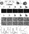
- FIG. 3a shows the fluorescence spectra of biMGA-2Kt-33bp (left) and biMGA-2Kt-33bp (right) in the presence (red) and absence (black) of L7Ae.
- FIG. 3b shows AFM images of biMGA-2Kt-33bpRNA nanostructures in the presence (left) and absence (right) of L7Ae.
- FIG. 3c shows AFM images of biMGA-2Kt-28bp RNA nanostructures in the presence (left) and absence (right) of L7Ae.
- FIG. 3d shows the measurement results of the number of open (black) and closed (red) nanostructures of biMGA-2Kt-33bp or biMGA-2Kt-28bp.
- FIG. 4a shows a schematic diagram of a protein driven RNA nanomachine for detecting intracellular FRET signals.
- FIG. 4b shows a schematic of a FRET assay based on flow cytometry.
- the lower right figure shows the result of plotting the FRET signal, [F 565 / F 665 (+ L7Ae)] / [F 565 / F 665 (-L7Ae)] against RNA duplex length.
- FIG. 4d shows the results of flow cytometry analysis in the activation of RNA nanomachine by L7Ae-K-tur interaction.
- FIG. 4e shows the measurement result of FRET signal when 2Kt-10bp-7bp or 2mKt-10bp-7bp RNA nanomachine in the presence of MS2CP or L7Ae was cotransfected into HeLa cells. All Cy3 / Cy5 mean intensities were normalized with the Cy3 / Cy5 mean intensities of cells co-transfected with MS2CP mRNA and 2mKt-10bp-7bp.
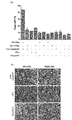
- FIG. 5a shows a schematic diagram of a protein driven RNA nanomachine for detecting intracellular caspase 8 aggregation.
- FIG. 5b shows an AFM image of each nanostructure (1Kt-33bp, 3Kt-33bp, 6Kt-33bp, 9Kt-33bp and 14Kt-33bp) in the presence of L7Ae-caspase-8 mRNA.
- FIG. 5c shows a micrograph of cells transfected with each nanostructure (1Kt-33bp, 3Kt-33bp, 6Kt-33bp, 9Kt-33bp and 14Kt-33bp) and L7Ae-caspase-8 mRNA.
- FIG. 5b shows an AFM image of each nanostructure (1Kt-33bp, 3Kt-33bp, 6Kt-33bp, 9Kt-33bp and 14Kt-33bp) in the presence of L7Ae-caspase-8 mRNA.
- FIG. 5d introduces each nanostructure (1Kt-33bp, 3Kt-33bp, 6Kt-33bp, 9Kt-33bp and 14Kt-33bp) (left) and the corresponding nanostructure with the mutant Kt (right)
- the analysis results are shown by flow cytometry after staining cells with Pacific Blue Annex V staining.
- FIG. 5e shows the result of plotting the number of dead cells against the number of K-turn motifs of the result of FIG. 5d.
- FIG. 6a shows the results of measurement of cell death induced by the interaction of RNA nanostructures having a 14 K-turn motif and L7Ae-caspase-8.
- FIG. 6b shows a microscopic image of the cells after co-transfection of RNA nanostructure (14Kt-33bp or 14mKt-33bp) and mRNA (L7Ae-caspase-8, L7Ae, or caspase-8).
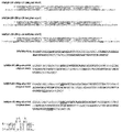
- FIG. 7a shows the structure of 2Kt-30bp.
- FIG. 7b shows the three-dimensional modeling results of the RNA-protein complex containing 2Kt-30bp (blue) and L7Ae protein (yellow).
- FIG. 8 shows the structure of double-stranded RNAN of 2Kt-33bp, 2mKt-33bp, 2Kt-28bp and 2mKt-28bp.
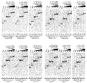
- Figure 9a shows 2Kt-33bp (lane 1-6) or 2mKt-33bp (lane 7-12) (left) or 2Kt-28bp (lane 1-6) or 2mKt-28bp (lane 7-12) (right) ) Shows the result of confirming the L7Ae-K-turn interaction by EMSA in the presence of L7Ae.
- FIG. 10 shows AFM images of 2Kt-33bp (upper figure) and 2Kt-28bp in the presence (right figure) and absence (left figure) of L7Ae.
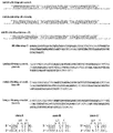
- FIG. 11 shows the structures of biMGA® (orginal® stem) -2Kt-33bp-U0, biMGA® (orginal® stem) -2Kt-33bp-U2, biMGA® (orginal® stem) -2Kt-33bp-U4 and the double-stranded RNAN of the orginal® stem part. Indicates.
- FIG. 11 shows the structures of biMGA® (orginal® stem) -2Kt-33bp-U0, biMGA® (orginal® stem) -2Kt-33bp-U2, biMGA® (orginal® stem) -2Kt-33bp-U4 and the double-stranded RNAN of the orginal® stem part. Indicates.
- FIG. 11 shows the structures of biMGA® (orginal® stem) -2Kt-33bp-U0, biMGA® (orginal® stem) -2Kt-33bp-U2, biMGA® (orginal® stem) -2
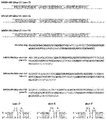
- FIG. 12 shows the structures of biMGA® (orginal® stem) -2Kt-28bp-U0, biMGA® (orginal® stem) -2Kt-28bp-U2, biMGA® (orginal® stem) -2Kt-28bp-U4 and the double-stranded RNAN of the orginal® stem part.
- FIG. 13 shows the measurement results of the fluorescence intensity (FI) of each uridine linker of the biMGA-bound RNA nanostructure in the presence (white) and absence (black) of L7Ae (upper figure).
- FIG. 14 shows biMGA (stem) A-2Kt-33bp-U0, biMGA (stem (B) -2Kt-33bp-U0 and biMGA (stem (C) -2Kt-33bp-U0, and stem (A), stem (B) and stem (C) parts.
- the structure of double-stranded RNAN is shown.
- FIG. 15 shows biMGA (stem D) -2Kt-33bp-U0, biMGA (stem E) -2Kt-33bp-U0 and biMGA (stem F) -2Kt-33bp-U0 and stem D, stem E and stem F parts.
- the structure of double-stranded RNAN is shown.
- FIG. 16 shows the fluorescence intensity (FI) measurement results for each biMGA stem sequence in the biMGA-binding RNA nanostructure in the presence (white) and absence (black) of 7Ae.
- FIG. 17 shows the results of EMSA confirming the L7Ae-K-turn interaction in the presence of each biMGA ⁇ stem sequence and each uridine linker L7Ae in the biMGA-binding RNA nanostructure.
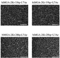
- FIG. 18 shows AFM images of biMGA-2Kt-33bp (upper figure) and biMGA-2Kt-28bp (lower figure) in the presence (right figure) and absence (left figure) of L7Ae. In the figure, the scale bar indicates 100 nm.
- FIG. 19 shows the structure of double-stranded RNAN of 2Kt-17bp-4bp, 2Kt-17bp-5bp, 2Kt-17bp-6bp, 2Kt-17bp-7bp, 2mKt-17bp-7bp and 2Kt-17bp-8bp.
- FIG. 20 shows the results of measuring the Casapase-8 activity when transfected with an RNA nanostructure having a 14 -K-turn motif and L7Ae-caspase-8. The caspase-8 activity of Hela cells transfected with RNA nanostructures and mRNA was measured using the fluorogenic substrate IETD-AMC.
- RNA Nanostructure 14Kt-33bp or 14mKt-33bp
- mRNA L7Ae, dcasp-8, L7Ae-dcasp-8 (L7Ae-caspase-8), L7Ae-dcasp-8-CS, DED-caspase-8, L7Ae -DED-caspase-8 or L7Ae-DED-caspase-8-CS
- HeLa cells PacificPBlue Annexin V staining and dead cell staining
- Pacific ⁇ ⁇ ⁇ Blue-positive cells are dead cells by flow cytometry As counted.
- the present invention provides an RNA-protein complex and a method for designing an RNA-protein complex for activating the function of a functional molecule added to the RNA or protein in the RNA-protein complex.
- RNA-protein complex of the present invention is a first RNA-protein complex comprising the following (1) and (2): (1) 2 comprising two RNA-protein binding motif sequences A heavy-chain RNA having functional molecules at both ends of the double-stranded RNA, and (2) a protein that binds to the RNA-protein binding motif sequence.
- RNA-protein complex of the present invention is a second RNA-protein complex comprising the following (1) and (2): (1) two RNA-protein binding motif sequences (2) a fusion protein of a protein that binds to the RNA-protein binding motif sequence and an aggregated protein.
- RNA-protein binding motif sequence contained in the double-stranded RNA of the present invention is the RNA sequence contained in the binding motif of RNA and protein in a natural or known RNA-protein complex, or in vitro evolution (in vitro selection method) RNA sequence contained in the artificial RNA-protein binding motif obtained by (Method).
- Natural RNA-protein binding motif sequences are usually composed of about 5 to 70 bases, and RNA having such sequences is non-covalently bonded to a protein having a specific amino acid sequence, that is, by hydrogen bonding. It is known to form specific bonds.
- Such natural RNA-protein binding motif sequences can be found in Tables 1 and 2 below and the database available on the website: http: // gibk26. bse. kyutech. ac. jp / jouhou / image / dna-protein / RNA / RNA. From html, it can be obtained by appropriately selecting from motifs that cause a desired structural change.
- RNA-protein binding motif preferably used in this embodiment can be estimated from the three-dimensional structure of a homologous protein that has already undergone an X-ray crystal structure analysis or NMR structure analysis or a structural analysis. Motif. Furthermore, it is desirable that the protein is a motif that specifically recognizes the secondary structure and base sequence of RNA.
- the RNA containing an artificial RNA-protein binding motif sequence is an RNA contained in one of the double-stranded RNAs in the binding motif between a double-stranded RNA and a protein in an artificially designed RNA-protein complex. It is.
- the base sequence of such RNA is usually composed of about 10 to 80 bases, and forms a specific bond with a specific amino acid sequence of a specific protein non-covalently, that is, by hydrogen bonding.
- An RNA aptamer that specifically binds to a specific protein is exemplified as the RNA containing such an artificial RNA-protein binding motif sequence.
- RNA aptamer that specifically binds to a desired target protein can be obtained by, for example, an evolutionary engineering technique known as an in vitro selection method or a SELEX method.
- the trigger protein at this time is a protein to which the RNA aptamer binds.
- the RNA sequences listed in Table 3 below are known, and these can also be used as sequences forming the RNA-protein binding motif of the present invention.
- the RNA-protein binding motif sequence preferably has a dissociation constant Kd with the corresponding protein of about 0.1 nM to about 1 ⁇ M.
- variants of such sequences are also encompassed by the sequences according to the present invention.
- the variant referred to in the present invention is a mutation having a dissociation constant Kd of 10%, 20%, 30%, 40% or 50% or more higher than a protein that specifically binds to a sequence forming an RNA-protein binding motif. Body or 10%, 20%, 30%, 40% or 50% or less mutant. Such mutants can be appropriately selected and used as long as an RNA-protein complex can be formed.
- the base sequence of such a variant can hybridize under stringent conditions with a nucleic acid (complementary strand) having a sequence complementary to the sequence (positive strand) forming the RNA-protein binding motif.
- a base sequence of a degree may be used.
- the stringent conditions here are the melting temperature (Tm) of the nucleic acid to be bound as taught by Berger and Kimmel (1987, GuideGto Molecular Cloning Techniques Methods in Enzymology, Vol.152, Academic Press, San Diego CA). Can be determined based on For example, as washing conditions after hybridization, the conditions of about “1 ⁇ SSC, 0.1% SDS, 37 ° C.” can be mentioned.
- the complementary strand is preferably one that maintains a hybridized state with the target positive strand even when washed under such conditions.
- washing is performed under more severe hybridization conditions such as “0.5 ⁇ SSC, 0.1% SDS, 42 ° C.”, more strictly “0.1 ⁇ SSC, 0.1% SDS, 65 ° C.”
- the conditions for maintaining the hybridized state between the positive strand and the complementary strand can be mentioned. Specifically, it comprises a base sequence having at least 90%, preferably at least 95%, 96%, 97%, 98% or 99% sequence identity with the RNA sequence contained in the RNA-protein binding motif described above. .
- Such mutants can maintain a constant bond with a protein that specifically binds to a sequence that forms an RNA-protein binding motif and contribute to the formation of an RNA-protein complex.
- RNA-protein binding motif is a sequence to which L7Ae (Moore T et al., Structure Vol. 12, pp. 807-818 (2004)) binds.
- K-turn (Kt) motif (5′-GGCGUGAUGAGC-3 ′ / 5′-GCUCUGACC-3 ′) (SEQ ID NO: 1 / SEQ ID NO: 2), kink-loop (SEQ ID NO: 3), kink-loop2 (SEQ ID NO: 4) ).
- 5′-NNCRUGAUNNNNN-3 ′ / 5′-NNNNUNGANN-3 ′ N represents any base, R represents A or G as a mutant of the K-turn (Kt) motif retaining the binding activity to L7Ae
- N represents any base
- R represents A or G as a mutant of the K-turn (Kt) motif retaining the binding activity to L7Ae
- 5′-GGCAUGAUGAAC-3 ′ / 5′-GUUCUGACC-3 ′ SEQ ID NO: 5 / SEQ ID NO: 6
- MS2 stem loop motif 22: Keryer-Bibens C, Barreau C, Osborne HB (2008) Tethering of proteins to RNAs by bacteriophage proteins. Biol Cell 100: 125-138
- Fr15 24: Batey RT, Williamson JR (1996) Interaction of the Bacillus stearothermophilus ribosomal protein S15 with 16 S rRNA: I. Defining the minimal RNA . J Mol Biol 261: 536-549).
- a further specific example is an enzyme that performs aminoacylation, which is known to have Threonyl-tRNA synthetase (Cell (Cambridge, Mass.) V97, which binds to its own mRNA and has feedback inhibition that inhibits translation.
- Cell Cell (Cambridge, Mass.) V97, which binds to its own mRNA and has feedback inhibition that inhibits translation.
- pp.371-381 (1999) which is a sequence to which 5′-GGCGGUAUGUGAUCUUGUCGUUGGGUCACCACUGCGCC-3 ′ (SEQ ID NO: 31), and variants thereof are present.
- R9-2 5′-GGGGUGUCGAGCUAGGGAAGAAAGCCGGGGGCUGCAGAUAAUGUUAUAGC-3 ′ (SEQ ID NO: 32), which is a base sequence forming an RNA-protein binding motif derived from Bcl-2 family CED-9, which is a cancer cell-specific endogenous protein , And variants thereof, nucleotide sequences derived from aptamers of RNA sequences that bind to NF-kappaB, and variants thereof.
- RNA sequence between the RNA-protein binding motif sequences in the double-stranded RNA contained in the RNA-protein complex of the present invention may be any sequence, for example, complementary sequences such as palindrome.
- a sequence that does not include a sequence and does not take tertiary structure by RNA sequence alone is preferable.
- the distance between RNA-protein binding motif sequences is the number of base pairs between RNA-protein binding motif sequences contained in the double-stranded RNA, and the double strands contained in the RNA-protein binding motif sequences. The part that forms is also counted as the distance.
- Kt K-turn
- the RNA-protein binding motif sequence is 5 ′ -NNCRUGAU- (N) n-CRUGAUNNNNN-3 ', where n is an integer, in which case the distance is n + 1.
- the distance between the RNA-protein binding motif sequences of the double-stranded RNA contained in the first RNA-protein complex of the present invention is 33 base pairs in order to retain the first RNA-protein complex as a triangular structure.
- it since it rotates 360 ° with 11 base pairs, it may be 11 base pairs and 22 base pairs and 44 bases, 55 base pairs, and multiples of 11 more.
- 9 base pairs to 13 base pairs, 20 base pairs to 24 base pairs, 31 base pairs to 35 base pairs, 42 bases Examples include 46 to 50 base pairs and 53 to 57 base pairs.
- the first RNA-protein complex is retained as a Z-shaped structure. 28 base pairs, but since it rotates 360 ° with 11 base pairs, it is 6 base pairs and 17 base pairs and 39 bases, 50 base pairs, and multiples of 11 + 6 base pairs. There may be. Further, since it is highly possible that the increase and decrease of 2 base pairs have the same effect, 4 base pairs to 8 base pairs, 15 base pairs to 19 base pairs, 26 base pairs to 30 base pairs, 37 bases Examples are 41 base pairs from pairs and 52 base pairs from 48 base pairs.
- the distance between RNA-protein binding motif sequences is preferably 25 base pairs or less in order to prevent interferon activity.
- the distance of the RNA sequence between the RNA-protein binding motif sequences can be adjusted as appropriate.
- FIG. 7 shows the structure of a first RNA-protein complex having 30 base pairs between RNA-protein binding motif sequences.
- the functional molecules described later are put into a functional state by binding the ends on both sides in the absence of the corresponding protein. Furthermore, in order to maintain the loop structure, an overhang of 6 base pairs or more that are complementary strands may be added to the ends.
- the overhang means a state in which a protruding single-stranded structure is added at the 3 'end or 5' end in RNA having a double-stranded structure.
- a functional molecule is added to the 3 'end and 5' end of the double-stranded RNA of the first RNA-protein complex of the present invention.
- a functional molecule is a molecule that enables coloration, fluorescence, luminescence, specific binding to other molecules, specific binding inhibition between molecules, enzyme activity, enzyme activity inhibition, and the like. May be a low-molecular compound or a polymer that functions in vivo, such as DNA, RNA, or protein. Examples include fluorescent dyes, antibodies, RNA interference, and RNA aptamers.
- the functional molecules added to the 3 ′ end and 5 ′ end of the double-stranded RNA of the first RNA-protein complex of the present invention include molecules added to the 3 ′ end and molecules added to the 5 ′ end. It is preferable to exhibit the function by interacting with each other.
- an RNA-protein complex is designed as a triangular structure, the function is exhibited in the presence of the corresponding protein, but cannot be performed in the absence of the corresponding protein, so the corresponding protein is present. Alternatively, the presence of the function can be controlled by the absence.
- the RNA-protein complex is designed as a Z-type structure, it does not exhibit the function in the presence of the corresponding protein, but it does the function in the absence of the corresponding protein. It is possible to control the performance of the function by the presence or absence of.
- a fluorescent dye causing fluorescence resonance energy transfer is added to the 5 'end and the 3' end.
- fluorescent dyes include Cy3 and Cy5.
- Cy3 and Cy5 interact in the presence of the corresponding protein to cause fluorescence resonance energy transfer, but in the absence of the corresponding protein, fluorescence resonance Since energy transfer does not occur, the function (fluorescence resonance energy transfer) can be controlled by the presence or absence of the corresponding protein.
- RNA-protein complex when the RNA-protein complex is designed as a Z-shaped structure, Cy3 and Cy5 do not interact and fluorescence resonance energy transfer does not occur in the presence of the corresponding protein, but in the absence of the corresponding protein, Since Cy3 and Cy5 interact to cause fluorescence resonance energy transfer, the function (fluorescence resonance energy transfer) can be controlled by the presence or absence of the corresponding protein.
- RNA aptamer when used as a functional molecule, an embodiment in which a sense strand of the RNA aptamer is added to the 5 'end and an antisense strand is added to the 3' end is exemplified.
- RNA aptamers include malachite green aptamer, and any one sequence selected from the group consisting of SEQ ID NOs: 44, 46, 48, 50, 52, 54 and 56 is added to the 5 ′ end, and 3 It is exemplified to add any one sequence selected from the group consisting of SEQ ID NOs: 45, 47, 49, 51, 53, 55 and 57 to the end.
- RNA-protein complex when the RNA-protein complex is designed as a triangular structure, an RNA aptamer is formed in the presence of the corresponding protein, but no RNA aptamer is formed in the absence of the corresponding protein. It is possible to control the function (binding ability as an RNA aptamer) by the presence or absence of.
- an RNA-protein complex is designed as a Z structure, an RNA aptamer is not formed in the presence of the corresponding protein, but an RNA aptamer is formed in the absence of the corresponding protein. By making it exist or not present, it becomes possible to control the function (binding ability as an RNA aptamer).
- the interaction of functional molecules added as described above to both ends of the double-stranded RNA contained in the first RNA-protein complex of the present invention is controlled by the presence or absence of the corresponding protein. Can do. Therefore, the present invention binds a double-stranded RNA having two RNA-protein binding motif sequences and having functional molecules at both ends to a protein that binds to the RNA-protein binding motif sequence, thereby binding the functional molecule.
- the double-stranded RNA containing two or more RNA-protein binding motif sequences used in the method is a double-stranded RNA contained in the first RNA-protein complex described above.
- the interaction between the RNA-protein binding motif sequences of the double-stranded RNA is adjusted in the presence of the corresponding protein.
- positive control means that the interaction of functional molecules added to both ends of the double-stranded RNA occurs, and similarly, negative control means that the interaction of functional molecules is the same. Means to stop.
- the functional molecule interaction is, for example, as described above, fluorescence resonance energy transfer when the functional molecule is a fluorescent dye, and RNA due to binding of complementary strands when the functional molecule is an RNA aptamer. It means the formation of aptamer.
- the distance between the RNA-protein binding motif sequences of the double-stranded RNA of the first RNA-protein complex is from 20 base pairs to 24 base pairs, or 31 base pairs to 35 base pairs, or 42 base pairs to 46 base pairs.
- the distance between the RNA-protein binding motif sequences of the double-stranded RNA of the first RNA-protein complex is 15 base pairs to 19 base pairs, or 26 base pairs to 30 base pairs, or 37 base pairs to 41 bases.
- the protein that binds to the RNA-protein binding motif sequence contained in the second RNA-protein complex of the present invention is a fusion protein with the aggregated protein, and the fusion protein binds to the RNA-protein binding motif sequence. It means that the protein and the aggregated protein are a single protein bound via a linker or the like.
- the aggregated protein means a protein that exhibits a function by aggregation, and includes, for example, apoptosis control protein, prion protein, caspase 9, caspase 8, amyloid ⁇ , tau, polyglutamine protein (Gatchel JR, Zoghbi HY. Nat Rev Gene 6, 743-755, 2005) or fragments of these proteins, as well as Apoptosis-associated speck-like protein containing caspase recruitment (domain (ASC) (Hara H, et al, Nat Immunol. 14, 1247- 55, 2013).
- ASC Apoptosis-associated speck-like protein containing caspase recruitment
- the distance of the RNA sequence between the RNA-protein binding motif sequences of the double-stranded RNA contained in the second RNA-protein complex of the present invention is not particularly limited, and examples thereof include 15 to 52 base pairs. . It is preferable to exhibit a repeating structure based on a triangle-like structure in the RNA-protein complex to which the corresponding protein is bound, and thus the distance of the RNA sequence between the RNA-protein binding motif sequences is 9 to 13 base pairs. Examples include base pairs, 20 base pairs to 24 base pairs, 31 base pairs to 35 base pairs, 42 base pairs to 46 base pairs, and 53 base pairs to 57 base pairs.
- the fusion protein of the aggregated protein and the protein corresponding to the RNA-protein complex may control the aggregation of the non-aggregated protein by the presence or absence of the second RNA-protein complex of the present invention. it can. Therefore, the present invention binds a double-stranded RNA containing two or more RNA-protein binding motif sequences, a protein that binds to the RNA-protein binding motif sequence, and a protein fused with the aggregated protein, thereby allowing the aggregated protein to bind.
- a method of controlling aggregation of The double-stranded RNA containing two or more RNA-protein binding motif sequences used in the method is a double-stranded RNA contained in the second RNA-protein complex described above.
- the double-stranded RNA used in the method for controlling the aggregation of the aggregated protein of the present invention in order to exert the function by aggregating many aggregated proteins contains 9 or more RNA-protein binding motif sequences. Are preferable, for example, 9 or 14 may be mentioned.
- the double-stranded RNA may be replaced with a modified base such as pseudouridine or 5-methylcytidine instead of ordinary uridine or cytidine in order to reduce cytotoxicity.
- a modified base such as pseudouridine or 5-methylcytidine instead of ordinary uridine or cytidine in order to reduce cytotoxicity.
- the positions of the modified bases can be all or part of the uridine and cytidine independently, and if they are part of the base, they can be random positions at an arbitrary ratio.
- RNA sequences used in this example are shown in Table 4. Mutant K-turns in 2mKt-33bp, 2mKt-28bp and 2mKt-17bp-7bp were prepared by appropriately replacing mKt in Table 4 in the long chain.
- biMGA-2Kt-28bp having biMGA-original stem biMGA-stem A, biMGA-stem B, biMGA-stem C, biMGA-stem D, biMGA-stem E and biMGA-stem F, 4 were prepared by appropriately replacing each of the stems.
- RNA nanomachines (1Kt-33bp, 3Kt-33bp, 6Kt-33bp, 9Kt-33bp, 14Kt-33bp and 1mKt-33bp, 3mKt-33bp, 6mKt-33bp, 9mKt-33bp, 14mKt-33bp) and mRNA introduced into living cells
- uridine triphosphate and cytidine triphosphate are substituted for pseudouridine-5′-triphosphate and methylcytidine-5′- Adjustments were made using triphosphate (TriLink BioTechnologies).
- Cy3 or Cy5 labeled RNA was prepared by ligation using T4 RNA ligase (Ambion) and pCp-Cy3 (Jena Bioscience) or pCp-Cy5 (Jena Bioscience). Cy3 or Cy5 labeling was performed by mixing 10 U of T4 RNA ligase, 3 nmol of pCp-Cy3 or pCp-Cy5 and 150 pmol of RNA at 16 ° C. for 36-48 hours in 10 ⁇ l of 10% (v / v) DMSO. . Cy3 or Cy5 labeled RNA was purified with RNeasy MinElute Cleanup Kit (Qiagen). After RNA recovery, Cy3 or Cy5 and RNA concentrations were measured with NanoDrop (Thermo Scienctific).
- ⁇ Electrophoretic mobility shift analysis (EMSA)> A mixture of L-RNA and S-RNA (50 nM each) in 20 mM phosphate buffer (pH 7.0) containing 1.5 mM MgCl 2 and 150 mM KCl was heated at 80 ° C. for 3 minutes, then at room temperature for 10 minutes. Cooled for minutes. After adding L7Ae (100, 300, and 600 nM), the mixture was incubated at room temperature for 10-30 minutes. The mixture was mixed with 10 ⁇ additive solution (0.25% bromophenol blue, 30% glycerol), and subjected to native polyacrylamide gel electrophoresis using 0.5 ⁇ Tris / Borate / EDTA buffer at room temperature. The gel after electrophoresis was stained with SYBR I and II (Lonza) for 10 minutes. Gel images were obtained using Gel Doc EZ Image r (BIO-RAD) or Typhoon FLA-7000 laser scanner (GE Healthcare).
- RNA and RNP nanostructures were observed in solution.
- This sample was prepared as described in EMSA.
- Small cantilevers (length 9 ⁇ m, width 2 ⁇ m and thickness 130 nm; spring constant about 0.1 N / m, underwater about 300-600 kHz and about 1500 kHz in the air, BL- AFM images were obtained using AC10DS, Olympus, Tokyo, Japan) and HS-AFM (Nano Live Vision, Research Institute of Biomolecules Metrology Co.).
- a sharp probe was placed on each cantilever and electron beam evaporation using a Nano-tool instrument (Munich) was used.
- a 320 x 240 pixel image was obtained at a scan rate of 0.2-1.0 frames per second (fps).
- a new mica surface was coated with 0.1% APTES (3-aminopropyltriethoxy silane).
- APTES 3-aminopropyltriethoxy silane
- Prepare this sample 50 nM RNA nanostructure with and without 600 nM protein
- RNP binding buffer 0.1%
- AFM observation buffer (20 mM Tris-HCl (pH 7.6) and 10 mM MgCl2). Diluted twice. Thereafter, it was applied to mica for 5 minutes at room temperature and washed with a buffer solution.
- Image sequences were analyzed with Image J and WSxM software.
- RNA nanostructure 100 nM was heated at 80 ° C. for 3 minutes and cooled in 20 mM phosphate buffer (pH 7.0) containing 1.5 mM MgCl 2 and 150 mM KCl for 10 minutes at room temperature.
- Malachite green (2 ⁇ M) protein buffer (20 mM phosphate buffer (pH 7.0), 1.5 mM MgCl 2 , 150 mM KCl, 40% glycerol) or L7Ae (600 nM) and 0.1% tween-20 And incubated at room temperature for 10-30 minutes.
- the excitation wavelength was 610 nm and the emission and excitation bandwidths were 10 and 20 nm, respectively. Fluorescence measurement was performed using Infinite M1000 Plate-Reader (TECAN).
- RNA transfection and flow cytometry analysis 5 ⁇ 10 4 cells were seeded in 12 well plates. After 24 hours of culture, RNA nanomachines (8 pmol) and mRNA (500 ng) were cotransfected into the cells using 2 ⁇ L Stem Fect (STEMGENT). After 2 hours, the medium was changed. Cells were washed with PBS 12 hours after transfection and incubated in 200 ⁇ L of 0.25% Tripsin-EDTA (Nacalai tesque) for 3 minutes at 37 ° C. After adding 100 ⁇ L of Dulbecco's PBS containing 2% fetal bovine serum, the cells were analyzed with a FACSAria cell sorter (BD Bioscience).
- a 532 nm laser and a 575/25 nm optical filter were used to detect Cy3 fluorescence.
- a 532 nm laser and a 670/30 nm optical filter were used to detect Cy5 fluorescence attributed to fluorescence resonance energy transfer (FRET).
- FRET fluorescence resonance energy transfer
- dead cells were removed using a dot plot of SSC against FSC.
- FRET fluorescence resonance energy transfer
- RNA nanomachines Two types of RNP nanostructures containing two L7Ae-K-turn mutual motifs were designed (FIG. 1a).
- the K-turn motif takes a flexible three-dimensional structure in the absence of L7Ae binding, but changes in the three-dimensional structure by bending at about 60 ° C in the K-turn motif due to the L7Ae-K-turn interaction. cause.
- Two K-turn motifs were placed between 3 double-stranded RNAs of 33, 30 and 28 base pairs to correspond to 3 and 2.5 helical turns (2Kt-33bp, respectively) 2Kt-30bp and 2Kt-28bp) (FIGS.
- RNA nanomachines consist of long chains that interact with each other (L chain: long in the K-turm motif) and short chains (S chain: short in the K-turm motif).
- L chain long in the K-turm motif
- S chain short in the K-turm motif
- RNA nanomachines In order to observe the structural changes of RNA nanomachines, analysis was performed with a high-speed atomic force microscope (HS-AFM) before and after the addition of L7Ae to 2Kt-33bp and 2Kt-28bp (FIGS. 2c and d). In the absence of L7Ae, 2Kt-33bp and 2Kt-28bp showed heterogeneous RNA structure, but in the presence of L7Ae showed a single structure. 2Kt-33bp and 2Kt-28bp cause conformational changes upon introduction of L7Ae, and are designed to form triangular and Z-shaped structures, respectively (FIGS. 2c, 2d and 10). The distance between the ends was measured (FIGS. 2e and f).
- RNA nanomachine was constructed by adding the divided binary malachite green aptamer (biMGA) to the ends. BiMGA probes were added with 2Kt-33bp and 2Kt-28bp, and activation and repression control of aptamers by RNP formation was investigated.
- biMGA binary malachite green aptamer
- BiMGA-added RNA nanomachines (referred to as biMGA-2Kt-33bp and biMGA-2Kt-28bp, respectively) were measured for fluorescence spectra before and after contact with L7Ae in the presence of malachite green (MG). We examined the regulation of RNA.
- the linker length (0-4 base pairs) between RNA nanomachine and biMGA and the biMGA stem sequence (biMGA-original stem, biMGA-stem A, biMGA-stem B, biMGA-stem C, biMGA-stem D, biMGA-stem E and biMGA-stem F), the linker length is 0 base pairs, stem A for biMGA-2Kt-33bp, stem D for biMGA-2Kt-28bp 2 biMGA-added RNA nanomachines (biMGA-2Kt-33bp and biMGA-2Kt-28bp) that can activate or suppress biMGA by RNP formation were produced (FIG.
- biMGA-2Kt-33bp and biMGA-2Kt-28bp were confirmed to form RNP nanostructures having a triangular structure or a Z-type structure, respectively (right figure in FIGS. 3b and c). .
- the closed and open fractions in the presence and absence of L7Ae were quantified (FIG. 3d).
- the fractions closed and open before and after RNP formation were 19% and 81%, respectively.
- the fraction of closed biMGA-2Kt-33bp increased to 92%.
- the open fraction of biMGA-2Kt-28bp increased from 47% to 75% due to RNP formation.
- RNA nanomachines by proteins expressed in living cells.
- Proteins can be used as biocompatible materials that can exhibit functional expression. Therefore, we examined whether RNA nanomachines can operate in response to L7Ae expressed in cells.
- flow cytometry was performed by FRET analysis using RNA nanomachines labeled with Cy3 and Cy5 (FIGS. 4a and b).
- RNA nanomachines labeled with Cy3 and Cy5 In order to suppress interferon activity by RNA nanomachine, 2Kt-17bp with two K-turn motifs including three 17 bp double-stranded RNAs was constructed.
- RNA nanomachines that change from a circular structure to a Z-like form by RNP interaction (each 2Kt-17bp- 4bp, 2Kt-17bp-5bp, 2Kt-17bp-6bp, 2Kt-17bp-7bp and 2Kt-17bp-8bp) (FIGS. 4a and 19).
- the fluorescence spectra of RNA nanomachines labeled with Cy3 and Cy5 were measured (FIG. 4c).
- RNA nanomachine (2Kt-17bp-7bp or 2mKt-17bp-7bp) and mRNA encoding L7Ae or MS2CP were simultaneously introduced into HeLa cells (FIG. 4b). Involvement of intracellular RNA nanomachines in the change of the FRET signal was analyzed using a flow cytometer (FIGS. 4d and e).
- RNA nanostructures with 1, 3, 6, 9 or 14 K-turn motifs (referred to as 1Kt-33bp, 3Kt-33bp, 6Kt-33bp, 9Kt-33bp and 14Kt-33bp, respectively) was designed (FIG. 5b). Mutants having the corresponding mutant K-turn motif (referred to as 1mKt-33bp, 3mKt-33bp, 6mKt-33bp, 9mKt-33bp and 14mKt-33bp, respectively) were used as negative controls.
- each RNA nanostructure and L7Ae-caspase-8 mRNA are simultaneously introduced into HeLa cells. did. Analysis of cell morphology showed that cell death was induced each time the number of K-turn motifs increased (upper figure in FIG. 5c). On the other hand, cell death was not confirmed in the RNA nanostructure having the mutant K-turn motif (lower figure in FIG. 5c). In order to quantify cell death, it was stained with Pacific Blue Annexin V and analyzed using a flow cytometer.
- 14Kt-33bp or 14mKt-33bp and L7Ae-caspase were transfected into Hela cells and fluorescence was generated.
- Caspase-8 activity was measured using the substrate IETD-AMC. The highest caspase-8 activity was observed when 14Kt-33bp was combined with L7Ae-caspase mRNA, and the activity was clearly reduced by the addition of caspase-8 inhibitor.
- RNA nanostructure 14Kt-33bp or 14mKt-33bp
- mRNA L7Ae [SEQ ID NO: 92], dcasp-8 [SEQ ID NO: 87], L7Ae-dcasp -8 (L7Ae-caspase-8) [SEQ ID NO: 65], L7Ae-dcasp-8-CS [SEQ ID NO: 88], DED-caspase-8 [SEQ ID NO: 89], L7Ae-DED-caspase-8 [SEQ ID NO: 90]
- L7Ae-DED-caspase-8-CS SEQ ID NO: 91]
- L7Ae-DED-caspase-8 is an mRNA obtained by adding a DED sequence to L7Ae-dcasp-8.
- L7Ae-dcasp-8-CS and L7Ae-DED-caspase-8-CS are mutants that do not cause caspase activation of the corresponding L7Ae-dcasp-8 and L7Ae-DED-caspase-8.
- High cell death activity was observed in 14Kt-33bp, L7Ae-dcasp-8 and L7Ae-DED-caspase-8.
- almost no cell death was confirmed in other combinations. From the above results, it was shown that caspase-8 was activated by the aggregation of L7Ae-caspase-8 via L7Ae-K-turn interaction on the RNA nanostructure and caused cell death.
Landscapes
- Health & Medical Sciences (AREA)
- Engineering & Computer Science (AREA)
- Chemical & Material Sciences (AREA)
- Life Sciences & Earth Sciences (AREA)
- Genetics & Genomics (AREA)
- Organic Chemistry (AREA)
- Biomedical Technology (AREA)
- Bioinformatics & Cheminformatics (AREA)
- Zoology (AREA)
- General Engineering & Computer Science (AREA)
- Wood Science & Technology (AREA)
- Biotechnology (AREA)
- Molecular Biology (AREA)
- General Health & Medical Sciences (AREA)
- Biochemistry (AREA)
- Biophysics (AREA)
- Microbiology (AREA)
- Medicinal Chemistry (AREA)
- Nanotechnology (AREA)
- Plant Pathology (AREA)
- Physics & Mathematics (AREA)
- Medical Informatics (AREA)
- Crystallography & Structural Chemistry (AREA)
- Pharmacology & Pharmacy (AREA)
- Gastroenterology & Hepatology (AREA)
- Proteomics, Peptides & Aminoacids (AREA)
- Peptides Or Proteins (AREA)
- Medicines That Contain Protein Lipid Enzymes And Other Medicines (AREA)
- Micro-Organisms Or Cultivation Processes Thereof (AREA)
- Medicinal Preparation (AREA)
- Pharmaceuticals Containing Other Organic And Inorganic Compounds (AREA)
Abstract
La présente invention concerne : un complexe ARN-protéine comprenant un ARN duplex qui comprend deux séquences à motif de liaison ARN-protéine et présente des molécules fonctionnelles au niveau des deux extrémités, et une protéine qui se lie aux séquences à motif de liaison ARN-protéine ; ou un complexe ARN-protéine comprenant une protéine obtenue par fusion d'une protéine à agglutiner et d'une protéine qui se lie à au moins deux séquences à motif de liaison ARN-protéine, ainsi qu'un ARN duplex comprenant lesdites au moins deux séquences à motif de liaison ARN-protéine.
Priority Applications (1)
| Application Number | Priority Date | Filing Date | Title |
|---|---|---|---|
| JP2017528737A JPWO2017010568A1 (ja) | 2015-07-16 | 2016-07-15 | Rna−タンパク質複合体とその使用 |
Applications Claiming Priority (2)
| Application Number | Priority Date | Filing Date | Title |
|---|---|---|---|
| JP2015-142315 | 2015-07-16 | ||
| JP2015142315 | 2015-07-16 |
Publications (1)
| Publication Number | Publication Date |
|---|---|
| WO2017010568A1 true WO2017010568A1 (fr) | 2017-01-19 |
Family
ID=57758033
Family Applications (1)
| Application Number | Title | Priority Date | Filing Date |
|---|---|---|---|
| PCT/JP2016/071029 Ceased WO2017010568A1 (fr) | 2015-07-16 | 2016-07-15 | Complexe arn-protéine et utilisation associée |
Country Status (2)
| Country | Link |
|---|---|
| JP (1) | JPWO2017010568A1 (fr) |
| WO (1) | WO2017010568A1 (fr) |
Cited By (1)
| Publication number | Priority date | Publication date | Assignee | Title |
|---|---|---|---|---|
| CN114040774A (zh) * | 2019-02-15 | 2022-02-11 | 阿特雷卡公司 | 结合肿瘤组织用于诊断和治疗的抗体 |
Citations (2)
| Publication number | Priority date | Publication date | Assignee | Title |
|---|---|---|---|---|
| WO2010010862A1 (fr) * | 2008-07-22 | 2010-01-28 | 独立行政法人科学技術振興機構 | Procédé de construction de nanostructure de prn artificielle en utilisant un motif d'interaction complexe arn-protéine |
| WO2016006628A1 (fr) * | 2014-07-09 | 2016-01-14 | 国立大学法人京都大学 | Complexe arn-protéine, et système d'administration d'arn et de protéine à l'aide dudit complexe |
-
2016
- 2016-07-15 JP JP2017528737A patent/JPWO2017010568A1/ja active Pending
- 2016-07-15 WO PCT/JP2016/071029 patent/WO2017010568A1/fr not_active Ceased
Patent Citations (2)
| Publication number | Priority date | Publication date | Assignee | Title |
|---|---|---|---|---|
| WO2010010862A1 (fr) * | 2008-07-22 | 2010-01-28 | 独立行政法人科学技術振興機構 | Procédé de construction de nanostructure de prn artificielle en utilisant un motif d'interaction complexe arn-protéine |
| WO2016006628A1 (fr) * | 2014-07-09 | 2016-01-14 | 国立大学法人京都大学 | Complexe arn-protéine, et système d'administration d'arn et de protéine à l'aide dudit complexe |
Non-Patent Citations (4)
| Title |
|---|
| OHNO H ET AL.: "Synthetic RNA-protein complex shaped like an equilateral triangle", NATURE NANOTECH, vol. 6, no. 2, February 2011 (2011-02-01), pages 116 - 120, XP055346447 * |
| OSADA E ET AL.: "Engineering RNA-Protein Complexes with Different Shapes for Imaging and Therapeutic Applications", ACS NANO, vol. 8, no. 8, 26 August 2014 (2014-08-26), pages 8130 - 8140, XP055346446 * |
| SHIBATA T ET AL.: "RNA-protein nanomachines for controlling both RNA structures and functions", THE 41ST INTERNATIONAL SYMPOSIUM ON NUCLEIC ACIDS CHEMISTRY PROGRAM & ABSTRACTS, 17 November 2014 (2014-11-17), pages 380 - 381 * |
| SHIBATA T ET AL.: "Synthetic RNA-protein nanomachines that function in vitro and in live cell", THE 42ND INTERNATIONAL SYMPOSIUM ON NUCLEIC ACIDS CHEMISTRY PROGRAM & ABSTRACTS, 16 December 2015 (2015-12-16), pages 72 - 73 * |
Cited By (3)
| Publication number | Priority date | Publication date | Assignee | Title |
|---|---|---|---|---|
| CN114040774A (zh) * | 2019-02-15 | 2022-02-11 | 阿特雷卡公司 | 结合肿瘤组织用于诊断和治疗的抗体 |
| JP2022520479A (ja) * | 2019-02-15 | 2022-03-30 | アトレカ インコーポレイテッド | 診断及び療法のための腫瘍組織に結合する抗体 |
| JP7365421B2 (ja) | 2019-02-15 | 2023-10-19 | アトレカ インコーポレイテッド | 診断及び療法のための腫瘍組織に結合する抗体 |
Also Published As
| Publication number | Publication date |
|---|---|
| JPWO2017010568A1 (ja) | 2018-04-26 |
Similar Documents
| Publication | Publication Date | Title |
|---|---|---|
| Autour et al. | Fluorogenic RNA Mango aptamers for imaging small non-coding RNAs in mammalian cells | |
| Estévez-Torres et al. | DNA compaction: fundamentals and applications | |
| EP3964583A1 (fr) | Molécule d'acide nucléique aptamère et application correspondante | |
| JP5503062B1 (ja) | ターゲット分析用蛍光センサ、ターゲット分析用キット、およびこれを用いたターゲットの分析方法 | |
| I Hernandez et al. | Aptamers overview: selection, features and applications | |
| Bi et al. | Self-assembled multifunctional DNA nanospheres for biosensing and drug delivery into specific target cells | |
| EP3211083A1 (fr) | Aptamères fluorescents et leurs applications | |
| US11434490B2 (en) | Fluorogen-binding RNA aptamers | |
| Sato et al. | The effect of backbone structure on polycation comb-type copolymer/DNA interactions and the molecular assembly of DNA | |
| Torelli et al. | Isothermal folding of a light-up bio-orthogonal RNA origami nanoribbon | |
| Bian et al. | Xeno nucleic acids as functional materials: from biophysical properties to application | |
| JP6969810B2 (ja) | Rna−タンパク質複合体及びこれによるrnaおよびタンパク質のデリバリーシステム | |
| WO2017010568A1 (fr) | Complexe arn-protéine et utilisation associée | |
| CN102245766B (zh) | 采用RNP基序构建蛋白应答shRNA/RNAi控制体系 | |
| Moriyama et al. | DNA assembly and re-assembly activated by cationic comb-type copolymer | |
| Zhan et al. | Sensitive spectrofluorometry of cellular prion protein based on the on–off interaction between fluorescent dye-labelled aptamers and multi-walled carbon nanotubes | |
| WO2015005431A1 (fr) | Composition pour transférer un gène vers une cellule | |
| CN114058622A (zh) | 一种新型rna检测与定量的方法 | |
| CN115704025A (zh) | 一种新型核酸分子检测与定量技术 | |
| CN103275978B (zh) | 修饰的小干扰核酸及其制备方法 | |
| GÜNER | Aptamers and biotechnological applications | |
| JP2022158252A (ja) | Rna蛍光体複合体 | |
| Afonin et al. | Functionally-interdependent shape-switching nanoparticles with controllable properties | |
| Kong | The development of orthogonal fluorogenic RNA aptamer pairs | |
| JP2017209030A (ja) | リボソーム侵入サイト(ires)を用いた人工リボスイッチ |
Legal Events
| Date | Code | Title | Description |
|---|---|---|---|
| 121 | Ep: the epo has been informed by wipo that ep was designated in this application |
Ref document number: 16824548 Country of ref document: EP Kind code of ref document: A1 |
|
| ENP | Entry into the national phase |
Ref document number: 2017528737 Country of ref document: JP Kind code of ref document: A |
|
| NENP | Non-entry into the national phase |
Ref country code: DE |
|
| 122 | Ep: pct application non-entry in european phase |
Ref document number: 16824548 Country of ref document: EP Kind code of ref document: A1 |