WO2012036094A1 - Marqueur d'identification du cancer du poumon - Google Patents
Marqueur d'identification du cancer du poumon Download PDFInfo
- Publication number
- WO2012036094A1 WO2012036094A1 PCT/JP2011/070635 JP2011070635W WO2012036094A1 WO 2012036094 A1 WO2012036094 A1 WO 2012036094A1 JP 2011070635 W JP2011070635 W JP 2011070635W WO 2012036094 A1 WO2012036094 A1 WO 2012036094A1
- Authority
- WO
- WIPO (PCT)
- Prior art keywords
- lung cancer
- glycoprotein
- sugar chain
- differentiation marker
- antibody
- Prior art date
- Legal status (The legal status is an assumption and is not a legal conclusion. Google has not performed a legal analysis and makes no representation as to the accuracy of the status listed.)
- Ceased
Links
Images




Classifications
-
- G—PHYSICS
- G01—MEASURING; TESTING
- G01N—INVESTIGATING OR ANALYSING MATERIALS BY DETERMINING THEIR CHEMICAL OR PHYSICAL PROPERTIES
- G01N33/00—Investigating or analysing materials by specific methods not covered by groups G01N1/00 - G01N31/00
- G01N33/48—Biological material, e.g. blood, urine; Haemocytometers
- G01N33/50—Chemical analysis of biological material, e.g. blood, urine; Testing involving biospecific ligand binding methods; Immunological testing
- G01N33/68—Chemical analysis of biological material, e.g. blood, urine; Testing involving biospecific ligand binding methods; Immunological testing involving proteins, peptides or amino acids
- G01N33/6893—Chemical analysis of biological material, e.g. blood, urine; Testing involving biospecific ligand binding methods; Immunological testing involving proteins, peptides or amino acids related to diseases not provided for elsewhere
-
- C—CHEMISTRY; METALLURGY
- C07—ORGANIC CHEMISTRY
- C07K—PEPTIDES
- C07K14/00—Peptides having more than 20 amino acids; Gastrins; Somatostatins; Melanotropins; Derivatives thereof
- C07K14/435—Peptides having more than 20 amino acids; Gastrins; Somatostatins; Melanotropins; Derivatives thereof from animals; from humans
- C07K14/46—Peptides having more than 20 amino acids; Gastrins; Somatostatins; Melanotropins; Derivatives thereof from animals; from humans from vertebrates
- C07K14/47—Peptides having more than 20 amino acids; Gastrins; Somatostatins; Melanotropins; Derivatives thereof from animals; from humans from vertebrates from mammals
- C07K14/4701—Peptides having more than 20 amino acids; Gastrins; Somatostatins; Melanotropins; Derivatives thereof from animals; from humans from vertebrates from mammals not used
- C07K14/4748—Tumour specific antigens; Tumour rejection antigen precursors [TRAP], e.g. MAGE
-
- C—CHEMISTRY; METALLURGY
- C07—ORGANIC CHEMISTRY
- C07K—PEPTIDES
- C07K16/00—Immunoglobulins [IG], e.g. monoclonal or polyclonal antibodies
- C07K16/18—Immunoglobulins [IG], e.g. monoclonal or polyclonal antibodies against material from animals or humans
- C07K16/28—Immunoglobulins [IG], e.g. monoclonal or polyclonal antibodies against material from animals or humans against receptors, cell surface antigens or cell surface determinants
-
- C—CHEMISTRY; METALLURGY
- C07—ORGANIC CHEMISTRY
- C07K—PEPTIDES
- C07K16/00—Immunoglobulins [IG], e.g. monoclonal or polyclonal antibodies
- C07K16/18—Immunoglobulins [IG], e.g. monoclonal or polyclonal antibodies against material from animals or humans
- C07K16/28—Immunoglobulins [IG], e.g. monoclonal or polyclonal antibodies against material from animals or humans against receptors, cell surface antigens or cell surface determinants
- C07K16/30—Immunoglobulins [IG], e.g. monoclonal or polyclonal antibodies against material from animals or humans against receptors, cell surface antigens or cell surface determinants from tumour cells
- C07K16/3023—Lung
-
- G—PHYSICS
- G01—MEASURING; TESTING
- G01N—INVESTIGATING OR ANALYSING MATERIALS BY DETERMINING THEIR CHEMICAL OR PHYSICAL PROPERTIES
- G01N33/00—Investigating or analysing materials by specific methods not covered by groups G01N1/00 - G01N31/00
- G01N33/48—Biological material, e.g. blood, urine; Haemocytometers
- G01N33/50—Chemical analysis of biological material, e.g. blood, urine; Testing involving biospecific ligand binding methods; Immunological testing
- G01N33/53—Immunoassay; Biospecific binding assay; Materials therefor
- G01N33/575—Immunoassay; Biospecific binding assay; Materials therefor for cancer
- G01N33/5752—Immunoassay; Biospecific binding assay; Materials therefor for cancer of the lungs
-
- G—PHYSICS
- G01—MEASURING; TESTING
- G01N—INVESTIGATING OR ANALYSING MATERIALS BY DETERMINING THEIR CHEMICAL OR PHYSICAL PROPERTIES
- G01N2440/00—Post-translational modifications [PTMs] in chemical analysis of biological material
- G01N2440/38—Post-translational modifications [PTMs] in chemical analysis of biological material addition of carbohydrates, e.g. glycosylation, glycation
-
- G—PHYSICS
- G01—MEASURING; TESTING
- G01N—INVESTIGATING OR ANALYSING MATERIALS BY DETERMINING THEIR CHEMICAL OR PHYSICAL PROPERTIES
- G01N33/00—Investigating or analysing materials by specific methods not covered by groups G01N1/00 - G01N31/00
- G01N33/48—Biological material, e.g. blood, urine; Haemocytometers
- G01N33/50—Chemical analysis of biological material, e.g. blood, urine; Testing involving biospecific ligand binding methods; Immunological testing
- G01N33/53—Immunoassay; Biospecific binding assay; Materials therefor
- G01N33/575—Immunoassay; Biospecific binding assay; Materials therefor for cancer
Definitions
- the present invention relates to a lung cancer differentiation marker glycoprotein having a sugar chain or a glycoprotein fragment thereof, and a method for determining the morbidity or lung cancer tissue type using the same.
- Lung cancer is a representative example of refractory cancer, accounting for first place in male cancer death in Japan and second place in female cancer death in 2009. Lung cancer is roughly divided into small cell cancer (lung small cell cancer), which accounts for about 10%, and non-small cell cancer, which accounts for about 90%. Non-small cell cancer is further classified into adenocarcinoma (lung adenocarcinoma) (60%). Squamous cell carcinoma (lung squamous cell carcinoma) (25%) and large cell carcinoma (large cell lung cancer) (5%).
- small cell lung cancer is a very high-grade cancer, it has a strong tendency to metastasize at an early stage, and it is highly likely that it has spread to the whole body at the time of discovery from previous cases. Therefore, usually non-surgical treatment is often selected even if metastasis to lymph or other tissues cannot be confirmed. On the other hand, because this cancer is highly sensitive to chemotherapy and radiation, chemotherapy is the main treatment among non-surgical treatments.
- non-small cell lung cancer which accounts for the majority of lung cancer, is less sensitive to chemotherapy and radiation, so it is important to detect it relatively early and remove the lesion by surgery. .
- lung cancer incidence determination that examines whether or not there is a possibility of being affected by lung cancer, and (2) if there is a possibility of being affected, it is lung cancer (3)
- lung cancer When lung cancer is confirmed, it can be roughly divided into three types: determination of the degree of progression of lung cancer and examination of the degree of progression of the cancer.
- chest X-ray examinations and CT examinations are used to confirm abnormal shadows in the lungs, followed by pathological diagnosis of biopsy samples using bronchoscopy or biopsy, etc.
- pathological diagnosis of biopsy samples using bronchoscopy or biopsy etc.
- a method of making a judgment is adopted.
- small cell cancer and non-small cell cancer coexist and in border region cancers, there are cases where the results of differentiation differ among pathologists, and an accurate definitive diagnosis has not yet been made.
- a tumor marker is a substance produced by cancer cells or a substance produced by cells in response to cancer cells. Since the amount of the tumor marker included in the serum reflects the tumor amount and tissue type, the tumor marker can be a discriminator for cancer determination, etc., assisting diagnosis, estimating the tissue type and the degree of progression, treatment It can be used to determine the effect, predict the recurrence, and estimate the prognosis.
- lung cancer several tumor markers such as CEA, CYFRA, NSE, ProGRP, SCC and SLX are currently known (Non-Patent Documents 1 to 3).
- each tumor marker is based on the difference in the expression level of protein in blood or tissue between healthy subjects and lung cancer patients, that is, based on the expression level, and is usually expressed in normal cells. Specificity is low. Therefore, it included the problem that the reliability and detection sensitivity of the obtained results were low. Furthermore, a useful lung cancer marker for determining which tissue type lung cancer belongs to has not been known so far.
- An object of the present invention is to develop a lung cancer differentiation marker that can easily and highly sensitively determine the onset of lung cancer without depending only on the amount of protein expression in cancer patients and healthy individuals. More specifically, an object of the present invention is to develop a lung cancer-specific differential marker glycoprotein and an glycoprotein fragment thereof that serve as an index when lung cancer is afflicted.
- Another object of the present invention is to develop a lung cancer differentiation marker that can distinguish tissue types in lung cancer.
- Another object of the present invention is to develop a glycan probe for distinguishing lung cancer and further distinguishing the tissue type in tissue staining.
- composition and structural diversity of sugar chains on proteins secreted from cells are controlled based on the expression balance of hundreds of sugar chain-related genes, and vary depending on the degree of cell differentiation and cancer progression.
- Glycoproteins whose sugar chain structure changes can be used as markers for disease state indicators including tumor markers.
- search for sugar chain-related tumor markers based on such proteomics has been actively pursued.
- candidate molecules are identified by large-scale analysis.
- candidate molecules are verified by quantitative analysis, and the candidates are narrowed down. Further, as phase 3, a validation test is performed.
- the present inventors searched for a lung cancer differentiation marker using glycoproteomics based on the marker search pipeline.
- a novel glycoprotein group or glycopeptide group having a lung cancer-specific structure detected in the lung cancer culture cell supernatant could be identified.
- the morbidity of lung cancer and the histological type of lung cancer can be distinguished using these glycoprotein groups or glycopeptide groups.
- the present invention is based on these findings and provides the following.
- glycoprotein identified in Table 1 or Table 2 wherein the sugar chain is added to the asparagine residue at the sugar addition position shown in Table 1 or Table 2.
- the sugar chain is selected from the group consisting of a sugar chain with fucose modification, a high mannose sugar chain, a hybrid sugar chain, a complex double chain sugar chain, chitin, polylactosamine and a ⁇ 1-3 galactose epitope
- the lung cancer differentiation marker glycoprotein according to (1) which is one or more sugar chains.
- the lung cancer differentiation marker glycoprotein according to (2) which is used for differentiation of small cell lung cancer or lung adenocarcinoma.
- the glycoprotein is selected from the group consisting of neural cell adhesion molecule (NCAM1), secretogranin III and insulin-like growth factor binding protein-L1 (IGFBP-L1)
- NCAM1 neural cell adhesion molecule
- IGFBP-L1 insulin-like growth factor binding protein-L1
- the lung cancer differentiation marker glycoprotein according to (3) which is one or more glycoproteins.
- the lung cancer differentiation marker glycoprotein according to (3) which is for lung adenocarcinoma differentiation, wherein the glycoprotein is fibronectin 1.
- one or more lung cancer differentiation marker glycoproteins according to Table 1 or Table 2 in which a sugar chain is added to an asparagine residue at the sugar addition position shown in Table 1 or Table 2 from a sample collected from a subject; and By detecting at least one glycoprotein fragment containing at least one asparagine residue at the glycosylation position shown in Table 1 or Table 2 to which a sugar chain has been added, the subject suffers from lung cancer.
- the sugar chain probe binds to a sugar chain with fucose modification, a high mannose sugar chain, a hybrid sugar chain, a complex double chain sugar chain, chitin, polylactosamine or ⁇ 1-3 galactose epitope.
- sugar chain probe is a lectin, an antibody or a phage antibody.
- the lung cancer differentiation marker glycoprotein is a nerve cell adhesion molecule (NCAM1), and it is determined that the tissue type of lung cancer is small cell cancer by detecting binding with AAL. the method of.
- the lung cancer differentiation marker glycoprotein is secretogranin III, and the binding to AAL and / or ConA is detected to determine that the lung cancer tissue type is small cell cancer. (11) The method described.
- the lung cancer differentiation marker glycoprotein is insulin-like growth factor binding protein-L1 (IGFBP-L1), and the tissue type of lung cancer is small cell cancer by detecting binding with ConA and / or PWM.
- lung cancer differentiation marker glycoprotein is fibronectin 1
- tissue type of lung cancer is determined to be adenocarcinoma by detecting binding with AAL and / or PNA.
- Binding result of the sugar chain probe to a sugar chain of the lung cancer differentiation marker glycoprotein and / or a fragment of the glycoprotein and the lung cancer differentiation marker glycoprotein and / or glycoprotein thereof described in Table 1 or Table 2 The method according to any one of (8) to (11), wherein the tissue type of lung cancer is determined to be a small cell cancer or an adenocarcinoma based on a binding mode of the fragment to the sugar chain probe.
- Table 1 or Table 2 wherein a sugar chain is added to the asparagine residue at the sugar addition position shown in Table 1 or Table 2, and / or a sugar chain added to the lung cancer differentiation marker glycoprotein of Table 1
- a lung cancer cell differentiation antibody for tissue staining that differentiates lung cancer by binding to the glycoprotein fragment containing at least one asparagine residue at the glycosylation position shown in Table 2.
- the lung cancer discrimination marker glycoprotein is a neuronal pentraxin receptor (NPR), and the lung cancer cell tissue type is a cell derived from small cell carcinoma. antibody.
- the presence or absence of lung cancer can be determined easily and with high reliability by examining body fluids, cells or lung lavage fluid. Furthermore, according to the lung cancer differentiation marker of the present application, the tissue type of lung cancer can be differentiated.
- lung cancer morbidity and lung cancer tissue type can be determined from body fluid, cells or lung lavage fluid with a higher accuracy than conventional tumor markers and with minimal invasiveness.
- lung cancer and further its tissue type can be differentiated in tissue staining.
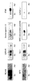
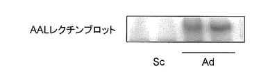
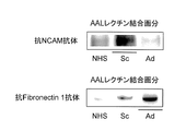
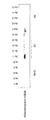
- Blotting diagram Lung cancer differentiation marker glycoprotein (NCAM, fibronectin 1) in the serum of small cell lung cancer patients (Sc) and lung adenocarcinoma patients (Ad) fractionated by AAL lectin was detected with each anti-lung cancer differentiation marker glycoprotein antibody
- NPR anti-neuropentraxin receptor
- Lung cancer differentiation marker glycoprotein and glycoprotein fragment thereof The first embodiment of the present invention is the lung cancer differentiation marker glycoprotein and the glycoprotein fragment thereof described in Table 1 or Table 2.
- the “lung cancer differential marker glycoprotein” of the present embodiment corresponds to the proteins shown as protein numbers # 1 to # 31 in Table 1 and protein # 1 to 48 in Table 2, respectively. All of these proteins have lung cancer in which a sugar chain is added to an asparagine residue at least at the position indicated by “sugar chain addition position” in the table (the position of the starting amino acid residue is 1). It is a specific glycoprotein.
- sugar chains are added to at least 390, 470 and 882 asparagine residues on the amino acid sequence of the protein. .
- glycoprotein a protein to which a sugar chain is added.
- Gi (ID) in the table indicates the ID number of the glycoprotein of the present embodiment. When multiple gi (ID) are registered in one protein, they are all listed in the table. Moreover, even when multiple isoforms exist in one protein, their gi (ID) is described together with their isoform numbers.
- the sugar chain added to the asparagine residue is not particularly limited as long as it is a sugar chain specifically added to lung cancer.
- examples include sugar chains with fucose modification (fucosylation), high mannose sugar chains, hybrid sugar chains, complex double-chain sugar chains, chitin, polylactosamine, or ⁇ 1-3 galactose epitopes.
- a sugar chain specifically added to lung cancer refers to a sugar chain added only to a protein derived from lung cancer cells at the asparagine residue indicated by “position of sugar chain addition” in the above table. Therefore, in principle, the lung cancer differentiation marker glycoprotein of the present embodiment is a glycoprotein produced from lung cancer cells. Therefore, using a sugar chain probe that recognizes these glycoproteins, for example, by detecting the presence or absence of the glycoprotein in the serum of a subject, it is determined that the individual having the glycoprotein is suffering from lung cancer. can do.
- the “sugar chain probe” refers to a discriminator that specifically recognizes and binds to a specific sugar chain and / or complex carbohydrate such as a glycoprotein.
- lectin, antibody or phage antibody can be mentioned.
- lung cancer is known to have a tissue type composed of small cell cancer and non-small cell adenocarcinoma, squamous cell carcinoma and large cell carcinoma.
- neuroendocrine cancers in the lung are also known, and many of them are classified as small cell cancers, but cases of other tissue type cancers are also known.
- the lung cancer differentiation marker glycoprotein of this embodiment can be identified as to which tissue type corresponds to the type of sugar chain added to the predetermined asparagine residue.
- acid alpha-glucosidase (acid ⁇ -glucosidase) shown as protein # 1 in Table 1
- a sugar chain with fucose modification is added to the predetermined asparagine residue only in a glycoprotein derived from small cell lung cancer.
- acid alpha-glucosidase can distinguish lung cancer as a lung cancer differentiation marker glycoprotein, and the lung cancer must be a small cell cancer. It can also be a marker for differentiating.
- biotinidase (biotinidase) indicated by protein # 2 in Table 1
- a sugar chain with fucose modification is added to the predetermined asparagine residue only in a glycoprotein derived from lung adenocarcinoma. Therefore, by using a lectin or antibody that binds to and recognizes a sugar chain with fucose modification, biotinidase can distinguish lung cancer as a lung cancer differentiation marker glycoprotein, and a marker that distinguishes that lung cancer is an adenocarcinoma Can also be.
- Sugar chains with fucose modification can be detected by AAL lectin
- high mannose-type sugar chains, mixed-type sugar chains and complex-type double-chain sugar chains can be detected by ConA lectin
- chitin and polylacto Samine can be detected by PWM lectin
- ⁇ 1-3 galactose epitope can be detected by PNA lectin.
- Table 1 when the lung cancer differentiation marker glycoprotein binds to AAL lectin or ConA lectin, it is indicated by “ ⁇ ”, and when it does not bind, it is indicated by “x”.
- each lung cancer differentiation marker glycoprotein (including the lung cancer differentiation marker glycoprotein fragment) shown in Tables 1 and 2 is AAL lectin and ConA for “small cell cancer” and “adenocarcinoma” in Tables 1 and 2. Based on the mode of binding to lectin, it can be a marker for differentiation of small cell lung cancer or lung adenocarcinoma.
- Lung Cancer Differential Marker Glycoprotein Fragment is an oligopeptide or polypeptide fragment consisting of a part of the lung cancer differential marker glycoprotein, and its amino acid sequence It contains at least one asparagine residue at the sugar addition position shown in Table 1 or Table 2, and the asparagine residue contains the lung cancer-specific sugar chain described in “1-1. Lung Cancer Differential Marker Glycoprotein”. It is added.
- the amino acid length of the lung cancer differentiation marker glycoprotein fragment is not particularly limited, but is preferably 5 to 100 amino acids, 8 to 80 amino acids, or 8 to 50 amino acids.
- the lung cancer differentiation marker glycoprotein fragment is hereinafter often referred to as “glycoprotein fragment” or “glycopeptide”.
- lung cancer differentiation marker when the lung cancer differentiation marker glycoprotein and its glycoprotein fragments are collectively expressed, they are hereinafter referred to as “lung cancer differentiation marker”.
- the lung cancer differentiation marker glycoprotein fragment include glycopeptides consisting of amino acid sequences represented by SEQ ID NOs: 1 to 223 described in Tables 1 and 2. These are glycoprotein fragments obtained by the IGOT method described later when identifying the lung cancer differentiation marker glycoprotein of the present embodiment, both as the lung cancer differentiation marker glycoprotein, It is possible to discriminate which tissue type corresponds to whether or not the patient is affected by lung cancer and the type of sugar chain added to the predetermined asparagine residue.
- the underlined asparagine residue (N) indicates an asparagine residue to which a sugar chain is added.
- 1-3-1 Large-scale identification of lung cancer-specific candidate glycoproteins
- Large-scale selective collection and enrichment of lung cancer-specific glycoproteins can be broadly divided into methods using probes having affinity for sugar chains, methods using chemical reactions with sugar chains (Zhang H. et al .Nat Biotechnol 21, 660-666 (2003)), and any method such as a method for introducing an affinity tag to a sugar chain can be used.
- the lung cancer differentiation marker sugars in Table 1 and Table 2 are used. A method using the probe used for protein acquisition will be described.
- a lectin or sugar chain antibody that reacts with a sugar chain characteristically produced by cancer cells is selected as a probe.
- Probe lectins can be selected by statistical analysis of sugar chain profiles using lectin microarrays.
- literature information (some of which can predict a probe lectin to be used, such as increased fucosylation associated with canceration) and tissue type discrimination can be selected. Basically, it is selected from statistical analysis of the profile, and the validity of the selection is judged from the binding specificity of the selected lectin.
- AAL lectin derived from Aleuria aurantia which can detect fucosylation, high mannose sugar chain, mixed sugar chain or complex double chain sugar bean Canavalia ensiformis
- An antibody probe or a phage antibody probe can be prepared after clarifying the structure of an antigen (sugar chain), but it is not a necessary condition and can be prepared even if the structure of the antigen sugar chain or glycopeptide remains unknown. .
- large-scale identification of a candidate glycoprotein is performed by first using a sugar chain probe (probe lectin and / or antibody probe, phage antibody probe) from a culture supernatant of a cultured cell line derived from lung cancer. To collect.
- a sugar chain probe probe lectin and / or antibody probe, phage antibody probe
- cultured cell supernatant derived from lung cancer it is possible to identify cancer cell-derived glycoproteins, so that it is possible to easily detect differences from sugar chains present on glycoproteins derived from serum of healthy individuals This can be useful information for selective acquisition of candidate molecules from serum.
- any lung cancer histological type of small cell cancer or non-small cell cancer (adenocarcinoma, squamous cell carcinoma, large cell carcinoma) may be used.
- a tissue type non-specific lung cancer differentiation marker and a tissue type specific lung cancer differentiation marker can also be obtained.
- the collected candidate glycoprotein for lung cancer differentiation marker is based on the IGOT method (isotope-coded glycosylation site-specific tagging) and mass spectrometry. Processed, the core glycopeptide of the candidate glycoprotein is identified. For example, JP 2004-233303 (Patent No. 4220257), Kaji H, et al. Mass spectrometric identification of N-linked glycopeptides using lectin-mediated affinity capture and glycosylation site-specific stable isotope tagging. Nature Protocols 1, 3019-3027 The glycopeptide can be analyzed by the Lec-IGOT-LC / MS method described in (2006). A specific method will be described below with an example.
- (1) Glycanectomy and IGOT method The candidate glycopeptide group obtained by digesting the candidate glycoprotein group collected with a probe lectin or probe antibody with a protease and using the same probe as a candidate glycopeptide group as a sample from the obtained peptide group Re-collect.
- the crude protein mixture can be directly digested with a probe lectin or a probe antibody from the obtained crude peptide group after protease digestion. Subsequently, the obtained candidate glycopeptide is labeled by the IGOT method.
- a sugar chain is dissociated by treatment with an enzyme such as glycopeptidase in water labeled with isotope oxygen, and asparagine at the sugar chain binding site becomes aspartic acid by this treatment.
- isotope oxygen (18 O) is incorporated into the candidate glycopeptide.
- Xaa is Lys / Arg and the identified peptide sequence is excised at this position, refer to the amino acid sequence of the entire protein, and the next residue of Xaa is [Ser / Thr].
- the candidate glycopeptide is also included in the glycoprotein fragment.
- the consensus sequence is a sugar chain binding position, but a site not following this has also been reported.
- the lung cancer differentiation marker glycoprotein fragment group selected by the above method and the lung cancer differentiation marker glycoprotein containing the fragment identified based on the amino acid sequence are the glycoprotein fragment group and glycoprotein shown in Tables 1 and 2. is there.
- glycoprotein fragment group and glycoprotein can be used as a lung cancer differentiation marker for determining the presence of lung cancer in a subject by, for example, verifying the presence or absence in the serum of the subject using a lectin or antibody that recognizes the fragment.
- 1-3-3 Verification of Lung Cancer Differential Marker
- the significance of the lung cancer differential marker selected in the section 1-3-2 can be further verified.
- Western blotting or immunoprecipitation of glycoproteins and / or glycoprotein fragments collected with the probe lectin from the lung cancer cultured cell supernatant using an antibody that specifically recognizes the above-identified lung cancer differentiation marker Can be detected and confirmed.
- 31 types of lung cancer differentiation marker glycoproteins and glycoprotein fragments thereof shown in Table 1 correspond to lung cancer differentiation markers further selected by the antibody detection.
- the lung cancer differentiation marker selected by the antibody detection may be further verified.
- it can be collected from the serum of lung cancer patients using a probe lectin and detected using the specific antibody.
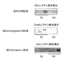
- the nerve cell adhesion molecule (NCAM1) (protein # 19 in Table 1) and fibronectin 1 (protein # 8 in Table 1) described in Table 1 shown in FIG. 5 and secretogranin III shown in FIGS. (Protein # 23 in Table 1) is a lung cancer differentiation marker glycoprotein whose usefulness has been confirmed by this further verification.
- the nerve cell adhesion molecule binds to AAL lectin only in small cell lung cancer according to Table 1, it can be a lung cancer differentiation marker capable of distinguishing small cell lung cancer.
- secretogranin III binds to AAL lectin and ConA lectin only in small cell lung cancer according to Table 1, it can be a lung cancer differentiation marker that can distinguish small cell lung cancer.
- fibronectin 1 binds to AAL lectin only in lung adenocarcinoma from Table 1, it can be a lung cancer differentiation marker that can distinguish lung adenocarcinoma.
- the specific antibody may be collected from the culture supernatant of a lung cancer cell line and detected using a lectin array.
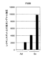
- IGFBP-L1 insulin-like growth factor binding protein-L1 shown in Table 1 shown in FIG.
- fibronectin 1 shown in Table 1 shown in FIG. 10 is a lung cancer differentiation marker glycoprotein whose usefulness has been confirmed by the further verification.
- insulin-like growth factor-binding protein-L1 IGFBP-L1
- IGFBP-L1 insulin-like growth factor-binding protein-L1
- fibronectin 1 can detect the fluorescent signal on the PNA lectin spot only in lung adenocarcinoma from FIG. 10, it can be a lung cancer differentiation marker that can distinguish lung adenocarcinoma.
- Lung cancer differentiation marker glycopeptide and glycoprotein can be detected using a mass spectrometer as a detector for a sample collected with a probe lectin or the like that binds to a sugar chain.
- Lung cancer differentiation marker glycopeptides are preferably detected by excision of the collected glycopeptides, followed by separation by liquid chromatography (LC), and the eluted peptides sequentially and directly on-line by a mass spectrometer (MS ) And can be detected.
- mass spectra can be acquired by acquiring MS / MS spectra using a fracture method such as collision-induced dissociation (CID), and only when preselected ions are detected. It is also possible to detect a plurality of fragment ions that are broken by a method such as single reaction monitoring or multi-reaction monitoring.
- CID collision-induced dissociation
- the core peptide part of the lung cancer differentiation marker glycopeptide was synthesized in the analysis sample, and the target peptide that caused a mass difference by incorporating a stable isotope into a part of the peptide was added, and the relative signal intensity was compared by comparing the respective signal intensities. Or absolute quantitative analysis can be performed. In a simple manner, the signal intensity of the detected ions can be easily quantified by comparing between a plurality of samples or a standard sample.
- the collected protein can be separated using one-dimensional or two-dimensional gel electrophoresis, and the detection intensity (staining, fluorescence, etc.) of the target spot can be compared with a standard sample and quantified.
- a detection method using a mass spectrometer a collected protein group can be digested with a protease, and the resulting peptide can be analyzed and detected by an LC / MS method.
- Lectin microarray (1) Glycan profiling by lectin microarray (a) Lectin microarray (also simply called lectin array) A lectin array is a glycan probe in which multiple types of lectins with different specificities are immobilized in parallel on one substrate (arrayed), and how much lectin interacts with the complex carbohydrate to be analyzed. Can be analyzed all at once. By using a lectin array, information necessary for sugar chain structure estimation can be acquired by a single analysis, and the operation steps from sample preparation to scanning can be performed quickly and easily.
- glycoproteins cannot be analyzed as they are, and must be processed in advance to the state of glycopeptides and free sugar chains.
- a lectin microarray has an advantage that analysis can be performed as it is by simply introducing a phosphor directly into the core protein portion.
- the lectin microarray technology has been developed by the present inventors, and its principle and basis are described in, for example, Kuno A., et al. Nat. Methods 2,851-856 (2005).
- Typical lectins used in the lectin array include, for example, those described in Table 3 below.
- a lectin array (LecChip manufactured by GP Bioscience) in which 45 types of lectins including the above 43 types of lectins are immobilized is already commercially available.
- the platform of the lectin microarray is basically as described above.
- the sample of the subject is not directly labeled with fluorescence, but indirectly with an antibody via a fluorescent group, etc.
- This is an application method that can simplify and speed up the analysis of multiple samples simultaneously by introducing the sample into the sample of the subject (“Kuno A, Kato Y, Matsuda A, Kaneko MK, Ito H, Amano K, Chiba Y, Narimatsu H, Hirabayashi J. Mol Cell Proteomics.
- the sugar chain part is recognized by the lectin on the lectin microarray, so that the test glycoprotein can be labeled by overlaying with an antibody against the core protein part. Or, it can be detected specifically with high sensitivity without being highly purified.
- Lectin overlay / antibody microarray method In place of the lectin microarray, an antibody microarray in which antibodies against the core protein are immobilized (arrayed) in parallel on a substrate such as a glass substrate is used. Only as many antibodies as the marker to be examined are needed. It is necessary to determine in advance a lectin for detecting a sugar chain change.
- a simple and inexpensive sandwich detection method can be designed based on the results of the lectin array. Basically, among the protocols used in the sandwich detection method using two kinds of antibodies, it can be applied by replacing one antibody with a lectin. Therefore, this method can also be applied to automation using an existing automatic immunodetection device. The only thing to consider is the reaction between the antibody used in the sandwich and the lectin. The antibody has at least two N-linked sugar chains. Therefore, when the lectin used recognizes the sugar chain on the antibody, background noise due to the binding reaction occurs during sandwich detection.
- a method of introducing a modification into the sugar chain part on the antibody or a method of using only a Fab that does not contain a sugar chain part can be considered.
- methods for modifying the sugar chain include Chen S et al. Nat Methods. 4, 437-44 (2007) and Consale MA et al. J Proteome Res. 8, 595-602 (2009). Include, for example, Matsumoto H et al. Clin Chem Lab Med 48, 505-512 (2010).
- Antibody overlay lectin arrays are the best way to statistically find lectins that best reflect disease-specific glycosylation on lung cancer differential marker glycoproteins.
- antibodies capable of immunoprecipitation and overlay detection are essential. However, such antibodies are not always available. Therefore, as a means for utilizing more candidate molecules for lung cancer detection, it is common to perform immunological quantitative detection of the target glycoprotein with respect to the glycoprotein collected by the probe lectin. Specifically, SDS-PAGE is performed, and immunological detection is performed by Western blot after membrane transfer. By comparing the signal intensities of the obtained bands, fluctuations between the samples can be quantitatively estimated.
- the AAL lectin used in the candidate molecule identification step is generally used as a probe protein even during verification.
- a report of Liu Y et al. J Proteome Res. 9, 798-805 (2010) can be cited as an example of such a strategy.
- serum proteins have different N-linked sugar chain structures (degree of branching, etc.) and fucose modifications (core fucose, blood group antigens, etc.) depending on the type. Therefore, it has been reported that even the same molecule may undergo different fucose modifications.
- ⁇ 1 antitrypsin is subjected to different fucose modifications. Because these differ in the type of disease, whether there is an increase depending on the degree of progression, and the timing, almost all fucose modifications can be recognized and collected using AAL as a probe to collect the entire fucose-containing glycoprotein Comparison is not ideal. Therefore, we considered separation and fractionation by continuous chromatography using two different fucose recognition lectins, and quantitative comparison analysis of each fraction.
- the lectins used at this time are LCA and AAL.
- LCA is known to be a lectin that recognizes N-linked sugar chains that have undergone low-branching core fucose modification based on lectin specificity analysis.
- AAL can recognize any branched core fucose of an N-linked sugar chain, and can also recognize fucose modifications on the non-reducing end side typified by ABO and Lewis antigens. . That is, LCA is highly specific and AAL is low. Therefore, as the first step, LCA column chromatography is performed to capture fucose-containing glycoprotein that binds to LCA. This is referred to as LCA-binding fucose-containing glycoprotein. At the time of chromatography, fucose-containing glycoproteins that do not have a low-branched N-linked sugar chain that has undergone core fucose modification do not bind to the LCA column but are fractionated into unbound fractions.
- AAL column chromatography is performed on the LCA-unbound fraction.
- a group of glycoproteins captured by AAL is defined as LCA non-binding / AAL-binding fucose-containing glycoprotein.
- the second embodiment of the present invention is a lung cancer incidence determination method.
- the method of this embodiment is characterized in that it is determined that the subject is suffering from lung cancer by detecting the lung cancer differentiation marker described in the first embodiment from a sample collected from the subject.
- subject refers to a person who is subjected to a test, that is, a person who provides a sample to be described later.
- the subject may be either a patient having some disease or a healthy person. Preferred are those who may have lung cancer or lung cancer patients.
- sample is collected from the subject and used in the determination method of the present embodiment, and corresponds to, for example, a body fluid, a cell such as a cancer tissue, or a lung lavage fluid collected during surgery.
- Body fluid refers to a liquid biological sample collected from a subject.
- blood including serum, plasma and interstitial fluid
- lymph fluid extract of each tissue or cell
- pleural effusion sputum
- cerebrospinal fluid tears
- nasal discharge saliva, urine, vaginal fluid, semen and the like
- the body fluid may be used after performing treatment such as dilution or concentration of a sample collected from a subject, addition of a blood coagulation inhibitor such as heparin as necessary, or such pretreatment. It may be used as it is.
- the body fluid may be collected based on a known method in the field.
- a known blood collection method may be followed.
- peripheral blood it can be collected by injection into a peripheral vein or the like.
- the bodily fluid may be used immediately after collection, or may be used after being frozen or refrigerated for a certain period of time and then subjected to processing such as thawing as necessary.
- a sufficient amount of lung cancer differential marker can be detected by using a volume of 10 ⁇ L to 100 ⁇ L, 20 ⁇ L to 80 ⁇ L, 30 ⁇ L to 70 ⁇ L, 40 ⁇ L to 60 ⁇ L, or 45 ⁇ L to 55 ⁇ L.
- the lung cancer marker used for detection in the determination method of the present embodiment is a lung cancer marker glycoprotein according to Table 1 or Table 2 in which a sugar chain is added to the asparagine residue at the sugar addition position shown in Table 1 or Table 2.
- Any lung cancer differentiation marker can be used as long as it is a glycoprotein fragment containing at least one asparagine residue at the sugar addition position shown in Table 1 or Table 2 to which a sugar chain has been added.
- These lung cancer differentiation markers may be used alone in the determination method of the present embodiment, or may be used in combination of two or more. For example, two or more different lung cancer differentiation marker glycoproteins may be used, or two or more different lung cancer differentiation marker glycoprotein fragments in the same lung cancer differentiation marker glycoprotein may be used.
- a lung cancer differentiation marker in Table 1 is used, and more preferably, a lung cancer differentiation marker having the same or different differentiation characteristics for small cell lung cancer and lung adenocarcinoma.
- a lung cancer differentiation marker having the same or different differentiation characteristics for small cell lung cancer and lung adenocarcinoma For example, neural cell adhesion molecule (NCAM1), secretogranin III and / or insulin-like growth factor binding protein-L1 (IGFBP-L1) listed in Table 1 for differentiation of small cell lung cancer, and lung adenocarcinoma differentiation Examples include a method of combining fibronectin 1 listed in Table 1 as a lung cancer differentiation marker of the present embodiment. This is convenient because it is possible to simultaneously determine whether the lung cancer is a small cell cancer or an adenocarcinoma at the same time as determining whether the subject has lung cancer.
- a lectin or antibody that binds to a sugar chain with fucose modification or a high-mannose sugar chain, a hybrid sugar chain, a complex double-chain sugar chain, chitin, polylactosamine and / or ⁇ 1-3 galactose epitope from a sample of a subject are used alone or in combination, and the lung cancer differentiation markers listed in Table 1 or 2 are detected by any method, the subject is suffering from or very likely to have lung cancer. Can be determined.
- AAL lectin that specifically binds to sugar chains with fucose modification can be detected, and specifically, it can be used to detect high-mannose sugar chains, mixed sugar chains, and complex double-chain sugar chains.
- the binding ConA lectin can be used to detect chitin and polylactosamine, PWM lectin specifically binding to it, or the detection of ⁇ 1-3 galactose epitope can be PNA lectin specifically binding to it. .
- the binding result of the lectin to the sugar chain of the lung cancer differentiation marker and the binding of the lung cancer differentiation marker glycoprotein and / or its glycoprotein fragment shown in Table 1 or Table 2 to the lectin Based on the embodiment, it can also be determined that the lung cancer tissue type in the subject is small cell carcinoma or adenocarcinoma.
- the sugar chain of acid alpha-glucosidase represented by protein # 1 binds to AAL lectin only in small cell lung cancer.
- the subject is determined to have lung cancer and the tissue type is small cell carcinoma. can do.
- the lung cancer differentiation marker glycoprotein is a neural cell adhesion molecule (NCAM1) or a glycoprotein fragment thereof and binding to AAL lectin is detected, it is determined that the lung cancer tissue type is small cell cancer. Can do.
- the lung cancer differentiation marker glycoprotein is fibronectin 1 or a glycoprotein fragment thereof, and by detecting the binding to the AAL lectin or antibody, it can be determined that the lung cancer tissue type is adenocarcinoma.
- Lung Cancer Differential Marker Detection Method As a method for detecting a lung cancer differential marker, for example, there are two methods, namely, a lectin that specifically binds to a sugar chain of a lung cancer differential marker (in this specification, for the sake of convenience, hereinafter referred to as lectin A).
- a combination of a method for selecting a lung cancer differential marker having the sugar chain and a method for detecting the lung cancer differential marker by detecting a portion (core protein) other than the sugar chain of the lung cancer differential marker, or a lectin A specific antibody against a lung cancer differentiation marker having a sugar chain that specifically binds to A, and a method for detecting the target lung cancer differentiation marker using an antibody having an epitope near the sugar chain binding portion can be mentioned .
- the method for detecting a sugar chain that specifically binds to lectin A and the method for detecting a core protein are respectively a method for measuring a sugar chain that specifically binds to lectin A, and a core protein. It may be a method.
- a lung cancer differentiation marker using an antibody against a core protein and lectin A
- lung cancer patients can be detected separately from healthy individuals.
- an antibody overlay method using a lectin array (Kuno A, Kato Y, Matsuda A, Kaneko MK, Ito H, Amano K, Chiba Y, Narimatsu H, Hirabayashi J. Mol Cell Proteomics. 8, 99-108 (2009 ))can be used.
- a lectin-antibody sandwich immunological detection method can be used.
- a lung cancer differentiation marker in serum obtained from a subject is separated using lectin A.
- a protein group having a sugar chain that specifically binds to lectin A is selected.
- the lung cancer differentiation marker detection is performed using an anti-lung cancer differentiation marker glycoprotein antibody that specifically recognizes a portion other than the sugar chain that specifically binds to lectin A.
- an anti-lung cancer differentiation marker glycoprotein antibody that specifically recognizes a portion other than the sugar chain that specifically binds to lectin A.
- a lung cancer differentiation marker having a sugar chain that specifically binds to the target lectin A can be detected. If this marker is detected, it is determined that the subject is suffering from lung cancer. .
- a lung cancer differentiation marker having a sugar chain that specifically reacts with lectin A for example, a method of measuring a sugar chain that specifically binds to lectin A, specifically, a column or an array on which lectin A is immobilized And a means for measuring a lung cancer differentiation marker, specifically, an antibody against a novel lung cancer differentiation marker glycoprotein or a fragment thereof.
- a lectin-antibody sandwich ELISA or an antibody overlay lectin array method can be used.
- LC-MS enzyme immunoassay using a monoclonal antibody or polyclonal antibody specific for a novel lung cancer differential marker glycoprotein having a sugar chain that specifically reacts with lectin A or a fragment thereof
- two-antibody sandwich ELISA method gold colloid method, radioimmunoassay method, latex agglutination immunoassay method, fluorescence immunoassay method, western blotting method, immunohistochemistry method, surface plasmon resonance method (SPR method) or quartz crystal microbalance (QCM) method
- SPR method surface plasmon resonance method
- QCM quartz crystal microbalance
- an anti-lung cancer differentiation marker glycopeptide polyclonal antibody can be prepared using a well-known method. Specifically, an adjuvant is added to the obtained lung cancer differentiation marker glycoprotein or glycopeptide for antigen.
- an adjuvant As the antigen, a lung cancer differential marker glycopeptide containing a sugar chain binding site (asparagine) may be synthesized and used. Examples of the adjuvant include Freund's complete adjuvant, Freund's incomplete adjuvant, and the like, and these can also be used as a mixture.
- An antibody-producing animal can be co-inoculated with an adjuvant together with the antigen to boost antibody production.
- this peptide may be covalently bound to commercially available keyhole limpet hemocyanin (KLH) and inoculated into antibody-producing animals.
- KLH keyhole limpet hemocyanin
- GM-CSF granulocyte-macrophage colony stimulating factor
- Mammals such as mice, rats, horses, monkeys, rabbits, goats, sheep and the like can be used as antibody-producing animals to inoculate the antigen.
- Any method can be used for immunization as long as it is an existing method, but it is mainly carried out by intravenous injection, subcutaneous injection, intraperitoneal injection or the like. Immunization intervals are not particularly limited, and immunization is performed at intervals of several days to several weeks, preferably at intervals of 4 to 21 days.
- a polyclonal antibody can be prepared by collecting whole blood from an immunized animal 2-3 days after the final immunization and separating the serum.
- an anti-lung cancer marker glycopeptide monoclonal antibody can be prepared by the method of Keller and Milstein (Nature Vol. 256, pp 495-497 (1975)).
- a hybridoma is prepared by cell fusion between an antibody-producing cell obtained from an animal immunized with an antigen and a myeloma cell, and a clone that produces an anti-lung cancer differential marker glycopeptide monoclonal antibody is selected from the resulting hybridoma. be able to.
- antibody production cells are collected 2 to 3 days after the last immunization in the production of the polyclonal antibody.
- antibody-producing cells include spleen cells, lymph node cells, and peripheral blood cells.
- myeloma (myeloma) cells to be fused with antibody-producing cells cell lines derived from various animals such as mice, rats, humans, and the like, which are generally available to those skilled in the art, are used.
- the cell line to be used those that have drug resistance and have the property that they cannot survive in a selective medium (for example, HAT medium) in an unfused state but can survive only in a fused state.
- An 8-azaguanine resistant strain is generally used, and this cell strain is deficient in hypoxanthine-guanine-phosphoribosyltransferase and cannot grow in hypoxanthine / aminopterin / thymidine (HAT) medium.
- HAT hypoxanthine-guanine-phosphoribosyltransferase
- Myeloma cells are various known cell lines such as P3 (P3x63Ag8.653) (J. Immunol. 123, 1548-1550 1979)), P3x63Ag8U.1 (Current Topics in Microbiology and Immunology 81, 1-7 ( 1978)), NS-1 (Kohler, G. and Milstein, C., Eur. J. Immunol. 6,511-519 (1976)), MPC-11 (Margulies, DH et al., Cell 8,405-415 (1976) ), SP2 / 0 (Shulman, M. et al., Nature 276,269-270 (1978)), FO (de St. Groth, SF et al., J. Immunol.
- cell fusion is performed between the myeloma cells and antibody-producing cells.
- Cell fusion is performed by mixing myeloma cells and antibody-producing cells in an animal cell culture medium such as MEM, DMEM, and RPMI-1640 medium at a mixing ratio of 1: 1 to 1:10 in the presence of a fusion promoter. By contacting at 37 ° C. for 1-15 minutes.
- a fusion promoter or fusion virus such as polyethylene glycol having an average molecular weight of 1,000 to 6,000, polyvinyl alcohol, or Sendai virus can be used.
- antibody-producing cells and myeloma cells can be fused using a commercially available cell fusion device utilizing electrical stimulation (for example, electroporation).
- Select the desired hybridoma from the cells after cell fusion treatment examples include a method utilizing selective growth of cells in a selective medium. That is, after the cell suspension is diluted with an appropriate medium, it is spread on a microtiter plate, a selective medium (HAT medium or the like) is added to each well, and thereafter, the selective medium is appropriately replaced and cultured. As a result, growing cells can be obtained as hybridomas.
- a selective medium HAT medium or the like
- Hybridoma screening is performed by the limiting dilution method, the fluorescence excitation cell sorter method, etc., and finally a monoclonal antibody-producing hybridoma is obtained.
- Examples of a method for collecting a monoclonal antibody from the obtained hybridoma include a normal cell culture method and ascites formation method.
- the third embodiment of the present invention is a lung cancer cell differentiation antibody for tissue staining.
- the antibody of the present embodiment is one or more of the lung cancer differentiation markers described in the first embodiment, that is, one or more of the sugar chains added to the asparagine residues at the sugar addition positions shown in Table 1 or Table 2. Specifically recognize one or more glycoprotein fragments comprising at least one asparagine residue at the glycosylation position shown in Table 1 or Table 2, to which is added a lung cancer differentiation marker glycoprotein and / or a sugar chain. It is an antibody that can specifically differentiate lung cancer cells in tissue staining by binding to it.
- the epitope of the lung cancer differentiation marker described in the first embodiment recognized by the antibody of the present embodiment is not particularly limited.
- it is a core protein or a core protein fragment thereof. More preferably, it is a part including both a sugar chain and a peptide sequence around the sugar chain addition.
- the length of the peptide sequence is 5-15 amino acids, 5-10 amino acids, or 5-8 amino acids.
- a phage antibody probe using a phage or the like can be a sugar chain probe for lung cancer cell identification.
- the antibody of the present embodiment can distinguish the tissue type of lung cancer cells.
- an antibody against a neuropentraxin receptor (NPR) represented by protein # 21 in Table 1 R & D, SYSTEM, Anti-human, Neuronal, Pentraxin, Receptor, and Antibody
- NPR neuropentraxin receptor
- FIG. 7 this anti-neuropentraxin receptor antibody can be specifically differentiated from cells derived from small cell carcinoma, and thus can be an antibody for differentiation of small cell carcinoma cells.
- the proteins collected and identified by the cancer sugar chain probe lectin usually have very little expression in other tissues and normal sites. Or what can become a tissue marker only by a phage antibody is also contained.
- Example 1 Selection of glycopeptide markers by glycoproteomics (IGOT-LC / MS method) Preparation method of human lung cancer cell supernatant Three types of lung adenocarcinoma (H358, H1975, LX-1) and three types of small cell lung cancer (H2171, H524, H526) were prepared with high glucose-containing 90% DMEM and 10% FBS, respectively. Using a medium of (PS +), the cells were cultured for 3 days in a dish having a diameter of 14 cm to be 80-90% confluent. The FBS serum-containing culture medium was aspirated and discarded, and washed with 10 mL of serum-free medium / dish (100% DMEM-high glucose, no additive).
- Serum-free medium was added at 30 mL / dish and cultured for 48 hours. After incubation, the mixture was centrifuged at 1000 rpm ⁇ 30 minutes, and the supernatant was further collected. The supernatant (culture supernatant) was collected at 3000 rpm ⁇ 30 minutes, and stored frozen at ⁇ 80 ° C. The culture supernatant stored at the above temperature was thawed before use, filtered through a 0.45 ⁇ m filter, and used in the following examples. NaN 3 was added so that the final concentration was 0.1%.
- the supernatant extract was recovered by high-speed centrifugation. Nitrogen gas was passed through or blown through the extract to remove dissolved oxygen, and then dithiothreitol (DTT) in an amount equal to the protein weight was dissolved in powder or a small amount of solubilization buffer and added. The reaction was carried out at room temperature for 1-2 hours in order to reduce disulfide bonds while passing nitrogen gas or under nitrogen gas atmosphere. Next, for S-alkylation, 2.5 times the protein weight of iodoacetamide was added, and the mixture was allowed to react at room temperature for 1-2 hours in the dark. After the reaction, dialyzed at 4 ° C.
- DTT dithiothreitol
- the candidate glycopeptide was eluted with 50% ethanol (v / v). Concentrate all candidate glycopeptide fractions by transferring a small amount of the candidate glycopeptide fraction to a microtube containing 2 ⁇ L of glycerol, removing the water by vacuum centrifugation, that is, vacuum centrifugation, and repeating the operation. did.
- IGOT reaction solution was diluted with 0.1% formic acid and subjected to LC / MS shotgun analysis.
- a nano LC system based on a direct nanoflow pump was used.
- the injected candidate glycopeptide is once collected on a trap column (reverse phase C18 silica gel-based carrier) for desalting, washed, and then a fritless microcolumn (in the form of a spray tip packed with the same resin) Using an inner diameter of 150 ⁇ m ⁇ 50 mL), separation was performed by the acetonitrile concentration gradient method.
- Mass spectrometry performed tandem mass spectrometry by collision induced dissociation (CID) while selecting up to two ions in a data dependent mode.
- CID collision induced dissociation
- the probability of identification (probability of coincidence: Expect value) is 0.05 or less.
- the number of fragment ions contributing to the identification is 4 or more.
- the identified peptide has a consensus sequence and has fewer than that number of Asn modifications (conversion to Asp and incorporation of 18 O).
- a glycoprotein containing this sequence can be defined. More specifically, based on the “peptide sequence” of the lung cancer differential marker glycopeptide represented by SEQ ID NOs: 1 to 223 in Tables 1 and 2, the entire amino acid sequence of the corresponding lung cancer differential marker glycoprotein from the amino acid sequence database NCBI-Refseq was identified.
- the lung cancer differentiation marker glycoprotein obtained by the above process is shown in Table 2.
- 5% skim milk or 5% BSA dissolved in PBSTx in which 1% Triton X-100 was added to PBS (Dulbecco) was used and blocked at room temperature for 1 hour.
- the membrane was reacted with a primary antibody (Table 4) biotinylated in advance with Biotin Labeling Kit-NH 2 (Dojindo Laboratories) for 1 hour.
- the mixture was reacted with the secondary antibody HRP-conjugated streptavidin (1: 3000 dilution, GE Healthcare) for 1 hour.
- the lung cancer differentiation marker glycoprotein obtained by the above process is shown in Table 1, FIG. 1 and FIG.
- the culture supernatant was fractionated using AAL lectin. Specifically, the AAL resin was washed 5 times with 3 times the amount of PBS and then prepared into a 50% slurry solution. 30 ⁇ L of the culture supernatant was added to 30 ⁇ L of the prepared AAL resin, and the mixture was shaken at 4 ° C. for 5 hours. After centrifugation (2,000 rpm, 2 minutes), the supernatant was removed, 50 ⁇ L of Wash Buffer (0.1% SDS in PBST, 0.1% Triton X-100) was added, and centrifugation (2,000 rpm, 2 minutes) was repeated twice.
- Wash Buffer (0.1% SDS in PBST, 0.1% Triton X-100
- the reacted culture supernatant and antibody were transferred to magnetic beads and shaken at 1,400 rpm for 1 hour at 20 ° C.
- the antibody bound to the magnetic beads was recovered using a magnetic stand.
- the collected magnetic beads were washed three times with 1 mL of TBSTx. After adding 20 ul of elution buffer (0.2% SDS in TBS) and stirring, the mixture was heated twice at 98 ° C. or 60 ° C. for 5 minutes for elution.
- 40 ⁇ L of magnetic beads washed in the same manner were added to the eluted sample and shaken at 1,400 rpm for 2 hours at 20 ° C.
- a sample obtained by removing antibody-magnetic beads with a magnetic stand was used as a sample.
- the prepared sample was developed by SDS-PAGE and then lectin blotted.
- Serum column fractionation with AAL lectin was fractionated using AAL lectin. Specifically, 1 mL of AAL resin is packed in a commercially available column, washed with 10 times the amount of resin TBS, twice the amount of resin 0.5 M NaCl, and washed with 10 times the amount of resin TBS. An AAL column was prepared. After the serum was added to the AAL column, the mixture was reacted in the resin for 1 hour, and then washed with 5 times the amount of TBS.
- Serum Fractionation by Continuous Chromatography Method Serum fractionation using continuous chromatography was performed at low temperature using LCA agarose (J-Oil Mills) and AAL agarose (J-Oil Mills). 100 ⁇ L of serum was diluted 4 times with PBS, and the serum sample was first developed in 5 mL of LCA agarose (0.7 ⁇ 13 cm) packed in a column. Next, the obtained unbound serum fraction was developed into 2.5 mL of AAL agarose (0.7 ⁇ 5.5 cm) packed in a column, washed thoroughly with PBS, and then eluted with PBS containing 0.2 M fucose. . The eluted fraction was concentrated with an ultra-free centrifugal concentration device (cut-off: 30 kDa, Millipore), and the obtained sample was subjected to comparative analysis of candidate molecules by immunoblotting as described above.
- the obtained unbound serum fraction was exposed to 250 ⁇ L of AAL agarose (0.8 ⁇ 0.5 cm) packed in an open column, washed thoroughly with PBS, and then eluted with PBS containing 0.02 M fucose. .
- the eluted fraction was concentrated with an ultra-free centrifugal concentration device (cut-off: 10 kDa, Millipore), and among the obtained samples, 10 ⁇ L of serum was subjected to comparative analysis of candidate molecules by immunoblotting as described above.
- Antibody overlay / lectin array Take an appropriate amount of the glycoprotein solution obtained in “5. Fractionation by immunoprecipitation” above and use 1% Triton X-100-containing Phosphate-buffered saline (PBSTx) as a lectin array reaction buffer. Prepared to 60 ⁇ L. This solution was added to each reaction vessel of the lectin array in which 8 reaction vessels were formed per glass, and reacted at 20 ° C. for 10 hours or more. The lectin array substrate composed of these eight reaction vessels was prepared according to the method of Uchiyama et al. (Proteomics 8, 3042-3050 (2008)).
- each reaction vessel was washed three times with 60 ⁇ L of PBSTx, and then array scan was performed with an array scanner GlycoStation manufactured by Moritex, and the fluorescence intensity on the lectin spot that specifically reacted with lung cancer tissue was compared.
- the lung cancer differentiation marker glycoprotein obtained by the above process is shown in FIG. 9 and FIG.
- Example 2 Tissue staining of lung cancer cells using lung cancer differentiation markers Tissue staining with antibodies was examined using lung cancer differentiation markers.
- paraffin covering a tissue section of a formalin-fixed lung cancer tissue section (5 ⁇ m thickness) on the tissue section was deparaffinized according to a conventional method.
- the deparaffinized tissue section was washed with PBS, air-dried, immersed in 10 mM citrate buffer, and autoclaved at 121 ° C. for 15 minutes to dissociate the intermolecular (intramolecular) cross-linking by formalin.
- the treated sections were allowed to stand at room temperature for a while and then immersed in PBS three times for 5 minutes to wash the tissue surface.
- the endogenous peroxidase blocking reaction was performed by treatment with 0.3% H 2 O 2 -MeOH for 10 minutes at room temperature.
- the lung cancer differentiation marker glycoprotein obtained by the above process is shown in FIG. It can be seen that only small cell carcinoma is stained specifically.
Landscapes
- Health & Medical Sciences (AREA)
- Life Sciences & Earth Sciences (AREA)
- Chemical & Material Sciences (AREA)
- Immunology (AREA)
- Molecular Biology (AREA)
- Engineering & Computer Science (AREA)
- Organic Chemistry (AREA)
- Biochemistry (AREA)
- General Health & Medical Sciences (AREA)
- Medicinal Chemistry (AREA)
- Urology & Nephrology (AREA)
- Biomedical Technology (AREA)
- Hematology (AREA)
- Proteomics, Peptides & Aminoacids (AREA)
- Cell Biology (AREA)
- Genetics & Genomics (AREA)
- Biophysics (AREA)
- Physics & Mathematics (AREA)
- Biotechnology (AREA)
- Microbiology (AREA)
- Food Science & Technology (AREA)
- Analytical Chemistry (AREA)
- General Physics & Mathematics (AREA)
- Pathology (AREA)
- Gastroenterology & Hepatology (AREA)
- Zoology (AREA)
- Toxicology (AREA)
- Peptides Or Proteins (AREA)
- Investigating Or Analysing Biological Materials (AREA)
Abstract
La présente invention concerne le problème consistant à développer et à fournir un marqueur d'identification du cancer du poumon pouvant déterminer simplement la présence d'un cancer du poumon avec une forte sensibilité sans compter uniquement sur la quantité d'expression protéique chez un patient cancéreux et chez une personne saine. De plus, la présente invention concerne le problème consistant à développer et à fournir un marqueur de chaîne saccharidique pouvant déterminer le type histologique du cancer du poumon. Parmi les glycoprotéines sériques, un groupe de glycopeptides et de glycoprotéines dans lequel la structure de la chaîne saccharidique est spécifiquement modifiée dans un surnageant de culture de cellules de cancer du poumon est identifié et donné comme marqueur d'identification du cancer du poumon.
Priority Applications (7)
| Application Number | Priority Date | Filing Date | Title |
|---|---|---|---|
| EP11825097.6A EP2617730B1 (fr) | 2010-09-17 | 2011-09-09 | Marqueur différentiel du cancer du poumon |
| EP17158414.7A EP3199542B1 (fr) | 2010-09-17 | 2011-09-09 | Marqueur d'identification du cancer du poumon |
| CN201180054286.6A CN103221419B (zh) | 2010-09-17 | 2011-09-09 | 肺癌鉴定标志物 |
| JP2012533980A JPWO2012036094A1 (ja) | 2010-09-17 | 2011-09-09 | 肺癌鑑別マーカー |
| EP18194507.2A EP3444267B1 (fr) | 2010-09-17 | 2011-09-09 | Marqueur différentiel du cancer du poumon |
| US13/823,922 US9696320B2 (en) | 2010-09-17 | 2011-09-09 | Lung cancer differential marker |
| US15/620,214 US10539576B2 (en) | 2010-09-17 | 2017-06-12 | Lung cancer differential marker |
Applications Claiming Priority (2)
| Application Number | Priority Date | Filing Date | Title |
|---|---|---|---|
| JP2010209932 | 2010-09-17 | ||
| JP2010-209932 | 2010-09-17 |
Related Child Applications (2)
| Application Number | Title | Priority Date | Filing Date |
|---|---|---|---|
| US13/823,922 A-371-Of-International US9696320B2 (en) | 2010-09-17 | 2011-09-09 | Lung cancer differential marker |
| US15/620,214 Division US10539576B2 (en) | 2010-09-17 | 2017-06-12 | Lung cancer differential marker |
Publications (1)
| Publication Number | Publication Date |
|---|---|
| WO2012036094A1 true WO2012036094A1 (fr) | 2012-03-22 |
Family
ID=45831557
Family Applications (1)
| Application Number | Title | Priority Date | Filing Date |
|---|---|---|---|
| PCT/JP2011/070635 Ceased WO2012036094A1 (fr) | 2010-09-17 | 2011-09-09 | Marqueur d'identification du cancer du poumon |
Country Status (5)
| Country | Link |
|---|---|
| US (2) | US9696320B2 (fr) |
| EP (3) | EP3199542B1 (fr) |
| JP (2) | JPWO2012036094A1 (fr) |
| CN (2) | CN105424942B (fr) |
| WO (1) | WO2012036094A1 (fr) |
Cited By (9)
| Publication number | Priority date | Publication date | Assignee | Title |
|---|---|---|---|---|
| WO2014010055A1 (fr) * | 2012-07-12 | 2014-01-16 | 独立行政法人産業技術総合研究所 | Marqueur de différentiation du cancer épithélial de l'ovaire |
| WO2014132869A1 (fr) * | 2013-02-26 | 2014-09-04 | 和光純薬工業株式会社 | Procédé de détermination du cancer du sein |
| JP2015105951A (ja) * | 2013-11-28 | 2015-06-08 | コリア ベーシック サイエンス インスティテュートKorea Basic Science Institute | 血液由来の癌診断用ペプチドマーカー及びそれを用いた癌診断方法 |
| WO2015098663A1 (fr) * | 2013-12-25 | 2015-07-02 | 東ソー株式会社 | Procédé de détermination de site auquel est ajouté une chaîne de sucre à liaison n ou de la proportion dudit ajout |
| JP2016531915A (ja) * | 2013-08-28 | 2016-10-13 | ステムセントリックス, インコーポレイテッド | 部位特異的抗体コンジュゲーション方法および組成物 |
| JP2017502048A (ja) * | 2014-01-08 | 2017-01-19 | ザ ボード オブ トラスティーズ オブ ザ レランド スタンフォード ジュニア ユニバーシティー | 小細胞肺癌に対する標的療法 |
| WO2017142025A1 (fr) * | 2016-02-19 | 2017-08-24 | 国立大学法人宮崎大学 | Procédé de détection d'adénocarcinome |
| CN107771183A (zh) * | 2015-05-06 | 2018-03-06 | 加马玛布斯制药公司 | 抗‑人‑her3抗体及其用途 |
| WO2025009551A1 (fr) * | 2023-07-05 | 2025-01-09 | 遠友ファーマ株式会社 | Glycopeptide, procédé d'analyse de glycopeptide et procédé d'analyse du cancer |
Families Citing this family (19)
| Publication number | Priority date | Publication date | Assignee | Title |
|---|---|---|---|---|
| WO2012036094A1 (fr) | 2010-09-17 | 2012-03-22 | 独立行政法人産業技術総合研究所 | Marqueur d'identification du cancer du poumon |
| JP2012121820A (ja) * | 2010-12-06 | 2012-06-28 | National Institute Of Advanced Industrial Science & Technology | 硫酸化糖鎖を有するタンパク質 |
| CN105792836A (zh) * | 2013-08-28 | 2016-07-20 | 施特姆森特克斯股份有限公司 | 新型sez6调节剂以及应用方法 |
| CN104655852B (zh) * | 2013-11-21 | 2017-02-08 | 苏州中赢医疗科技有限公司 | 一种系统性红斑狼疮的诊断标志 |
| TW201617368A (zh) | 2014-09-05 | 2016-05-16 | 史坦森特瑞斯公司 | 新穎抗mfi2抗體及使用方法 |
| CN108351359B (zh) * | 2015-09-18 | 2021-04-23 | 国立研究开发法人产业技术综合研究所 | 用于预测肝硬化患者的肝细胞癌发生风险和预后的方法 |
| CN105628934B (zh) * | 2016-02-05 | 2017-07-28 | 西北大学 | 特定凝集素组合在制备用以鉴别肺癌类型及分期的测试载体方面的用途 |
| JP6834747B2 (ja) * | 2016-05-18 | 2021-02-24 | 東ソー株式会社 | 肺癌を検出する方法及び検出用キット |
| EP3543701A4 (fr) * | 2016-10-28 | 2020-07-29 | Japanese Foundation For Cancer Research | Biomarqueur, procédé de recherche d'un gène associé à une maladie, et marqueur du cancer du rein |
| US12145966B2 (en) | 2017-02-10 | 2024-11-19 | University Of Louisville Research Foundation, Inc. | Actinohivin variant polypeptides and methods of treatment using them |
| CN111148759B (zh) * | 2017-09-30 | 2023-09-19 | 合肥立方制药股份有限公司 | 结合至纤维连接蛋白b结构域的蛋白 |
| CN109752546B (zh) * | 2017-11-03 | 2022-03-11 | 上海交通大学 | 血清免疫球蛋白g糖链修饰在肺部磨玻璃结节早期诊断中的用途 |
| CN109470859A (zh) * | 2018-11-04 | 2019-03-15 | 华东医院 | 一种外泌体蛋白作为鉴别肺结节良恶性标志物及其应用 |
| CN113474655B (zh) * | 2019-02-28 | 2025-02-25 | 希森美康株式会社 | 目标物质的检测方法、用于目标物质的检测的试剂、及用于目标物质的检测的试剂盒 |
| WO2020175688A1 (fr) * | 2019-02-28 | 2020-09-03 | 国立大学法人 東京大学 | Sonde fluorescente pour la détection du cancer |
| US20240092841A1 (en) * | 2021-01-19 | 2024-03-21 | University Of Louisville Research Foundation, Inc. | Actinohivin variant polypeptides and related methods |
| CN115078738A (zh) * | 2022-06-30 | 2022-09-20 | 复旦大学 | 一种l1cam蛋白n979位点特异性核心岩藻糖基化的抑制剂的应用 |
| CN115856300B (zh) * | 2022-08-01 | 2024-06-21 | 南京大学 | 一种与肺癌相关的血浆外泌体蛋白标志物、抗体及其应用 |
| CN115372618B (zh) * | 2022-09-19 | 2025-01-14 | 首都医科大学附属北京胸科医院 | 一种检测蛋白标志物水平的试剂在制备用于评估肿瘤治疗效果的诊断产品中的应用 |
Citations (2)
| Publication number | Priority date | Publication date | Assignee | Title |
|---|---|---|---|---|
| JP2004233303A (ja) | 2003-01-31 | 2004-08-19 | Hiroyuki Kaji | 糖ペプチドの糖鎖結合部位特定方法 |
| JP2010209932A (ja) | 2009-03-06 | 2010-09-24 | Mitsubishi Agricult Mach Co Ltd | 作業車両 |
Family Cites Families (8)
| Publication number | Priority date | Publication date | Assignee | Title |
|---|---|---|---|---|
| US5185432A (en) * | 1986-02-26 | 1993-02-09 | Oncogen | Monoclonal antibodies and antigen for human non-small cell lung carcinoma and other certain human carcinomas |
| AU3049189A (en) * | 1988-02-08 | 1989-08-25 | John Muir Cancer & Aging Institute | Monoclonal antibody specific to a novel mucin-like glycoprotein surface antigen on human carcinoma cells |
| CA2427643C (fr) * | 2000-11-02 | 2014-04-15 | Molecular Discoveries, L.L.C. | Anticorps monoclonaux et antigenes de surface pour la detection et le traitement du cancer a petites cellules (sclc) |
| CN1854313B (zh) * | 2002-09-30 | 2010-10-20 | 肿瘤疗法科学股份有限公司 | 非小细胞肺癌的诊断方法 |
| EP2015775B1 (fr) * | 2006-05-03 | 2011-06-01 | Bayer Schering Pharma Aktiengesellschaft | Combinaison d'un anticorps l19-sip anti domaine ed-b de la fibronectine et d'un anticorps anti-egfr |
| EP2305704B1 (fr) * | 2008-07-22 | 2016-10-19 | J-Oil Mills, Inc. | Lectine spécifique du fucose 1 6 |
| CN101806805A (zh) * | 2010-03-16 | 2010-08-18 | 华中师范大学 | 一组用于肺癌患者的血清代谢生物标志物 |
| WO2012036094A1 (fr) | 2010-09-17 | 2012-03-22 | 独立行政法人産業技術総合研究所 | Marqueur d'identification du cancer du poumon |
-
2011
- 2011-09-09 WO PCT/JP2011/070635 patent/WO2012036094A1/fr not_active Ceased
- 2011-09-09 CN CN201510755642.7A patent/CN105424942B/zh active Active
- 2011-09-09 JP JP2012533980A patent/JPWO2012036094A1/ja active Pending
- 2011-09-09 EP EP17158414.7A patent/EP3199542B1/fr active Active
- 2011-09-09 EP EP11825097.6A patent/EP2617730B1/fr active Active
- 2011-09-09 CN CN201180054286.6A patent/CN103221419B/zh active Active
- 2011-09-09 EP EP18194507.2A patent/EP3444267B1/fr active Active
- 2011-09-09 US US13/823,922 patent/US9696320B2/en active Active
-
2015
- 2015-04-20 JP JP2015085977A patent/JP6029218B2/ja active Active
-
2017
- 2017-06-12 US US15/620,214 patent/US10539576B2/en active Active
Patent Citations (3)
| Publication number | Priority date | Publication date | Assignee | Title |
|---|---|---|---|---|
| JP2004233303A (ja) | 2003-01-31 | 2004-08-19 | Hiroyuki Kaji | 糖ペプチドの糖鎖結合部位特定方法 |
| JP4220257B2 (ja) | 2003-01-31 | 2009-02-04 | 裕之 梶 | 糖ペプチドの糖鎖結合部位特定方法 |
| JP2010209932A (ja) | 2009-03-06 | 2010-09-24 | Mitsubishi Agricult Mach Co Ltd | 作業車両 |
Non-Patent Citations (37)
| Title |
|---|
| ATSUSHI KUNO; ATSUSHI MATSUDA; YOKO ITAKURA; HIDEKI MATSUZAKI; HISASHI NARIMATSU; JUN HIRABAYASHI: "Development of Application Technique for Lectin Microarray - Comparative Glycan Profiling and Statistical Analysis of Biological Sample", THE JAPANESE SOCIETY OF CARBOHYDRATE RESEARCH, 18 August 2008 (2008-08-18) |
| ATSUSHI KUNO; JUN HIRABAYASHI: "Development of Clinical Glycan Biomarker and Elucidation of Glycan Function", 2008, MEDICAL DO, INC., article "Application of Glycan Profiling System Based On Lectin Microarray to Glycan Biomarker Search'', and Genetic Medicine MOOK", pages: 34 - 39 |
| CHEN S. ET AL., NAT METHODS., vol. 4, 2007, pages 437 - 44 |
| COMUNALE MA ET AL., J PROTEOME RES., vol. 8, 2009, pages 595 - 602 |
| CURRENT TOPICS IN MICROBIOLOGY AND IMMUNOLOGY, vol. 81, 1978, pages 1 - 7 |
| EBE, Y ET AL., J. BIOCHEM., vol. 139, 2006, pages 323 - 327 |
| GALFRE, G. ET AL., NATURE, vol. 277, 1979, pages 131 - 133 |
| HERMANS M.M.P. ET AL.: "Human lysosomal alpha- glucosidase: functional characterization of the glycosylation sites.", BIOCHEM. J., vol. 289, 1993, pages 681 - 686, XP000609463 * |
| HOLDENRIEDER S ET AL., CLIN CANCER RES, vol. 14, 2008, pages 7813 - 7821 |
| J. IMMUNOL., vol. 123, 1979, pages 1548 - 1550 |
| JUN HIRABAYASHI; ATSUSHI KUNO; NOBORU UCHIYAMA: "Experimental Medicine, extra number ''Study on Cancer Diagnosis at Molecular Level - Challenge to Clinical Application", vol. 25, 2007, YODOSHA CO., LTD., article "Development of Application Technology for Glycan Profiling Using Lectin Microarray", pages: 164 - 171 |
| JUN IWAKI ET AL.: "Haigan Tosa Biomaker Kaihatsu ni Muketa Koho Bunshi no Dotei Oyobi Hatsugen no Hikaku Kaiseki", DAI 83 KAI ANNUAL MEETING OF THE JAPANESE BIOCHEMICAL SOCIETY DAI 33 KAI ANNUAL MEETING OF THE MOLECULAR BIOLOGY SOCIETY OF JAPAN GODO TAIKAI SHOROKU CD, December 2010 (2010-12-01), XP008166798 * |
| KAJI H ET AL.: "Mass spectrometric identification of N-linked glycopeptides using lectin-mediated affinity capture and glycosylation site-specific stable isotope tagging", NATURE PROTOCOLS, vol. 1, 2006, pages 3019 - 3027 |
| KISHNANI P.S. ET AL.: "Chinese hamster ovary cell-derived recombinant human acid alpha- glucosidase in infantile-onset Pompe disease.", J. PEDIATR., vol. 149, no. 1, 2006, pages 89 - 97, XP005581269 * |
| KOHLER, G.; MILSTEIN, C., EUR. J. IMMUNOL., vol. 6, 1976, pages 511 - 519 |
| KOHLER; MILSTEIN, NATURE, vol. 256, 1975, pages 495 - 497 |
| KOJI UEDA ET AL.: "Haigan ni Okeru Tosa Hyoteki Shuyo Marker no Tansaku", EXPERIMENTAL MEDICINE, vol. 25, no. 17, 2007, pages 2747 - 2753 * |
| KUNO A ET AL., J PROTEOMICS BIOINFORM., vol. 1, 2008, pages 68 - 72 |
| KUNO A. ET AL., NAT. METHODS, vol. 2, 2005, pages 851 - 856 |
| KUNO A; KATO Y; MATSUDA A; KANEKO MK; ITO H; AMANO K; CHIBA Y; NARIMATSU H; HIRABAYASHI J., MOL CELL PROTEOMICS., vol. 8, 2009, pages 99 - 108 |
| KUNO A; KATO Y; MATSUDA A; KANEKO MK; ITO H; AMANO K; CHIBA Y; NARIMATSU H; HIRABAYASHI, J. MOL CELL PROTEOMICS., vol. 8, 2009, pages 99 - 108 |
| LIU Y ET AL., J PROTEOME RES., vol. 9, 2010, pages 798 - 805 |
| MARGULIES, D.H. ET AL., CELL, vol. 8, 1976, pages 405 - 415 |
| MATSUDA A ET AL., BIOCHEM BIOPHYS RES COMMUN., vol. 370, 2008, pages 259 - 263 |
| MATSUMOTO H. ET AL., CLIN CHEM LAB MED, vol. 48, 2010, pages 505 - 512 |
| MIZUGUCHI S ET AL., ANN THORAC SURG, vol. 83, 2007, pages 216 - 221 |
| MOLINA R ET AL., ANTICANCER RES, vol. 25, 2005, pages 1773 - 1778 |
| NAKAGAWA T. ET AL., J. BIOL. CHEM., vol. 281, 2006, pages 29797 - 29806 |
| NARIMATSU H. ET AL.: "A strategy for discovery of cancer glyco-biomarkers in serum using newly developed technologies for glycoproteomics", FEBS J., vol. 277, no. 1, January 2010 (2010-01-01), pages 95 - 105, XP055082552 * |
| PILOBELLO, K.T. ET AL., PROC NATL ACAD SCI USA., vol. 104, 2007, pages 11534 - 11539 |
| See also references of EP2617730A4 |
| SHULMAN, M. ET AL., NATURE, vol. 276, 1978, pages 269 - 270 |
| ST. GROTH, S.F. ET AL., J. IMMUNOL. METHODS, vol. 35, 1980, pages 1 - 21 |
| TATENO, H. ET AL., GLYCOBIOLOGY, vol. 17, 2007, pages 1138 - 1146 |
| TROWBRIDGE, I.S., J. EXP. MED., vol. 148, 1978, pages 313 - 323 |
| UCHIYAMA ET AL., PROTEOMICS, vol. 8, 2008, pages 3042 - 3050 |
| ZHANG H. ET AL., NAT BIOTECHNOL, vol. 21, 2003, pages 660 - 666 |
Cited By (17)
| Publication number | Priority date | Publication date | Assignee | Title |
|---|---|---|---|---|
| WO2014010055A1 (fr) * | 2012-07-12 | 2014-01-16 | 独立行政法人産業技術総合研究所 | Marqueur de différentiation du cancer épithélial de l'ovaire |
| WO2014132869A1 (fr) * | 2013-02-26 | 2014-09-04 | 和光純薬工業株式会社 | Procédé de détermination du cancer du sein |
| JPWO2014132869A1 (ja) * | 2013-02-26 | 2017-02-02 | 和光純薬工業株式会社 | 乳癌の判定法 |
| JP2016531915A (ja) * | 2013-08-28 | 2016-10-13 | ステムセントリックス, インコーポレイテッド | 部位特異的抗体コンジュゲーション方法および組成物 |
| JP2015105951A (ja) * | 2013-11-28 | 2015-06-08 | コリア ベーシック サイエンス インスティテュートKorea Basic Science Institute | 血液由来の癌診断用ペプチドマーカー及びそれを用いた癌診断方法 |
| US9896713B2 (en) | 2013-12-25 | 2018-02-20 | Tosoh Corporation | Method for determining site having N-linked sugar chain added thereto or proportion of said addition |
| WO2015098663A1 (fr) * | 2013-12-25 | 2015-07-02 | 東ソー株式会社 | Procédé de détermination de site auquel est ajouté une chaîne de sucre à liaison n ou de la proportion dudit ajout |
| JP2015142555A (ja) * | 2013-12-25 | 2015-08-06 | 東ソー株式会社 | N型糖鎖が付加している部位又はその割合の測定方法 |
| JP2019196383A (ja) * | 2014-01-08 | 2019-11-14 | ザ ボード オブ トラスティーズ オブ ザ レランド スタンフォード ジュニア ユニバーシティー | 小細胞肺癌に対する標的療法 |
| JP2017502048A (ja) * | 2014-01-08 | 2017-01-19 | ザ ボード オブ トラスティーズ オブ ザ レランド スタンフォード ジュニア ユニバーシティー | 小細胞肺癌に対する標的療法 |
| US11046763B2 (en) | 2014-01-08 | 2021-06-29 | The Board Of Trustees Of The Leland Stanford Junior University | Targeted therapy for small cell lung cancer |
| JP7316862B2 (ja) | 2014-01-08 | 2023-07-28 | ザ ボード オブ トラスティーズ オブ ザ レランド スタンフォード ジュニア ユニバーシティー | 小細胞肺癌に対する標的療法 |
| JP7716299B2 (ja) | 2014-01-08 | 2025-07-31 | ザ ボード オブ トラスティーズ オブ ザ レランド スタンフォード ジュニア ユニバーシティー | 小細胞肺癌に対する標的療法 |
| CN107771183A (zh) * | 2015-05-06 | 2018-03-06 | 加马玛布斯制药公司 | 抗‑人‑her3抗体及其用途 |
| WO2017142025A1 (fr) * | 2016-02-19 | 2017-08-24 | 国立大学法人宮崎大学 | Procédé de détection d'adénocarcinome |
| JPWO2017142025A1 (ja) * | 2016-02-19 | 2018-12-27 | 国立大学法人 宮崎大学 | 腺癌の検出方法 |
| WO2025009551A1 (fr) * | 2023-07-05 | 2025-01-09 | 遠友ファーマ株式会社 | Glycopeptide, procédé d'analyse de glycopeptide et procédé d'analyse du cancer |
Also Published As
| Publication number | Publication date |
|---|---|
| CN103221419A (zh) | 2013-07-24 |
| JPWO2012036094A1 (ja) | 2014-02-03 |
| JP6029218B2 (ja) | 2016-11-24 |
| US9696320B2 (en) | 2017-07-04 |
| JP2015172583A (ja) | 2015-10-01 |
| EP3444267B1 (fr) | 2020-09-09 |
| US20170283506A1 (en) | 2017-10-05 |
| CN105424942A (zh) | 2016-03-23 |
| EP3199542B1 (fr) | 2018-11-14 |
| EP3444267A1 (fr) | 2019-02-20 |
| EP3199542A1 (fr) | 2017-08-02 |
| EP2617730A1 (fr) | 2013-07-24 |
| US10539576B2 (en) | 2020-01-21 |
| EP2617730B1 (fr) | 2018-03-07 |
| CN103221419B (zh) | 2015-12-09 |
| CN105424942B (zh) | 2017-07-18 |
| US20130288272A1 (en) | 2013-10-31 |
| EP2617730A4 (fr) | 2014-01-01 |
Similar Documents
| Publication | Publication Date | Title |
|---|---|---|
| JP6029218B2 (ja) | 肺癌鑑別マーカー | |
| JP6028960B2 (ja) | 肝疾患病態指標糖鎖マーカー | |
| US8557602B2 (en) | Method for detecting and distinguishing intrahepatic cholangiocarcinoma | |
| US20150293104A1 (en) | Epithelial ovarian cancer differentiation marker | |
| JP5906447B2 (ja) | 上皮性卵巣癌鑑別マーカー | |
| JPWO2016013597A1 (ja) | 肝細胞がんマーカー | |
| CN108351359B (zh) | 用于预测肝硬化患者的肝细胞癌发生风险和预后的方法 | |
| JP6060425B2 (ja) | 膀胱癌の診断 | |
| KR20150129932A (ko) | 보체인자 b 단백질에 특이적으로 결합하는 항체를 포함하는 췌장암 진단용 키트 | |
| EP4163634A1 (fr) | Méthode et réactif de détection de cancers pancréatiques | |
| JP6168625B2 (ja) | 上皮性卵巣癌鑑別マーカー | |
| KR102131860B1 (ko) | 아르기닌이 메틸화된 ggt1에 특이적으로 결합하는 대장암 진단용 바이오마커 조성물 | |
| EP4667940A1 (fr) | Biomarqueur pour la détection du cancer des cellules rénales | |
| HK1255344B (en) | Method for predicting prognosis and risk of developing hepatocellular carcinoma in liver cirrhosis patient | |
| Chen et al. | Research Elevated level of anterior gradient-2 in pancreatic juice from patients with pre-malignant pancreatic neoplasia |
Legal Events
| Date | Code | Title | Description |
|---|---|---|---|
| 121 | Ep: the epo has been informed by wipo that ep was designated in this application |
Ref document number: 11825097 Country of ref document: EP Kind code of ref document: A1 |
|
| WWE | Wipo information: entry into national phase |
Ref document number: 2012533980 Country of ref document: JP |
|
| NENP | Non-entry into the national phase |
Ref country code: DE |
|
| WWE | Wipo information: entry into national phase |
Ref document number: 2011825097 Country of ref document: EP |
|
| WWE | Wipo information: entry into national phase |
Ref document number: 13823922 Country of ref document: US |