WO2012081701A1 - Procédé pour l'enrichissement et la séparation d'une glycoprotéine du fluide spinal, procédé de recherche d'un marqueur pour des maladies du système nerveux central qui utilise le procédé mentionné ci-dessus, et marqueur pour des maladies du système nerveux central - Google Patents
Procédé pour l'enrichissement et la séparation d'une glycoprotéine du fluide spinal, procédé de recherche d'un marqueur pour des maladies du système nerveux central qui utilise le procédé mentionné ci-dessus, et marqueur pour des maladies du système nerveux central Download PDFInfo
- Publication number
- WO2012081701A1 WO2012081701A1 PCT/JP2011/079218 JP2011079218W WO2012081701A1 WO 2012081701 A1 WO2012081701 A1 WO 2012081701A1 JP 2011079218 W JP2011079218 W JP 2011079218W WO 2012081701 A1 WO2012081701 A1 WO 2012081701A1
- Authority
- WO
- WIPO (PCT)
- Prior art keywords
- nervous system
- central nervous
- glycoprotein
- sialic acid
- lectin
- Prior art date
- Legal status (The legal status is an assumption and is not a legal conclusion. Google has not performed a legal analysis and makes no representation as to the accuracy of the status listed.)
- Ceased
Links
Images




Classifications
-
- G—PHYSICS
- G01—MEASURING; TESTING
- G01N—INVESTIGATING OR ANALYSING MATERIALS BY DETERMINING THEIR CHEMICAL OR PHYSICAL PROPERTIES
- G01N33/00—Investigating or analysing materials by specific methods not covered by groups G01N1/00 - G01N31/00
- G01N33/48—Biological material, e.g. blood, urine; Haemocytometers
- G01N33/50—Chemical analysis of biological material, e.g. blood, urine; Testing involving biospecific ligand binding methods; Immunological testing
- G01N33/68—Chemical analysis of biological material, e.g. blood, urine; Testing involving biospecific ligand binding methods; Immunological testing involving proteins, peptides or amino acids
- G01N33/6893—Chemical analysis of biological material, e.g. blood, urine; Testing involving biospecific ligand binding methods; Immunological testing involving proteins, peptides or amino acids related to diseases not provided for elsewhere
- G01N33/6896—Neurological disorders, e.g. Alzheimer's disease
-
- G—PHYSICS
- G01—MEASURING; TESTING
- G01N—INVESTIGATING OR ANALYSING MATERIALS BY DETERMINING THEIR CHEMICAL OR PHYSICAL PROPERTIES
- G01N33/00—Investigating or analysing materials by specific methods not covered by groups G01N1/00 - G01N31/00
- G01N33/48—Biological material, e.g. blood, urine; Haemocytometers
- G01N33/50—Chemical analysis of biological material, e.g. blood, urine; Testing involving biospecific ligand binding methods; Immunological testing
- G01N33/68—Chemical analysis of biological material, e.g. blood, urine; Testing involving biospecific ligand binding methods; Immunological testing involving proteins, peptides or amino acids
- G01N33/6803—General methods of protein analysis not limited to specific proteins or families of proteins
- G01N33/6842—Proteomic analysis of subsets of protein mixtures with reduced complexity, e.g. membrane proteins, phosphoproteins, organelle proteins
-
- H—ELECTRICITY
- H01—ELECTRIC ELEMENTS
- H01J—ELECTRIC DISCHARGE TUBES OR DISCHARGE LAMPS
- H01J49/00—Particle spectrometers or separator tubes
- H01J49/0027—Methods for using particle spectrometers
- H01J49/0036—Step by step routines describing the handling of the data generated during a measurement
-
- G—PHYSICS
- G01—MEASURING; TESTING
- G01N—INVESTIGATING OR ANALYSING MATERIALS BY DETERMINING THEIR CHEMICAL OR PHYSICAL PROPERTIES
- G01N2333/00—Assays involving biological materials from specific organisms or of a specific nature
- G01N2333/435—Assays involving biological materials from specific organisms or of a specific nature from animals; from humans
- G01N2333/46—Assays involving biological materials from specific organisms or of a specific nature from animals; from humans from vertebrates
- G01N2333/47—Assays involving proteins of known structure or function as defined in the subgroups
-
- G—PHYSICS
- G01—MEASURING; TESTING
- G01N—INVESTIGATING OR ANALYSING MATERIALS BY DETERMINING THEIR CHEMICAL OR PHYSICAL PROPERTIES
- G01N2333/00—Assays involving biological materials from specific organisms or of a specific nature
- G01N2333/435—Assays involving biological materials from specific organisms or of a specific nature from animals; from humans
- G01N2333/46—Assays involving biological materials from specific organisms or of a specific nature from animals; from humans from vertebrates
- G01N2333/47—Assays involving proteins of known structure or function as defined in the subgroups
- G01N2333/4701—Details
- G01N2333/4728—Details alpha-Glycoproteins
-
- G—PHYSICS
- G01—MEASURING; TESTING
- G01N—INVESTIGATING OR ANALYSING MATERIALS BY DETERMINING THEIR CHEMICAL OR PHYSICAL PROPERTIES
- G01N2400/00—Assays, e.g. immunoassays or enzyme assays, involving carbohydrates
- G01N2400/10—Polysaccharides, i.e. having more than five saccharide radicals attached to each other by glycosidic linkages; Derivatives thereof, e.g. ethers, esters
- G01N2400/38—Heteroglycans, i.e. polysaccharides having more than one sugar residue in the main chain in either alternating or less regular sequence, e.g. gluco- or galactomannans, Konjac gum, Locust bean gum or Guar gum
- G01N2400/40—Glycosaminoglycans, i.e. GAG or mucopolysaccharides, e.g. chondroitin sulfate, dermatan sulfate, hyaluronic acid, heparin, heparan sulfate, and related sulfated polysaccharides
-
- G—PHYSICS
- G01—MEASURING; TESTING
- G01N—INVESTIGATING OR ANALYSING MATERIALS BY DETERMINING THEIR CHEMICAL OR PHYSICAL PROPERTIES
- G01N2800/00—Detection or diagnosis of diseases
- G01N2800/28—Neurological disorders
- G01N2800/285—Demyelinating diseases; Multipel sclerosis
Definitions
- the present invention relates to a method for enriching and separating cerebrospinal fluid glycoprotein, a method for searching a marker for central nervous system disease using the method, and a marker for central nervous system disease obtained by the method.
- Cerebrospinal fluid corresponds to the extracellular fluid of the central nervous system (CNS) and is separated from blood components by the blood-brain barrier.
- CNS central nervous system
- a protein derived from the central nervous system having a correlation between the expression level and a specific central nervous system disease can be a powerful diagnostic marker for the disease (Non-patent Document 1).
- Non-patent document 1 Even in cerebrospinal fluid, more than 80% of the components are serum proteins that have leaked into the cerebral fluid circulatory system, and there are actually very few protein components derived from the central nervous system ( Non-patent document 1). In addition, it is difficult to selectively separate a protein derived from the central nervous system from the cerebrospinal fluid using a known technique, and thus the search for the diagnostic marker is not easy.
- detecting a protein derived from the central nervous system in the blood can be a method for inspecting a powerful central nervous system disease with low invasiveness.
- the amount of protein derived from the central nervous system in the blood is much smaller than the amount of protein derived from the central nervous system in cerebrospinal fluid, making it more difficult than detecting proteins derived from the central nervous system from cerebrospinal fluid. I will master it.
- Non-patent document 2 As a currently known diagnostic marker for central nervous system diseases, for example, trace substances such as tau protein or A ⁇ 42 peptide that are causative factors of Alzheimer's disease (AD: Alzheimer's disease), or cytokines in inflammatory diseases are known ( Non-patent document 2).
- AD Alzheimer's disease
- cytokines in inflammatory diseases
- Non-patent document 2 Although tau protein is an excellent marker for AD diagnosis, the appearance of this protein means the death of nerve cells, which is insufficient for early diagnosis for the purpose of treating AD (avoiding nerve cell death) It is.
- some other dementias have increased tau protein, which is not a marker specific for AD.
- a ⁇ 42 peptide also has a problem that it does not change until after the disease has progressed.
- cytokines can be measured with high sensitivity, they have the disadvantage of poor disease specificity.
- the present inventor found an AD diagnostic agent focusing on the sugar chain of glycoprotein in serum in Patent Document 1 and disclosed it.
- most serum glycoproteins are derived from the liver, and their concentrations vary greatly depending on various diseases unrelated to AD.
- acute phase proteins such as C-reactive protein (CRP), mannose-binding protein, fibrinogen, haptoglobin, and ⁇ 1-antitrypsin. It has been. Therefore, there has been a problem that detection or enrichment of a trace amount of central nervous system markers mixed in serum proteins involves technical difficulties.
- the present inventors assume that a glycoprotein having a unique sugar chain structure exists in the central nervous system, and various proteins are used for proteins having different sugar chain portions between cerebrospinal fluid and serum. Screened. As a result, the cerebrospinal fluid does not contain known ⁇ 2,6-sialic acid (hereinafter referred to as “Sia- ⁇ 2,6-Gal” unless otherwise specified) that is also present in a large amount in serum.
- Sia- ⁇ 2,6-Gal ⁇ 2,6-sialic acid
- transferrin-2 N-acetylglucosamine
- GlcNAc N-acetylglucosamine
- this transferrin-1 is secreted from the choroid plexus, which is a cerebrospinal fluid producing tissue. Furthermore, it became clear that it can become an index marker of idiopathic normal pressure hydrocephalus (iNPH) which is a cerebrospinal fluid metabolic disorder (Patent Document 2). From these results, it was suggested that the cerebrospinal fluid glycoprotein derived from the central nervous system has GlcNAc at the non-reducing end of the sugar chain, but does not have Sia- ⁇ 2,6-Gal.
- PVL lectin is known as a lectin that binds strongly to the non-reducing end of GlcNAc. It is common technical knowledge in this field that PVL lectin hardly binds to the non-reducing end of Sia- ⁇ 2,6-Gal (Non-patent Document 3). Therefore, those skilled in the art can use a PVL lectin to have a GlcNAc at the non-reducing end contained in the cerebrospinal fluid and have no sialic acid residue, that is, a cerebrospinal fluid type derived from the central nervous system. It can be expected that glycoproteins can be selectively enriched.
- the terminal Sia- ⁇ 2,6-Gal-containing glycoprotein that is, the serotype glycoprotein can be selectively removed by using the PVL lectin.
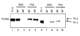
- PVL-lectin partially binds to transferrin-2 having terminal Sia- ⁇ 2,6-Gal. It was. For example, as shown in FIG. 11, transferrin-2 (TF-2) is recovered in both the bound fraction BF (lane 4) and the unbound fraction NF (lane 5) of the PVL-lectin column.
- An object of the present invention is to develop a method for selectively separating glycoproteins derived from the central nervous system from body fluids such as cerebrospinal fluid, development of a method for searching an index marker for central nervous system diseases based on the method, and a method thereof. It aims at providing the index marker for central nervous system diseases obtained by using.
- the present inventors also searched for glycoproteins derived from the central nervous system other than transferrin-1 by combining PVL lectin and various antibodies.
- the GlcNAc non-reducing terminal sugar chain is also present in many other cerebrospinal fluid glycoproteins derived from the central nervous system.
- This sugar chain structure is also present in serotype glycoproteins.
- serotype glycoproteins also have sialic acid at the non-reducing end, and there are structural differences from cerebrospinal fluid glycoproteins derived from the central nervous system that have only GlcNAc at the non-reducing end. Also turned out.
- a method for enriching or separating a terminal GlcNAc-containing glycoprotein from a body fluid or central nerve cell derived from a subject comprising (i) containing terminal sialic acid from a body fluid or central nerve cell using a sialic acid-binding substance.
- a removal step of removing the glycoprotein and (ii) a separation step of separating the complex formed by binding the terminal GlcNAc-containing glycoprotein present in the sample obtained in the removal step to a GlcNAc-binding substance.
- Said method comprising.
- sialic acid-binding substance is an anti-sialic acid antibody or an active fragment thereof or a sialic acid-binding lectin.
- ⁇ 2,6-sialic acid-binding lectin is selected from the group consisting of SSA lectin, SNA lectin and TJA-I lectin.
- albumin binding substance is blue sepharose, or an anti-albumin antibody or an active fragment thereof.
- the body fluid is cerebrospinal fluid, blood (including serum, plasma and interstitial fluid), lymph, perineural fluid, or tissue or cell extract, according to any one of (1) to (9) Method.
- a method for selecting an index marker for a central nervous system disease which is derived from a control body using the method according to any one of (1) to (10) and from an individual suffering from a specific central nervous system disease
- Enrichment / separation step for enriching or separating the terminal GlcNAc-containing glycoprotein from each body fluid or central nerve cell, measurement step for measuring the amount of the terminal GlcNAc-containing glycoprotein obtained in the enrichment / separation step,
- the terminal GlcNAc containing statistically significant quantitative differences by comparing the measured amount of terminal GlcNAc-containing glycoproteins from the control and from individuals suffering from certain central nervous system diseases
- Said method comprising a selection step of selecting a glycoprotein as an indicator marker for that particular central nervous system disease.
- a method for selecting an index marker for central nervous system disease comprising measuring the amount of terminal GlcNAc-containing glycoprotein shown in Table 1 in a control and an individual suffering from a specific central nervous system disease; and Comparing the amount of the corresponding glycoprotein derived from the control body and the individual suffering from a specific central nervous system disease, the glycoprotein showing a statistically significant quantitative difference is identified as the specific central nervous system.
- the method comprising the step of selecting as an index marker for disease.
- a kit for enriching or separating a GlcNAc-containing protein comprising a sialic acid-binding substance and a GlcNAc-binding substance.
- the sialic acid-binding substance is an anti-sialic acid antibody or an active fragment thereof or a sialic acid-binding lectin.
- index marker for central nervous system diseases according to any one of (24) to (27), wherein the index marker for central nervous system disease is ⁇ 2-macroglobulin described in Table 1.
- One or more central nervous system disease index markers according to (23) are detected from a body fluid or central nerve cell derived from a subject, and a disease related to a specific central nervous system disease in the subject is detected from the detection result. How to determine the presence or absence of
- the central nervous system-derived glycoprotein present in a trace amount in body fluids and / or central nerve cells is simply and efficiently enriched. Can be separated and selected.
- a glycoprotein as a novel index marker for central nervous system disease can be selected and separated.
- the obtained index marker for a specific central nervous system disease the onset of the disease can be diagnosed early, easily, and minimally invasively.
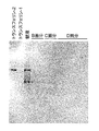
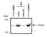
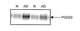
- SSA lectin blotting showing the presence or absence of transferrin-2 (TF-2), a serotype glycoprotein, in the fraction obtained in each step of the method of the first embodiment of the present invention, detected using an anti-transferrin antibody It is.
- SSA lectin-reactive TF-2 is not detected in the D fraction (enriched fraction), which is the fraction eluted from the PVL column.
- D fraction enriched fraction
- elution fraction an elution fraction from a spinal fluid and a PVL column.
- NMO optic neuromyelitis
- MS multiple sclerosis
- non-NMO affected individuals It is a figure which shows the result of the western blotting detected using.
- the first embodiment of the present invention is a method for enriching or isolating terminal GlcNAc (N-acetylglucosamine) -containing glycoproteins from body fluids or central nerve cells.
- terminal GlcNAc-containing glycoprotein is a glycoprotein having a GlcNAc residue on the non-reducing terminal side of the sugar chain.
- the term “terminal GlcNAc-containing glycoprotein” particularly refers to a cerebrospinal fluid type glycoprotein.
- the “sugar chain (of) non-reducing terminal” refers to terminal sugars other than the sugar chain terminal (reducing terminal) exhibiting reducibility when considering the structure of only the sugar chain (excluding the protein portion).
- the reducing sugar has a hemiacetal (or ketal) structure. For example, glucose takes a cyclic structure by reacting an aldehyde group at position 1 and a hydroxyl group at position 5.
- the aldehyde group can be regenerated by a reverse reaction to show reducibility.
- the hydroxyl group at the 1-position is linked to a glycoside as in the non-reducing terminal sugar of the sugar chain, a reaction that generates an aldehyde group does not occur and the reducing property is not exhibited. That is, it is the reducing end sugar that has a free hydroxyl group at the 1-position (1-hydroxyl group).
- the reducing end sugar is attached to the protein.
- Cerebrospinal fluid glycoprotein as used herein is expressed in cells of the central nervous system such as the brain and / or spinal cord, is mainly found in the cerebrospinal fluid, and is located on the non-reducing terminal side of the sugar chain. A glycoprotein having a GlcNAc residue.
- the glycoprotein having a sialic acid residue at the other non-reducing end of the sugar chain is included in the cerebrospinal fluid glycoprotein. Absent.
- the glycoprotein having one or more sialic acid residues at the non-reducing end belongs to a serotype glycoprotein described later.
- the number of GlcNAc terminal residues is not particularly limited.
- one or a plurality of sugar chains may be branched.
- the term “plurality” here means, for example, 2 or more, 3 or more, or 4 or more.
- the method of this embodiment includes a terminal sialic acid-containing glycoprotein removal step (0101) and a terminal GlcNAc-containing glycoprotein complex separation step (0102) as shown in FIG.
- a terminal GlcNAc-containing glycoprotein extraction step (0103) can optionally be included after the complex separation step (0102).
- an albumin removal step (0104) may optionally be included prior to the removal step (0101).
- Terminal sialic acid-containing glycoprotein removal process “Terminal sialic acid-containing glycoprotein removal step” (0101) (hereinafter, simply referred to as “removal step”) refers to a terminal sialic acid-containing sugar from a body fluid or a central nerve cell of a subject using a sialic acid binding substance. It refers to the process of removing proteins.
- terminal sialic acid-containing glycoprotein refers to a glycoprotein having at least one sialic acid residue at the non-reducing end of a sugar chain. Since many glycoproteins having a sugar chain having such a structure are usually found in serum, in this specification, “terminal sialic acid-containing glycoprotein” is also referred to as “serum-type glycoprotein”. However, as mentioned above, since many proteins in serum also leak into the cerebrospinal fluid, serotype glycoproteins are not proteins specific to serum and are the major proteins even in cerebrospinal fluid. Can be present as an ingredient.
- “Sialic acid” is a kind of acidic sugar present at the end of a sugar chain, and refers to a substance having a structure in which the amino group or hydroxyl group of neuraminic acid is substituted with another functional group. Examples thereof include N-acetylneuraminic acid (NeuAc).
- the binding mode in which sialic acid is linked to a sugar chain is not particularly limited, but in this specification, for example, ⁇ 2,6-linked ⁇ 2,6-sialic acid (Sia- ⁇ 2,6-Gal) sugar chain is used. Can be mentioned.
- the number of sialic acid residues as terminal sugar chains in the terminal sialic acid-containing glycoprotein is not particularly limited. For example, one or a plurality of sugar chains may be branched.
- the term “plurality” here means 2 or more, but may be 3 or more, or 4 or more.
- sialic acid-binding substance refers to a substance that binds to the sialic acid in an affinity manner, preferably specifically. Therefore, the sialic acid-binding substance does not bind to the terminal GlcNAc-containing glycoprotein of the present invention having no sialic acid in the terminal sugar chain.
- the bond may be a reversible bond or an irreversible bond.
- examples of the sialic acid binding substance include an anti-sialic acid antibody or an active fragment thereof or a sialic acid binding lectin.
- Anti-sialic acid antibody refers to a sugar chain antibody that recognizes sialic acid and can specifically bind to it.
- antibody refers to an immunoglobulin, a chimeric antibody, a humanized antibody, or a synthetic antibody (such as diabody). When the antibody is an immunoglobulin, it may be a polyclonal antibody or a monoclonal antibody.
- Specific examples of the anti-sialic acid antibody include an anti- ⁇ 2,6-sialic acid antibody that recognizes and specifically binds to an ⁇ 2,6-sialic acid sugar chain.
- the “active fragment” refers to a polypeptide chain or a complex thereof which is a partial region of the above-described antibody and has substantially the same activity as the antigen-specific binding activity of the antibody.
- a polypeptide chain having at least one light chain variable region (V L ) and at least one heavy chain variable region (V H ) or a complex thereof is applicable.
- Specific examples include antibody fragments produced by cleaving immunoglobulins with various peptidases. More specific examples include Fab, F (ab ′) 2 , Fab ′, and the like.
- the anti-sialic acid antibody of the present invention can be derived from any animal including birds and mammals. Examples include mice, rats, guinea pigs, rabbits, goats, donkeys, sheep, camels, horses, chickens or humans.
- anti-sialic acid antibodies may be prepared according to methods known in the art. For example, see the method described in Patent Document 3. Alternatively, a commercially available antibody may be used.
- “Sialic acid-binding lectin” refers to a protein other than an antibody having a binding activity to a terminal sialic acid-containing glycoprotein, that is, a serum-type glycoprotein.
- the type of the lectin is not particularly limited as long as it is a lectin that can recognize and bind to the sialic acid at the non-reducing end in the glycoprotein.
- an ⁇ 2,6-sialic acid-binding lectin capable of recognizing and binding to a glycoprotein having ⁇ 2,6-sialic acid as a terminal sugar chain can be mentioned.
- ⁇ 2,6-sialic acid-binding lectins include agglutinin SSA lectin from Sambucus sieboldiana, agglutinin SNA lectin from Sambucus nigra, and type I from Trichosanthes japonica An agglutinin TJA-I lectin can be mentioned. These may use commercially available lectins. For example, for SSA lectin, Biochemistry Biobusiness 300177 or MBL MJ1001014, for SNA lectin Sigma L6890 Lectin from Sambucus nigra (elder), and for TJA-I lectin, Biochemistry Biobusiness 300186 of the company can be used.
- subject refers to an individual to be examined, that is, a vertebrate that provides body fluids or central nerve cells, preferably a mammal, more preferably a human.
- the subject may be an individual having a certain disease or a healthy body.
- Body fluid refers to a liquid biological sample that can contain a terminal GlcNAc-containing glycoprotein collected from a subject.
- Examples include cerebrospinal fluid, blood (including serum, plasma and interstitial fluid), lymph fluid, perineural root fluid, extracts of tissues or cells, tear fluid, nasal discharge, saliva, urine, vaginal fluid, semen, and the like. . Cerebrospinal fluid, blood, lymph, or perineural root fluid is preferable.
- the body fluid collected from the subject may be used as it is, or may be diluted or concentrated as necessary, or added with a blood coagulation inhibitor such as heparin.
- the body fluid may be collected based on a known method in the field.
- a known blood collection method may be followed.
- peripheral blood it can be collected by injection into a peripheral vein or the like.
- cerebrospinal fluid it may be collected by a known lumbar puncture
- umbilical cord blood it may be collected by inserting a needle into the umbilical cord before delivery of the postpartum placenta.
- the bodily fluid may be used immediately after collection, or may be used after being frozen or refrigerated for a certain period of time and then subjected to processing such as thawing as necessary.
- a sufficient amount of terminal GlcNAc-containing glycoprotein is detected and enriched by using a volume of at least 0.1 mL, preferably at least 1 mL, more preferably at least 10 mL. be able to.
- a sufficient amount of the terminal GlcNAc-containing glycoprotein for detection can be enriched and separated by using a volume of at least 1 mL, preferably at least 10 mL, more preferably at least 30 mL.
- Central nerve cells refer to brain (including cerebrum, midbrain, diencephalon, pons, cerebellum, and medulla) cells or spinal cord cells collected from a subject, or tissues that are aggregates of these cells.
- the central nerve cell is preferably prepared in advance as a homogenized cell extract for use in the present method.
- a surfactant NP-40, TritonX-100, etc.
- media such as a physiological saline and PBS buffer, as needed.
- This step is formed by binding a terminal sialic acid-containing glycoprotein contained in a subject-derived body fluid or central nerve cell (hereinafter collectively referred to as “body fluid etc.”) with a sialic acid-binding substance. This can be achieved by removing the complex from the body fluid or the like.
- the method for forming the complex may be a method in which a body fluid or the like and the sialic acid binding substance are brought into contact with each other.
- a method of mixing a fluid containing a sialic acid binding substance with a body fluid or the like can be mentioned.
- Any method known in the art can be used as a method for removing the formed complex from body fluids and the like.
- a method in which a sialic acid-binding substance immobilized on a carrier surface or labeled with a compound is contacted with a body fluid or the like and then removed based on the characteristics of the carrier or compound.
- the “carrier” includes a polymeric polysaccharide support (eg, Sepharose or Sephadex), magnetic beads, silica, glass, metal (eg, gold, platinum, silver), plastic resin (eg, plastic such as polystyrene, Polyacryamide, etc.), ceramics, latex and the like.
- the shape of the carrier is not particularly limited, but a shape having a large surface area is preferable. This is to increase the chance of contact between the sialic acid-binding substance immobilized on the carrier surface and the terminal sialic acid-containing glycoprotein in the body fluid, etc., and to form a complex more efficiently.
- a bead shape, a porous particle shape, a fiber aggregate shape and the like are preferable.
- a shape that can be easily packed in a column or the like is more preferable.
- the “compound” includes fluorescent dyes (for example, Cy3, Cy5, FAM, HEX, VIC, TAMRA), luminescent substances, biotin or (strept) avidin, and the like.
- fluorescent dyes for example, Cy3, Cy5, FAM, HEX, VIC, TAMRA
- luminescent substances for example, biotin or (strept) avidin, and the like.
- the immobilization of the sialic acid-binding substance on the carrier or labeling with a compound in the present invention is preferably an irreversible bond, that is, a bond that is once formed but does not dissociate by a reverse reaction, or a bond that can be disregarded.
- irreversible bonds include covalent bonds by chemical reactions such as nucleophilic addition reactions between functional groups, nucleophilic substitution reactions or electrophilic substitution reactions, or non-covalent bonds with high affinity.
- a sialic acid-binding substance and a carrier or compound are each modified with an appropriate functional group to form a covalent bond by a chemical reaction between both functional groups. Can be achieved.
- both functional groups should be a combination that can form a covalent bond.
- combinations include an amino group and an aldehyde group, a thiol group and a maleimide group, an azide group and an acetylene group, an azide group and an amino group, a hydrazine group and a ketone group, and a hydrazine group and an aldehyde group.
- a method of forming a covalent bond by such a chemical reaction is a technique well known to those skilled in the art.
- Separation of the complex formed by the binding of the terminal sialic acid-containing glycoprotein and the sialic acid-binding substance may be appropriately performed by a known method utilizing the characteristics of the carrier or compound.
- the “characteristic” here is a characteristic property of the carrier or compound, and includes, for example, magnetic force, specific gravity, fluorescence, luminescence, affinity, and the like.
- a column chromatography (including HPLC) method can be used.
- a polymer polysaccharide support on which a sialic acid-binding substance is immobilized is packed into a column as a resin, a body fluid or the like is loaded (applied) on the column, and the sample liquid flowing out from the column, ie, terminal sialic The remaining fraction in which the acid-containing glycoprotein is captured by the resin may be collected.
- the sialic acid binding substance is immobilized on a carrier such as glass beads or Sephadex beads, the beads having the sialic acid binding substance immobilized thereon are put into a body fluid or the like and stirred as necessary.
- the sialic acid-binding substance-immobilized beads to which the terminal sialic acid-containing glycoprotein is bound are centrifuged by using the specific gravity of the carrier, and the upper layer liquid may be recovered. Furthermore, if the sialic acid-binding substance is immobilized on the magnetic beads, the sialic acid-binding substance-immobilized magnetic beads are put into a body fluid or the like, stirred as necessary, and then using magnetic force. What is necessary is just to isolate
- the sialic acid-binding substance is labeled with a compound such as a fluorescent dye or a luminescent substance
- the labeled sialic acid-binding substance is put into a body fluid or the like and stirred as necessary, and then a cell sorter or flow Cytometry can also be used to partition and remove the complex in which the labeled sialic acid binding substance is bound to the terminal sialic acid-containing glycoprotein. Any of these may follow a method known in the art.
- This step is performed by repeating the same removal method a plurality of times, for example, 2, 3, 4, 5 or 6 times or a combination of two or more different removal methods, so that the terminal sialic acid-containing sugar in body fluids, etc. Proteins can also be removed more reliably.
- PVL lectin binds strongly to the non-reducing end of GlcNAc and hardly binds to the non-reducing end of Sia- ⁇ 2,6-Gal, which is the main sugar chain end of blood group glycoproteins. Since it was technical common sense in this field, those skilled in the art assumed enrichment of cerebrospinal fluid glycoprotein using only PVL lectin with the aim of developing a simpler method. However, in practice, the present inventors found that PVL lectin also binds to the Sia- ⁇ 2,6-Gal non-reducing end, and in order to enrich for cerebrospinal fluid glycoprotein, It became clear that it was essential.
- This step makes it possible to remove terminal sialic acid-containing glycoproteins contained in body fluids or central nerve cells, that is, serotype glycoproteins.
- Terminal GlcNAc-containing glycoprotein complex separation process (configuration) “Terminal GlcNAc-containing glycoprotein complex separation step” (0102) (hereinafter simply referred to as “complex separation step”) is a terminal GlcNAc-containing glycoprotein present in the sample obtained in the removal step (0101). Is a process of separating and recovering the formed complex by binding to the GlcNAc binding substance.
- the terminal sialic acid-containing glycoprotein is not contained or is contained in a very small amount. Therefore, the glycoprotein having a GlcNAc residue at the sugar chain end in this sample is in principle the cerebrospinal fluid glycoprotein of the present invention. Therefore, this step is characterized in that the terminal GlcNAc-containing glycoprotein is separated from the sample that has undergone the removal step (0101) in the state of a complex bound to a GlcNAc-binding substance.
- the “GlcNAc-binding substance” refers to a substance that binds to GlcNAc or a GlcNAc terminal sugar chain with affinity, preferably specifically.
- the binding between GlcNAc and the GlcNAc-binding substance is preferably a reversible binding if the sample solution obtained in this step is subjected to the terminal GlcNAc-containing glycoprotein extraction step described later.
- the GlcNAc binding substance include an anti-GlcNAc antibody or an active fragment thereof, or a GlcNAc binding lectin.
- Anti-GlcNAc antibody refers to a sugar chain antibody that can specifically recognize and bind to GlcNAc.
- This anti-GlcNAc acid antibody may be prepared according to a method known in the art. For example, see the method described in Patent Document 3. For example, OMB4 (Archives of Biochemistry and Biophysics 1997, vol.342 (1), p48-57 Ozawa, H et al.) Can be used.
- GlcNAc-binding lectin refers to a protein other than an antibody having an activity of binding to GlcNAc or a glycoprotein having it at the sugar chain end.
- the type of the lectin is not particularly limited as long as it is capable of recognizing and binding to GlcNAc of the terminal sugar chain. Examples thereof include PVL lectin derived from Psathyrella velutina and WGA lectin derived from bread wheat. These may use commercially available lectins.
- PVL lectin Wako Pure Chemical Industries 165-17591 ⁇ ⁇ ⁇ ⁇ Psathyrella Velutina Lectin or MBL's recombinant PVL can be used, or if it is a WGA lectin, Biochemical Biobusiness 300191 or MBL's J1001016 can be used. .
- This step can be achieved by binding the terminal GlcNAc-containing glycoprotein contained in the sample solution obtained after the removing step with a GlcNAc-binding substance and recovering the formed complex.
- the method for forming the complex is in accordance with the method described in the removing step.
- the method for recovering the complex of the terminal GlcNAc-containing glycoprotein and the GlcNAc-binding substance thus formed is basically in accordance with the method described in the removing step.
- this step differs from the above step in that the complex of the terminal GlcNAc-containing glycoprotein and GlcNAc-binding substance formed is recovered and the other sample solution is removed.
- the terminal GlcNAc-containing glycoprotein in the sample solution can be more reliably obtained by subjecting the sample solution after recovering the complex to the same separation method or two or more different separation methods. It can also be recovered.
- the collected complex of the terminal GlcNAc-containing glycoprotein and the GlcNAc-binding substance may be removed by washing several times with an appropriate buffer (for example, PBS buffer).
- an appropriate buffer for example, PBS buffer
- This step can selectively enrich and separate terminal GlcNAc-containing glycoproteins, ie, cerebrospinal fluid glycoproteins, contained in body fluids or central nerve cells.
- Terminal GlcNAc-containing glycoprotein extraction process (configuration) “Terminal GlcNAc-containing glycoprotein extraction step” (0103) (hereinafter simply referred to as “extraction step”) means dissociation of the complex of the terminal GlcNAc-containing glycoprotein and the GlcNAc-binding substance, thereby producing a terminal GlcNAc-containing glycoprotein. Is a step of eluting. This step is an optional step that can be selected in the present invention.
- the GlcNAc-binding substance is an anti-GlcNAc antibody
- it can be dissociated by adding GlcNAc or the like having the same hapten and allowing the anti-GlcNAc antibody to compete with the terminal GlcNAc-containing glycoprotein.
- the complex may be dissociated under a high salt concentration condition (for example, Mg 2+ > 2M) or under a strongly acidic condition (for example, Gly-HCl pH3>).
- a high salt concentration condition for example, Mg 2+ > 2M
- a strongly acidic condition for example, Gly-HCl pH3>
- the GlcNAc-binding substance is a GlcNAc-binding lectin, it can be dissociated by adding GlcNAc, which is a hapten sugar.
- the “albumin removal step” (0104) is a step of removing albumin contained in a body fluid or the like using an albumin binding substance prior to the removal step. This step is also an optional step that can be selected in the present invention.
- albumin is albumin contained in a body fluid or the like and mainly means serum albumin.
- This step is achieved by bringing the albumin binding substance into contact with a body fluid or the like to form a complex of the albumin binding substance in the body fluid and the albumin binding substance, and then removing the complex. it can.
- albumin-binding substance refers to a substance that binds to albumin with affinity, preferably specifically. This bond may be either a reversible bond or an irreversible bond, but is desirably irreversible in order to prevent once bound albumin is dissociated and mixed again into a body fluid or the like.
- albumin binding substances examples include anti-albumin antibodies or active fragments thereof.
- Anti-albumin antibody refers to an antibody that can recognize and bind to albumin, mainly serum albumin.
- albumin mainly serum albumin.
- a commercially available antibody may be used.
- B80's A80-129A-Human-Albumin-Antibody-Affinity-Purified or KR-002 antibody from Cosmo Bio Inc. can be used.
- the method for forming a complex of albumin and an albumin binding substance in a body fluid and / or the method for removing the formed complex of albumin and an albumin binding substance are the same as those described in the removal step (0101).
- the body fluid from which albumin, which is a contaminant, has been removed can be subjected to the terminal sialic acid-containing glycoprotein removal step.
- ⁇ Embodiment 2 Index marker selection method for central nervous system disease> (Outline and configuration)
- the second embodiment of the present invention is a method for selecting an index marker for central nervous system diseases.
- This embodiment uses the method of Embodiment 1 to efficiently remove blood group glycoproteins present in large quantities in body fluids, that is, terminal sialic acid-containing glycoproteins, and to achieve the target cerebrospinal fluid type After enriching and separating glycoproteins, ie, terminal GlcNAc-containing glycoproteins, terminal GlcNAc-containing glycoproteins that serve as index markers for specific central nervous system diseases are obtained from the obtained terminal GlcNAc-containing glycoproteins. How to choose.
- “Indicator marker” refers to a substance having characteristics that are objectively measured and evaluated as an indicator of a pharmacological response to a pathological process or therapeutic intervention. Therefore, in the present specification, “(index) marker for central nervous system disease” is a characteristic that can be objectively measured and evaluated in response to the presence or severity of a specific central nervous system disease or the like. Or a fragment thereof. A specific example will be described in a fourth embodiment described later.
- central nervous system disease refers to a disease that develops due to abnormal function and / or morphological abnormality (including developmental abnormality) of the brain and / or spinal cord.
- neurodegenerative diseases include Alzheimer's disease (AD: Alzheimer's Disease and its previous lesion Mild Cognitive Impairment), frontotemporal dementia, Lewy body dementia, Parkinson's disease, progressive nuclear Examples include cerebral palsy or basal ganglia degeneration.
- Metabolic abnormalities include idiopathic normal pressure hydrocephalus (iNPH), obstructive hydrocephalus or infectious or metabolic encephalopathy, and autoimmune related diseases include multiple sclerosis (MS).
- Optic myelitis NMO: neuromyelitis optica
- Guillain-Barre syndrome peripheral motor nerves are often affected, but pharyngeal-cervical-brachial variant damages cranial nerves
- acute disseminated encephalomyelitis ADAM: acute disseminated encephalomyelitis etc.
- schizophrenia, bipolar disorder or major depression as psychiatric disorders neuroepithelial tissue tumors (glioma, neurocellular) as neoplastic diseases Tumors, etc.), schwannoma (neurinoma, neurofibroma, etc.), meningeal tumors (meningiomas, other mesenchymal tumors), Turkish groin tumors, metastatic tumors and the like.
- Infectious diseases include bacterial or viral meningitis.
- the method of this embodiment includes a terminal GlcNAc-containing glycoprotein enrichment / separation step (0201) and a comparative selection step (0202) as shown in FIG.
- a terminal GlcNAc-containing glycoprotein enrichment / separation step (0201) and a comparative selection step (0202) as shown in FIG.
- the method of each process is demonstrated concretely.
- Terminal GlcNAc-containing glycoprotein enrichment / separation step refers to a body fluid derived from a control body (hereinafter referred to as “control body sample”) using the method of Embodiment 1 above. And terminal GlcNAc-containing glycoproteins are separated from body fluids of individuals suffering from a specific central nervous system disease (hereinafter referred to as “affected individual samples”).
- control body refers to an individual who is not affected by a central nervous system disease (hereinafter referred to as “target disease”) as a target at the time of control body sampling.
- target disease a central nervous system disease
- individuals who are not affected by any central nervous system disease such as healthy subjects, but also individuals who are not affected by the target disease but are affected by other central nervous system diseases. It can be a control for affected individuals.
- An individual who is not affected by the target disease but suffers from another central nervous system disease whose symptoms are very similar to that of the specific central nervous system disease is particularly preferred as a control in the present embodiment. Described below are combinations of different central nervous system diseases that are difficult to distinguish because their symptoms are very similar.
- MS Multiple sclerosis
- MS is known as a representative autoimmune demyelinating disease.
- MS is a common disease among white women, and the number of patients is estimated to reach 400,000 in the United States (1 in 750) and 2.5 million worldwide.
- the incidence of MS in Japan is 10,000 (1 in 10,000), and it has been considered a disease with a low incidence in the past. Has increased significantly.
- Optic myelitis has historically been considered a subtype of multiple sclerosis.
- measurement of this antibody is technically difficult, and in Japan it can be measured only in 4 laboratories, and the positive rate is also 60-80%, which varies greatly depending on the facility.
- NMO and MS require strong prescriptions such as steroid therapy in the acute phase.
- interferon ⁇ In the chronic phase, interferon ⁇ is used in MS to prevent recurrence, whereas in NMO, interferon ⁇ may exacerbate symptoms, so a small amount of steroids and immunosuppressants are used. Therefore, differential treatment is an extremely important issue because the treatment methods differ between the two diseases. Therefore, there is a need for a diagnostic marker that can accurately diagnose both diseases.
- Some of the above listed neurological disorders can be diagnosed morphologically (by MRI sputum or CT examination) as they progress (MRI sputum or CT examination), but there are diseases that are difficult to differentiate in the early stages of the disease. Many. For example, Parkinson's disease and progressive supranuclear palsy or corticobasal degeneration show similar gait abnormalities with little morphological change. In addition, it is not easy to differentiate between early stage Alzheimer's disease and early Lewy body dementia or frontal-temporal dementia. Particularly in frontotemporal dementia, as in Alzheimer's disease, an increase in cerebrospinal fluid phosphorylated tau protein is observed, and the differential diagnostic marker that is most effective in the past cannot be used.
- idiopathic normal pressure hydrocephalus In advanced Alzheimer's disease, remarkable morphological changes such as dementia and ventricular enlargement (brain atrophy) are observed, but these two symptoms are the same as those of idiopathic normal pressure hydrocephalus. In fact, the number of patients with idiopathic normal pressure hydrocephalus is estimated to be 310,000, but only 1200 (0.4% of the expected number of patients) undergoing radical surgery for the disease described later. That is, it is highly likely that many patients with idiopathic normal pressure hydrocephalus have been mistakenly diagnosed with Alzheimer's disease and left without benefit from treatment.
- idiopathic normal pressure hydrocephalus is caused by excess of cerebrospinal fluid, it can be completely cured by draining excess cerebrospinal fluid by a simple minor operation (for example, shunting by bypassing the ventricle and abdominal cavity with a tube). In other words, since it is a “healing” dementia, an accurate diagnosis is required.
- an individual need not be a living body as long as it is clear that it is immediately after death and does not affect the central nervous system.
- the individual is preferably a mammal, more preferably a primate, particularly preferably a human.
- the details of the method of this step are described in the first embodiment, the details are omitted here.
- the terminal GlcNAc-containing glycoprotein group obtained by this step that is, the cerebrospinal fluid type glycoprotein group
- the cerebrospinal fluid type glycoprotein group shown in Table 1 separated in Example 2 described later can be mentioned. Any of these can be index marker candidates for central nervous system diseases in the present embodiment.
- Comparative selection process is a process in which the terminal GlcNAc-containing glycoprotein obtained from each of the control sample and the affected individual sample is quantified in the above-described process, When a statistically significant quantitative difference is found after comparing the quantities, it is a step of selecting the terminal GlcNAc-containing glycoprotein as an index marker for a specific central nervous system disease based on the affected individual sample .
- the “amount” is the amount of a specific terminal GlcNAc-containing glycoprotein in the terminal GlcNAc-containing glycoprotein group obtained in the above step, and may be a relative amount such as a concentration, or a weight or volume. Such an absolute amount may be used.
- the terminal GlcNAc-containing glycoprotein contained in the terminal GlcNAc-containing glycoprotein group obtained from each of the control body sample and the affected individual sample in the above step is quantified.
- the method for this detection and quantification is not particularly limited. For example, mass spectrometry, antigen-antibody reaction, electrophoresis and the like can be used.
- Mass spectrometry includes all known quantification methods using a mass spectrometer. For example, high performance liquid chromatography / mass spectrometry (LC-MS), high performance liquid chromatography / tandem mass spectrometry (LC / MS / MS), gas chromatography / mass spectrometry (GC-MS), gas chromatography / tandem mass spectrometry (GC) -MS / MS), capillary electrophoresis mass spectrometry (CE-MS) and ICP mass spectrometry (ICP-MS).
- LC-MS high performance liquid chromatography / mass spectrometry
- LC / MS / MS high performance liquid chromatography / tandem mass spectrometry
- GC-MS gas chromatography / mass spectrometry
- CE-MS capillary electrophoresis mass spectrometry
- ICP-MS ICP mass spectrometry
- the “antigen-antibody reaction method” includes all known quantification methods using antibodies. For example, ELISA (Enzyme-Linked ImmunoSorbent Assay) method or a similar method, flow cytometry method, surface plasmon resonance (SPR) method, or quartz crystal microbalance (QCM) method can be mentioned.
- ELISA Enzyme-Linked ImmunoSorbent Assay
- SPR surface plasmon resonance
- QCM quartz crystal microbalance
- Electrophoresis includes any known quantification method using electrophoresis.
- An example is two-dimensional electrophoresis.
- the amounts of glycoproteins containing terminal GlcNAc containing the same type, ie, the same type of terminal GlcNAc are compared based on the quantitative results.
- a known protein that is included in the terminal GlcNAc-containing glycoprotein group and is expected to have no quantitative difference in expression between the control body and the affected individual is used as the internal control, And the quantitative results of affected individuals can be corrected. Examples of such a protein for internal control include albumin.
- a terminal GlcNAc-containing glycoprotein showing a statistically significant quantitative difference is selected as an index marker for the specific central nervous system disease.
- the selected index marker for central nervous system disease may be an index marker specific for a specific central nervous system disease, or may be an index marker for a plurality of central nervous system diseases.
- Statistically significant means that when the quantitative difference between the two corresponding terminal GlcNAc-containing glycoproteins is statistically processed, there is a significant difference between the two. Specifically, for example, the risk rate (significance level) is less than 5%, 1%, or 0.1%.
- the test method for statistical processing is not particularly limited as long as a known test method capable of determining the presence or absence of significance is appropriately used. For example, Student's t test or multiple comparison test can be used.
- the statistically significant quantitative difference is that the difference between the two is 1.3 times or more, preferably 1.5 times or more, more preferably 1.7 times or more, and even more preferably 2 times or more.
- a statistically significant quantitative difference may be that a particular terminal GlcNAc-containing glycoprotein from an affected individual is more or less than the amount of that terminal GlcNAc-containing glycoprotein from a control. This is because any terminal GlcNAc-containing glycoprotein that specifically increases or decreases in a specific central nervous system disease can be a candidate for an index marker of the present invention.
- the terminal GlcNAc-containing glycoprotein present in the body fluid or the like of either the control or affected individual in the detection / quantitative results and not present in the other is a preferred index marker for the specific central nervous system disease. Can be.
- the central nervous system can be used as long as the terminal GlcNAc-containing glycoprotein enrichment / separation step is performed under the same conditions. This is convenient because it is only necessary to analyze body fluids derived from individuals suffering from systemic diseases, and it is not necessary to analyze body fluids derived from the control body each time.
- a terminal GlcNAc-containing glycoprotein that can serve as an index marker for a central nervous system disease from the body fluid of an individual suffering from a central nervous system disease, which has been difficult to search conventionally, particularly cerebrospinal fluid or central nerve cells Can be selected easily and efficiently.
- any of them can be an index marker for the specific central nervous system disease. If there are a plurality of index markers, it is possible to make a multifaceted diagnosis, which is advantageous in that a more accurate diagnosis can be performed.
- one selected index marker is an index marker for a plurality of central nervous system diseases
- ⁇ 2-macroglobulin may be an effective NMO indicator marker in diagnosing whether the patient is suffering from NMO or MS.
- one index marker is an index marker for a plurality of central nervous system diseases, and the plurality of central nervous system diseases exhibit similar symptoms, individual central nervous system diseases Can have a plurality of different index markers, each disease can be identified by the combination pattern of the index markers specific to the disease, so that it can also function as an index marker.
- index markers for specific central nervous system diseases are easily and comprehensively separated from cerebrospinal fluid glycoproteins enriched and separated from body fluids, for example, as shown in Table 1. can do.
- the third embodiment of the present invention is a kit that can selectively enrich and separate terminal GlcNAc-containing glycoproteins, that is, cerebrospinal fluid glycoproteins from body fluids and the like.
- kits of the present embodiment include a sialic acid binding substance and a GlcNAc binding substance as essential components.
- sialic acid-binding substance examples include an anti-sialic acid antibody and / or a sialic acid-binding lectin.
- the terminal sialic acid-containing glycoprotein to be used in this kit preferably has ⁇ 2,6 sialic acid at the terminal sugar chain. Therefore, the anti-sialic acid antibody included in the kit is preferably an anti- ⁇ 2,6-sialic acid antibody or an active fragment thereof.
- the sialic acid binding lectin is ⁇ 2,6 sialic acid binding lectin, for example, S177 lectin from Biochemical Biobusiness 300177 or MBL J1001014, and SNA lectin from Sigma L6890 Lectin from If Sambucus nigra (elder) is TJA-I lectin, 300186 of Biochemical Biobusiness can be used.
- the GlcNAc binding substance is preferably, for example, an anti-GlcNAc antibody or an active fragment thereof.
- the anti-GlcNAc antibody include OMB4 antibody (Archives of Biochemistry and Biophysics 1997, vol.342 (1), p48-57 Ozawa, H et. Al.).
- OMB4 antibody As the GlcNAc-binding lectin, 165-17591165Psathyrella Velutina Lectin of Wako Pure Chemical Industries or recombinant PVL of MBL can be used, or 300191 of Biochemical Biobusiness or J1001016 of MBL can be used for WGA lectin.
- the kit of the present embodiment can also include a complex dissociation buffer.
- the “complex dissociation buffer” refers to a buffer having a chaotropic effect that can dissociate a complex of a terminal GlcNAc glycoprotein and a GlcNAc-binding substance.
- a buffer containing a high salt concentration (eg, Mg 2+ > 2M) buffer, a strongly acidic buffer (pH> 2.5), or a chaotropic agent (eg, GlcNAc) is applicable.
- the kit of this embodiment may further contain an albumin binding substance.
- the albumin binding substance include blue sepharose, anti-albumin antibody or an active fragment thereof.
- the sialic acid binding substance, GlcNAc binding substance and / or albumin binding substance may be immobilized on a carrier as necessary, or may be labeled with a fluorescent dye or a luminescent substance.
- the kit of the present embodiment includes a column (spin column or the like) for filling the sialic acid binding substance, GlcNAc binding substance or albumin binding substance, a washing buffer (PBS buffer, physiological Saline, etc.) and / or instructions describing the protocol and the like.
- a column spin column or the like
- PBS buffer physiological Saline, etc.
- the kit of the present embodiment may include a specific central nervous system disease index marker binding substance.
- the “specific central nervous system disease index marker binding substance” is a specific index marker (terminal GlcNAc glycoprotein) obtained by the method of Embodiment 2 or the like in addition to a known specific central nervous system disease index marker A substance that can recognize and bind to it.
- an antibody against a specific terminal GlcNAc glycoprotein or an active fragment thereof is applicable.
- Specific examples include antibodies (anti-transferrin-1 antibody and anti-transferrin-2 antibody) or active fragments thereof against transferrin-1 and transferrin-2 (Patent Document 3), which are index markers for idiopathic normal pressure hydrocephalus (iNPH). Can be mentioned.
- the kit of this embodiment can be a kit for diagnosing the specific central nervous system disease. That is, in a group of terminal GlcNAc glycoproteins in a body fluid collected from a subject, an indicator marker related to a specific disease (iNPH in the above example) is detected and quantified using the attached indicator marker binding substance for the disease. Can do. If the quantification results show a statistically significant quantitative difference from the control, the subject may have a central nervous system disease corresponding to the disease marker marker binding substance. Can be diagnosed as high.
- the terminal GlcNAc glycoprotein which is a cerebrospinal fluid glycoprotein, can be easily and efficiently separated and added from body fluids and the like.
- the fourth embodiment of the present invention is an index marker for central nervous system diseases.
- the “index marker for central nervous system disease” of this embodiment is one or more sugars selected from the glycoprotein group shown in Table 1 obtained by using the method of Embodiment 2 in Example 2 described later. Based on protein or fragment thereof.
- All of the 13 glycoproteins listed in Table 1 are cerebrospinal fluid glycoproteins according to the present invention, that is, sugar chains containing N-acetylglucosamine residues at the non-reducing end and not containing ⁇ 2,6 sialic acid. It is a glycoprotein having
- the “fragment thereof” refers to a glycoprotein fragment having a sugar chain containing a GlcNAc residue and not containing ⁇ 2,6-sialic acid in each glycoprotein shown in Table 1.
- index marker for central nervous system disease examples include, for example, ⁇ 2-macroglobulin shown in Table 1 or a fragment thereof if the central nervous system disease is optic neuromyelitis (NMO). It is done. As described in Embodiment 2, ⁇ 2-macroglobulin having a sugar chain containing a GlcNAc residue and not containing ⁇ 2,6-sialic acid or a fragment thereof increases in NMO patients but does not increase in MS patients (described later). See Example 3). Therefore, ⁇ 2-macroglobulin or a fragment thereof can be a very useful index marker for NMO or MS in determining whether a subject suffers from NMO or multiple sclerosis (MS).
- NMO optic neuromyelitis
- the fifth embodiment of the present invention is a method for determining the presence or absence of a specific central nervous system disease.
- the method of this embodiment includes a central nervous system disease index marker detection step and a disease determination step.
- the central nervous system disease index marker detection step is a step of detecting the central nervous system disease index marker of the embodiment 4 from a body fluid or a central nerve cell derived from a subject. Say.
- a method for detecting an index marker for central nervous system disease from a body fluid or a central nerve cell derived from a subject uses the method for enriching and separating terminal GlcNAc-containing glycoproteins described in the first embodiment to obtain a body fluid or a central nerve cell. It is preferable to enrich / separate an indicator marker for central nervous system disease contained therein, that is, a cerebrospinal fluid glycoprotein or a fragment thereof.
- Whether or not how many of the marker markers for central nervous system disease of Embodiment 4, ie, the glycoprotein group described in Table 1 or a fragment thereof, are detected from body fluids or central nerve cells derived from the subject is determined. What is necessary is just to determine suitably according to the kind of specific central nervous system disease which should be done, and the objective. “Purpose” as used herein means, for example, to determine whether a subject suffers from any central nervous system disease or is healthy as in a medical examination, that is, the central nervous system disease is completely specified.
- the method for detecting the index marker for central nervous system disease is not particularly limited. Any method known in the art can be used as long as it can detect the target marker for central nervous system disease.
- an antigen-antibody reaction method particularly a sandwich ELISA method can be preferably used.
- JP 2010-121980 Patent Document 2
- Western blotting or lectin blotting was used for quantification of cerebrospinal fluid transferrin. More preferably, it is possible to use a high-throughput sandwich ELISA method, which has better measurement sensitivity and quantitativeness than the blotting method.
- Patent Document 2 As an (antibody / lectin) sandwich ELISA method, an anti-transferrin antibody (capture antibody) is adsorbed on a plate, and the sugar chain portion of the captured cerebrospinal fluid transferrin is PVL. A method for detection with lectins was described. However, although it is presumed that the binding property of PVL lectin is weak, it has been found that quantitative measurement is difficult. On the other hand, in the present invention, a molecule having a cerebrospinal fluid sugar chain is adsorbed on a plate. After capturing all of the above, a method for detecting with an antibody against the target molecule was developed (lectin / antibody sandwich ELISA method).
- the “morbidity discrimination step” is a step of discriminating whether or not the subject suffers from a specific central nervous system disease.
- This step is a step of finally determining whether or not the subject suffers from a specific central nervous disease from the detection result of the index marker detecting step for the central nervous system disease.
- the detection pattern that is, the increase or decrease in the content
- a specific central nervous system disease is previously known according to the second embodiment.
- the specific central nervous system disease is NMO
- ⁇ 2-macroglobulin as an index marker for central nervous system disease is increased in NMO patients.
- MS ⁇ 2-macroglobulin is not increased from MS patients.
- the cerebrospinal fluid derived from the subject (11 mL, 0.47 mg / mL) was dialyzed against phosphate buffer (20 mM, pH 7.0) before chromatography for desalting. After dialysis, it was applied to a blue sepharose column (HiTrap Blue HP; GE Healthcare) (5 mL) for albumin removal, and the unbound fraction (A fraction) that flowed out, that is, the fraction from which albumin was removed was collected ( Albumin removal step). This A fraction is rich in serotype glycoproteins with ⁇ 2,6-sialic acid.
- the bound cerebrospinal fluid glycoprotein was specifically eluted with 0.2 M GlcNAc (Sigma), and the terminal GlcNAc-containing glycoprotein-enriched fraction (D fraction) Min: PVL column elution fraction).
- the human cerebrospinal fluid equivalent to 20 ⁇ g protein and the BD fractions were mixed with LaemmLi sample buffer and heated, and then subjected to SDS / PAGE using 4-20% gradient polyacrylamide gel. Transferrin-2 and transferrin-1 were used as indicators of serotype glycoprotein and cerebrospinal fluid type glycoprotein, respectively. Electrophoresis was performed at a constant current of 40 mA for 55 minutes.
- biotinylated PVL is not commercially available, unlabeled PVL lectin (Wako Pure Chemicals, Cat. No.165-17591) was purchased and biotinylated in the presence of 10 mM GlcNAc. Ez-Link®NHS-Biotin (Pierce, Cat. No. 21336) was used as the biotinating agent. 10 mM GlcNAc was used to interact with the sugar binding site and prevent this site from being biotinylated and inactivated.
- the band detected here is derived from blood group glycoprotein.
- cerebrospinal fluid contains a large number of serum proteins, that is, blood group glycoproteins.
- many bands of blood group glycoproteins in the cerebrospinal fluid disappeared, confirming that most of the blood group glycoproteins were removed during the terminal sialic acid-containing glycoprotein removal step. It was done.
- a few bands derived from blood group glycoproteins were still observed in the B fraction, but disappeared completely in the eluted D fraction after adsorption to the PVL lectin column.
- LaemmLi sample buffer was added to the human cerebrospinal fluid equivalent to 0.04 ⁇ g protein and the B and D fractions and heated, and then subjected to SDS / PAGE using 4-20% gradient polyacrylamide gel. Transferrin-2 and transferrin-1 were used as indicators of serotype glycoprotein and cerebrospinal fluid type glycoprotein, respectively, as in (2). Electrophoresis was performed at a constant current of 40 mA for 55 minutes.
- the separated protein was electrically transferred to a PVDF membrane at a constant current of 260 mA for 50 minutes by a blotting method known in the art. After the transfer, the membrane was blocked with Tris buffered saline (TBS) containing 5% skim milk-0.1% Tween-20 for 1 hour or longer.
- TBS Tris buffered saline
- an antibody (1-2 ⁇ g / mL) diluted 1: 1000 in 3% skim milk-TBS was added and allowed to react for 1 hour.
- the membrane was washed three times for 15 minutes each with a TBS washing solution containing 0.1% Tween-20.
- the mixture was diluted 1: 1000 in 3% skim milk-TBS and reacted with anti-rabbit IgG, Horseradish®Peroidase (Amersham, ® # NA934V) (1 ⁇ g / mL) for 1 hour.
- the membrane was washed again with the washing solution three times for 15 minutes each.
- a chemiluminescent substrate PierceSuperSignal West DuraExtended Duration Substrate
- LAS-1000plus: Fuji Film For Western blotting of transferrin, as anti-human transferrin antibody, BETHYL® Laboratories, Cat. No. A80-128A of Cat. No. A80-128A of inc. Was used.
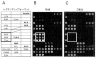
- Glycan profiling of glycoproteins in unpurified cerebrospinal fluid and enriched fractions by lectin microarray is basically performed by Kuno, A. et al., Nature Methods. 2, 851-856 (2005), Uchiyama N., Proteomics 8, The method described in 3042-3050 (2008) was followed. Unpurified cerebrospinal fluid (0.47 mg / l) and D fraction concentrate (1.42 mg / mL) were diluted 10-fold with PBS buffer.
- Protein equivalent to 200 ng (4.3 ⁇ L and 1.4 ⁇ L, respectively) is prepared to 10 ⁇ L with 1% Triton X-100-containing PBS buffer, and 20 ⁇ g of fluorescent labeling reagent (Cy3-SE, GE Healthcare) is added thereto. Fluorescence labeling reaction was performed at room temperature for a period of time. The excess fluorescent labeling reagent was inactivated by adding 90 ⁇ L of glycine-containing buffer to the reaction product and reacting at room temperature for 2 hours. This was subjected to a lectin microarray as a fluorescently labeled glycoprotein solution. A lectin microarray having 43 different lectins immobilized thereon was used.
- a 4-point dilution series was prepared for each sample and applied to the lectin microarray.
- the binding reaction between the lectin and the glycoprotein to be analyzed was performed at 20 ° C. for 12 hours. After the reaction, the sample solution on the array was removed, washed 3 times with a dedicated buffer, and then scanned with a lectin microarray scanner GlycoStation TM Reader 1200 manufactured by GP Bioscience. Data obtained by scanning was saved as jpeg files and TIFF files. A jpeg file was used to create the image diagram.
- the PHAE and UDA signals indicated the presence of bisecting beta1,4-GlcNAc, and the PSA, LCA, and AAL signals indicated the presence of core fucose. All of these showed the characteristics of cerebrospinal fluid type sugar chains. Jacalin, ACA, and MAH also showed increased signal after enrichment, indicating that the abundance was high. This is because N-in the glycoprotein enriched with PVL lectin. This means that not only a linking sugar chain but also an O-linked sugar chain is present. From the above results, glycoproteins having ⁇ 2,6 sialic acid ends are efficiently removed from the D fraction obtained by the enrichment / separation method of the present invention, and only cerebrospinal fluid glycoproteins having GlcNAc ends are concentrated. It was shown that.
- trypsin solution (2 ⁇ g trypsin was dissolved in 50 ⁇ L of 50 mM ammonium bicarbonate solution) 1) and incubate overnight at 37 ° C, heat at 100 ° C for 5 minutes, bring the reaction mixture to room temperature, add 10 ⁇ L of 5 mU PNGaseF (TaKaRa) solution, and overnight at 37 ° C. Incubated.
- the obtained sample was transferred to a glass vial and dried. After adding 50 ⁇ L of sodium hydroxide suspension obtained by grinding about 50 mg of sodium hydroxide (Fluka) with 250 ⁇ L of dimethyl sulfoxide (Wako) in a mortar and mixing it lightly, 50 ⁇ L of iodinated Methyl (Wako) was added and allowed to react for 30 minutes on a shaker. Next, after neutralization by adding 150 ⁇ L of 50% acetic acid solution on ice, the solution with further added 850 ⁇ L of distilled water was purified using a simple column SepPakC18 (50 mg / mL, Waters) and eluted with 600 ⁇ L of acetonitrile. Then, it concentrated under reduced pressure.
- sodium hydroxide suspension obtained by grinding about 50 mg of sodium hydroxide (Fluka) with 250 ⁇ L of dimethyl sulfoxide (Wako) in a mortar and mixing it lightly
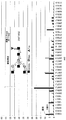
- the cerebrospinal fluid type glycoprotein group obtained by the enrichment / separation method of the present invention was identified by a mass spectrometer.
- Example 2 The enriched fraction D obtained in Example 1 was dissolved in a sample buffer for electrophoresis, reacted at 100 ° C. for 5 minutes, and separated and developed on a 5-20% gradient gel. The developed gel was subjected to silver staining according to the protocol for mass spectrometry shown below.
- the composition of the solution used is as follows.
- the solution was replaced with a silver nitrate solution, shaken at a low temperature for 20 minutes, then shaken with pure water for 1 minute, and a developer was put therein. After confirming color development, the gel was stored after changing to a stop solution. After cutting out the band to be analyzed, in-gel digestion was performed according to the following procedure, and protein identification was performed by analysis with a mass spectrometer. After cutting out the protein band to be analyzed, the stained gel was decolorized by the following procedure.
- the composition of the used solution is as follows.
- the sample solution was desalted and concentrated with ZipTipu-C18 (Millipore), mixed with CHCA (alpha-cyano-4-hydroxycinnamic acid (BRUKER)), and MALDI-TOF.
- Peptide mass was measured by analysis using a mass spectrometer (ultraflex III, Bruker). Proteins were identified by MS-Fit in the program package ProteinProspector developed by UCSF.
- cerebrospinal fluid glycoproteins can be separated by changing the separation conditions (for example, the type of lectin) and the quantitative conditions.
- these cerebrospinal fluid glycoproteins can be index marker candidates for various central nervous system diseases.
- MS Multiple sclerosis
- NMO Optic neuromyelitis
- cerebrospinal fluid glycoproteins were enriched from the cerebrospinal fluid of NMO patients and controls in accordance with the method of Example 1 (1). Subsequently, Western blotting was performed according to the method of Example 1 (3) using antibodies against various cerebrospinal fluid glycoproteins shown in Table 1.
- ⁇ 2-macroglobulin which is an easy-to-obtain antibody, as an NMO index marker candidate cerebrospinal fluid glycoprotein.
- an antibody an anti-human ⁇ 2-macroglobulin antibody (ICN, Cat. No. 55113) (1-2 ⁇ g / mL) was used.
- FIG. 7 shows ⁇ 2-macroglobulin in the cerebrospinal fluid sample after enrichment.
- ⁇ 2-macroglobulin showed a clear difference in expression levels between NMO patients and controls, ie non-NMO affected MS patients. Similar results were obtained using anti-human ⁇ 2-macroglobulin antibodies from other companies (eg, abcam cat. No. ab84176, SantaCruz cat. No. sc-8514, and Dako cat. No. Q0102). . Since ⁇ 2-macroglobulin was hardly detected in MS patients, it was shown that ⁇ 2-macroglobulin having a cerebrospinal fluid sugar chain is an NMO indicator marker for MS.
- NMO uses drugs with strong side effects such as steroids as therapeutic drugs, if this marker is used as an index for determining drug effects, it can be used as a measure of short-term treatment without side effects.
- ⁇ 2-macroglobulin has proteolytic enzyme (protease) inhibitory activity, and thus expression is considered to increase in order to prevent the spread of NMO inflammatory foci.
- Example 3 a sample enriched with cerebrospinal fluid glycoprotein was used from the control or patient's cerebrospinal fluid according to the method of Example 1 (1).
- the amount of cerebrospinal fluid glycoprotein in the body fluid may be significantly higher compared to the control body.
- the cerebrospinal fluid glycoprotein as an index marker for central nervous system diseases, there is an advantage that central nervous system diseases can be distinguished quickly and easily without undergoing enrichment treatment from the collected body fluid. .
- the central nervous system can distinguish two diseases that are difficult to distinguish from each other, such as NMO and MS.
- the search for the disease marker was performed.
- Acute disseminated encephalomyelitis (hereinafter referred to as “ADEM”) Acute inflammatory demyelinating disease that occurs after vaccination, often with a monophasic course and rarely recurs like MS.
- the lesions are symmetrical and have characteristics such as fever and meningeal irritation symptoms and more consciousness and convulsions than MS, but the lesion itself is very similar to MS. Therefore, it is very difficult to differentiate from acute symptoms at the time of the onset of MS. Therefore, there is a need for the development of diagnostic markers that can clearly differentiate ADEM and MS.
- Guillain-Barre syndrome is a disease caused by autoantibodies against nerves.
- the Pharyngeal-Cervical-Brachial variant is known to damage cranial nerves. For this reason, differentiation from polyneuritis is important.
- Polyneuritis is a disease in which peripheral nerves of the hands and feet are damaged by vaginal drug addiction, metabolic abnormalities, genetic diseases and the like. In many cases, numbness or the like appears at the end of the limb, and then movement disorders such as difficulty walking may occur.
- Guillain-Barre syndrome is a type of autoimmune disease
- treatment with steroids has been considered effective.
- both oral administration and intravenous therapy have hardly been effective, but rather symptoms have been observed. Almost no longer used due to growing concern.
- plasma exchange therapy or massive infusion (infusion) of gamma globulin is performed. About 20% of patients show severe sequelae, but early diagnosis is important because early treatment can reduce the frequency and extent of complications.
- the central nervous system capable of diagnosing MS and ADEM, Guillain-Barre syndrome and polyneuritis from cerebrospinal fluid glycoproteins as index marker candidates for central nervous system diseases described in Table 1 identified in Example 2 An index marker for system diseases was searched.
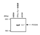
- cerebrospinal fluid was punctured by lumbar puncture according to a standard method, ADEM patients, Guillain-Barre syndrome patients, and individuals without central nervous system disease as controls (patients without ADEM and without Guillain-Barre syndrome) From.
- LaemmLi sample buffer was added to 2 ⁇ L of cerebrospinal fluid and heated, and then subjected to SDS / PAGE using 5-20% gradient polyacrylamide gel. Electrophoresis was performed at a constant current of 40 mA for 55 minutes. The separated protein was electrically transferred to a PVDF membrane at a constant current of 260 mA for 50 minutes by a blotting method known in the art.
- the membrane was blocked with 1% BSA-PBS for 1 hour or longer. Subsequently, among the cerebrospinal fluid glycoproteins listed in Table 1, those for which antibodies can be used (for example, goat anti-human ⁇ -2-macloglobulin antibody, cappel, Cat.No. 55113; cat.No. ab84176; SantaCruz cat.No. sc-8514; and Dako cat.No. Q0102)) was diluted with 1% BSA-PBS (final concentration: 1 to 2 ⁇ g / mL) and reacted for 1 hour. The membrane was washed three times for 15 minutes each with a PBS washing solution containing 0.1% Tween-20.
- a PBS washing solution containing 0.1% Tween-20.
- FIG. 8 shows the detection result of ⁇ 2-macroglobulin in the cerebrospinal fluid sample. Compared to the control, ADEM showed an increase in ⁇ 2-macroglobulin. On the other hand, as shown in Example 3, almost no ⁇ 2-macroglobulin is detected in MS. Therefore, ⁇ 2-macroglobulin was shown to be an effective index marker for central nervous system diseases in differentiation between ADEM and MS.
- ⁇ 2-macroglobulin is increased, indicating that it can be differentiated from polyneuritis in which the lesion is confined to the peripheral nerve. Similar results were obtained using anti-human ⁇ 2-macroglobulin antibodies from other companies.
- ⁇ 2-macroglobulin can be a useful diagnostic marker that can differentiate ADEM and MS, and Guillain-Barre syndrome from polyneuritis.
- Menitis includes bacterial and viral meningitis, both of which can be diagnosed by cerebrospinal fluid examination.
- bacterial meningitis can be diagnosed by increasing neutrophils in the cerebrospinal fluid, decreasing glucose concentration, and viral meningitis by increasing mononuclear cells.
- standard treatments for each meningitis have been established.
- “Encephalopathy” is a disease secondary to bacterial or viral infection. Although there are many unclear points in the pathology, it is said to be based on brain energy and electrolyte metabolism abnormality, and is a serious disease accompanied by swelling of the brain, disturbance of consciousness, convulsions and fever. For example, mortality in influenza encephalopathy reaches 50-60%.
- an index marker for central nervous system disease capable of distinguishing convulsive intussusception and encephalopathy from cerebrospinal fluid type glycoprotein which is a candidate index marker for central nervous system disease shown in Table 1 identified in Example 2 Explored.
- the basic method is the same as (1) above. Cerebrospinal fluid was collected from patients with intussusception, encephalopathy, and patients with viral meningitis. Here, the encephalopathy patient was diagnosed by follow-up after hospitalization, and was collected from the same patient at the time of first hospitalization and after treatment (7 days later).
- FIG. 9 shows the detection result of ⁇ 2-macroglobulin in the cerebrospinal fluid sample.
- the amount of ⁇ 2-macroglobulin in the cerebrospinal fluid is only slightly increased, whereas in patients with encephalopathy on admission, both diseases can be differentiated by ⁇ 2-macroglobulin. Indicated.
- encephalopathy For encephalopathy, powerful treatments such as the use of steroids are performed, but by using ⁇ 2-macroglobulin as an index marker for central nervous system diseases of encephalopathy, it is possible to determine the timing of discontinuation of steroid treatment, which can lead to overdose of drugs. The accompanying side effects can be reduced.
- ⁇ 2-macroglobulin can be an index marker for central nervous system diseases that can distinguish encephalopathy patients from convulsive intussusception patients and encephalopathy patients.
- AD Alzheimer's disease
- PWD2S prostaglandin D2 synthase
- PGD2S contained a relatively large amount in the cerebrospinal fluid, it was directly analyzed by Western blotting without performing enrichment.
- anti-human PGD2S antibody (Thermo scientific, Cat. No. PA1-46023) (1 ⁇ g / ml) was used, and two individuals were verified.
Landscapes
- Life Sciences & Earth Sciences (AREA)
- Health & Medical Sciences (AREA)
- Engineering & Computer Science (AREA)
- Molecular Biology (AREA)
- Biomedical Technology (AREA)
- Chemical & Material Sciences (AREA)
- Urology & Nephrology (AREA)
- Immunology (AREA)
- Hematology (AREA)
- Physics & Mathematics (AREA)
- Analytical Chemistry (AREA)
- General Health & Medical Sciences (AREA)
- Biochemistry (AREA)
- Cell Biology (AREA)
- Proteomics, Peptides & Aminoacids (AREA)
- Pathology (AREA)
- Microbiology (AREA)
- General Physics & Mathematics (AREA)
- Food Science & Technology (AREA)
- Medicinal Chemistry (AREA)
- Biotechnology (AREA)
- Bioinformatics & Cheminformatics (AREA)
- Bioinformatics & Computational Biology (AREA)
- Biophysics (AREA)
- Neurology (AREA)
- Neurosurgery (AREA)
- Peptides Or Proteins (AREA)
- Investigating Or Analysing Biological Materials (AREA)
Abstract
Priority Applications (6)
| Application Number | Priority Date | Filing Date | Title |
|---|---|---|---|
| JP2012548848A JP6074846B2 (ja) | 2010-12-16 | 2011-12-16 | 髄液型糖タンパク質の富化及び分離方法、並びにその方法を用いた中枢神経系疾患用マーカーの探索方法及び中枢神経系疾患用マーカー |
| EP17184546.4A EP3279668B1 (fr) | 2010-12-16 | 2011-12-16 | Procédé d'enrichissement et de séparation de fluide spinal glycoprotéine, procédé de recherche de marqueur pour les maladies du système nerveux central qui utilise le procédé susmentionné et marqueur pour les maladies du système nerveux central |
| US13/994,435 US20140323324A1 (en) | 2010-12-16 | 2011-12-16 | Method for Enrichment and Separation of Spinal Fluid Glycoprotein, Method for Searching for Marker for Central Nervous System Diseases Which Utilizes the Aforementioned Method, and Marker for Central Nervous System Diseases |
| EP11847931.0A EP2653476B1 (fr) | 2010-12-16 | 2011-12-16 | Procédé pour l'enrichissement et la séparation d'une glycoprotéine du fluide spinal, procédé de recherche d'un marqueur pour des maladies du système nerveux central qui utilise le procédé mentionné ci-dessus, et marqueur pour des maladies du système nerveux central |
| EP16173307.6A EP3101426B1 (fr) | 2010-12-16 | 2011-12-16 | Procédé d'enrichissement et de séparation de fluide spinal glycoprotéine, procédé de recherche de marqueur pour les maladies du système nerveux central qui utilise le procédé susmentionné et marqueur pour les maladies du système nerveux central |
| US15/071,419 US10156576B2 (en) | 2010-12-16 | 2016-03-16 | Method for enrichment and separation of spinal fluid glycoprotein, method for searching for marker for central nervous system diseases which utilizes the aforementioned method, and marker for central nervous system diseases |
Applications Claiming Priority (2)
| Application Number | Priority Date | Filing Date | Title |
|---|---|---|---|
| JP2010280753 | 2010-12-16 | ||
| JP2010-280753 | 2010-12-16 |
Related Child Applications (3)
| Application Number | Title | Priority Date | Filing Date |
|---|---|---|---|
| US13/994,435 A-371-Of-International US20140323324A1 (en) | 2010-12-16 | 2011-12-16 | Method for Enrichment and Separation of Spinal Fluid Glycoprotein, Method for Searching for Marker for Central Nervous System Diseases Which Utilizes the Aforementioned Method, and Marker for Central Nervous System Diseases |
| EP16173307.6A Previously-Filed-Application EP3101426B1 (fr) | 2010-12-16 | 2011-12-16 | Procédé d'enrichissement et de séparation de fluide spinal glycoprotéine, procédé de recherche de marqueur pour les maladies du système nerveux central qui utilise le procédé susmentionné et marqueur pour les maladies du système nerveux central |
| US15/071,419 Division US10156576B2 (en) | 2010-12-16 | 2016-03-16 | Method for enrichment and separation of spinal fluid glycoprotein, method for searching for marker for central nervous system diseases which utilizes the aforementioned method, and marker for central nervous system diseases |
Publications (1)
| Publication Number | Publication Date |
|---|---|
| WO2012081701A1 true WO2012081701A1 (fr) | 2012-06-21 |
Family
ID=46244793
Family Applications (1)
| Application Number | Title | Priority Date | Filing Date |
|---|---|---|---|
| PCT/JP2011/079218 Ceased WO2012081701A1 (fr) | 2010-12-16 | 2011-12-16 | Procédé pour l'enrichissement et la séparation d'une glycoprotéine du fluide spinal, procédé de recherche d'un marqueur pour des maladies du système nerveux central qui utilise le procédé mentionné ci-dessus, et marqueur pour des maladies du système nerveux central |
Country Status (4)
| Country | Link |
|---|---|
| US (2) | US20140323324A1 (fr) |
| EP (3) | EP2653476B1 (fr) |
| JP (1) | JP6074846B2 (fr) |
| WO (1) | WO2012081701A1 (fr) |
Cited By (4)
| Publication number | Priority date | Publication date | Assignee | Title |
|---|---|---|---|---|
| CN104884958A (zh) * | 2012-08-10 | 2015-09-02 | 独立行政法人产业技术综合研究所 | 糖链异构体检测方法及糖链异构体检测装置 |
| JPWO2016013597A1 (ja) * | 2014-07-22 | 2017-05-25 | 国立研究開発法人産業技術総合研究所 | 肝細胞がんマーカー |
| CN114660290A (zh) * | 2022-03-25 | 2022-06-24 | 中国医学科学院北京协和医院 | 预测甲状腺癌术后复发的糖链标志物及其应用 |
| JP2022542376A (ja) * | 2019-07-26 | 2022-10-03 | ザ バインディング サイト グループ リミティド | イオン化コントロール |
Families Citing this family (3)
| Publication number | Priority date | Publication date | Assignee | Title |
|---|---|---|---|---|
| WO2017139975A1 (fr) * | 2016-02-19 | 2017-08-24 | Huiru Wang | Anticorps dirigés contre la n-acétylglucosamine et la n-acétylgalactosamine |
| CN107782896B (zh) * | 2017-10-09 | 2020-05-01 | 广东医科大学 | 乳腺癌标志物 |
| CN107782897B (zh) * | 2017-10-09 | 2020-03-17 | 广东医科大学 | 乳腺癌标志物 |
Citations (4)
| Publication number | Priority date | Publication date | Assignee | Title |
|---|---|---|---|---|
| JPH11513372A (ja) * | 1995-09-22 | 1999-11-16 | コノート ラボラトリーズ リミテッド | パラインフルエンザウイルスの糖タンパクおよびワクチン |
| JP2008184404A (ja) * | 2007-01-29 | 2008-08-14 | Gekkeikan Sake Co Ltd | N−結合型の1〜3本側鎖糖鎖に結合するレクチン |
| JP4385149B2 (ja) | 2005-05-11 | 2009-12-16 | 独立行政法人理化学研究所 | 血清糖蛋白質をバイオマーカーとするアルツハイマー病の診断薬 |
| JP2010121980A (ja) | 2008-11-17 | 2010-06-03 | Institute Of Physical & Chemical Research | 糖鎖バイオマーカーによる特発性正常圧水頭症の診断 |
Family Cites Families (7)
| Publication number | Priority date | Publication date | Assignee | Title |
|---|---|---|---|---|
| ES2288877T3 (es) * | 1999-11-08 | 2008-02-01 | EISAI R&D MANAGEMENT CO., LTD. | Metodo y reactivo para detectar la muerte celular. |
| AU2002225219A1 (en) * | 2001-01-26 | 2002-08-06 | Oxford Glycosciences (Uk) Ltd | Diagnosis and treatment of multiple sclerosis |
| JP2004264066A (ja) * | 2003-02-27 | 2004-09-24 | Hiroshi Kido | 脳炎・脳症の診断 |
| GB2428240A (en) * | 2005-07-14 | 2007-01-24 | Univ Gen Ve | Diagnostic method for brain damage-related disorders |
| CA2680333C (fr) * | 2006-03-15 | 2016-10-25 | The Brigham And Women's Hospital, Inc. | Utilisation de la gelsoline pour traiter la sclerose en plaques et pour diagnostiquer des maladies neurologiques |
| WO2008016707A2 (fr) * | 2006-08-02 | 2008-02-07 | The Johns Hopkins University | Utilisation de l'il-6 pour le diagnostic et le traitement de pathologies neuro-inflammatoires |
| CN102460153A (zh) * | 2009-04-06 | 2012-05-16 | 里奇诊断学股份有限公司 | 监控神经精神疾病治疗的生物标记 |
-
2011
- 2011-12-16 EP EP11847931.0A patent/EP2653476B1/fr not_active Not-in-force
- 2011-12-16 US US13/994,435 patent/US20140323324A1/en not_active Abandoned
- 2011-12-16 WO PCT/JP2011/079218 patent/WO2012081701A1/fr not_active Ceased
- 2011-12-16 JP JP2012548848A patent/JP6074846B2/ja active Active
- 2011-12-16 EP EP16173307.6A patent/EP3101426B1/fr not_active Not-in-force
- 2011-12-16 EP EP17184546.4A patent/EP3279668B1/fr not_active Not-in-force
-
2016
- 2016-03-16 US US15/071,419 patent/US10156576B2/en not_active Expired - Fee Related
Patent Citations (4)
| Publication number | Priority date | Publication date | Assignee | Title |
|---|---|---|---|---|
| JPH11513372A (ja) * | 1995-09-22 | 1999-11-16 | コノート ラボラトリーズ リミテッド | パラインフルエンザウイルスの糖タンパクおよびワクチン |
| JP4385149B2 (ja) | 2005-05-11 | 2009-12-16 | 独立行政法人理化学研究所 | 血清糖蛋白質をバイオマーカーとするアルツハイマー病の診断薬 |
| JP2008184404A (ja) * | 2007-01-29 | 2008-08-14 | Gekkeikan Sake Co Ltd | N−結合型の1〜3本側鎖糖鎖に結合するレクチン |
| JP2010121980A (ja) | 2008-11-17 | 2010-06-03 | Institute Of Physical & Chemical Research | 糖鎖バイオマーカーによる特発性正常圧水頭症の診断 |
Non-Patent Citations (12)
| Title |
|---|
| KOROLAINEN MA.; 2010 ET AL., J. NEUROCHEM., vol. 112, no. 6, pages 1386 - 1414 |
| KUNO, A. ET AL., NATURE METHODS, vol. 2, 2005, pages 851 - 856 |
| MATSUDA ET AL., HEPATOLOGY, vol. 52, no. 1, July 2010 (2010-07-01), pages 174 - 82 |
| MATTSSON N ET AL., JAMA, vol. 302, no. 4, 22 July 2009 (2009-07-22), pages 385 - 93 |
| OZAWA, H ET AL., ARCHIVES OF BIOCHEMISTRY AND BIOPHYSICS, vol. 342, no. 1, 1997, pages 48 - 57 |
| OZAWA, H, ARCHIVES OF BIOCHEMISTRY AND BIOPHYSICS, vol. 342, no. 1, 1997, pages 48 - 57 |
| See also references of EP2653476A4 * |
| UCHIYAMA N., PROTEOMICS, vol. 8, 2008, pages 3042 - 3050 |
| UEDA H. ET AL., J. BIOL. CHEM., vol. 277, no. 28, 2002, pages 24916 - 24925 |
| UEDA H. ET AL.: "Interaction of a lectin from Psathyrella velutina mushroom with N-acetylneuraminic acid", FEBS LETT., vol. 448, no. 1, 1999, pages 75 - 80, XP004259469 * |
| UEDA H. ET AL.: "Psathyrella velutina Mushroom Lectin Exhibits High Affinity toward Sialoglycoproteins Possessing Terminal N-Acetylneuraminic Acid a2,3-Linked to Penultimate Galactose Residues of Trisialyl N-Glycans. Comparison with other sialic acid- specific lectins", J. BIOL. CHEM., vol. 277, no. 28, 2002, pages 24916 - 24925, XP055109193 * |
| YASUHIRO HASHIMOTO ET AL.: "To Tanpakushitsu no Tosa o Marker to suru Seijoatsu Suitosho no Shindanho no Kaihatsu", KOSEI RODO KAGAKU KENKYUHI HOJOKIN NANJISEI SHIKKAN KOKUFUKU KENKYU JIGYO SEIJOATSU SUITOSHO NO EKIGAKU BYOTAI TO CHIRYO NI KANSURU KENKYU HEISEI 21 NENDO SOKATSU BUNTAN KENKYU HOKOKUSHO, March 2010 (2010-03-01), pages 23 - 24, XP008169818 * |
Cited By (6)
| Publication number | Priority date | Publication date | Assignee | Title |
|---|---|---|---|---|
| CN104884958A (zh) * | 2012-08-10 | 2015-09-02 | 独立行政法人产业技术综合研究所 | 糖链异构体检测方法及糖链异构体检测装置 |
| CN104884958B (zh) * | 2012-08-10 | 2017-07-11 | 独立行政法人产业技术综合研究所 | 糖链异构体检测方法及糖链异构体检测装置 |
| JPWO2016013597A1 (ja) * | 2014-07-22 | 2017-05-25 | 国立研究開発法人産業技術総合研究所 | 肝細胞がんマーカー |
| JP2022542376A (ja) * | 2019-07-26 | 2022-10-03 | ザ バインディング サイト グループ リミティド | イオン化コントロール |
| JP7738546B2 (ja) | 2019-07-26 | 2025-09-12 | ザ バインディング サイト グループ リミティド | イオン化コントロール |
| CN114660290A (zh) * | 2022-03-25 | 2022-06-24 | 中国医学科学院北京协和医院 | 预测甲状腺癌术后复发的糖链标志物及其应用 |
Also Published As
| Publication number | Publication date |
|---|---|
| JP6074846B2 (ja) | 2017-02-08 |
| EP2653476A1 (fr) | 2013-10-23 |
| EP3101426A2 (fr) | 2016-12-07 |
| EP3101426A3 (fr) | 2017-02-08 |
| US20140323324A1 (en) | 2014-10-30 |
| EP3279668B1 (fr) | 2019-05-15 |
| US10156576B2 (en) | 2018-12-18 |
| EP3279668A1 (fr) | 2018-02-07 |
| US20160187356A1 (en) | 2016-06-30 |
| EP2653476B1 (fr) | 2017-04-26 |
| EP2653476A4 (fr) | 2015-10-14 |
| JPWO2012081701A1 (ja) | 2014-05-22 |
| EP3101426B1 (fr) | 2019-05-15 |
Similar Documents
| Publication | Publication Date | Title |
|---|---|---|
| JP6074846B2 (ja) | 髄液型糖タンパク質の富化及び分離方法、並びにその方法を用いた中枢神経系疾患用マーカーの探索方法及び中枢神経系疾患用マーカー | |
| US10352949B2 (en) | Biomarker and methods for early diagnosis of Alzheimer's disease | |
| KR101559101B1 (ko) | 혈액유래 암 진단용 펩티드 마커 및 이를 이용한 암 진단방법 | |
| CN113176411B (zh) | 利用唾液检测新型冠状病毒感染的生物标志物及其应用 | |
| KR101219519B1 (ko) | 렉틴을 이용한 암 진단 방법 | |
| KR20180023955A (ko) | 뇌척수액의 검출 | |
| JP2008175814A (ja) | 尿中タンパク質分子の検出・定量による糖尿病性腎症の検査方法及びそれに使用するキット | |
| CN108093648A (zh) | 簇集蛋白同种型的特异性检测 | |
| KR20150062915A (ko) | 혈액 당단백질을 이용하는 암 마커 및 이를 이용하는 암 진단방법 | |
| WO2010032458A1 (fr) | Biomarqueur inédit de la stéatose hépatique non alcoolique et méthode de détection d'une stéatose hépatique non alcoolique au moyen dudit biomarqueur | |
| EP3035058A1 (fr) | Procédé de criblage de marqueur de cancer par l'intermédiaire d'une détection de déglycosylation de glycoprotéine et de marqueur de cancer hépatocellulaire | |
| CN108896771B (zh) | Guca2a蛋白在骨关节炎中的用途 | |
| KR20110038778A (ko) | 다중렉틴을 이용한 체액 유래 단백질 동정 방법 및 이 방법에 의하여 탐지된 간암 바이오마커 | |
| JP7376048B2 (ja) | 大腸癌の診断を補助する方法、キット及びバイオマーカー | |
| KR101143891B1 (ko) | 단백질의 비정상적인 당쇄화를 이용하는 암진단 마커 | |
| CN118584103B (zh) | 中枢神经精神狼疮脑脊液诊断标志物及其应用 | |
| JP7425447B2 (ja) | 膵臓癌の判定用マーカー | |
| KR101100809B1 (ko) | 암 진단용 펩티드 마커 및 이를 이용한 암 진단방법 | |
| US20170299589A1 (en) | Methods to Diagnose and Treat Multiple Sclerosis via Detection of Altered Protein Components of Serum | |
| JP2024113271A (ja) | 癌の腫瘍マーカー | |
| CN121013988A (zh) | IgA肾病的判定方法 | |
| JP2014070037A (ja) | アルツハイマー病の診断薬および診断方法 | |
| JP2021162484A (ja) | 脳型n−アセチルグルコサミン−トランスフェリンの検出方法 | |
| NZ736200B2 (en) | Specific detection of clusterin isoforms |
Legal Events
| Date | Code | Title | Description |
|---|---|---|---|
| 121 | Ep: the epo has been informed by wipo that ep was designated in this application |
Ref document number: 11847931 Country of ref document: EP Kind code of ref document: A1 |
|
| ENP | Entry into the national phase |
Ref document number: 2012548848 Country of ref document: JP Kind code of ref document: A |
|
| NENP | Non-entry into the national phase |
Ref country code: DE |
|
| REEP | Request for entry into the european phase |
Ref document number: 2011847931 Country of ref document: EP |
|
| WWE | Wipo information: entry into national phase |
Ref document number: 2011847931 Country of ref document: EP |
|
| WWE | Wipo information: entry into national phase |
Ref document number: 13994435 Country of ref document: US |