WO2012153616A1 - Structure de membrane lipidique capable de migrer vers une cellule cible et procédé de production associé, et procédé de criblage d'une substance en présentant les effets dans une cellule cible - Google Patents
Structure de membrane lipidique capable de migrer vers une cellule cible et procédé de production associé, et procédé de criblage d'une substance en présentant les effets dans une cellule cible Download PDFInfo
- Publication number
- WO2012153616A1 WO2012153616A1 PCT/JP2012/060622 JP2012060622W WO2012153616A1 WO 2012153616 A1 WO2012153616 A1 WO 2012153616A1 JP 2012060622 W JP2012060622 W JP 2012060622W WO 2012153616 A1 WO2012153616 A1 WO 2012153616A1
- Authority
- WO
- WIPO (PCT)
- Prior art keywords
- lipid
- group
- peptide
- membrane structure
- lipid membrane
- Prior art date
- Legal status (The legal status is an assumption and is not a legal conclusion. Google has not performed a legal analysis and makes no representation as to the accuracy of the status listed.)
- Ceased
Links
Images


Classifications
-
- G—PHYSICS
- G01—MEASURING; TESTING
- G01N—INVESTIGATING OR ANALYSING MATERIALS BY DETERMINING THEIR CHEMICAL OR PHYSICAL PROPERTIES
- G01N33/00—Investigating or analysing materials by specific methods not covered by groups G01N1/00 - G01N31/00
- G01N33/48—Biological material, e.g. blood, urine; Haemocytometers
- G01N33/50—Chemical analysis of biological material, e.g. blood, urine; Testing involving biospecific ligand binding methods; Immunological testing
- G01N33/5005—Chemical analysis of biological material, e.g. blood, urine; Testing involving biospecific ligand binding methods; Immunological testing involving human or animal cells
- G01N33/5008—Chemical analysis of biological material, e.g. blood, urine; Testing involving biospecific ligand binding methods; Immunological testing involving human or animal cells for testing or evaluating the effect of chemical or biological compounds, e.g. drugs, cosmetics
-
- A—HUMAN NECESSITIES
- A61—MEDICAL OR VETERINARY SCIENCE; HYGIENE
- A61K—PREPARATIONS FOR MEDICAL, DENTAL OR TOILETRY PURPOSES
- A61K47/00—Medicinal preparations characterised by the non-active ingredients used, e.g. carriers or inert additives; Targeting or modifying agents chemically bound to the active ingredient
- A61K47/50—Medicinal preparations characterised by the non-active ingredients used, e.g. carriers or inert additives; Targeting or modifying agents chemically bound to the active ingredient the non-active ingredient being chemically bound to the active ingredient, e.g. polymer-drug conjugates
- A61K47/51—Medicinal preparations characterised by the non-active ingredients used, e.g. carriers or inert additives; Targeting or modifying agents chemically bound to the active ingredient the non-active ingredient being chemically bound to the active ingredient, e.g. polymer-drug conjugates the non-active ingredient being a modifying agent
- A61K47/54—Medicinal preparations characterised by the non-active ingredients used, e.g. carriers or inert additives; Targeting or modifying agents chemically bound to the active ingredient the non-active ingredient being chemically bound to the active ingredient, e.g. polymer-drug conjugates the non-active ingredient being a modifying agent the modifying agent being an organic compound
- A61K47/543—Lipids, e.g. triglycerides; Polyamines, e.g. spermine or spermidine
- A61K47/544—Phospholipids
-
- A—HUMAN NECESSITIES
- A61—MEDICAL OR VETERINARY SCIENCE; HYGIENE
- A61K—PREPARATIONS FOR MEDICAL, DENTAL OR TOILETRY PURPOSES
- A61K47/00—Medicinal preparations characterised by the non-active ingredients used, e.g. carriers or inert additives; Targeting or modifying agents chemically bound to the active ingredient
- A61K47/50—Medicinal preparations characterised by the non-active ingredients used, e.g. carriers or inert additives; Targeting or modifying agents chemically bound to the active ingredient the non-active ingredient being chemically bound to the active ingredient, e.g. polymer-drug conjugates
- A61K47/51—Medicinal preparations characterised by the non-active ingredients used, e.g. carriers or inert additives; Targeting or modifying agents chemically bound to the active ingredient the non-active ingredient being chemically bound to the active ingredient, e.g. polymer-drug conjugates the non-active ingredient being a modifying agent
- A61K47/56—Medicinal preparations characterised by the non-active ingredients used, e.g. carriers or inert additives; Targeting or modifying agents chemically bound to the active ingredient the non-active ingredient being chemically bound to the active ingredient, e.g. polymer-drug conjugates the non-active ingredient being a modifying agent the modifying agent being an organic macromolecular compound, e.g. an oligomeric, polymeric or dendrimeric molecule
- A61K47/59—Medicinal preparations characterised by the non-active ingredients used, e.g. carriers or inert additives; Targeting or modifying agents chemically bound to the active ingredient the non-active ingredient being chemically bound to the active ingredient, e.g. polymer-drug conjugates the non-active ingredient being a modifying agent the modifying agent being an organic macromolecular compound, e.g. an oligomeric, polymeric or dendrimeric molecule obtained otherwise than by reactions only involving carbon-to-carbon unsaturated bonds, e.g. polyureas or polyurethanes
- A61K47/60—Medicinal preparations characterised by the non-active ingredients used, e.g. carriers or inert additives; Targeting or modifying agents chemically bound to the active ingredient the non-active ingredient being chemically bound to the active ingredient, e.g. polymer-drug conjugates the non-active ingredient being a modifying agent the modifying agent being an organic macromolecular compound, e.g. an oligomeric, polymeric or dendrimeric molecule obtained otherwise than by reactions only involving carbon-to-carbon unsaturated bonds, e.g. polyureas or polyurethanes the organic macromolecular compound being a polyoxyalkylene oligomer, polymer or dendrimer, e.g. PEG, PPG, PEO or polyglycerol
-
- A—HUMAN NECESSITIES
- A61—MEDICAL OR VETERINARY SCIENCE; HYGIENE
- A61P—SPECIFIC THERAPEUTIC ACTIVITY OF CHEMICAL COMPOUNDS OR MEDICINAL PREPARATIONS
- A61P3/00—Drugs for disorders of the metabolism
- A61P3/04—Anorexiants; Antiobesity agents
-
- A—HUMAN NECESSITIES
- A61—MEDICAL OR VETERINARY SCIENCE; HYGIENE
- A61P—SPECIFIC THERAPEUTIC ACTIVITY OF CHEMICAL COMPOUNDS OR MEDICINAL PREPARATIONS
- A61P43/00—Drugs for specific purposes, not provided for in groups A61P1/00-A61P41/00
-
- C—CHEMISTRY; METALLURGY
- C07—ORGANIC CHEMISTRY
- C07K—PEPTIDES
- C07K7/00—Peptides having 5 to 20 amino acids in a fully defined sequence; Derivatives thereof
- C07K7/04—Linear peptides containing only normal peptide links
- C07K7/08—Linear peptides containing only normal peptide links having 12 to 20 amino acids
-
- A—HUMAN NECESSITIES
- A61—MEDICAL OR VETERINARY SCIENCE; HYGIENE
- A61K—PREPARATIONS FOR MEDICAL, DENTAL OR TOILETRY PURPOSES
- A61K38/00—Medicinal preparations containing peptides
-
- G—PHYSICS
- G01—MEASURING; TESTING
- G01N—INVESTIGATING OR ANALYSING MATERIALS BY DETERMINING THEIR CHEMICAL OR PHYSICAL PROPERTIES
- G01N2500/00—Screening for compounds of potential therapeutic value
- G01N2500/10—Screening for compounds of potential therapeutic value involving cells
-
- G—PHYSICS
- G01—MEASURING; TESTING
- G01N—INVESTIGATING OR ANALYSING MATERIALS BY DETERMINING THEIR CHEMICAL OR PHYSICAL PROPERTIES
- G01N2800/00—Detection or diagnosis of diseases
- G01N2800/04—Endocrine or metabolic disorders
- G01N2800/044—Hyperlipemia or hypolipemia, e.g. dyslipidaemia, obesity
Definitions
- the present invention relates to a lipid membrane structure having the ability to migrate to a target cell, a method for producing the same, and a screening method for a substance having an effect on a target cell, and more specifically, a lipid membrane structure having the ability to migrate to a target cell, A single lipid membrane comprising a peptide having the ability to migrate to a target cell, a long-chain polyethylene glycol and a lipid formed by binding lipids in this order and a lipid formed by binding a short-chain polyethylene glycol as constituent lipids Lipid membrane structure possessed as an obesity inhibitor / therapeutic agent using the same, adipose tissue inflammation inhibitor / therapeutic agent and agent for inhibiting / treating fat accumulation in non-adipose tissue, method for producing the lipid membrane structure, lipid Screening method of substance having effect on target cell using membrane structure, obesity inhibitor / therapeutic agent, adipose tissue inflammation inhibitor / therapeutic agent, and non-adipose group Accumulating a screening method of inhibit
- Patent Document 1 and Non-Patent Document 1 disclose a peptide having the ability to migrate adipose tissue to vascular endothelial cells, and by administering a peptide in which an apoptosis-inducing peptide is linked to this peptide, It has been disclosed that apoptosis can be induced in tissue vascular endothelial cells.
- a lipid membrane structure typified by a liposome can include (i) a substance, and the included substance is protected from degradation and metabolism in the living body, (ii) includes the substance. And (iii) excellent in biocompatibility and biodegradability, (iv) polyethylene on its surface, which can prevent the contained substance from acting outside the target cells (side effects)
- a functional molecule such as glycol (hereinafter sometimes referred to as “PEG” in this specification), antibody, protein, peptide, sugar chain, etc., the ability to migrate to target cells, fusion ability, pH responsiveness Therefore, it is expected to be an ideal vector for drugs and physiologically active substances.
- Non-Patent Document 2 Discloses a liposome having a lipid membrane containing, as a constituent lipid, a lipid obtained by binding the above-described peptide having the ability to migrate to adipose tissue vascular endothelial cells, PEG and lipid in this order.
- apoptosis-inducing peptide is bound to the peptide having the ability to migrate to adipose tissue vascular endothelial cells described in Patent Document 1 and Non-Patent Document 1 is not a lipid membrane structure, It is difficult to protect against metabolic effects, and it is difficult to prevent actions other than the target cells, adipose tissue vascular endothelial cells. Stealth liposomes are capable of migrating to cells of tumor tissues with enhanced vascular permeability, but are capable of migrating to cells of other tissues that do not have enhanced vascular permeability. Absent.
- Non-Patent Document 2 having a lipid membrane containing a lipid having a lipid tissue vascular endothelial cell migration ability, PEG, and a lipid formed by binding lipids in this order as a constituent lipid is a cultured cell (in vitro). ) Is only shown for the ability to migrate to adipose tissue vascular endothelial cells, and is not shown for the ability to migrate to adipose tissue vascular endothelial cells in the living body (in vivo). There is no description, nor disclosure, of having a lipid membrane containing a lipid formed by binding PEG as a constituent lipid.
- the present invention has been made to solve such problems, and is a lipid membrane structure having a target cell migration ability, a peptide having a target cell migration ability, a long-chain PEG and a lipid.
- a lipid membrane structure comprising as a single membrane a lipid membrane comprising a lipid formed by binding in this order and a lipid formed by binding a short chain length PEG as a constituent lipid, an obesity-suppressing / treating agent using the same, An agent for inhibiting / treating adipose tissue inflammation and an agent for inhibiting / treating fat accumulation in non-adipose tissue, a method for producing the lipid membrane structure, a method for screening a substance having an effect on target cells using the lipid membrane structure, and It is an object of the present invention to provide a screening method for agents for suppressing / treating obesity suppressing / therapeutic agents, adipose tissue inflammation suppressing / therapeutic agents and fat accumulation in non-adipose tissue.
- the present inventors have constructed a peptide having a target cell migration ability, a long-chain PEG and a lipid formed by combining lipids in this order and a lipid formed by combining a short-chain PEG.
- a lipid membrane structure having the ability to migrate to a target cell the lipid membrane structure having a lipid membrane containing the lipids of (a) and (b) below as a constituent lipid as a single membrane; (a) Lipid formed by binding a peptide having the ability to migrate to a target cell, polyethylene glycol and lipid in this order, and lipid formed by binding polyethylene glycol having a smaller number average molecular weight than polyethylene glycol constituting (b) (a) .
- the polyethylene glycol constituting (a) is a polyethylene glycol having a molecular weight Ma of 3500 ⁇ Ma ⁇ 6500
- the polyethylene glycol constituting (b) is a polyethylene glycol having a molecular weight Mb of 500 ⁇ Mb ⁇ 3500.
- the lipid membrane structure according to (1) or (2) which is a negatively charged lipid membrane structure or an uncharged lipid membrane structure.
- lipid membrane structure according to any one of (1) to (4), wherein the peptide having the ability to migrate to target cells is a peptide having ability to migrate to adipose tissue vascular endothelial cells.
- a peptide having the ability to migrate to adipose tissue vascular endothelial cells is KGGRAKD (wherein K is a lysine residue, G is a glycine residue, R is an arginine residue, A is an alanine residue, D is The lipid membrane structure according to (5), which is a peptide having an amino acid sequence of aspartic acid residues.
- An obesity-suppressing and / or therapeutic agent comprising the lipid membrane structure according to (5) or (6) in which an apoptosis-inducing agent is encapsulated as an active ingredient.
- apoptosis-inducing agent is the following (i) and / or (ii); (i) KLAKLAKKLAKLAK (where K is a lysine residue, L Represents a leucine residue, and A represents an alanine residue.) (Ii) Cytochrome c.
- An adipose tissue inflammation inhibitory and / or therapeutic agent comprising as an active ingredient the lipid membrane structure according to (5) or (6) in which an apoptosis-inducing agent is encapsulated.
- apoptosis-inducing agent is the following (i) and / or (ii); (i) KLAKLAKKLAKLAK (wherein K represents a lysine residue) , L represents a leucine residue, and A represents an alanine residue.) (Ii) Cytochrome c.
- An agent for suppressing and / or treating fat accumulation in non-adipose tissue comprising as an active ingredient the lipid membrane structure according to (5) or (6) in which an apoptosis-inducing agent is encapsulated.
- apoptosis-inducing agent is the following (i) and / or (ii); (i) KLAKLAKKLAKLAK, wherein K is a lysine residue, L is a leucine residue, A represents an alanine residue, respectively.) (Ii) Cytochrome c.
- a method for producing a lipid membrane structure having target cell migration ability comprising the step of preparing a single membrane lipid membrane containing the lipids of (a) and (b) below as constituent lipids: Method: (a) a lipid formed by binding a target cell migration ability, polyethylene glycol and lipid in this order; (b) a polyethylene glycol having a smaller number average molecular weight than the polyethylene glycol constituting (a) Combined lipids.
- the polyethylene glycol constituting (a) is a polyethylene glycol having a molecular weight Ma of 3500 ⁇ Ma ⁇ 6500, and the polyethylene glycol constituting (b) is a polyethylene glycol having a molecular weight Mb of 500 ⁇ Mb ⁇ 3500.
- a method for screening a substance having an effect on a target cell wherein the peptide having the ability to migrate to one target cell in the lipid membrane structure according to any one of (1) to (6) is selected and the target substance
- the method comprising: encapsulating and transferring to the target cell; and evaluating whether the target substance exhibits an effect in the target cell.
- lipid membrane structure of the present invention a lipid membrane structure that specifically migrates to a target cell can be obtained, and the encapsulated substance is delivered to the target cell without being damaged, and the target cell receives the lipid membrane structure.
- the encapsulated substance In addition to exerting the function of the substance, it has low toxicity and can be safely applied to living bodies.
- the obesity inhibitor / therapeutic agent using the lipid membrane structure according to the present invention it is possible to effectively suppress body weight increase due to a high-fat diet without inhibiting normal growth.
- adipose tissue inflammation suppressing / treating agent using the lipid membrane structure according to the present invention it is possible to effectively suppress or extinguish inflammation of the adipose tissue despite ingestion of a high fat diet. Therefore, diseases caused by inflammation of adipose tissue, such as inflammatory diseases such as type 2 diabetes and atherosclerosis, can be prevented or treated.
- the agent for suppressing / treating fat accumulation in non-adipose tissue using the lipid membrane structure according to the present invention fat accumulation in non-adipose tissue despite intake of a high fat diet It is possible to prevent or treat diseases caused by fat accumulation in non-adipose tissue, such as fatty liver, type 2 diabetes, myocardial injury, etc. it can. Moreover, according to the manufacturing method of the lipid membrane structure which concerns on this invention, the lipid membrane structure which has the above effects can be obtained. Furthermore, according to the screening method for a substance having an effect on the target cell according to the present invention, it is possible to simply and efficiently evaluate whether or not the target substance has an effect on the target cell, and the lipid membrane according to the present invention.
- an adipose tissue inflammation-suppressing and / or therapeutic agent or an agent that suppresses and / or treats fat accumulation in non-adipose tissue using a structure Easily and efficiently evaluate whether it is possible to suppress or treat, suppress or treat adipose tissue inflammation, or suppress or treat fat accumulation in non-adipose tissue be able to.
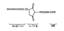
- FIG. 3 is a schematic diagram showing the structure of a lipid (Pep-PEG5000-DSPE) formed by binding at a point.
- (S) shows the thiol group of cysteine in which the carboxyl group which is the C-terminus of the migratory peptide is amidated.
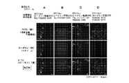
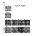
- Liposome A having a lipid membrane containing Pep-PEG2000-DSPE as a constituent lipid and fluorescently labeled with rhodamine as a single membrane, and one lipid membrane containing Pep-PEG5000-DSPE as a constituent lipid and fluorescently labeled with rhodamine
- Liposome B having a membrane, liposome C containing Pep-PEG5000-DSPE and PEG2000-DSPE as constituent lipids and a lipid membrane fluorescently labeled with rhodamine as a single membrane, and PEG5000-DSPE and PEG2000-DSPE as constituent lipids
- mouth the liposome D which has the lipid membrane which contains and was fluorescently labeled with rhodamine as a single membrane
- FITC (green) fluorescence is observed in the upper row
- rhodamine (red) fluorescence is observed in the middle row
- FITC (green) fluorescence and rhodamine (red) are superimposed.
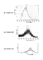
- the horizontal axis represents molecular weight (mass-to-charge ratio m / z)
- the vertical axis represents intensity (arbitrary unit).
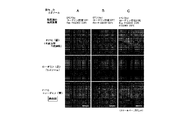
- the left column is the result of observing the fluorescence of FITC (green)
- the middle column is the result of observing the fluorescence of rhodamine (red)
- the right column is the result of superposing the left column and the middle column, It is the result of observing the overlap (yellow) of the fluorescence of FITC (green) and the fluorescence of rhodamine (red).
- Liposome E having PEG5000-DSPE and PEG2000-DSPE as constituent lipids and a lipid membrane fluorescently labeled with NBD as a single membrane, and containing Pep-PEG5000-DSPE and PEG2000-DSPE as constituent lipids and fluorescently labeled with NBD
- the lower diagram is an enlarged view of the frame shown in the upper diagram.
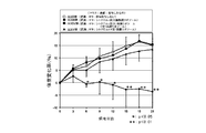
- High-fat were administered to healthy mice with fusion peptide (group IV) or apoptosis-inducing peptide-encapsulated single membrane liposome (group V) once every three days or without any administration (group VI). It is a figure which shows the result of having raised while giving a diet (HFD) and having calculated the weight change rate.
- Administer empty single membrane liposomes (Group VIII), fusion peptides (Group IX) or apoptosis-inducing peptide-encapsulated single membrane liposomes (Group X) to obese mice at intervals of once every 3 days, or none (Group VII) is a diagram showing the results of calculating the amount of change in body weight while feeding each with a high fat diet (HFD).
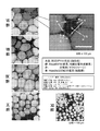
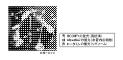
- FIG. 1 It is a figure which shows the observation result of a lipid droplet, a capillary endothelial cell, and a cell nucleus in the epididymal fat tissue of the obese mouse of a VII group, a VIII group, a IX group, and a X group, and the healthy mouse
- the right figure is an enlarged view of the portion shown in the left figure.
- the points where the fluorescence of BODIPY (blue), the fluorescence of Alexa 647 (red) and the fluorescence of Hoechst 33342 (green) are detected are illustrated by arrows.
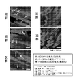
- FIG. 1 It is a figure which shows the observation result of a macrophage, a lipid droplet, a capillary endothelial cell, and a cell nucleus in the epididymal fat tissue of the obese mouse of a VII group, a VIII group, a IX group, and a X group, and the healthy mouse
- the right figure is an enlarged view of the portion shown in the left figure.
- arrows indicate the locations where Alexa 568 (red) fluorescence, BODIPY (light blue) fluorescence, Alexa 647 (green) fluorescence and Hoechst 33342 (blue) fluorescence are detected.
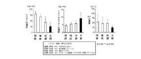
- the locations where BODIPY (green) fluorescence is detected are illustrated by arrows. It is a figure which shows the result of having calculated the energy intake amount for 24 hours of the obese mouse
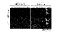
- HEPES buffer only (G) mixed lipid solution (H) containing 0.65 ⁇ mol (lipid) amount of empty single membrane liposome, final concentrations of 0.5 nmol / mL, 1 nmol / mL, 2 nmol / mL or 4 nmol / HEPES buffer (I1-4) in which cytochrome c is dissolved, or cytochrome c-encapsulated single membrane liposomes having a final concentration of cytochrome c of 0.5 nmol / mL, 1 nmol / mL, 2 nmol / mL, or 4 nmol / mL, respectively.
- FIG. 6 is a diagram showing the results of observing cell morphology after adding and incubating a mixed lipid solution (J1 to 4) containing a mixed lipid solution and an appropriate amount of empty single membrane liposomes to adipose tissue capillary endothelial cells.
- a mixed lipid solution J1 to 4
- J1 to 4 a mixed lipid solution
- an appropriate amount of empty single membrane liposomes to adipose tissue capillary endothelial cells.
- the locations where spherical cell morphology was confirmed are indicated by arrows.
- cytochrome c amount was 0.5 mmol / kg (XIX group), 0.1 mmol / kg (XX group) or 0.02 mmol / kg (XXI) of cytochrome c-encapsulated single membrane liposomes were administered to healthy mice at an interval of once every 3 days, and normal diet (ND) was administered to group IV, XVII group, XIX group, XX group and XXI It is a figure which shows the result of having raised with giving the high fat diet (HFD) to each group, and calculating the weight change rate.
- HFD high fat diet
- the lipid membrane structure according to the present invention is a lipid membrane structure having a target cell migration ability, (a) a lipid formed by binding a peptide having a target cell migration ability, polyethylene glycol and lipid in this order, and (B) It has a lipid membrane containing as a constituent lipid a lipid formed by binding polyethylene glycol having a smaller number average molecular weight than that of polyethylene glycol constituting (a).
- long-chain PEG refers to PEG constituting the above (a).
- short chain length PEG means a PEG having a smaller number average molecular weight than the PEG constituting (a), and means the PEG constituting the above (b). Therefore, in the present invention, the long chain length means that the chain length is long (number average molecular weight is large) compared to the chain length of the PEG constituting (b), and the short chain length means (a) It means that the chain length is short (number average molecular weight is small) compared to the chain length of the PEG to be constructed.
- the structure of the lipid membrane structure according to the invention is shown in FIG.
- the difference in the number average molecular weight between the long-chain PEG and the short-chain PEG is not particularly limited, but for example, 10 to 100,000, 50 to 750,000, 100 to 50000, 500 to 25000, 800 to 20000, 1000 to 15000, 1200 to 10000, 1400 to 8000, preferably 1600 to 6000, more preferably 1800 to 5000, and still more preferably 2000 to 4000.
- the molecular weight Ma of the long chain PEG in the present invention is, for example, 1000 ⁇ Ma ⁇ 10000000, 1200 ⁇ Ma ⁇ 5000000, 1300 ⁇ Ma ⁇ 1000000, 1400 ⁇ Ma ⁇ 500000, 1500 ⁇ Ma ⁇ 100000, 1600 ⁇ Ma ⁇ 50000, 1700 ⁇ Ma ⁇ 30000, 1800 ⁇ Ma ⁇ 20000, 1900 ⁇ Ma ⁇ 15000, 2000 ⁇ Ma ⁇ 12000, 2100 ⁇ Ma ⁇ 10000, 2200 ⁇ Ma ⁇ 9000, 2300 ⁇ Ma ⁇ 8500, 2400 ⁇ Ma ⁇ 8000, 2500 ⁇ Ma ⁇ 7500, 2600 ⁇ Ma ⁇ 7400, 2700 ⁇ Ma ⁇ 7300, 2800 ⁇ Ma ⁇ 7200, 2900 ⁇ Ma ⁇ 7100, 3000 ⁇ Ma ⁇ 7000, 3100 ⁇ Ma ⁇ 6900, 3200 ⁇ Ma Can be exemplified 6800, preferably 3300 ⁇ Ma ⁇ 6700, more
- the molecular weight Mb of the short chain PEG is, for example, 5 ⁇ M ⁇ 20000, 10 ⁇ M ⁇ 15000, 50 ⁇ M ⁇ 10000, 100 ⁇ M ⁇ 8000, 200 ⁇ M ⁇ 7000, 300 ⁇ M ⁇ 5000, 400 ⁇ M ⁇ 4500, 450 ⁇ M ⁇ 4000, 470 ⁇ M ⁇ 3800, preferably 480 ⁇ M ⁇ 3700, more preferably 490 ⁇ M ⁇ 3600, and even more preferably 500 ⁇ Mb ⁇ 3500.
- 5 ⁇ M ⁇ 20000 10 ⁇ M ⁇ 15000, 50 ⁇ M ⁇ 10000, 100 ⁇ M 8000, 200 ⁇ M ⁇ 7000, 300 ⁇ M ⁇ 5000, 400 ⁇ M ⁇ 4500, 450 ⁇ M ⁇ 4000, 470 ⁇ M ⁇ 3800, preferably 480 ⁇ M ⁇ 3700, more preferably 490 ⁇ M ⁇ 3600, and even more preferably 500
- examples of the PEG in the present invention include PEGs having many shapes, for example, linear, branched, forked or multi-armed PEG, and linear PEG is preferred.
- the PEG in the present invention includes PEG to which a functional group or a protective group necessary for binding to a peptide or lipid such as a maleimide group or a methoxy group is added or modified.
- target tissue refers to a tissue or cell to which the lipid membrane structure according to the present invention is to be transferred.
- transfer to target cells or “transfer ability to target cells” means the “transfer to tissues containing target cells” or the “transfer ability to tissues containing target cells”.
- the phrase “transfer to target tissue” or “transfer ability to target tissue” does not necessarily mean “transfer to cells contained in target tissue” or “transfer ability to cells contained in target tissue”.
- the type of “target tissue” is not particularly limited, and examples thereof include tissues belonging to epithelial tissue, connective tissue, muscle tissue, and nerve tissue.
- tissue belonging to the connective tissue examples include a loose connective tissue, a dense connective tissue, an adipose tissue, and a reticulum tissue.
- the cell type of the “target cell” may be any cell type, whether it is a somatic cell or a germ cell, or may be a cell separated from a living body (in vitro cell), or a cell in a living body (in vivo cell). But you can.
- cell types include adipose tissue vascular endothelial cells and epithelial cells, epidermal cells, epidermal basal cells, keratinocytes, root sheath cells, hair matrix cells, mucosal epithelial cells, mammary cells, lacrimal gland cells, ears Malignant gland cells, sweat gland cells, prostate cells, endometrial cells, goblet cells, mucous epithelial cells, panel cells, type II alveolar cells, anterior pituitary cells, pituitary mesothelial cells, posterior pituitary cells, parathyroid glands Cells, gallbladder epithelial cells, hepatocytes, adipocytes, type I alveolar cells, pancreatic duct cells, acinar center cells, duct cells, synovial cells, choroid plexus cells, corneal endothelial cells, contractile cells, skeletal muscle cells, Cardiomyocytes, smooth muscle cells, myoepithelial cells, red blood cells, mega
- a known peptide known to have the ability to migrate to the target cell can be used, and a phage display method or the like can be used depending on the type of the target cell. Those appropriately identified or designed according to the law can be used.
- Specific examples of known peptides known to have target cell migration ability include, for example, amino acid sequences such as KGGRAKD (SEQ ID NO: 1), VMGSVTG (SEQ ID NO: 2), RGEVLWS (SEQ ID NO: 3), and the like.
- a peptide having the ability to migrate to adipose tissue vascular endothelial cells MG Kolonin et al., Nature Medicine, Vol. 10, No.
- NGR Arap W. et al., Science, No. 1) 279, 377-380, 1998; SEQ ID NO: 4
- amino acids of RGD Hirano Y. et al., J. Biomed Mater Res., 25, 1523-1534, 1991; SEQ ID NO: 5
- a peptide having the ability to migrate tumor tissue vascular endothelial cells polyarginine peptide (for example, cell membrane-permeable peptides such as International Patent Application Publication No. WO 2005/032593), GALA peptides (for example, T. Kakudo et al., Biochemistry, 2004, Vol. 43, pages 5618-5623) and the like. Mention may be made of membrane-fusogenic peptides and transcytosis-inducing peptides (for example, Japanese Patent Application No. 2006-179955).
- the “peptide having the ability to migrate to the target cell” in the present invention includes a peptide consisting of an amino acid sequence having one or more conservative amino acid substitutions in the amino acid sequence as long as it has the ability to migrate to the target cell.
- conservative amino acid substitutions are those that can generally be made without changing the physiological activity of the resulting molecule, ie, those that are recognized within the range of conservative substitutions (Watson et al., Molecular® Biology® of Gene), etc.
- Yes for example, acidic amino acids of aspartic acid and glutamic acid; basic amino acids of lysine, arginine and histidine; nonpolar amino acids of alanine, valine, leucine, isoleucine, proline, phenylalanine, methionine and tryptophan; glycine, asparagine, cysteine, glutamine, Serine, threonine and tyrosine polar uncharged side chain amino acids; phenylalanine, tryptophan, and tyrosine aromatic amino acids Replacement can be mentioned that occurs in the family inside) of Amino Acids.
- acidic amino acids of aspartic acid and glutamic acid acidic amino acids of aspartic acid and glutamic acid; basic amino acids of lysine, arginine and histidine, aliphatic amino acids of glycine, alanine, valine, leucine, isoleucine, serine and threonine (classified as aliphatic-hydroxyamino acids of serine and threonine Can be classified); phenylalanine, tyrosine and tryptophan aromatic amino acids; asparagine and glutamine amides; cysteine and methionine sulfur amino acids.
- the “peptide having the ability to migrate to the target cell” in the present invention as long as it has the ability to migrate to the target cell, one or several amino acids are deleted, substitutions other than the above-mentioned conservative amino acid substitution, insertion and / or Added peptides are included.
- the specific range for deletion is usually 1 to 3, preferably 1 to 2, more preferably 1, and the specific range for substitution excluding conservative amino acid substitution is usually 1 to 3, preferably
- the specific range for insertion is usually 1 to 5, preferably 1 to 3, more preferably 1 to 2, and still more preferably 1.
- the specific range for is usually 1 to 5, preferably 1 to 3, more preferably 1 to 2, and still more preferably 1.
- a glycine residue on the N-terminal side of the amino acid sequence a glycine residue and a cysteine on the C-terminal side
- One or several amino acids may be added while retaining the ability to migrate to the target cell, such as by adding a residue, but such peptides are still included in the peptides of the present invention.
- the “peptide having the ability to migrate to a target cell” in the present invention can be synthesized using a method that can be appropriately selected by those skilled in the art based on the sequence.
- a method for example, in addition to a peptide synthesis method in which each amino acid is chemically polymerized to synthesize a polypeptide, a recombinant vector containing a DNA encoding the peptide in the present invention is prepared and prepared.
- a transformant obtained by introducing the obtained vector into an appropriate host cell is cultured in a medium, and collected from the obtained culture, or a DNA encoding the peptide of the present invention is obtained in a cell-free protein synthesis system. Examples thereof include a method obtained by expression.
- any synthesis method other than a general method widely known by those skilled in the art can be used for any synthesis method.
- the number of amino acid residues of the peptide is not particularly limited, and for example, 2-4 residues, 5-7 residues, 8-10 residues, 11-15 residues, 16-20 residues, Examples include 21-30 residues, 31-40 residues, 41-55 residues, 56-75 residues, 76-100 residues, and 101 or more amino acid residues, depending on the function. Are appropriately selected.
- the “lipid membrane” refers to a membrane having a lipid membrane structure, the main component of which is lipid.
- the lipid membrane in the present invention may be composed of a lipid bilayer in which lipid molecules are associated with each other to form a hydrophobic portion, and the hydrophobic portion is formed inward or outward. It may consist of a single layer.
- lipid membrane structure in the present invention a closed vesicle having a lipid membrane composed of a lipid bilayer can be mentioned, and as such a lipid membrane structure, for example, a liposome can be mentioned.
- the lipid membrane structure in the present invention may be any of positive chargeability, nonchargeability, both (positive and negative) chargeability, and negative chargeability, but is preferably negatively chargeable or nonchargeable.
- the “negatively charged lipid membrane structure” in the present invention refers to a lipid membrane structure that is negatively charged as a whole.
- positively charged lipids or both (positive and negatively) chargeable lipids as lipids constituting the lipid membrane. Because it contains lipids, non-charged lipids, or is positively charged or both (positive and negative) charged, or modified with a non-chargeable substance, it is locally positively charged or both (positive and negative) charged. Even those that are non-charged are included in the “negatively-charged lipid membrane structure” if they are negatively charged as a whole.
- the “uncharged lipid membrane structure” in the present invention refers to a lipid membrane structure that is uncharged as a whole.
- a positively charged lipid a negatively charged lipid, both Because it contains a (positive and negative) chargeable lipid, or it is modified with a negatively chargeable, positively chargeable, or both (positive and negative) chargeable substance, it is locally negatively charged or positively charged. Even if it is charged, it is included in the “non-charged lipid membrane structure” if it is non-charged as a whole.
- lipid membrane structure having a surface potential of about ⁇ several mV to + several mV is a “non-charged lipid membrane structure” in vivo (Md. Vietnameser Hossen et al., Journal of Controlled Release, 147, 261-268, 2010; Sjoard Hak et al., European Journals pharmaceutics and Biopharmaceutics, Vol. 22, Vol. 104, No. 9266-9271, 2007). Further, in the present invention, “non-charging” is used interchangeably with “neutral”.
- the lipid constituting the lipid membrane structure in the present invention may be any of positively charged lipids, neutral (including both positive and negative) charged lipids and uncharged lipids, and negatively charged lipids. Examples thereof include phospholipids, glycolipids, sterols, long-chain aliphatic alcohols and glycerin fatty acid esters, and one or more of these can be used.
- phospholipid examples include phosphatidylcholine (for example, dioleoylphosphatidylcholine, dilauroylphosphatidylcholine, dimyristoylphosphatidylcholine, dipalmitoylphosphatidylcholine, distearoylphosphatidylcholine), phosphatidylglycerol (for example, dioleoylphosphatidylglycerol, dilauroylphosphatidylglycerol, Dimyristoyl phosphatidylglycerol, dipalmitoyl phosphatidylglycerol, distearoyl phosphatidylglycerol, etc., phosphatidylethanolamine (eg dioleoylphosphatidylethanolamine, dilauroylphosphatidylethanolamine, dimyristoylphosphatidylethanolamine, dipasto) Mitoylphosphatidylethanolamine, di
- glycolipids examples include glyceroglycolipids such as sphingomyelin, sulfoxyribosyl glyceride, diglycosyl diglyceride, digalactosyl diglyceride, galactosyl diglyceride and glycosyl diglyceride, and sphingoglycolipids such as galactosyl cerebroside, lactosyl cerebroside and ganglioside. 1 type, or 2 or more types of these can be used.
- glyceroglycolipids such as sphingomyelin, sulfoxyribosyl glyceride, diglycosyl diglyceride, digalactosyl diglyceride, galactosyl diglyceride and glycosyl diglyceride
- sphingoglycolipids such as galactosyl cerebroside, lactosyl cerebroside and ganglioside. 1 type, or 2
- sterols examples include sterols derived from animals such as cholesterol, cholesterol succinic acid, lanosterol, dihydrolanosterol, desmosterol, dihydrocholesterol, sterols derived from plants such as stigmasterol, sitosterol, campesterol, and brassicasterol (tytosterol). And sterols derived from microorganisms such as ergosterol, and one or more of these can be used. In addition, these sterols can generally be used to physically or chemically stabilize the lipid bilayer or to adjust the fluidity of the membrane.
- long-chain fatty acid or long-chain aliphatic alcohol a fatty acid having 10 to 20 carbon atoms or an alcohol thereof can be used.
- long-chain fatty acids or long-chain aliphatic alcohols include palmitic acid, stearic acid, lauric acid, myristic acid, pentadecylic acid, arachidic acid, margaric acid, tuberculostearic acid and other saturated fatty acids, palmitoleic acid, Mention of unsaturated fatty acids such as oleic acid, arachidonic acid, vaccenic acid, linoleic acid, linolenic acid, arachidonic acid, eleostearic acid, oleyl alcohol, stearyl alcohol, lauryl alcohol, myristyl alcohol, cetyl alcohol, linolyl alcohol 1 type, or 2 or more types of these can be used.
- glycerin fatty acid ester examples include monoacyl glycerides, diacyl glycerides, and triacyl glycerides, and one or more of these can be used.
- Examples of the positively charged lipid include dioctadecyldimethylammonium chloride (DODAC), N- (2,3-oleyloxy) propyl-N, N, N-trimethylammonium (N-) in addition to the above-described lipids.
- DODAC dioctadecyldimethylammonium chloride
- N- N- (2,3-oleyloxy) propyl-N
- N- N-trimethylammonium
- Examples of neutral lipids including both (positive and negative) charged lipids and non-charged lipids include diacylphosphatidylcholine, diacylphosphatidylethanolamine, ceramide and the like in addition to the above-described lipids. Or 2 or more types can be used.
- Examples of the negatively charged lipid include diacylphosphatidylserine, diacylphosphatidic acid, N-succinylphosphatidylethanolamine (N-succinylPE), phosphatidylethylene glycol, cholesteryl hemisuccinate (CHEMS), etc. 1 type, or 2 or more types of these can be used.
- the lipid membrane of the lipid membrane structure according to the present invention has positive charges such as tocopherol, propyl gallate, ascorbyl palmitate, butylated hydroxytoluene, stearylamine, oleylamine and the like.
- Positive charges such as tocopherol, propyl gallate, ascorbyl palmitate, butylated hydroxytoluene, stearylamine, oleylamine and the like.
- Charged substances to be added, charged substances to give negative charges such as dicetyl phosphate, membrane proteins such as membrane surface proteins and integral membrane proteins, and peptides that impart cell permeability and nuclear translocation ability to lipid membrane structures It can be combined or contained, and the amount and content of the bond can be adjusted as appropriate.
- the lipid membrane structure according to the present invention is produced using a known method such as a hydration method, an ultrasonic treatment method, an ethanol injection method, an ether injection method, a reverse phase evaporation method, a surfactant method, or a freezing / thawing method. can do.
- the lipid membrane structure according to the present invention can be used by being dispersed in an appropriate aqueous solvent such as physiological saline, phosphate buffer, citrate buffer, and acetate buffer.
- Additives such as saccharides, polyhydric alcohols, water-soluble polymers, nonionic surfactants, antioxidants, pH adjusters, and hydration accelerators may be appropriately added to the dispersion.
- the lipid membrane structure according to the present invention can be stored in a state in which the dispersion is dried.
- the lipid membrane structure according to the present invention may contain, for example, specific antibodies, targeting ligands and other functional elements for drug delivery according to known methods.
- the present invention provides an anti-obesity and / or therapeutic agent using the lipid membrane structure according to the present invention.
- the anti-obesity and / or therapeutic agent according to the present invention is the above-described lipid membrane structure according to the present invention in which an apoptosis-inducing agent is encapsulated, and the peptide having the ability to migrate to target cells is capable of migrating to adipose tissue vascular endothelial cells
- a lipid membrane structure, which is a peptide having the above, is used as an active ingredient, and obesity is suppressed / treated by inducing apoptosis in vascular endothelial cells of adipose tissue.
- apoptosis inducer for example, apoptosis of KLAKLAKKLAKLAK peptide (SEQ ID NO: 7; Ellerby HM et al., Nature Medicine, Vol. 5, pp 1032-1038, 1999) can be induced.
- cytochrome c cytochrome c
- Actinomycin D Anisomycin
- Antibiotic A23187 Apoptosis Activator 2
- Aristoforin Betulinic Acid
- Camptothecin Cisplatin
- Colchicine cycloheximide
- Daunorubicin HCl Dexamethasone
- Doxorubicin HCl Etopo side, Forskolin, Genistin, Okadaic acid, Phorbol-12-myristate 13-acetate, Staurosporine, Tamoxifen Citrate, Tapsigargin and the like.
- the peptide constituting the apoptosis inducer in the present invention is the same as the “peptide having the ability to migrate to a target cell” in the present invention described above.
- a lipid membrane structure in which an apoptosis-inducing agent is encapsulated can be produced according to a conventional method.
- a solution of an organic solvent in which lipid is dissolved and apoptosis induction It can be produced by mixing an aqueous solution in which the agent is dissolved and preparing a W / O emulsion by ultrasonic treatment, and then distilling off the organic solvent under reduced pressure.
- organic solvent for dissolving lipid examples include hydrocarbons such as pentane, hexane, heptane, and cyclohexane, halogenated hydrocarbons such as methylene chloride and chloroform, aromatic hydrocarbons such as benzene and toluene, methanol, and ethanol.
- Lower alcohols such as methyl acetate, esters such as methyl acetate and ethyl acetate, ketones such as acetone and the like can be used alone or in combination of two or more.
- the present invention provides an agent for suppressing and / or treating adipose tissue inflammation using the lipid membrane structure according to the present invention.
- the agent for inhibiting and / or treating adipose tissue inflammation according to the present invention is the aforementioned lipid membrane structure according to the present invention in which an apoptosis-inducing agent is encapsulated, wherein the peptide having the ability to migrate to target cells is adipose tissue vascular endothelial cell
- a lipid membrane structure, which is a peptide having a migratory ability, is used as an active ingredient, and apoptosis is induced / induced in vascular endothelial cells of adipose tissue to suppress / treat inflammation of adipose tissue.
- the description of the same or equivalent configuration as that of the lipid membrane structure and obesity suppression / treatment agent according to the present invention described above will be o
- the present invention provides an agent for suppressing and / or treating fat accumulation in non-adipose tissue using the lipid membrane structure according to the present invention.
- the agent for suppressing and / or treating fat accumulation in the non-adipose tissue according to the present invention is the above-mentioned lipid membrane structure according to the present invention in which an apoptosis-inducing agent is encapsulated, and having a target cell migration ability Inhibits / treats fat accumulation in non-adipose tissue by using as an active ingredient a lipid membrane structure, which is a peptide having the ability to migrate to adipose tissue vascular endothelial cells, and inducing apoptosis in vascular endothelial cells of adipose tissue.
- the structure of the lipid membrane structure, obesity inhibiting / treating agent and adipose tissue inflammation inhibiting / treating agent according to the present invention described above The description of the same or corresponding configuration will be omitted.
- non-adipose tissue refers to a tissue other than adipose tissue, and particularly refers to a tissue in which ectopic fat can accumulate.
- the non-adipose tissue in the present invention can preferably include a tissue in which a fat component can migrate and accumulate, that is, a tissue other than the brain.
- a tissue other than the brain Specifically, for example, the liver, skeletal muscle, pancreas, bone marrow, heart, Examples include kidneys and blood vessels.
- the “non-adipose tissue” in the present invention may or may not contain a fat component, and may or may not contain, for example, fat droplets or fat cells.
- “non-adipose tissue” is used interchangeably with “non-adipose tissue”, “lean body tissue”, and “lean body tissue”.
- ectopic fat means fat accumulated in tissues other than adipose tissue. That is, the agent for suppressing and / or treating fat accumulation in non-adipose tissue according to the present invention can inhibit and / or treat ectopic fat accumulation.
- the present invention provides a method for producing a lipid membrane structure.
- the method for producing a lipid membrane structure according to the present invention is a method for producing a lipid membrane structure having a target cell migration ability, and comprises a single membrane comprising the following lipids (a) and (b) as constituent lipids: (A) a peptide having the ability to migrate to a target cell, a lipid formed by binding PEG and lipid in this order, (b) a number average compared to the PEG constituting (a) Lipid formed by PEG with low molecular weight.
- a description of the same or corresponding configuration as that of the lipid membrane structure or obesity suppressing / treating agent according to the present invention is omitted.
- a single membrane lipid membrane containing the lipids of (a) and (b) as constituent lipids is prepared.
- a single membrane lipid membrane may be prepared using the lipids of (a) and (b), or a single membrane lipid membrane may be prepared using lipids, and then the lipid.
- a peptide having the ability to migrate to a target cell and a long-chain PEG and a short-chain PEG may be bound to the constituent lipids of the membrane.
- a peptide having the ability to migrate to a target cell may be bound to a constituent lipid formed by binding a chain-length PEG.
- a lipid formed by binding a peptide having a target cell migration ability, PEG and lipid in this order can be prepared according to a conventional method.
- a solution in which a peptide having the ability to migrate to a target cell is dissolved and a solution in which a lipid formed by binding PEG is dissolved can be mixed and incubated while shaking. .
- a screening method for a substance having an effect on a target cell according to the present invention comprises: (I) a step of selecting a peptide having the ability to migrate to one target cell in the lipid membrane structure according to the present invention and encapsulating a target substance to migrate to the target cell; (Ii) a step of evaluating whether or not the target substance shows an effect in the target cell; The step (i) or (ii) is included.
- the method for encapsulating the target substance in the lipid membrane structure according to the present invention includes the same method as the method for producing the lipid membrane structure in which the apoptosis-inducing agent is encapsulated. it can.
- step (i) as a method of selecting a peptide having a target cell migration ability and transferring a lipid membrane structure encapsulating a target substance to a target cell, for example, a tissue in which the target cell is separated from a living body,
- a method of adding a lipid membrane structure to the culture solution can be mentioned.
- the target cell is a cell in a living body (in vivo tissue or cell)
- examples thereof include a method of orally administering a solution containing a lipid membrane structure as it is or dissolving it, or administering it parenterally, such as intravenously, intraperitoneally, subcutaneously, or nasally.
- the method for evaluating whether or not the target substance shows an effect in the target cell can be appropriately set according to the effect expected of the target substance.
- the target cell is separated from the living body. If the tissue or cells are in vitro (tissues or cells in vitro), the target cells are stained by immunostaining or nuclear staining as necessary, and then the morphology and color intensity of the tissue or cells are observed using a microscope. Examples thereof include a method and a method of extracting RNA and analyzing the gene expression level.
- the target cell is a tissue or cell in a living body (in-vivo tissue or cell)
- the target cell is separated and collected, and the target cell is separated from the living body or cell (in-vitro tissue or cell).
- the effect is indirectly evaluated by collecting blood from a living body and analyzing the abundance of various marker molecules, measuring body weight and body fat percentage, etc. You can also do it.
- the present invention suppresses and / or treats fat accumulation in an obesity-suppressing and / or therapeutic agent, adipose tissue inflammation-suppressing and / or therapeutic agent or non-adipose tissue using the lipid membrane structure according to the present invention.
- a screening method for an agent to be used is provided.
- the method for screening for obesity suppression and / or treatment agent, adipose tissue inflammation suppression and / or treatment agent or agent for suppressing and / or treating fat accumulation in non-adipose tissue comprises: (Iii) The lipid membrane structure according to the present invention, wherein a target substance is encapsulated in a lipid membrane structure in which a peptide having a target cell migration ability is a peptide having a fat tissue vascular endothelial cell migration ability, Transferring to cells, (Iv) evaluating whether the target substance induces apoptosis in adipose tissue vascular endothelial cells; It has the above process (iii) or (iv).
- the obesity suppressing and / or treating agent, adipose tissue inflammation suppressing and / or therapeutic agent or the agent for suppressing and / or treating fat accumulation according to the present invention in the screening method of the agent for suppressing and / or treating fat accumulation in non-adipose tissue according to the present invention described above, the obesity suppressing and / or treating agent, adipose tissue inflammation suppressing and / or therapeutic agent or the agent for suppressing and / or treating fat accumulation according to the present invention.
- the description of the structure that is the same as or equivalent to the structure of the lipid membrane structure, the anti-obesity agent / therapeutic agent, the method of manufacturing the lipid membrane structure, or the screening method for substances that are effective in target cells will be omitted.
- lipid membrane structure having the ability to migrate to a target cell according to the present invention a method for producing the same, and a method for screening a substance having an effect on the target cell will be described based on examples. Note that the technical scope of the present invention is not limited to the features shown by these examples.
- PCT / US02 / 27836) which is known to have the ability of migrating, has 1 residue glycine (G) at the N-terminus and 2 residues glycine at the C-terminus ( G) and a peptide to which one residue of cysteine (C) has been added, wherein the C-terminal cysteine carboxyl group is amidated (GKGGRAKDGGC-NH 2 ; SEQ ID NO: 6) This was synthesized and this was designated as “migration ability peptide”.
- Mal-PEG2000 a lipid (Mal-PEG2000-DSPE; Nippon Oil & Fats Co., Ltd.) bound to L-distearoylphosphatidylethanolamine (DSPE) and a long chain length PEG (PEG5000) having a number average molecular weight of 5000 and maleimide
- a lipid (Mal-PEG5000-DSPE; Nippon Oil & Fats Co., Ltd.) obtained by binding a group to which a group is added (Mal-PEG5000) to DSPE was added to distilled water to 5 mmol / L, and a bath sonicator was used. Dissolve by sonication at room temperature for 1 minute, and transfer ability A peptide solution, Mal-PEG2000-DSPE solution and Mal-PEG5000-DSPE solution were obtained.
- the Mal-PEG2000-DSPE solution or the Mal-PEG5000-DSPE solution was added to the migrating peptide solution with gentle stirring so that the molar ratios were 1: 1. Then, while shaking using a bioshaker, incubation was performed at 30 ° C. for 24 hours to transfer 2.5 mmol / L of a solution of Mal-PEG2000-DSPE (Pep-PEG2000-DSPE) to which the transferable peptide was bound, A 2.5 mmol / L solution of Mal-PEG5000-DSPE (Pep-PEG5000-DSPE) to which a functional peptide was bound was prepared.
- a schematic diagram showing Pep-PEG5000-DSPE is shown in FIG.
- EPC Egg yolk phosphatidylcholine
- a 10 mmol / L EPC / Chol solution was prepared.
- rhodamine-labeled DOPE solution was prepared by dissolving rhodamine-labeled DOPE (Avanti Polar Lipid Co., Ltd.) in which rhodamine was bound to dioleoylglycerophosphoethanolamine (DOPE) in chloroform to be 3.8 mmol / L. .
- DSPE PEG2000-DSPE; Nippon Oil & Fats Co., Ltd.
- DSPE PEG5000-DSPE; Nippon Oil & Fats Co., Ltd.
- HEPES buffer pH 7.4; HEPES buffer
- EPC / Chol solution, rhodamine-labeled DOPE solution, PEG2000-DSPE solution, PEG5000-DSPE solution, Pep-PEG2000-DSPE solution and Pep-PEG5000-DSPE solution of Example 1 [1-2] A total of four mixed lipid solutions (lipid concentration: 10 mmol / L) of a, b, c, and d were prepared by mixing so that the mol% ratio was the ratio shown below.
- EPC / Chol Rhodamine labeled DOPE: Pep-PEG2000-DSPE: Pep-PEG5000-DSPE: PEG2000-DSPE: PEG5000-DSPE a; 94: 1: 5: 0: 0: 0 b; 94: 1: 0: 5: 0: 0 c; 93: 1: 0: 5: 1: 0 d; 93: 1: 0: 0: 1: 5
- Example 1 (1) [1-3] 500 ⁇ L of the mixed lipid solution of Example 1 (1) [1-3] and 500 ⁇ L of HEPES buffer were mixed, and sonicated at 4 ° C. for 15 seconds using a probe type sonicator. After evaporating and distilling off chloroform by flowing nitrogen gas using an evaporator, the mixture was sonicated for 1 minute using a bath sonicator to prepare a mixed lipid solution (lipid concentration 10 mmol / L) containing liposomes.
- lipid concentration 10 mmol / L lipid concentration 10 mmol / L
- the mixed lipid solution a contains Pep-PEG2000-DSPE (hereinafter, the same as the order of binding) formed by binding migratory peptides, Mal-PEG2000 and DSPE in this order, and is fluorescently labeled with rhodamine.
- Liposome A having a single lipid membrane as a single membrane
- Liposome B having a lipid membrane labeled with rhodamine as a single membrane and containing Pep-PEG5000-DSPE as a constituent lipid in the mixed lipid solution
- Liposome C containing Pep-PEG5000-DSPE and PEG2000-DSPE as constituent lipids in c and a lipid membrane fluorescently labeled with rhodamine as a single membrane, and PEG5000-DSPE and PE in mixed lipid solution
- Liposomes D having a fluorescence-labeled lipid membrane as a single film by rhodamine together including 2000-DSPE as a component lipid, were prepared, respectively.
- FITC-GSI-B4 FITC-labeled Griffoniaonsimplicifolia Lectin I-B4 Ilectin
- the FITC (green) fluorescence and the rhodamine (red) fluorescence were higher in mice administered with liposome C than in mice administered with liposomes A, B, and D.
- the area (yellow) showing the overlap was significantly large. That is, it was confirmed that the mice administered with liposome C had significantly more liposomes present in the capillary endothelial cells of adipose tissue than the mice administered with liposomes A, B and D.
- mice administered with liposome C a portion (yellow) showing an overlap between the fluorescence of FITC (green) and the fluorescence of rhodamine (red) was hardly detected. That is, it was confirmed that in mice administered with liposome C, there were significantly fewer liposomes present in vascular endothelial cells of major organs such as the heart and lungs (not shown).
- lipid membrane comprising a peptide having a target cell migration ability, a lipid formed by binding a long-chain PEG and a lipid in this order, and a lipid formed by binding a short-chain PEG as constituent lipids It was revealed that lipid membrane structures having a specific migration to target cells.
- Mal-PEG2000-DSPE had a molecular weight of about 1500 to about 4500.
- Mal-PEG5000-DSPE had a molecular weight of about 4500 to about 7500.
- the molecular weight of DSPE is about 760 and the molecular weight of maleimide group is about 170
- the molecular weight of PEG constituting Mal-PEG2000-DSPE is about 500 to about 3500, and its number average molecular weight is about 2000. It can be seen that the molecular weight of PEG constituting Mal-PEG5000-DSPE is about 3500 to about 6500, and the number average molecular weight is about 5000.
- Pep-PEG5000-DSPE had a molecular weight of about 5500 to about 8500.
- Example 2 (1) [1-1] was reversed based on the method described in Example 1 (1) [1-4].
- Liposomes single membrane liposomes
- liposomes were prepared by the simple hydration method. It is known that liposomes prepared by the simple hydration method are generally multilamellar liposomes having a plurality of lipid membranes (Danilo D., Biochem. J., Vol. 256, Vol. 1-11). Page, 1988).
- chloroform was distilled off from 500 ⁇ L of the mixed lipid solution of Example 2 (1) [1-1], 375 ⁇ L of chloroform was added and redissolved, and evaporated again. After the lipid film was prepared, 500 ⁇ L of HEPES buffer was added to hydrate the lipid film, and the mixture was stirred using a vortex mixer for 2 to 3 minutes to prepare a mixed lipid solution containing liposomes (lipid concentration 10 mmol / L). ) was prepared.
- multilamellar liposomes having multiple lipid membranes containing Pep-PEG5000-DSPE and PEG2000-DSPE as constituent lipids and fluorescently labeled with rhodamine were prepared.
- Example 1 (1) [1-2] single membrane liposome and Example 2 (1) [1-3] multilamellar liposome The average particle diameter, surface potential and PDI were measured by the method described in Example 1 (1) [1-5], and the measurement was further repeated 5 to 8 times in Example 2 (1) [1-1]. By repeating, each standard deviation was calculated. The results are shown in Table 2.
- the average particle diameter of each of the single membrane liposome and the multilamellar liposome was about 100 nm, the surface potential was about -5 mV, and the PDI was about 0.25. From this result, it was revealed that the single membrane liposome and the multilamellar liposome are negatively charged liposomes or uncharged liposomes having the same physical properties.
- Example 2 (1) [1-2] and the multilamellar liposome of Example 2 (1) [1-3] were administered to mice by the method described in), and fluorescence observation of subcutaneous fat tissue was performed.
- the dose of the liposome contained in each mixed lipid solution was the body weight (kg) x 0.2 mmol (lipid) of the individual mouse.
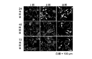
- the administration of FITC-GSI-B4 is 23.5 hours, 47.5 hours, and 71.5 hours after the administration of the mixed lipid solution (liposome), and the subcutaneous adipose tissue is removed by mixing lipid solution ( 24 hours, 48 hours and 72 hours after the administration of the liposome). The result is shown in FIG.
- the area of the portion (yellow) showing the overlap of the fluorescence of FITC (green) and the fluorescence of rhodamine (red) is the same as that of the mouse administered with the single membrane liposome and the mouse administered with the multiple membrane liposome.
- the values were almost the same after 24 hours, but were significantly larger in the mice administered with single membrane liposomes after 48 hours and 72 hours. That is, it was confirmed that in mice administered with single membrane liposomes, there were significantly more liposomes present in capillary endothelial cells of adipose tissue than in mice administered with multiple membrane liposomes.
- a lipid membrane comprising a peptide having a target cell migration ability, a lipid formed by binding a long-chain PEG and a lipid in this order, and a lipid formed by binding a short-chain PEG as constituent lipids It was revealed that a lipid membrane structure having a single membrane has a higher ability to migrate to a target cell than a lipid membrane structure having a plurality of similar lipid membranes.
- Example 3 Examination of liposome target cell migration ability (incorporation into target cells) (1) Preparation of mixed lipid solution Example 1 (1) Method described in [1-1] to [1-3] A mixed lipid solution was prepared as e and f. However, in place of rhodamine-labeled DOPE, DOPE (NBD-labeled DOPE) bound with Nitro-2-1,3-Benzoxadiazol-4-yl (NBD) was used. The mole% ratio of each lipid in e and f was as follows.
- EPC / Chol NBD-labeled DOPE: Pep-PEG5000-DSPE: PEG2000-DSPE: PEG5000-DSPE e; 93: 1: 0: 1: 5 f; 93: 1: 5: 1: 0
- a buffer ⁇ 123 mmol / L NaCl, 9.8 mmol / L KCl, 1.3 mmol / L CaCl 2 , 5 mmol / L D-(+)-glucose, 100 mmol / L HEPES, 2% (v Collagenase was added to / w) BSA ⁇ to 1 mg / mL to prepare a collagenase solution. 10 mL of collagenase solution per 1 g of subcutaneous adipose tissue was added and incubated at 37 ° C. for 30 minutes with shaking. During this incubation, every 5 minutes, the mixture was stirred for 5 seconds using a vortex mixer.
- the mixture was centrifuged at room temperature and 1500 rpm for 10 minutes, the upper layer was removed, and the lower layer was recovered.
- the cell fraction of endothelial cells After washing these cell fractions with Hank's equilibration buffer, the entire amount was seeded on a culture dish with a diameter of 35 mm, and 37 ° C., 5% (v / v) CO using EGM2-MV medium (Lonza). 2. Incubated for 2.5 hours in an environment of 100% relative humidity.
- Alexa647-labeled Griffonia simplicifolia Lectin I-B4 Iselectin Alexa647-GSI-B4 was added to the medium to a concentration of 5 ⁇ g / mL, and incubated for 0.5 hour to fluorescently stain capillary endothelial cells.
- Hoechst 33342 was added to the medium so as to be 2.5 ⁇ g / mL, and the cells were incubated for 0.2 hours to fluorescently stain the cell nucleus. After washing with Hanks equilibration buffer, Alexa647 (red) fluorescence, Hoechst 33342 (blue) fluorescence and NBD (green) fluorescence were observed using a confocal laser scanning microscope. A representative one of the observation results is shown in FIG.
- a lipid membrane containing a peptide having the ability to migrate to a target cell, a lipid formed by binding a long chain PEG and a lipid in this order and a lipid formed by binding a short chain PEG as constituent lipids is 1 It has been clarified that incorporation into the target cell is promoted by using a lipid membrane structure as a sheet membrane.
- Example 4 Examination of Apoptosis Inducing Effect of Liposomes Encapsulating Apoptosis Inducing Peptide (1) Chemical Synthesis of Apoptosis Inducing Peptide and Fusion Peptide M. et al., Nature Medicine, Vol. 5, pp. 1032-1038 (1999), wherein the carboxyl group of the C-terminal lysine is amidated (SEQ ID NO: 7; KLAKLAKKLAKLAK-NH 2 ) to Toray Industries, Inc. It was commissioned and chemically synthesized, and this was used as an apoptosis-inducing peptide.
- Example 4 (3) Preparation of liposomes by reverse phase evaporation method
- liposomes were prepared by the reverse phase evaporation method based on the method described in Example 1 (1) [1-4]. This was designated as empty single membrane liposome.
- the apoptosis-inducing peptide of Example 4 (1) is 190.4 mg / L (125 ⁇ mol / L) instead of the HEPES buffer.
- liposomes were prepared by the reverse phase evaporation method, and this was used as an apoptosis-inducing peptide-encapsulated single membrane liposome.
- an empty single membrane liposome is a liposome having a lipid membrane containing Pep-PEG5000-DSPE and PEG2000-DSPE as a constituent lipid, in which an apoptosis-inducing peptide is not encapsulated, and one apoptosis-inducing peptide-encapsulating membrane.
- the membrane liposome is a liposome having a lipid membrane containing Pep-PEG5000-DSPE and PEG2000-DSPE as a constituent lipid, in which an apoptosis-inducing peptide is encapsulated, as a single membrane.
- Example 4 The average particle size of the apoptosis-inducing peptide-encapsulated monolayer liposome of Example 4 (3) was determined by the method described in Example 1 (1) [1-5]. When the diameter, surface potential and PDI were measured, the average particle size was 109.2 ⁇ 7.8 nm, the surface potential was 6.0 ⁇ 0.9 mV, and the PDI was about 0.2 to 0.3. From this result, it became clear that the apoptosis-inducing peptide-encapsulated monolayer liposome is an uncharged liposome.
- Group I Empty single membrane liposome amount; (lipid) mouse body weight (kg) x 0.2 mmol
- Group II Fusion peptide amount; (Fusion peptide) Body weight (kg) of mouse individual x 3 mg (equivalent to 1.8 mg of apoptosis-inducing peptide)
- Group III Apoptosis-inducing peptide-encapsulated single membrane liposome amount; (apoptosis-inducing peptide) body weight of individual mouse (kg) ⁇ 1 mg, (lipid) body weight of individual mouse (kg) ⁇ 0.2 mmol
- cells containing activated caspase are caused by using a caspase 3,7 assay kit (Immunochemistry Technology) with a sulforhodamine fluorescently labeled caspase inhibitor (FLICA; fluorescent-labeled inhibitors of caspase).
- FLICA sulforhodamine fluorescently labeled caspase inhibitor
- Alexa647 (red) fluorescence and sulforhodamine (green) fluorescence were observed using a confocal laser scanning microscope.
- Alexa647 (red) fluorescence and sulforhodamine (green) fluorescence were observed using a confocal laser scanning microscope.
- apoptotic capillary endothelial cells were hardly observed when obese mice were administered empty monolayer liposomes, and were observed 24 hours after administration when a fusion peptide was administered, but thereafter Was hardly observed, but when the apoptosis-inducing peptide-encapsulated single membrane liposome was administered, it was continuously observed after administration.
- a lipid membrane structure having one membrane as a membrane and encapsulated with an apoptosis-inducing peptide can induce apoptosis in adipose tissue vascular endothelial cells, and its apoptosis-inducing effect is higher than that of a fusion peptide. Became more sustainable.
- Example 5 Examination of obesity suppression / treatment effect of liposome encapsulating apoptosis-inducing peptide (1) Preparation of liposome and fusion peptide Based on the method described in Example 4 (1) to (3), fusion peptide Empty single membrane liposomes and apoptosis-inducing peptide-encapsulated single membrane liposomes were prepared.
- the dose per one time of the fusion peptide in the HEPES buffer administered in the IV group or the V group or the apoptosis-inducing peptide-encapsulated single membrane liposome in the mixed lipid solution is as follows.
- HFD PMI
- water and feed were allowed to freely ingest, and the breeding temperature was 23 ° C.
- Group IV Fusion peptide amount; (Fusion peptide) Body weight (kg) of mouse individual ⁇ 3 mg (corresponding to 1.175 ⁇ mol of fusion peptide)
- Group V Apoptosis-inducing peptide-encapsulated monolayer liposome amount; (apoptotic fusion peptide) body weight (kg) of mouse individual ⁇ 1 mg (corresponding to 0.657 ⁇ mol of apoptosis-inducing peptide), (lipid) body weight of mouse individual (kg) ⁇ 0.2 mmol
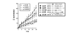
- the body weight change rate is a value indicating the rate of weight gain or weight loss when the body weight at the start of administration of the fusion peptide or apoptosis-inducing peptide-encapsulated single membrane liposome is 100%.
- Rate of weight change (%) ⁇ (weight on day 0, 3, 6, 9 or 12 ⁇ weight on day 0) / weight on day 0 ⁇ ⁇ 100
- the weight change rate value of the IV group is approximated on any of the 0th, 3rd, 6th, 9th, and 12th days as compared to the weight change rate value of the VI group. There was no significant difference on days 3, 6, 9, and 12.
- the value of the weight change rate of the V group is somewhat smaller on the 6th day than the value of the weight change rate of the VI group, but is slightly smaller on the 6th day, and on the 9th and 12th days. It was remarkably small. Significant differences between group V and group VI were not on days 3 and 6, but p ⁇ 0.05 on day 9 and p ⁇ 0.01 on day 12.
- a lipid having the ability to migrate to adipose tissue vascular endothelial cells, a long chain-length PEG and a lipid formed by combining lipids in this order and a lipid formed by combining a short chain-length PEG are included as constituent lipids. It was revealed that a lipid membrane structure having a lipid membrane as a single membrane and having an apoptosis-inducing peptide encapsulated can suppress obesity.
- the group VIII contains a mixed lipid solution containing empty unilamellar liposomes of Example 5 (1)
- the group IX contains a HEPES buffer in which the fusion peptide of Example 5 (1) is dissolved
- the group X contains The mixed lipid solution containing the apoptosis-inducing peptide-encapsulated unilamellar liposome of Example 5 (1) was administered to the tail vein 10 times at an interval of once every 3 days, and reared for 30 days.
- the group VII was reared for 30 days without any administration.
- the amount of change in body weight is a value indicating the amount of increase or decrease in body weight based on the body weight at the start of administration of empty single membrane liposome, fusion peptide or apoptosis-inducing peptide-encapsulated single membrane liposome.
- the average value and the standard deviation were calculated for each group of the body weight change amount, and the weight change amount of the VIII group, the IX group, and the X group was subjected to a significant difference test with respect to the body weight change amount of the IIV group.
- the significant difference test was performed by one-way analysis of variance (one-way ANOVA) and Dunnett's method. The result is shown in FIG. 10a.
- the weight change of the VIII group was approximated on the 3-18th day and smaller on the 21-30th day than the weight change of the VII group. There was no significant difference between group VIII and group VII at any time point on days 3-30. In addition, the change in body weight of the IX group was smaller at any time point on the 3-30th day than the change in weight of the VII group. Significant differences between group IX and group VII were not on days 3-12, but were p ⁇ 0.05 on day 15 and p ⁇ 0.005 on days 18-30. In contrast, the change in body weight of Group X was significantly smaller at any time point on days 3-30 than the change in weight of Group VII. There was no significant difference between group X and group VII on days 3 and 6, but p ⁇ 0.05 on days 9 and 12, p ⁇ 0.005 on day 15, and On day 18-30, p ⁇ 0.0005.
- the weight change of the VII group was positive on any of the 3rd to 30th days, and increased with the passage of the breeding days. Therefore, the weight of the VII group continuously increased during the breeding period.
- the weight change of the group VIII was positive on any of the 3-30th days, increased with the passage of the breeding days on the 3-12th day, and almost the same value on the 12-30th day. Therefore, in the group VIII, the body weight increased until the 12th day during the breeding period and was maintained thereafter.
- the weight change of the IX group was positive on the 3rd to 9th days, 0 on the 12th day and negative on the 15th to 30th days, and slightly changed with the passage of the breeding days on the 3rd to 9th days.
- the body weight increased slightly until the 9th day during the breeding period, and then the body weight gradually decreased.
- the change in body weight of group X was negative on any of days 3 to 30 and decreased with the passage of the breeding days. Therefore, in group X, the body weight continuously decreased during the breeding period.
- the weight on the 30th day was not significantly changed in the VII group and the IX group with respect to the weight on the 0th day, but increased significantly in the VIII group, In, it decreased significantly. That is, from the results shown in FIGS. 10a and 10b, it has been clarified that, when an apoptosis-inducing peptide-encapsulated monolayer liposome is administered to obese mice, the body weight is decreased despite being fed with a high fat diet. It was.
- the sizes of the inguinal subcutaneous fat tissue and epididymal fat tissue of group X were significantly smaller than those of group VII, group VIII, and IX. That is, it has been clarified that, when an apoptosis-inducing peptide-encapsulated single membrane liposome is administered, fat tissue hypertrophy of obese mice is suppressed or adipose tissue is reduced under high fat diet.
- BODIPY boron dipyrromethene
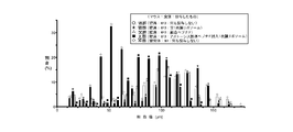
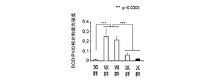
- the size of fat droplets indicated by the fluorescence of BODIPY is used as the cell diameter of adipocytes, and the cell diameter is measured for 300 lipid droplets for each mouse in each group. The percentage (%) of how much is present was calculated and represented in the distribution map. The result is shown in FIG.
- the average value and standard deviation of the cell diameter are calculated for each group, and the significant difference test for the IX group of the VII group, the VIII group, the IX group, and the X group, and the X group of the VII group, the VIII group, and the IX group Significance test was performed and displayed in a histogram. Significant difference tests were performed by the one-way ANOVA, Tukey-Kramer's Honestly Significant Difference (HSD) method. The result is shown in FIG. 11c.
- the cell diameter of the X group was slightly larger than that of the XI group, but was significantly smaller than those of the VII group, VIII group and IX group. That is, it was clarified that when an apoptosis-inducing peptide-encapsulated monolayer liposome is administered to obese mice, fat cell hypertrophy is suppressed or adipocytes shrink under high-fat diet intake.
- BODIPY blue fluorescence
- Alexa647 red fluorescence
- Hoechst 33342 green fluorescence
- FIG. A representative one of the observation results is shown in FIG.
- the BODIPY fluorescent color is displayed in green and the Hoechst fluorescent color is displayed in blue.
- the entire field of view is very bright green. Since it is difficult to discriminate the blue color shown, pseudo-coloring processing is performed on the computer.
- the fluorescent color of BODIPY is blue.
- the lipid droplets indicated by the fluorescence of BODIPY blue
- the periphery of the fat cells and the capillary endothelial cells indicated by the fluorescence of Alexa647 (red) and Hoechst 33342 red
- Many structures hereinafter referred to as “angiogenic-adipogenic cluster structure” surrounded by cell nuclei indicated by the green fluorescence were observed, but were hardly observed in the X group and the XI group.
- the angiogenic-adipogenic cluster structure is a site where angiogenesis and adipogenesis occur and is known to appear characteristically in enlarged adipose tissue (NishimuramS. Et al., Diabetes). 56, 1517-1526, 2007). From this, the apoptosis-inducing peptide-encapsulated single membrane liposome can suppress angiogenesis and cell neoplasia or abolish new blood vessels and adipocytes in the adipose tissue of obese mice under high-fat diet intake. It was revealed.
- Example 5 From the results of the above Example 5 (3) [3-2], [3-3] and [3-4], the peptide having the ability to migrate to adipose tissue vascular endothelial cells, long-chain PEG and lipid were found to be A lipid membrane structure having a lipid membrane containing a lipid formed by binding in order and a lipid formed by binding a short chain length PEG as a constituent lipid and having an apoptosis-inducing peptide encapsulated as a single membrane, It became clear that obesity can be treated.
- the mixed lipid solution containing the apoptosis-inducing peptide-encapsulated single membrane liposome of Example 5 (3) was administered, and neither was administered to the XII group or XVI group.
- HFD PMI
- ND PMI
- the size of the adipose tissue was observed based on the method described in Example 5 (3) [3-2].
- the result is shown in FIG. 13b.
- the weight of the adipose tissue was measured, the ratio of the adipose tissue weight to the body weight at the end of the breeding period (adipose tissue weight ratio) was calculated, the average value and the standard deviation were calculated for each group, and the XIV group and the XV group A significant difference test was performed on the XII group. The significant difference test was performed by the method described in Example 5 (3) [3-2]. The result is shown in FIG. 13c.
- the rate of weight change in the XV group is small at any time point on day 3-30 compared to the XII group, XIII group and XIV group, but 3% when compared with the XVI group. It was approximated at any point on day 30. That is, when apoptosis-inducing peptide-encapsulated monolayer liposomes are administered to healthy mice, weight gain due to high-fat diet is suppressed, but weight gain corresponding to weight gain under normal diet intake, ie normal growth It became clear that weight gain due to was hardly suppressed.
- the inguinal subcutaneous fat tissue and epididymal fat tissue of the XV group are significantly smaller than those of the XII group and the XIV group, and as shown in FIG.
- the adipose tissue weight ratio was significantly smaller compared to the XII group and the XIV group. That is, when an apoptosis-inducing peptide-encapsulated single membrane liposome is administered, it becomes clear that fat tissue enlargement due to a high-fat diet is suppressed, and the proportion of the weight of adipose tissue in the body weight decreases. It was.
- a lipid having the ability to migrate to adipose tissue vascular endothelial cells, a long chain-length PEG and a lipid formed by combining lipids in this order and a lipid formed by combining a short chain-length PEG are included as constituent lipids.
- a lipid membrane structure having a lipid membrane as a single membrane and encapsulated with an apoptosis-inducing peptide does not inhibit normal growth and specifically suppresses the enlargement of adipose tissue caused by ingestion of a high-fat diet It became clear.
- Example 6 Inhibition of adipose tissue inflammation / treatment effect of liposome encapsulated with apoptosis-inducing peptide Based on the method described in Example 5 (3) [3-3], group VII, group VIII, group IX Small pieces of epididymal adipose tissue of group X and group XI were prepared. These small pieces of epididymal adipose tissue have been previously reported (Nishimura S. et al., Diabetes, 56, 1517-1526, 2007 and Nishimura S. et al., Nat. Med., 15, 914-94). 920 (2009), macrophages were immunostained with anti-F4 / 80 antibody.
- lipid droplets, capillary endothelial cells and cell nuclei were fluorescently stained based on the method described in Example 5 (3) [3-4], and Alexa 568 (red) was analyzed using a confocal laser scanning microscope. Fluorescence, BODIPY (light blue) fluorescence, Alexa 647 (green) fluorescence and Hoechst 33342 (blue) fluorescence were observed. Of the observation results, a representative one is shown in FIG. In general, the BODIPY fluorescent color is displayed in green and the Alexa fluorescent color is displayed in red. However, the entire field of view is very bright green. Since it is difficult to discriminate blue indicating the color, pseudo-coloring processing is performed on the computer.
- the fluorescent color of BODIPY is blue.
- the three primary colors of red, blue, and green are assigned so that their localization is as easy to distinguish as possible. A color that has no effect is selected.
- the fluorescent color of BODIPY is light blue, and the fluorescent color of Alexa 647 is green.
- the lipid droplets indicated by the fluorescence of BODIPY (light blue), that is, the periphery of the fat cells, the capillary endothelial cells indicated by the fluorescence of Alexa647 (green) and the Hoechst 33342 (blue).
- BODIPY light blue
- Alexa647 green
- Hoechst 33342 blue
- Many macrophages indicated by Alexa568 (red) fluorescence were observed around the structure surrounded by the cell nucleus indicated by fluorescence (angiogenic-adipogenic cluster structure) and its surroundings.
- a lipid having the ability to migrate to adipose tissue vascular endothelial cells, a long chain-length PEG and a lipid formed by combining lipids in this order and a lipid formed by combining a short chain-length PEG are included as constituent lipids. It was revealed that a lipid membrane structure having a lipid membrane as a single membrane and encapsulating an apoptosis-inducing peptide can suppress / treat inflammation in adipose tissue.
- Example 7 Examination of effect of suppressing / treating fat accumulation in non-adipose tissue of liposome encapsulated with apoptosis-inducing peptide
- Example 5 (3) [3-1] Group VII, VIII, IX and Liver and skeletal muscle were excised from group X and group XI of Example 5 (3) [3-3], and sections with a thickness of about 100 ⁇ m were prepared using microslicer DSK-1000. These sections were fluorescently stained for lipid droplets using BODIPY, F-actin using rhodamine-labeled phalloidin, and cell nuclei using Hoechst 33342.
- BODIPY green fluorescence
- rhodamine red fluorescence
- Hoechst 33342 blue fluorescence
- the fluorescence intensity of BODIPY and Hoechst 33342 was measured using Image-Pro Plus-4.5 software according to the previous report (shahen SM, et al., Nucleic Acids Res., 39, e48, 2011).
- the fluorescence intensity (relative fluorescence intensity) of BODIPY with respect to the fluorescence intensity was calculated.
- the average value and standard deviation were computed for every group, the significant difference test was performed, and it represented on the histogram.
- the significant difference test was performed according to the method described in Example 5 (3) [3-3].
- the significant difference test for the X group or the XI group of the VII group, the VIII group, and the IX group, and the VII group and the VIII group were performed.
- a significant difference test for the XI group of the VII group and the VIII group and a significant difference test for the IX group and the X group of the VII group and the VIII group were performed for skeletal muscle, respectively.
- the results for the liver are shown in FIG. 15b, and the results for skeletal muscle are shown in FIG. 16b.
- the result is similar to that in the liver in skeletal muscles, and many lipid droplets indicated by BODIPY (green) fluorescence are observed in the VII and VIII groups and to some extent in the IX group. In contrast, little was observed in the X and XI groups.
- the relative fluorescence intensity of BODIPY in skeletal muscle that is, the amount of accumulated fat, was significantly smaller in the IX, X, and XI groups than in the VII and VIII groups.
- the relative fluorescence intensity of BODIPY of group X was similar to that of group XI.
- a lipid membrane comprising a lipid having a structure capable of migrating adipose tissue vascular endothelial cells, a lipid having a long chain length PEG and a lipid bonded in this order and a lipid having a short chain length PEG bonded as a constituent lipid. It was revealed that a lipid membrane structure having a single membrane and having an apoptosis-inducing peptide encapsulated can suppress / treat fat accumulation in non-adipose tissue.
- Example 8 Evaluation of effects when administering liposome encapsulating apoptosis-inducing peptide (1) Examination of energy intake Example 5 (3) [VII] group VII, VIII, IX and X For the group, after the liposome or peptide was administered 6 times (after 15 days from the start of administration), the amount of HFD (PMI) ingested during 24 hours was measured, and the amount of energy ingested from the measured result (PMI) Intake energy amount; kcal) was calculated. Moreover, the average value and the standard deviation were calculated for each group of the intake energy amount, and the significant difference test was performed on the intake energy amount of the VII group with respect to the intake energy amounts of the VIII group, the IX group, and the X group. The significant difference test was performed by the method described in Example 5 (3) [3-2]. The result is shown in FIG.
- the intake energy amount of the IX group was significantly smaller than that of the VII group, whereas the intake energy amount of the VIII group and the X group was not significantly changed compared to the VII group. . That is, when the fusion peptide is administered, the energy intake is reduced compared to when the fusion peptide is not administered, whereas when the apoptosis-inducing peptide-encapsulated single membrane liposome is administered, it is compared with the case where it is not administered. And it became clear that the amount of energy consumed does not change.
- Forward primer 5′-atgctgactgcaatgtgctg-3 ′ (SEQ ID NO: 9), reverse primer: 5′-cagacttagacctgggaactct-3 ′ (SEQ ID NO: 10) Npy; forward primer: 5′-atgctagggtaacaagcgaatgg-3 ′ (SEQ ID NO: 11), reverse primer: 5′-tgtcgcagaggcggagtagtat-3 ′ (SEQ ID NO: 12) Gal; forward primer: 5′-ggcaggcgttatcctgcttag-3 ′ (SEQ ID NO: 13), reverse primer: 5′-ctgtcagggtccaactct-3 ′ (SEQ ID NO: 14) Pomc; forward primer: 5′-ctggagagccccgtgtttc-3 ′ (SEQ ID NO: 15), reverse primer: 5′-tggactcggctctggactg-3 ′ (SEQ ID
- 18S rRNA was amplified using Quantum RNA Classic II II 18S standard (Ambion) as an internal control. After separating the PCR product by gel electrophoresis, the gel was stained with ethidium bromide, and the signal was detected using an image analyzer LAS-400 mini (Fuji Film). The intensity of the detected signal was measured using Multi-Gage software, and a value divided by the signal intensity of 18S rRNA was calculated, and this was used as the relative expression level of each gene. For the relative expression level, the average value and the standard deviation were calculated for each group, and the relative expression level of the VIII group, the IX group, and the X group was subjected to a significant difference test with respect to the relative expression level of the VII group. The significant difference test was performed by the method described in Example 5 (3) [3-2]. The result is shown in FIG.
- the relative expression levels of Agrp, Npy, Gal, and Pomc were hardly changed between the groups.
- the relative expression level of Trh was not significantly changed in the VIII group and the X group as compared to the VII group, but was significantly larger in the IX group than the VII group. That is, it is clear that when the apoptosis-inducing peptide-encapsulated single membrane liposome is administered to obese mice, the expression level of both the feeding enhancement gene and the feeding suppression gene is hardly changed compared to the case where it is not administered. Became.
- a lipid membrane structure that has a lipid membrane containing a lipid that is bound as a constituent lipid as a single membrane and encapsulated with an apoptosis-inducing peptide has little effect on appetite It became clear.
- Leptin and adiponectin are both hormones secreted from adipocytes, and it is known that leptin blood concentration increases and adiponectin blood concentration decreases with obesity.
- ALT also called glutamate pyruvate transferase (GPT)
- GPT glutamate pyruvate transferase
- the concentration of leptin was not significantly changed in the VIII group compared to the VII group, while the IX group and the X group were significantly smaller than the VII group. It was remarkably small.
- the concentration of adiponectin was not significantly changed in the VIII group and the IX group as compared with the VII group, while the X group was significantly higher than that in the VII group.
- the ALT concentration did not change significantly in the VIII group, the IX group, and the X group as compared with the VII group, but the X group tended to be smaller than the VII group.
- a lipid having the ability to migrate to adipose tissue vascular endothelial cells, a long chain-length PEG and a lipid formed by combining lipids in this order and a lipid formed by combining a short chain-length PEG are included as constituent lipids.
- lipid membrane structure having a lipid membrane as a single membrane and encapsulating an apoptosis-inducing peptide normalizes the balance of hormones secreted from adipocytes and normalizes liver function It has been suggested.
- Example 9 (1) Preparation of cytochrome c-encapsulated single membrane liposomes by the reverse phase evaporation method
- the mixed lipid solution of Example 9 (1) was subjected to reverse phase based on the method described in Example 1 (1) [1-4].
- Liposomes were prepared by the evaporation method.
- a HEPES buffer in which cytochrome c was dissolved at 100 ⁇ mol / L was used instead of the HEPES buffer. That is, a liposome having a single lipid membrane containing Pep-PEG5000-DSPE and PEG2000-DSPE as a constituent lipid, in which cytochrome c is encapsulated, was prepared, and this was designated as a single membrane liposome encapsulating cytochrome c.
- Example 9 (1) Preparation of Empty Single Membrane Liposomes by Reverse Phase Evaporation Method
- the reverse phase evaporation method Liposomes were prepared by That is, a liposome having a single lipid membrane containing Pep-PEG5000-DSPE and PEG2000-DSPE as a constituent lipid, in which cytochrome c is not encapsulated, was prepared, and this was defined as an empty single membrane liposome.
- the lipid concentration of the mixed lipid solution containing empty single membrane liposomes was 10 mmol / L.
- Example 9 (3) Liposome addition and observation of cell morphology G, H, I1, I2, I3, I4, J1, and the like of Example 9 (3) so that the final concentration of cytochrome c and the amount of lipid added are as follows. In the medium of J2, J3 and J4, only HEPES buffer, mixed lipid solution containing empty single membrane liposome of Example 9 (3), HEPES buffer in which cytochrome c is dissolved, or of Example 9 (2) A mixed lipid solution containing cytochrome c-encapsulated single-membrane liposomes was added.
- apoptosis is not induced when cytochrome c is added to adipose tissue vascular endothelial cells as it is, but apoptosis is induced when cytochrome c-encapsulated monolayer liposome is added, and apoptosis is induced in that case. It was revealed that the number of cells increased depending on the final concentration of cytochrome c.
- a lipid having the ability to migrate to adipose tissue vascular endothelial cells, a long chain-length PEG and a lipid formed by combining lipids in this order and a lipid formed by combining a short chain-length PEG are included as constituent lipids. It was revealed that a lipid membrane structure having a lipid membrane as a single membrane and encapsulating cytochrome c can induce apoptosis in adipose tissue vascular endothelial cells.
- Example 10 Examination of obesity suppression / treatment effect of liposome encapsulated with cytochrome c (1) Preparation of cytochrome c-encapsulated monolayer liposome by reverse phase evaporation method Based on the method described in Example 9 (2), Cytochrome c-encapsulated single membrane liposomes were prepared by the reverse phase evaporation method.
- the dose per one time of cytochrome c in the mixed lipid solution is as follows. Also, during the breeding period, ND (PMI) was given to the XVII group, HFD (PMI) was given to the XVIII group, XIX group, XX group and XXI group, and water and food were freely ingested. The temperature was 23 ° C.
- Group XVII administered; (does not administer any liposome) Group XVIII: administered; (does not administer any liposome) XIX group: administered; cytochrome c-encapsulated monolayer liposomal cytochrome c amount; body weight of individual mouse (kg) ⁇ 0.5 mmol (6.16 mg) Group XX: administered; cytochrome c-encapsulated monolayer liposomal cytochrome c amount; body weight (kg) of mouse individual ⁇ 0.1 mmol (1.23 mg) XXI group: administered; cytochrome c-encapsulated monolayer liposome cytochrome c amount; body weight of mouse individual (kg) ⁇ 0.02 mmol (0.25 mg)
- Example 10 (3) Calculation of body weight change rate For the XVII group, XVIII group, XIX group, XX group, and XXI group of Example 10 (2), from the start of administration of the mixed lipid solution (cytochrome c-encapsulated monolayer liposome), The body weight was measured on days 3, 6, 7, 9, 12, 14, 15, 18, 21, 24, 27, 28 and 30 and the method described in Example 5 (2) [2-2]. The body weight change rate was calculated, and the weight change rates of the XVII group, the XIX group, the XX group, and the XXI group were subjected to a significant difference test with respect to the body weight change rate of the XVIII group. The result is shown in FIG.
- the values of the weight change rate of the XVIII group, the XIX group, the XX group, and the XXI group are 3, 6, 7, 9, 12, 14, 15 compared with the weight change rate of the XVII group. , 18, 21, 24, 27, 28 and 30 days. From this result, it was confirmed that weight gain was caused by the high fat diet.
- the value of the weight change rate of the XIX group is 3, 6, 7, 9, 12, 14, 15, 18, 21, 24, 27, 28 and 30 days compared with the value of the weight change rate of the XVIII group. It was small in all eyes. The significant difference of the XIX group from the XVIII group was not on days 3, 6, 7, 9, 12 and 15 but p ⁇ 0.05 on days 14 and 18 and 21, 24, 27, 28. On the 30th day, p ⁇ 0.01.
- weight change rate values of the XX group are approximated on the third, sixth, and twelfth days compared with the value of the weight change rate of the XVIII group, and are larger on the seventh and ninth days. , 18, 21, 24, 27, 28 and 30 days. Significant differences between group XX and group XVIII were not on days 3, 6, 7, 9, 12, 14, 15, 18 and 21 but p ⁇ 0.05 on day 24 and 27, 28. On the 30th day, p ⁇ 0.01.
- the value of the weight change rate of the XXI group is approximated on the third, sixth, twelfth, and fourteenth days compared with the value of the weight change rate of the XVIII group, and larger on the seventh and ninth days, Small on days 18, 21, 24, 27, 28 and 30.
- the significant difference of the XXI group from the XVIII group was not on days 3, 6, 7, 9, 12, 14, 15, 18, 21 and 24, but p ⁇ 0.05 on days 27 and 28, In addition, p ⁇ 0.01 on the 30th day.
- the XIX group ⁇ XX group / XXI group on the third, sixth, seventh, ninth and twelfth days, , 18, 21, 24, 27, 28, and 30 days, XIX group ⁇ XX group ⁇ XXI group. That is, the administration of cytochrome c-encapsulated monolayer liposomes can suppress the body weight gain of healthy mice due to the intake of a high-fat diet, and the inhibitory effect can be increased depending on the dose of cytochrome c. It was revealed.
- a lipid having the ability to migrate to adipose tissue vascular endothelial cells, a long chain-length PEG and a lipid formed by combining lipids in this order and a lipid formed by combining a short chain-length PEG are included as constituent lipids. It was revealed that a lipid membrane structure having a lipid membrane as a single membrane and encapsulating cytochrome c can suppress obesity.
- Example 11 (1) Preparation of cytochrome c-encapsulated multilamellar liposomes by simple hydration method
- the mixed lipid solution of Example 11 (1) was prepared by the simple hydration method according to the method described in Example 2 (1) [1-3]. Liposomes were prepared. However, instead of the HEPES buffer, a HEPES buffer in which cytochrome c (Sigma Aldrich) was dissolved so as to be 100 ⁇ mol / L was used. That is, cytochrome c-encapsulated multilamellar liposomes having a plurality of lipid membranes containing Pep-PEG5000-DSPE and PEG2000-DSPE as constituent lipids and encapsulating cytochrome c were prepared.
- cytochrome c-encapsulated multilamellar liposomes having a plurality of lipid membranes containing Pep-PEG5000-DSPE and PEG2000-DSPE as constituent lipids and encapsulating cytochrome c were
- Example 11 The cytochrome c-encapsulated single membrane liposome of Example 11 (2) and the cytochrome c-encapsulated multilamellar liposome of Example 11 (3) were obtained in Example 1 (1).
- the average particle diameter, the surface potential and the PDI were measured by the method described in [1-5], and each standard deviation was determined by repeating the measurement of Example 11 (1) to the above measurement 3 to 6 times. Calculated. The results are shown in Table 3.
- both the cytochrome c-encapsulated single membrane liposome and the cytochrome c-encapsulated multilamellar liposome had an average particle size of about 105 nm, a surface potential of about ⁇ 9 mV, and a PDI of about 0.25. From this result, it was revealed that the cytochrome c1 single membrane liposome and the cytochrome c multiple membrane liposome are negatively charged liposomes or non-charged liposomes having the same physical properties.
- Each recovered lower layer was suspended in 100 ⁇ L of distilled water to recover cytochrome c-encapsulated single membrane liposomes and cytochrome c-encapsulated multilamellar liposomes.
- BCA assay was performed using BCA protein assay kit (Pierce), and cytochrome c was quantified. Further, EPC was quantified for each collected liposome using a phospholipid C-Test Wako kit (Wako Pure Chemical Industries, Ltd.). Based on these quantitative results, cytochrome c recovery rate, lipid recovery rate, and cytochrome c encapsulation rate were calculated using the following formula 3, formula 4 and formula 5, respectively.
- Cytochrome c recovery rate (%) ⁇ quantitative cytochrome c amount / cytochrome c amount used in the preparation of liposomes in Example 11 (2) or Example 11 (3) ⁇ ⁇ 100
- lipid recovery rate (%) ⁇ quantitative EPC amount / EPC amount used in preparing liposome in Example 11 (2) or Example 11 (3) ⁇ 100
- Cytochrome c encapsulation rate (%) (Cytochrome c recovery rate / lipid recovery rate) ⁇ 100
- the cytochrome c recovery rate was about 15% for both cytochrome c-encapsulated single membrane liposomes and cytochrome c-encapsulated multilamellar liposomes.
- the lipid recovery rate of cytochrome c-encapsulated multilamellar liposomes is about 1.5 times that of cytochrome c-encapsulated single-membrane liposomes
- the cytochrome c-encapsulated rate of cytochrome c-encapsulated multilamellar liposomes is cytochrome c-encapsulated 1 The ratio was about 2/3 compared with the membrane liposome.
- the cytochrome c-encapsulated single membrane liposome and the cytochrome c-encapsulated multilamellar liposome have the same physical properties (average particle diameter, surface potential and PDI), but the cytochrome c encapsulation rate is determined by the cytochrome c-encapsulated monolayer membrane. It was revealed that the liposome was larger than the cytochrome c-encapsulated multilamellar liposome.
- Example 12 Examination of obesity suppression / treatment effect of liposomes with different numbers of lipid membranes encapsulating cytochrome c (1) Preparation of liposomes One cytochrome c encapsulated based on the method described in Example 9 (2) Membrane liposomes were prepared. Moreover, empty unilamellar liposomes were prepared based on the method described in Example 9 (3). In addition, cytochrome c-encapsulated multilamellar liposomes were prepared by the method described in Example 11 (3).
- cytochrome c was “weight of individual mouse (kg) ⁇ 6.16 mg (cytochrome c) in any of the XXIII, XXIV and XXV groups. ) Amount ”and the lipid was“ the body weight of the individual mouse (kg) ⁇ 0.19 mmol (lipid) amount ”.
- HFD PMI
- the value of the weight change rate of the XXIII group is approximated on any of the third, sixth, ninth, twelfth, fifteenth, eighteenth, and twenty-first days compared to the value of the weight change rate of the XXIV group.
- the XXIII group and the XXIV group were not significant difference between the XXIII group and the XXIV group on days 3, 6, 9, 12, 15, 18, and 21.
- the value of the weight change rate of the XXV group is approximated on the third day as compared with the value of the weight change rate of the XXII group, and slightly on the 6, 9, 12, 15, 18 and 21 days. It was small. There was no significant difference between the XXV group and the XXII group on days 3, 6, 9, 12, 15, 18, and 21.
- the value of the weight change rate of the XXIV group was remarkably small on the third, sixth, ninth, twelfth, fifteenth, eighteenth, and twenty-first days compared with the value of the weight change rate of the XXII group. .
- Significant differences between the XXIV group and the XXII group were not on day 3, but p ⁇ 0.05 on days 6, 9 and 12, and p ⁇ 0.01 on days 15, 18 and 21 there were.
- the value of the weight change rate of the XXII group, the XXIII group, and the XXV group was positive on any of the third, sixth, ninth, twelfth, fifteenth, eighteenth, and twenty-first days, and increased with the passage of the breeding days. That is, in the XXII group, the XXIII group, and the XXV group, the body weight continuously increased during the breeding period.
- the weight change rate value of the XXIV group was positive on the 3rd and 9th days, but negative on the 6th, 12th, 15th, 18th and 21st days, and decreased with the passage of the breeding days. became. That is, in the XXIV group, body weight continuously decreased during the breeding period.
- a lipid having the ability to migrate to adipose tissue vascular endothelial cells, a long chain-length PEG and a lipid formed by combining lipids in this order and a lipid formed by combining a short chain-length PEG are included as constituent lipids.
- a lipid membrane structure having a lipid membrane as a single membrane, in which cytochrome c is encapsulated, is formed by binding a peptide capable of migrating adipose tissue vascular endothelial cells, a long-chain PEG and a lipid in this order.
- a lipid membrane structure having a plurality of lipid membranes containing lipids and lipids formed by binding short-chain PEG as constituent lipids, in which cytochrome c is encapsulated, or encapsulated in a lipid membrane structure It was revealed that the effect of suppressing and treating obesity is extremely high compared to cytochrome c that has not been obtained.
- Liposomes P and Liposomes C of Example 1 were each administered in the tail vein to four normal 14-week-old male C57BL / 6J mice (Charles River) at a volume of 0.1 mmol lipid / kg body weight, 4.5 hours later, a HEPES buffer in which FITC-labeled Griffoniaisimplicifolia Lectin I-B4 Ilectin (FITC-GSI-B4; Vector Laboratories) was dissolved was administered to the tail vein. Furthermore, 30 minutes later, the subcutaneous adipose tissue of each mouse's liver and groin was excised to produce small pieces, and fluorescence observation was performed using a confocal laser scanning microscope. The observation results are shown in FIG.
- the molecular weight Ma of the PEG bound to the migratory peptide can be set to 500 ⁇ Mb ⁇ 3500 instead of 3500 ⁇ Ma ⁇ 6500, and the molecular weight Ma3500 ⁇ Ma ⁇ 6500 of the PEG bound to the peptide is more It has been confirmed to provide enhanced in vivo targeting.
- the liposome Q is a single membrane liposome of the present invention in which rhodamine is encapsulated, and the liposome R has the same configuration as the liposome Q except that it does not have a migratory peptide, that is, has two types of PEGs having different chain lengths.
- Single membrane liposome Single membrane liposome.
- the adipose tissue was immersed in Alexa647-GSIB4 solution for 1 hour to stain the blood vessels to produce small pieces, and fluorescence observation was performed using a confocal laser scanning microscope. The observation results are shown in FIG. In FIG. 24, blood vessel staining with Alexa 647 is shown in red, and fluorescence of TAMRA and rhodamine is shown in green.
- Delivery efficiency of the encapsulated rhodamine (green) to the target site (adipose tissue vascular endothelial cells) in the second group administered with the liposome Q of the present invention is the third group administered with the liposome R having no migratory peptide. Compared to that of it was dramatically improved. This is consistent with the results shown in other examples. On the other hand, when administered to obese mice, it was observed that Liposome R having no migratory peptide also accumulates to some extent in adipose tissue.
- liposomes having no migratory peptide when administered to healthy mice do not accumulate in adipose tissue, as shown in the preceding examples, accumulation of liposome R in adipose tissue in obese mice is It was inferred that it was based on a mechanism different from active targeting by migratory peptides.
- liposome R was not a vascular endothelial cell in the adipose tissue, but the angiogenetic-adipogenic cluster described in Example 5 (3) [3-4]. It was observed that it accumulated in (FIG. 25).
- the fluorescence of Alexa647 indicating blood vessels is green
- the fluorescence of rhodamine indicating liposomes is red
- the fluorescence of BODIPY indicating lipid droplets is blue.
- the angiogenic-adipogenic cluster formed in obese adipose tissue is a site where new adipocyte formation and angiogenesis occur, and therefore it can be expected that the vascular structure is sparse compared to normal blood vessels. Therefore, the accumulation of liposome R in an angiogenic-adipogenic cluster is thought to be the result of passive targeting due to the liposome R passing through the gaps in the sparse vascular wall.
- accumulation of substances across the blood vessel wall in a tissue with a sparse vascular structure has been proposed as an EPR effect (Enhanced permability and Retention, Matsumura et al., Cancer Res., 1986) in a tumor tissue.
- a tumor tissue is an example of a site where tumor cell formation and angiogenesis occur, and it has been reported that nanoparticles of about several tens to 200 nm exhibit the above EPR effect. Since liposomes R having an average particle diameter of around 100 nm accumulate in an angiogenetic-adipogenic cluster, it is considered that liposomes having an average particle diameter of several tens to 200 nm are advantageous for accumulation in an anionic-adipogenic cluster.
- Example 4 3 groups of obese model mice weighing 43 g or more were prepared.
- 3 groups of healthy mice (6 weeks old) were prepared by feeding normal food to C57BL / 6J mice.
- Liposomes S and T were administered to obese and healthy mice at a volume of 0.1 mmol lipid / kg body weight, respectively, via tail vein, and 24 hours later, the adipose tissue was excised and immersed in Alexa647-GSIB4 solution for 1 hour. Small pieces were prepared after staining, and fluorescence observation was performed using a confocal laser scanning microscope. The observation results are shown in FIG. In FIG. 26, blood vessel staining with Alexa 647 is shown in red, and fluorescence of a substance administered with rhodamine is shown in green.
- Reference Example 1 single-membrane liposomes having no migratory peptide and a PEG modification amount of 6 mol% or more based on the total lipid
- Reference Example 2 having no migratory peptide and one kind of PEG) Modified single-membrane liposomes
- passive targeting liposomes for an anionic-adipogenic cluster consisting of the lipid membrane structure of 1) or 2) below.
- a lipid having an increased blood retention having a lipid membrane containing a lipid formed by binding polyethylene glycol as a constituent lipid as a single membrane, and having a modified amount of PEG of 6 mol% or more based on the total lipid.
- Membrane structure 3) The lipid membrane structure according to 1) having an average particle diameter of several tens to 200 nm. Liposomes encapsulating anti-obesity drugs that act directly on adipose tissue, especially adipocytes, and substances that cause apoptosis of adipocytes, deliver the drug not only to adipose tissue vascular endothelial cells but also to adipocytes themselves. This is advantageous in that
Landscapes
- Health & Medical Sciences (AREA)
- Life Sciences & Earth Sciences (AREA)
- Chemical & Material Sciences (AREA)
- Engineering & Computer Science (AREA)
- General Health & Medical Sciences (AREA)
- Medicinal Chemistry (AREA)
- Molecular Biology (AREA)
- Bioinformatics & Cheminformatics (AREA)
- Organic Chemistry (AREA)
- Pharmacology & Pharmacy (AREA)
- Animal Behavior & Ethology (AREA)
- Public Health (AREA)
- Veterinary Medicine (AREA)
- Biomedical Technology (AREA)
- Immunology (AREA)
- Hematology (AREA)
- Epidemiology (AREA)
- General Chemical & Material Sciences (AREA)
- Biophysics (AREA)
- Urology & Nephrology (AREA)
- Biochemistry (AREA)
- Nuclear Medicine, Radiotherapy & Molecular Imaging (AREA)
- Chemical Kinetics & Catalysis (AREA)
- Toxicology (AREA)
- Analytical Chemistry (AREA)
- Biotechnology (AREA)
- Cell Biology (AREA)
- Genetics & Genomics (AREA)
- Microbiology (AREA)
- Proteomics, Peptides & Aminoacids (AREA)
- Food Science & Technology (AREA)
- Physics & Mathematics (AREA)
- Tropical Medicine & Parasitology (AREA)
- General Physics & Mathematics (AREA)
- Pathology (AREA)
- Child & Adolescent Psychology (AREA)
- Diabetes (AREA)
- Obesity (AREA)
- Medicinal Preparation (AREA)
- Medicines That Contain Protein Lipid Enzymes And Other Medicines (AREA)
Abstract
La présente invention vise à fournir une structure de membrane lipidique capable de migrer vers une cellule cible ; un agent de prévention/traitement de l'obésité, un agent de prévention/traitement des inflammations du tissu adipeux, et un agent de prévention/traitement de l'accumulation de graisse dans un tissu non adipeux, chacun comprenant la structure de membrane lipidique ; un procédé de production de la structure de membrane lipidique ; un procédé de criblage d'une substance présentant un effet de celle-ci dans une cellule cible, qui utilise la structure de membrane lipidique ; et un procédé de criblage d'un agent de prévention/traitement de l'obésité, d'un agent de prévention/traitement des inflammations des tissus adipeux ou d'un agent de prévention/traitement de l'accumulation de graisse dans un tissu non adipeux.
La présente invention concerne par conséquent une structure de membrane lipidique capable de migrer vers une cellule cible, et qui comprend une membrane lipidique en tant qu'unique membrane, la membrane lipidique comprenant, en tant que lipides constitutifs, un lipide comprenant un peptide capable de migrer vers une cellule cible, un PEG à chaîne longue et un lipide liés ensemble dans cet ordre et un lipide auquel est lié un PEG à chaîne courte.
Applications Claiming Priority (4)
| Application Number | Priority Date | Filing Date | Title |
|---|---|---|---|
| JP2011-104816 | 2011-05-10 | ||
| JP2011104816 | 2011-05-10 | ||
| JP2011244878 | 2011-11-08 | ||
| JP2011-244878 | 2011-11-08 |
Publications (1)
| Publication Number | Publication Date |
|---|---|
| WO2012153616A1 true WO2012153616A1 (fr) | 2012-11-15 |
Family
ID=47139102
Family Applications (1)
| Application Number | Title | Priority Date | Filing Date |
|---|---|---|---|
| PCT/JP2012/060622 Ceased WO2012153616A1 (fr) | 2011-05-10 | 2012-04-19 | Structure de membrane lipidique capable de migrer vers une cellule cible et procédé de production associé, et procédé de criblage d'une substance en présentant les effets dans une cellule cible |
Country Status (1)
| Country | Link |
|---|---|
| WO (1) | WO2012153616A1 (fr) |
Cited By (3)
| Publication number | Priority date | Publication date | Assignee | Title |
|---|---|---|---|---|
| JP2014514283A (ja) * | 2011-03-15 | 2014-06-19 | インダストリー−アカデミック コーポレーション ファウンデーション,ヨンセイ ユニバーシティ | バイオピン |
| JP2017078027A (ja) * | 2015-10-19 | 2017-04-27 | 国立大学法人 東京大学 | 培養基材への細胞の固定を促進する化合物 |
| WO2025251525A1 (fr) * | 2024-06-05 | 2025-12-11 | 四川农业大学 | Liposome ciblant les matières grasses, son procédé de préparation et son utilisation |
Citations (4)
| Publication number | Priority date | Publication date | Assignee | Title |
|---|---|---|---|---|
| JP2007169223A (ja) * | 2005-12-22 | 2007-07-05 | Osaka Univ | アポトーシス促進剤およびそれを用いた医薬製剤 |
| JP2008538105A (ja) * | 2005-03-10 | 2008-10-09 | メビオファーム株式会社 | 新規リポソーム組成物 |
| JP2009501237A (ja) * | 2005-03-14 | 2009-01-15 | ボード オブ リージェンツ オブ ザ ユニバーシティー オブ テキサス システム | 生物活性fus1ペプチドおよびナノ粒子−ポリペプチド複合体 |
| JP2011057561A (ja) * | 2009-09-05 | 2011-03-24 | Ucc Ueshima Coffee Co Ltd | メタボリックシンドローム予防剤 |
-
2012
- 2012-04-19 WO PCT/JP2012/060622 patent/WO2012153616A1/fr not_active Ceased
Patent Citations (4)
| Publication number | Priority date | Publication date | Assignee | Title |
|---|---|---|---|---|
| JP2008538105A (ja) * | 2005-03-10 | 2008-10-09 | メビオファーム株式会社 | 新規リポソーム組成物 |
| JP2009501237A (ja) * | 2005-03-14 | 2009-01-15 | ボード オブ リージェンツ オブ ザ ユニバーシティー オブ テキサス システム | 生物活性fus1ペプチドおよびナノ粒子−ポリペプチド複合体 |
| JP2007169223A (ja) * | 2005-12-22 | 2007-07-05 | Osaka Univ | アポトーシス促進剤およびそれを用いた医薬製剤 |
| JP2011057561A (ja) * | 2009-09-05 | 2011-03-24 | Ucc Ueshima Coffee Co Ltd | メタボリックシンドローム予防剤 |
Non-Patent Citations (1)
| Title |
|---|
| HOSSEN, M.N. ET AL.: "Ligand-based targeted delivery of a peptide modified nanocarrier to endothelial cells in adipose tissue", J CONTROL RELEASE, vol. 147, no. 2, 2010, pages 261 - 268 * |
Cited By (3)
| Publication number | Priority date | Publication date | Assignee | Title |
|---|---|---|---|---|
| JP2014514283A (ja) * | 2011-03-15 | 2014-06-19 | インダストリー−アカデミック コーポレーション ファウンデーション,ヨンセイ ユニバーシティ | バイオピン |
| JP2017078027A (ja) * | 2015-10-19 | 2017-04-27 | 国立大学法人 東京大学 | 培養基材への細胞の固定を促進する化合物 |
| WO2025251525A1 (fr) * | 2024-06-05 | 2025-12-11 | 四川农业大学 | Liposome ciblant les matières grasses, son procédé de préparation et son utilisation |
Similar Documents
| Publication | Publication Date | Title |
|---|---|---|
| Kim et al. | Nanoparticle-assisted transcutaneous delivery of a signal transducer and activator of transcription 3-inhibiting peptide ameliorates psoriasis-like skin inflammation | |
| AU2013308245B2 (en) | Nanoparticle formulation | |
| Wu et al. | Cell penetrating peptide TAT-functionalized liposomes for efficient ophthalmic delivery of flurbiprofen: penetration and its underlying mechanism, retention, anti-inflammation and biocompatibility | |
| Han et al. | Inhibition of SerpinB9 to enhance granzyme B-based tumor therapy by using a modified biomimetic nanoplatform with a cascade strategy | |
| Papadia et al. | Multifunctional LUV liposomes decorated for BBB and amyloid targeting. A. In vitro proof-of-concept | |
| Fu et al. | Combination of oxaliplatin and POM-1 by nanoliposomes to reprogram the tumor immune microenvironment | |
| Zhang et al. | Hierarchical drug release of pH-sensitive liposomes encapsulating aqueous two phase system | |
| CN114042147A (zh) | 靶向调控线粒体呼吸链的微纳水凝胶微球及其制备与应用 | |
| US20100166840A1 (en) | Liposome having lipid membrane containing bacterial cell component | |
| Barui et al. | Systemic codelivery of a homoserine derived ceramide analogue and curcumin to tumor vasculature inhibits mouse tumor growth | |
| JPWO2013180253A1 (ja) | pH感受性担体およびその製造方法、並びに該担体を含むpH感受性医薬、pH感受性医薬組成物およびこれを用いた培養方法 | |
| JP2013245190A (ja) | 脂質膜構造体にpH依存性カチオン性を付与する剤、それによりpH依存性カチオン性が付与された脂質膜構造体および脂質膜構造体の製造方法 | |
| Martinelli et al. | Development of nanostructured lipid carriers for the delivery of idebenone in autosomal recessive spastic ataxia of charlevoix-saguenay | |
| WO2013140643A1 (fr) | Support pour l'administration intracellulaire d'une protéine fonctionnelle | |
| WO2012153616A1 (fr) | Structure de membrane lipidique capable de migrer vers une cellule cible et procédé de production associé, et procédé de criblage d'une substance en présentant les effets dans une cellule cible | |
| ES2897983T3 (es) | Liposomas cargados con IPA-3 y procedimientos de uso de los mismos | |
| Qi et al. | Flower-shaped lipid nanoparticles evade Apolipoprotein E-mediated liver tropism for safe and enhanced cytokine-based cancer immunotherapy | |
| JP6238366B2 (ja) | 非極性溶媒に分散性を有する細菌菌体成分を内封する脂質膜構造体およびその製造方法 | |
| Kunte et al. | Evaluation of transfection efficacy, biodistribution, and toxicity of branched amphiphilic peptide capsules (BAPCs) associated with mRNA | |
| KR102043248B1 (ko) | 세포투과성 물질이 융합된 앱타이드가 포집된 지질 나노 입자 복합체 및 이의 용도 | |
| EP2514760B1 (fr) | Peptides conférant une perméabilité cellulaire à une structure de membrane lipidique | |
| JP5253716B2 (ja) | pH応答性分子集合体 | |
| Azhari | Surface modified cubosomes for drug delivery across the blood-brain barrier | |
| JP2008031142A (ja) | 脂肪組織標的化ペプチド及び該ペプチドを有するリポソーム | |
| Denieva et al. | Vesicle Delivery Systems of Biologically Active Compounds: From Liposomes to Cerasomes |
Legal Events
| Date | Code | Title | Description |
|---|---|---|---|
| 121 | Ep: the epo has been informed by wipo that ep was designated in this application |
Ref document number: 12782011 Country of ref document: EP Kind code of ref document: A1 |
|
| NENP | Non-entry into the national phase |
Ref country code: DE |
|
| 122 | Ep: pct application non-entry in european phase |
Ref document number: 12782011 Country of ref document: EP Kind code of ref document: A1 |
|
| NENP | Non-entry into the national phase |
Ref country code: JP |