WO2013105363A1 - 細胞の封入方法及び細胞の観察方法 - Google Patents
細胞の封入方法及び細胞の観察方法 Download PDFInfo
- Publication number
- WO2013105363A1 WO2013105363A1 PCT/JP2012/081328 JP2012081328W WO2013105363A1 WO 2013105363 A1 WO2013105363 A1 WO 2013105363A1 JP 2012081328 W JP2012081328 W JP 2012081328W WO 2013105363 A1 WO2013105363 A1 WO 2013105363A1
- Authority
- WO
- WIPO (PCT)
- Prior art keywords
- cells
- adhesive tape
- transparent adhesive
- transparent
- contrast microscope
- Prior art date
- Legal status (The legal status is an assumption and is not a legal conclusion. Google has not performed a legal analysis and makes no representation as to the accuracy of the status listed.)
- Ceased
Links
Images


Classifications
-
- A—HUMAN NECESSITIES
- A61—MEDICAL OR VETERINARY SCIENCE; HYGIENE
- A61B—DIAGNOSIS; SURGERY; IDENTIFICATION
- A61B10/00—Instruments for taking body samples for diagnostic purposes; Other methods or instruments for diagnosis, e.g. for vaccination diagnosis, sex determination or ovulation-period determination; Throat striking implements
- A61B10/02—Instruments for taking cell samples or for biopsy
-
- G—PHYSICS
- G02—OPTICS
- G02B—OPTICAL ELEMENTS, SYSTEMS OR APPARATUS
- G02B21/00—Microscopes
- G02B21/34—Microscope slides, e.g. mounting specimens on microscope slides
-
- G—PHYSICS
- G01—MEASURING; TESTING
- G01N—INVESTIGATING OR ANALYSING MATERIALS BY DETERMINING THEIR CHEMICAL OR PHYSICAL PROPERTIES
- G01N1/00—Sampling; Preparing specimens for investigation
- G01N1/28—Preparing specimens for investigation including physical details of (bio-)chemical methods covered elsewhere, e.g. G01N33/50, C12Q
- G01N1/2813—Producing thin layers of samples on a substrate, e.g. smearing, spinning-on
- G01N2001/2833—Collecting samples on a sticky, tacky, adhesive surface
Definitions
- the present invention relates to a cell encapsulation method for use in observation with a phase contrast microscope and a new cell observation method using a phase contrast microscope.
- Patent Document 1 the stratum corneum cells collected from the skin are stained with a pigment, and the stratum corneum cells are observed with an optical microscope, and this result is discriminated as to whether the skin barrier function is good or not. It is described to apply.
- Non-Patent Document 1 In the case of using an optical microscope, it is necessary to use a staining solution in order to observe a colorless and transparent sample such as a cell, and in Patent Document 1, Sudan Black B is also used. However, if you want to observe the sample alive, the sample may be killed by staining. In such a case, a phase contrast microscope can be used without staining a sample that is nearly colorless and transparent. Observation is possible (see Non-Patent Document 1).
- the stratum corneum cells can be observed easily because the cosmetics selection is performed mainly by the department store booth, the store, or the customer of the cosmetics user by providing counseling and advice on the skin condition.
- the phase contrast microscope is used, a colorless and transparent sample can be observed, and it is not necessary to use a staining solution. Therefore, this is a preferable method for simple observation of stratum corneum cells.
- a halo called “halo” appears around the object on the observed image when the cells are observed without being enclosed. Therefore, when observing cells using a phase contrast microscope, it is necessary to encapsulate the cells with a liquid encapsulant and a cover glass. Such an enclosing operation takes time and effort at a department store booth or a store or a customer of a cosmetic user who actually observes the stratum corneum cells of the skin and selects the cosmetic based on the result, and the cover glass is thin. Difficult to work such as easy to break.
- the present invention has been made under such circumstances, and a cell encapsulation method that can be easily handled by a salesperson at a department store booth, a store, or a customer of a cosmetic user, and observation of cells using a phase contrast microscope A method is provided.
- the inventors of the present invention conducted an eager study to solve the above-described problems and conducted an eager study. Using a pressure-sensitive adhesive tape having a pressure-sensitive adhesive layer, a simple method of sandwiching and sticking cells from both sides with the pressure-sensitive adhesive tape. It was found that the problem can be solved by adopting the above, and the present invention has been completed.
- the present invention is as follows.
- a cell encapsulation method for use in observation with a phase contrast microscope A method for encapsulating cells, comprising placing cells between an adhesive layer of a transparent adhesive tape and an adhesive layer of the transparent adhesive tape, and sandwiching and adhering the cells between the adhesive layers of the two adhesive tapes.
- a cell observation method using a phase contrast microscope Preparing a transparent adhesive tape in which cells are adhered to the adhesive layer; A step of sticking a pressure-sensitive adhesive layer of the transparent pressure-sensitive adhesive tape to which the cells are stuck and a pressure-sensitive adhesive layer of another transparent pressure-sensitive adhesive tape to prepare a transparent pressure-sensitive adhesive tape laminate in which the cells are encapsulated, and a layer prepared in the step Observing the body with a phase contrast microscope, A method for observing cells using a phase contrast microscope.
- a transparent adhesive tape laminate in which a transparent adhesive tape composed of a film layer and an adhesive layer is adhered so that the adhesive layers face each other, and cells are enclosed between the adhesive layer and the adhesive layer. Transparent adhesive tape laminate.
- the transparent adhesive tape laminate according to (7) which is used for observation with a phase contrast microscope.
- the cell encapsulation method of the present invention makes it possible to easily produce an adhesive tape laminate in which cells are encapsulated, and even if it does not have specialized skills, it can be used in department store booths, stores or cosmetics users.
- the salesperson can easily observe stratum corneum cells.
- the adhesive tape laminate in which the cells of the present invention are encapsulated does not generate a halo when observed using a phase contrast microscope, and when corneal cells are observed, melanin and nuclei in the horny layer cells are sufficient. Since the arrangement regularity can be observed, information from the stratum corneum can be obtained simply and immediately.
- Example 1 of this invention it is a photograph of the result of having observed the horny layer cell with the phase-contrast microscope (drawing substitute photograph).
- Example 2 of this invention it is a photograph of the result of having observed the horny layer cell with the phase-contrast microscope (drawing substitute photograph).
- the first aspect of the present invention is a cell encapsulation method for use in observation with a phase contrast microscope. Since the encapsulation method of the present invention makes it possible to observe cells with a phase-contrast microscope, cells are arranged between the adhesive layer of the transparent adhesive tape and the adhesive layer of the transparent adhesive tape, and the two adhesive tapes It is sandwiched and stuck with an adhesive layer.
- the cells to be observed in the present invention are not particularly limited, but particularly cells used for evaluating skin conditions such as stratum corneum cells, keratinocytes (keratinocytes), and fibroblasts are preferably exemplified. Hereinafter, explanation will be made by taking horny layer cells as an example.
- the stratum corneum is a flat cell having a thickness of about 1 ⁇ m constituting the stratum corneum and has a lot of skin information. Therefore, the stratum corneum cells should be considered when selecting cosmetics by evaluating the stratum corneum cells. It becomes possible to know the characteristics of the skin.
- the stratum corneum cells are filled with keratin, which is a major component, forming a very strong cell structure that protects the skin from various external stimuli.
- keratin which is a major component
- the adhesive force between the stratum corneum cells is weakened, it can be easily collected.
- the horny layer cells already obtained from a person are used, the step of collecting the horny layer cells from a person is not included in the scope of the present invention.
- the present invention is a cell encapsulation method for use in observation with a phase contrast microscope.
- a phase-contrast microscope is an optical microscope that can be observed by converting the phase difference of a light beam into contrast, and can observe a sample without staining or noninvasively.
- the present invention is characterized in that a transparent adhesive tape is used to encapsulate cells.
- the term “transparent” means that light can be transmitted and the above can be seen, and as long as the object is observed with a phase-contrast microscope, the degree and coloring are not limited at all. Absent.
- the linear light transmittance of light is preferably 80% or more, and more preferably 90% or more.
- the transparent adhesive tape preferably has an optical property turbidity HAZE of less than 1.5%, more preferably less than 1.0%. Moreover, it is preferable that it is colorless.
- the turbidity HAZE (cloudiness) is a value (%) indicating the degree of cloudiness of an object, and the smaller the value, the clearer the transparent adhesive tape is and the higher the transparency.
- (Turbidity HAZE (%) Td / Tt ⁇ 100
- Td diffuse transmittance
- Tt total light transmittance
- “linear transmittance of light (parallel transmittance)” and “turbidity HAZE (cloudiness)” Represents a value measured based on Japanese Industrial Standards JISK7136 (International Standardization Organization Standard ISO14782), JISK7361 (International Standardization Organization Standard ISO13468), and can be measured using, for example, HASE METER NDH500 manufactured by Nippon Denshoku Industries Co., Ltd.
- “linear transmittance of light” and “turbidity HAZE” represent values measured in the cross-sectional direction of the transparent adhesive tape.
- the adhesive tape used for this invention can use the general adhesive tape which consists of a transparent film layer and an adhesive layer, and can use what is marketed.
- Scotch registered trademark
- transparent beautiful color manufactured by 3M
- Scotch (registered trademark) 313 manufactured by 3M
- Scotch (registered trademark) 3450 manufactured by 3M
- Diahalo registered trademark
- Mitsubishi Plastics manufactured by Mitsubishi Plastics
- Cellotape registered trademark
- Damplon registered trademark
- Sekisui Bercel Sekisui Prime
- Sekisui Chemical Co., Ltd. and the like.
- an adhesive tape having a refractive index different from that of the cell in consideration of the characteristics of the phase contrast microscope, it is preferable to use an adhesive tape having a refractive index different from that of the cell, and more preferably, the difference between the refractive index of the adhesive tape and the refractive index of the cell is 0. It is preferable to use an adhesive tape that is 0.03 or more.
- the upper limit of the refractive index is not particularly limited, but is usually 0.60 or less.
- the refractive index of the stratum corneum is 1.55.
- the material of the film layer of the adhesive tape is not particularly limited as long as it satisfies the above requirement for transparency, and the material of the adhesive layer is not particularly limited.
- the material of the film layer is generally polyethylene, polypropylene, cellophane (chemical name: cellulose), polyethylene terephthalate (PET), polyolefin, etc.
- the material of the adhesive layer is generally acrylic copolymer or olefin Copolymers, urethane copolymers, epoxy copolymers, synthetic rubber, natural rubber and the like are used.
- the thickness of the adhesive tape is not particularly limited as long as it can be observed with a phase contrast microscope.
- the encapsulating method of the present invention is characterized in that cells are arranged between two adhesive layers of adhesive tape, and the cells are sandwiched and adhered by the adhesive layers of two adhesive tapes.
- sandwiching and adhering the cells between the adhesive tapes it is possible to observe the cells with a phase contrast microscope without generating halos without using a liquid encapsulant and a cover glass.
- the transparent adhesive tape laminate in which cells are encapsulated by the encapsulation method of the present invention is the second aspect of the present invention. That is, the second aspect of the present invention is a transparent pressure-sensitive adhesive tape laminate in which a transparent pressure-sensitive adhesive tape comprising a film layer and a pressure-sensitive adhesive layer is bonded so that the pressure-sensitive adhesive layers face each other. It is a transparent adhesive tape laminated body in which cells are enclosed between.
- the third aspect of the present invention is a method for observing cells using a phase-contrast microscope, the step of preparing a transparent adhesive tape in which cells are adhered to an adhesive layer, and the transparent adhesive tape in which the cells are adhered.
- the adhesive layer and the adhesive layer of another transparent adhesive tape are pasted to prepare a transparent adhesive tape laminate in which cells are encapsulated, and the laminate prepared in the above step is observed with a phase contrast microscope
- a method for observing cells using a phase-contrast microscope will be described with reference to the drawings.
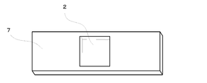
- FIG. 1 is a schematic cross-sectional view of a transparent adhesive tape laminate in which cells are encapsulated, showing a second embodiment of the present invention.
- the encapsulated cells are stratum corneum cells.
- the transparent adhesive tape laminated body 1 of this invention is stuck so that the mutual adhesion layer 6 of the adhesive tape 2 and the adhesive tape 3 may oppose.
- the stratum corneum cells 4 are encapsulated between the adhesive layer 6 and the adhesive layer 6.
- the manufacturing method of the transparent adhesive tape laminated body 1 of this invention and the observation method of the horny layer cell using a phase contrast microscope by the transparent adhesive tape laminated body 1 of this invention are demonstrated using FIG.
- FIG. 3 is a conceptual diagram showing an example of the steps of an observation method for stratum corneum cells using a phase contrast microscope.
- the transparent adhesive tape which the horny layer cell stuck to the adhesion layer is prepared.
- the transparent adhesive tape to which the stratum corneum cells are adhered may be prepared by any method, but it is convenient to use the tape strip kit 7 shown in FIG.
- the tape strip kit of FIG. 2 is made of a plastic plate and has a sampling hole in the center. The sampling hole is attached with an adhesive tape from the back side of the drawing, and the adhesive layer of the adhesive tape 2 is exposed at the sampling hole portion. Therefore, stratum corneum cells can be easily collected by pressing the collection hole of the tape strip kit 7 against the human skin.
- a transparent adhesive tape having stratum corneum cells adhered to the adhesive layer is prepared by pressing the tape strip kit 7 against a human cheek or the like.
- the step of pressing the tape strip kit 7 against a person's cheek is not included in the present invention.
- the adhesive layer of the transparent adhesive tape to which the horny layer cells are attached and the adhesive layer of another transparent adhesive tape are attached to prepare a transparent adhesive tape laminate in which the horny layer cells are encapsulated.
- the transparent adhesive tape in which the horny layer cells 4 are adhered to the adhesive layer is made to oppose the adhesive layer and the adhesive layer of another adhesive tape 3, and adheres the adhesive layers to each other.
- the existing horny layer cells 4 are encapsulated.
- the transparent adhesive tape laminated body 1 which is the 2nd aspect of this invention can be manufactured.
- the prepared laminate is observed with a phase contrast microscope.
- the transparent adhesive tape laminate 1 of the present invention is observed with the phase contrast microscope 9
- the horny layer cells can be observed by a simple method without using an encapsulating liquid or a cover glass.
- Example 1 Using the tape stripping kit (plastic plate-like member) shown in FIG. 2, stratum corneum cells were collected from the subject's cheek.
- the adhesive tape used in the tape stripping kit shown in FIG. 2 is a Diahalo tape (Mitsubishi Resin Co., Ltd.) with a refractive index of 1.48 to 1.50, a linear light transmittance of 92.374%, and a turbidity HAZE of 0.55%. Met.
- the linear transmittance (parallel transmittance) and turbidity HAZE of light are average values obtained by measuring the cross-sectional direction of the Diahalo tape five times using Nippon Denshoku Industries Co., Ltd. HASE METER NDH500. is there.
- the collected stratum corneum cells were attached using the same adhesive tape so that the adhesive layer and the adhesive layer were opposed to each other to enclose the stratum corneum cells, thereby preparing an adhesive tape laminate 1. At this time, when visually confirmed, air bubbles were not mixed.
- Example 2 The stratum corneum cells were observed with a phase contrast microscope in the same manner as in Example 1 except that the adhesive tape was changed to the 3M Scotch transparent beautiful color. No halo was observed, and the stratum corneum cells could be clearly observed. It was. The linear transmittance of light of 3M Scotch transparent beautiful color was 91.882%, and turbidity HAZE was 0.82%.
- Example 3 When the horny layer cells were observed with a phase contrast microscope in the same manner as in Example 1 except that the adhesive tape was changed to a cello tape made by Nichiban, the horny layer cells could be clearly observed without the occurrence of halo.
- the linear transmittance of light of the cellophane manufactured by Nichiban Co. was 89.608%, and the turbidity HAZE was 2.31%.
- Example 4 When corneal cells were observed with a phase contrast microscope in the same manner as in Example 1 except that the adhesive tape was changed to Scotch 313 manufactured by 3M, horny cells were clearly observed without the occurrence of halos. Note that the light transmittance of Scotch 313 manufactured by 3M was 92.30%, and the turbidity HAZE was 0.88%.
- Example 5 When the stratum corneum was observed with a phase contrast microscope in the same manner as in Example 1 except that the adhesive tape was changed to Scotch 3450 manufactured by 3M, the stratum corneum could be clearly observed without the occurrence of halo. In addition, the linear transmittance of light of Scotch 3450 manufactured by 3M was 91.76%, and the turbidity HAZE was 1.29%.
- Example 6 The stratum corneum cells were observed with a phase contrast microscope in the same manner as in Example 1 except that the adhesive tape was changed to Sekisui Chemical Co., Ltd. transparent packaging tape P83T. I was able to observe. In addition, Sekisui Chemical Co., Ltd. transparent packaging tape P83T had a linear light transmittance of 92.45% and a turbidity HAZE of 0.48%.
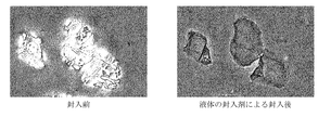
- the stratum corneum cells were observed by a method which is a method commonly used in phase contrast microscope observation. After collecting the stratum corneum cells using the tape stripping kit shown in FIG. 2, a drop of the liquid encapsulant was dropped into the collection hole and encapsulated using a cover glass. The observation photograph of a horny layer cell is shown in FIG. Even with a simple method using the adhesive tape of the present invention, stratum corneum cells could be clearly observed in the same manner as that conventionally performed by phase contrast microscopy.
- the encapsulation method, observation method, and adhesive tape laminate of the present invention enable a salesperson to easily observe stratum corneum cells when selecting cosmetics at a department store booth, a store, or a customer of a cosmetic user. Based on this observation result, an appropriate cosmetic can be selected and sold.
Landscapes
- Health & Medical Sciences (AREA)
- Physics & Mathematics (AREA)
- Life Sciences & Earth Sciences (AREA)
- Heart & Thoracic Surgery (AREA)
- Veterinary Medicine (AREA)
- Engineering & Computer Science (AREA)
- Medical Informatics (AREA)
- Molecular Biology (AREA)
- Surgery (AREA)
- Animal Behavior & Ethology (AREA)
- General Health & Medical Sciences (AREA)
- Public Health (AREA)
- Biomedical Technology (AREA)
- Chemical & Material Sciences (AREA)
- Analytical Chemistry (AREA)
- Pathology (AREA)
- General Physics & Mathematics (AREA)
- Optics & Photonics (AREA)
- Sampling And Sample Adjustment (AREA)
- Adhesives Or Adhesive Processes (AREA)
- Investigating Or Analysing Biological Materials (AREA)
- Microscoopes, Condenser (AREA)
Description
本発明は、位相差顕微鏡での観察に用いるための細胞の封入方法、および位相差顕微鏡を用いた、新たな細胞の観察方法に関する。
肌の状態に適した化粧料を選択する際に、その適用される肌の特性を知ることは非常に重要なことである。このような肌の特性を知るための方法として、角層細胞を用いる方法が検討されている。角層細胞は採取が容易であり、肌情報を多く有していることから、採取した角層細胞を観察し、得られた情報を化粧料の選択に利用しようとする試みが提案されている(例えば、特許文献1参照)。
特許文献1では、皮膚より採取した角層細胞を色素で染色し、光学顕微鏡によって角層細胞を観察することで、この結果を皮膚バリア機能が良好か否かを鑑別し、化粧料の選択に適用することが記載されている。
光学顕微鏡を用いる場合、細胞のような無色透明な試料を観察するために、染色液を用いる必要があり、特許文献1においてもズダンブラックBが用いられている。しかしながら、生きたまま試料を観察したい場合には、染色することで試料を殺してしまう場合があり、このような場合には位相差顕微鏡を用いることで、無色透明に近い試料を染色することなく観察が可能となる(非特許文献1参照)。
"位相差顕微鏡"、科学のつまみ食い[平成23年8月26日検索]、インターネット<URL:http://www.rikagaku.info/microscope9908/microscope07.htm>
肌状態のカウンセリングやアドバイスを行って化粧品の選択が行われるのは、主にデパートのブースや店舗または化粧品ユーザーの客先であることから、簡便に角層細胞の観察ができることが好ましい。上記位相差顕微鏡を用いると、無色透明な試料を観察することが可能となり、染色液を使用する必要がなくなることから、簡便な角層細胞の観察には好ましい方法である。
一方、位相差顕微鏡を用いて角層などの各種細胞を観察する場合、細胞を封入することなくそのまま観察すると、観察像上の物体の周辺に「ハロー」と呼ばれる光輪が現れる。そのため、位相差顕微鏡を用いて細胞を観察する場合には、液体の封入剤及びカバーガラスにより細胞を封入する必要がある。このような封入作業は、実際に肌の角層細胞を観察し、その結果に基づいて化粧料を選択するデパートのブースや店舗または化粧品ユーザーの客先では手間がかかり、また、カバーガラスが薄く割れやすいなど作業の難易度も高い。本発明はこのような状況下行われたものであり、デパートのブースや店舗または化粧品ユーザーの客先において販売員でも簡単に取り扱うことができる細胞の封入方法、および位相差顕微鏡を用いた細胞の観察方法を提供するものである。
本発明者らは、上記課題を解決すべく簡単な封入法を求め鋭意研究を行ったところ、粘着層を有する粘着テープを用い、該粘着テープにより両側から細胞を挟持貼着するという簡便な方法を採用することで問題を解決できることを見出し、本発明を完成させた。
即ち本発明は以下のとおりである。
(1)位相差顕微鏡での観察に用いるための細胞の封入方法であって、
透明粘着テープの粘着層と透明粘着テープの粘着層との間に細胞を配置し、該2つの粘着テープの粘着層で細胞を挟持貼着することを特徴とする、細胞の封入方法。
(2)前記透明粘着テープは、光の直線透過率が80%以上である、(1)に記載の細胞の封入方法。
(3)前記透明粘着テープの光屈折率と前記細胞の光屈折率との差が0.03以上である、(1)または(2)に記載の細胞の封入方法。
(4)前記2つの透明粘着テープの間に気泡を有しない、(1)~(3)のいずれかに記載の細胞の封入方法。
(5)前記細胞が角層細胞である(1)~(4)のいずれかに記載の細胞の封入方法。
(6)位相差顕微鏡を用いた細胞の観察方法であって、
細胞が粘着層に貼着した透明粘着テープを準備する工程、
前記細胞が貼着した透明粘着テープの粘着層と、別の透明粘着テープの粘着層とを貼着し、細胞が封入された透明粘着テープ積層体を準備する工程、および
前記工程で準備した積層体を位相差顕微鏡で観察する工程、
を含む、位相差顕微鏡を用いた細胞の観察方法。
(7)フィルム層と粘着層からなる透明粘着テープを、互いの粘着層が対向するように貼着した透明粘着テープ積層体であって、粘着層と粘着層との間に細胞が封入された透明粘着テープ積層体。
(8)位相差顕微鏡の観察に用いられる、(7)に記載の透明粘着テープ積層体。
本発明の細胞の封入方法により、細胞が封入された粘着テープ積層体を簡便に作成することが可能となり、専門的なスキルを有しなくても、デパートのブースや店舗または化粧品ユーザーの客先で販売員が簡単に角層細胞などを観察することができる。また、本発明の細胞を封入した粘着テープ積層体は、位相差顕微鏡を用いて観察した際にハローが発生せず、角層細胞を観察する場合には十分に角層細胞内のメラニン、核、配列規則性などの観察ができるため、簡便かつ即時に角層細胞からの情報を得ることが可能となる。
本発明の第一の態様は、位相差顕微鏡での観察に用いるための細胞の封入方法である。本発明の封入方法は、位相差顕微鏡で細胞を観察することを可能とするため、透明粘着テープの粘着層と透明粘着テープの粘着層との間に細胞を配置し、該2つの粘着テープの粘着層で挟持貼着するものである。本発明で観察する細胞は特に限定されるものではないが、特に角層細胞、角化細胞(ケラチノサイト)、線維芽細胞などの皮膚の状態を評価するために使用する細胞が好ましく例示される。以下、角層細胞を例に挙げて説明をする。
角層細胞は、角層を構成する厚さ1μm程度の扁平な細胞であり、肌情報を多く有していることから、角層細胞を評価することで化粧料の選択の際に考慮すべき肌の特性を知ることが可能となる。角層細胞内には主要な成分であるケラチンが繊維を形成して充満し、きわめて強固な細胞構造を構築しており、外界のさまざまな刺激から皮膚内部を守っている。しかしながら角層最外層においては、角層細胞同士の接着力が減弱するため容易に採取が可能となる。本発明では、既に人から取得した角層細胞を使用するため、人から角層細胞を採取する工程は本発明の範囲には含まれない。
本発明は位相差顕微鏡での観察に用いるための細胞の封入方法である。位相差顕微鏡は、光線の位相差をコントラストに変換して観察できる光学顕微鏡であり、標本を無染色・非侵襲的に観察することができるものである。
本発明は、細胞を封入するために透明粘着テープを用いることを特徴とするものである。本発明で透明とは、光を透過することが可能であり、先のものが見えるものを意味し、位相差顕微鏡にて対象が観察されるかぎりその程度や着色については何ら限定されるものではない。光の直線透過率が80%以上であるものが好ましく、90%以上であるものがより好ましい。また、透明粘着テープは光学特性の濁度HAZEが1.5%未満であるものが好ましく、1.0%未満であるものがより好ましい。また、無色であることが好ましい。なお、濁度HAZE(曇度)とは、物体の曇りの度合いを示す値(%)で、数値が小さいほど透明粘着テープに曇りが無く透明性が高いことを示す。(濁度HAZE(%)=Td/Tt × 100(Td:拡散透過率 Tt:全光線透過率)また、「光の直線透過率(平行透過率)」及び「濁度HAZE(曇度)」は、日本工業規格JISK7136(国際標準化機構規格ISO14782)、JISK7361(国際標準化機構規格ISO13468)に基づいて測定される値を表し、例えば日本電色工業株式会社製HASE METER NDH500を用いて測定することができる。但し、「光の直線透過率」及び「濁度HAZE」は、透明粘着テープの断面方向について測定した値を表すものとする。
本発明に用いる粘着テープは、透明なフィルム層および粘着層からなる、一般的な粘着テープを用いることができ、市販されているものを用いることができる。具体的にはScotch(登録商標)透明美色(3M社製)、Scotch(登録商標)313(3M社製)、Scotch(登録商標)3450(3M社製)、Scotch(登録商標)PRO(3M社製)、ダイアハロー(登録商標)(三菱樹脂社製)、セロテープ(登録商標)(ニチバン社製)、ダンプロン(登録商標)(日東電工社製)、セキスイオリエン・セキスイカットロン・セキスイタフライト・セキスイエバーセル・セキスイシュープリーム(登録商標)(積水化学社製)、などが挙げられる。
これらのうち、位相差顕微鏡の特性を考慮すると、細胞の屈折率と異なる屈折率を有する粘着テープを用いることが好ましく、より好ましくは、粘着テープの屈折率と細胞の屈折率との差が0.03以上である粘着テープを用いることが好ましい。屈折率の上限は特段限定されないが、通常0.60以下である。なお、角層細胞の屈折率は、1.55である。
これらのうち、位相差顕微鏡の特性を考慮すると、細胞の屈折率と異なる屈折率を有する粘着テープを用いることが好ましく、より好ましくは、粘着テープの屈折率と細胞の屈折率との差が0.03以上である粘着テープを用いることが好ましい。屈折率の上限は特段限定されないが、通常0.60以下である。なお、角層細胞の屈折率は、1.55である。
粘着テープのフィルム層の材質は、上記透明である要件を満たせば特段限定されず、また、粘着層の材質についても特段限定はされない。フィルム層の材質は、一般的にはポリエチレン、ポリプロピレン、セロハン(化学名:セルロース)、ポリエチレンテレフタラート(PET)、ポリオレフィン、などが用いられ、粘着層の材質は、一般的はアクリル系コポリマー、オレフィン系コポリマー、ウレタン系コポリマー、エポキシ系コポリマー、合成ゴム、天然ゴムなどが用いられる。また、粘着テープの厚さについても特段限定されず、位相差顕微鏡で観察可能であればよい。
本発明の封入方法では、2つの粘着テープの粘着層の間に細胞を配置し、そして2つの粘着テープの粘着層で細胞を挟持貼着することを特徴とする。このように、粘着テープの間に細胞を挟持し貼着することで、液体の封入剤及びカバーガラスを用いなくとも、ハローが発生することなく、位相差顕微鏡による細胞の観察が可能となる。
本発明の封入方法では、2つの粘着テープの粘着層の間に、気泡を有しないことが好ましい。粘着層の間に気泡が入る場合には、その気泡の位置によっては、気泡(空気)による屈折率の変化によりハローが生じる場合がある。
本発明の封入方法により細胞が封入された透明粘着テープ積層体は、本発明の第二の態様である。すなわち、本発明の第二の態様は、フィルム層と粘着層からなる透明粘着テープを、互いの粘着層が対向するように貼着した透明粘着テープ積層体であって、粘着層と粘着層との間に細胞が封入された透明粘着テープ積層体である。
また、本発明の第三の態様は、位相差顕微鏡を用いた細胞の観察方法であって、細胞が粘着層に貼着した透明粘着テープを準備する工程、前記細胞が貼着した透明粘着テープの粘着層と、別の透明粘着テープの粘着層とを貼着し、細胞が封入された透明粘着テープ積層体を準備する工程、および、前記工程で準備した積層体を位相差顕微鏡で観察する工程、を含む、位相差顕微鏡を用いた細胞の観察方法。
以下、図を用いてこれら本発明の第一の態様乃至は第三の態様を説明する。
以下、図を用いてこれら本発明の第一の態様乃至は第三の態様を説明する。
図1は、本発明の第二の態様を示す、細胞を封入した透明粘着テープ積層体の断面模式図である。なお、封入した細胞は角層細胞である。
本発明の透明粘着テープ積層体1は、粘着テープ2と粘着テープ3の互いの粘着層6が対向するように貼着されている。そして、粘着層6と粘着層6との間に、角層細胞4が封入されたものである。本発明の透明粘着テープ積層体1の製造方法、および本発明の透明粘着テープ積層体1による、位相差顕微鏡を用いた角層細胞の観察方法を、図3を用いて説明する。
本発明の透明粘着テープ積層体1は、粘着テープ2と粘着テープ3の互いの粘着層6が対向するように貼着されている。そして、粘着層6と粘着層6との間に、角層細胞4が封入されたものである。本発明の透明粘着テープ積層体1の製造方法、および本発明の透明粘着テープ積層体1による、位相差顕微鏡を用いた角層細胞の観察方法を、図3を用いて説明する。
図3は、位相差顕微鏡を用いた角層細胞の観察方法の工程の一例を示す概念図である。まず、本発明では、角層細胞が粘着層に貼着した透明粘着テープを準備する。角層細胞が貼着した透明粘着テープはどのような方法によって準備してもよいが、図2に示すテープストリップキット7を用いると簡便である。図2のテープストリップキットはプラスチック材質の板からなり、中央部に採取孔を有する。採取孔は図面奥側から粘着テープが貼られており、採取孔部分は粘着テープ2の粘着層が露出している。そのため、テープストリップキット7の採取孔を人の皮膚に押し当てることで、簡単に角層細胞が採取可能である。
図3の(a)に示すように、人のホホなどにテープストリップキット7を押し当てることにより、角層細胞が粘着層に貼着した透明粘着テープを準備する。なお、テープストリップキット7を人のホホに押し当てる工程は、本発明には含まれない。
次に、角層細胞が貼着した透明粘着テープの粘着層と、別の透明粘着テープの粘着層とを貼着し、角層細胞が封入された透明貼着テープ積層体を準備する。角層細胞4を粘着層に貼着した透明粘着テープは、図3の(b)に示すように、その粘着層と別の粘着テープ3の粘着層を対向させ、粘着層同士を貼着することで、存在する角層細胞4を封入する。この際、気泡が入らないように封入することが好ましい。このようにして、本発明の第二の態様である、透明粘着テープ積層体1を製造することができる。
次に、図3の(c)に示すように、準備した積層体を位相差顕微鏡で観察する。本発明の透明粘着テープ積層体1を位相差顕微鏡9で観察すると、封入液やカバーガラスを使用しなくても、簡便な方法で角層細胞が観察可能となる。
以下、実施例と比較例を挙げて、本発明を更に詳細に説明するが、以下の実施例に示す具体的な形態にのみ限定的に解釈されることはない。
<実施例1>
図2に示すテープストリッピングキット(プラスチックの板状部材)を用いて、被験者の頬部より角層細胞を採取した。図2に示すテープストリッピングキットに用いた粘着テープは、ダイアハローテープ(三菱樹脂社製)であり、屈折率が1.48~1.50であり、光の直線透過率は92.374%、濁度HAZEは0.55%であった。なお、光の直線透過率(平行透過率)及び濁度HAZEは、日本電色工業株式会社製HASE METER NDH500を使用し、ダイアハローテープの断面方向を5回測定して得られた平均値である。
採取した角層細胞は、同じ粘着テープを用いて、粘着層と粘着層を対向させて貼着して角層細胞を封入し、粘着テープ積層体1を作製した。この際、目視により確認したところ、空気の気泡の混入はなかった。
粘着テープ積層体1を位相差顕微鏡に設置し、角層細胞を観察したところ、ハローの発生もなく、はっきりと角層細胞が観察できた。角層細胞の観察写真を図4に示す。
<実施例2>
粘着テープを3M社製のScotch透明美色に変更した以外は実施例1と同様にして、位相差顕微鏡で角層細胞を観察したところ、ハローの発生もなく、はっきりと角層細胞が観察できた。なお、3M社製Scotch透明美色の光の直線透過率は91.882%、濁度HAZEは0.82%であった。
粘着テープを3M社製のScotch透明美色に変更した以外は実施例1と同様にして、位相差顕微鏡で角層細胞を観察したところ、ハローの発生もなく、はっきりと角層細胞が観察できた。なお、3M社製Scotch透明美色の光の直線透過率は91.882%、濁度HAZEは0.82%であった。
<実施例3>
粘着テープをニチバン社製のセロテープに変更した以外は実施例1と同様にして、位相差顕微鏡で角層細胞を観察したところ、ハローの発生もなく、はっきりと角層細胞が観察できた。なお、ニチバン社製のセロテープの光の直線透過率は89.608%、濁度HAZEは2.31%であった。
粘着テープをニチバン社製のセロテープに変更した以外は実施例1と同様にして、位相差顕微鏡で角層細胞を観察したところ、ハローの発生もなく、はっきりと角層細胞が観察できた。なお、ニチバン社製のセロテープの光の直線透過率は89.608%、濁度HAZEは2.31%であった。
<実施例4>
粘着テープを3M社製のScotch313に変更した以外は実施例1と同様にして、位相差顕微鏡で角層細胞を観察したところ、ハローの発生もなく、はっきりと角層細胞が観察できた。なお、3M社製Scotch313の光の直線透過率は92.30%、濁度HAZEは0.88%であった。
粘着テープを3M社製のScotch313に変更した以外は実施例1と同様にして、位相差顕微鏡で角層細胞を観察したところ、ハローの発生もなく、はっきりと角層細胞が観察できた。なお、3M社製Scotch313の光の直線透過率は92.30%、濁度HAZEは0.88%であった。
<実施例5>
粘着テープを3M社製のScotch3450に変更した以外は実施例1と同様にして、位相差顕微鏡で角層細胞を観察したところ、ハローの発生もなく、はっきりと角層細胞が観察できた。なお、3M社製Scotch3450の光の直線透過率は91.76%、濁度HAZEは1.29%であった。
粘着テープを3M社製のScotch3450に変更した以外は実施例1と同様にして、位相差顕微鏡で角層細胞を観察したところ、ハローの発生もなく、はっきりと角層細胞が観察できた。なお、3M社製Scotch3450の光の直線透過率は91.76%、濁度HAZEは1.29%であった。
<実施例6>
粘着テープを積水化学社製の透明包装用テープP83Tに変更した以外は実施例1と同様にして、位相差顕微鏡で角層細胞を観察したところ、ハローの発生もなく、はっきりと角層細胞が観察できた。なお、積水化学社製透明包装用テープP83Tの光の直線透過率は92.45%、濁度HAZEは0.48%であった。
粘着テープを積水化学社製の透明包装用テープP83Tに変更した以外は実施例1と同様にして、位相差顕微鏡で角層細胞を観察したところ、ハローの発生もなく、はっきりと角層細胞が観察できた。なお、積水化学社製透明包装用テープP83Tの光の直線透過率は92.45%、濁度HAZEは0.48%であった。
<参考例>
位相差顕微鏡観察で通常行われている方法である方法で角層細胞の観察を行った。図2に示すテープストリッピングキットを用いて角層細胞を採取した後、液体の封入剤を採取孔に一滴落とし、カバーガラスを用いて封入した。角層細胞の観察写真を図5に示す。
本発明の粘着テープを用いた簡易的な方法でも、従来位相差顕微鏡観察で行われていた方法と同様に、はっきりと角層細胞が観察できた。
位相差顕微鏡観察で通常行われている方法である方法で角層細胞の観察を行った。図2に示すテープストリッピングキットを用いて角層細胞を採取した後、液体の封入剤を採取孔に一滴落とし、カバーガラスを用いて封入した。角層細胞の観察写真を図5に示す。
本発明の粘着テープを用いた簡易的な方法でも、従来位相差顕微鏡観察で行われていた方法と同様に、はっきりと角層細胞が観察できた。
本発明の封入方法、観察方法、粘着テープ積層体は、デパートのブースや店舗または化粧品ユーザーの客先において化粧料を選択する際に、販売員でも容易に角層細胞を観察することが可能となり、この観察結果に基づいて適切な化粧料を選択し、販売することができる。
1 透明粘着テープ積層体
2、3 粘着テープ
4 角層細胞
5 フィルム層
6 粘着層
7 テープストリップキット
8 顔
9 位相差顕微鏡
2、3 粘着テープ
4 角層細胞
5 フィルム層
6 粘着層
7 テープストリップキット
8 顔
9 位相差顕微鏡
Claims (8)
- 位相差顕微鏡での観察に用いるための細胞の封入方法であって、
透明粘着テープの粘着層と透明粘着テープの粘着層との間に細胞を配置し、該2つの粘着テープの粘着層で細胞を挟持貼着することを特徴とする、細胞の封入方法。 - 前記透明粘着テープは、光の直線透過率が80%以上である、請求項1に記載の細胞の封入方法。
- 前記透明粘着テープの光屈折率と前記細胞の光屈折率との差が0.03以上である、請求項1または2に記載の細胞の封入方法。
- 前記2つの透明粘着テープの間に気泡を有しないことを特徴とする、請求項1~3のいずれか1項に記載の細胞の封入方法。
- 前記細胞が角層細胞である請求項1~4のいずれか1項に記載の細胞の封入方法。
- 位相差顕微鏡を用いた細胞の観察方法であって、
細胞が粘着層に貼着した透明粘着テープを準備する工程、
前記細胞が貼着した透明粘着テープの粘着層と、別の透明粘着テープの粘着層とを貼着し、細胞が封入された透明貼着テープ積層体を準備する工程、および
前記工程で準備した積層体を位相差顕微鏡で観察する工程、
を含む、位相差顕微鏡を用いた細胞の観察方法。 - フィルム層と粘着層からなる透明粘着テープを、互いの粘着層が対向するように貼着した透明粘着テープ積層体であって、粘着層と粘着層との間に細胞が封入された透明粘着テープ積層体。
- 位相差顕微鏡の観察に用いられる、請求項7に記載の透明粘着テープ積層体。
Priority Applications (1)
| Application Number | Priority Date | Filing Date | Title |
|---|---|---|---|
| JP2013553210A JP6126536B2 (ja) | 2012-01-13 | 2012-12-04 | 細胞の封入方法及び細胞の観察方法 |
Applications Claiming Priority (2)
| Application Number | Priority Date | Filing Date | Title |
|---|---|---|---|
| JP2012005335 | 2012-01-13 | ||
| JP2012-005335 | 2012-01-13 |
Publications (1)
| Publication Number | Publication Date |
|---|---|
| WO2013105363A1 true WO2013105363A1 (ja) | 2013-07-18 |
Family
ID=48754720
Family Applications (1)
| Application Number | Title | Priority Date | Filing Date |
|---|---|---|---|
| PCT/JP2012/081328 Ceased WO2013105363A1 (ja) | 2012-01-13 | 2012-12-04 | 細胞の封入方法及び細胞の観察方法 |
Country Status (4)
| Country | Link |
|---|---|
| JP (1) | JP6126536B2 (ja) |
| CN (2) | CN103207448B (ja) |
| TW (1) | TWI624654B (ja) |
| WO (1) | WO2013105363A1 (ja) |
Families Citing this family (4)
| Publication number | Priority date | Publication date | Assignee | Title |
|---|---|---|---|---|
| JP6126536B2 (ja) * | 2012-01-13 | 2017-05-10 | ポーラ化成工業株式会社 | 細胞の封入方法及び細胞の観察方法 |
| CN108369161B (zh) * | 2015-11-05 | 2021-10-01 | 株式会社资生堂 | 角质层采集工具和角质层采集检测用试剂盒 |
| CN105739076A (zh) * | 2016-04-08 | 2016-07-06 | 苏州雅睿生物技术有限公司 | 一种载玻片 |
| CN107991159A (zh) * | 2017-11-20 | 2018-05-04 | 珠海伊斯佳科技股份有限公司 | 一种通过皮肤角质细胞形态评估皮肤状况的方法 |
Citations (10)
| Publication number | Priority date | Publication date | Assignee | Title |
|---|---|---|---|---|
| JPS5943358A (ja) * | 1982-09-03 | 1984-03-10 | Kanebo Ltd | 皮膚表面形態の検査方法及び検査用具 |
| JPS6348137U (ja) * | 1986-09-17 | 1988-04-01 | ||
| JPS6453165A (en) * | 1987-08-24 | 1989-03-01 | Shiseido Co Ltd | Preparation of cutaneous cell sample |
| JPH0527670U (ja) * | 1991-04-26 | 1993-04-09 | 株式会社テクノメデイカ | ぎよう虫卵検査紙 |
| JPH11344489A (ja) * | 1998-06-02 | 1999-12-14 | Shiseido Co Ltd | 角層のメラニンの定量方法 |
| JP2000005134A (ja) * | 1998-06-17 | 2000-01-11 | Pola Chem Ind Inc | 角層標本の作成方法 |
| WO2002025272A1 (en) * | 2000-09-21 | 2002-03-28 | Shiseido Company, Ltd. | Method of evaluating the degree of maturity of corneous cells |
| JP2005168693A (ja) * | 2003-12-10 | 2005-06-30 | Pola Chem Ind Inc | 角層細胞の形状の鑑別方法 |
| JP2009115813A (ja) * | 2008-12-26 | 2009-05-28 | Tokiwa Yakuhin Kogyo Kk | アトピー性皮膚炎の局所病態の非侵襲的評価方法 |
| JP2009257954A (ja) * | 2008-04-17 | 2009-11-05 | Hitachi Chem Co Ltd | 光学顕微鏡観察用試料の調整方法及びそれを用いた、粒子の凝集状態・分散状態の観察方法、撮影方法 |
Family Cites Families (3)
| Publication number | Priority date | Publication date | Assignee | Title |
|---|---|---|---|---|
| JP2005249410A (ja) * | 2004-03-01 | 2005-09-15 | Menicon Co Ltd | 生体組織検査シートおよびそれを用いた生体組織検査方法 |
| EP1882040A4 (en) * | 2005-05-20 | 2008-06-11 | Premd Inc | DIRECT DETERMINATION OF SKIN PROTEIN IN SKIN SAMPLES TESTED BY TAPE-WRAPPING |
| JP6126536B2 (ja) * | 2012-01-13 | 2017-05-10 | ポーラ化成工業株式会社 | 細胞の封入方法及び細胞の観察方法 |
-
2012
- 2012-12-04 JP JP2013553210A patent/JP6126536B2/ja active Active
- 2012-12-04 WO PCT/JP2012/081328 patent/WO2013105363A1/ja not_active Ceased
-
2013
- 2013-01-11 TW TW102101011A patent/TWI624654B/zh active
- 2013-01-14 CN CN201310012849.6A patent/CN103207448B/zh active Active
- 2013-01-14 CN CN2013200171345U patent/CN203324569U/zh not_active Expired - Lifetime
Patent Citations (10)
| Publication number | Priority date | Publication date | Assignee | Title |
|---|---|---|---|---|
| JPS5943358A (ja) * | 1982-09-03 | 1984-03-10 | Kanebo Ltd | 皮膚表面形態の検査方法及び検査用具 |
| JPS6348137U (ja) * | 1986-09-17 | 1988-04-01 | ||
| JPS6453165A (en) * | 1987-08-24 | 1989-03-01 | Shiseido Co Ltd | Preparation of cutaneous cell sample |
| JPH0527670U (ja) * | 1991-04-26 | 1993-04-09 | 株式会社テクノメデイカ | ぎよう虫卵検査紙 |
| JPH11344489A (ja) * | 1998-06-02 | 1999-12-14 | Shiseido Co Ltd | 角層のメラニンの定量方法 |
| JP2000005134A (ja) * | 1998-06-17 | 2000-01-11 | Pola Chem Ind Inc | 角層標本の作成方法 |
| WO2002025272A1 (en) * | 2000-09-21 | 2002-03-28 | Shiseido Company, Ltd. | Method of evaluating the degree of maturity of corneous cells |
| JP2005168693A (ja) * | 2003-12-10 | 2005-06-30 | Pola Chem Ind Inc | 角層細胞の形状の鑑別方法 |
| JP2009257954A (ja) * | 2008-04-17 | 2009-11-05 | Hitachi Chem Co Ltd | 光学顕微鏡観察用試料の調整方法及びそれを用いた、粒子の凝集状態・分散状態の観察方法、撮影方法 |
| JP2009115813A (ja) * | 2008-12-26 | 2009-05-28 | Tokiwa Yakuhin Kogyo Kk | アトピー性皮膚炎の局所病態の非侵襲的評価方法 |
Also Published As
| Publication number | Publication date |
|---|---|
| CN103207448A (zh) | 2013-07-17 |
| JPWO2013105363A1 (ja) | 2015-05-11 |
| CN103207448B (zh) | 2017-11-24 |
| JP6126536B2 (ja) | 2017-05-10 |
| TW201333441A (zh) | 2013-08-16 |
| TWI624654B (zh) | 2018-05-21 |
| CN203324569U (zh) | 2013-12-04 |
Similar Documents
| Publication | Publication Date | Title |
|---|---|---|
| JP6126536B2 (ja) | 細胞の封入方法及び細胞の観察方法 | |
| US11696723B2 (en) | Devices, systems, and methods for monitoring hair | |
| JP2020128995A (ja) | ステップが簡略化され、試料が少なく、スピードアップし、使いやすい、生化学的/化学的なアッセイ装置及び方法 | |
| US20170224257A1 (en) | Epidermal Photonic Systems and Methods | |
| US8403582B2 (en) | Apparatus for treating a stain in clothing | |
| FR2831789A1 (fr) | Procede et dispositif pour l'evaluation de la secheresse cutanee notamment | |
| KR20090003188U (ko) | 인조속눈썹 | |
| US11448571B2 (en) | Specimen collection tip, specimen preparation container and specimen preparation kit | |
| CN102511005B (zh) | 测试带堆叠以及其制造方法 | |
| CN206792410U (zh) | 一种基于眼部识别的疲劳监测装置 | |
| Ma et al. | A taper-in-taper fiber optic biosensor based on Mach-Zehnder interferometer for human sweat pH detection | |
| CN117529274A (zh) | 皮肤表征方法和装备 | |
| JP7356326B2 (ja) | 皮膚成分の分析方法 | |
| CN107773275A (zh) | 一种皮肤检测采样包及其应用 | |
| JP2014198259A (ja) | メイク化粧料の塗布色チェック用具および塗布色チェック方法 | |
| FR2719830A1 (fr) | Paquet-gants. | |
| SE520948C2 (sv) | Allergitestelement | |
| JP2012061213A (ja) | メイク化粧料の塗布色チェック用具および塗布色チェック方法 | |
| US20190110737A1 (en) | Multiple well epicutaneous test patch array | |
| JPH11344489A (ja) | 角層のメラニンの定量方法 | |
| CN1100254C (zh) | 蛲虫卵取卵样用具 | |
| HK40105052A (zh) | 皮肤表徵方法和装备 | |
| Bloks | “Reconstructing” a colour theory: Unraveling the theoretical, material and methodological assumptions underlying James Sowerby’s" A New Elucidation of Colours"(1809) | |
| JP6330232B1 (ja) | ラベルおよびラベル貼付物並びに検体容器収納ケースおよび検体採取用キット | |
| Lee et al. | Gamma globulins-induced interaction between two red blood cells: forces measurement with optical tweezers |
Legal Events
| Date | Code | Title | Description |
|---|---|---|---|
| 121 | Ep: the epo has been informed by wipo that ep was designated in this application |
Ref document number: 12864883 Country of ref document: EP Kind code of ref document: A1 |
|
| ENP | Entry into the national phase |
Ref document number: 2013553210 Country of ref document: JP Kind code of ref document: A |
|
| 122 | Ep: pct application non-entry in european phase |
Ref document number: 12864883 Country of ref document: EP Kind code of ref document: A1 |
|
| NENP | Non-entry into the national phase |
Ref country code: DE |