WO2014005383A1 - Matériau de radiographie invisible pour le diagnostic précoce d'une tumeur et procédé de préparation associé - Google Patents
Matériau de radiographie invisible pour le diagnostic précoce d'une tumeur et procédé de préparation associé Download PDFInfo
- Publication number
- WO2014005383A1 WO2014005383A1 PCT/CN2012/082578 CN2012082578W WO2014005383A1 WO 2014005383 A1 WO2014005383 A1 WO 2014005383A1 CN 2012082578 W CN2012082578 W CN 2012082578W WO 2014005383 A1 WO2014005383 A1 WO 2014005383A1
- Authority
- WO
- WIPO (PCT)
- Prior art keywords
- contrast agent
- cancer
- early diagnosis
- contrast
- invisible
- Prior art date
- Legal status (The legal status is an assumption and is not a legal conclusion. Google has not performed a legal analysis and makes no representation as to the accuracy of the status listed.)
- Ceased
Links
Images


Classifications
-
- A—HUMAN NECESSITIES
- A61—MEDICAL OR VETERINARY SCIENCE; HYGIENE
- A61K—PREPARATIONS FOR MEDICAL, DENTAL OR TOILETRY PURPOSES
- A61K49/00—Preparations for testing in vivo
- A61K49/001—Preparation for luminescence or biological staining
- A61K49/0063—Preparation for luminescence or biological staining characterised by a special physical or galenical form, e.g. emulsions, microspheres
- A61K49/0069—Preparation for luminescence or biological staining characterised by a special physical or galenical form, e.g. emulsions, microspheres the agent being in a particular physical galenical form
- A61K49/0089—Particulate, powder, adsorbate, bead, sphere
- A61K49/0091—Microparticle, microcapsule, microbubble, microsphere, microbead, i.e. having a size or diameter higher or equal to 1 micrometer
- A61K49/0093—Nanoparticle, nanocapsule, nanobubble, nanosphere, nanobead, i.e. having a size or diameter smaller than 1 micrometer, e.g. polymeric nanoparticle
-
- A—HUMAN NECESSITIES
- A61—MEDICAL OR VETERINARY SCIENCE; HYGIENE
- A61K—PREPARATIONS FOR MEDICAL, DENTAL OR TOILETRY PURPOSES
- A61K49/00—Preparations for testing in vivo
- A61K49/001—Preparation for luminescence or biological staining
- A61K49/0013—Luminescence
- A61K49/0017—Fluorescence in vivo
- A61K49/0019—Fluorescence in vivo characterised by the fluorescent group, e.g. oligomeric, polymeric or dendritic molecules
- A61K49/0021—Fluorescence in vivo characterised by the fluorescent group, e.g. oligomeric, polymeric or dendritic molecules the fluorescent group being a small organic molecule
- A61K49/0032—Methine dyes, e.g. cyanine dyes
-
- A—HUMAN NECESSITIES
- A61—MEDICAL OR VETERINARY SCIENCE; HYGIENE
- A61K—PREPARATIONS FOR MEDICAL, DENTAL OR TOILETRY PURPOSES
- A61K49/00—Preparations for testing in vivo
- A61K49/001—Preparation for luminescence or biological staining
- A61K49/0013—Luminescence
- A61K49/0017—Fluorescence in vivo
- A61K49/0019—Fluorescence in vivo characterised by the fluorescent group, e.g. oligomeric, polymeric or dendritic molecules
- A61K49/0021—Fluorescence in vivo characterised by the fluorescent group, e.g. oligomeric, polymeric or dendritic molecules the fluorescent group being a small organic molecule
- A61K49/0041—Xanthene dyes, used in vivo, e.g. administered to a mice, e.g. rhodamines, rose Bengal
- A61K49/0043—Fluorescein, used in vivo
-
- A—HUMAN NECESSITIES
- A61—MEDICAL OR VETERINARY SCIENCE; HYGIENE
- A61K—PREPARATIONS FOR MEDICAL, DENTAL OR TOILETRY PURPOSES
- A61K49/00—Preparations for testing in vivo
- A61K49/04—X-ray contrast preparations
- A61K49/0409—Physical forms of mixtures of two different X-ray contrast-enhancing agents, containing at least one X-ray contrast-enhancing agent which is not a halogenated organic compound
- A61K49/0414—Particles, beads, capsules or spheres
-
- A—HUMAN NECESSITIES
- A61—MEDICAL OR VETERINARY SCIENCE; HYGIENE
- A61K—PREPARATIONS FOR MEDICAL, DENTAL OR TOILETRY PURPOSES
- A61K49/00—Preparations for testing in vivo
- A61K49/06—Nuclear magnetic resonance [NMR] contrast preparations; Magnetic resonance imaging [MRI] contrast preparations
- A61K49/08—Nuclear magnetic resonance [NMR] contrast preparations; Magnetic resonance imaging [MRI] contrast preparations characterised by the carrier
- A61K49/10—Organic compounds
- A61K49/101—Organic compounds the carrier being a complex-forming compound able to form MRI-active complexes with paramagnetic metals
- A61K49/103—Organic compounds the carrier being a complex-forming compound able to form MRI-active complexes with paramagnetic metals the complex-forming compound being acyclic, e.g. DTPA
- A61K49/105—Organic compounds the carrier being a complex-forming compound able to form MRI-active complexes with paramagnetic metals the complex-forming compound being acyclic, e.g. DTPA the metal complex being Gd-DTPA
-
- A—HUMAN NECESSITIES
- A61—MEDICAL OR VETERINARY SCIENCE; HYGIENE
- A61K—PREPARATIONS FOR MEDICAL, DENTAL OR TOILETRY PURPOSES
- A61K49/00—Preparations for testing in vivo
- A61K49/06—Nuclear magnetic resonance [NMR] contrast preparations; Magnetic resonance imaging [MRI] contrast preparations
- A61K49/18—Nuclear magnetic resonance [NMR] contrast preparations; Magnetic resonance imaging [MRI] contrast preparations characterised by a special physical form, e.g. emulsions, microcapsules, liposomes
- A61K49/1818—Nuclear magnetic resonance [NMR] contrast preparations; Magnetic resonance imaging [MRI] contrast preparations characterised by a special physical form, e.g. emulsions, microcapsules, liposomes particles, e.g. uncoated or non-functionalised microparticles or nanoparticles
- A61K49/1821—Nuclear magnetic resonance [NMR] contrast preparations; Magnetic resonance imaging [MRI] contrast preparations characterised by a special physical form, e.g. emulsions, microcapsules, liposomes particles, e.g. uncoated or non-functionalised microparticles or nanoparticles coated or functionalised microparticles or nanoparticles
- A61K49/1824—Nuclear magnetic resonance [NMR] contrast preparations; Magnetic resonance imaging [MRI] contrast preparations characterised by a special physical form, e.g. emulsions, microcapsules, liposomes particles, e.g. uncoated or non-functionalised microparticles or nanoparticles coated or functionalised microparticles or nanoparticles coated or functionalised nanoparticles
- A61K49/1827—Nuclear magnetic resonance [NMR] contrast preparations; Magnetic resonance imaging [MRI] contrast preparations characterised by a special physical form, e.g. emulsions, microcapsules, liposomes particles, e.g. uncoated or non-functionalised microparticles or nanoparticles coated or functionalised microparticles or nanoparticles coated or functionalised nanoparticles having a (super)(para)magnetic core, being a solid MRI-active material, e.g. magnetite, or composed of a plurality of MRI-active, organic agents, e.g. Gd-chelates, or nuclei, e.g. Eu3+, encapsulated or entrapped in the core of the coated or functionalised nanoparticle
- A61K49/1833—Nuclear magnetic resonance [NMR] contrast preparations; Magnetic resonance imaging [MRI] contrast preparations characterised by a special physical form, e.g. emulsions, microcapsules, liposomes particles, e.g. uncoated or non-functionalised microparticles or nanoparticles coated or functionalised microparticles or nanoparticles coated or functionalised nanoparticles having a (super)(para)magnetic core, being a solid MRI-active material, e.g. magnetite, or composed of a plurality of MRI-active, organic agents, e.g. Gd-chelates, or nuclei, e.g. Eu3+, encapsulated or entrapped in the core of the coated or functionalised nanoparticle having a (super)(para)magnetic core coated or functionalised with a small organic molecule
-
- A—HUMAN NECESSITIES
- A61—MEDICAL OR VETERINARY SCIENCE; HYGIENE
- A61K—PREPARATIONS FOR MEDICAL, DENTAL OR TOILETRY PURPOSES
- A61K49/00—Preparations for testing in vivo
- A61K49/06—Nuclear magnetic resonance [NMR] contrast preparations; Magnetic resonance imaging [MRI] contrast preparations
- A61K49/18—Nuclear magnetic resonance [NMR] contrast preparations; Magnetic resonance imaging [MRI] contrast preparations characterised by a special physical form, e.g. emulsions, microcapsules, liposomes
- A61K49/1818—Nuclear magnetic resonance [NMR] contrast preparations; Magnetic resonance imaging [MRI] contrast preparations characterised by a special physical form, e.g. emulsions, microcapsules, liposomes particles, e.g. uncoated or non-functionalised microparticles or nanoparticles
- A61K49/1821—Nuclear magnetic resonance [NMR] contrast preparations; Magnetic resonance imaging [MRI] contrast preparations characterised by a special physical form, e.g. emulsions, microcapsules, liposomes particles, e.g. uncoated or non-functionalised microparticles or nanoparticles coated or functionalised microparticles or nanoparticles
- A61K49/1824—Nuclear magnetic resonance [NMR] contrast preparations; Magnetic resonance imaging [MRI] contrast preparations characterised by a special physical form, e.g. emulsions, microcapsules, liposomes particles, e.g. uncoated or non-functionalised microparticles or nanoparticles coated or functionalised microparticles or nanoparticles coated or functionalised nanoparticles
- A61K49/1827—Nuclear magnetic resonance [NMR] contrast preparations; Magnetic resonance imaging [MRI] contrast preparations characterised by a special physical form, e.g. emulsions, microcapsules, liposomes particles, e.g. uncoated or non-functionalised microparticles or nanoparticles coated or functionalised microparticles or nanoparticles coated or functionalised nanoparticles having a (super)(para)magnetic core, being a solid MRI-active material, e.g. magnetite, or composed of a plurality of MRI-active, organic agents, e.g. Gd-chelates, or nuclei, e.g. Eu3+, encapsulated or entrapped in the core of the coated or functionalised nanoparticle
- A61K49/1866—Nuclear magnetic resonance [NMR] contrast preparations; Magnetic resonance imaging [MRI] contrast preparations characterised by a special physical form, e.g. emulsions, microcapsules, liposomes particles, e.g. uncoated or non-functionalised microparticles or nanoparticles coated or functionalised microparticles or nanoparticles coated or functionalised nanoparticles having a (super)(para)magnetic core, being a solid MRI-active material, e.g. magnetite, or composed of a plurality of MRI-active, organic agents, e.g. Gd-chelates, or nuclei, e.g. Eu3+, encapsulated or entrapped in the core of the coated or functionalised nanoparticle the nanoparticle having a (super)(para)magnetic core coated or functionalised with a peptide, e.g. protein, polyamino acid
- A61K49/1869—Nuclear magnetic resonance [NMR] contrast preparations; Magnetic resonance imaging [MRI] contrast preparations characterised by a special physical form, e.g. emulsions, microcapsules, liposomes particles, e.g. uncoated or non-functionalised microparticles or nanoparticles coated or functionalised microparticles or nanoparticles coated or functionalised nanoparticles having a (super)(para)magnetic core, being a solid MRI-active material, e.g. magnetite, or composed of a plurality of MRI-active, organic agents, e.g. Gd-chelates, or nuclei, e.g. Eu3+, encapsulated or entrapped in the core of the coated or functionalised nanoparticle the nanoparticle having a (super)(para)magnetic core coated or functionalised with a peptide, e.g. protein, polyamino acid coated or functionalised with a protein being an albumin, e.g. HSA, BSA, ovalbumin
-
- A—HUMAN NECESSITIES
- A61—MEDICAL OR VETERINARY SCIENCE; HYGIENE
- A61K—PREPARATIONS FOR MEDICAL, DENTAL OR TOILETRY PURPOSES
- A61K51/00—Preparations containing radioactive substances for use in therapy or testing in vivo
- A61K51/02—Preparations containing radioactive substances for use in therapy or testing in vivo characterised by the carrier, i.e. characterised by the agent or material covalently linked or complexing the radioactive nucleus
- A61K51/04—Organic compounds
- A61K51/0491—Sugars, nucleosides, nucleotides, oligonucleotides, nucleic acids, e.g. DNA, RNA, nucleic acid aptamers
-
- A—HUMAN NECESSITIES
- A61—MEDICAL OR VETERINARY SCIENCE; HYGIENE
- A61K—PREPARATIONS FOR MEDICAL, DENTAL OR TOILETRY PURPOSES
- A61K51/00—Preparations containing radioactive substances for use in therapy or testing in vivo
- A61K51/12—Preparations containing radioactive substances for use in therapy or testing in vivo characterised by a special physical form, e.g. emulsion, microcapsules, liposomes, characterized by a special physical form, e.g. emulsions, dispersions, microcapsules
- A61K51/1241—Preparations containing radioactive substances for use in therapy or testing in vivo characterised by a special physical form, e.g. emulsion, microcapsules, liposomes, characterized by a special physical form, e.g. emulsions, dispersions, microcapsules particles, powders, lyophilizates, adsorbates, e.g. polymers or resins for adsorption or ion-exchange resins
- A61K51/1244—Preparations containing radioactive substances for use in therapy or testing in vivo characterised by a special physical form, e.g. emulsion, microcapsules, liposomes, characterized by a special physical form, e.g. emulsions, dispersions, microcapsules particles, powders, lyophilizates, adsorbates, e.g. polymers or resins for adsorption or ion-exchange resins microparticles or nanoparticles, e.g. polymeric nanoparticles
Definitions
- the invention relates to nano contrast materials, in particular to a nano contrast material with high specific targeting function and a preparation method thereof.
- Malignant tumors are one of the major diseases that pose a serious threat to human health. So far, humans have not found an effective way to treat various malignant tumors. Recognized methods to reduce cancer mortality are early detection, early treatment, detection of cancer cells before they spread, and effective treatment. At present, the main diagnostic techniques for various types of malignant tumors can only find that the size is greater than 1 cm of tumor, but can not do anything for smaller tumors, such as magnetic resonance imaging (MRI) [Biomaterials, 2011; 32: 5167-5176], computer X Ray tomography (CT) and positron emission tomography (PET) ). Medical contrast agents can improve the detection sensitivity and spatial resolution of these diagnostic techniques. In order to achieve early detection and diagnosis of various malignant tumors, various high-performance medical contrast agents need to be developed [Nat. Nanotechnol., 2010; 5: 815-821].
- MRI magnetic resonance imaging
- CT computer X Ray tomography
- PET positron emission tomography
- the first commercially available MRI contrast agent is Gd-DTPA, developed by HJ Weinmenn of Schering, Germany, which is a complex of diethylenetriaminepentaacetic acid (DTPA) with Gd (III) [Angew. Chem. Int. Edit., 2010; 49: 1231-1233].
- DTPA diethylenetriaminepentaacetic acid
- III Gd
- superparamagnetic contrast agents The magnetic moment and magnetic susceptibility of the superparamagnetic contrast agent are much larger than the human tissue structure, and much larger than the paramagnetic chelate compound. However, because of its extremely low water solubility, it can only be administered in the form of homogenate or colloid.
- target molecules compounds that target cancer cells, such as monoclonal antibodies, ligands such as folic acid and galactosamine
- SPION superparamagnetic iron oxide nanoparticles
- PEGMA polyglycidyl methacrylate-polyethylene glycol methacrylate copolymer
- ATRP atom transfer radical polymerization
- PEGMA plays a role in stabilizing nanoparticles and prolonging the circulation time in the body.
- GMA can be used to graft the ligand folate that can target cancer cells, thereby producing an MRI contrast agent that can actively target tumor tissues [Langmuir] , 2012; 28: 563-571].
- CT Computerized tomography
- iodine has a high X-ray absorption coefficient [Advanced Drug Delivery Reviews, 1999; 37: 159-173].
- these iodine-containing substances are rapidly cleared by the kidneys, making the contrast time short and nephrotoxic, and, X The radiation induces the iodine-containing material to ionize the iodide ion, causing toxicity.
- new nanoparticle-based CT contrast agents are expected to improve these shortcomings [Small, 2007; 3: 333-341].
- Nanoparticles are small in size (1 to 100 nm) and can enter capillaries with micron-sized inner diameters [Journal of Artificial Organs, 2005; 8: 77-84], thus allowing more access to tissues; and, due to the increased permeability of capillaries in cancerous tissues [American Journal of Pathology, 2000; 156: 1363-1380], nanoparticles can be deposited more on cancerous tissue by leakage, thus better imaging cancerous areas [CT] Theory and Applied Research, 2009; 18: 15-25].
- positron emission tomography has been widely used in the diagnosis of a variety of malignant tumors, and the diagnostic value of cancer patients has attracted more and more attention.
- fluorine-labeled radionuclide 18 2- (fluorine-18) -2-deoxy-glucose (FDG) positron emission tomography (PET) gradually developed inspection method.
- FDG-PET can show the metabolic characteristics of the lesions, help to distinguish the benign and malignant properties of the lesions, and also help the staging of the tumor [Chinese Journal of Tuberculosis and Respiratory Diseases, 2005; 28: 221-224].
- optical imaging has the characteristics of no radiation and low cost, especially near-infrared fluorescence imaging, which can partially realize the detection and imaging of deep tissues and organs, and has certain advantages in optical imaging [Nature Methods, 2009; 6: 465-469] .
- the core of optical imaging is fluorescein contrast agent.
- the main problems include the transmission and collection of deep light and the luminescence quantum efficiency, anti-bleaching, good biocompatibility and targeting of fluorescein contrast agents. [Biomaterials, 2009; 30: 5592-5600] .
- Near-infrared fluorescent dyes such as phthalocyanine
- tetrapyrrolyl groups have been the focus of attention in recent years, and have good application prospects in near-infrared probes due to their good photophysical behavior [J. Am. Chem. Soc., 2009; 131: 2432-2433] .
- Targeting is a key issue in the design of fluorescein contrast agents. The most common is the use of cell surface receptors to design a drug delivery system that actively targets tumor cells [Journal of Chemical Industry of China, 2011; 32: 1010-1012].
- targeted contrast materials are typically prepared by binding target molecules (monoclonal antibodies or ligands) to a contrast agent to achieve specificity for cancer cells.
- target molecules monoclonal antibodies or ligands
- non-specific antigens or receptors are also present on the surface of normal cells. These antigens or receptors can also bind to target molecules (monoclonal antibodies or ligands), thus affecting the detection sensitivity and spatial resolution of various diagnostic techniques. Therefore, the development of contrast agents with higher specific targeting functions is an urgent problem to be solved.
- the invention provides a stealth contrast material for early diagnosis of tumor and a preparation method thereof, and the stealth contrast material can be targeted to the tumor site with high specificity.
- a stealth contrast material for early diagnosis of tumors comprising biodegradable nanospheres, a medical contrast agent embedded inside the nanospheres, a pH sensitive polymer attached to the surface of the nanospheres, and a target molecule.
- the pH sensitive polymer is at pH 5.0 ⁇ 6.0
- the transition is prone to change from a linearly stretched state to a contracted state, so that the target molecule can be exposed while having less toxicity to the cells
- the pH-sensitive molecule is preferably isopropylacrylamide- At least one of an acrylic copolymer and an isopropylacrylamide-methacrylic acid copolymer having a molecular weight in the range of 2k to 1000k.
- FIG. 1 Shown. Under normal physiological conditions, the pH of the body is between 7.3 and 7.4, and the target molecules on the surface of the nanosphere are hidden in a linearly stretched pH-sensitive polymer.
- the stealth contrast material cannot be non-specifically taken up by normal cells; in the tumor tissue environment, the pH is about 5.5, pH Sensitive polymer A phase transition occurs and shrinks, exposing the target molecules on the surface of the nanosphere, and specifically interacting with antigens or receptors on the surface of the tumor cells, and the invisible contrast material is taken up by the tumor cells, thereby achieving high-precision targeting of the contrast agent. Delivery to the tumor site, improved Detection sensitivity and spatial resolution reduce the dose, side effects and cost of the contrast agent.
- the medical contrast agent is used to increase the imaging resolution, which is well known to those skilled in the art.
- MRI contrast agent, CT contrast agent, PET contrast agent or fluorescein contrast agent can be applied to the invention, in order to enable the contrast agent to be smoothly embedded in the biodegradable nanosphere (referred to as nanosphere), contrast agent
- the particle diameter is preferably 15 nm or less.
- the MRI contrast agent is a material having an MRI contrast function, preferably a T 2 MRI contrast agent and a T 1 type MRI contrast agent, the selected material needs to have good water solubility, and the T 2 MRI contrast agent is further Preferably, it is superparamagnetic iron oxide nanoparticles (SPION), and the T 1 -type MRI contrast agent is further preferably small in size such as Gd 3+ , Dy 3+ , Mn 2+ or Fe 3+ having a large effective magnetic moment.
- SPION superparamagnetic iron oxide nanoparticles
- a molecular paramagnetic contrast agent or a macromolecular paramagnetic contrast agent that forms a stable chelate with a suitable ligand, such as at least one of a Gd-DTPA complex, a Gd-DOTA complex, and a Gd 2 O 3 nanoparticle.
- DTPA is diethylenetriaminepentaacetic acid
- DOTA is 1,4,7,10-tetraazacyclododecane-N,N',N",N-tetraacetic acid.
- the CT contrast agent is a material having a CT contrast function, and the selected material needs to have good water solubility, preferably a nanoparticle or a small molecule compound having a CT contrast function, further preferably a gold nanoparticle, a gold nanorod. At least one of gold nanocage, iohexol, and BaSO 4 .
- the PET contrast agent is a class of compounds capable of improving the resolution of PET imaging, preferably at least 18 F -FDG (2-(fluoro-18 )-2-deoxyglucose), 64 Cu, 124 I and 94 C One.
- the fluorescein contrast agent is a near-infrared fluorescent dye capable of improving near-infrared optical molecular imaging, preferably phthalocyanine.
- the nanosphere is used as a carrier to embed a contrast agent to achieve sustained release and controlled release.
- the non-toxic natural macromolecule or artificial synthetic polymer which is less toxic to human body and biodegradable can be applied to the present invention.
- the material is preferably at least one of a protein, an oligopeptide, a polysaccharide, a polyether or a polyester polymer, further preferably albumin and chitosan;
- the contrast material is generally used by intravenous injection, and the particle diameter cannot be too large, and the particle diameter of the nanosphere is preferably 500 nm or less, and at the same time, the pH is connected to the surface of the nanosphere.
- the particle size of the sensitive polymer and the target molecule is not too small, and the particle diameter of the nanosphere is further preferably 50-200 nm.
- the target molecule is a compound having specific interaction with tumor cells, and can be targeted to cancer cells, preferably At least one of a monoclonal antibody, folic acid or galactosamine.
- the ratio of the number of the pH-sensitive polymer to the target molecule on the surface of the nanosphere is 0.2-5.0.
- the target molecule can be concealed in the pH-sensitive polymer, and the target molecule can be exposed in the tumor tissue environment.
- the invention also provides a preparation method of the invisible material for early diagnosis of tumor, comprising the following steps:
- the biodegradable nanosphere embedded with the medical contrast agent in the step (1) can be prepared by a phacoemulsification method or a solvent removal method, wherein the ultrasonic emulsification method includes a water-in-oil method, an oil-in-water method, and a W/O/W complex.
- the milk method is introduced separately below.
- Water-in-oil method an aqueous solution in which a hydrophilic medical contrast agent and a hydrophilic membrane are dissolved is used as an aqueous phase, an organic solvent in which an oil-soluble emulsifier is dissolved is used as an oil phase, and the aqueous phase and the oil phase are mixed and stirred to be coarse. After dispersing, it is emulsified by ultrasonic cell crusher to obtain a water-in-oil type nanoemulsion, and then cross-linking is added to the obtained nano-emulsion by magnetic stirring to crosslink and solidify, and excess cross-linking agent and emulsifier can be removed. obtain.
- the oil-in-water method an organic solvent in which a hydrophobic medical contrast agent and a hydrophobic film are dissolved is used as an oil phase, and an aqueous solution in which a water-soluble emulsifier is dissolved is used as an aqueous phase, and the oil phase and the aqueous phase are mixed and stirred for coarse dispersion. Then, it is emulsified by ultrasonic cell crusher to obtain an oil-in-water type nano-emulsion, and then a cross-linking agent is added to the obtained nano-emulsion under magnetic stirring to cross-link and solidify, and excess cross-linking agent and emulsifier are removed to obtain a package. Biodegradable nanospheres embedded with medical contrast agents.
- the double emulsion method an aqueous solution in which a hydrophilic medical contrast agent is dissolved is used as an aqueous phase, and an organic solvent in which a hydrophobic membrane and an oil-soluble emulsifier are dissolved is used as an oil phase, and the aqueous phase and the oil phase are mixed and stirred for coarse dispersion.
- Desolvent method dissolving water-soluble medical contrast agent and nanosphere material oligopeptide or protein in NaCl
- ethanol is added dropwise, and the magnetic stirring is continued during the dropping process.
- glutaraldehyde cross-linking and solidifying the nanospheres is added, and excess cross-linking agent is removed to obtain a medical contrast agent.
- Biodegradable nanospheres are examples of materials that are used in the aqueous solution.
- Step ( 2 The covalent coupling reaction described in the reaction is a reaction in which a functional group at the end of the target molecule forms a covalent chemical bond with a functional group on the surface of the nanosphere, for example: in EDAC ( 1-ethyl - ( 3- A chemical reaction in which a carboxyl group and an amino group form an amide group catalyzed by dimethylaminopropyl)carbodiimide.
- the material of the nanosphere contains a carboxyl group (such as polyglutamic acid, polyaspartic acid, polypeptides and proteins containing glutamic acid and aspartic acid, and polysaccharides containing carboxyl groups), a monoclonal group containing an amino group may be selected.
- a carboxyl group such as polyglutamic acid, polyaspartic acid, polypeptides and proteins containing glutamic acid and aspartic acid, and polysaccharides containing carboxyl groups
- a ligand such as an antibody or galactosamine as a target molecule
- EDAC and NHS N- Hydroxyl succinimide
- a monoclonal antibody containing a carboxyl group or a ligand such as folic acid can be selected as a target.
- Molecules then adopt the above method to activate the carboxyl group of the target molecule and graft it onto the surface of the nanosphere; for biodegradable materials (such as polyethers and polyester polymers) which contain neither carboxyl groups nor amino groups,
- the copolymerization method is carried out with some amino groups or carboxyl groups, and after the nanospheres are formed, the target molecules can be grafted on the surface of the nanospheres by the same method as above.
- Step 3 The living radical polymerization technique (also referred to as controlled polymerization) is a relatively mature technology in the art, and can be carried out in the following steps in the present invention:
- the surface of the nanosphere obtained in step (2) is coupled with 4-chloromethylbenzoic acid (CBA) by catalysis of EDAC. Then, sodium diethyldithiocarbamate (NaDC) is fixed to the benzylic methyl group on the CBA, and finally the monomer is isopropyl acrylamide (NIPAM), acrylic acid (AA). Or active radical graft polymerization of methacrylic acid (MAA) on the surface of nanospheres. Among them, the pH point at which the polymer undergoes phase transformation and shrinks can pass AA (or MAA) and NIPAM.
- CBA 4-chloromethylbenzoic acid
- NaDC sodium diethyldithiocarbamate
- AA acrylic acid
- MAA active radical graft polymerization of methacrylic acid
- the molar ratio between the two when the molar ratio is in the range of 1: 1 ⁇ 50, the pH-sensitive polymer can be controlled at pH 5.0 ⁇ 6.0. A change takes place within the scope.
- the grafting density of the polymer can be controlled by the coupling amount of CBA on the surface of the nanosphere, and the coupling amount of CBA on the surface of the nanosphere can pass CBA.
- the initial concentration and reaction time are controlled; the chain length (molecular weight) of the polymer can be controlled by the concentration of the monomer at the time of synthesis and the polymerization time.
- Step (3) can also be achieved by a covalent coupling reaction, for example, by reacting a carboxyl functional group with an amino functional group.
- the sensitive polymer is covalently coupled to the surface of the nanosphere obtained in step (2).
- the synthesis method of pH-sensitive polymer uses a radical polymerization method using AIBN as an initiator, in which case block copolymerization is used.
- the pH-sensitive polymer has a plurality of acrylamines (or acrylic acids) at one end, so that the terminal of the thermosensitive polymer has a plurality of amino groups (carboxy groups).
- EDAC electroactive electros
- FIG. 1 is a schematic diagram of a mechanism for specifically targeting cancer cells by using a stealth contrast material for early diagnosis of tumors according to the present invention
- Figure 2 shows the synthesis of isopropyl acrylamide on the surface of biodegradable nanospheres by living radical graft polymerization in Example 1.
- Figure 3 is a transmission electron micrograph of the albumin nanosphere prepared in Example 1;
- FIG 4 shows the albumin nanospheres (a) prepared in Example 1 and the surface coupled with pH. Comparison of transmission electron micrographs of sensitive macromolecular albumin nanospheres (b);
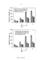
- Figure 5 is a view showing the invisible contrast material for early diagnosis of tumors prepared in Example 6 by hepatoma cells HepG2 Quantitative analysis of intake;
- Figure 6 shows the invisible angiographic material for early diagnosis of tumors prepared in Example 11 by cervical cancer cells HeLa Quantitative analysis of intake.
- the iron oleate complex (18 g, 20 mmol) and oleic acid (2.8 g, 10 mmol) were dissolved in 1-octadecene (30 g) and the mixture was heated to 320 o C (the rate of temperature increase was 3.3 o C/min), after reacting for 1 h under argon protection, cool the solution at room temperature, add ethanol (250 mL), and centrifuge (6000 rpm) for 10 minutes to obtain a monodisperse SPION with a particle size of about 14 nm. Finally, the resulting SPION is vacuum dried and stored at a low temperature (0 - 4 o C).
- - Methacrylic acid copolymer (PNIPAM-MAA), as shown in Figure 2.
- the sample (20,000 ⁇ g, 20 min), disperse the sample in 100 mL of ultrapure water, add 3.0 g of NIPAM and 200 mg of MAA, and use a 400 W UV lamp (UV wavelength 300-500). After irradiation for 1-5 hours at nm and peak value of 350 nm, the obtained composite nanoparticles are centrifuged, washed, and lyophilized to prepare a brain cancer, kidney cancer, breast cancer, lung cancer, ovarian cancer, uterine cancer, nasopharyngeal cancer, and the like. early diagnosis of cancer in invisible contrast material, may be used as T 2 weighted MRI contrast agent.
- Figure 3 is a transmission electron micrograph of the albumin nanosphere prepared in Example 1;
- Figure 4 is an albumin nanosphere (a) prepared in Example 1 and an albumin nanosphere having a pH-sensitive polymer coupled to it ( b) TEM photo comparison chart, (b) The imprinting circle on the surface of the nanosphere is evidence of successful coupling of pH-sensitive polymer on the surface of the nanosphere.
- step 1 (1) MRI contrast agent SPION was changed to PET contrast agent, 18 F -FDG was selected as medical contrast agent, preparation of biodegradable albumin nanosphere and its effect on PET contrast agent 18 F -FDG Embedding, coupling of folic acid on the surface of the albumin nanospheres, and synthesis of the pH-sensitive polymer on the surface of the albumin nanospheres are the same as in the first embodiment, and a brain cancer, kidney cancer, breast cancer, lung cancer, and ovary can be obtained.
- Invisible contrast materials for early diagnosis of cancer, uterine cancer, nasopharyngeal cancer, etc. can be used as PET contrast agents.
- Example 1 Step (1) MRI contrast agent SPION Changed to fluorescein contrast agent, selected near-infrared fluorescent dye phthalocyanine containing tetrapyrrolyl group as medical contrast agent, preparation of biodegradable albumin nanosphere and embedding of fluorescein contrast agent phthalocyanine, albumin Coupling of folic acid on the surface of nanospheres, surface of albumin nanospheres
- the synthesis of the pH-sensitive polymer is the same as in the first embodiment, and a stealth angiographic material for early diagnosis of tumors such as brain cancer, kidney cancer, breast cancer, lung cancer, ovarian cancer, uterine cancer, and nasopharyngeal cancer can be obtained.
- Fluorescein contrast agent .
- the target molecule coupled to the surface of the albumin nanosphere in the step (3) of Example 1 was changed to galactosamine, and the chemical reaction between the amino group of the galactosamine and the carboxyl group on the surface of the albumin nanosphere was catalyzed by EDAC.
- the ligand galactosamine capable of specifically targeting the liver cancer is coupled to the surface of the albumin nanosphere.
- the specific preparation method is as follows: prepare 500 ⁇ g/mL galactosamine solution with PBS as solvent, dissolve 50 mg EDAC in 10 mL galactosamine solution (ice bath), and then add 90 mL of PBS-embedded solution.
- the albumin nanosphere suspension (5.0 mg/mL) of the medical contrast agent was magnetically stirred at room temperature for 24 hours, and the sample was centrifuged (20,000 ⁇ g, 20 min), and the obtained sample was washed with PBS. Then, finally freeze-drying for 48 h can obtain albumin nanospheres with a surface coupled with ligand galactosamine and embedded with a medical contrast agent. Other steps are same as in Example 1, to prepare an early diagnosis of liver cancer in invisible contrast material, it may be used as T 2 weighted MRI contrast agent.
- SPION-AN-GAL-PM stands for internal embedding of contrast agent SPION, surface coupled with target molecule GAL and pH sensitive polymer PNIPAM-MAA (PM Albumin nanospheres (AN); SPION-AN represents albumin nanospheres (AN) encapsulating only the contrast agent SPION.
- PNIPAM-MAA PM Albumin nanospheres (AN)
- SPION-AN represents albumin nanospheres (AN) encapsulating only the contrast agent SPION.
- HepG2 cells are at pH 5.5.
- the amount of SPION-AN-GAL-PM ingested was significantly higher than that of SPION-AN-GAL-PM at pH 7.4, and significantly higher than that at pH 5.5.
- the amount of SPION-AN was significantly higher than that of SPION-AN-GAL-PM.
- the target molecule coupled to the surface of the albumin nanosphere in Example 2 was changed to galactosamine, and the preparation method was the same as that in Example 6. The other steps were the same as in Example 2, and a stealth angiography for early diagnosis of liver cancer was prepared.
- material may be used as T 1 weighted MRI contrast agent.
- the target molecule coupled to the surface of the albumin nanosphere in Example 3 was changed to galactosamine, and the specific preparation method and the embodiment 6 Similarly, the other steps are the same as in the third embodiment, and a stealth contrast material for early diagnosis of liver cancer can be obtained, which can be used as a CT contrast agent.
- the target molecule coupled to the surface of the albumin nanosphere in Example 4 was changed to galactosamine, and the specific preparation method and Example 6 Similarly, the other steps are the same as in the fourth embodiment, and a stealth contrast material for early diagnosis of liver cancer can be obtained, which can be used as a PET contrast agent.
- Example 5 The target molecule coupled to the surface of the albumin nanosphere in Example 5 was changed to galactosamine, and the specific preparation method and Example 6 Similarly, the other steps are the same as in the case of Example 5, and a stealth contrast material for early diagnosis of liver cancer can be obtained, which can be used as a fluorescein contrast agent.
- the monomeric methacrylic acid (MAA) in the synthesis of the pH-sensitive polymer in the step (5) of Example 1 was changed to acrylic acid (AA) to synthesize a pH-sensitive polymer isopropyl group on the surface of the albumin nanosphere.
- Acrylamide-acrylic acid copolymer (PNIPAM-AA) other experimental methods and conditions are the same as in Example 1, to obtain another brain cancer, kidney cancer, breast cancer, lung cancer, ovarian cancer, uterine cancer, nasopharyngeal carcinoma stealth early diagnosis of cancer and other contrast material, may be used as T 2 weighted MRI contrast agent.
- SPION-AN-FA-PA stands for internal embedding of contrast agent SPION, surface coupled with target molecule FA and pH sensitive polymer PNIPAM-AA (PA Albumin nanospheres (AN), SPION-AN represent albumin nanospheres (AN) with a contrast agent SPION embedded therein.
- PNIPAM-AA PA Albumin nanospheres
- SPION-AN represent albumin nanospheres (AN) with a contrast agent SPION embedded therein.
- HeLa cells are at pH 5.5.
- the amount of SPION-AN-FA-PA taken was significantly higher than that of SPION-AN-FA-PA at pH 7.4, and significantly higher than that at pH 5.5.
- the amount of SPION-AN was significantly higher than that of SPION-AN-FA-PA.
- the monomeric methacrylic acid (MAA) in the synthesis of the pH-sensitive polymer of Example 2 was changed to acrylic acid (AA) to synthesize a pH-sensitive polymer isopropylacrylamide-acrylic acid on the surface of albumin nanospheres.
- Copolymer (PNIPAM-AA) other experimental methods and conditions are the same as in Example 2, and another tumor diagnosis such as brain cancer, kidney cancer, breast cancer, lung cancer, ovarian cancer, uterine cancer, nasopharyngeal carcinoma, etc. can be obtained.
- stealth contrast material can be used as T 1 weighted MRI contrast agent.
- the monomeric methacrylic acid (MAA) in the synthesis of the pH-sensitive polymer of Example 3 was changed to acrylic acid (AA). ), thereby synthesizing a pH-sensitive polymer isopropylacrylamide-acrylic acid copolymer (PNIPAM-AA) on the surface of albumin nanospheres, and other experimental methods and conditions are the same as in Example 3
- PNIPAM-AA pH-sensitive polymer isopropylacrylamide-acrylic acid copolymer
- another kind of stealth contrast material for early diagnosis of tumors such as brain cancer, kidney cancer, breast cancer, lung cancer, ovarian cancer, uterine cancer and nasopharyngeal cancer can be obtained, which can be used as a CT contrast agent.
- the monomeric methacrylic acid (MAA) in the synthesis of the pH-sensitive polymer of Example 4 was changed to acrylic acid (AA). ), thereby synthesizing a pH-sensitive polymer isopropylacrylamide-acrylic acid copolymer (PNIPAM-AA) on the surface of albumin nanospheres, and other experimental methods and conditions are the same as in Example 4
- PNIPAM-AA pH-sensitive polymer isopropylacrylamide-acrylic acid copolymer
- another type of brain cancer, kidney cancer, breast cancer, lung cancer, ovarian cancer, uterine cancer, nasopharyngeal cancer and other invisible contrast materials for early diagnosis can be obtained, which can be used as a PET contrast agent.
- the monomeric methacrylic acid (MAA) in the synthesis of the pH-sensitive polymer in Example 5 was changed to acrylic acid (AA). ), thereby synthesizing a pH-sensitive polymer isopropylacrylamide-acrylic acid copolymer (PNIPAM-AA) on the surface of albumin nanospheres, and other experimental methods and conditions are the same as in Example 5
- PNIPAM-AA pH-sensitive polymer isopropylacrylamide-acrylic acid copolymer
- another kind of stealth contrast material for early diagnosis of tumors such as brain cancer, kidney cancer, breast cancer, lung cancer, ovarian cancer, uterine cancer and nasopharyngeal cancer can be obtained, which can be used as a fluorescein contrast agent.
- the monomeric methacrylic acid (MAA) in the synthesis of the pH-sensitive polymer of Example 6 was changed to acrylic acid (AA) to synthesize a pH-sensitive polymer isopropylacrylamide-acrylic acid on the surface of albumin nanospheres.
- AA acrylic acid
- PNIPAM-AA copolymer
- other experimental methods and conditions of Example 6 to obtain another early diagnosis of liver cancer in invisible contrast material, it may be used as T 2 weighted MRI contrast agent.
- the monomeric methacrylic acid (MAA) in the synthesis of the pH-sensitive polymer of Example 7 was changed to acrylic acid (AA) to synthesize a pH-sensitive polymer isopropylacrylamide-acrylic acid on the surface of albumin nanospheres.
- AA acrylic acid
- the monomeric methacrylic acid (MAA) in the synthesis of the pH-sensitive polymer of Example 8 was changed to acrylic acid (AA). ), thereby synthesizing a pH-sensitive polymer isopropylacrylamide-acrylic acid copolymer (PNIPAM-AA) on the surface of albumin nanospheres, and other experimental methods and conditions are the same as in Example 8
- PNIPAM-AA pH-sensitive polymer isopropylacrylamide-acrylic acid copolymer
- the monomeric methacrylic acid (MAA) in the synthesis of the pH-sensitive polymer of Example 9 was changed to acrylic acid (AA).
- AA acrylic acid
- PNIPAM-AA pH-sensitive polymer isopropylacrylamide-acrylic acid copolymer
- the monomeric methacrylic acid (MAA) in the synthesis of the pH-sensitive polymer of Example 10 was changed to acrylic acid (AA). ), thereby synthesizing a pH-sensitive polymer isopropylacrylamide-acrylic acid copolymer (PNIPAM-AA) on the surface of albumin nanospheres, and other experimental methods and conditions are the same as in Example 10
- PNIPAM-AA pH-sensitive polymer isopropylacrylamide-acrylic acid copolymer
- the biodegradable albumin nanospheres in Example 1 were changed to chitosan nanospheres, and the preparation thereof and the embedding method for the medical contrast agent were as follows: 0.2% (w/v) chitosan solution was prepared, and the solvent was 1% (w/v) acetic acid, the medical contrast agent (same as in Example 1) was dispersed into the chitosan solution, and the pH of the solution was adjusted to 4.7-4.8 with sodium hydroxide; 0.3% (w) was prepared.
- TPP sodium tripolyphosphate
- 0.1 mL of TPP solution was added to 0.5 mL of the above chitosan solution under magnetic stirring to prepare an ion-crosslinked shell embedded with a medical contrast agent.
- Glycan nanospheres Other experimental methods and conditions are the same as in the first embodiment, and another invisible angiographic material for early diagnosis of tumors such as brain cancer, kidney cancer, breast cancer, lung cancer, ovarian cancer, uterine cancer, and nasopharyngeal cancer can be obtained, which can be used as T. 2 weighted MRI contrast agents.
- the biodegradable albumin nanospheres of Example 2 were changed to chitosan nanospheres, and the preparation thereof and the embedding method for the medical contrast agent (same as in Example 2) were the same as in Example 21, and other experimental methods and conditions.
- the medical contrast agent (same as in Example 2) were the same as in Example 21, and other experimental methods and conditions.
- As in Example 2, can be prepared another early diagnosis of cancer, brain cancer, renal cancer, breast cancer, lung cancer, ovarian cancer, uterine cancer, nasopharyngeal stealth contrast material can be used as MRI contrast T 1 weighted Agent.
- Example 3 The biodegradable albumin nanospheres of Example 3 were changed to chitosan nanospheres, and their preparation and medical contrast agents (with Example 3)
- the same method of embedding is the same as in Example 21, other experimental methods and conditions and Example 3
- another kind of stealth contrast material for early diagnosis of tumors such as brain cancer, kidney cancer, breast cancer, lung cancer, ovarian cancer, uterine cancer and nasopharyngeal cancer can be obtained, which can be used as a CT contrast agent.
- the biodegradable albumin nanospheres of Example 4 were changed to chitosan nanospheres, and their preparation and medical contrast agents (with Example 4)
- the same method of embedding is the same as in Example 21, other experimental methods and conditions and Example 4
- another type of brain cancer, kidney cancer, breast cancer, lung cancer, ovarian cancer, uterine cancer, nasopharyngeal cancer and other invisible contrast materials for early diagnosis can be obtained, which can be used as a PET contrast agent.
- the biodegradable albumin nanospheres of Example 5 were changed to chitosan nanospheres, and their preparation and medical contrast agents (with Example 5)
- the same method of embedding is the same as in Example 21, other experimental methods and conditions and Example 5
- another kind of stealth contrast material for early diagnosis of tumors such as brain cancer, kidney cancer, breast cancer, lung cancer, ovarian cancer, uterine cancer and nasopharyngeal cancer can be obtained, which can be used as a fluorescein contrast agent.
- Example 11 The biodegradable albumin nanospheres of Example 11 were changed to chitosan nanospheres, and the preparation thereof and the embedding method for the medical contrast agent (same as in Example 11) were the same as in Example 21, and other experimental methods and conditions.
- the medical contrast agent (same as in Example 11) were the same as in Example 21, and other experimental methods and conditions.
- T 2 weighted MRI contrast Agent to obtain another early diagnosis of cancer, brain cancer, renal cancer, breast cancer, lung cancer, ovarian cancer, uterine cancer, nasopharyngeal invisible with contrast material.
- the biodegradable albumin nanospheres of Example 12 were changed to chitosan nanospheres, and the preparation thereof and the embedding method for the medical contrast agent (same as in Example 12) were the same as in Example 21, and other experimental methods and conditions.
- Example 12 to obtain another early diagnosis of cancer, brain cancer, renal cancer, breast cancer, lung cancer, ovarian cancer, uterine cancer, nasopharyngeal stealth contrast material can be used as MRI contrast T 1 weighted Agent.
- Example 13 The biodegradable albumin nanospheres of Example 13 were changed to chitosan nanospheres, and their preparation and medical contrast agents (and Example 13)
- the same method of embedding is the same as in Example 21, other experimental methods and conditions and Example 13
- another kind of stealth contrast material for early diagnosis of tumors such as brain cancer, kidney cancer, breast cancer, lung cancer, ovarian cancer, uterine cancer and nasopharyngeal cancer can be obtained, which can be used as a CT contrast agent.
- Example 14 The biodegradable albumin nanospheres of Example 14 were changed to chitosan nanospheres, their preparation and for medical contrast agents (and Example 14)
- the same method of embedding is the same as in Example 21, and other experimental methods and conditions and examples 14
- another type of brain cancer, kidney cancer, breast cancer, lung cancer, ovarian cancer, uterine cancer, nasopharyngeal cancer and other invisible contrast materials for early diagnosis can be obtained, which can be used as a PET contrast agent.
- the biodegradable albumin nanospheres of Example 15 were changed to chitosan nanospheres, and their preparation and medical contrast agents (with Example 15)
- the same method of embedding is the same as in Example 21, and other experimental methods and conditions and examples 15
- another kind of stealth contrast material for early diagnosis of tumors such as brain cancer, kidney cancer, breast cancer, lung cancer, ovarian cancer, uterine cancer and nasopharyngeal cancer can be obtained, which can be used as a fluorescein contrast agent.
- the MRI contrast agent Gd-DTPA complex in Example 2 was changed to a commercially available Gd-DOTA complex, the preparation of biodegradable albumin nanospheres and the embedding of the MRI contrast agent Gd-DOTA complex
- the coupling of folic acid on the surface of the albumin nanospheres and the synthesis of the pH-sensitive polymer on the surface of the albumin nanospheres are the same as in the second embodiment, and another brain cancer, kidney cancer, breast cancer, lung cancer, ovarian cancer can be obtained.
- uterine cancer, nasopharyngeal cancer early diagnosis invisible contrast material can be used as T 1 weighted MRI contrast agent.
- the MRI contrast agent Gd-DTPA complex in Example 7 was changed to a commercially available Gd-DOTA complex, the preparation of biodegradable albumin nanospheres and its embedding of MRI contrast agent Gd-DOTA complex
- the coupling of the galactosamine on the surface of the albumin nanospheres and the pH-sensitive polymer on the surface of the albumin nanospheres are the same as in the case of Example 7, and another invisible angiographic material for early diagnosis of liver cancer can be obtained.
- the MRI contrast agent Gd-DTPA complex in Example 12 was changed to a commercially available Gd-DOTA complex, the preparation of biodegradable albumin nanospheres and the embedding of the MRI contrast agent Gd-DOTA complex
- the coupling of folic acid on the surface of the albumin nanospheres and the pH-sensitive polymer on the surface of the albumin nanospheres are the same as in the case of Example 12, and another brain cancer, kidney cancer, breast cancer, lung cancer, ovarian cancer can be obtained.
- uterine cancer, nasopharyngeal cancer early diagnosis invisible contrast material can be used as T 1 weighted MRI contrast agent.
- the MRI contrast agent Gd-DTPA complex in Example 17 was changed to a commercially available Gd-DOTA complex, the preparation of biodegradable albumin nanospheres and the embedding of the MRI contrast agent Gd-DOTA complex
- the coupling of galactosamine on the surface of the albumin nanospheres and the pH-sensitive polymer on the surface of the albumin nanospheres are the same as in Example 17, and another invisible angiographic material for early diagnosis of liver cancer can be obtained.
- the MRI contrast agent Gd-DTPA complex in Example 22 was changed to a commercially available Gd-DOTA complex, and the preparation of the biodegradable chitosan nanosphere and its package for the MRI contrast agent Gd-DOTA complex
- the coupling of the surface ligand of the chitosan nanospheres and the synthesis of the pH-sensitive polymer on the surface of the albumin nanospheres are the same as in the case of Example 22, and another brain cancer, kidney cancer, breast cancer, lung cancer, ovarian cancer, uterine cancer, nasopharyngeal cancer early diagnosis invisible contrast material can be used as T 1 weighted MRI contrast agent.
- the MRI contrast agent Gd-DTPA complex in Example 27 was changed to a commercially available Gd-DOTA complex, and the preparation of the biodegradable chitosan nanosphere and its package for the MRI contrast agent Gd-DOTA complex
- the coupling of the surface ligand of the chitosan nanospheres and the synthesis of the pH-sensitive polymer on the surface of the albumin nanospheres are the same as in the case of Example 27, and another brain cancer, kidney cancer, breast cancer, lung cancer, ovarian cancer, uterine cancer, nasopharyngeal cancer early diagnosis invisible contrast material can be used as T 1 weighted MRI contrast agent.
- the MRI contrast agent Gd-DTPA complex in Example 2 was changed to Gd 2 O 3 nanoparticles, and the specific preparation method was as follows: 6.0 mmol GdCl 3 .6H 2 O was added to 30 mL of DEG, and magnetic stirring was continued for the silicone oil. Heat in the oil bath at 140-160 °C for 1 h, then add 30 mL of DEG solution containing 7.5 mmol NaOH. After the reaction is completely dissolved, the temperature of the oil bath is raised to 180 °C and reacted for 4 h with rapid stirring.
- Preparation of biodegradable albumin nanospheres and its application to MRI contrast agent Gd 2 O 3 nanoparticles, coupling of albumin nanosphere surface ligand folic acid, synthesis of albumin nanosphere surface pH sensitive polymer As in Example 2, can be prepared another early diagnosis of cancer, brain cancer, renal cancer, breast cancer, lung cancer, ovarian cancer, uterine cancer, nasopharyngeal stealth contrast material can be used as MRI contrast T 1 weighted Agent.
- Example 7 The MRI contrast agent Gd-DTPA complex in Example 7 was changed to Gd 2 O 3 nanoparticles, and the specific preparation method was the same as in Example 37.
- Preparation of Biodegradable Albumin Nanospheres and Encapsulation of MRI Contrast Agent Gd 2 O 3 Nanoparticles, Coupling of Albumin Nanosphere Surface Ligand Galactosamine, Albumin Nanosphere Surface pH Sensitive Polymer The synthesis of the same as in Example 7 can be used to obtain another invisible contrast material for early diagnosis of liver cancer, which can be used as a T1-weighted MRI contrast agent.
- the MRI contrast agent Gd-DTPA complex in Example 12 was changed to Gd 2 O 3 nanoparticles, and the specific preparation method was the same as in Example 37.
- to obtain another early diagnosis of cancer brain cancer, renal cancer, breast cancer, lung cancer, ovarian cancer, uterine cancer, nasopharyngeal stealth contrast material can be used as MRI contrast T 1 weighted Agent.
- the MRI contrast agent Gd-DTPA complex in Example 17 was changed to Gd 2 O 3 nanoparticles, and the specific preparation method was the same as in Example 37.
- Preparation of Biodegradable Albumin Nanospheres and Encapsulation of MRI Contrast Agent Gd 2 O 3 Nanoparticles, Coupling of Albumin Nanosphere Surface Ligand Galactosamine, Albumin Nanosphere Surface pH Sensitive Polymer synthesis same as in Example 17, to obtain another early diagnosis of liver cancer in invisible contrast material can be used as T 1 weighted MRI contrast agent.
- the MRI contrast agent Gd-DTPA complex in Example 22 was changed to Gd 2 O 3 nanoparticles, and the specific preparation method was the same as in Example 37.
- Preparation of biodegradable chitosan nanospheres and its embedding of MRI contrast agent Gd 2 O 3 nanoparticles, coupling of surface ligands of chitosan nanospheres, and pH sensitivity of chitosan nanospheres The synthesis of the molecule is the same as in Example 22, and another invisible contrast material for early diagnosis of tumors such as brain cancer, kidney cancer, breast cancer, lung cancer, ovarian cancer, uterine cancer, and nasopharyngeal cancer can be obtained, which can be used as T 1 weighting.
- MRI contrast agent is another invisible contrast material for early diagnosis of tumors such as brain cancer, kidney cancer, breast cancer, lung cancer, ovarian cancer, uterine cancer, and nasopharyngeal cancer.
- Example 27 The MRI contrast agent Gd-DTPA complex in Example 27 was changed to Gd 2 O 3 nanoparticles, and the specific preparation method was the same as in Example 37.
- Preparation of biodegradable chitosan nanospheres and its embedding of MRI contrast agent Gd 2 O 3 nanoparticles, coupling of surface ligands of chitosan nanospheres, and pH sensitivity of chitosan nanospheres The synthesis of the molecule is the same as in Example 27, and another invisible contrast material for early diagnosis of tumors such as brain cancer, kidney cancer, breast cancer, lung cancer, ovarian cancer, uterine cancer, and nasopharyngeal cancer can be obtained, which can be used as T 1 weighting.
- MRI contrast agent MRI contrast agent.
- the reducing agent NaBH 4 for preparing the gold nanoparticles in Example 3 was changed to sodium citrate, and the specific preparation method was as follows: 50 mL of ultrapure water was added to a 100 mL round bottom flask, and 2.5 mL of 5 was added under rapid magnetic stirring. mM HAuCl 4 solution, heated in boiling water, then add 0.5, 0.625, 1.0 or 2.0 mL of sodium citrate solution (1%), the reaction is continued in a boiling water bath for 10 min, and finally stirred at room temperature to cool down, you can get Gold nanoparticles with particle sizes of 38, 35, 16, and 14 nm, respectively. Larger gold nanoparticles (100 nm) can be synthesized from 14 nm gold nanoparticles.
- the specific preparation method is as follows: 0.75 mL of 14 nm gold nanoparticles are added to 97.25 mL of ultrapure water, then 1.0 mL is added. 1% (w/v) of HAuCl 4 •3H 2 O solution, stir rapidly at room temperature, add 0.22 mL of 1% sodium citrate, add 1.0 mL of 0.03 M hydroquinone, and stir overnight. 100 nm gold nanoparticles.
- the reducing agent NaBH 4 for preparing gold nanoparticles in Example 8 was changed to sodium citrate, and the specific preparation method was the same as in Example 43.
- Preparation of Biodegradable Albumin Nanospheres and Encapsulation of Five Different Particle Size Gold Nanoparticles, Coupling of Albumin Nanosphere Surface Ligand Galactosamine, Albumin Nanosphere Surface pH Sensitive The synthesis of the polymer is the same as in Example 8, and another four kinds of invisible contrast materials for early diagnosis of liver cancer can be obtained and used as a CT contrast agent.
- Example 3 Replace the CT contrast agent gold nanoparticles in Example 3 with a commercially available CT Preparation of contrast agent iohexol, biodegradable albumin nanospheres and its encapsulation of CT contrast agent iohexol, coupling of folic acid on the surface of albumin nanospheres, pH of albumin nanospheres
- CT contrast agent is the same as in Example 3, and another invisible angiographic material for early diagnosis of tumors such as brain cancer, kidney cancer, breast cancer, lung cancer, ovarian cancer, uterine cancer, and nasopharyngeal cancer can be obtained.
- CT contrast agent is the same as in Example 3, and another invisible angiographic material for early diagnosis of tumors such as brain cancer, kidney cancer, breast cancer, lung cancer, ovarian cancer, uterine cancer, and nasopharyngeal cancer.
- Example 8 Replace the CT contrast agent gold nanoparticles in Example 8 with a commercially available CT Preparation of contrast agent iohexol, biodegradable albumin nanospheres and its encapsulation of CT contrast agent iohexol, coupling of galactosamine on the surface of albumin nanospheres, pH of albumin nanospheres
- CT contrast agent iohexol a commercially available CT Preparation of contrast agent iohexol, biodegradable albumin nanospheres and its encapsulation of CT contrast agent iohexol, coupling of galactosamine on the surface of albumin nanospheres, pH of albumin nanospheres
- the synthesis of the sensitive polymer is the same as in the case of Example 8, and another stealth contrast material for early diagnosis of liver cancer can be obtained and used as a CT contrast agent.
- Example 13 Replace the CT contrast agent gold nanoparticles in Example 13 with a commercially available CT Preparation of contrast agent iohexol, biodegradable albumin nanospheres and its encapsulation of CT contrast agent iohexol, coupling of folic acid on the surface of albumin nanospheres, pH of albumin nanospheres
- CT contrast agent is the same as in Example 13, and another invisible angiographic material for early diagnosis of tumors such as brain cancer, kidney cancer, breast cancer, lung cancer, ovarian cancer, uterine cancer, and nasopharyngeal cancer can be obtained.
- CT contrast agent is the same as in Example 13, and another invisible angiographic material for early diagnosis of tumors such as brain cancer, kidney cancer, breast cancer, lung cancer, ovarian cancer, uterine cancer, and nasopharyngeal cancer.
- Example 18 Replace the CT contrast agent gold nanoparticles in Example 18 with a commercially available CT Preparation of contrast agent iohexol, biodegradable albumin nanospheres and its encapsulation of CT contrast agent iohexol, coupling of galactosamine on the surface of albumin nanospheres, pH of albumin nanospheres
- CT contrast agent iohexol Preparation of contrast agent iohexol, biodegradable albumin nanospheres and its encapsulation of CT contrast agent iohexol, coupling of galactosamine on the surface of albumin nanospheres, pH of albumin nanospheres
- the synthesis of the sensitive polymer is the same as in Example 18, and another invisible contrast material for early diagnosis of liver cancer can be obtained and used as a CT contrast agent.
- Example 23 Replace the CT contrast agent gold nanoparticles in Example 23 with a commercially available CT Preparation of contrast agent iohexol, biodegradable chitosan nanospheres and its encapsulation of CT contrast agent iohexol, coupling of surface ligands of chitosan nanospheres, pH of chitosan nanospheres
- CT contrast agent is the same as in Example 23, and another invisible angiographic material for early diagnosis of tumors such as brain cancer, kidney cancer, breast cancer, lung cancer, ovarian cancer, uterine cancer, and nasopharyngeal cancer can be obtained.
- CT contrast agent is the same as in Example 23, and another invisible angiographic material for early diagnosis of tumors such as brain cancer, kidney cancer, breast cancer, lung cancer, ovarian cancer, uterine cancer, and nasopharyngeal cancer.
- Example 28 Replace the CT contrast agent gold nanoparticles in Example 28 with a commercially available CT Preparation of contrast agent iohexol, biodegradable chitosan nanospheres and its encapsulation of CT contrast agent iohexol, coupling of surface ligands of chitosan nanospheres, pH of chitosan nanospheres
- CT contrast agent is the same as in Example 28, and another invisible angiographic material for early diagnosis of tumors such as brain cancer, kidney cancer, breast cancer, lung cancer, ovarian cancer, uterine cancer, and nasopharyngeal cancer can be obtained.
- CT contrast agent is the same as in Example 28, and another invisible angiographic material for early diagnosis of tumors such as brain cancer, kidney cancer, breast cancer, lung cancer, ovarian cancer, uterine cancer, and nasopharyngeal cancer.
- Example 3 Replace the CT contrast agent gold nanoparticles in Example 3 with a commercially available CT Contrast agent barium sulfate suspension, preparation of biodegradable albumin nanospheres and its encapsulation of CT contrast agent barium sulfate, folic acid coupling of albumin nanosphere surface ligand, albumin nanosphere surface pH
- CT contrast agent a commercially available CT Contrast agent barium sulfate suspension, preparation of biodegradable albumin nanospheres and its encapsulation of CT contrast agent barium sulfate, folic acid coupling of albumin nanosphere surface ligand, albumin nanosphere surface pH
- CT contrast agent is the same as in Example 3, and another invisible angiographic material for early diagnosis of tumors such as brain cancer, kidney cancer, breast cancer, lung cancer, ovarian cancer, uterine cancer, and nasopharyngeal cancer.
- Example 8 Replace the CT contrast agent gold nanoparticles in Example 8 with a commercially available CT Contrast agent barium sulfate suspension, preparation of biodegradable albumin nanospheres and its encapsulation of CT contrast agent barium sulfate, coupling of galactosamine on the surface of albumin nanospheres, surface pH of albumin nanospheres
- the synthesis of the sensitive polymer is the same as in the case of Example 8, and another stealth contrast material for early diagnosis of liver cancer can be obtained and used as a CT contrast agent.
- Example 13 Replace the CT contrast agent gold nanoparticles in Example 13 with a commercially available CT Contrast agent barium sulfate suspension, preparation of biodegradable albumin nanospheres and its encapsulation of CT contrast agent barium sulfate, folic acid coupling of albumin nanosphere surface ligand, albumin nanosphere surface pH
- CT contrast agent a commercially available CT Contrast agent barium sulfate suspension, preparation of biodegradable albumin nanospheres and its encapsulation of CT contrast agent barium sulfate, folic acid coupling of albumin nanosphere surface ligand, albumin nanosphere surface pH
- CT contrast agent is the same as in Example 13, and another invisible angiographic material for early diagnosis of tumors such as brain cancer, kidney cancer, breast cancer, lung cancer, ovarian cancer, uterine cancer, and nasopharyngeal cancer.
- Example 18 Replace the CT contrast agent gold nanoparticles in Example 18 with a commercially available CT Contrast agent barium sulfate suspension, preparation of biodegradable albumin nanospheres and its encapsulation of CT contrast agent barium sulfate, coupling of galactosamine on the surface of albumin nanospheres, surface pH of albumin nanospheres
- the synthesis of the sensitive polymer is the same as in Example 18, and another invisible contrast material for early diagnosis of liver cancer can be obtained and used as a CT contrast agent.
- Example 23 Replace the CT contrast agent gold nanoparticles in Example 23 with a commercially available CT Contrast agent barium sulfate suspension, preparation of biodegradable chitosan nanospheres and its encapsulation of CT contrast agent barium sulfate, coupling of surface ligands of chitosan nanospheres, and surface of chitosan nanospheres pH
- the synthesis of the sensitive polymer is the same as in Example 23, and another invisible angiographic material for early diagnosis of tumors such as brain cancer, kidney cancer, breast cancer, lung cancer, ovarian cancer, uterine cancer, and nasopharyngeal cancer can be obtained.
- CT contrast agent is the same as in Example 23, and another invisible angiographic material for early diagnosis of tumors such as brain cancer, kidney cancer, breast cancer, lung cancer, ovarian cancer, uterine cancer, and nasopharyngeal cancer.
- Example 28 Replace the CT contrast agent gold nanoparticles in Example 28 with a commercially available CT Contrast agent barium sulfate suspension, preparation of biodegradable chitosan nanospheres and its encapsulation of CT contrast agent barium sulfate, coupling of surface ligands of chitosan nanospheres, and surface of chitosan nanospheres pH
- the synthesis of the sensitive polymer is the same as in Example 28, and another invisible angiographic material for early diagnosis of tumors such as brain cancer, kidney cancer, breast cancer, lung cancer, ovarian cancer, uterine cancer, and nasopharyngeal cancer can be obtained.
- CT contrast agent is the same as in Example 28, and another invisible angiographic material for early diagnosis of tumors such as brain cancer, kidney cancer, breast cancer, lung cancer, ovarian cancer, uterine cancer, and nasopharyngeal cancer.
- the PET contrast agent 18 F -FDG in Example 4 was changed to 64 Cu, the preparation of biodegradable albumin nanospheres and the entrapment of PET contrast agent 64 Cu and the coupling of folic acid on the surface of albumin nanospheres.
- the synthesis of the pH-sensitive polymer on the surface of albumin nanospheres is the same as in the case of Example 4, and an early diagnosis of tumors such as brain cancer, kidney cancer, breast cancer, lung cancer, ovarian cancer, uterine cancer, and nasopharyngeal cancer can be obtained.
- Invisible contrast material can be used as a PET contrast agent.
- the preparation of the PET contrast agent 18 F -FDG in Example 9 was changed to 64 Cu, the preparation of biodegradable albumin nanospheres and the entrapment of the PET contrast agent 64 Cu, the surface ligand of the albumin nanospheres, galactosamine
- the synthesis of the pH-sensitive polymer on the surface of the coupled and albumin nanospheres is the same as in Example 9, and another invisible contrast material for early diagnosis of liver cancer can be obtained and used as a PET contrast agent.
- the PET contrast agent 18 F -FDG in Example 14 was changed to 64 Cu, the preparation of biodegradable albumin nanospheres and the entrapment of PET contrast agent 64 Cu and the coupling of folic acid on the surface of albumin nanospheres.
- the synthesis of the pH-sensitive polymer on the surface of albumin nanospheres is the same as in Example 14, and another tumor for brain cancer, kidney cancer, breast cancer, lung cancer, ovarian cancer, uterine cancer, nasopharyngeal cancer, etc. can be prepared for early diagnosis.
- Invisible contrast material can be used as a PET contrast agent.
- the PET contrast agent 18 F -FDG in Example 19 was changed to 64 Cu, the preparation of biodegradable albumin nanospheres and its encapsulation of PET contrast agent 64 Cu, albumin nanosphere surface ligand galactosamine
- the synthesis of the pH-sensitive polymer on the surface of the coupled and albumin nanospheres is the same as in Example 19, and another invisible contrast material for early diagnosis of liver cancer can be obtained and used as a PET contrast agent.
- PET contrast agent 18 F -FDG in Example 24 to 64 Cu preparation of biodegradable chitosan nanospheres and encapsulation of PET contrast agent 64 Cu, surface ligand of folic acid nanospheres folic acid
- the synthesis of the pH-sensitive polymer on the surface of the coupled chitosan nanosphere is the same as in Example 24, and another tumor of brain, kidney, breast, lung, ovarian, uterine, nasopharyngeal, etc. can be obtained.
- Invisible contrast materials for early diagnosis can be used as PET contrast agents.
- the PET contrast agent 18 F -FDG in Example 29 was changed to 64 Cu, the preparation of biodegradable chitosan nanospheres and the encapsulation of PET contrast agent 64 Cu and the surface ligand of chitosan nanospheres.
- the synthesis of the pH-sensitive polymer on the surface of the coupled chitosan nanospheres is the same as in Example 29, and another tumor such as brain cancer, kidney cancer, breast cancer, lung cancer, ovarian cancer, uterine cancer, nasopharyngeal carcinoma, etc. can be obtained.
- Invisible contrast materials for early diagnosis can be used as PET contrast agents.
- the PET contrast agent 18 F -FDG in Example 4 was changed to 124 I , the preparation of biodegradable albumin nanospheres and the entrapment of PET contrast agent 124 I and the coupling of albumin nanosphere surface ligand folic acid
- the synthesis of the pH-sensitive polymer on the surface of albumin nanospheres is the same as in the case of Example 4, and an early diagnosis of tumors such as brain cancer, kidney cancer, breast cancer, lung cancer, ovarian cancer, uterine cancer, and nasopharyngeal cancer can be obtained.
- Invisible contrast material can be used as a PET contrast agent.
- the PET contrast agent 18 F -FDG in Example 9 was changed to 124 I , the preparation of biodegradable albumin nanospheres and its encapsulation of PET contrast agent 124 I , albumin nanosphere surface ligand galactosamine
- the synthesis of the pH-sensitive polymer on the surface of the coupled and albumin nanospheres is the same as in Example 9, and another invisible contrast material for early diagnosis of liver cancer can be obtained and used as a PET contrast agent.
- the PET contrast agent 18 F -FDG in Example 14 was changed to 124 I , the preparation of biodegradable albumin nanospheres and the entrapment of PET contrast agent 124 I and the coupling of albumin nanosphere surface ligand folic acid
- the synthesis of the pH-sensitive polymer on the surface of albumin nanospheres is the same as in Example 14, and another tumor for brain cancer, kidney cancer, breast cancer, lung cancer, ovarian cancer, uterine cancer, nasopharyngeal cancer, etc. can be prepared for early diagnosis.
- Invisible contrast material can be used as a PET contrast agent.
- the PET contrast agent 18 F -FDG in Example 19 was changed to 124 I , the preparation of biodegradable albumin nanospheres and its encapsulation of PET contrast agent 124 I , albumin nanosphere surface ligand galactosamine
- the synthesis of the pH-sensitive polymer on the surface of the coupled and albumin nanospheres is the same as in Example 19, and another invisible contrast material for early diagnosis of liver cancer can be obtained and used as a PET contrast agent.
- the PET contrast agent 18 F -FDG in Example 24 was changed to 124 I , the preparation of biodegradable chitosan nanospheres and the entrapment of PET contrast agent 124 I , the surface ligand of folic acid nanospheres folic acid
- the synthesis of the pH-sensitive polymer on the surface of the coupled chitosan nanosphere is the same as in Example 24, and another tumor of brain, kidney, breast, lung, ovarian, uterine, nasopharyngeal, etc. can be obtained.
- Invisible contrast materials for early diagnosis can be used as PET contrast agents.
- the PET contrast agent 18 F -FDG in Example 29 was changed to 124 I , the preparation of biodegradable chitosan nanospheres and the entrapment of PET contrast agent 124 I , the surface ligand of folic acid nanospheres folic acid
- the synthesis of the pH-sensitive polymer on the surface of the coupled chitosan nanospheres is the same as in Example 29, and another tumor such as brain cancer, kidney cancer, breast cancer, lung cancer, ovarian cancer, uterine cancer, nasopharyngeal carcinoma, etc. can be obtained.
- Invisible contrast materials for early diagnosis can be used as PET contrast agents.
- the PET contrast agent 18 F -FDG in Example 4 was changed to 94 Tc , the preparation of biodegradable albumin nanospheres and the entrapment of PET contrast agent 94 Tc and the coupling of folic acid on the surface of albumin nanospheres.
- the synthesis of the pH-sensitive polymer on the surface of albumin nanospheres is the same as in the case of Example 4, and an early diagnosis of tumors such as brain cancer, kidney cancer, breast cancer, lung cancer, ovarian cancer, uterine cancer, and nasopharyngeal cancer can be obtained.
- Invisible contrast material can be used as a PET contrast agent.
- the PET contrast agent 18 F -FDG in Example 9 was changed to 94 Tc , the preparation of biodegradable albumin nanospheres and its encapsulation of PET contrast agent 94 Tc , albumin nanosphere surface ligand galactosamine
- the synthesis of the pH-sensitive polymer on the surface of the coupled and albumin nanospheres is the same as in Example 9, and another invisible contrast material for early diagnosis of liver cancer can be obtained and used as a PET contrast agent.
- the PET contrast agent 18 F -FDG in Example 14 was changed to 94 Tc , the preparation of biodegradable albumin nanospheres and the entrapment of PET contrast agent 94 Tc and the coupling of albumin nanosphere surface ligand folic acid
- the synthesis of the pH-sensitive polymer on the surface of albumin nanospheres is the same as in Example 14, and another tumor for brain cancer, kidney cancer, breast cancer, lung cancer, ovarian cancer, uterine cancer, nasopharyngeal cancer, etc. can be prepared for early diagnosis.
- Invisible contrast material can be used as a PET contrast agent.
- the PET contrast agent 18 F -FDG in Example 19 was changed to 94 Tc , the preparation of biodegradable albumin nanospheres and its encapsulation of PET contrast agent 94 Tc , albumin nanosphere surface ligand galactosamine
- the synthesis of the pH-sensitive polymer on the surface of the coupled and albumin nanospheres is the same as in Example 19, and another invisible contrast material for early diagnosis of liver cancer can be obtained and used as a PET contrast agent.
- the PET contrast agent 18 F -FDG in Example 24 was changed to 94 Tc , the preparation of biodegradable chitosan nanospheres and its encapsulation of PET contrast agent 94 Tc , the surface ligand of folic acid nanospheres folic acid
- the synthesis of the pH-sensitive polymer on the surface of the coupled chitosan nanosphere is the same as in Example 24, and another tumor of brain, kidney, breast, lung, ovarian, uterine, nasopharyngeal, etc. can be obtained.
- Invisible contrast materials for early diagnosis can be used as PET contrast agents.
- the PET contrast agent 18 F -FDG in Example 29 was changed to 94 Tc , the preparation of biodegradable chitosan nanospheres and its encapsulation of PET contrast agent 94 Tc , the surface ligand of folic acid nanospheres folic acid
- the synthesis of the pH-sensitive polymer on the surface of the coupled chitosan nanospheres is the same as in Example 29, and another tumor such as brain cancer, kidney cancer, breast cancer, lung cancer, ovarian cancer, uterine cancer, nasopharyngeal carcinoma, etc. can be obtained.
- Invisible contrast materials for early diagnosis can be used as PET contrast agents.
- the gold nanoparticles prepared in Example 3 were changed to gold nano cages, and the preparation method was as follows: 3 mL of 0.75 mM HAuCl 4 and 3 mL of 0.03 M HMT (hexamethylenetetramine) were mixed, and the solution color was light yellow. It became transparent. Then, add 3 mL of 0.3 M PVP (polyvinylpyrrolidone) and 100 ⁇ L of 0.01 M AgNO3. After gentle stirring, add 50 ⁇ L of 0.08 M ascorbic acid, stir the mixed solution for 10 s, and let stand at room temperature for 12 h. After centrifugation, a gold nanocage is obtained.
- 3 mL of 0.75 mM HAuCl 4 and 3 mL of 0.03 M HMT (hexamethylenetetramine) were mixed, and the solution color was light yellow. It became transparent. Then, add 3 mL of 0.3 M PVP (polyvinylpyrrolidone) and 100 ⁇ L
- the gold nanoparticles prepared in Example 8 were changed to gold nano cages, and the specific preparation method and examples 75 The same, the preparation of biodegradable albumin nanospheres and the embedding of the above gold nanocage, the coupling of the albumin nanosphere surface ligand galactosamine, the synthesis of the albumin nanosphere surface pH sensitive polymer and Example 8 In the same way, another invisible contrast material for early diagnosis of liver cancer can be obtained and used as a CT contrast agent.
- the gold nanoparticles prepared in Example 3 were changed to gold nanorods, and the preparation method was as follows: 20 mL of a 0.1 M NaBH 4 solution containing 0.25 mM HAuCl 4 and 0.25 mM sodium citrate solution and 0.6 mL ice bath while stirring was prepared. After adding NaBH 4 , the solution will quickly turn purple and gold nanoparticles will form.
- the gold nanoparticles can be used as gold nano seeds within 2-5 h after synthesizing the gold nanoparticles.
- the gold nanoparticles prepared in Example 8 were changed to gold nanorods, and the specific preparation method and examples 77 The same, the preparation of biodegradable albumin nanospheres and the embedding of the above gold nanorods, the coupling of galactosamine on the surface of albumin nanospheres, and the synthesis of pH-sensitive polymers on the surface of albumin nanospheres Example 8 In the same way, another invisible contrast material for early diagnosis of liver cancer can be obtained and used as a CT contrast agent.
Landscapes
- Health & Medical Sciences (AREA)
- Chemical & Material Sciences (AREA)
- Engineering & Computer Science (AREA)
- Nanotechnology (AREA)
- Life Sciences & Earth Sciences (AREA)
- General Health & Medical Sciences (AREA)
- Veterinary Medicine (AREA)
- Public Health (AREA)
- Epidemiology (AREA)
- Animal Behavior & Ethology (AREA)
- Nuclear Medicine, Radiotherapy & Molecular Imaging (AREA)
- Radiology & Medical Imaging (AREA)
- Medicinal Chemistry (AREA)
- Biomedical Technology (AREA)
- Pharmacology & Pharmacy (AREA)
- Optics & Photonics (AREA)
- Molecular Biology (AREA)
- Proteomics, Peptides & Aminoacids (AREA)
- Physics & Mathematics (AREA)
- Biochemistry (AREA)
- Biotechnology (AREA)
- Dispersion Chemistry (AREA)
- Medicines Containing Antibodies Or Antigens For Use As Internal Diagnostic Agents (AREA)
- Magnetic Resonance Imaging Apparatus (AREA)
Abstract
La présente invention concerne un matériau de radiographie invisible, destiné au diagnostic précoce d'une tumeur et un procédé de préparation associé. Le procédé comprend les étapes suivantes : l'emploi d'une nanosphère biodégradable afin d'intégrer un produit de contraste médical ; le couplage d'une molécule cible, qui interagit spécifiquement avec une cellule tumorale sur la surface de la nanosphère ; et la greffe d'une macromolécule sensible au pH, qui est un composé de macromolécules contracté entre un pH de 5,0 à 6,0.
Applications Claiming Priority (2)
| Application Number | Priority Date | Filing Date | Title |
|---|---|---|---|
| CN201210230833.8 | 2012-07-03 | ||
| CN201210230833.8A CN102743768B (zh) | 2012-07-03 | 2012-07-03 | 肿瘤早期诊断用隐形造影材料及其制备方法 |
Publications (1)
| Publication Number | Publication Date |
|---|---|
| WO2014005383A1 true WO2014005383A1 (fr) | 2014-01-09 |
Family
ID=47024489
Family Applications (1)
| Application Number | Title | Priority Date | Filing Date |
|---|---|---|---|
| PCT/CN2012/082578 Ceased WO2014005383A1 (fr) | 2012-07-03 | 2012-10-08 | Matériau de radiographie invisible pour le diagnostic précoce d'une tumeur et procédé de préparation associé |
Country Status (2)
| Country | Link |
|---|---|
| CN (1) | CN102743768B (fr) |
| WO (1) | WO2014005383A1 (fr) |
Families Citing this family (9)
| Publication number | Priority date | Publication date | Assignee | Title |
|---|---|---|---|---|
| CN103041407B (zh) * | 2012-12-19 | 2015-02-18 | 深圳先进技术研究院 | 核-壳型纳米造影剂、其制备方法及应用 |
| CN103446588B (zh) * | 2013-07-24 | 2015-12-02 | 中国科学院宁波材料技术与工程研究所 | 靶向型诊疗联用药物及其制备方法和应用 |
| CN104368007A (zh) * | 2013-08-12 | 2015-02-25 | 于湛 | 可注射蛋白基花样结构载药微纳材料与内耳跨膜给药缓释制剂及其制备方法 |
| CN104623697B (zh) * | 2015-02-08 | 2017-03-22 | 中国科学院武汉物理与数学研究所 | 一种基于人体生理温度响应的聚合物胶束型磁共振造影剂的制备方法 |
| CN108872172B (zh) * | 2018-06-21 | 2021-05-25 | 东南大学 | 一种基于bsa的超分辨成像探针及其制备方法和应用 |
| CN111558040B (zh) * | 2020-04-17 | 2021-03-26 | 华南农业大学 | 一种集磁共振成像和光热治疗于一体的高分子自愈凝胶及其制备与应用 |
| CN112338199B (zh) * | 2020-10-20 | 2022-05-31 | 华中科技大学同济医学院附属协和医院 | 金纳米笼的制备方法及其应用 |
| TWI856287B (zh) * | 2022-02-11 | 2024-09-21 | 國立臺灣科技大學 | 可溶解感溫型的微載體、其製備方法及其使用方法 |
| CN114949261B (zh) * | 2022-05-05 | 2024-06-04 | 南方医科大学南方医院 | 一种铁钆纳米复合物及其制备方法和应用 |
Citations (2)
| Publication number | Priority date | Publication date | Assignee | Title |
|---|---|---|---|---|
| WO2004012855A2 (fr) * | 2002-08-01 | 2004-02-12 | E.I. Du Pont De Nemours And Company | Nanoparticules protegees par une monocouche de glycol d'ethylene |
| CN102228425A (zh) * | 2011-07-01 | 2011-11-02 | 华南师范大学 | 一种肿瘤靶向磁性水凝胶纳米递药系统及其构建方法和应用 |
Family Cites Families (3)
| Publication number | Priority date | Publication date | Assignee | Title |
|---|---|---|---|---|
| DE10224352A1 (de) * | 2002-06-01 | 2003-12-11 | Mueller Schulte Detlef | Thermosensitive Polymerträger mit veränderbarer physikalischer Struktur für die biochemische Analytik, Diagnostik und Therapie |
| CA2668457C (fr) * | 2006-11-02 | 2016-10-04 | Veridex, Llc | Imagerie d'endothelium vasculaire active au moyen d'agents de contraste immunomagnetiques pour irm |
| CN101850128B (zh) * | 2010-05-18 | 2012-10-24 | 陈道桢 | 188Re-标记叶酸偶联的顺铂磁性白蛋白复合纳米粒及其制备方法与应用 |
-
2012
- 2012-07-03 CN CN201210230833.8A patent/CN102743768B/zh active Active
- 2012-10-08 WO PCT/CN2012/082578 patent/WO2014005383A1/fr not_active Ceased
Patent Citations (2)
| Publication number | Priority date | Publication date | Assignee | Title |
|---|---|---|---|---|
| WO2004012855A2 (fr) * | 2002-08-01 | 2004-02-12 | E.I. Du Pont De Nemours And Company | Nanoparticules protegees par une monocouche de glycol d'ethylene |
| CN102228425A (zh) * | 2011-07-01 | 2011-11-02 | 华南师范大学 | 一种肿瘤靶向磁性水凝胶纳米递药系统及其构建方法和应用 |
Non-Patent Citations (3)
| Title |
|---|
| CHAO HUANG ET AL.: "Combined ATRP and 'Click' Chemistry for Designing Stable Tumor-Targeting Superparamagnetic Iron Oxide Nanoparticles", LANGMUIR., vol. 28, no. 1, January 2012 (2012-01-01), pages 563 - 571 * |
| LIU, HAI ET AL.: "Evaluation of Effect and Targetability on Folate-conjugated Albumin anoparticles", JOURNAL OF GUANGDONG COLLEGE OF PHARMACY, vol. 23, no. 1, February 2007 (2007-02-01), pages 50 - 52 * |
| ZHANG HUIFANG.: "Study on Preparation and Properties of Albumin-based Biopolymer Carriers", CHINESE MASTER'S THESES FULL-TEXT DATABASE MEDICINE AND HEALTH SCIENCES, 15 June 2010 (2010-06-15) * |
Also Published As
| Publication number | Publication date |
|---|---|
| CN102743768B (zh) | 2014-07-09 |
| CN102743768A (zh) | 2012-10-24 |
Similar Documents
| Publication | Publication Date | Title |
|---|---|---|
| WO2014005383A1 (fr) | Matériau de radiographie invisible pour le diagnostic précoce d'une tumeur et procédé de préparation associé | |
| Shen et al. | Iron oxide nanoparticle based contrast agents for magnetic resonance imaging | |
| Zhang et al. | Surface modification of superparamagnetic magnetite nanoparticles and their intracellular uptake | |
| Xie et al. | Iron oxide nanoparticle platform for biomedical applications | |
| CN103721271B (zh) | 用于肝癌多模态影像和光热治疗的多功能纳米探针及应用 | |
| JP2009519316A (ja) | 磁気共鳴造影法用のターゲティングナノ粒子 | |
| Fakayode et al. | Photodynamic therapy evaluation of methoxypolyethyleneglycol-thiol-SPIONs-gold-meso-tetrakis (4-hydroxyphenyl) porphyrin conjugate against breast cancer cells | |
| CN104758955B (zh) | 一种拥有多重刺激药物释放以及mri造影能力的超分子胶囊的制备方法 | |
| US9757342B2 (en) | Method for preparing protein cage, and in situ method for preparing hydrophobic additive-supported core-shell structured polymer-protein particles | |
| Hemalatha et al. | Fabrication and characterization of dual acting oleyl chitosan functionalised iron oxide/gold hybrid nanoparticles for MRI and CT imaging | |
| KR101720046B1 (ko) | 광역학 치료용 자기조립형 약학 조성물 | |
| Wang et al. | Magneto-fluorescent nanoparticles with high-intensity NIR emission, T 1-and T 2-weighted MR for multimodal specific tumor imaging | |
| CN107789632A (zh) | 一种t7肽修饰的主动脑靶向纳米递药系统及其制备方法 | |
| EP0361960A2 (fr) | Procédé et compositions pour la production d'images par résonance magnétique | |
| CN110559454B (zh) | 一种用于诊疗阿尔兹海默症的纳米复合药物 | |
| CN108948152A (zh) | 一种两亲性穿膜肽键合物、其制备方法及用途 | |
| CN111097052B (zh) | 用于肿瘤主动靶向治疗的两亲性前药及其纳米颗粒的制备方法、应用 | |
| Yin et al. | Fluorescent oligo (p-phenyleneethynylene) contained amphiphiles-encapsulated magnetic nanoparticles for targeted magnetic resonance and two-photon optical imaging in vitro and in vivo | |
| KR101765335B1 (ko) | 암 세포 조영용 mri 조영제 조성물 | |
| CN107224590B (zh) | 一种可降解聚合物磁性纳米粒子及其制备方法 | |
| CN106924762B (zh) | 一种用于t1、t2双模态磁共振造影剂的磁性纳米颗粒复合物及其制备方法 | |
| KR101115799B1 (ko) | Dtpa 유도체, 금속 착물, mr 및 ct 조영제 및 이의 제조 방법 | |
| Yao et al. | Multifunctional ferritin nanocages for bimodal imaging and targeted delivery of doxorubicin into cancer cells | |
| CN105148291A (zh) | 多聚糖醛酸为载体的活体染色造影剂及制备方法和应用 | |
| Ouyang et al. | Dendrimer-based tumor-targeted systems |
Legal Events
| Date | Code | Title | Description |
|---|---|---|---|
| 121 | Ep: the epo has been informed by wipo that ep was designated in this application |
Ref document number: 12880414 Country of ref document: EP Kind code of ref document: A1 |
|
| NENP | Non-entry into the national phase |
Ref country code: DE |
|
| 122 | Ep: pct application non-entry in european phase |
Ref document number: 12880414 Country of ref document: EP Kind code of ref document: A1 |