WO2014199954A1 - イムノクロマト診断キット - Google Patents
イムノクロマト診断キット Download PDFInfo
- Publication number
- WO2014199954A1 WO2014199954A1 PCT/JP2014/065243 JP2014065243W WO2014199954A1 WO 2014199954 A1 WO2014199954 A1 WO 2014199954A1 JP 2014065243 W JP2014065243 W JP 2014065243W WO 2014199954 A1 WO2014199954 A1 WO 2014199954A1
- Authority
- WO
- WIPO (PCT)
- Prior art keywords
- particles
- diagnostic kit
- cellulose
- colored
- sample pad
- Prior art date
- Legal status (The legal status is an assumption and is not a legal conclusion. Google has not performed a legal analysis and makes no representation as to the accuracy of the status listed.)
- Ceased
Links
Images


Classifications
-
- G—PHYSICS
- G01—MEASURING; TESTING
- G01N—INVESTIGATING OR ANALYSING MATERIALS BY DETERMINING THEIR CHEMICAL OR PHYSICAL PROPERTIES
- G01N33/00—Investigating or analysing materials by specific methods not covered by groups G01N1/00 - G01N31/00
- G01N33/48—Biological material, e.g. blood, urine; Haemocytometers
- G01N33/50—Chemical analysis of biological material, e.g. blood, urine; Testing involving biospecific ligand binding methods; Immunological testing
- G01N33/53—Immunoassay; Biospecific binding assay; Materials therefor
- G01N33/543—Immunoassay; Biospecific binding assay; Materials therefor with an insoluble carrier for immobilising immunochemicals
- G01N33/54366—Apparatus specially adapted for solid-phase testing
- G01N33/54386—Analytical elements
- G01N33/54387—Immunochromatographic test strips
- G01N33/54388—Immunochromatographic test strips based on lateral flow
Definitions
- the present invention relates to an immunochromatographic diagnostic kit including a sample pad for in vitro diagnostics, and more particularly to a lateral flow type immunochromatographic diagnostic kit.
- a sandwich method As a measurement principle of the immunochromatography measurement method, there are a method called a sandwich method and a method called a competition method. As a measurement format, there are methods called a flow-through type and a lateral flow type.
- Various substances can be detected as test substances in the specimen.
- a typical example is measurement of detecting an antigen by a sandwich method, and the following operations are sequentially executed.
- An antibody that specifically binds to an antigen as a test target substance is immobilized on a predetermined portion of a chromatographic medium such as a nitrocellulose membrane, and a test line (hereinafter referred to as “TL”) is placed at an arbitrary position on the chromatographic medium.
- TL test line
- the labeled substance is captured by the antibody immobilized on the chromatographic medium at the reaction site via the antigen, and diagnosis by the immunochromatographic diagnostic kit is performed by detecting the signal of the labeled substance.
- diagnosis by the immunochromatographic diagnostic kit is performed by detecting the signal of the labeled substance.
- a general diagnosis is a qualitative diagnosis in which only the presence or absence of an antigen is examined, but in recent years, a quantitative diagnosis can also be performed by visually or mechanically detecting the intensity of the signal.
- Patent Document 1 reports that it is possible to speed up diagnosis time by using a specific cellulose-based nonwoven fabric for a sample pad that is a portion where a specimen sample containing a test target substance is dropped. Yes. Patent Document 1 also mentions the liquid absorption speed and developability, but there is no specific diagnosis time data, and no mention is made of a combination with specific coloring particles as in the present invention. Furthermore, no mention is made of the arrangement of the sample pad and the conjugate pad. Furthermore, the following Patent Document 2 reports that it is possible to speed up the diagnosis time by devising the structure of the diagnostic kit. For example, in the hCG assay, the determination is made in the shortest 2 minutes and 35 seconds. ing.
- Patent Document 4 discloses a semi-quantitative test method using immunochromatography, and it is described that the antigen concentration and the intensity of the test result signal can be made proportional within a certain range of antigen concentration. Has been. However, quantification in immunochromatography, in addition to the fact that the antigen concentration is proportional to the strength of the test result signal in a range of antigen concentrations, the reproducibility of the results, that is, the same strength signal when measuring the same concentration of antigen. It is generally said that such reproducibility is still not sufficient. Furthermore, the following Patent Document 5 discloses an immunochromatographic diagnostic kit using fluorescent particles as colored particles. Cellulose particles are used as examples of the colored particles, and cellulose nonwoven fabric is used as an example of the sample pad. None of them are specifically mentioned. Furthermore, the arrangement of the sample pad and the conjugate pad is not devised. Of course, the effect of the combination of the sample pad and the coloring particles as in the present invention is not described.
- JP 2012-108031 A Japanese Unexamined Patent Publication No. 7-55809 International Publication No.WO2011 / 062157 Specification International Publication No.WO2006 / 080438 Specification International Publication No.WO2013 / 151066 Specification
- the problem to be solved by the present invention is to provide an immunochromatographic diagnostic kit capable of more rapid diagnosis, high analytical sensitivity, and excellent reproducibility of test results. .
- the present inventors diligently studied to solve the above-mentioned problems, and as a result of repeated experiments, they used particles having a large color and a large particle diameter as color-developing particles, and a non-woven fabric made of specific regenerated cellulosic fibers was used as a sample pad. Using these in combination, it was surprisingly surprising that it was possible to speed up diagnosis and improve analytical sensitivity, and also to realize excellent reproducibility of test results, and completed the present invention. It is a thing.
- the present invention is as follows.
- the average particle diameter is 100 to 1000 nm
- the color intensity is 1.0 to 10.0
- 10 to 90% by weight of the particle weight is derived from cellulose
- 90 to 10% by weight is derived from the coloring component
- a non-woven fabric made of regenerated cellulosic fibers having a basis weight of 10 to 150 g / m 2 and a thickness of 0.07 to 1.00 mm.
- An immunochromatographic diagnostic kit comprising a sample pad for in vitro diagnostics.
- the immunochromatography diagnostic kit includes a predetermined coloring particle as a labeling substance, and uses a nonwoven fabric made of a predetermined regenerated cellulose fiber as a sample pad, thereby speeding up diagnosis and improving analytical sensitivity. Furthermore, the reproducibility of the inspection results is excellent.
- the present inventors can unexpectedly speed up the diagnostic time, improve the analysis sensitivity, and improve the reproducibility of the test. It is an invention made based on this.
- Patent Document 3 it has been found that colored particles having a large particle size and a high coloring intensity have high analytical sensitivity even when used alone.
- the diagnosis time can be shortened to some extent by such high analysis sensitivity.
- the immunochromatography diagnostic kit is composed of various porous structures such as a chromatographic medium, colored particles having a large particle diameter may interfere with the pores of the porous structure and hardly flow. If particles with a slow flow are generated, the diagnosis time may be delayed.
- the diagnosis time could be further shortened by facilitating the flow of colored particles having a large particle diameter.
- the unit per unit time supplied to the conjugate pad containing the colored particles It has been found that the diagnostic time can be shortened by increasing the flow rate to facilitate the flow of the colored particles contained in the conjugate pad.
- a chemical such as a surfactant may be added to the sample pad to facilitate the flow of the colored particles.
- the sample pad of the present invention instantly absorbs the dropped specimen, so that the conjugate pad or chromatographic medium is used.
- a drug for facilitating the flow of the colored particles can always be provided at a constant concentration. For this reason, it becomes easy for the colored particles to flow without interfering with the pores of the porous structure.
- the concentration of the drug for facilitating the flow of the colored particles may not be constant, and clogging of the particles may occur.
- all these colored particles can contribute to the inspection.
- the analysis sensitivity has been improved, the test reproducibility has been improved, and the arrangement of the sample pad and conjugate pad can be devised to make these effects more prominent. I also found what I can do.
- the “colored particle” in the present invention refers to a particulate substance that is insoluble in water, a buffer solution or the like and carries a pigment or a dye.
- the material constituting the particles is not particularly limited, but examples of such colored particles include metal colloid particles such as gold colloid, platinum colloid, silver colloid and selenium colloid, styrene latex such as polystyrene latex, and acrylic acid latex.
- Colored latex particles colored, etc. colored silica particles colored silica composed of a three-dimensional structure composed of silicon atoms and oxygen atoms, colored cellulose particles colored with cellulose, colored particles formed by directly coloring colored components such as carbon black , Magnetic particles, and the like.
- the color-developing particles may be fluorescent particles.
- Colored cellulose particles are preferred because of easy adjustment of particle characteristics such as particle diameter adjustment, color density adjustment, color type adjustment, particle surface condition adjustment and the like. Since cellulose has a large amount of hydroxyl groups, it has high hydrophilicity and excellent dispersion stability, and can contain a large amount of coloring components.
- the method for producing colored particles is not particularly limited.
- a method of first shaping particles and supporting coloring components such as pigments and dyes, a method of molding particles and supporting smaller colored particles such as metal colloids and pigments, dyes, dyes, pigments and metal colloids when molding particles A coloring method such as a method of adding and molding together is also included.
- the particles are first shaped and colored components such as pigments and dyes because of easy adjustment of particle characteristics such as particle diameter adjustment, color density adjustment, color type adjustment, particle surface condition adjustment, etc.
- a method of supporting is preferable.
- dye is preferable from the ease of carrying
- the “type of dye” is not particularly limited. Dyes such as reactive dyes, direct dyes, metal-containing dyes, acid dyes, basic dyes, disperse dyes, sulfur dyes, vegetable dyes, naphthol dyes, and fluorescent dyes can be used. Of course, any dye may be combined.
- a reactive dye that is covalently bonded to a hydroxyl group of cellulose is particularly preferable from the viewpoint of retaining a large amount of dye and stability.
- the “cellulosic particle molding method” is not particularly limited. Examples include a method of physically refining natural cellulose with a ball mill or a high-pressure homogenizer, a method of chemically treating and refining with natural acid or alkali, a method of dissolving cellulose once in its good solvent and forming into particles. It is done. Alternatively, cellulose particles may be prepared by dissolving derivatized cellulose, forming the particles into particles, and returning the derivatized substituents to hydroxyl groups. Furthermore, these molding methods may be combined.
- the “type of cellulose” is not particularly limited, and regenerated cellulose, purified cellulose, natural cellulose, cellulose derivatized with the above, cellulose in which a derivatized substituent is returned to a hydroxyl group, and the like can be used. .
- regenerated cellulose purified cellulose, natural cellulose, cellulose derivatized with the above, cellulose in which a derivatized substituent is returned to a hydroxyl group, and the like.
- a method in which it is once dissolved in a good solvent and formed into particles is preferred, and the type of cellulose is preferably regenerated cellulose.
- the “type of good solvent for dissolving cellulose” is not particularly limited, and copper ammonia solution, viscose solution, N-methylmorpholine, various types Various good solvents that can dissolve cellulose, such as an ionic liquid, can be used. Among these, a copper ammonia solution is preferable from the viewpoint of adjusting the particle diameter and adjusting the particle shape. Further, the method for forming the dissolved cellulose into particles is not particularly limited. In the present invention, a method by phase separation was selected.
- the “average particle diameter” of the colored particles refers to the volume average median diameter measured by the dynamic light scattering method, and the volume average median diameter is in the range of 100 to 1000 nm.
- the average particle diameter is in this range, the surface area of the particles is large, and therefore, when used as an immunochromatography diagnostic kit, TL becomes darker, that is, analysis sensitivity becomes higher. If the average particle size is too small, the surface area may be reduced, resulting in a decrease in analytical sensitivity or particle aggregation. From the above points, the particle diameter is preferably 200 nm or more, and more preferably 300 nm or more.
- the particle diameter is preferably 800 nm or less, more preferably 600 nm or less.
- the average particle diameter described here is an average value to the last, and a part of particle diameter distribution may remove
- volume average is used for particle size evaluation.
- too large particles in an immunochromatography diagnostic kit are clogged with chromatographic media such as nitrocellulose. This is because the effect is reflected even if it exists in
- chromatographic media such as nitrocellulose.
- the volume average there are various methods for expressing the particle diameter, such as number average and area average. Of course, if the representation is different, the value of the particle diameter also changes. In the present invention, the volume average is adopted.
- Color intensity is a value that defines the color intensity of the particles.
- the method of measuring the value is to prepare a pure water dispersion of colored particles of known concentration, measure the visible absorbance using an integrating sphere in the range of 400 to 800 nm with an optical path length of 10 mm, and obtain the peak of the obtained absorbance curve.
- the reason for performing visible absorbance measurement using an integrating sphere for measuring the color density of particles is that the color density of particles dispersed in a liquid can be measured most accurately.
- a method of measuring the color density of particles there is a method of measuring a solid obtained by drying particles with a colorimeter or the like, but such a method cannot accurately measure the color density of particles.
- metal colloids and the like have different color tones and maximum wavelengths depending on the particle diameter, and the dry aggregated state cannot accurately reflect the color density of the state dispersed in the liquid. Further, even when the particles are dispersed in the liquid at the same particle concentration, the color density becomes light when aggregation occurs.
- the reason for using an integrating sphere when performing visible absorbance measurement is to remove the influence of scattering of the particles themselves.
- Ordinary visible absorbance measurement is a method of measuring transmitted light, which reflects not only the absorption by colored components but also the effect of scattering of the particles themselves on the incident light.
- gold colloids generally used in immunochromatography may have a particle diameter of 40 nm to 60 nm, and sometimes 100 nm, but in any case, the particle diameter is small, so there is almost no influence of scattered light.
- polystyrene latex particles have a large particle size and are clearly affected by scattered light.
- visible absorbance measurement using an integrating sphere is employed in order to more accurately reflect the color density of the particles themselves when the particle diameters and particle materials are different.
- the “color intensity” is 1.0 to 10.0.
- the larger this value the darker the color of the particles, and the higher the sensitivity of analysis when used as an immunochromatographic diagnostic kit.
- the larger the value the better.
- Use a darker dye For example, it is possible to adopt a method that makes it easier for the dye to enter.
- the upper limit is preferably 7.0 or less, and more preferably 5.0 or less.
- the lower the value the lower the analytical sensitivity when used as an immunochromatography diagnostic kit, so the lower limit is preferably 1.5 or more, and more preferably 2.0 or more.
- the ratio of the coloring components of the coloring particles refers to the ratio of the coloring components in the total weight of the coloring particles. For example, when 1.0 g of colored particles is composed of 0.2 g of cellulose and 0.8 g of a coloring component, the ratio of the coloring component is 80% by weight. The ratio of the coloring component of the color developing particles is preferably 10 to 90% by weight. When it is within this range, the analytical sensitivity is high when used as an immunochromatography diagnostic kit. In addition, when particles obtained by dyeing cellulose with a dye are used as the colored particles, it is possible to impart appropriate hydrophobicity to the cellulose by holding the dye in this range in the cellulose, and it is specific to an object to be detected such as an antibody.
- the substance to be bound can be retained by adsorption.
- the ratio of the coloring component is small, sufficient color intensity cannot be obtained, and the analytical sensitivity becomes low when used as an immunochromatographic diagnostic kit.
- the lower limit is preferably 20% by weight or more, and more preferably 30% by weight or more.
- the ratio of the coloring component exceeds 90% by weight, it is preferably 85% by weight or less, more preferably 80% by weight or less from the economical viewpoint.
- the hydrophobicity is too strong, aggregation or non-specific reaction may occur.
- the “calculation method of the ratio of the coloring component of the colored particles” can be calculated from the weight change before and after the coloring.
- an operation of separating the colored component from the particle can be performed, and the colored component or particle can be isolated and calculated.
- cellulose particles are dyed with a reactive dye
- it can be calculated by cutting the covalent bond between cellulose and dye with acid or alkali, and collecting the cellulose particles by centrifugation. It can also be calculated by decomposing only cellulose using cellulase.
- the proportion of components derived from cellulose in the color developing particles is preferably 90 to 10% by weight. Within this range, the dispersion stability of the cellulose particles can be maintained.
- the lower limit of the cellulose-derived component of the colored particles is more preferably 15% by weight or more, particularly preferably 20% by weight or more, and the upper limit is more preferably 80% by weight or less. Particularly preferably, it is 70% by weight or less.
- Particle refers to a structure having a long diameter (L) and a short diameter (D) that are close in length and close to a sphere. Specifically, it refers to a structure having an L / D ratio represented by L ⁇ D of 1.0 to 3.0. When the L / D ratio is within this range, clogging is less likely to occur when used as an immunochromatography diagnostic kit, more preferably 1.0 to 2.0, still more preferably 1.0 to 1.5, and most preferably 1. 0.0 to 1.3. As a measuring method, an electron microscope image of the particles is taken, the major axis (L) and minor axis (D) of 100 particles are measured, and the average value of the 100 particles is calculated.
- Regenerated cellulosic fiber refers to a fiber mainly composed of regenerated cellulose.
- regenerated cellulose include cupra, lyocell, and rayon.
- Regenerated cellulosic fibers are preferable as a material constituting a sample pad, and more preferably cupra and lyocell, because they are highly hydrophilic and excellent in liquid absorbency due to the influence of orientation and surface molecular state when forming fibers. Especially preferred is cupra.
- These specific regenerated cellulose fibers may be composed of 100%, but they may be mixed and used.
- nonwoven fabric made of regenerated cellulose fiber refers to a non-woven fabric made of the regenerated cellulose fiber.
- a non-woven fabric By using a non-woven fabric, the structure can be easily formed, and it is excellent in handling such as being rollable.
- the form of the nonwoven fabric either a short fiber nonwoven fabric or a long fiber nonwoven fabric may be used, but a continuous long fiber nonwoven fabric is more preferable because it is excellent in water absorbency and also has fewer dropped fibers.
- synthetic fibers may be mixed within a range that does not impair the hydrophilicity. In that case, the content of the regenerated cellulosic fibers is 50% by weight or more in area ratio.
- the synthetic fiber is not particularly limited as long as the desired physical properties of the sample pad are satisfied.
- polyolefin-based, polyester-based, polyamide-based, and acrylic-based synthetic fibers may be used, and one or more of these may be used. Also good. These may be subjected to binder use or entanglement treatment as required.
- the basis weight of the nonwoven fabric made of regenerated cellulosic fibers is 10 to 150 g / m 2 .
- the basis weight can be changed as appropriate depending on the size of the sample pad, the amount of sample fluid, the type of test, etc. However, if the basis weight is within this range, the balance between the absorption of the sample fluid and the deployment to the conjugate pad will be good, After rapid absorption, the analyte can be quickly delivered to the conjugate pad.
- a nonwoven fabric with a fabric weight that is too low has a small thickness or a high porosity, resulting in poor movement of the sample liquid inside the sample pad and spreadability to the conjugate pad.
- the lower limit of the basis weight is preferably 15 g / m 2 or more, more preferably 20 g / m 2 or more, and particularly preferably 25 g / m 2 or more.
- the nonwoven fabric having a too high basis weight has a large thickness or a low porosity, so that the specimen liquid cannot permeate the sample pad well and the liquid slides on the sample pad.
- the upper limit of the basis weight is preferably 120 g / m 2 or less, more preferably 100 g / m 2 or less.
- the thickness of the nonwoven fabric made of regenerated cellulose fibers is 0.07 to 1.00 mm.
- the thickness can be changed as appropriate depending on the size of the sample pad, the amount of sample liquid, the type of test, etc., but if the thickness is within this range, the balance between the absorption of the sample liquid and the deployment to the conjugate pad will be good, and the sample will be removed. After rapid absorption, the analyte can be quickly delivered to the conjugate pad. If the thickness is too thin, the sample liquid cannot be received sufficiently. Moreover, handling becomes difficult in the manufacturing process of a diagnostic kit. From this viewpoint, the lower limit of the thickness is preferably 0.10 mm, and more preferably 0.20 mm. On the other hand, if the thickness is too thick, the sample liquid is retained too much, which is not preferable. From this viewpoint, the upper limit of the thickness is preferably 0.80 mm or less, and more preferably 0.70 mm or less.
- the “bulk density of the nonwoven fabric composed of regenerated cellulose fibers” is preferably 0.06 to 1.00 g / cm 3 .
- the bulk density can be changed as appropriate depending on the size of the sample pad, the amount of specimen liquid, the type of test, etc. However, if the bulk density is within this range, the balance between the specimen liquid absorption and the deployment to the conjugate pad will be good. After rapid absorption of the specimen, the specimen can be quickly supplied to the conjugate pad. If the bulk density is too low, the voids in the fiber will be high, so that the movement of the sample liquid inside the sample pad and the developability to the conjugate pad will deteriorate.
- the lower limit of the bulk density is preferably 0.07 g / cm 3 , more preferably 0.10 g / cm 3 .
- the upper limit of the bulk density is preferably 0.70 g / cm 3 or less, more preferably 0.50 g / cm 3 or less.
- the “number of fallen fibers” is calculated by putting a 25 cm ⁇ 25 cm sample in 300 ml of pure water, allowing to stand for 2 minutes, and filtering the remaining liquid from the sample with black filter paper (ADVANTECNO131). This refers to the number of shed fibers having a size of 100 ⁇ m or more per unit square meter counted and measured with a video microscope after being dried for 12 hours at 20 ° C. and 65% RH.
- the number of shed fibers is preferably less than 5000 / m 2 , more preferably less than 4000 / m 2 , and still more preferably less than 3000 / m 2 .
- the number of shed fibers exceeds 10,000 / m 2 , the fibers easily fall off, and depending on the type of the test solution, the shed fibers may collect at the interface between the sample pad and the conjugate pad, impairing the spreadability. In addition, the yield is deteriorated by contaminating the periphery of the production line during production, which is not preferable. Since the number of fallen fibers is preferably smaller, the lower limit is 1 / m 2 or more.
- the “sample pad” in the present invention is a portion that first receives a sample to be measured in immunochromatography.
- sample pads include cellulose filter paper, paper, glass fiber, glass fiber, acrylic fiber, nylon fiber, various fabrics, etc.
- regenerated cellulose fiber as described above.
- the nonwoven fabric which consists of is used. Nonwoven fabric made of regenerated cellulose fibers is excellent in liquid absorbency, so that the specimen can be quickly absorbed and transferred to the conjugate pad.
- the flow rate per unit time supplied to the conjugate pad is increased, and the colored particles contained in the conjugate pad are more early. It becomes easy to be released and the diagnosis time can be shortened.
- medical agent for making a color particle easy to flow can always be supplied with a fixed density
- a nonwoven fabric made of regenerated cellulosic fibers as long as it does not adversely affect the above physical properties and does not affect the antigen-antibody reaction or antibody stability.
- Various chemicals and powders may be contained in the resin, or a part of cellulose may be derivatized.
- the agent to be impregnated include surfactants, proteins, antibodies, resins, water-soluble polymers, antibacterial agents, preservatives, antioxidants, and the like.
- Examples of the derivatization of cellulose include carboxymethylation, carboxyethylation, primary amination, secondary amination, tertiary amination, quaternary amination, and oxidation.
- the sample pad may be pretreated as necessary.
- a treatment such as preliminarily containing a buffer solution, a surfactant, a protein, a reagent for trapping impurities in the specimen sample, an antiseptic, an antibacterial agent, an antioxidant, a hygroscopic agent, and the like may be performed.
- the shape of the sample pad is not particularly limited.
- the sample pad size (length through which the liquid flows) is about 10 to 25 mm in consideration of the connection from the sample liquid and the diagnosis time. It is preferable that the width (perpendicular to the liquid flow) is larger than the width of the conjugate pad. If the width is too narrow, there is a possibility that the test solution may wrap around from the end of the sample pad.
- the “immunochromatography diagnostic kit” in the present invention is a method for simply detecting the presence or absence of a test target substance in various specimens.
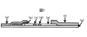
- Types of the diagnostic kit include a lateral flow type and a flow through type. Although it will not specifically limit if color developing particles and a sample pad are used, Preferably it is a lateral flow type. Further, among the lateral flow types, there are a dipstick type and a cassette type, but these types are not particularly limited.
- the configuration of the diagnostic kit is not particularly limited, and any configuration may be used as long as it is generally used in the field.
- the types of members other than the conjugate pad (b) and the sample pad (a) containing antibody-sensitized coloring particles are not particularly limited as long as they are used in this field.
- the “conjugate pad” is a pad containing a conjugate containing colored particles, and contains, for example, colored particles bound to an antibody that binds to a substance to be examined. Although it does not specifically limit as a raw material of a conjugate pad, A general glass fiber or resin fiber etc. can be used.
- resin fibers polyolefin fibers such as polyethylene, polyester fibers, polyamide resins, acrylic resins, and composite fibers of these resin fibers can be preferably used, but are not limited thereto. Absent.
- resin fiber is preferable.
- resin fibers a material that is hydrophobic to some extent is more preferable from the viewpoint of ease of release of the colored particles. If the hydrophobicity is too high, it may be used after pretreatment with a surfactant or the like. More preferably, a polyethylene fiber conjugate pad is pretreated with a surfactant.
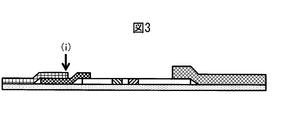
- the sample pad coverage is preferably 50 to 100% with respect to the conjugate pad.
- the coverage indicates how much the sample pad covers the upper area of the conjugate pad. For example, 0% when the sample pad does not overlap the upper part of the conjugate pad, and 100% when the sample pad is completely covered.
- this coverage is often 50% or less.
- the overlap of the sample pad is 5 mm with respect to the length of the conjugate pad of 15 mm, and the coverage is 33%.
- the coverage is 0%.
- the specimen supplied to the sample pad is transferred to the conjugate pad and further transferred to the chromatographic medium.
- the coverage is low, the specimen is transferred to the chromatographic medium without being sufficiently supplied to the downstream side of the conjugate pad in the portion not in contact with the sample pad, and as a result, the particle size as in the present invention is reduced. For large colored particles, release from the conjugate pad may be delayed.
- the lower limit of the preferred coverage is 60%, more preferably 70%, more preferably 80%, and particularly preferably 90%.
- the downstream side of the sample pad may float from the conjugate pad. This is because the sample pad adhered to the mount cannot follow the shape of the conjugate pad and jumps up.
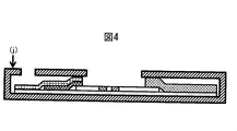
- a method of suppressing there are a method of placing an immunochromatography kit in a casing and suppressing it by a part of the casing, and a method of using a transparent sheet having an adhesive surface. The optimum degree of suppression depends on the structure of the immunochromatography diagnostic kit and cannot be generally stated.
- the most downstream portion of the sample pad is set to -5 to the total thickness of each immunochromatography diagnostic kit member. It is preferable to adjust to +2 mm.
- the position of the most downstream portion of the sample pad changes depending on the coverage of the sample pad. For example, the most downstream portion (h) when the coverage is 100% is shown in FIG. 2, and the most downstream portion (i) when the coverage is 50% is shown in FIG. 2 and 3 are merely examples and do not limit the present invention in any way.
- the total thickness of each immunochromatography diagnostic kit member is, for example, that the sample pad, conjugate pad, chromatographic medium, and mount overlap each other at the most downstream portion of the sample pad in FIG.
- the total thickness is preferably +1 mm or less, more preferably the total thickness + 0 mm or less.
- the total thickness of the members is preferably -3 mm or more, more preferably -1 mm or more.
- FIG. 4 illustrates a case where the thickness is adjusted using the housing (j).
- the casing is composed of an upper part and a lower part, and the thickness can be adjusted by adjusting the clearance.
- FIG. 4 is merely an example and does not limit the present invention.
- the “diagnostic method” using the immunochromatographic diagnostic kit in the present invention refers to various diagnoses performed using the immunochromatographic diagnostic kit.
- the diagnostic object is not particularly limited, and can be used for various diagnostic tests such as human, animal, food, plant, and other environmental tests.
- a sample is collected from the test subject, and if necessary, it is pre-treated, such as extraction and filtration, dripped onto the sample pad, waits for a predetermined time from the start of the test, The diagnosis result is judged from the color development depending on the presence or absence.
- the present invention is not limited to this procedure, and the same procedure and principle can also be used for diagnosis.
- extra foreign matters and contaminants can be removed by filtering the specimen sample in advance, so that further rapid diagnosis and improved diagnostic accuracy can be expected.
- a specimen obtained by subjecting the specimen to dilution treatment with a specimen treatment liquid that has been adjusted to a predetermined composition may be dripped.
- the purpose of using the sample treatment solution is to make it easier to react with the antigen in the sample, to decompose the impurities in the sample, to trap the impurities in the sample, and to suppress non-specific reactions Addition of a component that facilitates the flow of the colored particles, a component that improves the visibility when the colored particles are appropriately aggregated and captured on the test line, and the like.
- a buffer solution As an example, a buffer solution, a surfactant, a protein, an inorganic salt, a water-soluble polymer, a reducing agent, a chelating agent, and the like may be added.
- nonionic surfactants various water-soluble amino acids, proteins, inorganic salts, water-soluble polymers, and the like. Specific component types and amounts added vary depending on the test target and the type of antibody used. Examples of nonionic surfactants include poly (oxyethylene) alkyl ether, poly (oxyethylene) octylphenyl ether, Examples include poly (oxyethylene) nonylphenyl ether.
- water-soluble amino acids examples include asparagine, aspartic acid, alanine, arginine, isoleucine, glycine, glutamine, glutamic acid, cysteine, serine, tyrosine, tryptophan, threonine, valine, histidine, proline, phenylalanine, methionine, lysine, leucine, etc.
- proteins include casein, skim milk, casein degradation products, bovine serum albumin, fish gelatin, and the like.
- the inorganic salt compounds that generate alkali metal ions such as sodium, potassium, and lithium are preferable.
- water-soluble polymer polyethylene glycol, polyvinyl alcohol, polyvinyl pyrrolidone, carboxymethyl cellulose, and other cellulose derivatives are preferable. These components are not only added to the sample treatment solution, but may be added in advance to a sample pad, a conjugate pad, a chromatographic medium (nitrocellulose membrane), or the like.
- the target that can be diagnosed with the immunochromatography diagnostic kit of the present invention is not particularly limited, but specific examples include the following: cancer marker, hormone, infection, autoimmunity, plasma protein, TDM, coagulation / line Soluble, amino acid, peptide, protein, gene, cell, and the like.
- CEA AFP, ferritilin, ⁇ 2 micro, PSA, CA19-9, CA125, BFP, elastase 1, pepsinogen 1 and 2, fecal occult blood, urinary ⁇ 2 micro, PIVKA-2, urinary BTA, insulin E3, HCG, HPL, LH, HCV antigen, HBs antigen, HBs antibody, HBc antibody, HBe antigen, HBe antibody, HTLV-1 antibody, HIV antibody, HIV antigen, HIV virus gene, toxoplasma antibody, syphilis, ASO, A Type influenza antigen, type A influenza antibody, type B influenza antigen, type B influenza antibody, rota antigen, adenovirus antigen, rota adenovirus antigen, group A streptococcus, group B streptococcus, candida antigen, CD fungus, cryptolocus antigen Vibrio cholerae, Neisseria meningitidis antigen, E.
- the colored particles need to carry a substance that specifically binds to an object such as an antibody, but the carrying method is not particularly limited.
- a substance that specifically binds to an object such as an antibody
- the carrying method is not particularly limited.
- support by physical adsorption, support by covalent bond, support by a combination thereof, and the like can be mentioned.
- the type and amount of the substance to be supported are not particularly limited.
- Antibodies are the most common and preferred as the type of substance to be supported.
- supporting by physical adsorption is preferable from the viewpoint of ease
- supporting by covalent bond is preferable from the viewpoint of stability and performance.
- the chromatographic medium used in the immunochromatographic diagnostic kit is not particularly limited, and various commonly used chromatographic media can be used. More specifically, a nitrocellulose membrane can be mentioned. Commercially available nitrocellulose membranes are classified according to the time required to move a certain distance called a flow rate, and the faster the flow rate, the larger the pore size. In the present invention, a membrane having a large pore diameter is preferred because the specimen moves quickly and the colored particles are not easily clogged.
- a specific flow rate is preferably a film faster than 120 sec / 4 cm, more preferably a film faster than 100 sec / 4 cm, and still more preferably a film faster than 90 sec / 4 cm.
- Cellulose linter is dissolved in a good solvent for cellulose.
- a copper ammonia solution prepared by a known method is used as a good solvent.
- an organic solvent + water + ammonia mixed system is mainly used as the coagulation liquid. While the coagulation liquid is stirred, the prepared copper ammonia cellulose solution is added for coagulation. Further, by neutralizing and regenerating by adding sulfuric acid, a slurry containing the desired cellulose particles can be obtained.
- the slurry is acidic due to the residual of the acid used for the regeneration, and further contains impurities such as ammonium salts generated by neutralization, and therefore requires an operation for purification to a cellulose dispersion composed of cellulose particles and a medium.
- this purification operation uses repeated centrifugation, decantation, and dilution with a dispersion medium liquid. Since the cellulose particles in the obtained cellulose particle dispersion may be aggregated in the course of the purification operation, in this case, a dispersion treatment by shearing or the like can be performed.
- a high-pressure homogenizer is used as means for applying shear.
- Method for coloring cellulose particles Sodium sulfate and a reactive dye are added to the obtained aqueous dispersion of cellulose particles, and the temperature is raised to an appropriate temperature in a thermostatic bath while stirring with a magnetic stirrer. After heating, sodium carbonate is added as an alkali to start dyeing.
- a slurry containing the objective dyed cellulose particles can be obtained after a predetermined time. At this time, since the slurry is alkaline and further contains sodium sulfate, unreacted dye, etc., an operation for purification to a dyed cellulose particle dispersion comprising dyed cellulose particles and a medium is required.
- a dyed cellulose particle dispersion is obtained. Since the cellulose particles in the obtained dyed cellulose particle dispersion liquid may aggregate in the course of the purification operation, in this case, a dispersion treatment by shearing or the like can be performed.
- a high-pressure homogenizer is used as means for applying shear. Thereafter, various physical properties of the obtained colored cellulose particles are measured.
- the single fineness and the strength of the nonwoven fabric can be changed.
- the fiber arrangement to be sprinkled onto the net is changed evenly in 3 to 10 steps in the phase of the Sin curve in 3 to 10 layers.
- a cellulosic nonwoven fabric having a very uniform fiber arrangement can be obtained.
- a multilayer nonwoven fabric having such a uniform fiber arrangement has a uniform fiber gap and uniform average pore diameter as a nonwoven fabric. If a nonwoven fabric having such a uniform pore size is densified, it is thin and dense. A nonwoven fabric having a uniform pore size is obtained.
- the shape of the spinning funnel is preferably a rectangular shape, and the length of the spinning funnel to be flowed down is preferably 100 to 400 mm, and the slit width of the flow down outlet is preferably 2 to 5 mm.
- the diameter of the stock solution discharge hole of the spinning nozzle is preferably 0.1 to 0.5 mm, and the shape is preferably a round shape.
- the web after lamination is made by regenerating cellulose in a web state by a method described in, for example, Japanese Patent No. 787914 and Japanese Patent No.
- the obtained nonwoven fabric can be obtained as a dried or wound product. Since the process from spinning to winding is performed in a series of steps, the fibers are connected continuously without being cut. Thereafter, various physical properties of the obtained regenerated cellulose continuous long fiber nonwoven fabric are measured.
- a dispersion of colored cellulose particles adjusted to a predetermined concentration is prepared, a buffer solution and an antibody are added, and the mixture is stirred for a certain period of time while adjusting the temperature, so that the antibodies are adsorbed on the colored cellulose particles.
- the colored cellulose particles are blocked by adding a blocking agent and stirring for a certain time while adjusting the temperature.
- a blocking agent various blocking agents can be used according to the composition of the substance to be examined, the specimen, or a solution for diluting it.
- the casein used in the present invention is particularly preferable for blocking colored cellulose particles.
- Centrifugation is performed to wash the colored cellulose particles after antibody adsorption and blocking, the supernatant liquid containing excess antibody and blocking agent is separated from the precipitated particles, and the supernatant liquid is removed by decantation.
- a liquid such as a buffer solution is added to the settled particles, and if necessary, a dispersion treatment is performed using ultrasonic waves. Washing by a series of operations such as sedimentation by centrifugation, removal of supernatant, and addition of liquid is performed as many times as necessary to prepare a dispersion containing a predetermined concentration of particles subjected to antibody adsorption and blocking.
- Proteins, surfactants, sugars such as sucrose and trehalose are added to this dispersion as needed, and a certain amount of the resulting solution is applied to a polyethylene conjugate pad and dried to adjust the detection reagent-containing part. If necessary, apply a buffer solution, surfactant, protein, reagent for trapping impurities in the specimen, antiseptic, antibacterial agent, antioxidant, hygroscopic agent, etc. to the regenerated cellulose continuous long-fiber nonwoven fabric and dry it. And prepare a sample pad. Further, a chromatographic medium made of a nitrocellulose porous membrane in which an antibody is immobilized at a predetermined position, and an absorbent pad made of cellulose filter paper for absorbing the specimen are prepared.
- the immunochromatography diagnostic kit is prepared by fixing them on a backing sheet having an adhesion site called a backing sheet and cutting it into a predetermined size.
- a nanotrack particle size distribution measuring apparatus UPA-EX150 dynamic light scattering type manufactured by Nikkiso Co., Ltd. was used.
- a measurement sample a sample having 0.01% by weight of colored particles and 99.99% by weight of pure water was used.
- the number of integrations was 30 times, the measurement time per measurement was 30 seconds, and the median diameter was defined as the average particle diameter using a volume average particle size distribution.
- the CV value was calculated using the standard deviation and the average particle size of the particle size distribution obtained by 30 times of integration.
- JASCO V-650 an ultraviolet-visible near-infrared spectrophotometer manufactured by JASCO (optical system: single monochromator, Zellnitana mount, double beam system, light source: deuterium lamp (190 to 350 nm), halogen lamp (330 ⁇ 900 nm)) was used in which an integrating sphere unit ISV-722 manufactured by the same company was attached.
- concentration so that the colored particles are 0.01% by weight and pure water is 99.99% by weight using an aqueous dispersion or dried particles of colored particles of any concentration using distilled water as the dispersion medium. We used what we did.
- a non-woven fabric having an area of 0.5 m 2 or more is dried at 105 ° C. until a constant weight is reached, then left in a constant temperature room at 20 ° C. and 65% RH for 16 hours or more, and the weight is measured. The weight was measured (unit: g / m 2 ).
- a negative specimen was prepared by adjusting 66 mM, pH 7.4 PBS containing 1 wt% BSA. 120 ul of a negative sample was dropped onto the sample dropping part of the diagnostic kit, and the TL color intensity after 15 minutes was measured with an immunochromatographic reader. This measurement was performed 5 times, and if the average value of the obtained values was 5 mABS or less, it was determined that there was no false positive. The reason why the 5 mABS is used here is that there is an individual difference, but if it is 5 mABS or less, the presence of TL cannot be visually confirmed.
- Example 1 [Preparation of antibody-sensitized colored cellulose particles] 120 ⁇ l of 1.0% by weight of colored cellulose particles 1 (average particle size 352 nm, coloring intensity 2.9 ABS, coloring component ratio 49%) prepared by a known method was put in a 15 ml centrifuge tube, and further tris buffer (50 mM). , PH 7.0) and 240 ⁇ l of 0.1% anti-hCG- ⁇ mouse antibody (manufactured by Fitzgerald, 10-C25C) were added, and the mixture was stirred by vortexing for 10 seconds. Then, it put into the dryer adjusted to 37 degreeC, and left still for 120 minutes.
- borate buffer 50 mM boric acid, PH 10.0
- SSH ultrasonic disperser
- centrifugation at 10,000 g was performed for 15 minutes to settle the sensitized particles, and then the supernatant was removed.
- 0.6 g of sucrose manufactured by Wako Pure Chemical Industries, 196-00015
- 0.8 g of 1.0 wt% casein blocking solution were added, and a borate buffer solution (50 mM boric acid, PH 10.0) was used.
- the weight was adjusted to 4.0 g, 0.03% by weight of antibody-sensitized colored cellulose particle dispersion was prepared, and treated with an ultrasonic disperser for 10 seconds.
- a polyethylene conjugate pad (Pall, 6613) was dipped in a large excess of 0.05% by weight Tween-20 (Sigma Aldrich, T2700), and after removing excess liquid, it was dried at 50 ° C. for 60 minutes. I let you. Subsequently, it was cut into a shape having a height of 10 mm and a length of 300 mm. Subsequently, 1020 ⁇ l of 0.03% by weight antibody-sensitized colored cellulose particle dispersion was applied evenly using a micropipette and dried at 50 ° C. for 60 minutes.
- Regenerated cellulose continuous long-fiber nonwoven fabric 1 prepared by a known method (weight per unit: 57.2 g / m 2 , thickness: 0.52 mm, bulk density: 0.11 g / cm 3 , number of shed fibers: 2.0000) After impregnating with PBS buffer (66 mM, PH 7.4) containing 2.0 wt% BSA (Sigma Aldrich, A7906) and 2.0 wt% Tween-20, the excess liquid was removed. It was dried at 50 ° C. for 60 minutes. Subsequently, it was cut into a shape having a height of 20 mm and a length of 300 mm.
- PBS buffer 66 mM, PH 7.4
- BSA Sigma Aldrich, A7906
- a nitrocellulose membrane (manufactured by Millipore, SHF0900425) was cut into a shape having a height of 25 mm and a length of 300 mm.
- a liquid application apparatus Musashi Engineering Co., Ltd., 300DS
- 0.1 ⁇ l / mm of PBS solution 66 mM, PH 7.4
- 0.1 wt% anti-hCG- ⁇ mouse antibody MedixBiochemical, 6601
- a PBS solution (66 mM, PH 7.4) containing 0.1% by weight of anti-mouse-rabbit antibody (Daco, Z0259) was applied to a 12 mm high portion at a rate of 0.1 ⁇ l / mm. Subsequently, it was dried at 37 ° C. for 30 minutes.
- Example 2 An immunochromatographic diagnostic kit was prepared in the same manner as in Example 1 except that the colored particles were colored cellulose particles 2 (average particle size 588 nm, colored intensity 2.7 ABS, colored component ratio 45%), and the performance was evaluated. did. The results are shown in Table 1 below.
- Example 3 An immunochromatographic diagnostic kit was prepared in the same manner as in Example 1 except that the colored particles were colored cellulose particles 3 (average particle size 790 nm, colored intensity 2.6 ABS, colored component ratio 42%), and the performance was evaluated. did. The results are shown in Table 1 below.
- Example 4 An immunochromatographic diagnostic kit was prepared in the same manner as in Example 1 except that the colored particles were colored cellulose particles 4 (average particle size 185 nm, colored intensity 3.4 ABS, colored component ratio 61%), and the performance was evaluated. did. The results are shown in Table 1 below.
- Example 5 An immunochromatographic diagnostic kit was prepared in the same manner as in Example 1 except that the colored particles were colored cellulose particles 5 (average particle size 394 nm, coloring intensity 4.8 ABS, coloring component ratio 75%), and the performance was evaluated. did. The results are shown in Table 1 below.
- Example 6 An immunochromatographic diagnostic kit was prepared in the same manner as in Example 1 except that the colored particles were colored cellulose particles 6 (average particle size 332 nm, colored intensity 1.5 ABS, colored component ratio 32%), and the performance was evaluated. did. The results are shown in Table 1 below.
- Example 7 The non-woven fabric used for the sample pad is a regenerated cellulose continuous long-fiber non-woven fabric 2 (weight per unit area 72.0 g / m 2 , thickness 0.08 mm, bulk density 0.90 g / cm 3 , number of fallen fibers 1.1 thousand).
- An immunochromatographic diagnostic kit was prepared in the same manner as in Example 1, and its performance was evaluated. The results are shown in Table 1 below.
- Example 8 The nonwoven fabric used for the sample pad is a regenerated cellulose continuous long-fiber nonwoven fabric 3 (weight is 50.3 g / m 2 , thickness is 0.75 mm, bulk density is 0.07 g / cm 3 , and the number of fallen fibers is 2.1 thousand).
- An immunochromatographic diagnostic kit was prepared in the same manner as in Example 1, and its performance was evaluated. The results are shown in Table 1 below.
- Example 9 The non-woven fabric used for the sample pad is a regenerated cellulose continuous long-fiber non-woven fabric 4 (weight per unit: 17.9 g / m 2 , thickness 0.13 mm, bulk density 0.14 g / cm 3 , number of fallen fibers 1.000)
- An immunochromatographic diagnostic kit was prepared in the same manner as in Example 1, and its performance was evaluated. The results are shown in Table 1 below.
- the nonwoven fabric used for the sample pad is a regenerated cellulose continuous long fiber nonwoven fabric (weight 61.5 g / m 2 ) and a polyester nonwoven fabric (weight 32.0 g / m 2 ).
- an immunochromatographic diagnostic kit was prepared in the same manner as in Example 1 except that the thickness was 0.30 mm, the bulk density was 0.31 g / cm 3 , and the number of shed fibers was 0.9000), and the performance was evaluated. . The results are shown in Table 1 below.
- Recycled cellulose short fiber nonwoven fabric 1 (weight per unit: 49.2 g / weight) is obtained by kinocross method, which is one of the dry airlaid nonwoven fabric manufacturing methods, in which cupra ammonium fiber is cut into a fiber length of 30 mm and dispersed and laminated with a hydrophilic binder in the air.
- m 2 , thickness of 0.24 mm, bulk density of 0.21 g / cm 3 , and the number of fallen fibers of 105,000 were prepared, and the immunochromatographic diagnostic kit was prepared in the same manner as in Example 1 except that it was used as a sample pad. Were prepared and their performance was evaluated. The results are shown in Table 1 below.
- Example 12 Using lyocell fiber as the fiber to be used, regenerated cellulose short fiber nonwoven fabric 2 (weight per unit: 61.3 g / m 2 , thickness: 0.50 mm, bulk density: 0.12 g / cm 3 , number of fallen fibers: 8. (9,000) was prepared, and an immunochromatographic diagnostic kit was prepared in the same manner as in Example 1 except that it was used as a sample pad, and its performance was evaluated. The results are shown in Table 1 below.
- Example 13 Recycled cellulose short fiber nonwoven fabric 3 (weight per unit: 31.7 g / m 2 , thickness: 0.19 mm, bulk density: 0.17 g / cm 3 , dropping fiber number: 10.
- An immunochromatographic diagnostic kit was prepared in the same manner as in Example 1 except that it was used as a sample pad, and its performance was evaluated. The results are shown in Table 1 below.
- Example 1 An immunochromatographic diagnostic kit was prepared in the same manner as in Example 1 except that the colored particles were colored cellulose particles 7 having a small particle size (average particle size 69 nm, color developing intensity 2.8 ABS, coloring component ratio 48%), Its performance was evaluated. The results are shown in Table 1 below. As is apparent from the results shown in Table 1, when the average particle diameter of the colored particles was small, the line was colored for more than 1 minute, the diagnosis time was slow, and the analytical sensitivity was low.
- Example 2 An immunochromatographic diagnostic kit was prepared in the same manner as in Example 1 except that the colored particles were colored cellulose particles 8 having a large particle size (average particle size 1017 nm, color developing intensity 2.4 ABS, ratio of colored component 41%), Its performance was evaluated. The results are shown in Table 1 below. As is clear from the results shown in Table 1, when the average particle size of the colored particles is large, the diagnosis time is somewhat late, the results are not reproducible, and false positives occur because the particles are clogged. Furthermore, the conjugate pad and the nitrocellulose membrane were colored.
- Example 3 An immunochromatographic diagnostic kit was prepared in the same manner as in Example 1 except that the colored particles were colored latex particles (average particle size 351 nm, colored intensity 0.4 ABS, the ratio of the colored component is unknown), and its performance was evaluated. .
- the results are shown in Table 1 below. As is apparent from the results shown in Table 1, when the colored particles were latex, the colored intensity of the particles was low, the diagnosis time was slow, and the analytical sensitivity was also low.
- Example 4 An immunochromatographic diagnostic kit was prepared in the same manner as in Example 1 except that the colored particles were gold colloid (average particle size 63 nm, colored intensity 2.3 ABS), and the performance was evaluated. The results are shown in Table 1 below. As is apparent from the results shown in Table 1, when the colored particles were gold colloid, the particle size was small, the diagnosis time was slow, the analytical sensitivity was low, and the reproducibility of the results was poor.
- Example 5 Except that the sample pad is a regenerated cellulose continuous long-fiber non-woven fabric 5 having a low basis weight (9.1 g / m 2 , weight 0.12 mm, bulk density 0.08 g / cm 3 , number of shed fibers 2.9 thousand).
- An immunochromatographic diagnostic kit was prepared in the same manner as in Example 1, and its performance was evaluated. The results are shown in Table 1 below.
- Table 1 When the basis weight of the non-woven fabric made of regenerated cellulose fiber used for the sample pad is low, some of the diagnostic liquids leaked from the diagnostic kit and some of the liquids were not supplied to the conjugate pad. The particle release was bad. For this reason, some diagnostic kits have a slow diagnosis time, low analytical sensitivity, and the reproducibility of the results is poor. In addition, handling during wetness was poor and difficult to handle at the pretreatment stage of the sample pad.
- the sample pad is a regenerated cellulose continuous long-fiber nonwoven fabric 6 having a large basis weight and a large thickness (a basis weight of 170 g / m 2 , a thickness of 1.09 mm, a bulk density of 0.16 g / cm 3 , and a number of shed fibers of 2.9000). Except for this, an immunochromatographic diagnostic kit was prepared in the same manner as in Example 1, and its performance was evaluated. The results are shown in Table 1 below.
- the sample pad itself may retain a certain amount of liquid, or the release of particles from the conjugate pad will be poor, and the diagnosis time will It was slow and the reproducibility of the results was poor. Furthermore, the conjugate pad and the nitrocellulose membrane were colored.
- Example 7 Except that the sample pad is thin regenerated cellulose continuous long-fiber nonwoven fabric 7 (57.2 g / m 2 in basis weight, 0.05 mm in thickness, 1.14 g / cm 3 in bulk density, and 1.0000 dropped fibers).
- An immunochromatographic diagnostic kit was prepared in the same manner as in Example 1, and its performance was evaluated. The results are shown in Table 1 below. When the fiber nonwoven fabric made of regenerated cellulose used for the sample pad is thin, the bulk density is high and the liquid absorption rate is poor, and the flow rate per unit time is not sufficient. Was slow and the reproducibility of the results was poor. Furthermore, the conjugate pad and the nitrocellulose membrane were colored.
- Example 8 Similar to Example 1 except that the sample pad is a short pulp nonwoven fabric 1 (weight per unit: 68.0 g / m 2 , thickness: 0.16 mm, bulk density: 0.43 g / cm 3 , number of shed fibers: 92,000)
- An immunochromatography diagnostic kit was prepared by the method described above and its performance was evaluated. The results are shown in Table 1 below. When using short fiber nonwoven fabric for the sample pad, the hydrophilicity of the fiber itself is low and the flow rate per unit time is insufficient. It was bad too. Furthermore, the conjugate pad and the nitrocellulose membrane were colored.
- the sample pad is a pulp short fiber nonwoven fabric 2 (commercially available sample pad for immunochromatography, basis weight 179.0 g / m 2 , thickness 0.48 mm, bulk density 0.37 g / cm 3 , and number of dropped fibers 11.1 thousand) Except for the above, an immunochromatographic diagnostic kit was prepared in the same manner as in Example 1, and its performance was evaluated. The results are shown in Table 1 below. When using short fiber nonwoven fabric for the sample pad, the hydrophilicity of the fiber itself is low and the flow rate per unit time is insufficient. It was bad too. Furthermore, the conjugate pad and the nitrocellulose membrane were colored.
- Example 10 An immunochromatographic diagnostic kit was prepared in the same manner as in Example 1 except that the colored particles were the gold colloid used in Comparative Example 4 and the sample pad was the pulp short fiber nonwoven fabric 2 used in Comparative Example 9, and the performance was evaluated. did. The results are shown in Table 1 below. In this combination, the diagnosis time was very slow, the analytical sensitivity was low, and the reproducibility of the results was poor.
- Example 11 An immunochromatographic diagnostic kit was prepared in the same manner as in Example 1 except that the colored particles were the colored latex particles used in Comparative Example 3, and the sample pad was the pulp short fiber nonwoven fabric 2 used in Comparative Example 9, and the performance was measured. evaluated. The results are shown in Table 1 below. In this combination, the diagnosis time was very slow, the analytical sensitivity was low, and the reproducibility of the results was poor.
- Example 1 the color intensity is very high and the color intensity is stabilized quickly. This is an effect of combining the predetermined coloring particles and the predetermined sample pad according to the present invention, and it can be said that the particles are released from the conjugate pad at an earlier stage.
- Example 1 Comparative Example 4, and Comparative Example 9, when performance was evaluated under exactly the same conditions except that the hCG system was changed to an influenza system, the same effect as that of the hCG system was confirmed in the influenza system.
- Example 14 The immunochromatography diagnostic kit was prepared in the same manner as in Example 1 except that the sample pad coverage was 70% by setting the sample pad to a length of 17 mm, and its performance was evaluated. The results are shown in Table 2 below.
- Example 15 An immunochromatographic diagnostic kit was prepared in the same manner as in Example 1 except that the sample pad length was 15 mm, so that the coverage of the sample pad with respect to the conjugate pad was 50%, and its performance was evaluated. The results are shown in Table 2 below.
- Example 12 An immunochromatography diagnostic kit was prepared in the same manner as in Example 1 except that the sample pad coverage was 20% by changing the length of the sample pad to 12 mm, and its performance was evaluated. The results are shown in Table 2 below.
- the immunochromatography diagnostic kit according to the present invention is capable of rapid diagnosis, has high analytical sensitivity, and is excellent in reproducibility of test results, so that it can be suitably used as a diagnostic agent.
Landscapes
- Health & Medical Sciences (AREA)
- Immunology (AREA)
- Life Sciences & Earth Sciences (AREA)
- Engineering & Computer Science (AREA)
- Molecular Biology (AREA)
- Biomedical Technology (AREA)
- Chemical & Material Sciences (AREA)
- Hematology (AREA)
- Urology & Nephrology (AREA)
- Biotechnology (AREA)
- Biochemistry (AREA)
- Cell Biology (AREA)
- Food Science & Technology (AREA)
- Medicinal Chemistry (AREA)
- Physics & Mathematics (AREA)
- Analytical Chemistry (AREA)
- Microbiology (AREA)
- General Health & Medical Sciences (AREA)
- General Physics & Mathematics (AREA)
- Pathology (AREA)
- Investigating Or Analysing Materials By The Use Of Chemical Reactions (AREA)
- Investigating Or Analysing Biological Materials (AREA)
- Textile Engineering (AREA)
Abstract
Description
(1)検査対象物質である抗原に特異的に結合する抗体をニトロセルロース膜などのクロマトグラフ媒体の所定の部位に固定化し、クロマトグラフ媒体の任意の位置にテストライン(以下「TL」という。)と呼ばれる反応部位を形成する。
(2)酵素、発色粒子、蛍光発色粒子、磁性粒子などの標識物質に、検査対象物質と特異的に結合する抗体を担持させた検出試薬を調製し、コンジュゲートパッドなどに検出試薬を塗布乾燥し、検出試薬含有部を形成させ、前記クロマトグラフ媒体と組み合わせてイムノクロマト診断キットを形成する。
(3)抗原を含む検体そのもの、又はそれを任意の液体で希釈した溶液を前記イムノクロマト診断キットの所定の位置に、例えば、サンプルパッドに滴下し、抗原と検出試薬をクロマトグラフ媒体上に展開させる。
これらの操作によって、反応部位においてクロマトグラフ媒体上に固定化された抗体に、抗原を介して標識物質が捕捉され、標識物質の信号を検出することでイムノクロマト診断キットによる診断を行う。一般的な診断は抗原の有無のみを検査する定性診断だが、近年ではその信号の強度を目視あるいは機械で検出することで定量診断を行うこともできる。
また、以下の特許文献1には、検査対象物質を含む検体試料を滴下する部分であるサンプルパッドに特定のセルロース系繊維不織布を用いることで、診断時間の迅速化が可能なことが報告されている。特許文献1には、吸液速度や展開性についても触れられているが、具体的な診断時間のデータはなく、本願発明のような特定の発色粒子との組み合わせについては全く言及されていない。更にサンプルパッドとコンジュゲートパッドの配置の工夫についても言及されていない。
さらに、以下の特許文献2には、診断キットの構造などを工夫することで、診断時間の迅速化が可能なことが報告されており、例えば、hCGアッセイにおいて最短2分35秒で判定を行っている。
さらに、以下の特許文献5には、蛍光粒子を発色粒子として用いたイムノクロマト診断キットが開示されており、発色粒子の素材としてセルロース粒子が、サンプルパッドの素材としてセルロース不織布がそれぞれ一例として挙げられているが、いずれも具体的には何も述べられていない。更にサンプルパッドとコンジュゲートパッドの配置についての工夫もなされていない。当然ながら、本願発明のようなサンプルパッドと発色粒子の組み合わせによる効果も記載されていない。
[1]平均粒子径が100~1000nmであり、発色強度が1.0~10.0であり、粒子の重量の10~90重量%がセルロース由来、かつ、90~10重量%が着色成分由来である発色粒子を含むコンジュゲートを含有するコンジュゲートパッドと、目付が10~150g/m2であり、かつ、厚みが0.07~1.00mmである再生セルロース系繊維からなる不織布から構成される体外診断薬用サンプルパッドとを、含むイムノクロマト診断キット。
本発明における「発色粒子」とは、水、緩衝液などに不溶性であり、色素や染料等が担持された粒子状物質を指す。粒子を構成する素材は特に限定されないが、このような発色粒子としては、例えば、金コロイド、白金コロイド、銀コロイド、セレンコロイドなどの金属コロイド粒子、ポリスチレンラテックス等のスチレン系ラテックスやアクリル酸系ラテックス等を着色した着色ラテックス粒子、ケイ素原子及び酸素原子からなる3次元構造体からなるシリカを着色した着色シリカ粒子、セルロースを着色した着色セルロース粒子、カーボンブラックなどの着色成分をそのまま粒子化した発色粒子、磁性粒子、などが挙げられる。また、前記発色粒子は蛍光発光性粒子でもかまわない。粒子径の調整、色の濃さの調整、色の種類の調整、粒子表面状態の調整などの粒子の特徴の調整のしやすさから、着色セルロース粒子が好ましい。セルロースは大量の水酸基を有するため、親水性が高く分散安定性に優れ、着色成分を大量に含有することができる。
被覆率(%)=重なっている部分の面積(A)/全体の面積(B)×100
セルロースリンターをセルロースの良溶媒に溶解させる。本発明では良溶媒として公知の方法で調製した銅アンモニア溶液を用いる。そして凝固液としては有機溶媒+水+アンモニア混合系を主に用いる。この凝固液を攪拌しながら、調製しておいた銅アンモニアセルロ-ス溶液を加えて凝固を行う。さらに硫酸を加え中和、再生を行うことで、目的のセルロ-ス粒子を含有したスラリーを得ることができる。この際スラリーは再生に用いた酸の残留により酸性であり、さらに中和で発生したアンモニウム塩などの不純物を含んでいるため、セルロース粒子と媒体からなるセルロース分散液へと精製する操作が必要となる。本発明ではこの精製操作として遠心分離-デカンテーション-分散媒液体による希釈の処理の繰り返しを用いる。得られたセルロース粒子分散液中のセルロース粒子は、精製操作の過程において凝集する場合もあるので、この場合は剪断などによる分散処理を行うことができる。本発明では剪断を与える手段としては高圧ホモジナイザーを用いる。
得られたセルロース粒子の水分散体に対し、硫酸ナトリウム、反応性染料を加え、マグネティックスターラーで撹拌しながら恒温槽で適温に昇温する。昇温後にアルカリとして炭酸ナトリウムを加え染色を開始する。所定時間経過後に目的の染色セルロース粒子を含有したスラリーを得ることができる。この際スラリーはアルカリ性であり、さらに、硫酸ナトリウム、未反応の染料などを含んでいるため、染色セルロース粒子と媒体からなる染色セルロース粒子分散液へと精製する操作が必要となる。前記同様に遠心分離による精製を行い、染色セルロース粒子分散液を得る。得られた染色セルロース粒子分散液中のセルロース粒子は、精製操作の過程において凝集する場合もあるので、この場合は剪断などによる分散処理を行うことができる。本発明では剪断を与える手段としては高圧ホモジナイザーを用いる。その後、得られた着色セルロース粒子の各種物性を測定する。
異物を除去し、重合度を調整したコットンリンターを銅アンモニウム溶液に溶解させた原液を、細孔(原液吐出孔)を有した紡糸口金(紡口)から押し出し、水と共に漏斗内を落下させ、脱アンモニアさせることにより原液を凝固させつつ、延伸を行い、ネット上へ振り落としウェブ形成させる。この際、ネットを進行させながら進行方向と垂直方向へ振動させることにより、ネットへ振り落とされる繊維はSinカーブを描くことになる。紡糸時の延伸は100~500倍が可能であり、紡糸漏斗の形状と、その中を流下させる紡糸水量を変えることにより、延伸倍率の調整が任意に可能である。延伸倍率を変えることにより、単繊度や不織布の強度を変えることが可能である。不織布のウェブ形成において、繊維配列の均一性を得る上で、ネットへ振り落とされる繊維配列を、Sinカーブの位相を層単位で、3~10段階で均等に変え、3~10層の積層ウェブとすることで、極めて均一な繊維配列を有するセルロース系不織布を得ることができる。この様な均一な繊維配列を有する多層構造不織布は、繊維間隙が均一で、不織布としての平均孔径が揃っており、このような均一な孔径を有する不織布を緻密化すれば、薄く、緻密でしかも均一な孔径を有する不織布が得られる。また、紡糸水量や温度を変化させることにより、原液内に微量残留する低分子量セルロース、いわゆるヘミセルロースをコントロールすることも可能である。また、ネットの進行速度、振動幅を制御することにより、繊維配列方向を制御し、不織布としての強度や伸度等をコントロールすることが可能である。
所定の濃度に調整した着色セルロース粒子の分散液を準備し、緩衝液、抗体を加え、温度調整を行いながら一定時間撹拌し、着色セルロース粒子に抗体を吸着させる。一定時間撹拌後、更にブロッキング剤を加え温度調整を行いながら一定時間撹拌することで、着色セルロース粒子のブロッキングを行う。ブロッキング剤としては、検査対象物質や検体又はそれを希釈する溶液の組成などに応じ様々なブロッキング剤を用いることができる。本発明で用いたカゼインは、着色セルロース粒子のブロッキングに特に好ましい。抗体吸着&ブロッキング後の着色セルロース粒子を洗浄するため、遠心分離を行い、余剰な抗体とブロッキング剤が含まれた上澄み液と沈降した粒子を分離し、上澄み液をデカンテーションにて除去する。沈降した粒子に緩衝液などの液体を加え、必要に応じ超音波などで分散処理を行う。この遠心分離による沈降、上澄みの除去、液体の添加という一連の操作による洗浄を必要回数行い、抗体吸着&ブロッキングを行った粒子を所定の濃度含有した分散液を調整する。この分散液に必要に応じタンパク質、界面活性剤、スクロースやトレハロースなどの糖を加え、得られた溶液をポリエチレン製のコンジュゲートパッドに一定量塗布し、乾燥させ、検出試薬含有部を調整する。また再生セルロース連続長繊維不織布に必要に応じ緩衝液、界面活性剤、タンパク、検体試料中の夾雑物をトラップする試薬、防腐剤、抗菌剤、酸化防止剤、吸湿剤、などを塗布し、乾燥させ、サンプルパッドを調製する。更に所定の位置に抗体を固定化したニトロセルロース多孔膜製のクロマトグラフ媒体、検体を吸収するためのセルロース濾紙製の吸収パッドを調製する。それらをバッキングシートと呼ばれる接着部位を有する台紙に固定化し、所定のサイズに裁断することでイムノクロマト診断キットを作製する。
装置としては日機装社製のナノトラック粒度分布測定装置UPA-EX150(動的光散乱式)を用いた。測定サンプルとして、発色粒子が0.01重量%、純水99.99重量%のサンプルを用いた。測定条件としては積算回数を30回、1測定辺りの測定時間を30秒とし、体積平均の粒子径分布を用いそのメジアン径を平均粒子径とした。また、30回の積算によって得られた粒度分布の標準偏差と平均粒子径を用いCV値を算出した。
装置としては日本分光社製の紫外可視近赤外分光光度計JASCO V-650(光学系:シングルモノクロメータ、ツェルニターナマウント、ダブルビーム方式 光源:重水素ランプ(190~350nm)、ハロゲンランプ(330~900nm))に同社製の積分球ユニットISV-722を取り付けた装置を用いた。測定するサンプルは、任意の濃度の発色粒子の水分散液又は乾燥粒子を、分散媒として蒸留水を用いて発色粒子が0.01重量%、純水99.99重量%になるよう濃度を調整したものを使用した。この濃度調整した水分散液を、光路長10mmの石英セル(容量:3.5mL 光路幅:10mm)に2.5mL加え、この石英セルを紫外可視近赤外分光光度計のサンプルフォルダにセットし、その後、測定を実施した。得られた吸光度ピークのうち、400~800nm可視光範囲での最大値(ABS)を発色強度とした。
所定回数の着色操作を行った後の発色粒子の重量と、着色前の粒子の重量から計算し算出した。例えば、1.0gのセルロース粒子を着色し、2.5gの着色セルロース粒子を得た場合の着色成分は2.5g-1.0g=1.5gとして計算した。この場合の着色成分の割合は1.5g÷2.5g×100=60.0重量%となる。
前記の通り、「発色粒子のセルロース由来成分の割合」=100%-(発色粒子の着色成分の割合)の計算式によって計算した。
装置としては日本電子株式社製の走査型電子顕微鏡JSM-6700を用いた。発色粒子が0.01重量%、純水99.99重量%のサンプルを雲母板に滴下し、10秒経過させることで発色粒子を雲母板上に吸着させ、キムワイプで余分な液体を吸い取り乾燥させた。得られた雲母板をプラチナでコーティングし、電子顕微鏡測定用のサンプルを調製した。加速電圧1.6kV、測定倍率5万倍で観測を行い、粒子画像が100個以上になるように必要枚数の画像を撮影し、それぞれの粒子の長径(L)と短径(D)を測定し、粒子100個のL/Dの平均値を算出した。
JIS-L1096準拠の厚み試験にて荷重を1.96kPaとして測定した(単位はmm)。
0.5m2以上の面積の不織布を、105℃で一定重量になるまで乾燥後、20℃、65%RHの恒温室に16時間以上放置してその重量を測定し、不織布の単位面積当たりの重量を測定した(単位はg/m2)。
前記の不織布の単位面積当たりの重量を厚みで割り返すことで算出した(単位はg/cm3)。
純水300mlに25cm×25cmのサンプルを入れ2分間静置した。サンプルを取り出した残りの液を黒色濾紙(ADVANTEC NO131)で濾過し、濾過後の濾紙を恒温室(20℃、65%RH)に12時間入れて乾燥した後、ビデオマイクロスコープにて大きさ100μm以上の脱落繊維数の個数を測定した(単位は個/m2)。
5mm幅にカットしたイムノクロマト診断キットをプラスチックのハウジングに入れた。得られたハウジング入りの診断キットを、浜松ホトニクス社製のイムノクロマトリーダーC10066-10を用い測定した。用いる粒子の色に応じて装置の設定を行った。検査対象物質にはヒト絨毛性ゴナドトロピン(以下「hCG」という。)を用い、hCGを、1重量%の牛血清アルブミン(以下「BSA」という。)を含む66mM、PH7.4のリン酸緩衝液(以下「PBS」という。)で希釈し、hCG濃度が10mIU/mlの陽性検体を調製した。この陽性検体120μlを診断キットのサンプル滴下部に滴下し、以降20秒毎にイムノクロマトリーダーで測定を行い、TLの経時変化を測定した。ここで20秒毎とした理由は、測定1回につき20秒弱が必要なためである。イムノクロマトリーダーで得られるTLの発色強度(単位はmABS)が20mABS以上になった時間を測定した。ここで20mABSとした理由は、個人差もあるが20mABS以上になれば目視でもTLの存在を確認できるからである。この測定を5回行い、平均の時間を診断時間とした。
前記同様に120ulの陽性検体を診断キットのサンプル滴下部に滴下し、15分経過後のTLの発色強度をイムノクロマトリーダーで測定した。この測定を20回行い、得られた値の平均値をTL強度、その標準偏差をTL強度標準偏差とした。再現性を表す指標%CVは下記式(1)により算出した:
%CV=TL強度標準偏差/TL強度×100・・・式(1)
1重量%BSAを含む66mM、PH7.4のPBSを調整し陰性検体を調製した。120ulの陰性検体を診断キットのサンプル滴下部に滴下し、15分経過後のTLの発色強度をイムノクロマトリーダーで測定した。この測定を5回行い、得られた値の平均値が5mABS以下であれば偽陽性はないと判断した。ここで5mABSとした理由は、個人差もあるが5mABS以下であれば目視ではTLの存在が確認できないからである。
hCG濃度を3.20mIU/ml、1.60mIU/ml、0.80mIU/ml、0.40mIU/ml、0.20mIU/ml、0.10mIU/ml、0.05mIU/ml、0.025mIU/mlと段階的に薄くしていった陽性検体を調製した。前記同様に120μlを診断キットのサンプル滴下部に滴下し、15分経過後のTLの発色強度をイムノクロマトリーダーで測定した。この測定を各濃度で5回行い、得られた値の平均値が陰性検体を測定した時の値+20mABS以上の場合は陽性判定、以下の場合は検出限界以下と見なした。この陽性判定が得られる下限のhCG濃度を検出限界とした。
[抗体感作着色セルロース粒子の調製]
既知の方法で調製した1.0重量%の着色セルロース粒子1(平均粒子径352nm、発色強度2.9ABS、着色成分の割合49%)120μlを15mlの遠心管に入れ、更にトリス緩衝液(50mM、PH7.0)を240μl、0.1%の抗hCG-αマウス抗体(Fitzgerald社製、10-C25C)を120μl加え、ボルテックスで10秒撹拌した。続いて37℃に調整した乾燥機内に入れ120分間静置した。続いて1.0重量%のカゼイン(和光純薬工業社製、030-01505)を含有するブロッキング液(100mMホウ酸、PH8.5)を14.4ml加え、更に37℃の乾燥機内で60分間静置した。続いて遠心分離機(クボタ商事社製、6200)と遠心分離ローター(クボタ商事社製、AF-5008C)を用い、10,000gの遠心を15分間行い、感作粒子を沈降させた後に上澄みを除去した。続いてホウ酸緩衝液(50mMホウ酸、PH10.0)を14.4ml加え、超音波分散機(エスエムテー社製、UH-50)で10秒間処理した。続いて10,000gの遠心を15分間行い、感作粒子を沈降させた後に上澄みを除去した。続いてスクロース(和光純薬工業社製、196-00015)を0.6g、1.0重量%のカゼインブロッキング液を0.8g加え、ホウ酸緩衝液(50mMホウ酸、PH10.0)を用い重量を4.0gに調整し、0.03重量%の抗体感作着色セルロース粒子分散液を調整し、超音波分散機で10秒間処理した。
ポリエチレン製コンジュゲートパッド(Pall社製、6613)を大過剰の0.05重量%のTween-20(シグマアルドリッチ社製、T2700)に浸漬し、余分な液を取り除いた後に50℃で60分乾燥させた。続いて高さ10mm、長さ300mmの形状にカットした。続いてマイクロピペットを用い0.03重量%の抗体感作着色セルロース粒子分散液1020μlを均等に塗布し、50℃で60分乾燥させた。
既知の方法で調整した再生セルロース連続長繊維不織布1(目付57.2g/m2、厚み0.52mm、嵩密度0.11g/cm3、脱落繊維数2.0千個)を、大過剰の2.0重量%のBSA(シグマアルドリッチ社製、A7906)と2.0重量%のTween-20を含有するPBS緩衝液(66mM、PH7.4)に含浸し、余分な液を取り除いたのちに50℃で60分乾燥させた。続いて高さ20mm、長さ300mmの形状にカットした。
ニトロセルロース膜(Millipore社製、SHF0900425)を高さ25mm、長さ300mmの形状にカットした。液体塗布装置(武蔵エンジニアリング社製、300DS)を用い、0.1重量%抗hCG-βマウス抗体(MedixBiochemica社製、6601)を含むPBS溶液(66mM、PH7.4)を0.1μl/mmの割合で高さ7mmの部分に塗布した。続いて0.1重量%の抗マウス-ウサギ抗体(Daco社製、Z0259)を含むPBS溶液(66mM、PH7.4)を0.1μl/mmの割合で高さ12mmの部分に塗布した。続いて37℃で30分乾燥させた。
バッキングカード(Adhesives Reserch社製、AR9020)に、調整した捕捉抗体塗布ニトロセルロース膜、吸収パッド(Millipore社製、C083)、抗体感作着色セルロース粒子を含有したコンジュゲートパッド、再生セルロース連続長繊維不織布サンプルパッドを、図1のようなレイアウトで張り合わせた。続いて裁断機にて5mmの幅にカットし、幅5mm、高さ60mmのイムノクロマト診断キットを得た。
得られたイムノクロマト診断キットの性能を評価した。結果を以下の表1に示す。
発色粒子が着色セルロース粒子2(平均粒子径588nm、発色強度2.7ABS、着色成分の割合45%)であること以外は実施例1と同様の方法でイムノクロマト診断キットを調製し、その性能を評価した。結果を以下の表1に示す。
発色粒子が着色セルロース粒子3(平均粒子径790nm、発色強度2.6ABS、着色成分の割合42%)であること以外は実施例1と同様の方法でイムノクロマト診断キットを調製し、その性能を評価した。結果を以下の表1に示す。
発色粒子が着色セルロース粒子4(平均粒子径185nm、発色強度3.4ABS、着色成分の割合61%)であること以外は実施例1と同様の方法でイムノクロマト診断キットを調製し、その性能を評価した。結果を以下の表1に示す。
発色粒子が着色セルロース粒子5(平均粒子径394nm、発色強度4.8ABS、着色成分の割合75%)であること以外は実施例1と同様の方法でイムノクロマト診断キットを調製し、その性能を評価した。結果を以下の表1に示す。
発色粒子が着色セルロース粒子6(平均粒子径332nm、発色強度1.5ABS、着色成分の割合32%)であること以外は実施例1と同様の方法でイムノクロマト診断キットを調製し、その性能を評価した。結果を以下の表1に示す。
サンプルパッドに用いる不織布が再生セルロース連続長繊維不織布2(目付72.0g/m2、厚み0.08mm、嵩密度0.90g/cm3、脱落繊維数1.1千個)であること以外は実施例1と同様の方法でイムノクロマト診断キットを調製し、その性能を評価した。結果を以下の表1に示す。
サンプルパッドに用いる不織布が再生セルロース連続長繊維不織布3(目付50.3g/m2、厚み0.75mm、嵩密度0.07g/cm3、脱落繊維数2.1千個)であること以外は実施例1と同様の方法でイムノクロマト診断キットを調製し、その性能を評価した。結果を以下の表1に示す。
サンプルパッドに用いる不織布が再生セルロース連続長繊維不織布4(目付17.9g/m2、厚み0.13mm、嵩密度0.14g/cm3、脱落繊維数1.5千個)であること以外は実施例1と同様の方法でイムノクロマト診断キットを調製し、その性能を評価した。結果を以下の表1に示す。
サンプルパッドに用いる不織布が再生セルロース連続長繊維不織布(目付61.5g/m2)とポリエステル不織布(目付32.0g/m2)をウォータジェットで水流交絡させた複合不織布(目付92.5g/m2、厚み0.30mm、嵩密度0.31g/cm3、脱落繊維数0.9千個)であること以外は実施例1と同様の方法でイムノクロマト診断キットを調製し、その性能を評価した。結果を以下の表1に示す。
キュプラアンモニウム繊維を繊維長30mmにカットし、空気中に親水性バインダーと共に分散し積層させる、乾式エアレイド不織布の製法の一つであるキノクロス法にて、再生セルロース短繊維不織布1(目付49.2g/m2、厚み0.24mm、嵩密度0.21g/cm3、脱落繊維数10.5千個)を調製し、それをサンプルパッドに用いること以外は実施例1と同様の方法でイムノクロマト診断キットを調製し、その性能を評価した。結果を以下の表1に示す。
用いる繊維としてリヨセル繊維を用い、実施例11と同様の方法で再生セルロース短繊維不織布2(目付61.3g/m2、厚み0.50mm、嵩密度0.12g/cm3、脱落繊維数8.9千個)を調製し、それをサンプルパッドに用いること以外は実施例1と同様の方法でイムノクロマト診断キットを調製し、その性能を評価した。結果を以下の表1に示す。
用いる繊維としてレーヨン繊維を用い、実施例11と同様の方法で再生セルロース短繊維不織布3(目付31.7g/m2、厚み0.19mm、嵩密度0.17g/cm3、脱落繊維数10.0千個)を調製し、それをサンプルパッドに用いること以外は実施例1と同様の方法でイムノクロマト診断キットを調製し、その性能を評価した。結果を以下の表1に示す。
実施例1~13においてはいずれも、粒子径が大きく発色強度が高い所定の発色粒子と、嵩密度が低く厚みが薄い所定の再生セルロース系繊維からなる不織布のサンプルパッドを併用することにより、以下の表1に示すように、発色が早く、1分以内に線が発色し、診断時間が短く、分析感度が高く、結果の再現性に優れるイムノクロマト診断キットを得ることができた。
発色粒子が粒子径の小さい着色セルロース粒子7(平均粒子径69nm、発色強度2.8ABS、着色成分の割合48%)であること以外は実施例1と同様の方法でイムノクロマト診断キットを調製し、その性能を評価した。結果を以下の表1に示す。表1に示す結果から明らかなように発色粒子の平均粒子径が小さい場合、1分を超えて線が発色し、診断時間が遅く、分析感度も低かった。
発色粒子が粒子径の大きい着色セルロース粒子8(平均粒子径1017nm、発色強度2.4ABS、着色成分の割合41%)であること以外は実施例1と同様の方法でイムノクロマト診断キットを調製し、その性能を評価した。結果を以下の表1に示す。表1に示す結果から明らかなように発色粒子の平均粒子径が大きい場合は粒子が詰まるためか、診断時間がいくぶん遅く、結果の再現性が悪く、偽陽性も発生した。更にコンジュゲートパッドやニトロセルロース膜が着色していた。
発色粒子が着色ラテックス粒子(平均粒子径351nm、発色強度0.4ABS、着色成分の割合は不明)であること以外は実施例1と同様の方法でイムノクロマト診断キットを調製し、その性能を評価した。結果を以下の表1に示す。表1に示す結果から明らかなように発色粒子がラテックスの場合は粒子の発色強度が低く、診断時間が遅く、分析感度も低かった。
発色粒子が金コロイド(平均粒子径63nm、発色強度2.3ABS)であること以外は実施例1と同様の方法でイムノクロマト診断キットを調製し、その性能を評価した。結果を以下の表1に示す。表1に示す結果から明らかなように発色粒子が金コロイドの場合は粒子径が小さく、診断時間が遅く、分析感度も低く、結果の再現性も悪かった。
実施例1~13と比較例1~4との間の比較から明らかなように、発色粒子の粒子径が最適範囲にない場合や、発色強度が低い場合である比較例1~4においては、検査時間、分析感度、結果の再現性、偽陽性の有無などを全て兼ね備える診断キットを提供することはできなかった。
サンプルパッドが目付の低い再生セルロース連続長繊維不織布5(目付9.1g/m2、厚み0.12mm、嵩密度0.08g/cm3、脱落繊維数2.9千個)であること以外は実施例1と同様の方法でイムノクロマト診断キットを調製し、その性能を評価した。結果を以下の表1に示す。サンプルパッドに用いる再生セルロース系繊維からなる不織布の目付が低い場合、いくつかの診断キットは検体液の一部が診断キット上から漏れてしまい、コンジュゲートパッドに十分な液量が供給されなかったために粒子のリリースが悪かった。そのためいくつかの診断キットは診断時間が遅く、分析感度が低く、その影響で結果の再現性も悪かった。また、サンプルパッドの前処理の段階で湿潤時のハンドリングが悪く扱いにくかった。
サンプルパッドが、目付が大きく、厚みが厚い再生セルロース連続長繊維不織布6(目付170g/m2、厚み1.09mm、嵩密度0.16g/cm3、脱落繊維数2.9千個)であること以外は実施例1と同様の方法でイムノクロマト診断キットを調製し、その性能を評価した。結果を以下の表1に示す。サンプルパッドに用いる再生セルロース系繊維からなる不織布の目付が大きく、厚みが厚い場合、サンプルパッド自体がある程度の液量を保持してしまうためかコンジュゲートパッドからの粒子のリリースが悪く、診断時間が遅くなり、結果の再現性も悪かった。更にコンジュゲートパッドやニトロセルロース膜が着色していた。
サンプルパッドが厚みの薄い再生セルロース連続長繊維不織布7(目付57.2g/m2、厚み0.05mm、嵩密度1.14g/cm3、脱落繊維数1.0千個)であること以外は実施例1と同様の方法でイムノクロマト診断キットを調製し、その性能を評価した。結果を以下の表1に示す。サンプルパッドに用いる再生セルロース系からなる繊維不織布の厚みが薄い場合、嵩密度が高く吸液速度が悪くなり単位時間当たりの流量が十分でないためかコンジュゲートパッドからの粒子のリリースが悪く、診断時間が遅くなり、結果の再現性も悪かった。更にコンジュゲートパッドやニトロセルロース膜が着色していた。
サンプルパッドがパルプ短繊維不織布1(目付68.0g/m2、厚み0.16mm、嵩密度0.43g/cm3、脱落繊維数9.2千個)であること以外は実施例1と同様の方法でイムノクロマト診断キットを調製し、その性能を評価した。結果を以下の表1に示す。サンプルパッドにパルプ短繊維不織布を用いた場合、繊維自体の親水性が低く単位時間当たりの流量が十分でないためかコンジュゲートパッドからの粒子のリリースが悪く、診断時間が遅くなり、結果の再現性も悪かった。更にコンジュゲートパッドやニトロセルロース膜が着色していた。
サンプルパッドがパルプ短繊維不織布2(市販のイムノクロマト用サンプルパッド、目付179.0g/m2、厚み0.48mm、嵩密度0.37g/cm3、脱落繊維数11.1千個)であること以外は実施例1と同様の方法でイムノクロマト診断キットを調製し、その性能を評価した。結果を以下の表1に示す。サンプルパッドにパルプ短繊維不織布を用いた場合、繊維自体の親水性が低く単位時間当たりの流量が十分でないためかコンジュゲートパッドからの粒子のリリースが悪く、診断時間が遅くなり、結果の再現性も悪かった。更にコンジュゲートパッドやニトロセルロース膜が着色していた。
実施例1~13と比較例5~9との間の比較から明らかなように、サンプルパッドに用いる再生セルロース系繊維からなる不織布の目付や厚みが所定範囲外である場合や、再生セルロース系繊維以外の繊維からなる場合には、表1に示すように、診断時間、分析感度、結果の再現性、偽陽性の有無などを全て兼ね備える診断キットを提供することはできなかった。
発色粒子が比較例4で用いた金コロイド、サンプルパッドが比較例9で用いたパルプ短繊維不織布2であること以外は実施例1と同様の方法でイムノクロマト診断キットを調製し、その性能を評価した。結果を以下の表1に示す。この組み合わせの場合、診断時間は非常に遅く、分析感度は低く、結果の再現性も悪かった。
発色粒子が比較例3で用いた着色ラテックス粒子、サンプルパッドが比較例9で用いたパルプ短繊維不織布2であること以外は実施例1と同様の方法でイムノクロマト診断キットを調製し、その性能を評価した。結果を以下の表1に示す。この組み合わせの場合、診断時間は非常に遅く、分析感度は低く、結果の再現性も悪かった。
実施例1、比較例4、比較例9、比較例10で実施したイムノクロマト診断キットの診断時間の測定の結果を用い、TLの発色強度の経時変化を比較した。その結果を図5に示す。図5から明らかなように、発色粒子に金コロイドを用いた比較例4、比較例10はサンプルパッドの影響をあまり受けない。また、特定の発色粒子を用いたもののサンプルパッドに市販品のイムノクロマトサンプルパッドを用いた比較例9はある程度の時間が経過した後も緩やかに発色強度が増大している。これはサンプルパッドの吸液速度が十分ではなく、単位時間当たりの検体液の流量が少ないため、コンジュゲートパッドから発色粒子がリリースされ続けているためである。それに対し実施例1では発色強度が非常に高く、かつ発色強度が早く安定化している。これは本発明の奏する所定の発色粒子と所定のサンプルパッドを組み合わせた効果であり、より早い段階でコンジュゲートパッドから粒子がリリースされていることを裏付けるものといえる。
〔実施例14〕
サンプルパッドを長さ17mmにすることでコンジュゲートパッドに対するサンプルパッドの被覆率を70%とした以外は実施例1と同様の方法でイムノクロマト診断キットを調整し、その性能を評価した。結果を以下の表2に示す。
サンプルパッドの長さを15mmにすることでコンジュゲートパッドに対するサンプルパッドの被覆率を50%とした以外は実施例1と同様の方法でイムノクロマト診断キットを調整し、その性能を評価した。結果を以下の表2に示す。
サンプルパッドの長さを12mmにすることでコンジュゲートパッドに対するサンプルパッドの被覆率を20%とした以外は実施例1と同様の方法でイムノクロマト診断キットを調整し、その性能を評価した。結果を以下の表2に示す。

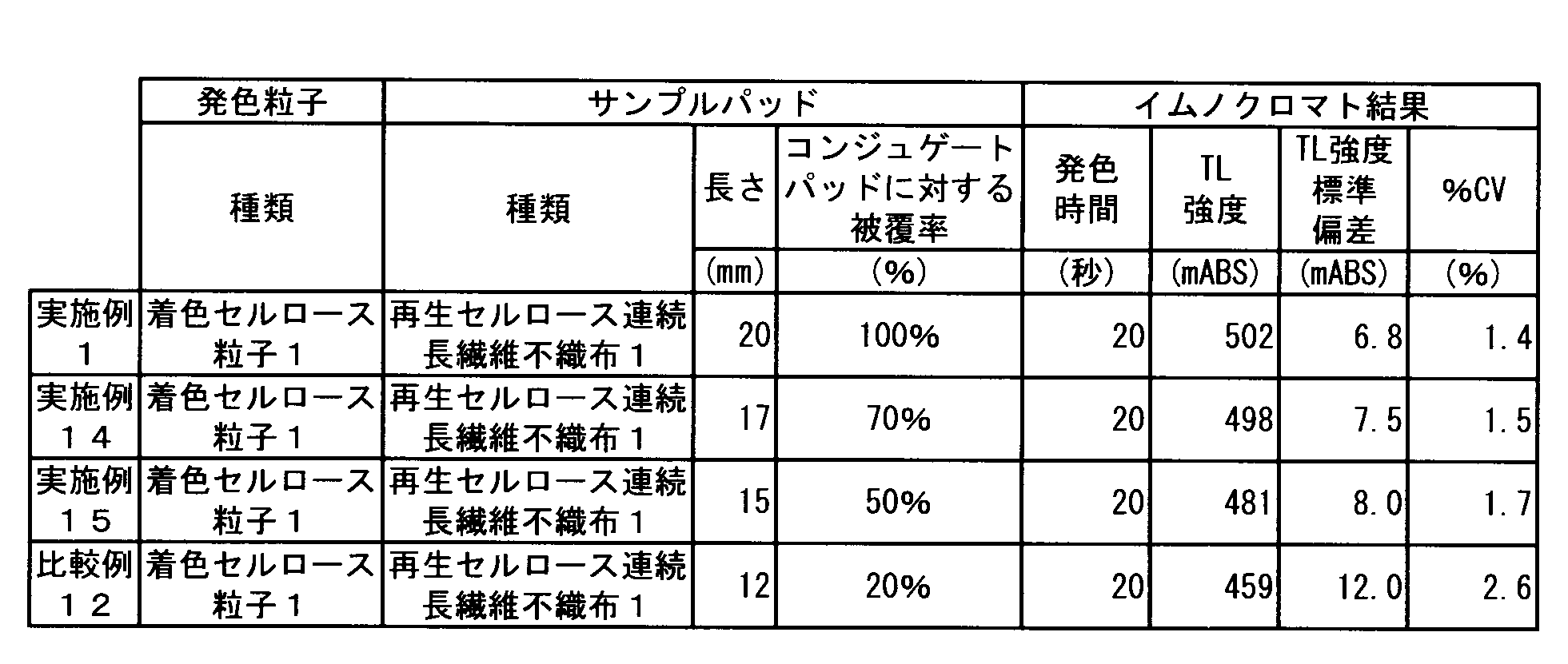
(b) 抗体感作発色粒子を含むコンジュゲートパッド
(c) 検出部A(TL)
(d) 検出部B(コントロールライン)
(e) クロマトグラフ媒体
(f) 吸収パッド
(g) 台紙
(h) コンジュゲートパッドに対するサンプルパッドの被覆率が100%の時の、サンプルパッドの最下流部分
(i) コンジュゲートパッドに対するサンプルパッドの被覆率が50%の時の、サンプルパッドの最下流部分
(j) 筐体
Claims (6)
- 平均粒子径が100~1000nmであり、発色強度が1.0~10.0であり、粒子の重量の10~90重量%がセルロース由来、かつ、90~10重量%が着色成分由来である発色粒子を含むコンジュゲートを含有するコンジュゲートパッドと、目付が10~150g/m2であり、かつ、厚みが0.07~1.00mmである再生セルロース系繊維からなる不織布から構成される体外診断薬用サンプルパッドとを、含むイムノクロマト診断キット。
- 前記再生セルロース系繊維がキュプラアンモニウムレーヨン繊維を主成分とするものである、請求項1に記載のイムノクロマト診断キット。
- 前記再生セルロース系繊維が連続長繊維である、請求項1又は2に記載のイムノクロマト診断キット。
- 前記再生セルロース系繊維からなる不織布の脱落繊維数が5000個/m2未満である、請求項1~3のいずれか1項に記載のイムノクロマト診断キット。
- 前記着色成分が反応性染料である、請求項1~4のいずれか1項に記載のイムノクロマト診断キット。
- 前記コンジュゲートパッドに対する前記体外診断薬用サンプルパッドの被覆率が50~100%である、請求項1~5のいずれか1項に記載のイムノクロマト診断キット。
Priority Applications (6)
| Application Number | Priority Date | Filing Date | Title |
|---|---|---|---|
| ES14810458.1T ES2695000T3 (es) | 2013-06-10 | 2014-06-09 | Kit de diagnóstico inmunocromatográfico |
| CN201480033270.0A CN105283761B (zh) | 2013-06-10 | 2014-06-09 | 免疫层析诊断试剂盒 |
| US14/896,519 US9863944B2 (en) | 2013-06-10 | 2014-06-09 | Immunochromatographic diagnosis kit |
| JP2015522769A JP6053927B2 (ja) | 2013-06-10 | 2014-06-09 | イムノクロマト診断キット |
| EP14810458.1A EP3009840B1 (en) | 2013-06-10 | 2014-06-09 | Immunochromatographic diagnosis kit |
| KR1020157034235A KR101822727B1 (ko) | 2013-06-10 | 2014-06-09 | 이뮤노크로마토 진단 키트 |
Applications Claiming Priority (2)
| Application Number | Priority Date | Filing Date | Title |
|---|---|---|---|
| JP2013-121898 | 2013-06-10 | ||
| JP2013121898 | 2013-06-10 |
Publications (1)
| Publication Number | Publication Date |
|---|---|
| WO2014199954A1 true WO2014199954A1 (ja) | 2014-12-18 |
Family
ID=52022247
Family Applications (1)
| Application Number | Title | Priority Date | Filing Date |
|---|---|---|---|
| PCT/JP2014/065243 Ceased WO2014199954A1 (ja) | 2013-06-10 | 2014-06-09 | イムノクロマト診断キット |
Country Status (7)
| Country | Link |
|---|---|
| US (1) | US9863944B2 (ja) |
| EP (1) | EP3009840B1 (ja) |
| JP (1) | JP6053927B2 (ja) |
| KR (1) | KR101822727B1 (ja) |
| CN (1) | CN105283761B (ja) |
| ES (1) | ES2695000T3 (ja) |
| WO (1) | WO2014199954A1 (ja) |
Cited By (9)
| Publication number | Priority date | Publication date | Assignee | Title |
|---|---|---|---|---|
| JP2019120497A (ja) * | 2017-12-28 | 2019-07-22 | 日鉄ケミカル&マテリアル株式会社 | 免疫学的測定用金属ナノ粒子−セルロース複合体、標識物質、免疫学的測定法、免疫学的測定用試薬、アナライトの測定方法、アナライト測定用キット、及び、ラテラルフロー型クロマト用テストストリップ |
| JPWO2018186267A1 (ja) * | 2017-04-06 | 2019-11-21 | 旭化成株式会社 | 親水化着色セルロース微粒子 |
| CN111108387A (zh) * | 2017-09-25 | 2020-05-05 | 旭化成株式会社 | 有机着色细颗粒、诊断药试剂盒及体外诊断方法 |
| WO2020196298A1 (ja) * | 2019-03-25 | 2020-10-01 | 積水メディカル株式会社 | イムノクロマト分析方法及び該方法に使用するテストストリップ |
| WO2020196296A1 (ja) * | 2019-03-25 | 2020-10-01 | 積水メディカル株式会社 | イムノクロマト分析方法及び該方法に使用するテストストリップ |
| JPWO2020196295A1 (ja) * | 2019-03-25 | 2020-10-01 | ||
| JPWO2020230826A1 (ja) * | 2019-05-14 | 2021-10-21 | 旭化成株式会社 | イムノクロマト診断キット用吸収パッド及びイムノクロマト診断キット |
| JP2021188960A (ja) * | 2020-05-27 | 2021-12-13 | 積水メディカル株式会社 | イムノクロマトグラフィー用テストストリップ |
| WO2022004685A1 (ja) * | 2020-07-02 | 2022-01-06 | パナソニックIpマネジメント株式会社 | 機能性部材とこれを備えた化学物質センサー |
Families Citing this family (10)
| Publication number | Priority date | Publication date | Assignee | Title |
|---|---|---|---|---|
| KR101994411B1 (ko) | 2017-02-01 | 2019-06-28 | 주식회사 제트바이오텍 | 고감도 체외 진단 키트 및 이를 이용한 진단 분석 방법 |
| CN107765002B (zh) * | 2017-11-17 | 2024-04-05 | 丹娜(天津)生物科技股份有限公司 | 一种胶体金免疫层析试纸条及其制备方法和应用 |
| CN108020666A (zh) * | 2018-02-07 | 2018-05-11 | 福州大学 | 一种可同时定量检测血液中cea和ca19-9的磁性免疫层析试纸条及制备方法 |
| JP7128911B2 (ja) * | 2019-01-31 | 2022-08-31 | 旭化成株式会社 | イムノクロマト診断キット用吸収パッド |
| CN109959783B (zh) * | 2019-04-01 | 2022-07-22 | 宁波大榭开发区综研化学有限公司 | 一种进样胶带及其制备方法 |
| CN111474351B (zh) * | 2020-04-28 | 2024-01-16 | 上海泰辉生物科技有限公司 | 一种可用于检测冠状病毒的免疫层析试纸条和试剂盒 |
| CN113219169B (zh) * | 2021-05-22 | 2021-12-17 | 北京金诺百泰生物技术有限公司 | 生物检测用封闭剂及其制备方法、包被板及应用该包被板的试剂盒 |
| CN114720682A (zh) * | 2022-04-15 | 2022-07-08 | 山东康华生物医疗科技股份有限公司 | 一种用于免疫层析法检测中的样品垫的制备方法 |
| CN118518872B (zh) * | 2024-07-18 | 2024-09-27 | 天津喜诺生物医药有限公司 | 一种真菌检测方法、检测装置 |
| CN119224297B (zh) * | 2024-11-02 | 2025-04-15 | 珠海科域生物工程股份有限公司 | 一种荧光微球标记的大便隐血定量试剂条及其制备工艺 |
Citations (13)
| Publication number | Priority date | Publication date | Assignee | Title |
|---|---|---|---|---|
| JPS61128979A (ja) * | 1984-11-26 | 1986-06-17 | 旭メデイカル株式会社 | 血液処理装置 |
| JPH04329965A (ja) * | 1991-05-27 | 1992-11-18 | Asahi Medical Co Ltd | 白血球の選択的除去方法 |
| JPH0755809A (ja) | 1993-08-20 | 1995-03-03 | Arakusu:Kk | 尿中ホルモン検出装置 |
| JPH0787914A (ja) | 1993-09-27 | 1995-04-04 | Kubota Corp | 飯混合装置 |
| JPH0877579A (ja) | 1994-09-08 | 1996-03-22 | Sony Corp | フォーカスバイアス調整方法及びフォーカスバイアス調整装置 |
| JPH1048215A (ja) * | 1996-07-31 | 1998-02-20 | Sekisui Chem Co Ltd | 着色粒子 |
| JPH11170413A (ja) * | 1997-12-11 | 1999-06-29 | Asahi Chem Ind Co Ltd | 不織布複合低密度織物 |
| JP2955405B2 (ja) * | 1990-08-31 | 1999-10-04 | ジェイエスアール株式会社 | イムノクロマトグラフ法 |
| WO2006080438A1 (ja) | 2005-01-28 | 2006-08-03 | Mochida Pharmaceutical Co., Ltd. | イムノクロマト用試験具およびこれを用いた半定量方法 |
| WO2008084854A1 (ja) * | 2007-01-12 | 2008-07-17 | Asahi Kasei Fibers Corporation | セルロース微粒子並びにその分散液及び分散体 |
| WO2011062157A1 (ja) | 2009-11-17 | 2011-05-26 | 旭化成せんい株式会社 | 有機着色微粒子、それを含む診断薬キット及びインビトロ診断方法 |
| JP2012108031A (ja) | 2010-11-18 | 2012-06-07 | Asahi Kasei Fibers Corp | 体外診断薬用サンプルパッド |
| WO2013151066A1 (ja) | 2012-04-06 | 2013-10-10 | コニカミノルタ株式会社 | アナライトの検出または定量方法、アナライトを検出または定量するためのキット、およびアナライトを検出または定量するためのラテラルフロー型クロマト法用テストストリップ |
Family Cites Families (8)
| Publication number | Priority date | Publication date | Assignee | Title |
|---|---|---|---|---|
| US5266497A (en) * | 1990-08-31 | 1993-11-30 | Japan Synthetic Rubber Co., Ltd. | Immunochromatographic assay with improved colored latex |
| JP3965699B2 (ja) | 2005-01-31 | 2007-08-29 | 横河電機株式会社 | 情報処理装置および情報処理方法 |
| JP5474784B2 (ja) * | 2008-06-30 | 2014-04-16 | 積水メディカル株式会社 | 結合アッセイ用多孔性固相及びこれを用いた結合アッセイ法 |
| CN101609093B (zh) * | 2009-07-17 | 2013-05-15 | 田伏洲 | 快速诊断胰腺创伤和急性胰腺炎的测试卡及其测试方法 |
| EP3095876A1 (en) * | 2010-11-02 | 2016-11-23 | Kypha, Inc. | Point of care lateral flow immunoassay for complement activation and methods of use for point-of-care assessment of complement-associated disorders |
| CN102183634A (zh) * | 2011-03-04 | 2011-09-14 | 河北省兽药监察所 | 快速检测四种硝基呋喃类药物的免疫纳米金试纸条 |
| CN102323262A (zh) * | 2011-08-10 | 2012-01-18 | 无锡安迪生物工程有限公司 | 一种苯乙醇胺a快速检测卡及其检测方法 |
| CN102411052B (zh) * | 2011-11-17 | 2014-01-29 | 成都创宜生物科技有限公司 | 以补体因子d为检测指标的子痫前期快速检测工具与试剂盒及制作方法 |
-
2014
- 2014-06-09 ES ES14810458.1T patent/ES2695000T3/es active Active
- 2014-06-09 CN CN201480033270.0A patent/CN105283761B/zh not_active Expired - Fee Related
- 2014-06-09 WO PCT/JP2014/065243 patent/WO2014199954A1/ja not_active Ceased
- 2014-06-09 JP JP2015522769A patent/JP6053927B2/ja not_active Expired - Fee Related
- 2014-06-09 US US14/896,519 patent/US9863944B2/en not_active Expired - Fee Related
- 2014-06-09 EP EP14810458.1A patent/EP3009840B1/en not_active Not-in-force
- 2014-06-09 KR KR1020157034235A patent/KR101822727B1/ko not_active Expired - Fee Related
Patent Citations (13)
| Publication number | Priority date | Publication date | Assignee | Title |
|---|---|---|---|---|
| JPS61128979A (ja) * | 1984-11-26 | 1986-06-17 | 旭メデイカル株式会社 | 血液処理装置 |
| JP2955405B2 (ja) * | 1990-08-31 | 1999-10-04 | ジェイエスアール株式会社 | イムノクロマトグラフ法 |
| JPH04329965A (ja) * | 1991-05-27 | 1992-11-18 | Asahi Medical Co Ltd | 白血球の選択的除去方法 |
| JPH0755809A (ja) | 1993-08-20 | 1995-03-03 | Arakusu:Kk | 尿中ホルモン検出装置 |
| JPH0787914A (ja) | 1993-09-27 | 1995-04-04 | Kubota Corp | 飯混合装置 |
| JPH0877579A (ja) | 1994-09-08 | 1996-03-22 | Sony Corp | フォーカスバイアス調整方法及びフォーカスバイアス調整装置 |
| JPH1048215A (ja) * | 1996-07-31 | 1998-02-20 | Sekisui Chem Co Ltd | 着色粒子 |
| JPH11170413A (ja) * | 1997-12-11 | 1999-06-29 | Asahi Chem Ind Co Ltd | 不織布複合低密度織物 |
| WO2006080438A1 (ja) | 2005-01-28 | 2006-08-03 | Mochida Pharmaceutical Co., Ltd. | イムノクロマト用試験具およびこれを用いた半定量方法 |
| WO2008084854A1 (ja) * | 2007-01-12 | 2008-07-17 | Asahi Kasei Fibers Corporation | セルロース微粒子並びにその分散液及び分散体 |
| WO2011062157A1 (ja) | 2009-11-17 | 2011-05-26 | 旭化成せんい株式会社 | 有機着色微粒子、それを含む診断薬キット及びインビトロ診断方法 |
| JP2012108031A (ja) | 2010-11-18 | 2012-06-07 | Asahi Kasei Fibers Corp | 体外診断薬用サンプルパッド |
| WO2013151066A1 (ja) | 2012-04-06 | 2013-10-10 | コニカミノルタ株式会社 | アナライトの検出または定量方法、アナライトを検出または定量するためのキット、およびアナライトを検出または定量するためのラテラルフロー型クロマト法用テストストリップ |
Non-Patent Citations (1)
| Title |
|---|
| See also references of EP3009840A4 |
Cited By (16)
| Publication number | Priority date | Publication date | Assignee | Title |
|---|---|---|---|---|
| JPWO2018186267A1 (ja) * | 2017-04-06 | 2019-11-21 | 旭化成株式会社 | 親水化着色セルロース微粒子 |
| CN111108387A (zh) * | 2017-09-25 | 2020-05-05 | 旭化成株式会社 | 有机着色细颗粒、诊断药试剂盒及体外诊断方法 |
| CN111108387B (zh) * | 2017-09-25 | 2023-08-29 | 旭化成株式会社 | 有机着色细颗粒、诊断药试剂盒及体外诊断方法 |
| JP2019120497A (ja) * | 2017-12-28 | 2019-07-22 | 日鉄ケミカル&マテリアル株式会社 | 免疫学的測定用金属ナノ粒子−セルロース複合体、標識物質、免疫学的測定法、免疫学的測定用試薬、アナライトの測定方法、アナライト測定用キット、及び、ラテラルフロー型クロマト用テストストリップ |
| JP7265315B2 (ja) | 2017-12-28 | 2023-04-26 | 日鉄ケミカル&マテリアル株式会社 | 免疫学的測定用金属ナノ粒子-セルロース複合体、標識物質、免疫学的測定法、免疫学的測定用試薬、アナライトの測定方法、アナライト測定用キット、及び、ラテラルフロー型クロマト用テストストリップ |
| WO2020196295A1 (ja) * | 2019-03-25 | 2020-10-01 | 積水メディカル株式会社 | 呼吸器感染ウイルスの免疫学的分析方法及び検出キット |
| JPWO2020196295A1 (ja) * | 2019-03-25 | 2020-10-01 | ||
| JPWO2020196296A1 (ja) * | 2019-03-25 | 2020-10-01 | ||
| WO2020196296A1 (ja) * | 2019-03-25 | 2020-10-01 | 積水メディカル株式会社 | イムノクロマト分析方法及び該方法に使用するテストストリップ |
| JPWO2020196298A1 (ja) * | 2019-03-25 | 2020-10-01 | ||
| WO2020196298A1 (ja) * | 2019-03-25 | 2020-10-01 | 積水メディカル株式会社 | イムノクロマト分析方法及び該方法に使用するテストストリップ |
| JP7544438B2 (ja) | 2019-03-25 | 2024-09-03 | 積水メディカル株式会社 | イムノクロマト分析方法及び該方法に使用するテストストリップ |
| JPWO2020230826A1 (ja) * | 2019-05-14 | 2021-10-21 | 旭化成株式会社 | イムノクロマト診断キット用吸収パッド及びイムノクロマト診断キット |
| JP2021188960A (ja) * | 2020-05-27 | 2021-12-13 | 積水メディカル株式会社 | イムノクロマトグラフィー用テストストリップ |
| WO2022004685A1 (ja) * | 2020-07-02 | 2022-01-06 | パナソニックIpマネジメント株式会社 | 機能性部材とこれを備えた化学物質センサー |
| JPWO2022004685A1 (ja) * | 2020-07-02 | 2022-01-06 |
Also Published As
| Publication number | Publication date |
|---|---|
| KR20160003240A (ko) | 2016-01-08 |
| CN105283761A (zh) | 2016-01-27 |
| KR101822727B1 (ko) | 2018-01-26 |
| JPWO2014199954A1 (ja) | 2017-02-23 |
| EP3009840A1 (en) | 2016-04-20 |
| JP6053927B2 (ja) | 2016-12-27 |
| ES2695000T3 (es) | 2018-12-28 |
| US9863944B2 (en) | 2018-01-09 |
| EP3009840B1 (en) | 2018-09-19 |
| CN105283761B (zh) | 2018-02-09 |
| EP3009840A4 (en) | 2016-06-29 |
| US20160123974A1 (en) | 2016-05-05 |
Similar Documents
| Publication | Publication Date | Title |
|---|---|---|
| JP6053927B2 (ja) | イムノクロマト診断キット | |
| JP7192146B2 (ja) | 免疫検査方法及び濃縮用治具 | |
| TWI696833B (zh) | 有機著色微粒子、診斷藥套組及體外診斷方法 | |
| CA3058871C (en) | Hydrophilic colored cellulose microparticles | |
| JP7128911B2 (ja) | イムノクロマト診断キット用吸収パッド | |
| JP7176915B2 (ja) | イムノクロマト診断キット | |
| WO2016031892A1 (ja) | イムノクロマトグラフィー用テストストリップ | |
| JP2010256309A (ja) | コンジュゲートパッドおよび体外診断薬 | |
| TW202109044A (zh) | 免疫層析診斷套組用吸收墊及免疫層析診斷套組 | |
| EP3842789A1 (en) | Immunochromatography in which carrier particles are used to amplify surface plasmon resonance | |
| JP2019174336A (ja) | 懸濁性に優れ、迅速診断が可能な着色セルロース微粒子 | |
| JP2013181935A (ja) | クロマトグラフ方法 | |
| JP2025072808A (ja) | 測定試料希釈液および免疫測定キット | |
| JP2018169373A (ja) | 免疫測定方法、免疫測定用キット及びラテラルフロー型クロマトテストストリップ |
Legal Events
| Date | Code | Title | Description |
|---|---|---|---|
| WWE | Wipo information: entry into national phase |
Ref document number: 201480033270.0 Country of ref document: CN |
|
| 121 | Ep: the epo has been informed by wipo that ep was designated in this application |
Ref document number: 14810458 Country of ref document: EP Kind code of ref document: A1 |
|
| ENP | Entry into the national phase |
Ref document number: 2015522769 Country of ref document: JP Kind code of ref document: A |
|
| ENP | Entry into the national phase |
Ref document number: 20157034235 Country of ref document: KR Kind code of ref document: A |
|
| WWE | Wipo information: entry into national phase |
Ref document number: 2014810458 Country of ref document: EP |
|
| WWE | Wipo information: entry into national phase |
Ref document number: 14896519 Country of ref document: US |
|
| NENP | Non-entry into the national phase |
Ref country code: DE |