WO2015012377A1 - 骨芽細胞及びその調製方法 - Google Patents
骨芽細胞及びその調製方法 Download PDFInfo
- Publication number
- WO2015012377A1 WO2015012377A1 PCT/JP2014/069628 JP2014069628W WO2015012377A1 WO 2015012377 A1 WO2015012377 A1 WO 2015012377A1 JP 2014069628 W JP2014069628 W JP 2014069628W WO 2015012377 A1 WO2015012377 A1 WO 2015012377A1
- Authority
- WO
- WIPO (PCT)
- Prior art keywords
- oct4
- oct4l
- oct4ml
- bone
- cells
- Prior art date
- Legal status (The legal status is an assumption and is not a legal conclusion. Google has not performed a legal analysis and makes no representation as to the accuracy of the status listed.)
- Ceased
Links
Images



















Classifications
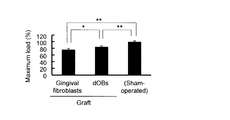
-
- C—CHEMISTRY; METALLURGY
- C12—BIOCHEMISTRY; BEER; SPIRITS; WINE; VINEGAR; MICROBIOLOGY; ENZYMOLOGY; MUTATION OR GENETIC ENGINEERING
- C12N—MICROORGANISMS OR ENZYMES; COMPOSITIONS THEREOF; PROPAGATING, PRESERVING, OR MAINTAINING MICROORGANISMS; MUTATION OR GENETIC ENGINEERING; CULTURE MEDIA
- C12N5/00—Undifferentiated human, animal or plant cells, e.g. cell lines; Tissues; Cultivation or maintenance thereof; Culture media therefor
- C12N5/06—Animal cells or tissues; Human cells or tissues
- C12N5/0602—Vertebrate cells
- C12N5/0652—Cells of skeletal and connective tissues; Mesenchyme
- C12N5/0654—Osteocytes, Osteoblasts, Odontocytes; Bones, Teeth
-
- A—HUMAN NECESSITIES
- A61—MEDICAL OR VETERINARY SCIENCE; HYGIENE
- A61P—SPECIFIC THERAPEUTIC ACTIVITY OF CHEMICAL COMPOUNDS OR MEDICINAL PREPARATIONS
- A61P19/00—Drugs for skeletal disorders
-
- C—CHEMISTRY; METALLURGY
- C12—BIOCHEMISTRY; BEER; SPIRITS; WINE; VINEGAR; MICROBIOLOGY; ENZYMOLOGY; MUTATION OR GENETIC ENGINEERING
- C12N—MICROORGANISMS OR ENZYMES; COMPOSITIONS THEREOF; PROPAGATING, PRESERVING, OR MAINTAINING MICROORGANISMS; MUTATION OR GENETIC ENGINEERING; CULTURE MEDIA
- C12N15/00—Mutation or genetic engineering; DNA or RNA concerning genetic engineering, vectors, e.g. plasmids, or their isolation, preparation or purification; Use of hosts therefor
- C12N15/09—Recombinant DNA-technology
- C12N15/63—Introduction of foreign genetic material using vectors; Vectors; Use of hosts therefor; Regulation of expression
- C12N15/79—Vectors or expression systems specially adapted for eukaryotic hosts
- C12N15/85—Vectors or expression systems specially adapted for eukaryotic hosts for animal cells
-
- C—CHEMISTRY; METALLURGY
- C12—BIOCHEMISTRY; BEER; SPIRITS; WINE; VINEGAR; MICROBIOLOGY; ENZYMOLOGY; MUTATION OR GENETIC ENGINEERING
- C12N—MICROORGANISMS OR ENZYMES; COMPOSITIONS THEREOF; PROPAGATING, PRESERVING, OR MAINTAINING MICROORGANISMS; MUTATION OR GENETIC ENGINEERING; CULTURE MEDIA
- C12N2501/00—Active agents used in cell culture processes, e.g. differentation
- C12N2501/60—Transcription factors
-
- C—CHEMISTRY; METALLURGY
- C12—BIOCHEMISTRY; BEER; SPIRITS; WINE; VINEGAR; MICROBIOLOGY; ENZYMOLOGY; MUTATION OR GENETIC ENGINEERING
- C12N—MICROORGANISMS OR ENZYMES; COMPOSITIONS THEREOF; PROPAGATING, PRESERVING, OR MAINTAINING MICROORGANISMS; MUTATION OR GENETIC ENGINEERING; CULTURE MEDIA
- C12N2501/00—Active agents used in cell culture processes, e.g. differentation
- C12N2501/60—Transcription factors
- C12N2501/603—Oct-3/4
-
- C—CHEMISTRY; METALLURGY
- C12—BIOCHEMISTRY; BEER; SPIRITS; WINE; VINEGAR; MICROBIOLOGY; ENZYMOLOGY; MUTATION OR GENETIC ENGINEERING
- C12N—MICROORGANISMS OR ENZYMES; COMPOSITIONS THEREOF; PROPAGATING, PRESERVING, OR MAINTAINING MICROORGANISMS; MUTATION OR GENETIC ENGINEERING; CULTURE MEDIA
- C12N2506/00—Differentiation of animal cells from one lineage to another; Differentiation of pluripotent cells
- C12N2506/13—Differentiation of animal cells from one lineage to another; Differentiation of pluripotent cells from connective tissue cells, from mesenchymal cells
- C12N2506/1307—Differentiation of animal cells from one lineage to another; Differentiation of pluripotent cells from connective tissue cells, from mesenchymal cells from adult fibroblasts
-
- C—CHEMISTRY; METALLURGY
- C12—BIOCHEMISTRY; BEER; SPIRITS; WINE; VINEGAR; MICROBIOLOGY; ENZYMOLOGY; MUTATION OR GENETIC ENGINEERING
- C12N—MICROORGANISMS OR ENZYMES; COMPOSITIONS THEREOF; PROPAGATING, PRESERVING, OR MAINTAINING MICROORGANISMS; MUTATION OR GENETIC ENGINEERING; CULTURE MEDIA
- C12N2510/00—Genetically modified cells
Definitions
- the present invention relates to an osteoblast and a method for preparing the same, and more particularly to a method for preparing an osteoblast by direct reprogramming.
- osteoblasts can be transplanted into the affected area to promote bone formation and functional morphology. Prognosis can be expected to improve. Actually, for example, a treatment for autologous transplantation of bone marrow cells collected from a patient's cancellous bone is performed, and its effectiveness is known. In this case, it is considered that osteoblasts are induced to differentiate from mesenchymal stem cells contained in autologous bone marrow cells and contribute to bone formation and remodeling. On the other hand, the prevalence of osteoporosis is increasing with the aging of the population.
- osteoblast transplantation can promote the healing of fractures associated with osteoporosis, trauma, etc., refractory fractures and pseudo-fractures.
- Osteoblast transplantation also includes rheumatoid arthritis, sudden femoral head necrosis, osteoarthritis, lumbar osteoarthritis, spinal stenosis, disc herniation, spondylolysis, spondylolisthesis, scoliosis, cervical spine Myelopathy, posterior longitudinal ligament ossification, spinal cord injury, hip osteoarthritis, knee osteoarthritis, femoral slippage, osteomalacia, bone repair after surgery (sternal repair after cardiac surgery, etc.) ), May be useful for repairing defects associated with artificial ankle surgery, osteomyelitis, osteonecrosis, and the like.
- periodontal disease is also called the fourth lifestyle-related disease, has a very high morbidity rate, and causes various systemic diseases. As periodontal disease progresses, bone resorption of alveolar bone occurs, and if osteoblasts can be efficiently supplied to the local bone resorbing part, it is considered that this will lead to regenerative treatment of alveolar bone.
- osteoblast transplantation is used in combination with bone transplantation, artificial bone transplantation, artificial joints or implants, the therapeutic effect may be enhanced.
- bone marrow mesenchymal stem cells and bone marrow cells including bone marrow mesenchymal stem cells have been used.
- the collection of bone marrow has problems such as the invasion to a patient is large and a sufficient number of bone marrow cells may not be supplied.
- ES cells human embryonic stem cells
- ES cells human embryonic stem cells
- iPS cells it is not necessary to collect bone marrow from the patient, and a sufficient number of osteoblasts may be supplied.
- there is a risk that the remaining iPS cells may become tumor after transplantation.
- Non-patent document 1 introduces the differentiation of osteoblasts into osteoblasts using Osterix Lentivirus vector introduction + Osteogenic medium into human ES cells.
- Non-Patent Documents 2 and 3 obtain osteoblasts by inducing differentiation from mouse iPS cells through Osteogenic medium via MSC.
- Non-Patent Document 4 introduces Runx2 Adenovirus vector into mouse iPS cells and induces differentiation in Osteogenic medium to obtain osteoblasts. As shown in Non-Patent Documents 1 to 4, osteoblasts are produced by inducing differentiation from pluripotent stem cells such as ES cells and iPS cells. There was a danger.
- the present invention can be applied to the repair of bone defects associated with various tumors, trauma, surgery, etc., bone resorption typified by periodontal disease, fractures, osteoporosis, etc., and has a low risk of canceration It is an object to provide a method for preparing osteoblasts.
- a specific gene is introduced into a mammalian somatic cell in combination to directly obtain (direct reprogramming) osteoblasts without going through pluripotent stem cells such as ES cells and iPS cells.
- the present invention provides the following osteoblasts and methods for preparing the same.
- Item 1 A method for preparing osteoblasts from a somatic cell by introducing a reprogramming related gene or an expression product thereof into a mammalian somatic cell, wherein the reprogramming related gene is an Oct family, c-Myc (M), A method for preparing osteoblasts, which is at least one selected from the group consisting of L-Myc (L), GLIS family, Klf family, Lin-28, and Sox2.
- a method for preparing osteoblasts from somatic cells by introducing a bone-related gene or an expression product thereof and a reprogramming-related gene or an expression product thereof into a somatic cell of a mammal, wherein the bone-related gene is Runx2 ( R), Osterix (O), Dlx5 (D), at least one selected from the group consisting of Oct family, c-Myc (M), L-Myc (L), GLIS family, A method for preparing osteoblasts, which is at least one selected from the group consisting of Klf family, Lin-28, and Sox2.
- Item 3. The method according to Item 1 or 2, wherein the somatic cells are fibroblasts or gingival cells.
- a bone-related gene or its expression product introduced into a somatic cell and a combination of a reprogramming-related gene or its expression product are ROD Oct4L, RD Oct4L, RO Oct4ML, D Oct4ML, ROD Oct4M, OD Oct4L, O Oct4ML, O Oct4L, O Oct4M, OD Oct4, D Oct4L and Oct4ML (Where R represents “Runx2”, O represents “Osterix”, D represents “Dlx5”, M represents “c-Myc”, and L represents “L-Myc”) Item 4.
- a combination of a bone-related gene or its expression product and a reprogramming-related gene or its expression product introduced into a somatic cell is any combination selected from the group consisting of ROD Oct4L, RD Oct4L, RO Oct4ML, and D Oct4ML.
- Item 4 The method according to Item 2 or 3.
- the present invention is a technique for preparing osteoblasts from somatic cells by introducing a reprogramming-related gene or an expression product thereof, or a bone-related gene or an expression product thereof and a reprogramming-related gene or an expression product thereof.
- at least one bone-related gene can be selected from the group consisting of Runx2 (R), Osterix (O), and Dlx5 (D). Desirably, at least one of Oxterix and Dlx5 is included.
- Reprogramming-related genes include Oct4 family, c-Myc (M), L-Myc (L), Glis family, Klf family (KLF1, KLF2, KLF3, KLF4, KLF5, KLF6, KLF7, KLF8, KLF9, At least one selected from the group consisting of KLF10, KLF11, KLF12, KLF13, KLF14, KLF15, KLF16, KLF17), Lin-28, and Sox2 can be selected. Desirably, any of the Oct4 families should contain at least one, and more desirably, any of the Oct4 families should contain at least one and L-Myc and / or c-Myc. Good.
- the Oct4 family is preferably Oct4. Therefore, those containing both Oct4 and L-Myc and / or c-Myc are most desirable.
- Oct4 is sometimes expressed as Oct3 / 4, but in this specification, it is expressed as “Oct4”.
- “Oct4” is used as a representative of the Oct family, but other genes of the Oct family (Oct1A, Oct6) and the like can be used in the same manner as Oct4.
- Oct4 is a factor that can induce pluripotency by reprogramming somatic cells by introducing genes into somatic cells together with Sox2, Klf4, and c-Myc (Takahashi K, Yamanaka S.
- factors such as GATA3, GATA6, SOX7, PAX1, GATA4, CEBPa, HNF4a, GRB2, which are not reprogramming factors and are not Oct families but can substitute for Oct functions in reprogramming are also used in the present invention.
- KLF-4 can be replaced with genes from other Klf families (KLF1, KLF2, KLF3, KLF5, KLF6, KLF7, KLF8, KLF9, KLF10, KLF11, KLF12, KLF13, KLF14, KLF15, KLF16, KLF17) .
- KLF-4 is used as a representative of the Klf family.
- other genes of the Klf family can be used in the same manner as KLF-4.
- Examples of the GLIS family include GLIS1 (GLIS family zinc finger 1).
- IRX family members IRX6 (iroquois homeobox protein 6), etc.
- PTX family members such as ILF cell induction
- PITX2 paired-like homeodomain transcription factor 2
- DMRTB1 DMRT-like family B with proline-rich C-terminal 1
- KLF-4 a gene that can substitute for the function of KLF-4 in somatic cell reprogramming (iPS cell induction) may be used in the same manner as KLF-4. it can.
- mRNA can also be used.
- SOX family genes can be used in the same manner instead of SOX2.
- SOX2 is used as a representative of the SOX family.
- other genes of the SOX family can be used in the same manner as SOX2. In the present invention, all are called SOX2 for simplicity.
- bone-related genes and reprogramming-related genes can be replaced with genes and their expression products can be substituted.
- one or more genes can be replaced with a molecule such as a drug that induces or mimics the gene.
- GATA3, GATA6, SOX7, PAX1, GATA4, CEBPa, HNF4a, GRB2 are known as genes that can substitute for the function of Oct4 in iPS cell induction (Jian Shu et al., Cell 153, 963 -975, 2013), such compounds can be used in place of the Oct4 gene introduction.
- BIX-01294 As a small molecule compound that can replace the function of Oct4 in reprogramming neural progenitor cells, BIX-01294 is known (Bo Feng et al., Cell Stem Cell 4: 301, 2009). Such a compound can be used instead of the introduction of.
- kenpaullone As a compound that can substitute for the function of Klf-4 in iPS cell induction, kenpaullone (Lyssiotis, CA, et al., Proc Natl Acad Sci US A. 2009 June 2; 106 (22): 8912-8917.) As known, such compounds can be used in place of the Klf-4 gene introduction.
- a gene and its expression product may be collectively referred to as a “gene”.
- “gene transfer” is read as “addition of molecule”, “addition of drug”, “addition of compound”, “administration of molecule”, “administration of drug”, “administration of compound”, and the like. It is also possible to add other genes to the gene combination of the present invention.
- the present invention includes a method of inducing osteoblasts from somatic cells. Also included are osteoblasts prepared by this method. In addition, therapeutic cell preparations and transplantation materials containing these osteoblasts are included. If this technology is applied to somatic cells in the body, it is possible to make osteoblasts directly in the body. This method, a formulation for direct induction, is also included in the present invention.
- osteoblasts can be provided from somatic cells in a short period of time by direct reprogramming. Since these osteoblasts can be easily derived from the somatic cells of the individual to be transplanted, problems such as immunological rejection response do not occur even when osteoblasts themselves or bone tissue prepared therefrom are transplanted. In addition, since osteoblasts can be directly induced from somatic cells without going through iPS cells or ES cells, problems caused by pluripotent stem cells such as canceration can be avoided.
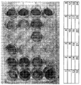
- FIG. 3A is a macroscopic image of a dish. Stained in red is a calcified bone matrix, indicating that functional osteoblasts have been induced.
- FIG. 3H (“1” in the table of FIG. 3H indicates that a retroviral vector containing each gene was infected, and a blank indicates that a retroviral vector containing each gene was not infected. Represents that).
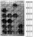
- Alizarin Red S staining Fig. 3B is a macroscopic image of a dish. Stained in red is a calcified bone matrix, indicating that functional osteoblasts have been induced.
- FIG. 3H For numbers of wells, refer to FIG. 3H (“1” in the table of FIG. 3H indicates that a retroviral vector containing each gene was infected, and a blank indicates that a retroviral vector containing each gene was not infected. Represents that).
- Alizarin Red S staining Fig. 3C is a macroscopic image of a dish. Stained in red is a calcified bone matrix, indicating that functional osteoblasts have been induced.
- FIG. 3H (“1” in the table of FIG. 3H indicates that a retroviral vector containing each gene was infected, and a blank indicates that a retroviral vector containing each gene was not infected. Represents that).
- Alizarin Red S staining Fig. 3D is a macroscopic image of the dish. Stained in red is a calcified bone matrix, indicating that functional osteoblasts have been induced. For numbers of wells, refer to FIG. 3H (“1” in the table of FIG. 3H indicates that a retroviral vector containing each gene was infected, and a blank indicates that a retroviral vector containing each gene was not infected. Represents that).
- Alizarin Red S staining ( Figure 3E is a macroscopic image of a dish). Stained in red is a calcified bone matrix, indicating that functional osteoblasts have been induced. For numbers of wells, refer to FIG. 3H (“1” in the table of FIG.
- FIG. 3H indicates that a retroviral vector containing each gene was infected, and a blank indicates that a retroviral vector containing each gene was not infected. Represents that).
- Alizarin Red S staining (Figure 3F is a macroscopic image of a dish). Stained in red is a calcified bone matrix, indicating that functional osteoblasts have been induced.
- FIG. 3H (“1” in the table of FIG. 3H indicates that a retroviral vector containing each gene was infected, and a blank indicates that a retroviral vector containing each gene was not infected. Represents that).
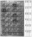
- FIG. 3G is a macroscopic photograph of a 96-well plate (see FIG. 3H for well numbers).
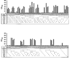
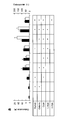
- FIG. 3H shows the results of measuring the absorbance (550 nm-650 nm) of the reaction solution using a microplate reader.
- the vertical axis of the graph represents absorbance, and the higher the absorbance, the more calcified bone matrix was produced, indicating that many fibroblasts were converted to functional osteoblasts.
- “1” in the table indicates that a retroviral vector containing each gene was infected, and a blank indicates that a retroviral vector containing each gene was not infected.
- well 27 was infected with a retroviral vector containing genes for Osterix, Runx2, Oct4, L-Myc and Dlx5, but not for a retroviral vector containing genes for c-Myc, Glis1 and EGFP. It can be seen that there are no cells, producing a lot of calcified bone matrix.
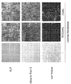
- ALP activity test ALP stained image Immunostaining (a) control, (b) RO 4 Oct4M, (c) RO Oct4L: ⁇ 100 Alizarin Red S staining (a) Control, (b) RO Oct4M, (c) RO Oct4L: ⁇ 1 (d) Control, (e) RO Oct4M, (f) RO Oct4L: ⁇ 40 von Kossa staining Outline of 3D culture experiment Giemsa-stained 3D culture) (a) background, (b) RO ⁇ Oct4L: ⁇ 1 Alizarin Red S staining in 3D culture (a) background, (b) control, (c) RO Oct4M, (d) RO : Oct4L: ⁇ 1 ALP stained image (aHDF) (a) control, (b) RO Oct4G, (c) RO Oct4L: ⁇ 1 (d) control, (e) RO Oct4G, (f) RO Oct4L: ⁇ 40 Alizarin Red S staining (aHDF).
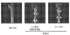
- the arrow is the bone defect.
- Bone transplanted with osteoblasts (dOB) induced by direct reprogramming has a higher radioopacity at the bone defect than bone transplanted with human gingival fibroblasts.
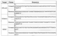
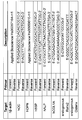
- List of primers used for RT-PCR® A list of primers used for sequencing.
- Direct reprogramming of osteoblasts with episomal vectors Direct reprogramming of mouse osteoblasts Measurement of mRNA expression level of hOsteopontin ( ⁇ ) and hOsteocalcin ( ⁇ ) by Real-time RT-PCR, and the introduced gene is indicated by “+”.
- Reprogramming of human fibroblasts into osteoblasts by gene transfer using plasmid vectors Reprogramming of human fibroblasts into osteoblasts in medium without the addition of heterologous proteins
- Reprogramming of human fibroblasts into osteoblasts in medium without the addition of heterologous proteins Viability after freeze-thawing of human osteoblasts induced by direct reprogramming from human fibroblasts in a medium without the addition of heterologous proteins.
- pre-osteoblasts pre-osteoblasts, immature osteoblasts, mature osteoblasts, bone cells and the like can also be prepared. In the present specification, these are all called osteoblasts for simplicity.
- Examples of diseases to be treated using osteoblasts (transplant material) obtained by the present invention include bone tumors associated with bone tumors, trauma, osteomyelitis, etc., bone defects after fractures such as bone tumors, fractures, Osteoporosis, periodontal disease, alveolar bone resorption, rheumatoid arthritis, idiopathic femoral head necrosis, osteoarthritis, lumbar spondylotic spondylosis, spinal canal stenosis, intervertebral disc herniation, spondylolysis, spondylolisthesis, spine Destroyed by complicated fractures such as scoliosis, cervical spondylotic myelopathy, posterior longitudinal ligament ossification, spinal cord injury, hip osteoarthritis, knee osteoarthritis, femoral head slippery, osteomalacia, mandibular reconstruction Examples include bone fracture reconstruction, bone repair after surgery (sternal repair after cardiac surgery, etc.), defect repair associated with artificial
- osteoblasts are transplanted, there is a possibility that the therapeutic effect can be enhanced in combination with bone grafts, artificial bone grafts, artificial joints and implants. It is also possible to treat the above-mentioned diseases by culturing osteoblasts using a three-dimensional scaffold or the like to produce various forms of bone tissue outside the body and transplanting the bone tissue. In addition, various diseases related to osteoblast deficiency, deficiency or reduced function are targeted.
- treatment intends a treatment performed while a patient is suffering from a particular disease or disorder, whereby the severity of the disease or disorder, or one or It means that a plurality of its symptoms are alleviated or the progression of the disease or disorder is delayed or slowed down.
- treatment includes “prevention”.
- the osteoblasts obtained by the present invention can also be used not only for treatment of diseases but also for cosmetic purposes.
- a bone matrix can be produced to repair the deficient site and become plump and inconspicuous.
- treatment for humans is also referred to as treatment in this specification for convenience, and “patient” can be read as “healthy” or “human”, and “disease” can be read as “beauty”.
- the present invention can also be used to treat not only humans but also mammals including pets such as dogs and cats and livestock such as cows, horses, pigs, sheep and chickens.
- “patient” is read as “patient” or “mammal”.
- the transplant material refers to a material containing osteoblasts that is introduced into a living body for bone tissue repair and reconstruction.
- the transplant material includes a material in which bone tissue is regenerated partially or completely in vitro and transplanted to the same or another individual.
- the osteoblast obtained by this invention can be used for preparation of a transplant material. Osteoblasts themselves are also transplant material.
- osteoblasts can be transplanted into patients as cell preparations, transplanted with a substrate made of artificial materials such as hydroxyapatite or bioabsorbable ceramic (scaffold), or cultured with the scaffold.
- a substrate made of artificial materials such as hydroxyapatite or bioabsorbable ceramic (scaffold)
- the scaffold can have various three-dimensional shapes depending on the purpose of implantation.
- Somatic cells may be derived from mammals. When transplanting osteoblasts into a living body, it is preferable to use somatic cells (autologous cells) derived from the transplanted subject in order to reduce the risk of infection, rejection response, and the like.
- somatic cells autologous cells
- osteoblasts prepared in advance from somatic cells of another person or another animal can be used for transplantation instead of autologous cells.
- osteoblasts can be prepared from somatic cells of other people or other animals prepared in advance and used for transplantation. That is, an osteoblast bank or an osteoblast precursor cell bank can be prepared and used for transplantation purposes.
- MHC can be typed in advance in order to reduce the risk of a rejection response or the like.
- the osteoblast character and tumorigenicity can be confirmed in advance.
- examples of mammals include mice, rats, hamsters, humans, dogs, cats, monkeys, rabbits, cows, horses, pigs, and the like, and particularly humans.
- the present invention can also be used for various researches and technical developments using osteoblasts. For example, it is useful for basic research such as analysis of bone development and aging, morphogenesis, remodeling mechanisms, mechanical stress on these, nutrition, immunity, nerves, and hormone effects. It is also useful for analyzing the effects of radioactive materials such as strontium 90 on internal bones and developing a technique for removing strontium 90 from bone.
- osteoblasts can be established easily, quickly and inexpensively from humans and animals having various diseases and genetic backgrounds, so that osteoblast abnormalities related to diseases and genetic backgrounds can be biochemically detected. It can be analyzed by molecular biology, immunology, etc., and this can be used for research such as elucidation of the pathogenesis of diseases and development of diagnostic methods. If such osteoblasts are used for drug development, drug toxicity tests, etc., it can be used for development of new treatments for various diseases.
- the somatic cells to be subjected to the method of the present invention are not particularly limited, but for example, fibroblasts, keratinocytes, oral mucosal epithelial cells, airway mucosal epithelial cells, gastric mucosal epithelial cells, intestinal mucosal epithelial cells Vascular endothelial cells, smooth muscle cells, fat cells, gingival cells (gingival fibroblasts, gingival epithelial cells), pulp cells, periodontal ligament cells, leukocytes, lymphocytes, muscle cells, conjunctival epithelial cells, osteoclasts, etc.
- fibroblasts, keratinocytes, oral mucosal epithelial cells, gingival cells, leukocytes, lymphocytes, osteoclasts and the like can be mentioned.
- At least one reprogramming-related gene, or at least one bone-related gene or an expression product thereof, and at least one reprogramming-related gene or an expression product thereof are introduced into a somatic cell.
- the “expression product” include mRNA or protein of genes such as bone-related genes and reprogramming-related genes.
- a bone-related gene is a gene that is introduced in order for a reprogrammed osteoblast to function as an osteoblast, and specifically, Runx2 (hereinafter sometimes abbreviated as ⁇ R ''), Osterix (hereinafter referred to as ⁇ R ''). O ”), and Dlx5 (hereinafter sometimes abbreviated as“ D ”), at least one kind can be selected. Preferably, at least one of Oxterix and Dlx5 is included. It is preferable to introduce one or more of these genes or their expression products into somatic cells in combination with other bone-related genes or their expression products as necessary.
- Reprogramming-related genes are genes that are introduced into somatic cells to convert somatic cells into osteoblasts.
- Bone-related genes such as Runx2, Osterix, and Dlx5 have been known to be involved in bone cell differentiation, development, proliferation, survival, etc., but they can be used to generate osteoblasts from somatic cells such as fibroblasts. There is no known technique for directly inducing cells (not via pluripotent stem cells). In addition, it is known to induce iPS cells from somatic cells by a combination of reprogramming-related genes, but it is not known to induce osteoblasts. The present inventors use a combination of one or more bone-related genes and one or more reprogramming-related genes, or one or more reprogramming-related genes, so that osteoblasts can be transformed from somatic cells such as fibroblasts.
- Oct4L are more desirable.
- ROD Oct4L, RD Oct4L, RO Oct4ML, and D Oct4ML are more preferable. These desirable combinations are particularly useful for direct reprogramming of human cells.
- reprogramming-related genes introduced into somatic cells for inducing osteoblasts with only reprogramming-related genes include Oct4, Oct4L, Oct4M, Oct4LM, Oct4LGlis1, and Oct4LMGlis1.
- any of the above genes is a gene that is highly conserved in vertebrates, and in the present specification, unless the name of an animal is indicated, it means a gene including a homolog. Moreover, even if it is a gene which has a variation
- the method of the present invention can be carried out according to a known direct reprogramming technique except that a specific gene is selected, and can be carried out, for example, according to any of the following literature methods: Reference: 1 Direct Reprogramming of Fibroblasts into Functional Cardiomyocytes by Defined Factors; Masaki Ieda, Ji-Dong Fu, Paul Delgado-Olguin, Vasanth Vedantham, Yohei Hayashi, Benoit G. Bruneau, and Deepak Srivastava Cell 142: 375-386, 2010. 2 Direct conversion of fibroblasts to functional neurons by defined factors.Thomas Vierbuchen, Austin Ostermeier, Zhiping P. Pang, Yuko Kokubu, Thomas C.
- a transgene (a combination of a bone-related gene and a reprogramming-related gene or a reprogramming-related gene alone) to be converted into osteoblasts is incorporated into an expression vector, and the expression vector is introduced into the target somatic cell. However, it is preferably expressed in cells.
- Methods for introducing genes include infecting viral vectors such as retrovirus vectors, adenovirus vectors, lentivirus vectors, adeno-associated virus vectors, herpes virus vectors, Sendai virus vectors, and genes and their expression products.
- viral vectors such as retrovirus vectors, adenovirus vectors, lentivirus vectors, adeno-associated virus vectors, herpes virus vectors, Sendai virus vectors, and genes and their expression products.
- non-viral vectors such as cationic liposomes, cationic polymers, electroporation, etc., plasmid vectors, episomal vectors, and gene transfection products (mRNA, protein) can also be used. it can. It is also possible to introduce mRNA. All these means for gene transfer are comprehensively referred to as a vector in this specification.
- the gene for drug selection (puromycin resistance, blasticidin S resistance, neomycin resistance, hygromycin resistance, etc.) is introduced together with the therapeutic target gene, and then the therapeutic gene is expressed by selecting the drug. It can be used after selecting cells.
- the transduction factor is an expression product of a bone-related gene and an expression product of a reprogramming-related gene (for example, a protein)
- a peptide called Protein Transduction Domain (PTD) is bound to the protein that is the expression product and added to the medium. Then, it may be introduced into somatic cells.
- PTD Protein Transduction Domain
- a part of bone-related gene is expressed in a somatic cell that is a raw material of osteoblast, it is not necessary to introduce the protein from the outside.
- osteoblasts can be induced instead of small molecules without introducing reprogramming factors or reprogramming factor genes.
- the differentiation induction medium for differentiating osteoblasts is not particularly limited, and for example, the following bone induction medium can be used.
- Bone induction medium normal medium with 50 ⁇ g / ml ascorbic acid, 10 ⁇ M ⁇ -Glycerophosphate, 100 nM dexamethasone (final concentrations).
- the osteoblasts were obtained by ALP staining, measurement of osteocalcin and osteopontin mRNA by real-time PCR, staining with alizarin red S (production of mineralized bone matrix), von Kossa staining, etc. Can be confirmed.
- the gene of the present invention may be introduced using a plasmid, or a viral vector such as a retroviral vector may be used. From the viewpoint of introduction efficiency and stable retention of the transgene, a viral vector is preferable, and a plasmid is preferable in order to suppress the risk of canceration.
- a gene introduced into a somatic cell can be transcribed by an LTR promoter, or can be expressed from another promoter in the vector.
- a constitutive expression promoter such as CMV promoter, EF-1 ⁇ promoter, CAG promoter, or a desired inducible promoter can be used.
- a chimeric promoter in which a part of the LTR is replaced with another promoter may be used.
- FIGS. 3 to 11, FIGS. 14 to 16, FIGS. 18 to 22, FIGS. 26, 28 to 32, FIG. 34, and FIGS. 37 to 38 used Gin-1, which is a human normal gingival fibroblast cell line. Results are shown.
- FIGS. 12 to 13, FIGS. 17, 33, 35 to 36, and 39 to 42 show the results using aHDF (human normal skin fibroblasts).
- FIG. 27 shows the results using mouse fetal fibroblasts.
- Example 1 Construction of pMXs vector encoding target gene (Fig. 1)
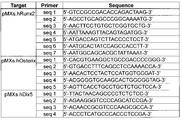
- the target gene was amplified by PCR using a coding region primer from a plasmid containing the target gene (Runx2 or the like) (the base sequence of the primer is shown in FIG. 23).
- the pMXs puro vector was digested with EcoRI. After each electrophoresis and separation, the gene and vector back bone were extracted from the electrophoresis gel.
- the pMXs vector encoding the target gene was prepared by ligating both using the Gene art system. The base sequences of these vectors were confirmed using the primers shown in FIG.
- pMXs vectors containing various genes were introduced in various combinations with the pCMV VSV vector into the packaging cell Plat GP using X-tremeGENE 9 at a 1: 3 ratio by lipofection ( Add 5 cm of transgene, 2.5 ⁇ g of pCMV.VSV, 500 ⁇ l of Opti-MEM, 22.5 ⁇ l of X-tremeGENE 9 in a 10 cm dish containing 10 ml of medium). After 24 hours, the medium was replaced with an antibiotic-free normal medium.
- the human normal gingival fibroblast cell line Gin-1 and the human normal skin fibroblast cell line aHDF are cultured at 2 ⁇ 10 4 to 2 ⁇ 10 5 cells / ml in a dish or culture plate (for example, immunostained 12 well plate, 24 well plate for ALP activity, 12 well plate for PCR purpose, 6 well plate for ALP staining purpose, 24 well plate, 6 well plate, 35mm dish, 60mm dish for Aizarin Red S staining purpose
- the day was set as the first day of culture.
- the Plat GP culture supernatant was passed through a syringe filter with a pore diameter of 0.45 ⁇ m, and then mixed with polybrene (final concentration 4 ⁇ g / ml) (virus solution).
- virus solution 500 ⁇ l for 24 well, 1 ml for 12 well, 1.5 ml for 6 well and 35 mm, 1.5 ml for 60 mm, 60 mm) 2.5 ml
- cells not infected with virus were also prepared.
- the culture supernatant was removed by aspiration, and osteoinduction medium (50 ⁇ g / ml ascorbic acid, 10 mM ⁇ -Glycerophosphate, 100 nM dexamethasone (all final concentrations) was added to the normal medium. 1) and cultured. Thereafter, the culture medium was changed every 2-3 days. 14 days after gene introduction, ALP staining, ALP activity test, Real-time RT-PCR, immunostaining after 20 days, Alizarin Red S staining after 20 or 28 days, von Kossa staining after 28 days went. The cells cultured in the same manner without infection with a retroviral vector were designated as Control.
- 3I indicates that a retroviral vector containing each gene was infected, and the blank indicates a retro It means that the virus vector is not infected).
- the well No. 27 in FIG. 3B (the top well, the third well from the left) is infected with a retroviral vector containing Osterix, Runx2, Oct4, L-Myc, and Dlx5 genes as shown in FIG. 3H. It can be seen that these cells are producing a lot of calcified bone matrix. Further, Alizarin Red S staining solution was removed from all wells, washed with sterilized distilled water, added with 10% Triton X, and reacted at room temperature for 1 hour. The liquid was collected from each well and transferred to a 96 well plate.
- the macrophotograph is shown in FIG. 3G (see FIG. 3H for well numbers). Thereafter, the absorbance (550 nm-650 nm, FIG. 3H; 490 nm-650 nm, FIG. 3I) of the reaction solution was measured using a microplate reader, and the results are shown in the graphs of FIGS. 3H and 3I.
- the vertical axis of the graph represents absorbance, and the higher the absorbance, the more calcified bone matrix was produced, indicating that many fibroblasts were converted to functional osteoblasts.
- the 27th cell infected with a retroviral vector containing the genes Osterix, Runx2, Oct4, L-Myc, and Dlx5 produced the most calcified bone matrix.
- ALP activity test ( Figure 4) A normal human gingival fibroblast cell line Gin-1 was cultured in a 12-well plate and experimented as shown in FIG. 14 days after gene transfer, the culture medium is removed from the cell culture dish by suction and washed twice with physiological saline. Cells are lysed with physiological saline containing 1% NP-40 and centrifuged at 12000 rpm for 5 minutes. The supernatant was collected, reacted with an ALP buffer containing p-nitrophenol phosphate, and measured with an absorptiometer at 405 nm. At the same time, the total protein amount was measured, corrected by the ALP activity per total protein amount, and displayed. The results are shown in FIG. ROOct4, ROOct4M, ROOct4L, ROOct4G, etc. showed significantly higher ALP activity compared to control. ROOct4L showed the highest ALP activity among all groups.
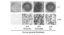
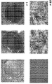
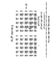
- ALP stained image ( Figure 5) A normal human gingival fibroblast cell line Gin-1 was cultured in a 6-well plate and experimented as shown in FIG. 14 days after gene introduction, the culture medium was aspirated and removed from the wells, washed twice with physiological saline, added with fixative, and fixed for 5 minutes. Washed twice with sterile distilled water. ALP staining solution was added, left to stand at room temperature for 1 hour, protected from light, washed twice with sterilized distilled water, and then observed with the naked eye and an inverted phase contrast microscope. The results are shown in FIG. Some of the cells into which ROOct4M, ROOct4L, or Oct4L were introduced became ALP positive. In particular, in cells into which ROOct4L was introduced, ALP staining positive cells were observed over the entire bottom surface of the well.
- FIG. 12 A human normal dermal fibroblast cell line aHDF was cultured in a 6-well plate and experimented as shown in FIG. 14 days after gene introduction, the culture medium was aspirated and removed from the wells, washed twice with physiological saline, added with fixative, and fixed for 5 minutes. The plate was washed twice with sterilized distilled water, added with ALP staining solution, allowed to stand at room temperature for 1 hour in the dark. After washing twice with sterilized distilled water, it was observed with the naked eye and an inverted phase contrast microscope. The results are shown in FIG.
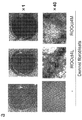
- FIG. 13a Human normal dermal fibroblast cell line aHDF was cultured in a 6-well plate and experimented as shown in FIG. 28 days after gene introduction, the culture medium was removed from the well by aspiration, washed twice with PBS, and fixed with 95% ethanol. After washing with sterilized distilled water, Alizarin Red S staining solution was added and allowed to stand at room temperature for 15 minutes. After washing with sterilized distilled water, it was observed with the naked eye and an inverted phase contrast microscope. The result is shown in FIG. 13a.
- FIG. 13b An inverted phase contrast microscope image obtained by conducting the same experiment as in a, and performing ALP staining (top) 14 days after gene introduction and von Kossa staining (bottom) 28 days after gene introduction is shown in FIG. 13b.
- the magnification is x40.
- Human osteoblasts are NHost cells purchased from Lonza Walkersville, Inc. Alizarin red S staining was performed as follows. Aspirate the culture medium from the culture dish, wash twice with PBS, and fix with 95% ethanol. After washing with sterilized distilled water, add Alizarin Red S staining solution and let stand at room temperature for 15 minutes. After washing with sterilized distilled water, it was observed and photographed with an inverted phase contrast microscope. Cells infected with ROOct4L and Oct4L produce a large amount of calcified bone matrix, and even cells infected with ROOct4M produce calcified bone matrix, although weaker than ROOct4L. ND: Not determined.
- FIG. 15 Properties of human osteoblasts induced by direct reprogramming (FIG. 15) Similar to FIG. 14, human gingival fibroblasts (Gin-1) were mixed with retrovirus vectors containing human Runx2, Osterix, Oct4 and L-Myc genes (ROOct4L), or Runx2, Osterix, Oct4 and A mixture of retroviral vectors (ROOct4M) each containing the c-Myc gene was infected and cultured for 28 days. (-) Is gingival fibroblast Gin-1 not infected with a retroviral vector. In addition, NHost cells purchased from Lonza Walkersville, Inc. were used as human osteoblasts. a, Quantification of calcium deposition by chelate method.
- the cells were washed thoroughly with PBS, detached with a scraper, dissolved with 0.5 M hydrochloric acid, and sonicated. The lysate was centrifuged at 10,000 rpm for 5 minutes, and the supernatant was collected. The 2 microliters were mixed with 240 microliters of Chlorophosphonazo-III solution (LS-MPR CPZ III, AKJ Global Technology, Chiba, Japan) and incubated for 10 minutes. The absorbance at 690 nm was measured with a microplate reader, and the calcium concentration (mg / dL) was calculated by comparison with a standard curve.
- Chlorophosphonazo-III solution LS-MPR CPZ III, AKJ Global Technology, Chiba, Japan
- RNA was recovered from the cells using ISOGEN II (Nippon Gene), and reverse transcription was performed using ReverTra Ace qPCR RT Master Mix (TOYOBO).
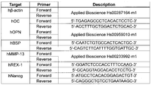
- Real-time RT-PCR analysis was performed using 7300 Real Time PCR System (Applied Biosystems) using primers specific to each gene (shown in FIG. 25) and Real-time PCR Master Mix (TOYOBO).
- the mRNA level of each sample was expressed as a relative value to the value of human gingival fibroblasts after correction with the ⁇ -actin mRNA level.
- Cells infected with ROOct4L can deposit calcium more than human osteoblasts.
- Cells infected with ROOct4M can deposit calcium to the same extent as human osteoblasts.
- Both ROOct4L infected cells and ROOct4M infected cells express osteoblast-specific genes.
- cells infected with ROOct4L have sufficient proliferation ability.
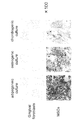
- FIG. 16 Analysis of human osteoblasts induced by direct reprogramming (FIG. 16) a, human gingival fibroblasts (Gin-1), cells cultured for 20 days after infection of human gingival fibroblasts (Gin-1) with ROOct4L (referred to as dOB in the following text and figures), and human Osteoblasts (NHost cells purchased from Lonza Walkersville, Inc.) were immunostained with FITC-labeled anti-human osteocalcin antibody and anti-osteopontin antibody. Magnification x 100. b, RNA was collected from the same cells as described above, and DNA microarray analysis was performed using GeneChip (registered trademark) human Gene 1.0 ST (Affymetrix).
- GeneChip registered trademark
- human Gene 1.0 ST Affymetrix
- All genes were divided into 7 groups by the ratio of the expression level in osteoblasts to the expression level in human gingival fibroblasts (less than 0.2, less than 0.2 and less than 0.33, more than 0.33 and less than 0.5, more than 0.5 and less than 2.0, more than 2.0 Less than 3.0, 3.0 or more and less than 5.0, 5.0 or more) (X axis).
- the number of genes belonging to each group is shown in parentheses.
- the ratio of the expression level in dOB to the expression level in human gingival fibroblasts (mean value ⁇ SD) was plotted.
- human dOB produces a large amount of osteocalcin and osteopontin (a). It can also be seen that human dOB has similar gene expression profiles to human osteoblasts (b). It can be seen that human dOB is close to human osteoblasts in that the epigenetic mark of chromosomal DNA is different from that of fibroblasts (c). In addition, during the transition from fibroblasts to dOB due to ROOct4L infection, no significant expression of REX-1 (d) and Nanog (e) gene mRNA was observed at any time point, via iPS cell-like cells. (D, e).
- FIG. 18 Bone regeneration in bone defect in vivo (FIG. 18) dOB (directly reprogrammed osteoblasts) contributes to bone regeneration in vivo.
- Animal experiments were performed with the approval of Kyoto Medical University. Pentobarbital was injected into the abdominal cavity of 8-week-old male NOD / SCID mice (Charles River) and anesthetized. A partial bone defect having a diameter of about 4 mm was created in the left femoral shaft using a dental drill under water injection. Incubate for 14 days after infecting human gingival fibroblasts (Gin-1) and a mixture of retroviral vectors containing human Runx2, Osterix, Oct4 and L-Myc genes in Gin-1 (ROOct4L).
- Gin-1 gingival fibroblasts
- ROOct4L a mixture of retroviral vectors containing human Runx2, Osterix, Oct4 and L-Myc genes in Gin-1
- dOB dBeton Dickinasar cells
- Matrigel BD Bioscience, San Jose, Calif.
- Mice were transplanted at a concentration of individuals / mouse. Mice were also prepared in which the same operation was performed without bone loss and transplantation (sham surgery).
- ⁇ CT Micro computed tomography
- the mice were anesthetized with pentobarbital injected into the abdominal cavity. The thigh was excised, fixed with neutral formalin, and then photographed with an X-ray CT device (Scan Xmate-L090, Com Scan Techno, Yokohama, Japan).
- a continuous tomographic image of 10 ⁇ m is shown.
- a triangle indicates a bone defect, and an arrowhead indicates a regenerated trabecular bone.
- b Bone defect tissue was embedded with SCEM (Leica Microsystem) compound and snap frozen. After slicing into 6 ⁇ m sections, serial sections were stained with hematoxylin and eosin (H & E) (top) and Alizarin Red S (bottom).
- H & E hematoxylin and eosin
- bottom Alizarin Red S
- a triangle indicates a bone defect, and an arrowhead indicates a regenerated trabecular bone. Magnification is x40.
- FIG. 19 Bone regeneration in bone defect in vivo
- the mouse was euthanized 21 days after the transplantation, and the femur was collected. A three point bending test was performed and the maximum loading value was measured.
- dOB osteoblasts
- FIG. 20 Bone regeneration in bone defect in vivo
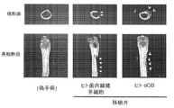
- a microscopic image of the bone defect of the femur on day 21 (upper) (magnification 10 times), and This is a superposition of explanations (bottom).
- * Indicates bone defect and # indicates regenerated bone.
- ossified bone composed of aligned trabeculae is formed, and the defect is completely covered and cannot be seen.
- fibroblasts only a small amount of fossils are formed, and a large bone defect remains.
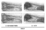
- FIG. 22 ⁇ CT transmission image of the experiment of FIG. 18a (FIG. 22).
- 18 shows a ⁇ CT transmission image of the same experiment as in FIG. 18a.
- the arrow is the bone defect.
- Bone transplanted with osteoblasts (dOB) induced by direct reprogramming has a higher radioopacity at the bone defect than bone transplanted with human gingival fibroblasts.
- dOB osteoblasts
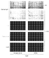
- a mixture of episomal vectors of the above four genes (0.5 ⁇ g each), Extreme GENE9 DNA Transfection Regent (6 ⁇ L) and Opti-MEM (200 ⁇ l) were mixed and added to the above cells for transfection. After culturing for 1 day, discard the medium and replace with osteoinduction medium (usual medium supplemented with 50 ⁇ g / ml ascorbic acid, 10 mM ⁇ -Glycerophosphate, 100 nM dexamethasone (all final concentrations)), and further culture did. ALP staining was performed 14 days after gene transfer. An inverted phase contrast microscope image is shown (b).
- CAG promoter CAG promoter
- polyA polyA addition signal
- oriP Epstein-Barr virus oriP sequence
- EBNA1 Epstein-Barr virus Nuclear antigen 1 gene
- KanR kanamycin resistance gene.
- FIG. 27 Mouse fetal fibroblasts were infected with a mixture of retroviral vectors with Runx2, Klf4 and c-Myc (RKM) or a mixture of retroviral vectors with Runx2, Klf4 and Glis1 (RKG) and then cultured for X days . Some cells are not infected. a, alkaline phosphatase (ALP) staining, alzaline red S staining and von Kossa staining. The magnification is x40.
- ALP alkaline phosphatase
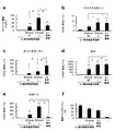
- RNA was collected from the cells, and the expression level of each gene was quantified by Real-time RT-PCR analysis. The mRNA level of each sample was corrected to the ⁇ -actin mRNA level, and then the non-transgenic mouse fetal fibroblasts were introduced. It was expressed as a relative value to the cell value. * P ⁇ 0.05, ** P ⁇ 0.01 (significant difference from non-transgenic mouse fetal fibroblasts). # P ⁇ 0.05, ## P ⁇ 0.01. Values are average values ⁇ SD (n 4)
- RNA was collected from the cells, and osteocalcin (black bar) and ALP (white bar) mRNA were quantified by real-time RT-PCR. Bars are mean ⁇ standard deviation. N 3. ** P ⁇ 0.01 (compared to uninfected cells).
- c 28 days after gene introduction, the culture medium is aspirated and removed from the culture dish, washed twice with PBS, and fixed with 10% formalin. After washing with sterilized distilled water, add 5% Silver nitrate solution and let stand under UV light for 30 minutes.
- the mRNA level of each sample was expressed as a relative value to the value of human gingival fibroblasts after correction with the ⁇ -actin mRNA level.
- # P ⁇ 0.05, ## P ⁇ 0.01. Values are mean ⁇ SD (n 4). Both ROOct4L-infected and Oct4L-infected cells express osteoblast-specific genes.
- RNA was recovered from the following cells.
- dOBs Osteoblasts derived by infecting human gingival fibroblasts (Gin-1) with a mixture of retroviral vectors (ROOct4L) containing human Runx2, Osterix, Oct4 and L-Myc genes, respectively.
- Fibroblasts Uninfected gingival fibroblasts Gin-1.
- Osteoblasts Human osteoblasts (NHost cells) purchased from Lonza Walkersville, Inc. Exhaustive gene expression analysis was performed using GeneChip (registered trademark) human Gene 1.0 ST of Affymetrix.
- MSCs human bone marrow mesenchymal stem cells.
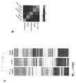
- MSC-OBs Osteoblasts derived from human bone marrow mesenchymal stem cells cultured in osteoblast medium. a) Heat map and cluster analysis results for genes whose expression levels increased and decreased more than 2-fold compared to Fibroblasts. b) Heat map of all genes. From these results, it can be seen that the gene expression profile of dOBs is very different from the original fibroblasts and is similar to osteoblasts. The similarity between dOBs and osteoblasts is higher than that between MSC-OBs and osteoblasts.
- Fig. 31 A) Osteoblasts derived from human gingival fibroblasts (Gin-1) by culturing 21 days after infection with a mixture of retroviral vectors containing Runx2, Osterix, Oct4 and L-Myc genes (ROOct4L) ( dOBs) were stained with anti-human osteocalcin and Alexa fluor 488 labeled secondary antibody and DAPI. DAPI stains the nuclei of all cells, but it can be seen that most DAPI-positive cells produce osteocalcin. b) The above-mentioned osteocalcin (+) DAPI (+) cell number and DAPI (+) cell number were counted.
- Reprogramming from human fibroblasts to osteoblasts is a direct conversion without going through a pluripotent stem cell-like stage (FIG. 32).
- Human gingival fibroblasts (Gin-1) were infected with a mixture of retrovirus vectors (ROOct4L) containing Runx2, Osterix, Oct4 and L-Myc genes, respectively. Cells were fixed with paraformaldehyde at 4 ° C. for 30 minutes every other day from day 1 to day 15 after infection. After permeabilizing with 0.2% Triton X-100 at room temperature for 15 minutes, staining was performed with anti-Nanog antibody, Alexa fluor 488-labeled secondary antibody and DAPI.
- ROOct4L retrovirus vectors
- Nanog positive cells When observed with a fluorescence microscope and more than 1,000 DAPI positive cells were observed for each sample, the number of Nanog positive cells was zero at any time point. A typical fluorescence microscope image is shown (magnification is x100). As a positive control, human iPS cells were stained in the same manner, and strong Nanog expression was observed in all cells.
- Human fibroblasts are not contaminated with mesenchymal stem cells (MSCs) (FIG. 34)
- MSCs mesenchymal stem cells
- FIG. 34 Human gingival fibroblasts (Gin-1) and human adipose tissue-derived mesenchymal stem cells MSC as a positive control were cultured in the following media, respectively.
- a medium for inducing differentiation into adipocytes left
- a medium for inducing differentiation into osteoblasts (middle)
- a medium for inducing differentiation into chondrocytes right.
- Oil O Red staining left
- Alizarin Red S staining center
- Alcian blue staining right
- FIG. 36 Properties of human osteoblasts derived from human fibroblasts by direct reprogramming (FIG. 36)
- RNA was collected from cells using ISOGEN II (Nippon Gene), and reverse transcription was performed using ReverTra Ace qPCR RT Master Mix (TOYOBO).
- Real-time RT-PCR analysis was performed using 7300 Real Time PCR System (Applied Biosystems) using primers specific to each gene (shown in FIG.
- dOBs directly contributed to bone regeneration by producing bone matrix after transplantation in vivo
- the GFP gene was introduced into osteoblasts (dOBs) induced by culturing after introducing ROOct4L into normal human gingival fibroblasts using a retroviral vector. These were transplanted into an artificial bone defect of the femur of a NOD / SCID mouse (right) as in FIG. As a negative control, only Matrigel was transplanted into the artificial bone defect of the femur of NOD / SCID mice (left). Three weeks later, femurs were collected, tissue sections were prepared, and anti-human osteocalcin antibody (not reactive with mouse OC) and DAPI immunostaining were performed.
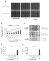
- Human fibroblasts can be reprogrammed into osteoblasts by gene transfer using plasmid vectors (FIGS. 39a and 39b).
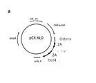
- a plasmid vector, pCX.OXL (FIG. 39a) was constructed as follows. A chimeric protein in which three genes, Oct4, Osterix, and L-myc are connected in this order from the N-terminus, and a self cleaving 2A peptide is inserted between Oct4 and Osterix, and between Osterixd and L-myc. was incorporated into the plasmid vector pCX.
- This pCX.OXL was introduced into human normal skin fibroblasts by electroporation (Neon) (middle) or lipofection (X-treme Gene 9) (right), and cultured in induction medium for 28 days. These cells and human normal dermal fibroblasts without gene introduction (left) were subjected to Aalizarin Red S (top) and von Kossa staining (bottom) (FIG. 39b). It was found that fibroblasts were converted into osteoblasts that produce a large amount of mineralized bone matrix by introducing three factors, Oct4, Osterix, and L-myc, by gene transfer using a plasmid vector.
- Human fibroblasts can be reprogrammed into osteoblasts by gene transfer using a plasmid vector (FIGS. 40a and b).
- a plasmid vector, pCX.XLO (FIG. 40a) was constructed as follows. Three genes of Osterix, L-myc and Oct4 are connected in this order from the N-terminal, and self cleaving 2A peptide is inserted between Osterix and L-myc and between L-myc and Oct4. The chimeric protein expression unit was incorporated into the plasmid vector pCX. pCX.XLO and pCX.OXL (FIG.
- FIG. 41 Human fibroblasts can be reprogrammed into osteoblasts in a medium without the addition of heterologous proteins.
- the pCX.OXL constructed in FIG. 39a was introduced into human normal skin fibroblasts by electroporation (Neon) method. Thereafter, the cells were cultured in an osteoinductive medium supplemented with 2% human serum without adding fetal bovine serum. After 5 days, ALP staining was performed in the same manner as in FIG. 5 (FIG. 41). It can be seen that human fibroblasts can be reprogrammed into osteoblasts in a medium without the addition of heterologous proteins.
- Human osteoblasts induced by direct reprogramming from human fibroblasts can be cryopreserved in a medium without the addition of heterologous proteins (FIG. 42).
- the pCX.OXL constructed in FIG. 39 was introduced into human normal skin fibroblasts by electroporation (Neon) method. After 14 days of culture, some cells were frozen in liquid nitrogen, stored in a freezer at ⁇ 80 ° C., thawed the next day, and then continued for another 5 days (right). Another part of the cells was cultured without freezing and thawing (middle).
Landscapes
- Health & Medical Sciences (AREA)
- Engineering & Computer Science (AREA)
- Life Sciences & Earth Sciences (AREA)
- Biomedical Technology (AREA)
- Genetics & Genomics (AREA)
- Organic Chemistry (AREA)
- Bioinformatics & Cheminformatics (AREA)
- Chemical & Material Sciences (AREA)
- Zoology (AREA)
- Wood Science & Technology (AREA)
- Biotechnology (AREA)
- General Engineering & Computer Science (AREA)
- General Health & Medical Sciences (AREA)
- Microbiology (AREA)
- Biochemistry (AREA)
- Orthopedic Medicine & Surgery (AREA)
- Cell Biology (AREA)
- Rheumatology (AREA)
- Biophysics (AREA)
- Physics & Mathematics (AREA)
- Plant Pathology (AREA)
- Molecular Biology (AREA)
- Chemical Kinetics & Catalysis (AREA)
- Medicinal Chemistry (AREA)
- General Chemical & Material Sciences (AREA)
- Animal Behavior & Ethology (AREA)
- Physical Education & Sports Medicine (AREA)
- Nuclear Medicine, Radiotherapy & Molecular Imaging (AREA)
- Veterinary Medicine (AREA)
- Pharmacology & Pharmacy (AREA)
- Public Health (AREA)
- Micro-Organisms Or Cultivation Processes Thereof (AREA)
- Pharmaceuticals Containing Other Organic And Inorganic Compounds (AREA)
Abstract
Description
一方、歯周病は第4の生活習慣病とも呼ばれ、罹患率が極めて高く、またさまざまな全身疾患の原因になっている。歯周病の進行にともなって、歯槽骨の骨吸収が起こるので、骨芽細胞を効率良く局所の骨吸収部に供給出来れば、歯槽骨の再生治療につながると考えられる。
このような移植目的の骨芽細胞として、これまで骨髄間葉系幹細胞や骨髄間葉系幹細胞を含む骨髄細胞などが用いられてきた。しかし骨髄の採取は患者への侵襲が大きく、また十分な数の骨髄細胞が供給できない場合があるなどの問題点がある。一方、ヒト胚性幹細胞(ES細胞)を用いれば、患者から骨髄を採取する必要はなく、また十分な数の骨芽細胞を供給できる可能性があるが、倫理的問題に加えて移植後に残存ES細胞が腫瘍化する危険性がある。またiPS細胞を用いても、患者から骨髄を採取する必要はなく、また十分な数の骨芽細胞を供給できる可能性があるが、移植後に残存iPS細胞が腫瘍化する危険性がある。
非特許文献2,3は、マウスiPS細胞からMSCを経てOsteogenic培地で分化誘導して骨芽細胞を得ている。
非特許文献1~4に示されるように、骨芽細胞はES細胞やiPS細胞などの多能性幹細胞から分化誘導して作製されているため、長期間の培養を要し、また癌化の危険性があった。
体細胞に、組織特異的な転写因子の遺伝子群を導入して、iPS細胞を経ずに直接その組織細胞に分化誘導できること(ダイレクト・リプログラミング(ダイレクト・コンヴァージョン))について、たとえば、以下の報告がある:
マウス線維芽細胞→軟骨細胞(SOX9 + Klf4 + c‐Myc遺伝子を導入)
マウス線維芽細胞→心筋細胞(GATA4 + Mef2c + Tbx5遺伝子を導入)
マウス線維芽細胞→肝細胞(Hnf4α+(Foxa1またはFoxa2またはFoxa3)遺伝
子を導入)
マウス線維芽細胞→神経幹細胞(Sox2 + FoxG1遺伝子を導入など)、
マウス、ヒト細胞→造血幹細胞など
しかし、体細胞を骨芽細胞にダイレクト・コンヴァージョンしたという報告はない。
本発明は、以下の骨芽細胞及びその調製方法を提供するものである。
項1. 哺乳動物の体細胞にリプログラミング関連遺伝子又はその発現産物を導入することで、前記体細胞から骨芽細胞を調製する方法であって、リプログラミング関連遺伝子がOctファミリー、c-Myc(M)、L-Myc(L)、GLIS ファミリー、Klfファミリー、Lin-28、Sox2からなる群から選択される少なくとも1種である、骨芽細胞を調製する方法。
項2. 哺乳動物の体細胞に骨関連遺伝子又はその発現産物とリプログラミング関連遺伝子又はその発現産物を導入することで、前記体細胞から骨芽細胞を調製する方法であって、前記骨関連遺伝子がRunx2(R)、Osterix(O)、Dlx5(D)からなる群から選択される少なくとも1種であり、リプログラミング関連遺伝子がOctファミリー、c-Myc(M)、L-Myc(L)、GLIS ファミリー、Klfファミリー、Lin-28、Sox2からなる群から選択される少なくとも1種である、骨芽細胞を調製する方法。
項3. 前記体細胞が線維芽細胞または歯肉細胞である、項1又は2に記載の方法。
項4. リプログラミング関連遺伝子又はその発現産物がOct4を含む、項1~3のいずれか1項に記載の方法。
項5. 体細胞に導入されるリプログラミング関連遺伝子又はその発現産物がOct4、Oct4L、Oct4M、Oct4LM、Oct4LGlis1、Oct4LMGlis1(ここで、Mは「c-Myc」を示し、Lは「L-Myc」を示す)
のいずれかである、項4に記載の骨芽細胞を調製する方法。
項6. 体細胞に導入される骨関連遺伝子又はその発現産物とリプログラミング関連遺伝子又はその発現産物の組み合わせが、
Oct4、Oct4LMGlis1、ROD Oct4L、RD Oct4L、RO Oct4ML、D Oct4ML、ROD Oct4M、OD Oct4L、O Oct4ML、O Oct4L、O Oct4M、OD Oct4、D Oct4L、Oct4ML、ROD Oct4ML、RD Oct4ML、OD Oct4ML、O Oct4MLGlis1、RD Oct4M、R Oct4L Glis1、R Oct4ML、OD Oct4M、O Oct4L Glis1、ROD Oct4、RO Oct4M、RO Oct4L、RO Oct4、O Oct4 Glis1、RD Oct4、Oct4L Glis1、D Oct4M、D Oct4 Glis1、O Oct4、Oct4L、Oct4M、D Oct4、RO Oct4 K、RO Oct4Sox2及びRO Oct4Lin28
(ここで、Rは「Runx2」を示し、Oは「Osterix」を示し、Dは「Dlx5」を示し、Mは「c-Myc」を示し、Lは「L-Myc」を示す)
からなる群から選ばれるいずれかの組み合わせである、項1~3のいずれかに記載の方法。
項7. 体細胞に導入される骨関連遺伝子又はその発現産物とリプログラミング関連遺伝子又はその発現産物の組み合わせが、ROD Oct4L、RD Oct4L、RO Oct4ML、D Oct4ML、ROD Oct4M、OD Oct4L、O Oct4ML、O Oct4L、O Oct4M、OD Oct4、D Oct4L及びOct4ML
(ここで、Rは「Runx2」を示し、Oは「Osterix」を示し、Dは「Dlx5」を示し、Mは「c-Myc」を示し、Lは「L-Myc」を示す)
からなる群から選ばれるいずれかの組み合わせである、項1~3のいずれかに記載の方法。
項8. 体細胞に導入される骨関連遺伝子又はその発現産物とリプログラミング関連遺伝子又はその発現産物の組み合わせが、ROD Oct4L、RD Oct4L、RO Oct4ML、D Oct4MLからなる群から選ばれるいずれかの組み合わせである、項2又は3に記載の方法。
項9. 哺乳動物の体細胞に由来し、リプログラミング関連遺伝子又はその発現産物を有する骨芽細胞であって、リプログラミング関連遺伝子がOct4、c-Myc(M)、L-Myc(L)、GLIS ファミリー、Klfファミリー、Lin-28、Sox2からなる群から選択される少なくとも1種である、骨芽細胞。
項10. 哺乳動物の体細胞に由来し、骨関連遺伝子又はその発現産物とリプログラミング関連遺伝子又はその発現産物を有する骨芽細胞であって、前記骨関連遺伝子がRunx2(R)、Osterix(O)、Dlx5(D)からなる群から選択される少なくとも1種であり、リプログラミング関連遺伝子がOct4、c-Myc(M)、L-Myc(L)、GLIS ファミリー、Klfファミリー、Lin-28、Sox2からなる群から選択される少なくとも1種である、骨芽細胞。
本発明は、リプログラミング関連遺伝子又はその発現産物、或いは骨関連遺伝子又はその発現産物とリプログラミング関連遺伝子又はその発現産物を導入することで、体細胞から骨芽細胞を調製する技術である。このうち骨関連遺伝子としては、Runx2(R)、Osterix(O)、Dlx5(D)からなる群から少なくとも1種を選択することができる。望ましくはOxterixとDlx5のいずれかが少なくとも1種類が含まれているのがよい。また、リプログラミング関連遺伝子としては、Oct4ファミリー、c-Myc(M)、L-Myc(L)、Glisファミリー、Klfファミリー(KLF1, KLF2, KLF3, KLF4, KLF5, KLF6, KLF7, KLF8, KLF9, KLF10, KLF11, KLF12, KLF13, KLF14, KLF15, KLF16, KLF17)、Lin-28、Sox2からなる群から選ばれる少なくとも1種を選択することができる。望ましくはOct4ファミリーのいずれかが少なくとも1種類が含まれているのがよく、さらに望ましくは、Oct4ファミリーのいずれかが少なくとも1種類とL-Myc及び/又はc-Mycが含まれているのがよい。このOct4ファミリーとしては望ましくはOct4である。したがって、Oct4とL-Myc及び/又はc-Mycの両方を含むものがもっとも望ましい。
Oct4は、Sox2、Klf4、およびc-Mycとともに体細胞に遺伝子導入することで、体細胞をリプログラミングして多能性を誘導できる因子である(Takahashi K, Yamanaka S. Cell、 126(4):663-76、 2006; Takahashi K, Tanabe K, Ohnuki M, Narita M, Ichisaka T, Tomoda K, Yamanaka S. Cell. 131(5):861-72, 2007)。Oct4の替わりに、GATA3, GATA6, SOX7, PAX1, GATA4, CEBPa,HNF4a, GRB2 等のいずれかを、Sox2、Klf4、およびc-Mycとともに体細胞に遺伝子導入することで、体細胞をリプログラミングして多能性を誘導できることが知られている(Shu et al. Cell 153:963, 2013)。GATA3, GATA6, SOX7, PAX1, GATA4, CEBPa,HNF4a, GRB2のように、リプログラミング因子ではなく、Octファミリーでもないが、リプログラミングにおけるOctの機能を代替することのできる因子についても、本発明においてはOct4の代わりに使用することができる。すなわち本明細書中では、リプログラミングにおけるOct4の機能を果たすことのできる因子の代表として「Oct4」を使用して説明しているが、GATA3, GATA6, SOX7, PAX1, GATA4, CEBPa,HNF4a, GRB2等の、リプログラミングにおけるOctの機能を代替することのできる因子も、Oct4と同様に使用することができる。
GLIS ファミリーとしては、GLIS1(GLIS family zinc finger 1)などが挙げられる。
体細胞リプログラミング(iPS細胞誘導)におけるKLF-4の機能を代替し得る遺伝子として、KLFファミリーの遺伝子以外にも、IRX ファミリーのメンバー(IRX6(iroquois homeobox protein 6)など)、PTXファミリーのメンバー(PITX2(paired-like homeodomain transcription factor 2)など)、DMRTB1(DMRT-like family B with proline-rich C-terminal 1)などが知られている(WO2010/098419 (2010/9/2))。本明細書では「KLF-4」を使用して説明しているが、体細胞リプログラミング(iPS細胞誘導)におけるKLF-4の機能を代替し得る遺伝子をKLF-4と同様に使用することができる。これらの遺伝子の産物、mRNAも使用することができる。
SOX2に代えて他のSOXファミリーの遺伝子を同様に使用することも可能である。本明細書ではSOXファミリーの代表として「SOX2」を使用して説明しているが、SOXファミリーの他の遺伝子もSOX2と同様に使用することができる。
本発明では簡単のため、すべてSOX2と呼ぶ。
本発明の遺伝子の組み合わせに、さらに他の遺伝子を加えることも可能である。
本発明は、体細胞から骨芽細胞を誘導する方法を含む。またこの方法により作成した骨芽細胞を含む。またこの骨芽細胞を含む治療用の細胞製剤や移植用材料をふくむ。
本技術を、体内の体細胞に応用すれば、体内で直接骨芽細胞を作ることが可能である。この方法、直接誘導のための製剤も本発明に含まれる。
移植材料とは、骨組織の修復、再建のために生体内に導入する、骨芽細胞を含有する材料をいう。移植材料は、インビトロで部分的もしくは完全に骨組織を再生させて、同一または別の個体に移植する材料を包含する。本発明で得られた骨芽細胞は、移植材料の作製に使用することができる。骨芽細胞自体も移植材料になる。したがって、骨芽細胞を細胞製剤として患者に移植することもできるし、ヒドロキシアパタイトや生体吸収性セラミックなどの人工材料からなる基材(スキャホルド(scaffold))とともに移植したり、スキャホルドとともに培養してから移植することができる。これらの場合、スキャホルドは移植目的に応じて様々な3次元的な形状を作らせることができる。
本発明はまた、骨芽細胞を用いたさまざまな研究や技術開発等に用いることができる。たとえば骨の発生と老化、形態形成、リモデリングの機構、これらに対する力学的ストレス、栄養、免疫、神経、ホルモンの影響の解析などの基礎研究に有用である。またストロンチウム90等の放射性物質の内部被爆における骨への影響の解析と骨からのストロンチウム90の除去技術の開発等にも有用である。
文献:1 Direct Reprogramming of Fibroblasts into Functional Cardiomyocytes
by Defined Factors; Masaki Ieda, Ji-Dong Fu, Paul Delgado-Olguin, Vasanth Vedantham, Yohei Hayashi, Benoit G. Bruneau, and Deepak Srivastava Cell 142: 375-386, 2010.
2 Direct conversion of fibroblasts to functional neurons by defined factors. Thomas Vierbuchen, Austin Ostermeier, Zhiping P. Pang, Yuko Kokubu, Thomas C. Sudhof & Marius Wernig. Nature 463: 1035-1041, 2010
3 Induction of human neuronal cells by defined transcription factors. Pang ZP, Yang N, Vierbuchen T, Ostermeier A, Fuentes DR, Yang TQ, Citri A, Sebastiano V, Marro S, Sudhof TC, Wernig M. Nature 476: 220-223, 2011.
4 Generation of hyaline cartilaginous tissue from mouse adult dermal fibroblast culture by defined factors Kunihiko Hiramatsu, Satoru Sasagawa, Hidetatsu Outani, Kanako Nakagawa, Hideki Yoshikawa, and Noriyuki Tsumaki, Journal of Clinical Investigation, 121: 640-657, 2011.
5 Induction of functional hepatocyte-like cells from mouse fibroblasts by defined factors. Pengyu Huang, Zhiying He, Shuyi Ji, Huawang Sun, Dao Xiang, Changcheng Liu, Yiping Hu, XinWang & Lijian Hui, . Nature 475:386-389, 2011.
6 Direct conversion of mouse fibroblasts to hepatocyte-like cells by defined factors. Sayaka Sekiya & Atsushi Suzuki. Nature 475:390-393, 2011.
具体的には、骨芽細胞に変換するための導入遺伝子(骨関連遺伝子とリプログラミング関連遺伝子の組み合わせ、或いはリプログラミング関連遺伝子単独)を発現ベクターに組み込み、対象とする体細胞に発現ベクターを導入し、細胞内で発現させることが好ましい。
本発明の遺伝子の導入は、プラスミドで行ってもよく、ウイルスベクター、たとえばレトロウイルスベクターを用いてもよい。導入効率と導入遺伝子の安定保持の観点からはウイルスベクターが好ましく、癌化のリスクを抑えるためにはプラスミドが好ましい。
なお、図3~図11、図14~16,図18~22、図26、図28~32、図34、図37~38、はヒト正常歯肉線維芽細胞株であるGin-1を用いた結果を示す。図12~13、図17、図33、図35~36、39~42、はaHDF(ヒト正常皮膚線維芽細胞)を用いた結果を示す。図27は、マウスの胎仔線維芽細胞を用いた結果を示す。
(1)目的遺伝子をコードしたpMXsベクターの作製(図1)
目的の遺伝子(Runx2など)を含んだプラスミドからコード領域のプライマーを用いてPCRにて目的遺伝子を増幅した(プライマーの塩基配列を図23に示す)。またpMXs puroベクターをEcoRIで切断した。それぞれについて電気泳動を行い分離した後、電気泳動ゲルより遺伝子とベクターのBack boneを抽出。両者をGene artシステムによりライゲーションすることで目的遺伝子をコードしたpMXsベクターを作製した。これらのベクターの塩基配列は図24に示すプライマーを用いて確認した。
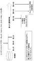
3×10⁶ 個の Plat GP細胞を、ケラチンコートした10cmディシュに播種、100U/ml Penicillinならびに100μg/ml Streptomycinを含んだ1% NEAA 10% FBS DMEM(通常培地)で培養。24時間後、種々の遺伝子を含むpMXsベクターを、種々の組み合わせで、pCMV VSVベクターと伴に、パッケージング細胞であるPlat GPにX-tremeGENE 9を用いて1:3比でリポフェクション法により導入(導入遺伝子5μg、pCMV.VSV 2.5μg、Opti-MEM 500μl、X-tremeGENE 9 22.5μlの混和液を10mlの培地入りの10cmディシュの添加)。24時間後、抗生剤フリーの通常培地に培地交換。同日にヒト正常歯肉線維芽細胞株であるGin-1およびヒト正常皮膚線維芽細胞株であるaHDFを2×10⁴~2×10⁵ cells/mlで培養ディッシュまたは培養プレート(たとえば、免疫染色の目的では12 wellプレート、24 wellプレート、ALP活性、PCRの目的では12 wellプレート、ALP染色の目的では6wellプレート、Alizarin Red S染色の目的では、24wellプレート、6wellプレート、35mmディッシュ、60mmディッシュのいずれか)に播種、この日を培養第-1日目とした。1日後(培養第0日目)、Plat GP培養上清を、ポアの直径が0.45μmのシリンジフィルターを通した後、ポリブレン(最終濃度4μg/ml)と混和した(ウイルス液)。Gin-1およびaHDF の培養上清を吸引除去した後、すばやく上記のウイルス液を加え(24 wellの場合は500μl、12 wellの場合は1ml、6wellならびに35mmの場合は1.5ml、60mmの場合は2.5ml)、24時間培養した(感染)。コントロール群として、ウイルス感染を行わない細胞も準備した。1日後(培養第1日目)、培養上清を吸引除去し、骨誘導培地(通常培地に50 μg/mlアスコルビン酸、10 mM β-Glycerophosphate、100 nMデキサメタゾン(いずれも最終濃度)を加えたもの)を加え培養した。その後2~3日間隔で培養液を交換した。遺伝子を導入して14日後に、ALP染色、ALP活性試験、Real-time RT-PCRを、20日後に免疫染色を、20日または28日後にAlizarin Red S染色を、28日後にvon Kossa染色を行った。レトロウイルスベクターを感染させずに、同じ培養を行った細胞をControlとした。
ヒト正常歯肉線維芽細胞株Gin-1を、24 wellプレートに培養し、図2のように実験した。遺伝子導入28日後、培養ディッシュから培養液を吸引除去し、PBSで2回洗浄を行い、95%エタノールで固定。滅菌蒸留水で洗浄した後、アリザリンレッドS染色液を加え、室温で15分間静置。図3A-Fはディッシュの肉眼像写真である。赤く染まっているのは石灰化骨基質であり、機能性骨芽細胞が誘導されたことを示す。ウェルの数字は図3H、図3Iを参照(図3H、図3Iの表中の「1」は、それぞれの遺伝子を含むレトロウイルスベクターを感染させたことを、空欄は、それぞれの遺伝子を含むレトロウイルスベクターを感染させていないことを表す)。たとえば、図3Bの27番のウェル(一番上段、左から3番目のウェル)は、図3Hに示されるとおり、Osterix、Runx2、Oct4、L-Myc、Dlx5の遺伝子を含むレトロウイルスベクターを感染させた細胞であり、多くの石灰化骨基質を産生しているのが分かる。さらにすべてのウェルからアリザリンレッドS染色液を取り除き、滅菌蒸留水で洗浄後、10%Triton Xを加え、室温で1時間反応させた。各ウェルから液を採取し、96 well plateに移した。その肉眼写真を図3Gに示す(ウェルの数字は図3Hを参照)。その後反応液の吸光度(550nm-650nm、図3H;490nm-650nm、図3I)をマイクロプレートリーダーを用いて測定した結果を図3H、図3Iのグラフに示す。グラフの縦軸は吸光度であり、吸光度が高い程、石灰化骨基質が多量に産生されたことを表し、多くの線維芽細胞が機能性骨芽細胞にコンヴァートしたことを示す。たとえば27番目の、Osterix、Runx2、Oct4、L-Myc、Dlx5の遺伝子を含むレトロウイルスベクターを感染させた細胞は、もっとも多くの石灰化骨基質を産生したことが分かる。
ヒト正常歯肉線維芽細胞株Gin-1を、12wellプレートに培養し、図2のように実験した。遺伝子導入14日後、細胞培養ディッシュから、培養液を吸引除去し、生理食塩水で2回洗浄。1%NP-40含有生理食塩水で細胞を溶解し、12000rpmで5分間遠心。上清を回収し、p-nitrophenol phosphateを含むALP緩衝液と反応させ、405nmで吸光度計にて測定した。また、同時に総タンパク量も測定し、総タンパク質量あたりのALP活性で補正し、表示した。結果を図4に示す。
ROOct4、ROOct4M、ROOct4L、ROOct4G等でcontrolと比較し、有意に高いALP活性を示した。また、ROOct4Lは全てのグループの中で最も高いALP活性を示した。
ヒト正常歯肉線維芽細胞株Gin-1を、6wellプレートに培養し、図2のように実験した。遺伝子導入14日後、ウェルから培養液を吸引除去し、生理食塩水で2回洗浄を行い、固定液を添加し、5分間固定した。滅菌蒸留水で、2回洗浄した。ALP染色液を加え、遮光して室温で1時間静置し、滅菌蒸留水で、2回洗浄した後、肉眼および倒立位相差顕微鏡で観察した。結果を図5に示す。
ROOct4M、またはROOct4L、またはOct4Lを導入した細胞の一部がALP陽性となった。とくにROOct4Lを導入した細胞では、ウェルの底面全体にわたってALP染色陽性細胞が認められた。
ヒト正常歯肉線維芽細胞株Gin-1を、24wellプレートに培養し、図2のように実験した。遺伝子導入21日後、培養ディッシュから培養液を吸引除去し、PBSで2回洗浄を行い、4%パラホルムアルデヒドで30分間固定した。3回洗浄を行った後、室温で1時間ブロッキングした。1次抗体(anti-hOsteocalcin)を4℃で24時間反応させ、3回洗浄した後、FITCがラベルしてある2次抗体を室温で1時間反応させた。3回洗浄した後、蛍光顕微鏡で観察した。結果を図6に示す。
(a)control、(b) ROOct4M、(c) ROOct4L:×100
ROOct4M、ROOct4Lを導入した細胞においてOsteocalcinの発現を確認した。またROOct4Lを導入した細胞でより多数のOsteocalcin陽性細胞を認めた。
ヒト正常歯肉線維芽細胞株Gin-1を、35 mmディッシュに培養し、図2のように実験した。遺伝子導入28日後、培養ディッシュから培養液を吸引除去し、PBSで2回洗浄を行い、95%エタノールで固定。滅菌蒸留水で洗浄した後、アリザリンレッドS染色液を加え、室温で15分間静置。滅菌蒸留水で洗浄した後、肉眼および倒立位相差顕微鏡で観察した。結果を図7に示す。
(a)control、(b) ROOct4M、(c) ROOct4L:×1
(d)control、(e) ROOct4M、(f) ROOct4L:×40
ROOct4M、またはROOct4Lを導入した細胞のディッシュで石灰化基質の沈着を認めた。とくにROOct4Lを導入した細胞のディッシュでは、培養ディッシュの底面全体にわたって大量の石灰化骨基質の沈着を認めた。
ヒト正常歯肉線維芽細胞株Gin-1を、35 mmディッシュに培養し、図2のように実験した。遺伝子導入28日後、培養ディッシュから培養液を吸引除去し、PBSで2回洗浄を行い、10%ホルマリンで固定。滅菌蒸留水で洗浄した後、5% Silver nitrate solutionを加え、UVライト下で30分間静置。その後滅菌蒸留水で洗浄し、5% Thiosulfate solutionを加えて2分間反応させた。滅菌蒸留水で洗浄した後、肉眼および倒立位相差顕微鏡で観察した。結果を図8に示す。
ROOct4Mを導入した細胞では散在したリン酸カルシウムの沈着を認めた。ROOct4LとOct4Lを導入した細胞では、培養ディッシュの底面全体に密集したリン酸カルシウムの沈着を認めた。
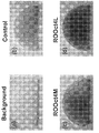
図9のように、体細胞を第-1日に培養ディッシュまたは培養プレートに撒き、第0日に遺伝子を導入し、第1日に培地を骨分化誘導培地に交換した。この第―1日目の細胞の播種、第0日目の遺伝子導入、第1日目の培地交換の詳細は図2と同様である。第4日に細胞をディッシュから剥がし、5×10⁵個を、Scaffold(3D insert-PCL)上に播種して3次元培養を行った。第28日にギムザ染色またはAlizarin Red S染色を行った。レトロウイルスベクターを感染させずに、同じ培養を行った細胞をControlとした。また、細胞を加えずにスキャホルドだけで同じ培養を行ったものをBackgroundとした。
ヒト正常歯肉線維芽細胞株Gin-1を、60 mmディッシュに培養し、図9のように実験した。遺伝子導入から28日後、培養ディッシュから培養液を吸引し、PBSで2回洗浄を行い、Scaffoldごと細胞をメタノールで固定。滅菌蒸留水で洗浄した後、ギムザ染色液を加え、室温で15分間静置。滅菌蒸留水で洗浄した後、肉眼で観察した。結果を図10に示す。
(a) Background、(b) ROOct4L:×1
誘導細胞は、このScaffoldに生着し、増殖することが判明した。
ヒト正常歯肉線維芽細胞株Gin-1を、60 mmディッシュに培養し、図9のように実験した。遺伝子導入から28日後、培養ディッシュから培養液を吸引し、PBSで2回洗浄を行い、Scaffoldごと細胞を95%エタノールで固定した。滅菌蒸留水で洗浄した後、アリザリンレッドS染色液を加え、室温で15分間静置し、滅菌蒸留水で洗浄した後、肉眼で観察した。結果を図11に示す。
(a)Background、(b)control、(c) ROOct4M、(d) ROOct4L:×1
誘導細胞は、このScaffold上で石灰化骨基質産生能を示すことが判明した。
また石灰化骨基質の産生は、ROOct4Lを導入した細胞で顕著であった。
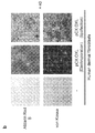
ヒト正常皮膚線維芽細胞株aHDFを6 well plateに培養し、図2のように実験した。遺伝子導入14日後、ウェルから培養液を吸引除去し、生理食塩水で2回洗浄を行い、固定液を添加し、5分間固定した。滅菌蒸留水で、2回洗浄し、ALP染色液を加え、遮光して室温で1時間静置した。滅菌蒸留水で、2回洗浄した後、肉眼および倒立位相差顕微鏡で観察した。結果を図12に示す。
(a)control、(b)ROOct4 M、(c) ROOct4L:×1
(d)control、(e)ROOct4 M、(f) ROOct4L:×40
ROOct4M、またはROOct4Lを導入した細胞の一部がALP染色陽性となった。とくにROOct4Lを導入した細胞では、ウェルの底面全体にわたって多数のALP染色陽性細胞を認めた。
a)ヒト正常皮膚線維芽細胞株aHDFを6 well plateに培養し、図2のように実験した。遺伝子導入28日後、ウェルから培養液を吸引除去し、PBSで2回洗浄を行い、95%エタノールで固定した。滅菌蒸留水で洗浄した後、アリザリンレッドS染色液を加え、室温で15分間静置した。滅菌蒸留水で洗浄した後、肉眼および倒立位相差顕微鏡で観察した。結果を図13aに示す。
(左)control、(中央)ROOct4 M、(右) ROOct4L:×1
ROOct4 M、またはROOct4Lを導入した細胞のウェルで石灰化基質の沈着を認めた。とくにROOct4Lを導入した細胞では、ウェルの底面全体にわたって大量の石灰化骨基質の沈着を認めた。
ヒト正常歯肉線維芽細胞Gin-1を、6ウェルプレートに培養し、ヒトのRunx2, Osterix, Oct4とL-Myc の遺伝子をそれぞれ含むレトロウイルスベクターの混合(ROOct4L) 、または Runx2, Osterix, Oct4とc-Myc の遺伝子をそれぞれ含むレトロウイルスベクターの混合(ROOct4M) 、または Oct4とL-Myc の遺伝子をそれぞれ含むレトロウイルスベクターの混合(Oct4L)を感染させた。感染後、記載の日数(1日~28日)培養した。(-)はレトロウイルスベクターを感染していない歯肉線維芽細胞Gin-1である。またヒト骨芽細胞は、Lonza Walkersville, Inc.より購入したNHost細胞である。アリザリンレッドS染色は以下の様に行った。培養ディッシュから培養液を吸引除去し、PBSで2回洗浄を行い、95%エタノールで固定。滅菌蒸留水で洗浄した後、アリザリンレッドS染色液を加え、室温で15分間静置。滅菌蒸留水で洗浄した後、倒立位相差顕微鏡で観察、撮影した。ROOct4LとOct4Lを感染させた細胞は、多量に石灰化骨基質を産生し、ROOct4Mを感染させた細胞でも、ROOct4Lよりは弱いが石灰化骨基質を産生する。ND: Not determined.
図14と同様に、ヒト歯肉線維芽細胞(Gin-1) に、ヒトのRunx2, Osterix, Oct4とL-Myc の遺伝子をそれぞれ含むレトロウイルスベクターの混合(ROOct4L) 、または Runx2, Osterix, Oct4 とc-Myc の遺伝子をそれぞれ含むレトロウイルスベクターの混合(ROOct4M) を感染させ、28日間培養した。(-)はレトロウイルスベクターを感染させていない歯肉線維芽細胞Gin-1である。またヒト骨芽細胞として、Lonza Walkersville, Inc.より購入したNHost細胞を用いた。a、カルシウム沈着のキレート法による定量。細胞をPBSでよく洗浄した後、スクレーパーで剥離し、0.5Mの塩酸で溶解し、ソニケーションを行った。ライセートを10,000 rpmで5 分間遠心し、上清を回収した。その2マイクロリットルを、240マイクロリットルのChlorophosphonazo-III 溶液 (LS-MPR CPZ III, AKJ Global Technology, 千葉、日本) と混ぜ、10分間インキュベートした。マイクロプレートリーダーで690 nmの吸光度を測定し、標準曲線と比較してカルシウム濃度(mg/dL) を計算した。b-e、細胞からISOGEN II (Nippon Gene)を用いてRNAを回収し、ReverTra Ace qPCR RT Master Mix (TOYOBO)を用いて逆転写を行った。それぞれの遺伝子に特異的なプライマー(図25に示す)とReal-time PCR Master Mix (TOYOBO) を用い、7300 Real Time PCR System (Applied Biosystems)を使ってreal-time RT-PCR 解析を行った。各サンプルのmRNAレベルは、β-アクチンmRNA レベルで補正後、ヒト歯肉線維芽細胞の値に対する相対値としてあらわした。f、細胞に2-(2-methoxy-4-nitrophenyl)-3-(4-nitrophenyl)-5-(2, 4-disulfophenyl)-2H-tetrazolium monosodium salt 溶液 (WST-8) (Cell Count Reagent SF; Nacalai Tesque) を加え、37℃で1時間培養した。上清の450 nm と650 nmの吸光度を測定し、遺伝子非導入ヒト歯肉線維芽細胞の値を100%として、各細胞のviabilityを計算した。*P<0.05 および **P<0.01,(遺伝子非導入ヒト歯肉線維芽細胞(Gin-1)に対する有意差)。#P < 0.05 、##P < 0.01。値は平均値 ± S.D. (n=4).
a、ヒト歯肉線維芽細胞(Gin-1) 、ヒト歯肉線維芽細胞(Gin-1)にROOct4Lを感染させた後20日間培養した細胞(以下の文章と図中ではdOBと呼ぶ)、およびヒト骨芽細胞(Lonza Walkersville, Inc.より購入したNHost細胞)を、FITCラベルした抗ヒトオステオカルシン抗体および抗オステオポンチン抗体で免疫染色した。倍率x 100。b、上記と同様の細胞からRNAを回収し、GeneChip(登録商標) human Gene 1.0 ST (Affymetrix社) を用いてDNAマイクロアレイ解析を行った。ヒト歯肉線維芽細胞での発現量に対する骨芽細胞での発現量の比で、全遺伝子を7群に分けた(0.2未満、0.2以上0.33未満、0.33以上0.5未満、0.5以上2.0未満、2.0以上3.0未満、3.0以上5.0未満、5.0以上)(X軸)。各群に属する遺伝子の数を括弧内に示す。それぞれの群に属する遺伝子について、ヒト歯肉線維芽細胞での発現量に対するdOBでの発現量の比(平均値±S.D.)をプロットした。ヒト歯肉線維芽細胞に比して骨芽細胞で強く発現する遺伝子は、dOBでも強く発現し、逆に骨芽細胞で弱く発現する遺伝子は、dOBでも弱く発現することが分かり、有意な相関がみられる(相関係数R = 0.70, P<0.01). c, 上記と同様の細胞からDNAを回収し、オステオカルシン遺伝子上流域のCpGメチル化を解析した。DNAの回収は、Genomic DNA purification kit (Mag Extractor, Toyobo Life Science, Tokyo, Japan)を用いて行い、EZ DNA methylation kit (ZYMO research, Irvine, CA)を用いてバイサルファイド処理を行った後、オステオカルシン遺伝子上流域の配列を、センスプライマー(5’-GTGTATTTGGTAGTTATAGTTATTTGG) とアンチセンスプライマー(5’-CCTCAAATTAAACACTAACTAAACTC) を用いてPCRで増幅した。pTA2ベクターにクローニングした後、T7とT3ユニバーサルプライマーを用いてシーケンシングした。黒はメチル化、白は非メチル化CpGを示す。d-e, ヒト歯肉線維芽細胞(Gin-1)にROOct4L を感染後、感染前(-)、および感染後7, 14, 21日目に細胞からRNAを回収した。ポジティブコントロールとしてヒトiPS細胞からもRNAを回収した。逆転写後REX-1 (d) および Nanog (e) 特異的なプライマーを用いてreal time RT-PCRを行った。各サンプルのmRNAレベルはβ-アクチンmRNA レベルで補正後、ヒト歯肉線維芽細胞(Gin-1) の値に対する相対値としてあらわしている。**P<0.01, (遺伝子非導入ヒト歯肉線維芽細胞に対する有意差). 値は平均値 ± S.D. (n=4).
ヒト皮膚線維芽細胞に、ヒトのRunx2, Osterix, Oct4とL-Myc の遺伝子をそれぞれ含むレトロウイルスベクターの混合(ROOct4L) 、または Runx2, Osterix, Oct4とc-Myc の遺伝子をそれぞれ含むレトロウイルスベクターの混合(ROOct4M) を感染させ、14日間の培養した(d0)。(-)はレトロウイルスベクターを感染させていない歯肉線維芽細胞Gin-1である。図15b-eと同様に、各遺伝子の発現量をReal-time RT-PCR 解析で定量した. 各サンプルのmRNAレベルはβ-アクチンmRNA レベルで補正後、ヒト歯肉線維芽細胞(Gin-1) の値に対する相対値としてあらわした。*P<0.05 、 **P<0.01 (遺伝子非導入ヒト歯肉線維芽細胞に対する有意差). #P<0.05、 ##P<0.01. 値は平均値 ± S.D. (n=4)。
dOB (ダイレクト・リプログラミングされた骨芽細胞)は生体内で骨再生に寄与する。
動物実験は、京都府立医科大学の認可を得て行った。8週齢オスのNOD/SCIDマウス(Charles River)の腹腔内にペントバルビタールを注射し麻酔した。注水下に歯科ドリルを用いて左大腿骨骨幹に直径約4mmの部分骨欠損を作成した。ヒト歯肉線維芽細胞(Gin-1) 、および、Gin-1にヒトのRunx2, Osterix, Oct4とL-Myc の遺伝子をそれぞれ含むレトロウイルスベクターの混合(ROOct4L) を感染させた後14日間培養して誘導した細胞(以下の文章と図中ではdOBと呼ぶ)を、25マイクロリッターの培地と75マイクロリッターのマトリゲル (BD Bioscience, San Jose, CA)に懸濁し、骨欠損部に5×10⁵ 個 /マウスの濃度で移植した.骨欠損と移植は行わずにそれ以外の同様の手術を行ったマウスも作成した(偽手術)。a, マイクロ・コンピューター断層撮影(μCT) イメージ。移植後21日目にマウスの腹腔内にペントバルビタールを注射し麻酔した。大腿を切除し、中性ホルマリンで固定後、X-ray CT device (Scan Xmate-L090, Com Scan Techno, Yokohama, Japan)で撮影した。10 μmの連続断層像を示す。三角は骨欠損を、矢頭は再生骨梁を示す。b, 骨欠損部の組織をSCEM (Leica Microsystem) compoundで包埋し、急速凍結した。6 μmの切片にスライス後、連続切片をヘマトキシリン・エオジン (H&E)(上)およびAlizarin Red S (下)で染色した。三角は骨欠損を、矢頭は再生骨梁を示す。倍率は×40。c, 上記の6 μmの切片を4%パラホルムアルデヒドで固定し、ヒト核特異的マウスモノクローナル抗体(Cat: MAB1281; clone: 235-1; Millipore, Billerica, MA)で免疫染色した。#は再生骨梁、*は骨髄を示す。倍率は×100。
マイクロCT画像(a)と組織像(b)では、ヒトdOBを移植した骨において、整列した骨梁からなる骨性化骨が形成され、欠損部は完全に被覆されていることが分かる。線維芽細胞を移植した骨では化骨はわずかに形成しているのみで、骨欠損が残存している。免疫蛍光像(c)では、ヒトdOBを移植した骨において、移植したdOBが多数、骨再生部に生着していることが分かる。線維芽細胞を移植した骨では、移植した線維芽細胞は少数のみ生着している。
図18と同様の移植実験を行ったのち、移植後21日目に、マウスを安楽死させ、大腿骨を採取した。three point bending testを行い、maximum loading valueを計測した。RO Oct4L を感染させた後14日間培養して誘導した骨芽細胞(dOB)を移植した群では、歯肉線維芽細胞を移植した群に比べて、有意に大腿骨の力学的強度が増強している。Sham operatedは骨欠損と移植は行わずにそれ以外の同様の手術を行ったマウスの大腿骨である。*P<0.05、 **P<0.01. 値は平均値 ± S.D. (n=3)。
図18と同様の実験で、ヒト歯肉線維芽細胞(左)およびdOB(右)を移植後、21日目の大腿骨の骨欠損部の実態顕微鏡像(上)(倍率10倍)、および、これに説明書きを重ね合せたもの(下)を示す。*は骨欠損、#は再生化骨である。
ヒトdOBを移植した骨において、整列した骨梁からなる骨性化骨が形成され、欠損部は完全に被覆されて視認できない。線維芽細胞を移植した骨では化骨はわずかに形成しているのみで、骨欠損が大きく残存している。
三角は骨欠損を、矢頭は再生骨梁を示す。
ヒトdOBを移植した骨において、整列した骨梁からなる骨性化骨が形成され、欠損部は完全に被覆されている。線維芽細胞を移植した骨では化骨はわずかに形成しているのみで、骨欠損が残存している。
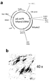
ヒトのRunx2, Osterix, Oct4とL-Myc の遺伝子を、それぞれpG.oriPP9.EBNA1エピゾーマル・ベクターのEcoRIサイトに挿入した(図26のaに、Runx2を挿入したpG.oriPP9.hRunx2.EBNA1を示す)。2×10⁵個のヒト歯肉線維芽細胞(Gin-1)を、図1の実験と同様の通常培地(100U/ml Penicillinならびに100μg/ml Streptomycinを含んだ1% NEAA 10% FBS DMEM)に再縣濁し、35 mmディシュに播種して1日間培養した。上記の4つの遺伝子のエピゾーマル・ベクターの混合(各0.5 μgずつ)、Extreme GENE9 DNA Transfection Regent(6μL)およびOpti-MEM(200 μl)を混和し、上記の細胞に添加してトランスフェクションした。1日間の培養後、培地を棄て、骨誘導培地(通常培地に50 μg/mlアスコルビン酸、10 mM β-Glycerophosphate、100 nMデキサメタゾン(いずれも最終濃度)を加えたもの)に交換し、さらに培養した。遺伝子導入の14日後にALP染色を施行した。倒立位相差顕微鏡像を示す(b)。aの図中、CAG promoter:CAGプロモーター、polyA: ポリA付加シグナル、oriP:エプスタイン・バール・ウイルスoriP配列、EBNA1:エプスタイン・バール・ウイルスNuclear antigen 1遺伝子、KanR:カナマイシン耐性遺伝子。
マウスの胎仔線維芽細胞に、 Runx2, Klf4 およびc-Myc を有するレトロウイルスベクターの混合 (RKM) または Runx2, Klf4および Glis1を有するレトロウイルスベクターの混合(RKG)を感染させ、その後X日間培養した。一部の細胞は感染させていない。a 、アルカリフォスファターゼ(ALP)染色、アルザリンレッドS染色およびフォン・コッサ染色を行った。倍率はx 40である。b、細胞よりRNAを採取し、記載の各遺伝子の発現量をReal-time RT-PCR 解析で定量した.各サンプルのmRNAレベルはβ-アクチンmRNA レベルで補正後、遺伝子非導入マウス胎仔線維芽細胞の値に対する相対値としてあらわした。*P<0.05 、 **P<0.01 (遺伝子非導入マウス胎仔線維芽細胞に対する有意差)。 #P<0.05、 ##P<0.01. 値は平均値 ± S.D. (n=4)
ヒト正常歯肉線維芽細胞株Gin-1を、 35 mmディッシュに培養し、図2のように実験した。図中の「+」は、それぞれの遺伝子を含むレトロウイルスベクターを感染させたことを、空欄は、それぞれの遺伝子を含むレトロウイルスベクターを感染させていないことを表す。
a、遺伝子導入14日後、細胞からRNAを回収し、オステオカルシン(黒バー)とオステオポンチン(白バー)のmRNAをリアルタイムRT-PCRで定量した。バーは平均±標準偏差。N=3。*P<0.05 および **P<0.01(非感染細胞との比較)。
b、遺伝子導入14日後、細胞からRNAを回収し、オステオカルシン(黒バー)とALP(白バー)のmRNAをリアルタイムRT-PCRで定量した。バーは平均±標準偏差。N=3。**P<0.01(非感染細胞との比較)。
c、遺伝子導入28日後に、培養ディッシュから培養液を吸引除去し、PBSで2回洗浄を行い、10%ホルマリンで固定。滅菌蒸留水で洗浄した後、5% Silver nitrate solutionを加え、UVライト下で30分間静置。その後滅菌蒸留水で洗浄し、5% Thiosulfate solutionを加えて2分間反応させた。滅菌蒸留水で洗浄した後、肉眼および倒立位相差顕微鏡で観察した。
Osterix+Oct4+L-Myc や Runx2+Osterix+Oct4+L-Myc の遺伝子を含むレトロウイルスベクターを感染させた細胞は、多くの石灰化骨基質を産生したことが分かる。また、 Oct4+L-Myc の遺伝子を含むレトロウイルスベクターを感染させた細胞も、多くの石灰化骨基質を産生した。
ヒト歯肉線維芽細胞(Gin-1) に、ヒトのRunx2, Osterix, Oct4とL-Myc の遺伝子をそれぞれ含むレトロウイルスベクターの混合(ROOct4L) 、または Oct4 とL-Myc の遺伝子をそれぞれ含むレトロウイルスベクターの混合(Oct4L) を感染させ、14日間培養した。(-)はレトロウイルスベクターを感染させていない歯肉線維芽細胞Gin-1である。またヒト骨芽細胞として、Lonza Walkersville, Inc.より購入したNHost細胞を用いた。図15b~eと同様に、細胞からISOGEN II (Nippon Gene)を用いてRNAを回収し、ReverTra Ace qPCR RT Master Mix (TOYOBO)を用いて逆転写を行った。それぞれの遺伝子に特異的なプライマー(図29bに示す)とReal-time PCR Master Mix (TOYOBO) を用い、7300 Real Time PCR System (Applied Biosystems)を使ってreal-time RT-PCR 解析を行った。結果を図29aに示す。各サンプルのmRNAレベルは、β-アクチンmRNA レベルで補正後、ヒト歯肉線維芽細胞の値に対する相対値としてあらわした。*P<0.05 および **P<0.01,(遺伝子非導入ヒト歯肉線維芽細胞(Gin-1)に対する有意差)。#P < 0.05 、##P < 0.01。値は平均値 ± S.D. (n=4).
ROOct4Lを感染させた細胞もOct4Lを感染させた細胞も、骨芽細胞特異的な遺伝子を発現する。
RNAを以下の細胞から回収した。dOBs:ヒト歯肉線維芽細胞(Gin-1) に、ヒトのRunx2, Osterix, Oct4とL-Myc の遺伝子をそれぞれ含むレトロウイルスベクターの混合(ROOct4L)を感染させて誘導した骨芽細胞。Fibroblasts:感染させていない歯肉線維芽細胞Gin-1。Osteoblasts: Lonza Walkersville, Inc.より購入したヒト骨芽細胞(NHost細胞)。Affymetrix社のGeneChip(登録商標) human Gene 1.0 STを用いて網羅的遺伝子発現解析を行った。MSCs:ヒト骨髄間葉系幹細胞。MSC-OBs:ヒト骨髄間葉系幹細胞を骨芽細胞培地で培養して誘導した骨芽細胞。a)、 Fibroblastsと比較して発現量が2倍より大きく増加および減少した遺伝子のHeat mapとクラスター解析結果。b)、全遺伝子のHeat map。これらの結果から、dOBsの遺伝子発現プロファイルは、元の線維芽細胞とは大きく異なり、骨芽細胞に類似していることがわかる。またdOBsと骨芽細胞の類似性は、MSC-OBsと骨芽細胞の類似性よりも高い。
A)、ヒト歯肉線維芽細胞(Gin-1)にRunx2, Osterix, Oct4とL-Myc の遺伝子をそれぞれ含むレトロウイルスベクターの混合(ROOct4L)を感染後21日間培養して誘導した骨芽細胞(dOBs)を、抗ヒトオステオカルシンおよびAlexa fluor 488標識二次抗体とDAPIで染色した。DAPIはすべての細胞の核を染めるが、DAPI陽性の細胞のほとんどがオステオカルシンを産生することがわかる。b)、上記のオステオカルシン(+)DAPI(+)細胞数とDAPI(+)細胞数をカウントした。オステオカルシン産生細胞率=オステオカルシン(+)DAPI(+)細胞数/DAPI(+)細胞数x100として計算した。約80%の線維芽細胞が骨芽細胞にconvertしたことがわかる。means ± S.D. (n=5). **P<0.01.
ヒト歯肉線維芽細胞(Gin-1)にRunx2, Osterix, Oct4とL-Myc の遺伝子をそれぞれ含むレトロウイルスベクターの混合(ROOct4L)を感染した。感染後day 1からday 15まで1日おきに、細胞をパラホルムアルデヒドで4℃、30分間固定した。0.2% Triton X-100で室温で15分間permeabilizeした後、抗Nanog抗体とAlexa fluor 488標識二次抗体とDAPIにて染色した。蛍光顕微鏡で観察し、各サンプルごとに1,000個以上のDAPI陽性細胞を観察したところ、Nanog陽性の細胞はどの時点でも0個であった。典型的な蛍光顕微鏡像を示す(倍率はx100)。ポジティブコントロールとして、ヒトiPS細胞も同様に染色したところ、すべての細胞で強力なNanogの発現を認めた。
ヒト線維芽細胞にRunx2, Osterix, Oct4とL-Myc の遺伝子をそれぞれ含むレトロウイルスベクターの混合(ROOct4L)を感染した。14日後、Karyotype解析を行ったところ、正常のKaryotypeが示された。
ヒト歯肉線維芽細胞(Gin-1)、および、ポジティブコントロールとしてヒト脂肪組織由来の間葉系幹細胞MSCを、以下の培地内でそれぞれ培養した。脂肪細胞に分化誘導させる培地(左)、骨芽細胞に分化誘導させる培地(中央)、および軟骨細胞に分化誘導させる培地(右)。21日間培養後、Oil O Red染色(左)、Alizarin Red S染色(中央)、Alcian blue染色(右)を行った。MSCからと異なり、Gin-1からは、脂肪細胞、骨芽細胞、軟骨細胞に分化した細胞は全く認められなかった。
これより、線維芽細胞にMSCsが混入していたのでそのMSCsから骨芽細胞が分化した、という可能性が排除できる。
ヒト正常皮膚線維芽細胞に、ヒトのRunx2, Osterix, Oct4とL-Myc の遺伝子をそれぞれ含むレトロウイルスベクターの混合(ROOct4L) 、または Oct4 とL-Myc の遺伝子をそれぞれ含むレトロウイルスベクターの混合(Oct4L) を感染させ培養した。(-)はレトロウイルスベクターを感染させていない正常皮膚線維芽細胞である。遺伝子導入後14日後に、ALP染色を、図5と同様の方法で行った。また遺伝子導入後28日後に、Alizarin red S染色を、図14と同様の方法で行った。また遺伝子導入後28日後に、von Kossa染色を、図8と同様の方法で行った。
ヒト正常皮膚線維芽細胞に、ヒトのRunx2, Osterix, Oct4とL-Myc の遺伝子をそれぞれ含むレトロウイルスベクターの混合(ROOct4L) 、またはOct4 とL-Myc の遺伝子をそれぞれ含むレトロウイルスベクターの混合(Oct4L) を感染させ培養した。図15b~eと同様に、細胞からISOGEN II (Nippon Gene)を用いてRNAを回収し、ReverTra Ace qPCR RT Master Mix (TOYOBO)を用いて逆転写を行った。それぞれの遺伝子に特異的なプライマー(図25に示す)とReal-time PCR Master Mix (TOYOBO) を用い、7300 Real Time PCR System (Applied Biosystems)を使ってreal-time RT-PCR 解析を行った。各サンプルのmRNAレベルは、β-アクチンmRNA レベルで補正後、ヒト正常皮膚線維芽細胞の値に対する相対値としてあらわした。**P<0.01,(遺伝子非導入ヒト正常皮膚線維芽細胞に対する有意差)。値は平均値 ± S.D. (n=4).
ROOct4Lを感染させたヒト正常皮膚線維芽細胞もOct4Lを感染させたヒト正常皮膚線維芽細胞も、骨芽細胞特異的な遺伝子を発現する。
ヒト線維芽細胞、および、ヒト線維芽細胞にROOct4Lを導入後培養して誘導した骨芽細胞(dOBs)を、図18と同様に、NOD/SCIDマウスの大腿骨の人為的骨欠損部に移植した。3週間後、大腿骨を採取し、組織切片作成し、HE染色を行った。その組織像から、欠損を作成した領域の長軸方向の距離と、そのうちの化骨が形成されていた距離を計測し、% of callus formation を、以下の数式で求めた。% of callus formation=欠損を作成した領域の長軸方向の距離に占める、化骨が形成されていた距離の割合(%)。値は平均値 ± S.D. **P<0.01.
ヒト正常歯肉線維芽細胞にROOct4Lを導入後培養して誘導した骨芽細胞(dOBs)に、レトロウイルスベクターにてGFP遺伝子を導入した。これらを図18と同様に、NOD/SCIDマウスの大腿骨の人為的骨欠損部に移植した(右)。ネガティブコントロールとして、NOD/SCIDマウスの大腿骨の人為的骨欠損部にマトリゲルのみを移植した(左)。3週間後、大腿骨を採取し、組織切片を作成し、抗ヒトオステオカルシン抗体(マウスOCには反応しない)およびDAPIの免疫染色を行った。dOBs移植群では、骨欠損部に著明な化骨形成が認められ、移植したdOBsの生着が化骨部の骨周囲に見られる。また化骨部位および化骨部の骨周囲にヒトオステオカルシンを認める。これらのことから、移植したヒトdOBsが、生理的な骨再生と同様に化骨部の骨周囲に生着することがわかる。またdOBsが、産生したヒト骨基質によって、直接化骨形成に貢献したことがわかる。
プラスミドベクター、pCX.OXL(図39a)を以下の様に構築した。Oct4、Osterix、L-mycの3つの遺伝子が、N末からこの順につながり、かつOct4とOsterix の間とOsterixdとL-mycの間にはself cleaving 2Aペプチドが挿入されているような、キメラタンパクの発現ユニットを、プラスミドベクターpCXに組み込んだ。このpCX.OXLを、エレクトロポレーション(Neon)(中央)またはリポフェクション(X-treme Gene 9)(右)法にて、ヒト正常皮膚線維芽細胞に導入し、28日間誘導培地にて培養した。これらの細胞、および遺伝子を導入しないヒト正常皮膚線維芽細胞(左)を、Aalizarin Red S(上)とvon Kossa染色(下)に供した(図39b)。プラスミドベクターによる遺伝子導入でOct4、Osterix、L-mycの3因子を導入することで、線維芽細胞がミネラル化骨基質を大量に産生する骨芽細胞にconvertしたことがわかった。
プラスミドベクター、pCX.XLO(図40a)を以下の様に構築した。Osterix、L-myc、Oct4の3つの遺伝子が、N末からこの順につながり、かつOsterix とL-mycの間とL-mycとOct4の間にはself cleaving 2Aペプチドが挿入されているような、キメラタンパクの発現ユニットを、プラスミドベクターpCXに組み込んだ。pCX.XLOとpCX.OXL(図39a)を、ヒト正常皮膚線維芽細胞に、エレクトロポレーション(Neon)法にて導入し、28日間誘導培地にて培養した。これらの細胞、および遺伝子を導入しないヒト正常皮膚線維芽細胞(左)を、Alizarin Red S染色に供した(図40b)プラスミドベクターによってOct4、Osterix、L-mycの3因子を導入した場合、発現ユニット内での3つの遺伝子の並び順によって、骨芽細胞にconvertする効率が異なることがわかった。pCX.OXLの方がpCX.XLOよりも効率が高かった。
図39aで構築したpCX.OXLを、ヒト正常皮膚線維芽細胞に、エレクトロポレーション(Neon)法にて導入した。その後、ウシ胎仔血清を添加せずヒト血清を2%添加した骨誘導培地で培養した。5日後、ALP染色を、図5と同様の方法で行った(図41)。異種たんぱくを添加しない培地で、ヒト線維芽細胞を骨芽細胞にリプログラミングできることがわかる。
図39で構築したpCX.OXLを、ヒト正常皮膚線維芽細胞に、エレクトロポレーション(Neon)法にて導入した。14日間培養後、一部の細胞は液体窒素で凍結させ、フリーザー内で-80℃で保存し、その翌日解凍してから、さらに5日間培養を続けた(右)。別の一部の細胞は凍結融解をせずに培養した(中央)。これらの細胞、および遺伝子を導入しないヒト正常皮膚線維芽細胞(左)を、WST8を用いたテトラゾリウム塩アッセイに供し、細胞のviabilityを定量した(図42)。ヒト線維芽細胞からダイレクト・リプログラミングで誘導した骨芽細胞は、凍結融解を行ってもviabilityに大きな低下はないことがわかる。
Claims (10)
- 哺乳動物の体細胞にリプログラミング関連遺伝子又はその発現産物を導入することで、前記体細胞から骨芽細胞を調製する方法であって、リプログラミング関連遺伝子がOctファミリー、c-Myc(M)、L-Myc(L)、GLIS ファミリー、Klfファミリー、Lin-28、Sox2からなる群から選択される少なくとも1種である、骨芽細胞を調製する方法。
- 哺乳動物の体細胞に骨関連遺伝子又はその発現産物とリプログラミング関連遺伝子又はその発現産物を導入することで、前記体細胞から骨芽細胞を調製する方法であって、前記骨関連遺伝子がRunx2(R)、Osterix(O)、Dlx5(D)からなる群から選択される少なくとも1種であり、リプログラミング関連遺伝子がOctファミリー、c-Myc(M)、L-Myc(L)、GLIS ファミリー、Klfファミリー、Lin-28、Sox2からなる群から選択される少なくとも1種である、骨芽細胞を調製する方法。
- 前記体細胞が線維芽細胞または歯肉細胞である、請求項1又は2に記載の方法。
- リプログラミング関連遺伝子又はその発現産物がOct4を含む、請求項1~3のいずれか1項に記載の方法。
- 体細胞に導入されるリプログラミング関連遺伝子又はその発現産物がOct4、Oct4L、Oct4M、Oct4LM、Oct4LGlis1、Oct4LMGlis1(ここで、Mは「c-Myc」を示し、Lは「L-Myc」を示す)
のいずれかである、請求項4に記載の骨芽細胞を調製する方法。 - 体細胞に導入される骨関連遺伝子又はその発現産物とリプログラミング関連遺伝子又はその発現産物の組み合わせが、
Oct4、Oct4LMGlis1、ROD Oct4L、RD Oct4L、RO Oct4ML、D Oct4ML、ROD Oct4M、OD Oct4L、O Oct4ML、O Oct4L、O Oct4M、OD Oct4、D Oct4L、Oct4ML、ROD Oct4ML、RD Oct4ML、OD Oct4ML、O Oct4MLGlis1、RD Oct4M、R Oct4L Glis1、R Oct4ML、OD Oct4M、O Oct4L Glis1、ROD Oct4、RO Oct4M、RO Oct4L、RO Oct4、O Oct4 Glis1、RD Oct4、Oct4L Glis1、D Oct4M、D Oct4 Glis1、O Oct4、Oct4L、Oct4M、D Oct4、RO Oct4 K、RO Oct4Sox2及びRO Oct4Lin28
(ここで、Rは「Runx2」を示し、Oは「Osterix」を示し、Dは「Dlx5」を示し、Mは「c-Myc」を示し、Lは「L-Myc」を示す)
からなる群から選ばれるいずれかの組み合わせである、請求項1~3のいずれかに記載の方法。 - 体細胞に導入される骨関連遺伝子又はその発現産物とリプログラミング関連遺伝子又はその発現産物の組み合わせが、ROD Oct4L、RD Oct4L、RO Oct4ML、D Oct4ML、ROD Oct4M、OD Oct4L、O Oct4ML、O Oct4L、O Oct4M、OD Oct4、D Oct4L及びOct4ML
(ここで、Rは「Runx2」を示し、Oは「Osterix」を示し、Dは「Dlx5」を示し、Mは「c-Myc」を示し、Lは「L-Myc」を示す)
からなる群から選ばれるいずれかの組み合わせである、請求項1~3のいずれかに記載の方法。 - 体細胞に導入される骨関連遺伝子又はその発現産物とリプログラミング関連遺伝子又はその発現産物の組み合わせが、ROD Oct4L、RD Oct4L、RO Oct4ML、D Oct4MLからなる群から選ばれるいずれかの組み合わせである、請求項2又は3に記載の方法。
- 哺乳動物の体細胞に由来し、リプログラミング関連遺伝子又はその発現産物を有する骨芽細胞であって、リプログラミング関連遺伝子がOct4、c-Myc(M)、L-Myc(L)、GLIS ファミリー、Klfファミリー、Lin-28、Sox2からなる群から選択される少なくとも1種である、骨芽細胞。
- 哺乳動物の体細胞に由来し、骨関連遺伝子又はその発現産物とリプログラミング関連遺伝子又はその発現産物を有する骨芽細胞であって、前記骨関連遺伝子がRunx2(R)、Osterix(O)、Dlx5(D)からなる群から選択される少なくとも1種であり、リプログラミング関連遺伝子がOct4、c-Myc(M)、L-Myc(L)、GLIS ファミリー、Klfファミリー、Lin-28、Sox2からなる群から選択される少なくとも1種である、骨芽細胞。
Priority Applications (9)
| Application Number | Priority Date | Filing Date | Title |
|---|---|---|---|
| CN201480048547.7A CN105555955B (zh) | 2013-07-26 | 2014-07-24 | 成骨细胞和制备成骨细胞的方法 |
| ES14829754T ES2929549T3 (es) | 2013-07-26 | 2014-07-24 | Osteoblasto y método de preparación del mismo |
| CA2919324A CA2919324A1 (en) | 2013-07-26 | 2014-07-24 | Osteoblast and method for preparing same |
| EP14829754.2A EP3026112B1 (en) | 2013-07-26 | 2014-07-24 | Osteoblast and method for preparing same |
| JP2015528348A JP6516672B2 (ja) | 2013-07-26 | 2014-07-24 | 骨芽細胞及びその調製方法 |
| AU2014294010A AU2014294010A1 (en) | 2013-07-26 | 2014-07-24 | Osteoblast and method for preparing same |
| KR1020167004965A KR102250027B1 (ko) | 2013-07-26 | 2014-07-24 | 골아 세포 및 그 조제 방법 |
| BR112016001487A BR112016001487A2 (pt) | 2013-07-26 | 2014-07-24 | Osteoblasto e método para preparar o mesmo |
| US14/906,948 US10557120B2 (en) | 2013-07-26 | 2014-07-24 | Osteoblast and method for preparing same |
Applications Claiming Priority (4)
| Application Number | Priority Date | Filing Date | Title |
|---|---|---|---|
| JP2013-156025 | 2013-07-26 | ||
| JP2013156025 | 2013-07-26 | ||
| JP2014012441 | 2014-01-27 | ||
| JP2014-012441 | 2014-01-27 |
Publications (1)
| Publication Number | Publication Date |
|---|---|
| WO2015012377A1 true WO2015012377A1 (ja) | 2015-01-29 |
Family
ID=52393410
Family Applications (1)
| Application Number | Title | Priority Date | Filing Date |
|---|---|---|---|
| PCT/JP2014/069628 Ceased WO2015012377A1 (ja) | 2013-07-26 | 2014-07-24 | 骨芽細胞及びその調製方法 |
Country Status (10)
| Country | Link |
|---|---|
| US (1) | US10557120B2 (ja) |
| EP (1) | EP3026112B1 (ja) |
| JP (1) | JP6516672B2 (ja) |
| KR (1) | KR102250027B1 (ja) |
| CN (1) | CN105555955B (ja) |
| AU (1) | AU2014294010A1 (ja) |
| BR (1) | BR112016001487A2 (ja) |
| CA (1) | CA2919324A1 (ja) |
| ES (1) | ES2929549T3 (ja) |
| WO (1) | WO2015012377A1 (ja) |
Cited By (5)
| Publication number | Priority date | Publication date | Assignee | Title |
|---|---|---|---|---|
| WO2017033863A1 (ja) * | 2015-08-21 | 2017-03-02 | 京都府公立大学法人 | 骨芽細胞及びその調製方法 |
| WO2018181960A1 (ja) * | 2017-03-30 | 2018-10-04 | 国立大学法人東北大学 | ヒトiPS細胞を用いた骨芽細胞塊の作製法 |
| JP2020534019A (ja) * | 2017-09-21 | 2020-11-26 | コリア リサーチ インスティチュート オブ バイオサイエンス アンド バイオテクノロジーKorea Research Institute Of Bioscience And Biotechnology | ナチュラルキラー細胞の製造方法及びその用途 |
| WO2021095811A1 (ja) * | 2019-11-12 | 2021-05-20 | 学校法人順天堂 | 体細胞の直接分化転換方法 |
| KR20220054315A (ko) | 2019-08-30 | 2022-05-02 | 세루아쿠시아 가부시키가이샤 | 요로 상피 세포로의 유도제 및 요로 상피 세포의 유도 방법 |
Families Citing this family (7)
| Publication number | Priority date | Publication date | Assignee | Title |
|---|---|---|---|---|
| US11174464B2 (en) | 2014-04-18 | 2021-11-16 | Kyoto Prefectural Public University Corporation | Method for preparing osteoblasts and osteoblast inducer |
| US11242541B2 (en) * | 2016-12-28 | 2022-02-08 | Kyoto Prefectural Public University Corporation | Skeletal muscle cells and method for inducing same |
| WO2020009320A1 (ko) * | 2018-07-04 | 2020-01-09 | 서울대학교 산학협력단 | Oct4를 포함하는 골 관련 세포 직접교차분화 유도용 조성물 |
| JP2022512727A (ja) * | 2018-10-19 | 2022-02-07 | エフ.ホフマン-ラ ロシュ アーゲー | 高抵抗性経内皮バリアを誘導する相乗的転写因子 |
| CN111849879A (zh) * | 2020-08-07 | 2020-10-30 | 张娇 | 一种细胞培养基及细胞培养方法 |
| CN112708592A (zh) * | 2021-01-12 | 2021-04-27 | 赵彬 | 一种成骨细胞蛋白质的3d结构 |
| CN117044680B (zh) * | 2023-07-11 | 2025-10-10 | 吉林大学重庆研究院 | 一种runx2基因不表达的异种移植供体猪及制备方法 |
Citations (1)
| Publication number | Priority date | Publication date | Assignee | Title |
|---|---|---|---|---|
| WO2010098419A1 (en) | 2009-02-27 | 2010-09-02 | Kyoto University | Novel nuclear reprogramming substance |
Family Cites Families (15)
| Publication number | Priority date | Publication date | Assignee | Title |
|---|---|---|---|---|
| GB0329310D0 (en) * | 2003-12-18 | 2004-01-21 | Univ Keele | Method |
| CA2604580A1 (en) * | 2005-04-15 | 2006-10-26 | President And Fellows Of Harvard College | Methods for modulating bone formation and mineralization by modulating krc activity |
| US9012219B2 (en) | 2005-08-23 | 2015-04-21 | The Trustees Of The University Of Pennsylvania | RNA preparations comprising purified modified RNA for reprogramming cells |
| HUE043492T2 (hu) | 2005-08-23 | 2019-08-28 | Univ Pennsylvania | Módosított nukleozidokat tartalmazó RNS és eljárások az alkalmazására |
| EP1996013A4 (en) * | 2005-09-21 | 2010-03-10 | Dask Technologies Llc | METHODS AND COMPOSITIONS FOR FUNCTIONALITY OF ORGANS AND TISSUES |
| US20090297579A1 (en) * | 2006-06-01 | 2009-12-03 | Massachusetts Institute Of Technology | Control of Cells and Cell Multipotentiality in Three Dimensional Matrices |
| CA2683816A1 (en) * | 2007-04-26 | 2008-11-06 | President And Fellows Of Harvard College | Assays for the identification of compounds that modulate bone formation and mineralization |
| US20110236971A2 (en) * | 2007-09-25 | 2011-09-29 | Maksym Vodyanyk | Generation of Clonal Mesenchymal Progenitors and Mesenchymal Stem Cell Lines Under Serum-Free Conditions |
| US8404442B2 (en) * | 2009-07-08 | 2013-03-26 | New York University | Methods for identification of bone anabolic agents |
| JP5835772B2 (ja) * | 2009-08-06 | 2015-12-24 | 国立大学法人 長崎大学 | 骨芽細胞特異的発現を誘導するdna及びその塩基配列 |
| US20120164110A1 (en) | 2009-10-14 | 2012-06-28 | Feinberg Andrew P | Differentially methylated regions of reprogrammed induced pluripotent stem cells, method and compositions thereof |
| US20130189741A1 (en) | 2009-12-07 | 2013-07-25 | Cellscript, Inc. | Compositions and methods for reprogramming mammalian cells |
| KR101878502B1 (ko) | 2009-12-07 | 2018-07-13 | 더 트러스티스 오브 더 유니버시티 오브 펜실바니아 | 세포 리프로그래밍을 위한 정제된 변형 rna를 포함하는 rna 제제 |
| WO2011102680A2 (ko) * | 2010-02-18 | 2011-08-25 | 주식회사 강스템홀딩스 | Pi3k/akt/gsk3 경로를 통해 성체줄기세포의 증식, 다분화능 및 재프로그래밍을 촉진하는 cd49f |
| EP4721752A2 (en) | 2011-12-30 | 2026-04-08 | Cellscript, Llc | Herstellung und verwendung von in vitro-synthetisierter ssrna zur einführung in säugetierzellen zur induktion einer biologischen oder biochemischen wirkung |
-
2014
- 2014-07-24 ES ES14829754T patent/ES2929549T3/es active Active
- 2014-07-24 AU AU2014294010A patent/AU2014294010A1/en not_active Abandoned
- 2014-07-24 WO PCT/JP2014/069628 patent/WO2015012377A1/ja not_active Ceased
- 2014-07-24 JP JP2015528348A patent/JP6516672B2/ja active Active
- 2014-07-24 CN CN201480048547.7A patent/CN105555955B/zh active Active
- 2014-07-24 KR KR1020167004965A patent/KR102250027B1/ko active Active
- 2014-07-24 BR BR112016001487A patent/BR112016001487A2/pt not_active IP Right Cessation
- 2014-07-24 CA CA2919324A patent/CA2919324A1/en not_active Abandoned
- 2014-07-24 US US14/906,948 patent/US10557120B2/en active Active
- 2014-07-24 EP EP14829754.2A patent/EP3026112B1/en active Active
Patent Citations (1)
| Publication number | Priority date | Publication date | Assignee | Title |
|---|---|---|---|---|
| WO2010098419A1 (en) | 2009-02-27 | 2010-09-02 | Kyoto University | Novel nuclear reprogramming substance |
Non-Patent Citations (27)
| Title |
|---|
| BILOUSSOVA G ET AL., STEM CELLS, 2011 |
| BO FENG ET AL., CELL STEM CELL, vol. 4, 2009, pages 301 |
| JIAN SHU ET AL., CELL, vol. 153, 2013, pages 963 - 975 |
| KARNER E ET AL., J CELL PHYSIOL., 2009 |
| KATO, S. ET AL.: "BONE MORPHOGENETIC PROTEIN-2 INDUCES THE DIFFERENTIATION OF A MESENCHYMAL PROGENITOR CELL LINE, ROB-C26, INTO MATURE OSTEOBLASTS AND ADIPOCYTES", LIFE SCIENCES, vol. 84, 2009, pages 302 - 310, XP055315564 * |
| KATSUHISA TASHIRO: "OPTIMIZATION OF ADENOVIRUS VECTORS FOR TRANSDUCTION IN EMBRYONIC STEM CELLS AND INDUCED PLURIPOTENT STEM CELLS", YAKUGAKU ZASSHI, vol. 131, no. 9, 2011, pages 1333 - 1338, XP055315475 * |
| KENJI KAWABATA ET AL., YAKUGAKU ZASSHI, vol. 130, no. 11, 2010, pages 1527 - 1534, XP055316728 * |
| KIM D; KIM CH; MOON JI; CHUNG YG; CHANG MY; HAN BS; KO S; YANG E; CHA KY; LANZA R: "Generation of human induced pluripotent stem cells by direct delivery of reprogramming proteins.", CELL STEM CELL, vol. 4, no. 6, 5 June 2009 (2009-06-05), pages 472 - 6, XP002564507, DOI: doi:10.1016/j.stem.2009.05.005 |
| KUNIHIKO HIRAMATSU; SATORU SASAGAWA; HIDETATSU OUTANI; KANAKO NAKAGAWA; HIDEKI YOSHIKAWA; NORIYUKI TSUMAKI: "Generation of hyaline cartilaginous tissue from mouse adult dermal fibroblast culture by defined factors", JOURNAL OF CLINICAL INVESTIGATION, vol. 121, 2011, pages 640 - 657 |
| LEE, S.-J. ET AL.: "ENHANCEMENT OF BONE REGENERATION BY GENE DELIVERY OF BMP2/RUNX2 BICISTRONIC VECTOR INTO ADIPOSE-DERIVED STROMAL CELLS", BIOMATERIALS, vol. 31, 2010, pages 5652 - 5659, XP027059066 * |
| LI F ET AL., J CELL BIOCHEM., 2010 |
| LYSSIOTIS, CA ET AL., PROC NATL ACAD SCI U S A., vol. 106, no. 22, 2 June 2009 (2009-06-02), pages 8912 - 8917 |
| MASAKI IEDA; JI-DONG FU; PAUL DELGADO-OLGUIN; VASANTH VEDANTHAM; YOHEI HAYASHI; BENOIT G. BRUNEAU; DEEPAK SRIVASTAVA, CELL, vol. 142, 2010, pages 375 - 386 |
| NORIYUKI TSUMAKI, JOURNAL OF CLINICAL AND EXPERIMENTAL MEDICINE, vol. 239, no. 14, 2011, pages 1332 - 1337, XP008182186 * |
| PANG ZP; YANG N; VIERBUCHEN T; OSTERMEIER A; FUENTES DR; YANG TQ; CITRI A; SEBASTIANO V; MARRO S; SUDHOF TC: "Induction of human neuronal cells by defined transcription factors", NATURE, vol. 476, 2011, pages 220 - 223 |
| PENGYU HUANG; ZHIYING HE; SHUYI JI; HUAWANG SUN; DAO XIANG; CHANGCHENG LIU; YIPING HU; XINWANG; LIJIAN HUI: "Induction of functional hepatocyte-like cells from mouse fibroblasts by defined factors", NATURE, vol. 475, 2011, pages 386 - 389, XP055086662, DOI: doi:10.1038/nature10116 |
| SAYAKA SEKIYA; ATSUSHI SUZUKI: "Direct conversion of mouse fibroblasts to hepatocyte-like cells by defined factors", NATURE, vol. 475, 2011, pages 390 - 393, XP055086663, DOI: doi:10.1038/nature10263 |
| See also references of EP3026112A4 |
| SHU ET AL., CELL, vol. 153, 2013, pages 963 |
| TAKAHASHI K; TANABE K; OHNUKI M; NARITA M; ICHISAKA T; TOMODA K; YAMANAKA S, CELL, vol. 131, no. 5, 2007, pages 861 - 72 |
| TAKAHASHI K; YAMANAKA S, CELL, vol. 126, no. 4, 2006, pages 663 - 76 |
| TAKAHASHI, K. ET AL.: "INDUCTION OF PLURIPOTENT STEM CELLS FROM ADULT HUMAN FIBROBLASTS BY DEFINED FACTORS", CELL, vol. 131, 2007, pages 861 - 872, XP008155962 * |
| TAKAHASHI, K. ET AL.: "INDUCTION OF PLURIPOTENT STEM CELLS FROM MOUSE EMBRYONIC AND ADULT FIBROBLAST CULTURES BY DEFINED FACTORS", CELL, vol. 126, 2006, pages 663 - 676, XP008157317 * |
| TAKEYASU, M. ET AL., J. ORAL TISSUE ENGIN., vol. 2, no. 1, 2004, pages 25 - 30, XP008182178 * |
| TASHIRO K ET AL., STEM CELLS, 2009 |
| THOMAS VIERBUCHEN; AUSTIN OSTERMEIER; ZHIPING P. PANG; YUKO KOKUBU; THOMAS C. SUDHOF; MARIUS WERNIG: "Direct conversion of fibroblasts to functional neurons by defined factors", NATURE, vol. 463, 2010, pages 1035 - 1041, XP055023150, DOI: doi:10.1038/nature08797 |
| ZHOU H; WU S; JOO JY; ZHU S; HAN DW; LIN T; TRAUGER S; BIEN G; YAO S; ZHU Y: "Generation of induced pluripotent stem cells using recombinant proteins", CELL STEM CELL, vol. 4, no. 5, 8 May 2009 (2009-05-08), pages 381 - 4 |
Cited By (16)
| Publication number | Priority date | Publication date | Assignee | Title |
|---|---|---|---|---|
| CN108350433B (zh) * | 2015-08-21 | 2021-11-30 | 京都府公立大学法人 | 成骨细胞及其制备方法 |
| KR20180056658A (ko) | 2015-08-21 | 2018-05-29 | 교토후고리츠다이가쿠호진 | 골아세포 및 이의 제조 방법 |
| JPWO2017033863A1 (ja) * | 2015-08-21 | 2018-07-12 | 京都府公立大学法人 | 骨芽細胞及びその調製方法 |
| CN108350433A (zh) * | 2015-08-21 | 2018-07-31 | 京都府公立大学法人 | 成骨细胞及其制备方法 |
| KR102737833B1 (ko) | 2015-08-21 | 2024-12-03 | 교토후고리츠다이가쿠호진 | 골아세포 및 이의 제조 방법 |
| WO2017033863A1 (ja) * | 2015-08-21 | 2017-03-02 | 京都府公立大学法人 | 骨芽細胞及びその調製方法 |
| JP7102004B2 (ja) | 2017-03-30 | 2022-07-19 | 国立大学法人東北大学 | ヒトiPS細胞を用いた骨芽細胞塊の作製法 |
| JPWO2018181960A1 (ja) * | 2017-03-30 | 2020-02-13 | 国立大学法人東北大学 | ヒトiPS細胞を用いた骨芽細胞塊の作製法 |
| US11459547B2 (en) | 2017-03-30 | 2022-10-04 | Tohoku University | Method for producing osteoblast cluster using human iPS cells |
| WO2018181960A1 (ja) * | 2017-03-30 | 2018-10-04 | 国立大学法人東北大学 | ヒトiPS細胞を用いた骨芽細胞塊の作製法 |
| JP2020534019A (ja) * | 2017-09-21 | 2020-11-26 | コリア リサーチ インスティチュート オブ バイオサイエンス アンド バイオテクノロジーKorea Research Institute Of Bioscience And Biotechnology | ナチュラルキラー細胞の製造方法及びその用途 |
| JP7058728B2 (ja) | 2017-09-21 | 2022-04-22 | コリア リサーチ インスティチュート オブ バイオサイエンス アンド バイオテクノロジー | ナチュラルキラー細胞の製造方法及びその用途 |
| KR20220054315A (ko) | 2019-08-30 | 2022-05-02 | 세루아쿠시아 가부시키가이샤 | 요로 상피 세포로의 유도제 및 요로 상피 세포의 유도 방법 |
| WO2021095811A1 (ja) * | 2019-11-12 | 2021-05-20 | 学校法人順天堂 | 体細胞の直接分化転換方法 |
| JPWO2021095811A1 (ja) * | 2019-11-12 | 2021-05-20 | ||
| US12606799B2 (en) | 2019-11-12 | 2026-04-21 | Juntendo Educational Foundation | Method for direct transdifferentiation of somatic cell |
Also Published As
| Publication number | Publication date |
|---|---|
| KR102250027B1 (ko) | 2021-05-07 |
| ES2929549T3 (es) | 2022-11-30 |
| CN105555955B (zh) | 2020-06-16 |
| EP3026112A1 (en) | 2016-06-01 |
| US20160160180A1 (en) | 2016-06-09 |
| KR20160034416A (ko) | 2016-03-29 |
| JP6516672B2 (ja) | 2019-05-22 |
| CA2919324A1 (en) | 2015-01-29 |
| EP3026112B1 (en) | 2022-09-21 |
| BR112016001487A2 (pt) | 2017-08-29 |
| JPWO2015012377A1 (ja) | 2017-03-02 |
| AU2014294010A1 (en) | 2016-03-03 |
| US10557120B2 (en) | 2020-02-11 |
| EP3026112A4 (en) | 2017-03-29 |
| CN105555955A (zh) | 2016-05-04 |
Similar Documents
| Publication | Publication Date | Title |
|---|---|---|
| JP6516672B2 (ja) | 骨芽細胞及びその調製方法 | |
| RU2619221C2 (ru) | Иммортализованная стволовая клетка и лекарственная композиция и лекарственный препарат, содержащие ее в качестве активного ингредиента | |
| US9725737B2 (en) | Chondrocyte-like cell, and method for producing same | |
| Dai et al. | LncRNA H19 regulates BMP2-induced hypertrophic differentiation of mesenchymal stem cells by promoting Runx2 phosphorylation | |
| US8748179B2 (en) | Method for efficient production of induced pluripotent stem cells utilizing cells derived from oral mucosa | |
| CN110291190B (zh) | 骨骼肌细胞及其诱导方法 | |
| US11021685B2 (en) | Osteoblasts and method for generating same | |
| JP5963670B2 (ja) | 遺伝子細胞療法のための移植材料 | |
| Go et al. | Engineering functional BMP-2 expressing teratoma-derived fibroblasts for enhancing osteogenesis | |
| JPWO2019189947A1 (ja) | ヒト臍帯由来間葉系幹細胞から骨芽細胞の製造を目的としたアクチン重合阻害剤による分化誘導技術 | |
| Lin et al. | Oral mucosal fibroblasts overexpressing BMP-2 differentiate into osteoblasts and participate in new bone formation during bone regeneration | |
| Rad | Characteristics of dental follicle stem cells and their potential application for treatment of craniofacial defects | |
| Rad | LSU Scholarly Repositor y | |
| Rezai Rad | Characteristics of Dental Follicle Stem Cells and Their Potential Application for Treatment of Craniofacial Defects |
Legal Events
| Date | Code | Title | Description |
|---|---|---|---|
| WWE | Wipo information: entry into national phase |
Ref document number: 201480048547.7 Country of ref document: CN |
|
| 121 | Ep: the epo has been informed by wipo that ep was designated in this application |
Ref document number: 14829754 Country of ref document: EP Kind code of ref document: A1 |
|
| WWE | Wipo information: entry into national phase |
Ref document number: 14906948 Country of ref document: US |
|
| ENP | Entry into the national phase |
Ref document number: 2919324 Country of ref document: CA |
|
| ENP | Entry into the national phase |
Ref document number: 2015528348 Country of ref document: JP Kind code of ref document: A |
|
| NENP | Non-entry into the national phase |
Ref country code: DE |
|
| REG | Reference to national code |
Ref country code: BR Ref legal event code: B01A Ref document number: 112016001487 Country of ref document: BR |
|
| WWE | Wipo information: entry into national phase |
Ref document number: 2014829754 Country of ref document: EP |
|
| ENP | Entry into the national phase |
Ref document number: 20167004965 Country of ref document: KR Kind code of ref document: A |
|
| ENP | Entry into the national phase |
Ref document number: 2014294010 Country of ref document: AU Date of ref document: 20140724 Kind code of ref document: A |
|
| ENP | Entry into the national phase |
Ref document number: 112016001487 Country of ref document: BR Kind code of ref document: A2 Effective date: 20160122 |