WO2017010381A1 - Agent de marquage de n-terminal, protéine à marquage fluorescent l'utilisant, et son procédé de production - Google Patents
Agent de marquage de n-terminal, protéine à marquage fluorescent l'utilisant, et son procédé de production Download PDFInfo
- Publication number
- WO2017010381A1 WO2017010381A1 PCT/JP2016/070058 JP2016070058W WO2017010381A1 WO 2017010381 A1 WO2017010381 A1 WO 2017010381A1 JP 2016070058 W JP2016070058 W JP 2016070058W WO 2017010381 A1 WO2017010381 A1 WO 2017010381A1
- Authority
- WO
- WIPO (PCT)
- Prior art keywords
- group
- protein
- fluorescent
- antibody
- labeling agent
- Prior art date
- Legal status (The legal status is an assumption and is not a legal conclusion. Google has not performed a legal analysis and makes no representation as to the accuracy of the status listed.)
- Ceased
Links
Images



Classifications
-
- C—CHEMISTRY; METALLURGY
- C07—ORGANIC CHEMISTRY
- C07D—HETEROCYCLIC COMPOUNDS
- C07D311/00—Heterocyclic compounds containing six-membered rings having one oxygen atom as the only hetero atom, condensed with other rings
- C07D311/02—Heterocyclic compounds containing six-membered rings having one oxygen atom as the only hetero atom, condensed with other rings ortho- or peri-condensed with carbocyclic rings or ring systems
- C07D311/78—Ring systems having three or more relevant rings
- C07D311/80—Dibenzopyrans; Hydrogenated dibenzopyrans
-
- C—CHEMISTRY; METALLURGY
- C07—ORGANIC CHEMISTRY
- C07K—PEPTIDES
- C07K1/00—General methods for the preparation of peptides, i.e. processes for the organic chemical preparation of peptides or proteins of any length
- C07K1/13—Labelling of peptides
-
- G—PHYSICS
- G01—MEASURING; TESTING
- G01N—INVESTIGATING OR ANALYSING MATERIALS BY DETERMINING THEIR CHEMICAL OR PHYSICAL PROPERTIES
- G01N33/00—Investigating or analysing materials by specific methods not covered by groups G01N1/00 - G01N31/00
- G01N33/48—Biological material, e.g. blood, urine; Haemocytometers
- G01N33/50—Chemical analysis of biological material, e.g. blood, urine; Testing involving biospecific ligand binding methods; Immunological testing
- G01N33/53—Immunoassay; Biospecific binding assay; Materials therefor
- G01N33/531—Production of immunochemical test materials
- G01N33/532—Production of labelled immunochemicals
- G01N33/533—Production of labelled immunochemicals with fluorescent label
-
- G—PHYSICS
- G01—MEASURING; TESTING
- G01N—INVESTIGATING OR ANALYSING MATERIALS BY DETERMINING THEIR CHEMICAL OR PHYSICAL PROPERTIES
- G01N33/00—Investigating or analysing materials by specific methods not covered by groups G01N1/00 - G01N31/00
- G01N33/48—Biological material, e.g. blood, urine; Haemocytometers
- G01N33/50—Chemical analysis of biological material, e.g. blood, urine; Testing involving biospecific ligand binding methods; Immunological testing
- G01N33/53—Immunoassay; Biospecific binding assay; Materials therefor
- G01N33/536—Immunoassay; Biospecific binding assay; Materials therefor with immune complex formed in liquid phase
- G01N33/542—Immunoassay; Biospecific binding assay; Materials therefor with immune complex formed in liquid phase with steric inhibition or signal modification, e.g. fluorescent quenching
-
- G—PHYSICS
- G01—MEASURING; TESTING
- G01N—INVESTIGATING OR ANALYSING MATERIALS BY DETERMINING THEIR CHEMICAL OR PHYSICAL PROPERTIES
- G01N33/00—Investigating or analysing materials by specific methods not covered by groups G01N1/00 - G01N31/00
- G01N33/48—Biological material, e.g. blood, urine; Haemocytometers
- G01N33/50—Chemical analysis of biological material, e.g. blood, urine; Testing involving biospecific ligand binding methods; Immunological testing
- G01N33/58—Chemical analysis of biological material, e.g. blood, urine; Testing involving biospecific ligand binding methods; Immunological testing involving labelled substances
Definitions
- the present invention relates to an N-terminal labeling agent for simply labeling a protein, a fluorescent-labeled protein labeled with such an N-terminal labeling agent, and a method for producing the same, and a fluorescent-labeled antibody used in a fluorescence immunoassay and the like is prepared.
- an N-terminal labeling agent for simply labeling a protein
- a fluorescent-labeled protein labeled with such an N-terminal labeling agent and a method for producing the same
- a fluorescent-labeled antibody used in a fluorescence immunoassay and the like is prepared. Useful as technology.
- a fluorescently labeled antibody is quenched in a state where no antigen is bound (in this specification, it may be referred to as quenching or quenching), whereas when the antigen is bound, the quenching is performed.
- quenching or quenching There is known a “homogeneous fluorescence immunoassay method” using a phenomenon in which is canceled (for example, see Patent Document 1 below). This method is carried out by mixing a fluorescently labeled antibody fragment or a fluorescently labeled single-chain antibody with a test sample solution for examining whether or not an antigen is contained.
- the presence of the antigen is concluded when the fluorescence intensity of the fluorescent dye and the antigen concentration are positively correlated, and the concentration of the antigen can be measured based on the degree of correlation.
- the main cause of such quenching and its release is that the highly conserved tryptophan residue in the antibody molecule interacts with the fluorescent dye to quench the fluorescent dye and bind to the antigen in an antigen-dependent manner. Is to be resolved.
- VL the antibody light chain variable region
- VH the antibody heavy chain variable region
- a single chain antibody (abbreviated as “scFv”) formed by binding a fluorescently labeled VL polypeptide and a VH polypeptide. May be used).
- the above two types of fluorescently labeled antibodies may be collectively referred to as “Quenchbody (registered trademark, hereinafter the same)” or “Q-body (registered trademark, hereinafter the same)”.
- the former when two antibody fragments are used
- the latter when a single chain antibody is used
- scFv type Q-body May be called.
- Patent Document 1 The features of the method described in Patent Document 1 are mainly in the following three points.
- the first feature is that no cleaning process is required. Thus, since the measurement is completed simply by mixing a small amount of sample and Q-body and measuring the fluorescence intensity, this method can be said to be an extremely simple measurement technique.
- the second feature is that tryptophan having a high degree of preservation present in the antibody is used for quenching. That is, since the quenching effect can be obtained even if the type of antibody is changed, it can be said that this technique is excellent in versatility in that it can be applied to detection of various substances using various antibodies.
- the third feature is that only one antigenic site is required. For this reason, it can be applied to a low molecular compound in principle.
- this method is a simple measurement method that does not require a washing step. For this reason, it is possible to reduce the size of the measurement device based on this method to a portable size, for example, and by using this measurement device, ordinary people who have not received specialized education can make measurements on-site. It is assumed that this is possible.
- a Fab antibody Fram, antigen binding
- a fluorescent labeling complex has been developed in which the same or different fluorescent dyes are labeled on each of VL and VH (Patent Document 2).
- the quenching effect (H-dimer) between the dyes is added in addition to the quenching due to the interaction between the fluorescent dye and the tryptophan residue, which is high.
- Detection sensitivity can be obtained (FIG. 7, FIG. 8, etc. of Patent Document 2).
- fluorescent labeling complex in which each of VL and VH is labeled with a different color fluorescent dye, in addition to quenching effect due to interaction between the fluorescent dye and tryptophan residue and quenching effect between the dyes, a quenching effect due to the FRET effect is added.
- High detection sensitivity can be obtained (FIGS. 9 and 10 in Patent Document 2).
- Such fluorescently labeled Fab antibody complexes are sometimes collectively referred to as “UQ-body (registered trademark, the same applies hereinafter)”.
- Quenching when antigens and ligands are not bound, and releasing the quenching when bound is popular not only in the medical field but also in various fields such as environmental pesticide detection and toxin detection It is expected that.
- a technique for easily producing a fluorescently labeled protein such as Q-body or UQ-body using a protein containing an existing antibody or receptor.
- an object of the present invention is to provide a fluorescently labeled protein such as Q-body or UQ-body in a short time and in a short time using a protein containing an existing antibody or receptor.
- the present inventors have made it easy to use fluorescent dyes for proteins such as existing antibodies by binding a fluorescent group to an aldehyde group via a linker having a certain length. It was found that this can be used for fluorescent immunoassay and the like.
- the present invention has been completed by further studies based on such knowledge.
- the present invention [1] The following general formula (1): (In the formula, F is a fluorescent group, and R is a linear soft segment composed of a linear hydrocarbon having 5 or more carbon atoms, which may have a substituent, or a part of the main chain carbon. A linker group having a linear soft segment substituted with -O-, -N-, -S-, -CO-, -CONH-, -COO- or -POO-).
- the fluorescent group is a rhodamine type, coumarin type, oxazine type, carbopyronine type, cyanine type, pyromesene type, naphthalene type, biphenyl type, anthracene type, phenanthrene type, pyrene type, carbazole type, Cy type, EvoBlue type,
- a fluorescent group can be bound to the N-terminus in a short time using a protein such as an existing antibody in a short time. Since the existing protein can be used, it is not necessary to prepare a hybridoma or analyze a gene sequence, and a protein that can be used for a homogeneous fluorescent immunoassay can be easily and efficiently produced.
- N-terminal labeling agent The present invention provides the following general formula (1): (In the formula, F is a fluorescent group, and R is a linear soft segment composed of a linear hydrocarbon having 5 or more carbon atoms, which may have a substituent, or a part of the main chain carbon. A linker group having a linear soft segment substituted with -O-, -N-, -S-, -CO-, -CONH-, -COO- or -POO-). It is a protein N-terminal labeling agent.
- the N-terminal labeling agent in the present invention has an aldehyde group at the terminal, and is bonded to a fluorescent group represented by F via a linker group represented by R.
- the linker group may have a linear soft segment and other linking groups.
- the linear soft segment refers to a segment having a linear group in the main chain and having flexibility.
- a linear group is preferably a group in which the main chain rotation and flexibility are not easily inhibited.
- the straight chain hydrocarbon include an alkylene group, an alkenylene group, and an alkynylene group.
- the straight chain hydrocarbon has 5 or more carbon atoms, preferably 6 or more, more preferably 7 or more, still more preferably 8 or more, and even more preferably 9 or more.
- the above is more preferable, 11 or more is more preferable, 12 or more is particularly preferable, and 13 or more is most preferable.
- the carbon number in the straight chain hydrocarbon can be 100 or less, preferably 90 or less, more preferably 80 or less, and 70 or less. More preferably.
- the straight chain soft segment may have a part of the main chain carbon substituted with —O—, —N—, —S—, —CO—, —CONH—, —COO— or —POO—. This includes those having a repeating unit such as an alkylene group.
- the tryptophan residue and the fluorescent group interact to quench the fluorescent group.
- the protein is an antibody
- fluorescent groups can interact with tryptophan residues in the antigen binding pocket.
- the principle of quenching is not limited to the interaction between tryptophan residues and fluorescent groups, but the structure and length of linker groups can affect the interaction between tryptophan residues and fluorescent groups. is assumed. For example, if the linker group is too short or too long, the tryptophan residue and the fluorescent group cannot sufficiently interact.
- the linker group includes a plurality of cyclic structures, it is assumed that the rotation and mobility of the linker group are inhibited and the tryptophan residue and the fluorescent group cannot sufficiently interact.
- the linear soft segment made of a linear hydrocarbon having 5 or more carbon atoms may have a substituent.
- the substituent is not limited as long as the rotation and flexibility of the main chain are not hindered.
- a methyl group, an ethyl group, an n-propyl group, an i-propyl group, a butyl group are not limited.
- the linker group may contain another linking group as long as the function of the linear soft segment is not easily inhibited.
- another linking group for example, an arylene group, a heteroarylene group, C 4 -C 10 Selected from the group consisting of cycloalkyl groups and combinations thereof.
- the linker group may have a substituent, imino group, succinylamino group, acetamide group, 2-aminopentanamide, 2 (2 ′)-alkyl-aminoacetyl group, carbamoyl group, Aminoalkyl group, aminoalkylol group, glutaramide group, ornitino group, theopropanoyl group, N- (mercaptomethyl) propionamide group, 2- (1,2-dihydroxy-3-mercaptopropylthio) propanoyl group, It may contain a linking group such as aminoethanethiol group, mercaptopropanol group, (hydroxypropylthio) propanenoyl group, oxy group, succinyl group, acetyl group or oxopentanoyl group.
- a linking group such as aminoethanethiol group, mercaptopropanol group, (hydroxypropylthio) propanenoyl group, oxy
- the linker group may have a substituent, and may include a binding group such as an amino acid linker, a disulfide linker, a phosphate ester linker, or the like containing one or more amino acid repeating units. Good.
- the linker group includes an alkylene group
- it is not limited.
- a C 2 to C 20 alkylene group is preferable, and a C 3 to C 18 alkylene group is preferable. Is more preferable, and a C 4 to C 16 alkylene group is more preferable.
- the linker group includes a PEG group
- the number of repeating units of the PEG group is preferably 3 to 20, more preferably 4 to 18, more preferably 5 to 15 is more preferable, 6 to 12 is particularly preferable, and 2 to 10 is most preferable.
- the protein labeled with the N-terminal labeling agent is not particularly limited, but is preferably a protein that can bind to a target substance, and is not limited, but examples thereof include antibodies, protein tags, receptors, and cell adhesion molecules. It is done.
- an antibody is a glycoprotein produced by B cells and has a function of recognizing and binding to an antigen.
- An antibody also called an immunoglobulin, has two long polypeptide chains (also referred to herein as antibody heavy chains) and two short polypeptide chains (also referred to herein as antibody light chains). Are connected by a disulfide bond.
- Such an antibody having two antibody heavy chains and two antibody light chains is referred to as a complete antibody in the present specification.
- an antibody when an antibody is used as a protein labeled with an N-terminal labeling agent, either a polyclonal antibody or a monoclonal antibody can be used. Also good.
- an antibody isotype any of IgG, IgA, IgM, IgE, and IgD can be used, and IgG is preferable from the viewpoint of availability.
- the origin of the antibody is not particularly limited, but is preferably derived from a mammal, more preferably from a human, mouse, rat, dog, cat, monkey, pig, cow, sheep, goat, horse, or dolphin. More preferably derived from human, mouse, rat, sheep or goat, particularly preferably derived from human or mouse, and most preferably derived from human.
- the antibody used may be an antibody against various antigens, and examples of the target antigen include proteins, peptides, carbohydrates, lipids, glycolipids, low molecular compounds, and the like. Protozoa, fungi, bacteria, mycoplasma, rickettsia, chlamydia, viruses, animal tissues and the like containing these substances can also be targeted. In addition, chemical substances including low-molecular compounds such as narcotics, explosives, agricultural chemicals, fragrances, and pollutants can be targeted.
- cannabinoids such as tetrahydrocannabinol (THC), tetrahydrocannabinolic acid (THC-A), cannabinol (CBN), cannabidiol (CBD); amphetamine, methamphetamine, morphine , Heroin, codeine and other stimulants and narcotics; fungal toxins such as aflatoxin, sterigmatocystin, neosolanil, nivalenol, fumonisin, ochratoxin, and endophyte-producing toxins; sex hormones such as testosterone and estradiol; feeds such as clenbuterol and ractopamine Additives illegally used in foods; PCBs, gossypol, histamine, benzpyrene, melamine, acrylamide, dioxins and other harmful substances; acetamiprid, imidaclopri Chlorfenapyr
- a complete antibody can be used by cleaving a part thereof by known means.
- a complete antibody can be separated into two Fab portions and one Fc portion by cleaving with, for example, papain.
- a complete antibody can be separated into one F (ab ′) 2 portion and one Fc ′ portion by cleaving with, for example, pepsin.
- a part of a complete antibody is referred to as an antibody fragment, and a single chain antibody [single-chain Fv (scFv)], Fab part, F (ab ′) 2 part, variable region (Fv) part, Fc part , Fc ′ portion and the like.
- the fragment of the antibody is preferably scFv, Fab portion, F (ab ′) 2 portion or Fv portion from the viewpoint that it can bind to the antigen. Further, from the viewpoint of being able to bind to an Fc receptor, the antibody fragment is preferably an Fc portion or an Fc ′ portion. Antibody fragments can also be synthesized and used by known means.
- the scFv part and the Fab part consist of one polypeptide containing the antibody light chain variable region and one polypeptide containing the antibody heavy chain variable region, and the F (ab ′) 2 part and the complete antibody contain the antibody light chain variable region. It consists of two polypeptides containing and two polypeptides containing antibody heavy chain variable regions.
- Antibody light chain variable region polypeptides, antibody light chain variable region polypeptides, and single chain antibody polypeptides comprising both antibody light chain variable regions and antibody light chain variable regions are known chemical synthesis methods, gene sets It can be prepared using a replacement technique, a method for degrading antibody molecules with proteolytic enzymes, etc., but among them, it is preferable to prepare by a gene recombination technique that can be prepared in large quantities by a relatively easy operation. .
- the desired polypeptide can be expressed by an expression system using bacteria, yeast, insects, animal or plant cells as a host, or a cell-free translation system.
- a cell-free translation system for example, in a reaction solution in which nucleotide triphosphate and various amino acids are added to a cell-free extract such as Escherichia coli, wheat germ, rabbit reticulocyte, etc., the polypeptide is used. Can be expressed.
- the antibody light chain variable region is the number 35 in the Kabat numbering system. It is preferred to have an amino acid sequence in which the second amino acid is tryptophan.
- the antibody heavy chain variable region preferably has an amino acid sequence in which the 36th, 47th, or 103rd amino acid is tryptophan in the Kabat numbering system.
- a protein tag refers to a peptide or protein that utilizes affinity with other molecules.
- the protein tag is not particularly limited as long as it is a protein tag having affinity with other molecules.
- Histidine tag His tag
- glutathione-S-transferase GST
- MBP maltose binding protein
- influenza virus examples include hemagglutinin peptide sequence tags (HA tags), myc tags, FLAG tags, Biotin Carboxyl Carrier Protein (BCCP) tags (biotinylated peptides), and the like. From the viewpoint of remarkably exhibiting the quenching effect of the fluorescent group, His tag, HA tag, and FLAG tag are preferable.
- the protein tag may be commercially available or may be obtained by synthesis.
- a receptor refers to a protein having a binding property to a ligand.
- the receptor is not particularly limited as long as it is a receptor capable of binding to a ligand, and any of a secretory receptor, a membrane-bound receptor, and an intracellular receptor can be used. Such receptors may be naturally occurring or modified.
- the receptor may be a commercially available product or a synthesized product.
- the origin of the receptor is not particularly limited, but is preferably derived from mammals, and more preferably derived from humans, mice, rats, dogs, cats, monkeys, pigs, cows, sheep, goats, horses, or dolphins.
- the receptor is derived from human, mouse, rat, sheep or goat, more preferably derived from human or mouse, and most preferably derived from human.
- a fragment of the receptor can be used as long as it has binding ability to the ligand.
- the secretory receptor refers to a receptor having a structure obtained by removing a transmembrane region and an intracellular region from a transmembrane receptor, and is not particularly limited as long as it has a binding property to a ligand.
- the transmembrane receptor is not particularly limited as long as it has a binding property to a ligand.
- a ligand for example, an ion channel-linked receptor, a receptor not linked to an ion channel, a kinase type receptor, Examples include kinase type receptors.
- the nuclear receptor is not particularly limited as long as it has a binding property with a ligand, and examples thereof include a thyroid hormone receptor type, a retinoid X receptor type, an estrogen receptor type, and a steroid hormone receptor type.
- the cell adhesion molecule refers to a molecule involved in adhesion between cells or between a cell and an extracellular matrix.
- the cell adhesion molecule is not particularly limited as long as it is a receptor having a binding property with a ligand, and examples thereof include an immunoglobulin superfamily, a cadherin superfamily, an integrin superfamily, a selectin family, and a MAM superfamily.
- Such cell adhesion molecules may be naturally occurring or modified.
- Cell adhesion molecules may be commercially available or synthesized.
- the origin of the cell adhesion molecule is not particularly limited, but is preferably derived from a mammal, and preferably derived from a human, mouse, rat, dog, cat, monkey, pig, cow, sheep, goat, horse or dolphin. More preferably, it is derived from human, mouse, rat, sheep or goat, particularly preferably derived from human or mouse, and most preferably derived from human.
- a cell adhesion molecule a fragment of a cell adhesion molecule can be used as long as it has binding ability to a ligand.
- the fluorescent group is not limited as long as the effects of the present invention are exhibited, but the rhodamine system, coumarin system, oxazine system, carbopyronine system, cyanine system, pyromesene system, naphthalene system, biphenyl system, anthracene system, phenanthrene system , Pyrene series, carbazole series, Cy series, EvoBlue series, fluorescein series or derivatives thereof.
- the fluorescent group is preferably a rhodamine fluorescent group, an oxazine fluorescent group, a Cy system, or a fluorescein system.
- the fluorescent group is a fluorescent group having, for example, rhodamine, coumarin, oxazine, carbopyronine, cyanine, pyromesene, naphthalene, biphenyl, anthracene, phenanthrene, pyrene, carbazole, Cy, EvoBlue, fluorescein or the like as a basic skeleton, or Derivatives of these fluorescent groups can be mentioned.
- the fluorescent group is, for example, CR110: carboxyrhodamine 110: Rhodamine Green (trade name), TAMRA: carbocytetremethlrhodamine: TMR, Carboxyrhodamine 6G: CR6G, ATTO655 (trade name), BODIPY FL (trade name): 4, 4-difluoro-5,7-dimethyl-4-bora-3a, 4a-diaza-s-indancene-3-propionic acid, BODIPY 493/503 (trade name): 4,4-difluoro-1,3,5, 7-tetramethyl-4-bora-3a, 4a-diaza-s-indancene-8-propionicacid, BODIPY R6G (trade name): 4,4-difluoro-5- (4-phenyl-1,3-butadienyl) -4 -bora-3a, 4a-diaza-s-indancene-3-propionic acid,
- the fluorescent group has a rhodamine-based fluorescent group TAMRA, CR110, Rhodamine ⁇ Green (trade name), an oxazine-based fluorescent group, ATTO, and a Cy-based fluorescent group, Cy3, from the viewpoint of remarkably exhibiting the quenching effect of the fluorescent group.
- TAMRA rhodamine-based fluorescent group
- CR110 Rhodamine ⁇ Green
- ATTO oxazine-based fluorescent group
- Cy3 Cy-based fluorescent group
- Cy3 Cy-based fluorescent group, Cy3 from the viewpoint of remarkably exhibiting the quenching effect of the fluorescent group.
- carboxyfluorescein which is a fluorescein series, are preferable.
- 5 (6) -TAMRA-X (trade name), 5TAMRA (trade name), ATTO 655 (trade name), Cy3 (trade name) ), Cy5 (trade name), 5-carboxyfluorescein, 6-carboxyfluorescein, and 5,6-dicarboxyfluorescein are more preferable.
- a reaction between the reactive end of the compound having the fluorescent group and the end of the compound having the linker group examples include amidation by a condensation reaction.
- a compound having a linker group may have an aldehyde group at the other end, but after reacting in the form of a hydroxyl group or the like, it may be aldehyded by a reaction such as desmartin oxidation. Is preferred.
- the present invention relates to a fluorescently labeled protein in which the fluorescent group is introduced at the N-terminus of the protein using the N-terminal labeling agent. Since the N-terminal labeling agent of the present invention has an aldehyde group at one end, it can cause a reductive amination reaction with the N-terminus of the protein in the presence of a reducing agent.
- a quencher capable of quenching the fluorescence of the labeled fluorescent group may be further added to the fluorescently labeled protein.
- a quencher a combination in which the quencher effectively quenches the fluorescent group in the absence of a target substance such as an antigen and does not inhibit the emission of the fluorescent group in the presence of the target substance is appropriately selected.
- examples of such quenchers include NBD: 7-nitrobenzofurazane, DABCYL, BHQ, ATTO, QXL, QSY, Cy, Lowa 'Black, IRDYE, etc., quenching dyes, derivatives of these quenching dyes, and the like. It is done.
- NBD DABCYL
- BHQ-1 trade name
- BHQ-2 trade name
- BHQ-3 trade name
- ATTO 540Q trade name
- ATTO 580Q trade name
- ATTO 612Q trade name
- QXL490 trade name
- QXL520 trade name
- QXL 570 trade name
- QXL 610 trade name
- QXL 670 trade name
- QXL 680 trade name
- QSY-35 trade name
- QSY-7 Trade name
- QSY-9 trade name
- QSY-21 trade name
- Cy5Q trade name
- Cy7Q trade name
- Lowa Black Black FQ trade name
- Lowa Black Black RQ trade name
- IRDYE QC -1 trade name
- NBD is preferable from the viewpoint of remarkably exhibiting the effect as a quencher.
- a combination with NBD can be exemplified.
- a fluorescent group when a fluorescent group is introduced into an antibody using an N-terminal labeling agent, in the absence of an antigen, the fluorescent group is caused by the interaction between the fluorescent group and a tryptophan residue conserved in the antibody variable region. Quenching occurs. When multiple fluorescent groups of the same color are introduced into the antibody, a quenching effect between the fluorescent groups can be obtained. In addition, when a plurality of differently colored fluorescent groups are introduced into an antibody, in addition to quenching by the above tryptophan residues and quenching between fluorescent groups, there is a quenching effect due to the fluorescence resonance energy transfer (FRET) effect. can get.
- FRET fluorescence resonance energy transfer
- FIG. 1 shows an example in which a fluorescent group is introduced into a complete antibody using an N-terminal labeling agent.
- fluorescence is quenched (middle) in the absence of antigen (middle).
- the quench is eliminated and the fluorescent group is exposed from the protein (right side).
- the generated fluorescence intensity can be positively correlated with the antigen concentration.
- the hydrophobic group in the protein tag, receptor or cell adhesion molecule is hydrophobically or electrically fluorescent in the absence of the target substance.
- the part capable of interacting with the fluorescent substance acts as a quencher, and quenching of the fluorescent group can occur.
- a quenching effect between the fluorescent groups can be obtained.
- FRET fluorescence resonance energy transfer
- quenching of the fluorescent group may occur in the presence of the target substance.
- the target substance can interact with the fluorescent group mainly hydrophobicly and electrically, it can be caused by more quenching in the presence of the target substance.
- the generated fluorescence intensity can be negatively correlated with the antigen concentration.
- the production method of the N-terminal labeling agent can be a known method.
- the protective group for the fluorescent group is a compound containing the above linker group.
- the terminal can be substituted with an aldehyde group by causing desmartin oxidation using a desmartin reagent, for example. Specifically, it will be described in detail in Examples.
- a light source and a measuring device used for fluorescence detection can be usually used for measurement of fluorescence.
- the light source any light source capable of irradiating the excitation light wavelength may be used. Specific examples include a mercury lamp, a xenon lamp, an LED (light emitting diode), and laser light. At this time, excitation light having a specific wavelength can be obtained using an appropriate fluorescent filter.
- the fluorescence measuring apparatus for example, a fluorescence microscope equipped with a light source of excitation light and its irradiation system, a fluorescence image acquisition system, and the like can be used.
- MF20 / FluoroPoint-Light manufactured by Olympus
- FMBIO- III manufactured by Hitachi Software Engineering Co., Ltd.
- the fluorescence detection may be detection of a fluorescence spectrum or detection of fluorescence intensity at a specific wavelength.
- a fluorescently labeled protein when administered to a non-human animal, in addition to collecting the body fluid, tissue, etc., irradiate the detection target region of the non-human animal with BR> Ano excitation light, and the fluorescence of the fluorescent dye 2 Measurement and / or detection can be performed three-dimensionally or three-dimensionally.
- examples using a fluorescence microscope, a fluorescence image analyzer, an endoscope equipped with a light source, and the like can be given.
- an image showing the structure of an individual, tissue, or cell of a non-human animal can also be acquired using an endoscope, X-ray, CT, MRI, ultrasound, a microscope, or the like. preferable.
- the localization (position) of the target substance and / or based on the two-dimensional or three-dimensional image of the detected fluorescence The amount can be known and compared with an image showing the structure.
- a sample for measurement that does not contain a fluorescently labeled protein or does not contain a sample is prepared as a negative control and measured together. And / or can be detected.
- the amount of target substance can be measured using the fluorescence intensity ratio obtained by dividing the fluorescence measurement value in the negative control by the fluorescence measurement value in the measurement sample.
- the excitation light to be irradiated and the wavelength of fluorescence to be measured and / or detected may be appropriately selected according to the type of fluorescent group to be used.
- the type of fluorescent group to be used for example, when TAMRA is used for the fluorescent group, an excitation light wavelength of 530 nm and a fluorescence wavelength of 580 nm can be used, and when ATTO655 is used for the fluorescent group, an excitation light wavelength of 630 nm and a fluorescence wavelength of 680 nm can be used.
- the combination of an excitation light wavelength and a fluorescence wavelength can be selected suitably, and can be used.
- the method for producing a fluorescently labeled protein of the present invention includes a step (a) of reacting the N-terminal labeling agent with the N-terminus of the protein.
- the N-terminal labeling agent can bond the terminal aldehyde group to the N-terminal of the protein by causing a reductive amination reaction.
- the pH of the solution is preferably pH 3-7.
- the pH of the above solution is more preferably pH 3.5 to 6, further preferably pH 4 to pH 5.5, and pH 4.5 to pH 5. It is particularly preferable to do this.
- the temperature condition can be, for example, 1 to 30 ° C., preferably 2 to 10 ° C., more preferably 3 to 5 ° C.
- the reaction time can be, for example, 1 minute to 3 days, preferably 30 minutes to 2 days, and more preferably 1 hour to 1 day.
- the reducing agent used in step (a) is not limited as long as it can cause a reductive amination reaction.
- sodium cyanoborohydride, sodium borohydride, sodium triacetoxyborohydride , 2-picoline borane and the like are preferred. From the viewpoint of efficient reaction, sodium cyanoborohydride and 2-picoline borane are preferred.
- the method for producing a fluorescently labeled protein can further include a purification step (b).
- a known method can be used as a purification method of the fluorescently labeled protein, and is not limited. For example, gel filtration chromatography, ultrafiltration, ion exchange chromatography, hydrophobic chromatography, affinity chromatography, adsorption Chromatography, reverse phase chromatography and the like can be mentioned. From the viewpoint of efficient reaction, it is preferable to use gel filtration chromatography and ultrafiltration.
- the present invention also relates to a kit for producing a fluorescently labeled protein, the kit having the above N-terminal labeling agent.
- a kit for producing a fluorescently labeled protein the kit having the above N-terminal labeling agent.
- Such a kit can further contain usual reagents (reducing agent, pH adjuster, etc.) used for the reductive amination reaction, instruments (equipment relating to purification, etc.), instruction manuals, and the like.
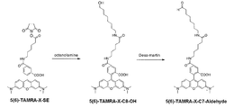
- Example 1 Synthesis of N-terminal labeling agent using fluorescent group TAMRA-X 1.5 Synthesis of 5 (6) -TAMRA-X-C8-OH 5 (6) -TAMRA- Using X-SE, 5 (6) -TAMRA-X-C8-OH was synthesized. Specifically, it is as follows.
- 5 (6) -TAMRA-X-C7-Aldehyde As shown in FIG. 2, using the obtained 5 (6) -TAMRA-X-C8-OH, 5 (6) -TAMRA- X-C7-Aldehyde was synthesized. Specifically, it is as follows. Add 175 ⁇ L of 2 mM 5 (6) -TAMRA-X-C8-OH, 175 ⁇ L of DMSO and 350 ⁇ L of 500 mM Dess-Martin reagent (manufactured by Tokyo Chemical Industry Co., Ltd.) to a 1.5 mL low adsorption tube and shake at 37 ° C. for 1 hour. Incubated while allowing.
- Dess-Martin reagent manufactured by Tokyo Chemical Industry Co., Ltd.
- Example 2 Fluorescent labeling of protein Fluorescence labeling reaction (reductive amination reaction) Using the N-terminal labeling agent synthesized in Example 1, fluorescent labeling of the N-terminal of the antibody was performed. 9 ⁇ L of 1.0 mg / mL antibody solution, 4.5 ⁇ L of 0.96 mM 5 (6) -TAMRA-X-C7-aldehyde (50% DMSO solution), 4.5 ⁇ L of 192 mM picoline borane (18% DMSO solution), 9 ⁇ L of 50 mM quencher Sodium acid buffer (pH 4.8) was added to a 1.5 mL low adsorption tube. This solution was incubated at 4 ° C. for 24 hours to perform a reductive alkylation reaction.
- Anti-thyroxine antibody Anti-Thyroxine: HyTest, clone # 1H1
- Anti-influenza hemagglutinin antibody Anti-Influenza Hemagglutinin: MBL, clone # 5D8
- Anti-influenza hemagglutinin antibody Anti-Influenza Hemagglutinin: MBL, clone # TANA2
- Anti-FLAG antibody Sigma, clone # M2
- Anti-His Tag antibody Novagen
- scFv derived from anti-BGP antibody expressed in E. coli with MetAla (methionine and alanine) added to the N-terminus
- Thyroxine antigen Sigma
- Influenza hemagglutinin tag peptide (Wako Pure Chemical Industries, Ltd.) (The same antigen was used for the fluorescence-labeled proteins using anti-influenza hemagglutinin antibodies clone # 5D8 and TANA2)
- FLAG peptide antigen (Wako Pure Chemical Industries, Ltd.)
- His peptide antigen (Wako Pure Chemical Industries, Ltd.) 5) BGP peptide antigen (Genscript)
- Fluorescence measurement These fluorescently labeled protein samples were subjected to fluorescence intensity measurement using a fluorescence spectrophotometer (Fluorolog-3; manufactured by Horiba, Ltd.).
- the excitation wavelength (Ex) was set to 550 nm, and the fluorescence intensity at the fluorescence wavelength (Em) of 565 to 700 nm was measured.
- the fluorescence intensity at 580 nm which is the maximum wavelength, was used.
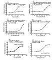
- the measurement results of the fluorescence intensity of fluorescently labeled protein samples using various antibodies are shown in FIG.
- the horizontal axis represents the antigen concentration
- the vertical axis represents the ratio of the fluorescence intensity at each antigen concentration to the fluorescence intensity without the antigen as the fluorescence intensity ratio.
- the fluorescent group was quenched in the absence of the antigen, but it was confirmed that the quenching of the fluorescent group was canceled and the fluorescence was emitted as the antigen concentration increased.
- the amplification rate of the fluorescence intensity after addition of the antigen is 1.75 times for the fluorescence-labeled protein sample using the anti-thyroxine antibody, and the anti-influenza hemagglutinin antibody (clone # 5D8).
- Example 3 Measurement using N-terminal labeling agents having various linker lengths
- n in the linker group was changed to 2, 5, 7, 9 by the same method as in Example 1.
- Various N-terminal labeling agents were prepared.
- n The N-terminal labeling agent for 7 is described as 5 (6) -TAMRA-X-C7-CHO
- Fluorescent labeled protein samples of each linker length were reacted with an antigen, irradiated with excitation light having a wavelength of 550 nm, and the fluorescence spectrum was measured using a fluorescence spectrophotometer (Fluorolog-3).
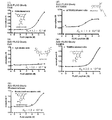
- FIG. 4 or FIG. 5 shows the measurement results of the fluorescence intensity of the fluorescently labeled protein samples of each linker length.
- FIG. 4 shows the results of using anti-FLAG antibody as an antibody solution and FLAG peptide antigen as an antigen
- FIG. 5 shows the results of using anti-His tag antibody as an antibody solution and His peptide antigen as an antigen. .
- Example 4 Measurement using N-terminal labeling agent having various linker groups N-terminal labeling agents having a PEG group as a linker group were synthesized. This synthesis was the same as the synthesis of TAMRA-X-C2-CHO in Example 3 except that TAMRA-PEO8-SE (manufactured by Biotium) was used instead of 5 (6) -TAMRA-X-SE. I went there.
- the protein was fluorescently labeled using an N-terminal labeling agent having a PEG group as a linker group.
- the antibody solution used was an anti-thyroxine antibody, and the antigen used was a thyroxine antigen.
- a fluorescently labeled protein sample having a PEG group as a linker group was reacted with an antigen, irradiated with excitation light having a wavelength of 550 nm, and a fluorescence spectrum was measured using a fluorescence spectrophotometer (Fluorolog-3).
- Microspin® G25 column was equilibrated with a citric acid equilibration buffer (50 mM sodium citrate buffer (pH 4.8), 100 mM KCl, 0.1% PEG8000, 0.1% Brij35). 300 ⁇ L of HKM equilibration buffer was added to the column and centrifuged at 3000 rpm for 1 minute at room temperature. This operation was performed 4 times in total. After that, 1 mg / mL anti-FLAG antibody / 9FLAG ⁇ L and 50 mM sodium citrate buffer (pH 4.8) 9 ⁇ L mixed solution was added to the column and centrifuged at 3000rpm for 1 minute to exchange the buffer. .
- a citric acid equilibration buffer 50 mM sodium citrate buffer (pH 4.8), 100 mM KCl, 0.1% PEG8000, 0.1% Brij35.
- 300 ⁇ L of HKM equilibration buffer was added to the column and centrifuged at 3000 rpm for 1 minute
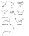
- Fluorescently labeled protein samples with various fluorescent groups are reacted with antigens, and the maximum absorption wavelength of each fluorescent group (TAMRA 550nm, FAM 495nm, RhodamineRed 570nm, RhodamineGreen 495nm, BODIPY FL 495nm, IC3 540nm, ATTO655 650nm, Cy3 540nm ) And the fluorescence spectrum was measured using a fluorescence spectrophotometer (Fluorolog-3).
- the horizontal axis represents the antigen concentration
- the vertical axis represents the ratio of the fluorescence intensity at each antigen concentration to the fluorescence intensity without the antigen as the fluorescence intensity ratio.
- the fluorescent group was quenched in the absence of antigen, but as the antigen concentration increased, the quenching of the fluorescent group was released, It was confirmed to emit fluorescence.
- N-terminal labeling agents having various fluorescent groups shown in FIG. 8 were used, fluorescently labeled protein samples were obtained.
Landscapes
- Health & Medical Sciences (AREA)
- Life Sciences & Earth Sciences (AREA)
- Chemical & Material Sciences (AREA)
- Immunology (AREA)
- Engineering & Computer Science (AREA)
- Molecular Biology (AREA)
- Hematology (AREA)
- Urology & Nephrology (AREA)
- Biomedical Technology (AREA)
- Organic Chemistry (AREA)
- General Health & Medical Sciences (AREA)
- Biochemistry (AREA)
- Analytical Chemistry (AREA)
- Medicinal Chemistry (AREA)
- Physics & Mathematics (AREA)
- Food Science & Technology (AREA)
- Microbiology (AREA)
- Cell Biology (AREA)
- General Physics & Mathematics (AREA)
- Pathology (AREA)
- Biotechnology (AREA)
- Genetics & Genomics (AREA)
- Proteomics, Peptides & Aminoacids (AREA)
- Biophysics (AREA)
- Peptides Or Proteins (AREA)
- Pyrane Compounds (AREA)
- Investigating Or Analysing Biological Materials (AREA)
- Investigating, Analyzing Materials By Fluorescence Or Luminescence (AREA)
Abstract
L'invention concerne un agent de marquage de N-terminal pour une protéine, comprenant un composé représenté par la formule générale (1) (où F est un groupe fluorescent, et R est un groupe de liaison éventuellement substitué ayant un segment souple linéaire formé à partir d'un hydrocarbure linéaire en C5 ou plus, ou un segment souple linéaire où les carbones de la chaîne principale ont été substitués en partie par -O-, -N-, -S-, -CO-, -CONH-, -COO-, ou -POO-).
Applications Claiming Priority (2)
| Application Number | Priority Date | Filing Date | Title |
|---|---|---|---|
| JP2015138743A JP2017020896A (ja) | 2015-07-10 | 2015-07-10 | N末端標識剤、これを用いた蛍光標識タンパク質及びその製造方法 |
| JP2015-138743 | 2015-07-10 |
Publications (1)
| Publication Number | Publication Date |
|---|---|
| WO2017010381A1 true WO2017010381A1 (fr) | 2017-01-19 |
Family
ID=57757313
Family Applications (1)
| Application Number | Title | Priority Date | Filing Date |
|---|---|---|---|
| PCT/JP2016/070058 Ceased WO2017010381A1 (fr) | 2015-07-10 | 2016-07-07 | Agent de marquage de n-terminal, protéine à marquage fluorescent l'utilisant, et son procédé de production |
Country Status (2)
| Country | Link |
|---|---|
| JP (1) | JP2017020896A (fr) |
| WO (1) | WO2017010381A1 (fr) |
Cited By (2)
| Publication number | Priority date | Publication date | Assignee | Title |
|---|---|---|---|---|
| WO2018147018A1 (fr) * | 2017-02-08 | 2018-08-16 | 国立大学法人東京工業大学 | Kit de détection ou de mesure d'antigène |
| IT202100013634A1 (it) * | 2021-05-25 | 2022-11-25 | Alda S R L | Nuovi reagenti per microscopia ottica |
Citations (8)
| Publication number | Priority date | Publication date | Assignee | Title |
|---|---|---|---|---|
| JPH06172312A (ja) * | 1992-03-04 | 1994-06-21 | Wako Pure Chem Ind Ltd | 1級アミノ基を有する化合物の蛍光標識化方法 |
| JP2005501228A (ja) * | 2001-06-04 | 2005-01-13 | アマシャム バイオサイエンス ユーケイ リミテッド | 生物材料の蛍光検出用標識試薬としてのキナクリドン誘導体 |
| JP2005013119A (ja) * | 2003-06-26 | 2005-01-20 | Japan Science & Technology Agency | Rna−ペプチド複合体を用いた蛍光センサー |
| US20090149631A1 (en) * | 2007-11-09 | 2009-06-11 | Massachusetts Institute Of Technology | Methods and compositions for protein labeling using lipoic acid ligases |
| JP4414654B2 (ja) * | 2001-05-14 | 2010-02-10 | ザ インスティチュート フォー システムズ バイオロジー | サンプル分子を単離および標識するための方法 |
| WO2013065314A1 (fr) * | 2011-11-02 | 2013-05-10 | ウシオ電機株式会社 | Procédé de dosage fluoro-immunologique faisant appel à un complexe polypeptidique contenant une région variable d'anticorps fluoromarquée |
| JP2013528185A (ja) * | 2010-05-27 | 2013-07-08 | ジーイー・ヘルスケア・バイオサイエンス・アクチボラグ | タンパク質の標識法及びキット |
| JP2014156428A (ja) * | 2013-02-15 | 2014-08-28 | Univ Of Tokyo | 抗体結合タンパク質 |
-
2015
- 2015-07-10 JP JP2015138743A patent/JP2017020896A/ja active Pending
-
2016
- 2016-07-07 WO PCT/JP2016/070058 patent/WO2017010381A1/fr not_active Ceased
Patent Citations (8)
| Publication number | Priority date | Publication date | Assignee | Title |
|---|---|---|---|---|
| JPH06172312A (ja) * | 1992-03-04 | 1994-06-21 | Wako Pure Chem Ind Ltd | 1級アミノ基を有する化合物の蛍光標識化方法 |
| JP4414654B2 (ja) * | 2001-05-14 | 2010-02-10 | ザ インスティチュート フォー システムズ バイオロジー | サンプル分子を単離および標識するための方法 |
| JP2005501228A (ja) * | 2001-06-04 | 2005-01-13 | アマシャム バイオサイエンス ユーケイ リミテッド | 生物材料の蛍光検出用標識試薬としてのキナクリドン誘導体 |
| JP2005013119A (ja) * | 2003-06-26 | 2005-01-20 | Japan Science & Technology Agency | Rna−ペプチド複合体を用いた蛍光センサー |
| US20090149631A1 (en) * | 2007-11-09 | 2009-06-11 | Massachusetts Institute Of Technology | Methods and compositions for protein labeling using lipoic acid ligases |
| JP2013528185A (ja) * | 2010-05-27 | 2013-07-08 | ジーイー・ヘルスケア・バイオサイエンス・アクチボラグ | タンパク質の標識法及びキット |
| WO2013065314A1 (fr) * | 2011-11-02 | 2013-05-10 | ウシオ電機株式会社 | Procédé de dosage fluoro-immunologique faisant appel à un complexe polypeptidique contenant une région variable d'anticorps fluoromarquée |
| JP2014156428A (ja) * | 2013-02-15 | 2014-08-28 | Univ Of Tokyo | 抗体結合タンパク質 |
Non-Patent Citations (5)
| Title |
|---|
| KEISUKE FUKUNAGA: "Monoclonal IgG no N-mattan Sentakuteki na Keiko Hyoshiki to Kogen no Keiko Kenshutsu", ANNUAL MEETING OF THE CHEMICAL SOCIETY OF JAPAN IN SPRING KOEN YOKOSHU, vol. 96 th, 10 March 2016 (2016-03-10), pages ROMBUNNO.4c5 - 10 * |
| KENSUKE YOSHIKOSHI: "Shuju no Keikoki o Mochiita Niju Keiko Hyoshiki Ipponsa Kotai ni yoru Kogen no Ratio Kenshutsu", CSJ: THE CHEMICAL SOCIETY OF JAPAN KOEN YOKOSHU, vol. 95 th, no. 3, 11 March 2015 (2015-03-11), pages 822 1J3 - 33 * |
| MASANORI MIURA: "Kaishi Codon no Kakucho ni yoru Shuju no Keiko Hyoshiki Karubonsan no Tanpakushitsu N-mattan eno Donyu", PROTEIN SCIENCE SOCIETY OF JAPAN NENKAI PROGRAM . YOSHISHU, vol. 9 th, 2009, pages 79 1P-123 * |
| TAKAYOSHI WATANABE: "Hi Tennen Amino-san Donyu Gijutsu o Riyo shita Tanpakushitsu no Bui Tokuiteki Shushoku Gijutsu no Kaihatsu", CSJ: THE CHEMICAL SOCIETY OF JAPAN KOEN YOKOSHU, vol. 94, no. 3, 2014, pages 938 4G5 - 12 * |
| TAKAYOSHI WATANABE: "Keiko Hyoshiki Hydroxy-san o Mochiita Shinki N-mattan Tokuiteki Tanpakushitsu Keiko Hyoshikiho no Kaihatsu", JOURNAL OF JAPANESE BIOCHEMICAL SOCIETY, 2007, pages 2P-1533 * |
Cited By (4)
| Publication number | Priority date | Publication date | Assignee | Title |
|---|---|---|---|---|
| WO2018147018A1 (fr) * | 2017-02-08 | 2018-08-16 | 国立大学法人東京工業大学 | Kit de détection ou de mesure d'antigène |
| JPWO2018147018A1 (ja) * | 2017-02-08 | 2019-12-12 | 国立大学法人東京工業大学 | 抗原検出又は測定用キット |
| US11499965B2 (en) | 2017-02-08 | 2022-11-15 | Tokyo Institute Of Technology | Kit for detecting antigen or measuring its amount |
| IT202100013634A1 (it) * | 2021-05-25 | 2022-11-25 | Alda S R L | Nuovi reagenti per microscopia ottica |
Also Published As
| Publication number | Publication date |
|---|---|
| JP2017020896A (ja) | 2017-01-26 |
Similar Documents
| Publication | Publication Date | Title |
|---|---|---|
| JP6070769B2 (ja) | 蛍光標識抗体可変領域含有ポリペプチド複合体を用いた蛍光免疫測定キット | |
| Zhang et al. | A broad-spectrum sensing strategy for the tetracycline family of antibiotics based on an ovalbumin-stabilized gold nanocluster and its application in a pump-free microfluidic sensing platform | |
| Prakashan et al. | Design and fabrication of a competitive lateral flow assay using gold nanoparticle as capture probe for the rapid and on-site detection of penicillin antibiotic in food samples | |
| Yang et al. | Fluorescent immunosorbent assay for the detection of alpha lactalbumin in dairy products with monoclonal antibody bioconjugated with CdSe/ZnS quantum dots | |
| CN102667480A (zh) | 荧光免疫测定方法 | |
| JP7561364B2 (ja) | 蛍光偏光免疫分析法 | |
| Kong et al. | An ultrasensitive ELISA for medroxyprogesterone residues in fish tissues based on a structure-specific hapten | |
| Zhou et al. | Gold nanoparticle probe-based immunoassay as a new tool for tetrodotoxin detection in puffer fish tissues | |
| Wu et al. | Sensitive enzyme‐linked immunosorbent assay and gold nanoparticle immunochromatocgraphic strip for rapid detecting chloramphenicol in food | |
| WO2017010381A1 (fr) | Agent de marquage de n-terminal, protéine à marquage fluorescent l'utilisant, et son procédé de production | |
| JP5949890B2 (ja) | 蛍光標識された抗体可変領域を含むポリペプチドを含む抗原結合タンパク質を用いた蛍光免疫測定方法 | |
| CN100552452C (zh) | 使用荧光化抗体的荧光分析方法 | |
| Chen et al. | Fluorescence quenching immunoaffinity test column with quantum dots as fluorescence donors for the quick detection of malachite green and crystal violet in aquatic products | |
| JP2016200545A (ja) | 検査対象物質を識別して検出する方法 | |
| CN106018793B (zh) | 一种六次甲基四胺的检测方法 | |
| He et al. | Sample-to-answer Aggregation-Induced Emission Lateral Flow Immunoassay (ALFIA) for rapid and sensitive on-site detection of Toxoplasma gondii antibodies across multiple host species | |
| Xi et al. | Preparation and characterization of artificial antigens for cadmium and lead | |
| Du et al. | Determination of diethylstilbestrol based on biotin–streptavidin‐amplified time‐resolved fluoro‐immunoassay | |
| JP7461661B2 (ja) | 複数の蛍光色素を結合させたペプチドを用いるホモジニアス免疫測定法 | |
| Yoohanngoh et al. | Carbon dots-based fluorescent immunosensor for highly sensitive detection of leucomalachite green in fish and shrimp samples | |
| JP2017049184A (ja) | 抗体のヌクレオチド結合部位(nbs)を利用して蛍光標識された抗体 | |
| US20230393123A1 (en) | Concentration estimation kit and concentration estimation method | |
| WO2016208466A1 (fr) | Procédé de détection d'une substance d'intérêt | |
| Perez | Development of Fast Photochemical Oxidation of Proteins for in Vivo Modification in Caenorhabditis elegans | |
| Bu et al. | A sensitive enzyme‐linked immunosorbent assay amplified by biotin–streptavidin system for detecting non‐steroidal anti‐inflammatory drug ketoprofen |
Legal Events
| Date | Code | Title | Description |
|---|---|---|---|
| 121 | Ep: the epo has been informed by wipo that ep was designated in this application |
Ref document number: 16824361 Country of ref document: EP Kind code of ref document: A1 |
|
| NENP | Non-entry into the national phase |
Ref country code: DE |
|
| 122 | Ep: pct application non-entry in european phase |
Ref document number: 16824361 Country of ref document: EP Kind code of ref document: A1 |