WO2017141987A1 - Procédé d'essai non clinique caractérisé par l'évaluation quantitative d'échantillon d'animal de laboratoire - Google Patents
Procédé d'essai non clinique caractérisé par l'évaluation quantitative d'échantillon d'animal de laboratoire Download PDFInfo
- Publication number
- WO2017141987A1 WO2017141987A1 PCT/JP2017/005588 JP2017005588W WO2017141987A1 WO 2017141987 A1 WO2017141987 A1 WO 2017141987A1 JP 2017005588 W JP2017005588 W JP 2017005588W WO 2017141987 A1 WO2017141987 A1 WO 2017141987A1
- Authority
- WO
- WIPO (PCT)
- Prior art keywords
- tumor
- test method
- profile
- cells
- clinical test
- Prior art date
- Legal status (The legal status is an assumption and is not a legal conclusion. Google has not performed a legal analysis and makes no representation as to the accuracy of the status listed.)
- Ceased
Links
Images


Classifications
-
- G—PHYSICS
- G01—MEASURING; TESTING
- G01N—INVESTIGATING OR ANALYSING MATERIALS BY DETERMINING THEIR CHEMICAL OR PHYSICAL PROPERTIES
- G01N33/00—Investigating or analysing materials by specific methods not covered by groups G01N1/00 - G01N31/00
- G01N33/48—Biological material, e.g. blood, urine; Haemocytometers
- G01N33/50—Chemical analysis of biological material, e.g. blood, urine; Testing involving biospecific ligand binding methods; Immunological testing
- G01N33/5005—Chemical analysis of biological material, e.g. blood, urine; Testing involving biospecific ligand binding methods; Immunological testing involving human or animal cells
- G01N33/5008—Chemical analysis of biological material, e.g. blood, urine; Testing involving biospecific ligand binding methods; Immunological testing involving human or animal cells for testing or evaluating the effect of chemical or biological compounds, e.g. drugs, cosmetics
- G01N33/5082—Supracellular entities, e.g. tissue, organisms
- G01N33/5088—Supracellular entities, e.g. tissue, organisms of vertebrates
-
- G—PHYSICS
- G01—MEASURING; TESTING
- G01N—INVESTIGATING OR ANALYSING MATERIALS BY DETERMINING THEIR CHEMICAL OR PHYSICAL PROPERTIES
- G01N33/00—Investigating or analysing materials by specific methods not covered by groups G01N1/00 - G01N31/00
- G01N33/48—Biological material, e.g. blood, urine; Haemocytometers
- G01N33/50—Chemical analysis of biological material, e.g. blood, urine; Testing involving biospecific ligand binding methods; Immunological testing
- G01N33/53—Immunoassay; Biospecific binding assay; Materials therefor
- G01N33/575—Immunoassay; Biospecific binding assay; Materials therefor for cancer
-
- G—PHYSICS
- G01—MEASURING; TESTING
- G01N—INVESTIGATING OR ANALYSING MATERIALS BY DETERMINING THEIR CHEMICAL OR PHYSICAL PROPERTIES
- G01N33/00—Investigating or analysing materials by specific methods not covered by groups G01N1/00 - G01N31/00
- G01N33/48—Biological material, e.g. blood, urine; Haemocytometers
- G01N33/50—Chemical analysis of biological material, e.g. blood, urine; Testing involving biospecific ligand binding methods; Immunological testing
- G01N33/58—Chemical analysis of biological material, e.g. blood, urine; Testing involving biospecific ligand binding methods; Immunological testing involving labelled substances
- G01N33/588—Chemical analysis of biological material, e.g. blood, urine; Testing involving biospecific ligand binding methods; Immunological testing involving labelled substances with semiconductor nanocrystal label, e.g. quantum dots
Definitions
- the present invention relates to a non-clinical test method for evaluating the efficacy or side effects of a drug or a candidate substance thereof performed using a laboratory animal.
- a model animal produced by transplanting human cancer (tumor) cells or tissues into a laboratory animal for example, a tumor bearing mouse produced using a mouse as an experimental animal, May be used as an experimental system that reproduces.
- tumor-bearing mice cultured cells derived from tumor cells removed from patients are transplanted into cells and grown in mice, and tumor tissues or tumor cells removed from patients are used.
- a patient tumor tissue-transplanted mouse or the like prepared by planting in a mouse and growing in the mouse is known.
- the drug efficacy or safety (toxicity) of the drug or its candidate substance can be examined even during the drug discovery research or non-clinical trial stage that is conducted prior to the human study. Can do.
- mice derived from cancer patients with various properties can be prepared, it is possible to find effective patient profiles by comparative evaluation.
- Cultured cancer cell transplanted mice can be produced using cells that have been stabilized by culturing in a test tube and can be easily transplanted into mice, and thus have been established as experimental animals. Mice transplanted with the cloned cells are suitable for a test in which clonal elements are inherited and tumor-bearing mice with small individual differences are desired to be used.
- a patient tumor tissue-transplanted mouse is produced by directly implanting a tumor tissue (tumor part) or tumor cell taken out from a patient into the mouse.
- a patient tumor tissue-transplanted mouse prepared by planting a tumor tissue derived from a patient (human) into an acquired immunodeficient mouse and growing it in a body such as a mouse for a certain period of time is PDX (Patient-derived tumor xenograft (patient-derived tumor xenograft) One piece)) It is called a model mouse.
- the PDX model mouse produced in this way does not use cells cultured in vitro, it is more effective in reproducing the actual human pathology than conventional tumor-bearing mice. It is thought that it can be evaluated.
- diagnosis / treatment methods methods for obtaining an index for diagnosis or treatment
- non-clinical test methods using PDX model mice has become active. Recognizing the importance of testing with tumor-bearing mice in which the complexity of the cancer cell is maintained, including the cause (gene mutation, etc.) that produces it, it is desirable to ensure that the complexity is repeated It is because it came to be considered.
- the PDX model can be created not only for mice but also for various experimental animals.
- Non-Patent Document 1 describes that a PDX model mouse for bladder cancer has been prepared. However, depending on the patient and the type of protein, the trait changes greatly from the original patient specimen. Various aspects are shown, such as those that do not, and those that gradually change with passage (see Figure IV-3, for example).
- the experimental animal of the PDX model inherits the genetic information of the tumor by passage, it has not been cloned as much as the cultured cancer cell transplantation model, and thus tumor cells are not found between the primary PDX model animal and the passaged PDX model animal. These traits are not necessarily completely maintained, and even individual PDX model animals of the same strain will vary from individual to individual.
- Non-Patent Document 1 the expression level of protein in tumor tissue of PDX model mice is evaluated by the IHC (immunohistochemistry) method.
- IHC immunohistochemistry
- an enzyme-labeled antibody is bound to a target protein (antigen) by a direct method or an indirect method, and then a substrate is reacted to develop a color, for example, a DAB staining method using peroxidase and diaminobenzidine. It was common to use.
- staining with an enzyme such as DAB staining in the IHC method has a problem in that it is difficult to accurately estimate the amount of an actual antigen or the like from the staining concentration because the staining concentration greatly depends on environmental conditions such as temperature and time. There is.
- the evaluation is often expressed by a score of several stages based on the staining concentration and the like, which is close to qualitative evaluation rather than quantitative.
- Non-Patent Document 1 In present, in the study of academia, as in Non-Patent Document 1, only the qualitative analysis of protein expression level in the tumor tissue of patients and PDX mice by the IHC (immunohistochemistry) method is performed. In addition, mouse providers and contract research companies have little interest in accurately evaluating protein expression levels. It can be said that the technical significance of quantitatively and accurately grasping the protein expression level in the tumor tissue and the like of PDX mice is not yet known.
- phosphor integrated particles sometimes called fluorescent substance integrated nanoparticles
- the present invention has been made in view of the above problems, and more accurately performs quality control of experimental animals having a tumor site transplanted like PDX mice and analysis of test results using such experimental animals. It is an object of the present invention to provide means for performing the above. Such means are not limited to the problems in tumor-bearing mice including PDX mice, but other lesion model animals (particularly mice) such as Alzheimer's disease model mice, diabetes model mice, genetic disease model mice, and infection disease model mice. It can be a means to solve the same problem in.
- the inventors used a specimen collected from a laboratory animal to quantitatively analyze the tumor profile of the transplanted laboratory animal, preferably immunostaining using fluorescent nanoparticles such as phosphor-aggregated particles.
- fluorescent nanoparticles such as phosphor-aggregated particles.
- the inventors have found that the quality control and test result analysis as described above can be performed more accurately. Furthermore, it has been found that such a non-clinical test method for tumor bearing animals (mouse) can be applied to other model animals as described above.
- the present invention uses a specimen collected from an experimental animal (lesion model animal) to identify a profile of a lesioned part of the experimental animal, for example, a transplanted tumor part, by a quantitative technique. To provide a non-clinical test method.
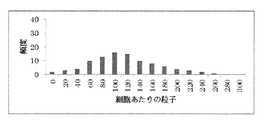
- FIG. 1 is a histogram of the number of particles per cell and the frequency (number of cells) measured for the tumor part of a first generation mouse (P1) transplanted with a breast cancer tissue of a patient in Example 3.
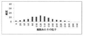
- FIG. 2 is a histogram of the number of particles per cell and frequency (number of cells) measured in Example 3 for the tumor part of the second generation mouse 1 (P2-1).
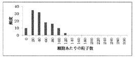
- FIG. 3 is a histogram of the number of particles per cell and the frequency (number of cells) measured for the tumor part of the second generation mouse 2 (P2-2) in Example 3.
- FIG. 4 is a histogram of the number of particles per cell and the frequency (number of cells) measured for the tumor area of the third generation mouse 1 (P3-1) prepared from P2-1 in Example 3.
- FIG. 5 is a histogram of the number of particles per cell and the frequency (number of cells) measured for the tumor area of the third generation mouse 2 (P3-2) prepared from P2-2 in Example 3.
- the non-clinical test method of the present invention uses a specimen collected from an experimental animal to specify a lesion profile of the experimental animal using a quantitative technique (referred to herein as “lesion profile specifying step”). ").
- the lesioned part is a transplanted tumor part when, for example, the experimental animal is a tumor-bearing animal model such as a PDX model mouse.
- the “lesion site profile specifying step” can be referred to as a “post-transplant profile specifying step”.
- the “tumor part” is a part containing “human tumor tissue or tumor cell”, from which “sample” is collected.
- the tumor part usually contains “substance other than human-derived tumor tissue” together with human-derived tumor tissue or tumor cells.
- specimens collected from experimental animals are generally targeted by immunostaining in, for example, pathological diagnosis, as is commonly used when evaluating the expression level of a target protein by immunostaining. It takes the form of a specimen slide prepared according to a predetermined procedure, which is conventionally used when evaluating the expression level of protein.
- the non-clinical test method of the present invention is carried out in vitro in laboratory animals using such specimens.
- the “profile” in the present invention refers to the expression level of the target protein, the type, number, form, and expression site of cells expressing the target protein (in the case of using a tumor-bearing animal model as an experimental animal, tumor tissue or tumor). This is a feature formed by information such as the distribution within the department and the exclusive area.
- the target protein identified in the profile of the present invention is not particularly limited as long as it is expressed by cells contained in a specimen, but is preferably expressed in a large amount in those cells.
- One specific type of protein may be the target protein, and two or more types of proteins may be the target protein.
- the target protein is a marker that is a target of a molecular target drug that has already been developed, and the profile to be identified is the expression level of the marker and the expression in the tumor tissue or tumor part. Includes distribution.
- markers are known, and examples thereof include HER2, HER3, PD-L1, PD-1, CTLA-4, EGFR, and VEGFR.
- the target protein is a phosphorylated protein
- the profile to be specified includes the expression level of the phosphorylated protein and the expression distribution in the tumor tissue or tumor part.
- phosphorylated proteins include HER2, HER3, EGFR, VEGFR and the like.
- the phosphorylated protein can be quantified by using an antibody that specifically recognizes only the phosphorylated protein of the target protein during immunostaining. In addition, when it is desired to quantify the entire target protein regardless of whether it is phosphorylated or non-phosphorylated, an antibody that recognizes them without distinction may be used.
- the cells contained therein include not only tumor cells but also cells other than tumor cells, for example, cells such as immune cells that interact with tumor cells.
- the target protein in the present specification preferably includes a cancer-related protein in tumor cells and / or a protein in immune cells.
- Cancer-related proteins typically include “immune system proteins in cancer cells”, “pathway proteins in cancer cells”, and “metastatic proteins in cancer cells”.
- Various cancer-related proteins classified into each are known, and appropriate proteins can be selected according to the purpose of diagnosis or treatment, and are not particularly limited.
- the cancer-related gene expression panel provided by nCounter covers 770 genes for each of the immune gene panel, pathway gene panel, and metastasis gene panel, The proteins encoded by these genes correspond to immune system proteins, pathway proteins, and metastasis proteins in cancer cells, respectively. Mutant proteins corresponding to mutant genes of these genes can also be included in immune system proteins, pathway proteins, and transfer proteins.
- Immuno system proteins in cancer cells include, for example, immune checkpoint proteins CD40, TL1A, GITR-L, 4-188-L, CX4D-L, CD70, HHLA2, ICOS-L, CD85, CD86. , CD80, MHC-II, PDL1, PDL2, VISTA, BTNL2, B7-H3, B7-H4, CD48, HVEM, CD40L, TNFRSF25, GITR, 4-188, OX40, CD27, TMIGD2, ICOS, CD28, TCR, LAG3 CTLA4, PD1, CD244, TIM3, BTLA, CD160, LIGHT, and the like.
- pathway proteins in cancer cells include, for example, EGFR (HER1), HER2, HER3, HER4, IGFR, HGFR, which are cancer cell growth factors or cancer cell growth factor receptors; cell surface antigens, blood vessel growth Factor or vascular growth factor receptor VEGF-A, VEGF-B, VEGF-C, VEGF-D, VEGF-E, PlGF-1, PlGF-2; cytokine or cytokine receptor interferon, interleukin, G -CSF, M-CSF, EPO, SCF, EGF, FGF, IGF, NGF, PDGF, TGF and the like.
- EGFR HER1
- HER2, HER3, HER4, IGFR HGFR
- HGFR which are cancer cell growth factors or cancer cell growth factor receptors
- cell surface antigens include blood vessel growth Factor or vascular growth factor receptor VEGF-A, VEGF-B, VEGF-C, VEGF-D, VEGF-E,
- metastatic proteins in cancer cells include cancer metastasis markers ACTG2, ALDOA, APC, BRMS1, CADM1, CAMK2A, CAMK2B, CAMK2D, CCL5, CD82, CDKN1A, CDKN2A, CHD4, CNN1, CST7, CTSL, CXCR2, YBB, DCC, DENR, DLC1, EGLN2, EGLN3, EIF4E2, EIF4EBP1, ENO1, ENO2, ENO3, ETV4, FGFR4, GSN, HK2, HK3, HKDC1, HLA-DPB1, HUNILDH, LD LIFR, MED23, MET, MGAT5, MAP2K4, MT3, MTA1, MTBP, MTOR, MYCL, MYH11, NDRG1, NF2, NF B1, NME1, NME4, NOS2, NR4A3, PDK1, PEBP4, PFKFB1, PFKFB4, PGK1, PLAUR, PTTG
- proteins in immune cells include PD-1, CTLA-4, TIM3, Foxp3, CD3, CD4, CD8, CD25, CD27, CD28, CD70, CD40, CD40L, CD80, CD86, CD160, CD57, CD226 CD112, CD155, OX40 (CD134), OX40L (CD252), ICOS (CD278), ICOSL (CD275), 4-1BB (CD137), 4-1BBL (CD137L), 2B4 (CD244), GITR (CD357), B7 -H3 (CD276), LAG-3 (CD223), BTLA (CD272), HVEM (CD270), GITRL, Galectin-9 (Galectin-9), B7-H4, B7-H5, PD-L2, KLRG-1, EC Adherin, N-Cadherin, R-Cadherin and IDO, TDO, CSF-1R, HDAC, CXCR4, FLT-3, T
- the target protein in the present specification may be a protein expressed in cells other than tumor cells and immune cells.
- proteins expressed in cells other than tumor cells and immune cells include proteins contained in the stroma.
- “Stromal” mainly consists of stromal cells such as fibroblasts, endothelial cells, leukocytes (lymphocytes, monocytes, neutrophils, eosinophils, basophils) and proteins such as collagen and proteoglycans. Composed of an extracellular matrix. Either the amount of stromal cells or extracellular matrix may be measured, but the amount of stromal cells that are considered to have a greater effect on the traits of human-derived tumor cells carried by experimental animals may be measured. Preferably, it is more preferable to measure the amount of protein expressed in a typical stromal cell fibroblast.
- the “stroma” contained in the tumor part is typically “interstitium derived from experimental animals” or “between tumor tissues derived from humans”. "Quality".
- the stroma derived from an experimental animal is a substance mixed in the stroma of a human-derived tumor tissue while the tumor part is held in the living body of the experimental animal.
- the stroma of a human-derived tumor tissue is derived from what was included when a tumor tissue collected from a human (patient) was transplanted into an experimental animal. The greater the proportion of the tumor tissue or the interstitium derived from the experimental animal in the specimen, the smaller the proportion of the human tissue-derived stroma.
- an appropriate protein can be selected and used from the following membrane proteins that are stromal cell markers.
- CD140a is a membrane protein expressed on the surface of cells such as fibroblasts, megakaryocytes, monocytes, erythrocytes, myeloid progenitor cells, and endothelial cells, and is preferable as a stromal cell marker in the present invention.
- CD106 VCAM-1, INCAM-110; CD49d / CD29-L
- DC CD109 Platinum activation factor, 8A3, E123
- Activated T cells platelets, vascular endothelium, megakaryocytes, CD34 + progenitor cell subset CD140a (PDGF-R, PDGFR2) ... fibroblasts, megakaryocytes, monocytes, erythrocytes, myeloid progenitor cells, endothelial cells CD140b (PDGF-R, PDGFR1) ...
- endothelial cells stromal cells CD141 (Thrombomodulin): Vascular endothelium, myeloid cells, platelets, smooth muscle CD142 (Tissue Factor (TF), Thromboplastin): Epithelial cells, activated monocytes, activated vascular endothelium CD143 (ACE; angiotensin converting enzyme) ... vascular endothelium, epithelial cells, activated macrophages CD144 (VE-Cadherin, Cadherin-5: involved in permeability of vascular endothelium) ... vascular endothelium CD145 (7E9, P7A5) ... endothelial cells CD146 (MUC18, s-endo, Mel-CAM) ...
- vascular endothelium activated T cells, melanoma CD147 (Basigin, M6, EMMRRIN) ... white blood cells, red blood cells, vascular endothelium, platelets CD201 (EPCR: protein C receptor) ... vascular endothelium CD202 (TIE2, TEK; Angiopoietin-1-R) ...
- Vascular endothelium, hematopoietic stem cell subset CD280 (Endo180, TEM22, uPARAP (uPAR-associated protein); bone marrow progenitor cells, fibroblasts, endothelial cell subsets, macrophage subsets CD299 (DC-SIGN-related, L-SIGN (Liver / Lympho node specific ICAM3-grabbing nonintegrin)) ...
- Endothelial cells CD309 VGFR2: Vascular endothelial growth factor receptor 2, KDR
- endothelial cells megakaryocytes, platelets, stem cell subsets CD322 (JAM2: Junctional adhesion molecule 2) ...
- endothelial cells monocytes, B cells, T cell subsets CD331 (FGFR1: Fibroblast growth factor receptor1) ... fibroblasts, epithelial cells CD332 (FGFR2, Keratinocyte growth factor receptor) ... epithelial cells CD333 (FGFR3, JTK4; Achondroplasia, Thanatophoric dwarfism) ... fibroblasts, epithelial cells CD334 (FGFR4, JTK2, TKF) ... fibroblasts, epithelial cells CD339 (Jagged-1, JAG1; Alagille syndrome) ... Stroma cells, epithelial cells.
- FGFR1 Fibroblast growth factor receptor 1
- fibroblasts epithelial cells CD332 (FGFR2, Keratinocyte growth factor receptor) ... epithelial cells CD333 (FGFR3, JTK4; Achondroplasia, Thanatophoric dwarfism) ... fibroblasts
- epithelial cells CD334 FGFR4, JTK
- mice As the “experimental animal” in the present invention, various experimental animals, particularly lesion model animals, can be used depending on the purpose of the non-clinical test method. Examples of experimental animals include tumor-bearing animals that have previously retained a tumor portion derived from tumor cells or tumor tissue and generated in vivo. In addition, Alzheimer's disease model mice, diabetes model mice, Various lesion model animals such as genetic disease model mice and infectious disease model mice can be targeted by the present invention. Examples of animal species include mice, rats, rabbits, guinea pigs, gerbils, hamsters, ferrets, dogs, minipigs, monkeys, cows, horses, sheep, etc. Animals that have requirements are listed.
- the technique for causing the experimental animal to retain the tumor part is not particularly limited, and a known technique can be used.
- tumor-bearing model mice can be broadly classified into three types: naturally induced tumor mice, cultured cancer cell transplanted mice, and patient tumor tissue transplanted mice (see table below; Kohrt et al., InDefining the optimal murine models to investigate immune checkpoint) blockers and their combination with other immunotherapies. Annals of Oncology 00: 1-9, 2016).
- Cultured cancer cell transplanted mice are prepared by transplanting cultured cells derived from tumor cells removed from patients.
- Examples of mice transplanted with cultured cancer cells derived from humans include CDX (Cell-line derived xenograft) model mice, and mice transplanted with tumor tissues derived from humans (patients) include PDX (Patient In addition to derived xenograft model mice, there are Imuno-avatar model mice, Hemato-lymphoid humanized model mice, and Imune-PDX model mice.
- PDX mice are prepared by transplanting patient-derived tumor tissue into acquired immunodeficient mice as described above.
- Immuno-avatar model mice, hematopoietic lymphoid humanized model mice and Imune-PDX model mice have human peripheral blood mononuclear cells, CD34 + human hematopoietic stem cells and their progenitor cells (HSPC) or tumor infiltrating lymphocytes, respectively.
- HSPC progenitor cells
- mice transplanted with patient tumor tissue were transplanted (passaged) with a mouse (first generation: 0th generation) that had been grown for a certain period after transplanting a patient-derived tumor tissue and a 0th generation tumor site.
- a mouse and all of the (n + 1) generation mice transplanted (passaged) with a subsequent n generation (n ⁇ 1) tumor site are included.
- the “transplanted tumor part” in the present invention includes all tumor parts of 0th generation to (n + 1) th generation experimental animals transplanted with a patient's tumor cells or tumor tissues or cultured cells.
- a tumor tissue transplant animal When creating a patient tumor tissue transplant animal, an incision is made to transplant a tumor block, a tumor tissue injected into a frozen needle is inserted by injection, a matrix is introduced at the same time as a tumor, or a hormone such as estrogen is transplanted
- Various techniques have been tried, including pre-administration. Among these, a place where a tumor is transplanted varies greatly depending on the place, the state of the tumor-bearing animal after preparation and the state of the tumor. The place of transplantation can be roughly classified into subcutaneous transplantation and orthotopic transplantation (a tumor generated in one organ is transplanted into the same organ of a donor animal).
- the former can be visually confirmed as the transplanted tumor grows gradually, it can be used for testing after confirming that it has become a certain size, and it is usually easy to handle. It is an option.
- the environment where the tumor originally occurred and the transplantation site (subcutaneous) are greatly different, there is a risk that a coating is formed around the tumor or that the tumor does not grow due to slow blood vessel formation.
- Profile information Among the information included in the profile specified in the non-clinical test method of the present invention, information on the expression level of the target protein and the number of expressed cells is, for example, (i) per cell of the target protein in a specimen (specimen slide). Average expression level, (ii) Expression level of target protein per unit area of tissue, (iii) Histogram expressed by expression level of target protein per cell and corresponding number of cells, (iv) Per target protein per cell Information on the expression level and the curve represented by the number of cells corresponding thereto. These information may be included in the profile by combining not only one of them but also a plurality, or a combination of each piece of information selected from the target protein is included in the profile. Also good.
- the specimen can be immunostained with fluorescent nanoparticles, and the shape of the cell can be specified by a morphology observation stain (eg, eosin) Stain as follows.
- a morphology observation stain eg, eosin Stain
- An image in which fluorescent nanoparticles labeled with the target protein appear as bright spots is obtained by observation and imaging in a dark field performed while irradiating excitation light having a predetermined wavelength corresponding to the fluorescent nanoparticles.
- an image that is stained so as to represent the shape of a cell is acquired by observation and imaging in a bright field.
- the target protein expressed for individual cells contained in the entire image or in a specific region (for example, only tumor tissue) in the image is displayed.
- the number of bright spots to be represented can be measured.
- the number of bright spots may be used as the index value.
- the brightness (luminance, fluorescence intensity) of one bright spot is measured separately for each fluorescent nanoparticle.
- the number of fluorescent nanoparticles contained in the bright spot can be calculated by dividing by the brightness of the light, and the number of particles may be used as an index value for the expression level of the target protein.
- (Iii) When creating a histogram represented by the expression level of the target protein per cell and the number of cells corresponding thereto, first, as in (i), a specific region included in the entire image or in the image The number of bright spots or particles representing the target protein expressed is determined for each individual cell contained in (for example, only tumor tissue). Then, the expression level of the target protein per cell is divided into predetermined numbers on the horizontal axis (for example, as in the examples described in this specification, 1 to 300 is expressed every 20 A histogram can be created by measuring the number of cells (frequency) corresponding to each division and taking it on the vertical axis.
- the profile may include information other than information related to cells expressing the target protein, such as blood vessel occupancy in a specimen (for example, a tumor tissue or a tumor part).
- a quantitative method means a profile including the above-described profile, in particular, the amount of protein expression, the number of expressed cells, and the area occupied by the expressed cells in the tissue. Is identified using a “quantitative” rather than “qualitative” approach.
- the “qualitative” method correlates with the expression level of the protein and the number of expressed cells, but does not directly handle those numbers or index values closely related to them, but within a predetermined range.
- a certain number or index value is collectively expressed as one score, and such a score is about several, for example, about 2 to 5, and is typically based on the subjective and empirical factors of the observer Say.
- an IHC method using HER2 protein expressed in cell membranes such as breast cancer cells and using DAB staining which is 3+ (strongly completely complete based on the staining property and staining intensity (staining pattern) of cancer cell membranes.
- the technique used in Non-Patent Document 1 page 20527, Figure 3 to express the protein expression level with a four-stage score based on the stained image by the IHC method is also a “qualitative” technique.
- the “quantitative” method directly deals with the expression level of the protein, the number of expressed cells, or index values closely related to them, and typically relies on the objective measurement result using the apparatus.
- a technique Typically, fluorescent nanoparticles, that is, quantum dots (not integrated), or particles in which fluorescent materials such as fluorescent dyes and quantum dots are integrated as a base material (phosphor integrated particles, Phosphor Integrated Dot : PID), a method of labeling and quantifying the target protein using particles having a nano-sized diameter is used.
- the quantitative method (also referred to as “PID method” in the present specification) performed using phosphor-aggregated particles, which is also employed in the examples of the present specification, is the “quantitative method” used in the present invention.
- This method is particularly suitable.
- the “quantitative” method that can be used in the present invention is not limited to the method using phosphor-aggregated particles such as the PID method, and may be another method having the same degree of accuracy. .
- Patent Document 1 International Publication WO2012 / 029752
- Patent Document 2 International Publication WO2013 / 035703
- the PID method can be performed in an embodiment according to the case of performing pathological diagnosis using a specimen slide.
- the “histogram” is, for example, divided into a predetermined number of expression levels of the target protein per cell (for example, 1 to 300 is divided into 20 pieces, as in the examples described in the present specification). It is expressed with the number of cells (frequency) corresponding to each division, and originally expressed in the amount of protein expression (number of bright spots or particles) and expression. Since the number of cells is measured and those numbers are handled directly and then graphed, the histogram is categorized as information obtained using a “quantitative” method rather than a “qualitative” method.
- the non-clinical test method of the present invention uses a specimen collected from an experimental animal when the lesion is a transplanted tumor, that is, when the experimental animal is a tumor-bearing animal.
- the profile identification step using a specimen collected from a patient or cultured cells, that is, the tumor before being transplanted to a laboratory animal using a specimen collected from the patient or cultured cells
- a step of specifying a profile of a cell or tumor tissue using a quantitative method (sometimes referred to herein as a “pre-transplant profile specifying step”), and a profile specified by the pre-transplant profile specifying step
- profile the profile identified by the post-transplant profile identification step
- yl comparison step ".) Further comprises a. According to such an embodiment, it is possible to evaluate to what extent the tumor part after being transplanted into the experimental animal has inherited the characteristics of the tumor cell or the tumor tissue before being transplanted into the experimental animal. Quality control and medicinal effects analysis can be performed more accurately.
- Tumor cell or tumor tissue before transplantation includes both tumor tissue or tumor cells removed from a patient and cultured cells derived from tumor cells removed from a patient. From the viewpoint of being able to eliminate the influence of cell alteration during the culture, it is preferable to use a tumor tissue or tumor cell taken out from a patient as a tumor cell or tumor tissue before being transplanted to a laboratory animal, That is, it is preferable that the non-clinical test method of the present invention uses a laboratory animal transplanted with a tumor tissue or tumor cell taken out from a patient.
- the non-clinical test method of the present invention may include a step of comparing the profile of the transplanted tumor portion between experimental animals of the same or different generations.
- the non-clinical test method of the present invention uses a specimen collected from a 0th generation or 1st generation laboratory animal and a donor collected from a 2nd generation or later laboratory animal.
- the method further includes a step of comparing the profile of the tumor part of the experimental animal. According to such an embodiment, it is possible to evaluate how much the characteristics of the transplanted part have been inherited by passage in experimental animals, but to perform more accurate quality control and drug efficacy analysis of experimental animals. This is useful.
- the non-clinical test method of the present invention includes a step of collecting a specimen a plurality of times from a lesioned part of a transplanted experimental animal, for example, a tumor part, and specifying a profile over time.
- a profile can be specified for each.
- the PID method is excellent in the accuracy of quantifying the target protein, and only a very small amount of sample is used. Therefore, the sample can be collected from the tumor site without sacrificing the experimental animal.
- the needle biological needle
- the needle for collecting the sample is frozen or heated to freeze or bake the tumor site, thereby minimizing bleeding from the collection site. It is preferable to apply the method because damage to the experimental animal can be reduced and the possibility of continuing to survive after collection of the specimen is increased.
- the linker reagent “Maleimide-PEG2-Biotin” (Thermo Scientific, product number 21901) was adjusted to 0.4 mM using DMSO. 8.5 ⁇ L of this linker reagent solution was added to the antibody solution, mixed, and reacted at 37 ° C. for 30 minutes to bind biotin to the anti-rabbit IgG antibody via the PEG chain.
- the reaction solution was purified through a desalting column.
- the absorbance at a wavelength of 300 nm was measured using a spectrophotometer (Hitachi “F-7000”) to calculate the concentration of the protein (biotin-modified secondary antibody) in the reaction solution.
- a solution in which the concentration of the biotin-modified secondary antibody was adjusted to 250 ⁇ g / mL using a 50 mM Tris solution was used as a biotin-modified secondary antibody solution.
- the cooled solution was dispensed into a plurality of centrifuge tubes and centrifuged at 12,000 rpm for 20 minutes to precipitate Texas red-integrated melamine resin particles contained in the solution as a mixture.
- the supernatant was removed and the precipitated particles were washed with ethanol and water.
- the average particle size was 152 nm.
- the Texas red-integrated melamine resin particles thus produced were surface-modified with streptavidin according to the following procedure, and the resulting streptavidin-modified Texas red-integrated melamine resin particles were converted into the phosphor-integrated particles in Examples 1 and 3 ( PID).
- the particles subjected to the above surface amination treatment are adjusted to 3 nM using PBS (phosphate buffered saline) containing 2 mM of EDTA (ethylenediaminetetraacetic acid), and the final concentration of this solution is 10 mM.
- SM (PEG) 12 manufactured by Thermo Scientific, succinimidyl-[(N-maleimidopropionamido) -dodecaethyleneglycol] ester
- streptavidin manufactured by Wako Pure Chemical Industries, Ltd.
- SATA N-succinimidyl S-acetylthioacetate
- the above Texas Red-integrated melamine resin particles and streptavidin were mixed in PBS containing 2 mM of EDTA and reacted at room temperature for 1 hour. 10 mM mercaptoethanol was added to stop the reaction. After the obtained solution was concentrated with a centrifugal filter, unreacted streptavidin and the like were removed using a gel filtration column for purification to produce streptavidin-modified Texas red integrated melamine resin particles.
- Nanoparticles quantitative dot integrated melamine resin particles having an average particle diameter of 150 nm were prepared.
- the thus prepared quantum dot-integrated melamine resin particles are surface-modified with streptavidin according to the procedure of Preparation Example 5 below, and the obtained streptavidin-modified quantum dot-integrated melamine resin particles are phosphor-integrated in Example 2. Used as particles (PID).
- the particles subjected to the above surface amination treatment are adjusted to 3 nM using PBS (phosphate buffered saline) containing 2 mM of EDTA (ethylenediaminetetraacetic acid), and the final concentration of this solution is 10 mM.
- SM (PEG) 12 manufactured by Thermo Scientific, succinimidyl-[(N-maleimidopropionamido) -dodecaethyleneglycol] ester
- quantum dot integrated melamine resin particles having maleimide groups at the ends were obtained.
- streptavidin manufactured by Wako Pure Chemical Industries, Ltd.
- SATA N-succinimidyl S-acetylthioacetate
- Example 1 (1) 6 patients (A ⁇ F) respectively of breast cancer tissue PDX mice transplanted (volume approximately 200 mm 3) were purchased by the five. These PDX mice were intravenously administered with lapatinib (trade name: Tykerb) 100 mg / kg twice a day for a total of 42 times. The tumor volume was measured before the first administration and 21 days after the first administration, and tissue sections were collected using a frozen needle at each timing. The collected tissue sections were subjected to formalin fixation treatment and paraffin embedding treatment according to a conventional method, and then sliced to prepare a specimen slide. This specimen slide was used for quantification of the expression level using HER2 as the target protein.
- lapatinib trade name: Tykerb
- the HER2 score was in accordance with standard standards (see, for example, “HER2 Test Guide 3rd Edition” (prepared by Trastuzumab Pathology Committee, September 2009)).
- Example 1 one specimen (A) having a region with a score of ⁇ (A), one specimen having a region with a score of 1+ (B), one specimen having a region with a score of 2+ (C), There were 3 specimens (D, E and F) with a region with a score of 3+.
- the specimen slide was deparaffinized and then washed with water.
- the washed specimen slide was autoclaved at 121 ° C. for 15 minutes in 10 mM citrate buffer (pH 6.0) to carry out antigen activation treatment.
- the tissue array slide after the activation treatment was washed with PBS, and the washed tissue array slide was subjected to blocking treatment with PBS containing 1% BSA for 1 hour.
- the primary reaction process for the first immunostaining of the target protein HER2 uses PBS containing 1 W / W% of BSA.
- a primary reaction treatment solution containing anti-HER2 rabbit monoclonal antibody “4B5” (Ventana) at a concentration of 0.05 nM was prepared.
- the specimen prepared in the step (1) was immersed in this primary reaction treatment solution and reacted at 4 ° C. overnight.
- the sample was irradiated with excitation light corresponding to the Texas Red dye used for the fluorescent labeling of the target protein HER2, to emit fluorescence, and an immunostained image in that state was photographed.
- the wavelength of the excitation light was set to 575 to 600 nm using the excitation light optical filter provided in the fluorescence microscope, and the wavelength of the fluorescence to be observed was set to 612 to 692 nm using the fluorescence optical filter.
- the intensity of the excitation light at the time of observation and image photographing with a fluorescence microscope was such that the irradiation energy near the center of the visual field was 900 W / cm 2 .
- the exposure time at the time of image shooting was adjusted within a range in which the luminance of the image was not saturated, and set to, for example, 4000 ⁇ sec.
- the bright spot representing was extracted. Since HER2 is not expressed in the stromal cell region, the bright spot located in the stromal cell was treated as a nonspecific signal, that is, noise.
- the number of base points on the cell membrane whose brightness is equal to or higher than a predetermined value is measured, and the brightness of the bright spot is divided by the brightness per one phosphor integrated particle (PID) particle to be converted into the number of particles. It was handled as the expression level of HER2 in the cells.
- PID phosphor integrated particle
- the expression level (number of particles) of HER2 was measured for 1000 cells per specimen slide (5 fields of view), and the average value was calculated as the “PID score” of the specimen slide. Furthermore, the average value of the PID scores of the five specimen slides corresponding to each patient (AF) was calculated.
- Example 2 Five PDX mice (samples X and Y) each transplanted with breast cancer tissues (volume: about 200 mm 3 ) of two patients determined to be 3+ by DAB staining were prepared. To these PDX mice, trastuzumab (trade name: Herceptin) 15 mg / kg was intravenously administered once a day for a total of 3 times, before the first administration, 3 days after the first administration, 1 week and 2 weeks later (sample) X), or 1 week, 2 weeks and 3 weeks after the first administration (sample Y), the tumor volume was measured, and tissue sections were collected using a frozen needle at each timing.
- trastuzumab trade name: Herceptin
- the collected tissue sections were subjected to formalin fixation treatment and paraffin embedding treatment according to a conventional method, and then sliced to prepare a specimen slide.
- This specimen slide was used for quantification of the expression level using HER2 as the target protein.
- the immunostaining of the specimen slide with the phosphor-aggregated particles and the analysis of the expression level (number of particles) of the HER2 protein were replaced with the streptavidin-modified Texas red-integrated melamine resin nanoparticles produced in Production Example 3 in the fluorescence labeling treatment of immunostaining
- the same procedure as in Example 1 was performed except that the streptavidin-modified quantum dot integrated melamine resin particles prepared in Preparation Example 5 were used.
- Results are as shown in Tables 3 and 4.
- the change in the expression level of the protein, in which the difference cannot be seen in the DAB score is expressed in the PID score.
- two types of mice have different drug efficacy (tumor reduction effect) and protein expression level behavior, which may be useful for analyzing the mechanism.
- Example 3 A breast cancer tissue (volume of about 200 mm 3 ) of a patient determined to be 3+ by DAB staining was transplanted, and five first-generation PDX mice (sample P1) were subcultured. Further, five second-generation PDX mice (P2-1, P2-2) were prepared, respectively, and then the third-generation PDX mice (P2-1, P2-2 were used using tumor tissues collected from P2-1 and P2-2, respectively. Five samples P3-1 and P3-2) were prepared.
- Each PDX mouse was intravenously administered 15 mg / kg of trastuzumab (trade name: Herceptin) once a week for a total of 5 times, and the tumor volume before the first administration and one month after the first administration was measured.
- Tissue sections were collected using a freezing needle. The collected tissue sections were subjected to formalin fixation treatment and paraffin embedding treatment according to a conventional method, and then sliced to prepare a specimen slide. This specimen slide was used for quantification of the expression level using HER2 as the target protein.
- the immunostaining of the specimen slide with the phosphor-aggregated particles and the analysis of the expression level (number of particles) of the HER2 protein are performed in the same manner as in Example 1. Further, using the average value of the number of particles for each sample, a frequency curve ( A histogram) was also created.
- mice P2-1 (FIG. 2) and P3-1 (FIG. 4) whose histograms are consistent with the patient's breast cancer tissue (FIG. 1) have a high drug effect (tumor reduction effect).
- the histograms were judged to be in agreement when all of (i) average value: within 15%, (ii) number of peaks: the same number, and (iii) difference in CV values: within 5%. Based on such a result, it is possible to select a mouse that can maintain the trait to the next generation by comparing histograms of protein expression levels per cell.
- CD140a is a membrane protein expressed on the surface of cells such as fibroblasts, megakaryocytes, monocytes, erythrocytes, myeloid progenitor cells, and endothelial cells, and is used as a stromal cell marker. Mice were selected by measuring the expression level of CD140a in the tumor tissue of PDX mice, and the efficacy of anticancer agents in the selected mice was determined.
- the primary reaction treatment solution was applied to the prepared specimen slide. Except for the preparation, the primary reaction of the immune reaction was performed in the same manner as in Example 1. After washing with PBS, as in Example 1 (2-2-3), the streptavidin-modified Texas Red integrated melamine resin body integrated particles (PID) prepared in Preparation Example 3 were subjected to fluorescent labeling, The observation staining treatment and the sample post-treatment were performed to obtain a specimen slide (CD140a / PID stained specimen slide) used for observation.
- PID Texas Red integrated melamine resin body integrated particles
- mice each having a low CD140a PID score or DAB score were selected from each group of 5 mice transplanted with tumor tissue of each patient. It used for the following drug efficacy tests. By selecting mice using any marker in this way, experiments with higher reproducibility become possible, and the use of mice that were not selected in the medicinal efficacy test can be omitted, thereby eliminating the waste of experimental animals. This is desirable from the viewpoint of animal protection.
- Example 5 Hematopoietic lymphoid humanized model mouse By transplanting a tumor tissue into a hematopoietic lymphoid humanized mouse produced by transplanting human hematopoietic stem cells into an acquired immunodeficient mouse. Then, hematopoietic lymphoid humanized model mice, which are one of the tumor tissue transplanted mice, were prepared. Tumor tissues collected from 6 lung cancer patients were purchased from SofiaBio, and 2 mm square tumor tissues were transplanted subcutaneously into hematopoietic lymphoid humanized mice (five mice were prepared for each patient's tumor tissue).
- Example 1 As in the case of Example 1, the results confirmed that PDX mice with a higher PID score of the PD-L1 protein had a tendency to greatly reduce the tumor size and increase the efficacy of nivolumab (the effect of reducing the tumor size) by administration of lavatinib. .
- Example 6 Orthotopic tumor transplantation in Imune-PDX model mice Tumor tissues collected from 3 lung cancer patients with advanced lymph node metastasis were purchased from SofiaBio, and each 2 mm square tumor tissue was tumor infiltrating lymphocytes. Immun-PDX model mice were prepared by transplanting into acquired subcutaneous tissue or lung tissue of acquired immunodeficient mice (5 mice were prepared for each patient's tumor tissue). Cancer tissues and lymph node tissues that had grown to about 300 mm 3 after one month were collected from each mouse, and specimen slides were prepared in the same manner as in Example 1.
- the tissue slides obtained from orthotopic transplanted mice were generally able to observe more than twice the number of bright spots as compared to those transplanted subcutaneously, and were orthotopic transplanted with the subcutaneously transplanted tumor. It was found that there was a difference in the expression of MET, which is a cancer metastasis marker, with tumors.
- MET which is a cancer metastasis marker
Landscapes
- Health & Medical Sciences (AREA)
- Life Sciences & Earth Sciences (AREA)
- Engineering & Computer Science (AREA)
- Biomedical Technology (AREA)
- Immunology (AREA)
- Chemical & Material Sciences (AREA)
- Urology & Nephrology (AREA)
- Hematology (AREA)
- Molecular Biology (AREA)
- Cell Biology (AREA)
- Physics & Mathematics (AREA)
- Pathology (AREA)
- Food Science & Technology (AREA)
- Medicinal Chemistry (AREA)
- Biotechnology (AREA)
- Analytical Chemistry (AREA)
- Biochemistry (AREA)
- General Health & Medical Sciences (AREA)
- General Physics & Mathematics (AREA)
- Microbiology (AREA)
- Bioinformatics & Cheminformatics (AREA)
- Toxicology (AREA)
- Tropical Medicine & Parasitology (AREA)
- Crystallography & Structural Chemistry (AREA)
- Materials Engineering (AREA)
- Nanotechnology (AREA)
- Investigating Or Analysing Biological Materials (AREA)
- Hospice & Palliative Care (AREA)
- Oncology (AREA)
- Measuring Or Testing Involving Enzymes Or Micro-Organisms (AREA)
Abstract
Priority Applications (3)
| Application Number | Priority Date | Filing Date | Title |
|---|---|---|---|
| JP2018500172A JPWO2017141987A1 (ja) | 2016-02-19 | 2017-02-15 | 実験動物検体を定量評価することを特徴とする、非臨床試験方法 |
| EP17753243.9A EP3418744A1 (fr) | 2016-02-19 | 2017-02-15 | Procédé d'essai non clinique caractérisé par l'évaluation quantitative d'échantillon d'animal de laboratoire |
| US15/999,859 US20210311024A1 (en) | 2016-02-19 | 2017-02-15 | Non-clinical test method characterized by quantitative evaluation of experimental animal specimen |
Applications Claiming Priority (2)
| Application Number | Priority Date | Filing Date | Title |
|---|---|---|---|
| JP2016-030419 | 2016-02-19 | ||
| JP2016030419 | 2016-02-19 |
Publications (1)
| Publication Number | Publication Date |
|---|---|
| WO2017141987A1 true WO2017141987A1 (fr) | 2017-08-24 |
Family
ID=59625113
Family Applications (1)
| Application Number | Title | Priority Date | Filing Date |
|---|---|---|---|
| PCT/JP2017/005588 Ceased WO2017141987A1 (fr) | 2016-02-19 | 2017-02-15 | Procédé d'essai non clinique caractérisé par l'évaluation quantitative d'échantillon d'animal de laboratoire |
Country Status (4)
| Country | Link |
|---|---|
| US (1) | US20210311024A1 (fr) |
| EP (1) | EP3418744A1 (fr) |
| JP (1) | JPWO2017141987A1 (fr) |
| WO (1) | WO2017141987A1 (fr) |
Cited By (3)
| Publication number | Priority date | Publication date | Assignee | Title |
|---|---|---|---|---|
| WO2018164262A1 (fr) * | 2017-03-10 | 2018-09-13 | コニカミノルタ株式会社 | Méthode d'estimation de l'efficacité thérapeutique |
| CN110687283A (zh) * | 2019-08-26 | 2020-01-14 | 中国医学科学院肿瘤医院 | 自身抗体在诊断和/或治疗肿瘤中的应用 |
| WO2024162815A1 (fr) * | 2023-02-03 | 2024-08-08 | 서울대학교산학협력단 | Procédé d'évaluation non clinique de lymphocytes t récepteurs antigéniques chimériques pour le traitement du cancer du sang |
Families Citing this family (1)
| Publication number | Priority date | Publication date | Assignee | Title |
|---|---|---|---|---|
| CN115910214B (zh) * | 2022-10-13 | 2023-10-13 | 南京普恩瑞生物科技有限公司 | 一种利用肿瘤活组织生物样本库模拟临床试验评估抗肿瘤药物药效的方法及其应用 |
Citations (8)
| Publication number | Priority date | Publication date | Assignee | Title |
|---|---|---|---|---|
| WO2007049620A1 (fr) * | 2005-10-24 | 2007-05-03 | Fujifilm Ri Pharma Co., Ltd. | Agent diagnostique et therapeutique de maladies associees au récepteur du facteur de croissance des hépatocytes |
| WO2007058021A1 (fr) * | 2005-11-16 | 2007-05-24 | Keio University | Modele animal et procede de production de ce modele |
| JP2007274950A (ja) * | 2006-04-05 | 2007-10-25 | Yokohama City Univ | 肺癌移植モデル動物 |
| WO2008001614A1 (fr) * | 2006-06-29 | 2008-01-03 | Hiroshima Industrial Promotion Organization | Modèle animalier de stéatohépatite non alcoolique et modèle animalier de stéatose hépatique |
| JP2008169132A (ja) * | 2007-01-10 | 2008-07-24 | Chiba Univ | Lin7cの癌の転移診断への応用 |
| WO2012029752A1 (fr) | 2010-08-31 | 2012-03-08 | コニカミノルタエムジー株式会社 | Procédé de détection de substance biologique |
| WO2013035703A1 (fr) | 2011-09-09 | 2013-03-14 | コニカミノルタエムジー株式会社 | Procédé de détection d'une substance biologique |
| JP2014510075A (ja) * | 2011-03-07 | 2014-04-24 | エフ・ホフマン−ラ・ロシュ・アクチェンゲゼルシャフト | 治療用抗体についてのインビボ試験の手段および方法 |
Family Cites Families (6)
| Publication number | Priority date | Publication date | Assignee | Title |
|---|---|---|---|---|
| HUP0600340A3 (en) * | 2002-07-15 | 2011-06-28 | Genentech Inc | Methods for identifying tumors that are responsive to treatment with anti-erbb2 antibodies |
| WO2005096953A1 (fr) * | 2004-03-31 | 2005-10-20 | Wilson-Cook Medical Inc. | Systeme d'aiguille de biopsie |
| JP4688483B2 (ja) * | 2004-04-15 | 2011-05-25 | 株式会社テクノネットワーク四国 | フォリスタチン変異体ポリペプチド |
| JP4982869B2 (ja) * | 2005-11-30 | 2012-07-25 | 国立大学法人信州大学 | Lmp2を用いた子宮平滑筋肉腫の検出 |
| WO2011104692A2 (fr) * | 2010-02-25 | 2011-09-01 | Robin Medical Inc. | Système et procédé de biopsie cryogénique |
| BR112013002167A2 (pt) * | 2010-08-24 | 2016-05-31 | Roche Glycart Ag | anticorpo biespecífico, composição farmacêutica, uso, método de tratamento de um paciente que sofre de câncer e de um paciente que sofre de inflamação |
-
2017
- 2017-02-15 JP JP2018500172A patent/JPWO2017141987A1/ja active Pending
- 2017-02-15 WO PCT/JP2017/005588 patent/WO2017141987A1/fr not_active Ceased
- 2017-02-15 US US15/999,859 patent/US20210311024A1/en not_active Abandoned
- 2017-02-15 EP EP17753243.9A patent/EP3418744A1/fr not_active Withdrawn
Patent Citations (8)
| Publication number | Priority date | Publication date | Assignee | Title |
|---|---|---|---|---|
| WO2007049620A1 (fr) * | 2005-10-24 | 2007-05-03 | Fujifilm Ri Pharma Co., Ltd. | Agent diagnostique et therapeutique de maladies associees au récepteur du facteur de croissance des hépatocytes |
| WO2007058021A1 (fr) * | 2005-11-16 | 2007-05-24 | Keio University | Modele animal et procede de production de ce modele |
| JP2007274950A (ja) * | 2006-04-05 | 2007-10-25 | Yokohama City Univ | 肺癌移植モデル動物 |
| WO2008001614A1 (fr) * | 2006-06-29 | 2008-01-03 | Hiroshima Industrial Promotion Organization | Modèle animalier de stéatohépatite non alcoolique et modèle animalier de stéatose hépatique |
| JP2008169132A (ja) * | 2007-01-10 | 2008-07-24 | Chiba Univ | Lin7cの癌の転移診断への応用 |
| WO2012029752A1 (fr) | 2010-08-31 | 2012-03-08 | コニカミノルタエムジー株式会社 | Procédé de détection de substance biologique |
| JP2014510075A (ja) * | 2011-03-07 | 2014-04-24 | エフ・ホフマン−ラ・ロシュ・アクチェンゲゼルシャフト | 治療用抗体についてのインビボ試験の手段および方法 |
| WO2013035703A1 (fr) | 2011-09-09 | 2013-03-14 | コニカミノルタエムジー株式会社 | Procédé de détection d'une substance biologique |
Non-Patent Citations (4)
| Title |
|---|
| JAGER ET AL.: "Patient-derived bladder cancer xenografts in the preclinical development of novel target therapies", ONCOTARGET, vol. 6, no. 25, pages 21522 - 21532 |
| KOHRT ET AL.: "Defining the optimal murine models to investigate immune checkpoint blockers and their combination with other immunotherapies", ANNALS OF ONCOLOGY, vol. 00, 2016, pages 1 - 9 |
| PROC. NATL. ACAD. SCI. USA, vol. 89, June 1992 (1992-06-01), pages 5645 - 5649 |
| See also references of EP3418744A4 |
Cited By (4)
| Publication number | Priority date | Publication date | Assignee | Title |
|---|---|---|---|---|
| WO2018164262A1 (fr) * | 2017-03-10 | 2018-09-13 | コニカミノルタ株式会社 | Méthode d'estimation de l'efficacité thérapeutique |
| US12339285B2 (en) | 2017-03-10 | 2025-06-24 | Konica Minolta, Inc. | Method for estimating therapeutic efficacy |
| CN110687283A (zh) * | 2019-08-26 | 2020-01-14 | 中国医学科学院肿瘤医院 | 自身抗体在诊断和/或治疗肿瘤中的应用 |
| WO2024162815A1 (fr) * | 2023-02-03 | 2024-08-08 | 서울대학교산학협력단 | Procédé d'évaluation non clinique de lymphocytes t récepteurs antigéniques chimériques pour le traitement du cancer du sang |
Also Published As
| Publication number | Publication date |
|---|---|
| JPWO2017141987A1 (ja) | 2018-12-06 |
| EP3418744A4 (fr) | 2018-12-26 |
| US20210311024A1 (en) | 2021-10-07 |
| EP3418744A1 (fr) | 2018-12-26 |
Similar Documents
| Publication | Publication Date | Title |
|---|---|---|
| JP6648842B2 (ja) | 抗体−薬物複合体の構成成分の検出方法 | |
| JPWO2017141987A1 (ja) | 実験動物検体を定量評価することを特徴とする、非臨床試験方法 | |
| JP2014524013A (ja) | 合成校正用粒子を用いた細胞に基づく免疫蛍光アッセイによる免疫蛍光病巣を自動的に判定するための方法及びシステム | |
| JP7140103B2 (ja) | 治療有効性の予測方法 | |
| CN115508550A (zh) | 免疫细胞免疫刺激应答性的测定、免疫细胞免疫突触形成能力的判定、以及细胞分析装置 | |
| Yang et al. | Emerging insights into intravital imaging, unraveling its role in cancer immunotherapy | |
| JP6443581B2 (ja) | がんまたは免疫系が関係する疾患の診断または治療のための情報取得方法 | |
| JP6922444B2 (ja) | 蛍光ナノ粒子を用いた、病理学的完全奏効(pCR)の予測を支援するための検査支援方法 | |
| EP3734277A1 (fr) | Procédé pour évaluer un médicament | |
| WO2018030193A1 (fr) | Procédé d'évaluation de tissu tumoral d'un animal de laboratoire | |
| US11275087B2 (en) | Test support method for supporting prediction of pathological complete response (pCR) using fluorescent nanoparticles | |
| JP7238792B2 (ja) | 情報取得方法 | |
| KR102180623B1 (ko) | 암의 골전이 진단용 조성물 및 이를 이용한 암의 골전이 진단방법 | |
| WO2025090900A1 (fr) | Systèmes et procédés ex vivo servant à évaluer des informations cellulaires dans un tissu vivant | |
| WO2026052764A1 (fr) | Procédé de notation pour une thérapie par conjugué anticorps anti-trop2-médicament | |
| Tárnok | Cytometry in Cardiovascular Research | |
| Singh | Selection and high-throughput immunofluorescence detection of cell lines with a hybrid epithelial/mesenchymal phenotype: towards improved characterization of Circulating Tumor Cells |
Legal Events
| Date | Code | Title | Description |
|---|---|---|---|
| 121 | Ep: the epo has been informed by wipo that ep was designated in this application |
Ref document number: 17753243 Country of ref document: EP Kind code of ref document: A1 |
|
| WWE | Wipo information: entry into national phase |
Ref document number: 2018500172 Country of ref document: JP |
|
| NENP | Non-entry into the national phase |
Ref country code: DE |
|
| WWE | Wipo information: entry into national phase |
Ref document number: 2017753243 Country of ref document: EP |
|
| ENP | Entry into the national phase |
Ref document number: 2017753243 Country of ref document: EP Effective date: 20180919 |