WO2017142095A1 - Dispositif de source lumineuse pour endoscope et système endoscopique - Google Patents
Dispositif de source lumineuse pour endoscope et système endoscopique Download PDFInfo
- Publication number
- WO2017142095A1 WO2017142095A1 PCT/JP2017/006122 JP2017006122W WO2017142095A1 WO 2017142095 A1 WO2017142095 A1 WO 2017142095A1 JP 2017006122 W JP2017006122 W JP 2017006122W WO 2017142095 A1 WO2017142095 A1 WO 2017142095A1
- Authority
- WO
- WIPO (PCT)
- Prior art keywords
- light
- wavelength
- light source
- phosphor
- emitted
- Prior art date
- Legal status (The legal status is an assumption and is not a legal conclusion. Google has not performed a legal analysis and makes no representation as to the accuracy of the status listed.)
- Ceased
Links
Images





Classifications
-
- A—HUMAN NECESSITIES
- A61—MEDICAL OR VETERINARY SCIENCE; HYGIENE
- A61B—DIAGNOSIS; SURGERY; IDENTIFICATION
- A61B1/00—Instruments for performing medical examinations of the interior of cavities or tubes of the body by visual or photographical inspection, e.g. endoscopes; Illuminating arrangements therefor
- A61B1/06—Instruments for performing medical examinations of the interior of cavities or tubes of the body by visual or photographical inspection, e.g. endoscopes; Illuminating arrangements therefor with illuminating arrangements
- A61B1/0655—Control therefor
Definitions
- Patent Document 1 Japanese Unexamined Patent Application Publication No. 2009-165889
- Patent Document 1 describes a specific configuration of a light source device used in this type of endoscope system.
- the light source device described in Patent Document 1 uses a xenon lamp as a light source.
- solid-state light-emitting elements such as light-emitting diodes (LEDs: Light Emitting Diodes) have been increasing in brightness, and there is a demand for using a solid-state light-emitting element having a longer life as a light source.
- the wavelength band of a solid state light emitting device is narrower than that of a xenon lamp. Therefore, for example, pseudo white light is generated by combining an LED emitting blue LED light and a phosphor emitting yellow fluorescence.
- pseudo white light is generated by combining an LED emitting blue LED light and a phosphor emitting yellow fluorescence.
- the spectral intensity distribution of normal light does not become flat in the visible light band and the color reproducibility of the photographed image of the photographed subject is poor only by combining the LED and the phosphor in this way.
- the first phosphor and the second phosphor are excited by, for example, light in the first wavelength band emitted from the first solid state light emitting device.
- the first phosphor and the second phosphor are excited by, for example, light in the second wavelength band emitted from the second solid state light emitting device.
- the phosphor insertion / extraction means extracts the first phosphor from the optical path of the light emitted from the first solid state light emitting device, and the filter
- the phosphor insertion / extraction means inserts the first phosphor into the optical path of the light emitted from the first solid state light emitting device.
- an endoscope system includes the above-described endoscope light source device and an endoscope.
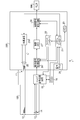
- FIG. 1 is a block diagram showing a configuration of an electronic endoscope system 1 including an endoscope light source device 201 according to the first embodiment of the present invention.
- the electronic endoscope system 1 is a system specialized for medical use, and includes an electronic scope 100, a processor 200, and a monitor 300.
- the processor 200 includes a system controller 21 and a timing controller 22.
- the system controller 21 executes various programs stored in the memory 23 and controls the entire electronic endoscope system 1 in an integrated manner.
- the system controller 21 is connected to the operation panel 24.
- the system controller 21 changes each operation of the electronic endoscope system 1 and parameters for each operation in accordance with an instruction from the operator input from the operation panel 24.
- the input instruction by the operator includes, for example, an instruction to switch the observation mode of the electronic endoscope system 1.
- the observation mode includes a normal observation mode and a special observation mode. Details of each observation mode will be described later.
- the timing controller 22 outputs a clock pulse for adjusting the operation timing of each unit to each circuit in the electronic endoscope system 1.
- the processor 200 includes a light source device 201.
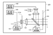
- FIG. 2 is a block diagram of the light source device 201 according to the first embodiment of the present invention.
- the light source device 201 includes a first light source unit 111 and a second light source unit 112.
- the first and second light source units 111 and 112 are individually controlled to emit light by the first and second light source drive circuits 141 and 142, respectively.
- the first light source unit 111 includes a purple light emitting diode (LED: Light Emitting Diode) 111a that emits light in a purple wavelength band (for example, a wavelength of 395 to 435 nm) and a blue phosphor 111b.
- the blue phosphor 111b is excited by the purple LED light emitted from the purple LED 111a and emits fluorescence in a blue wavelength band (for example, a wavelength of 430 to 550 nm).
- the second light source unit 112 includes a blue LED 112a that emits light in a blue wavelength band (for example, a wavelength of 420 to 480 nm), and a yellow phosphor 112b.
- the yellow phosphor 112b is excited by the blue LED light emitted from the blue LED 112a, and emits fluorescence in a yellow wavelength band (for example, a wavelength is 420 to 700 nm).
- the yellow phosphor 112b is attached on the light emitting surface of the blue LED 112a, and unlike the blue phosphor 111b, it cannot be inserted into and removed from the optical path of the blue LED light.
- Collimating lenses 121 and 122 are arranged in front of the light source units 111 and 112 in the light emission direction, respectively.
- the light emitted from the first light source unit 111 is converted into parallel light by the collimator lens 121 and is incident on the dichroic mirror 131.
- the light emitted from the second light source unit 112 is converted into parallel light by the collimator lens 122 and is incident on the dichroic mirror 131.
- the dichroic mirror 131 combines the optical path of the light emitted from the first light source unit 111 and the optical path of the light emitted from the second light source unit 112.
- the wavelength limiting filter 161 has a characteristic of transmitting only light in a specific wavelength band.
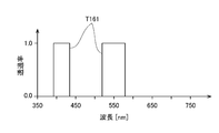
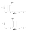
- FIG. 3 shows the spectral transmission characteristic T161 of the wavelength limiting filter 161.
- the horizontal axis of the graph shown in FIG. 3 indicates the wavelength (nm), and the vertical axis indicates the transmittance.
- the wavelength limiting filter 161 has a transmittance of about 1 (100%) for light having a wavelength of about 415 nm (more specifically, 390 nm to 430 nm) and a wavelength of about 550 nm (more specifically, 520 nm to 580 nm).
- the transmittance for light in other wavelength bands is approximately 0 (0%).
- FIG. 4 is a block diagram conceptually showing only the light source units 111 and 112, the dichroic mirror 131, and the wavelength limiting filter 161 in the light source device 201. Since the blue phosphor 111b is a separate body from the purple LED 111a, the blue phosphor 111b and the purple LED 111a are shown as separate blocks in FIG. On the other hand, since the yellow phosphor 112b is attached to the light emitting surface of the blue LED 112a and is configured integrally with the blue LED 112a, the yellow phosphor 112b and the blue LED 112a are shown in one block in FIG. Yes.
- the dichroic mirror 131 synthesizes optical paths of light having different wavelengths. Therefore, in FIG. 4, the dichroic mirror 131 is indicated by an addition symbol “+”. In FIG. 4, the collimating lenses 121 and 122 disposed in front of the light source units 111 and 112 are omitted.
- each arrow indicates an optical path of light.
- the purple LED light emitted from the purple LED 111a of the first light source unit 111 and the blue fluorescence emitted from the blue phosphor 111b are emitted in the same optical path.
- the blue LED light emitted from the blue LED of the second light source unit 112 and the yellow fluorescence emitted from the yellow phosphor are emitted in the same optical path.
- the optical path of the light emitted from the first light source unit 111 and the optical path of the light emitted from the second light source unit 112 are combined by the dichroic mirror 131.
- the light whose optical path is synthesized by the dichroic mirror 131 is emitted as the irradiation light L from the light source device 201.
- the solid-state imaging device 14 is a single-plate color CCD (Charge Coupled Device) image sensor having a Bayer pixel arrangement.
- the solid-state imaging device 14 accumulates an optical image formed by each pixel on the light receiving surface as a charge corresponding to the amount of light, and generates R (Red), G (Green), and B (Blue) image signals. Output.
- the solid-state imaging device 14 is not limited to a CCD image sensor, and may be replaced with a CMOS (Complementary Metal Oxide Semiconductor) image sensor or other types of imaging devices.
- the solid-state image sensor 14 may also be one having a complementary color filter mounted thereon.
- the driver signal processing circuit 15 also accesses the memory 16 and reads the unique information of the electronic scope 100.
- the unique information of the electronic scope 100 recorded in the memory 16 includes, for example, the number and sensitivity of the solid-state imaging device 14, an operable frame rate, a model number, and the like.
- the driver signal processing circuit 15 outputs the unique information read from the memory 16 to the system controller 21.
- the system controller 21 performs various calculations based on the unique information of the electronic scope 100 and generates a control signal.
- the system controller 21 uses the generated control signal to control the operation and timing of various circuits in the processor 200 so that processing suitable for the electronic scope 100 connected to the processor 200 is performed.
- the timing controller 22 supplies clock pulses to the driver signal processing circuit 15 according to the timing control by the system controller 21.
- the driver signal processing circuit 15 drives and controls the solid-state imaging device 14 at a timing synchronized with the frame rate of the video processed on the processor 200 side in accordance with the clock pulse supplied from the timing controller 22.
- the pre-stage signal processing circuit 26 performs predetermined signal processing such as demosaic processing, matrix calculation, and Y / C separation on the image signal input from the driver signal processing circuit 15 in one frame period, and outputs it to the image memory 27. To do.
- the image memory 27 buffers the image signal input from the upstream signal processing circuit 26 and outputs it to the downstream signal processing circuit 28 according to the timing control by the timing controller 22.
- the post-stage signal processing circuit 28 processes the image signal input from the image memory 27 to generate screen data for monitor display, and converts the generated screen data for monitor display into a predetermined video format signal.
- the converted video format signal is output to the monitor 300. Thereby, the image of the subject is displayed on the display screen of the monitor 300.
- the electronic endoscope system 1 of the present embodiment has a plurality of observation modes including a normal observation mode and a special observation mode.
- Each observation mode is switched manually or automatically depending on the subject to be observed. For example, when it is desired to observe the subject illuminated with normal light, the observation mode is switched to the normal observation mode.
- the normal light is, for example, white light or pseudo white light.
- White light has a flat spectral intensity distribution in the visible light band.
- the pseudo-white light has a spectral intensity distribution that is not flat, and light in a plurality of wavelength bands is mixed. For example, when it is desired to obtain a captured image in which a specific living tissue is emphasized by illuminating the subject with special light, the observation mode is switched to the special observation mode.
- ⁇ Blood containing hemoglobin flows in the surface blood vessels. It is known that hemoglobin has absorbance peaks near wavelengths of 415 nm and 550 nm. Therefore, by irradiating the subject with special light suitable for emphasizing the superficial blood vessels (specifically, light having a higher intensity near the wavelength of 415 nm where the absorbance of hemoglobin is peak than other wavelength bands). A captured image in which the superficial blood vessels are emphasized can be obtained. Special light having a high intensity near the wavelength of 550 nm has a relatively high absorbance even for the surface blood vessels. In other words, special light having a high intensity in the vicinity of a wavelength of 550 nm also contributes to highlighting of the surface blood vessels.
- the brightness of the photographed image is maintained while maintaining the state where the surface blood vessels are emphasized. Can be brightened.
- narrowband light special light
- normal observation mode blood vessels in each layer such as the surface layer, middle layer, and deep layer
- Narrow band observation suitable for clearly grasping the state can be performed.
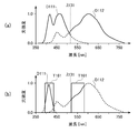
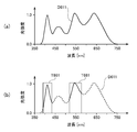
- FIG. 5 shows the spectral intensity distribution of the irradiation light L emitted from the light source device 201 in each observation mode.
- 5A shows a spectral intensity distribution of the irradiation light L (normal light) in the normal observation mode
- FIG. 5B shows a spectral intensity distribution of the irradiation light L (special light) in the special observation mode.
- the horizontal axis of the spectral intensity distribution shown in FIG. 5 indicates the wavelength (nm), and the vertical axis indicates the intensity of the irradiation light L. Note that the vertical axis is standardized so that the maximum intensity value is 1.
- the first light source unit 111 and the second light source unit 112 are driven to emit light after the blue phosphor 111b is inserted in the optical path. At this time, the wavelength limiting filter 161 is removed from the optical path of the irradiation light L.
- the spectral intensity distribution D111 of light emitted from the first light source unit 111 has peaks at a wavelength of about 415 nm and a wavelength of about 470 nm.
- a wavelength having the highest intensity among the specific wavelengths is referred to as a peak wavelength.
- the wavelength having the highest intensity is called the peak wavelength.
- the two wavelengths that are the intensity peaks of the spectral intensity distribution D111 are the peak wavelength of the light emitted from the purple LED 111a and the peak wavelength of the spectral intensity distribution of the fluorescence emitted from the blue phosphor 111b.
- the spectral intensity distribution D112 of light emitted from the second light source unit 112 has peaks at a wavelength of about 450 nm and a wavelength of about 600 nm. These two wavelengths are respectively the peak wavelength of the intensity distribution of the light emitted from the blue LED 112a and the peak wavelength of the spectral intensity distribution of the fluorescence emitted from the yellow phosphor 112b.
- the spectral intensity distribution D111 shown in FIG. 5A has substantially the same peak intensity of purple LED light and blue fluorescence, but the present invention is not limited to this.
- the ratio of the intensity of the purple LED light emitted from the first light source unit 111 to the intensity of blue fluorescence can be freely changed by changing the type and amount of use of the blue phosphor 111b.
- the spectral intensity distribution D112 shown in FIG. 5A has a larger ratio of the intensity of yellow fluorescence than the blue LED light, but the present invention is not limited to this.
- the ratio between the blue LED light emitted from the second light source unit 112 and the yellow fluorescence can be freely changed by changing the type and amount of use of the yellow phosphor 112b.
- the maximum intensity is set to 1, but the present invention is not limited to this.
- the intensity ratio of the light emitted from the light source units 111 and 112 can be arbitrarily set according to the subject to be observed, the photographing mode, and the operator's preference.
- the cutoff wavelength ⁇ 131 of the dichroic mirror 131 is indicated by a dotted line.
- the dichroic mirror 131 has a cutoff wavelength ⁇ 131 of about 520 nm, transmits light in a wavelength band shorter than the cutoff wavelength ⁇ 131, and reflects light in a wavelength band longer than the cutoff wavelength ⁇ 131. Therefore, in the spectral intensity distribution D111 shown in FIG. 5A, light in the wavelength band indicated by the solid line is transmitted through the dichroic mirror 131, and light in the wavelength band indicated by the broken line is reflected by the dichroic mirror 131. Also, in the spectral intensity distribution D112 shown in FIG.
- the optical paths of the light emitted from the light source units 111 and 112 are synthesized by the dichroic mirror 131, and the light source device 201 emits light having a wide wavelength band from the ultraviolet region (part of the near ultraviolet) to the red region.
- Light L normal light
- the spectral intensity distribution of the irradiation light L is the sum of the areas indicated by the solid lines in the spectral intensity distributions D111 and D112 shown in FIG.
- two phosphors 111b and 112b having a wide wavelength band and different wavelength bands are provided. Therefore, the spectral intensity distribution of the irradiation light L (normal light) is closer to flat in the visible light band than when no phosphor is used or when only one type of phosphor is used. Thereby, the subject is illuminated with the irradiation light L (normal light) close to natural white light, and a color photographed image with high color reproducibility can be obtained.
- the first light source unit 111 and the second light source unit 112 are driven to emit light after the blue phosphor 111b is removed from the optical path.
- the wavelength limiting filter 161 is inserted on the optical path of the irradiation light L.
- FIG. 5B shows the intensity distribution of light emitted from the light source units 111 and 112 and the spectral transmission characteristic T161 of the wavelength limiting filter 161.
- Light emitted from the light source device 201 as irradiation light L (special light) is light whose optical path is combined by the dichroic mirror 131 and transmitted through the wavelength limiting filter 161. Therefore, the spectral intensity distribution of the irradiation light L (special light) is the sum of the areas indicated by the solid lines in the spectral intensity distributions D111 and D112 shown in FIG.
- the electronic endoscope system 1 When the electronic endoscope system 1 is in the special observation mode, since the blue phosphor 111b is removed from the optical path, the light emitted from the purple LED 111a is not used to excite the blue phosphor 111b. It is possible to increase the intensity of light in the vicinity of a wavelength of 415 nm, which is the peak of the absorbance. Also, the wavelength limiting filter 161 irradiates the subject with only light having a wavelength of about 550 nm, which is another peak of hemoglobin absorbance, out of light emitted from the second light source unit 112. Thereby, the brightness
- the light paths of the light emitted from the light source units 111 and 112 are combined by the dichroic mirror 131. At this time, since the wavelength bands of the light emitted from the light source units 111 and 112 are different from each other, the loss of the light amount can be minimized when the optical paths in the dichroic mirror 131 are combined.
- the spectral intensity distribution of the irradiation light L emitted from the light source device 201 is switched by inserting / removing the wavelength limiting filter 161 on the optical path of the irradiation light L.
- the processor 200 does not need to have the plurality of light source devices 201 in accordance with the spectral intensity distribution of the desired irradiation light L, and the configuration of the processor 200 can be simplified and downsized.
- the peak intensities of the spectral intensity distributions D111 and D112 are all set to 1, but the present invention is not limited to this. It is not limited to.
- the second light source unit 112 may be driven to emit light so that the drive current is smaller and the intensity is lower than that in the normal observation mode.
- the intensity around the wavelength of 415 nm, which is the peak of hemoglobin absorbance is relatively higher than the intensity of other wavelength bands (that is, becomes narrowband light), and a captured image in which the surface blood vessels are more emphasized is obtained.
- the blue phosphor 111b is supported by the phosphor insertion / extraction mechanism 151 so that it can be inserted into and removed from the optical path.
- the present invention is not limited to this.
- the blue phosphor 111b is not insertable / removable on the optical path, and may be attached on the light emitting surface of the purple LED 111a.
- blue fluorescence is also emitted from the first light source unit 111.
- the blue fluorescence is cut by the wavelength limiting filter 161, the enhancement effect of the surface blood vessels is not reduced by the blue fluorescence.
- an endoscope light source device according to a second embodiment of the present invention will be described.
- the light source device according to the second embodiment is also used in the electronic endoscope system 1 in the same manner as the light source device 201 according to the first embodiment.
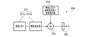
- FIG. 6 is a block diagram conceptually showing only a light source unit, a dichroic mirror, and a wavelength limiting filter in the light source device 202 according to the second embodiment.
- the light source device 202 includes a first light source unit 211, a second light source unit 212, a dichroic mirror 231, and a wavelength limiting filter 261.
- the light source units 211 and 212 are individually controlled to emit light by a first light source drive circuit and a second light source drive circuit (not shown).
- the first light source unit 211 has a purple LED 211a that emits light in a purple wavelength band (for example, a wavelength of 395 to 435 nm) and a blue phosphor 211b.
- the blue phosphor 211b is excited by the purple LED light emitted from the purple LED 211a and emits fluorescence in a blue wavelength band (for example, a wavelength of 430 to 550 nm).
- the blue phosphor 211b is supported by an unillustrated phosphor insertion / extraction mechanism so that the blue phosphor 211b can be inserted into and removed from the optical path of the purple LED light emitted from the purple LED 211a.
- the second light source unit 212 includes a blue LED, a green phosphor, and a red phosphor that emit light in a blue wavelength band (for example, a wavelength of 420 to 480 nm).
- the green phosphor is excited by blue LED light emitted from the blue LED, and emits fluorescence in a green wavelength band (for example, a wavelength of 510 to 630 nm).
- the red phosphor is excited by the blue LED light emitted from the blue LED and emits fluorescence in the red wavelength band (for example, the wavelength is 550 to 750 nm).
- the green phosphor and the red phosphor may be arranged side by side along the emission direction of the blue LED light, or may be arranged side by side in a direction perpendicular to the emission direction of the blue LED light.
- the green phosphor and the red phosphor may be prepared as a single phosphor by mixing the materials.
- a collimating lens (not shown) is arranged in front of the light source units 211 and 212 in the emission direction.
- the light emitted from the first light source unit 211 is converted into parallel light by the collimator lens and is incident on the dichroic mirror 231.
- the light emitted from the second light source unit 212 is converted into parallel light by the collimator lens and is incident on the dichroic mirror 231.
- the dichroic mirror 231 combines the optical path of the light emitted from the first light source unit 211 and the optical path of the light emitted from the second light source unit 212.
- the light whose optical path is synthesized by the dichroic mirror 231 is emitted as the irradiation light L toward the wavelength limiting filter 261.
- the spectral transmission characteristic of the wavelength limiting filter 261 is the same as the spectral transmission characteristic of the wavelength limiting filter 161 of the first embodiment.
- the wavelength limiting filter 261 is removed from the optical path of the irradiation light L.
- the irradiation light L is emitted from the light source device 202 without the wavelength being limited by the wavelength limiting filter 261.
- the wavelength limiting filter 261 is inserted in the optical path of the irradiation light L. In this case, of the irradiation light L, only light having a wavelength near 415 nm and a wavelength near 550 nm is emitted from the light source device 202.
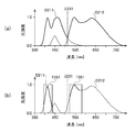
- FIG. 7 is a view similar to FIG. 5 and shows the spectral intensity distribution of the irradiation light L emitted from the light source device 202 in each observation mode.
- FIG. 7B the spectral transmission characteristic T261 of the wavelength limiting filter 261 is also shown.
- both the first light source unit 211 and the second light source unit 212 are driven to emit light after the blue phosphor 211b is inserted in the optical path. At this time, the wavelength limiting filter 261 is removed from the optical path of the irradiation light L.
- the spectral intensity distribution D211 of light emitted from the first light source unit 211 has peaks at a wavelength of about 415 nm and a wavelength of about 470 nm. These two wavelengths are the peak wavelength of the intensity distribution of the light emitted from the purple LED 211a and the peak wavelength of the spectral intensity distribution of the fluorescence emitted from the blue phosphor 211b.
- the spectral intensity distribution D212 of light emitted from the second light source unit 212 has peaks at wavelengths of about 450 nm, about 550 nm, and about 650 nm. These three wavelengths are respectively the peak wavelengths of the intensity distribution of the blue LED light, the fluorescence emitted by the green phosphor, and the fluorescence emitted by the red phosphor.
- the cutoff wavelength ⁇ 231 of the dichroic mirror 231 is indicated by a dotted line.
- the dichroic mirror 231 has a cutoff wavelength ⁇ 231 of about 510 nm, transmits light in a wavelength band shorter than the cutoff wavelength ⁇ 231, and reflects light in a wavelength band longer than the cutoff wavelength ⁇ 231. Therefore, in the spectral intensity distribution D211 shown in FIG. 7A, light in the wavelength band indicated by the solid line passes through the dichroic mirror 231 and light in the wavelength band indicated by the broken line is reflected by the dichroic mirror 231. In the spectral intensity distribution D212 shown in FIG. 7A, light in the wavelength band indicated by the solid line is reflected by the dichroic mirror 231 and light in the wavelength band indicated by the broken line passes through the dichroic mirror 231.
- the optical path of the light emitted from each of the light source units 211 and 212 is synthesized by the dichroic mirror 231, and the light source device 202 emits light having a wide wavelength band from the ultraviolet region (part of near ultraviolet) to the red region.
- Light L normal light
- the spectral intensity distribution of the irradiation light L (normal light) is the sum of the areas indicated by the solid lines in the spectral intensity distributions D211 and D212 shown in FIG.
- both the first light source unit 211 and the second light source unit 212 are driven to emit light after the blue phosphor 211b is removed from the optical path. .
- the wavelength limiting filter 261 is inserted on the optical path of the irradiation light L.
- the ratio of light in the vicinity of a wavelength of 415 nm, which is the peak of the absorbance of hemoglobin, in the irradiation light L (special light) is relatively high (that is, narrow-band light), and a captured image in which the surface blood vessels are emphasized is obtained.
- the second light source unit 212 has two phosphors, green and red. Therefore, the spectral intensity distribution of the irradiation light L (normal light) when the electronic endoscope system 1 is in the normal observation mode is more visible than when the second light source unit 212 has one phosphor. It approaches flat in the area. Thereby, in the normal observation mode, the subject can be illuminated with the irradiation light L (normal light) close to natural white light.
- the light source device according to the third embodiment is also used in the electronic endoscope system 1 in the same manner as the light source device 201 according to the first embodiment.
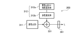
- FIG. 8 is a block diagram conceptually showing only a light source unit, a dichroic mirror, and a wavelength limiting filter in the light source device 203 according to the third embodiment.
- the light source device 203 includes a first light source unit 311, a second light source unit 312, a dichroic mirror 331, and a wavelength limiting filter 361.
- the light source units 311 and 312 are individually controlled to emit light by a first light source drive circuit and a second light source drive circuit (not shown).
- the first light source unit 311 is a purple LED that emits light in a purple wavelength band (for example, a wavelength of 395 to 435 nm).
- the second light source unit 312 includes a phosphor LED 312a and a red phosphor 312b.
- the phosphor LED 312a includes a blue LED that emits light in a blue wavelength band (for example, a wavelength of 430 to 490 nm), and a green phosphor that is attached on the light emitting surface of the blue LED. This green phosphor is excited by blue LED light emitted from a blue LED, and emits fluorescence in a green wavelength band (for example, a wavelength of 460 to 600 nm).
- the red phosphor 312b is excited by the blue LED light emitted from the blue LED, and emits fluorescence in the red wavelength band (for example, the wavelength is 550 to 750 nm).
- the red phosphor 312b is supported by an unillustrated phosphor insertion / extraction mechanism so that the red phosphor 312b can be inserted into and removed from the optical path of light emitted from the blue LED.
- a collimator lens (not shown) is arranged in front of the light source units 311 and 312 in the emission direction.
- the purple LED light emitted from the first light source unit 311 is converted into parallel light by the collimator lens and is incident on the dichroic mirror 331.
- the light emitted from the second light source unit 312, that is, the blue LED light and the green and red fluorescence are converted into parallel light by the collimator lens and are incident on the dichroic mirror 331.
- the dichroic mirror 331 combines the optical path of the light emitted from the first light source unit 311 and the optical path of the light emitted from the second light source unit 312.
- the light whose optical path is synthesized by the dichroic mirror 331 is emitted as irradiation light L toward the wavelength limiting filter 361.
- the spectral transmission characteristic of the wavelength limiting filter 361 is the same as the spectral transmission characteristic of the wavelength limiting filter 161 of the first embodiment.
- the wavelength limiting filter 361 is removed from the optical path of the irradiation light L.
- the irradiation light L is emitted from the light source device 203 without being limited in wavelength by the wavelength limiting filter 361.
- the wavelength limiting filter 361 is inserted into the optical path of the irradiation light L. In this case, of the irradiation light L, only light having a wavelength near 415 nm and a wavelength near 550 nm is emitted from the light source device 203.
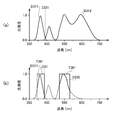
- FIG. 9 is a view similar to FIG. 5 and shows the spectral intensity distribution of the irradiation light L emitted from the light source device 203 in each observation mode.
- FIG. 9B also shows the spectral transmission characteristic T361 of the wavelength limiting filter 361.
- both the light source unit 311 and the light source unit 312 are driven to emit light after the red phosphor 312b is inserted in the optical path.
- the wavelength limiting filter 361 is removed from the optical path of the irradiation light L.
- the spectral intensity distribution D311 of light emitted from the first light source unit 311 has a steep intensity distribution having a peak wavelength of about 415 nm.
- the spectral intensity distribution D312 of light emitted from the second light source unit 312 has peaks at wavelengths of about 470 nm, about 550 nm, and about 630 nm. These three wavelengths are respectively the peak wavelengths of the intensity distribution of blue LED light, green fluorescence, and red fluorescence.
- the cutoff wavelength ⁇ 331 of the dichroic mirror 331 is indicated by a dotted line.
- the dichroic mirror 331 has a cutoff wavelength ⁇ 331 of about 430 nm, transmits light in a wavelength band shorter than the cutoff wavelength ⁇ 331, and reflects light in a wavelength band greater than or equal to the cutoff wavelength ⁇ 331. Therefore, in the spectral intensity distribution D311 shown in FIG. 9A, light in the wavelength band indicated by the solid line passes through the dichroic mirror 331, and light in the wavelength band indicated by the broken line is reflected by the dichroic mirror 331. In the spectral intensity distribution D312 shown in FIG. 9A, light in the wavelength band indicated by the solid line is reflected by the dichroic mirror 331, and light in the wavelength band indicated by the broken line passes through the dichroic mirror 331.
- the light source device 203 has a wide wavelength from the ultraviolet region (part of the near ultraviolet) to the red region.
- Irradiation light L (normal light) having a band is emitted.
- the spectral intensity distribution of the irradiation light L (normal light) is the sum of the areas indicated by the solid lines in the spectral intensity distributions D311 and D312 shown in FIG.
- both the first light source unit 311 and the second light source unit 312 are driven to emit light after the red phosphor 312b is removed from the optical path. .
- the wavelength limiting filter 361 is inserted on the optical path of the irradiation light L.
- the second light source unit 312 has two phosphors, green and red. Therefore, the spectral intensity distribution of the irradiation light L (normal light) when the electronic endoscope system 1 is in the normal observation mode is more visible than when the second light source unit 312 has one phosphor. It approaches flat in the area. Accordingly, the subject can be illuminated with the irradiation light L (normal light) close to natural white light.
- the red phosphor 312b is supported by the phosphor insertion / removal mechanism so that it can be inserted / removed on the optical path, but the present invention is not limited to this.
- the red phosphor 312b is not insertable / removable on the optical path, and may be attached together with the green LED on the light emitting surface of the blue LED.
- red fluorescence is also emitted from the second light source unit 312.
- the red fluorescence is cut by the wavelength limiting filter 361, the enhancement effect of the surface blood vessels is not reduced by the red fluorescence.
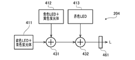
- FIG. 10 is a block diagram conceptually showing only the light source unit, the dichroic mirror, and the wavelength limiting filter in the light source device 204 according to the fourth embodiment of the present invention. Similarly to the light source device 201 according to the first embodiment, the light source device 204 according to the fourth embodiment is used in the electronic endoscope system 1, for example.
- the light source device 204 includes first to third light source units 411 to 413, first and second dichroic mirrors 431 and 432, and a wavelength limiting filter 461.
- the light source units 411 to 413 are individually controlled to emit light by first to third light source driving circuits (not shown).
- the first light source unit 411 emits light in a purple wavelength band (for example, a wavelength of 395 to 435 nm) and is excited by the purple LED light to emit blue (for example, a wavelength of 430 to 490 nm) fluorescence. It has a blue phosphor that emits light.
- the second light source unit 412 is excited by a blue LED that emits light in a blue wavelength band (for example, a wavelength of 430 to 470 nm) and a blue LED light emitted from the blue LED, and a yellow wavelength band (for example, It has a yellow phosphor that emits fluorescence having a wavelength of 500 to 720 nm.
- the third light source unit 413 is a red LED that emits light in a red wavelength band (for example, a wavelength of 620 to 680 nm).
- a collimator lens (not shown) is arranged in front of each of the light source units 411 to 413 in the emission direction. Purple LED light and blue fluorescence emitted from the first light source unit 411 are converted into parallel light by a collimating lens and are incident on a dichroic mirror 431. Further, the blue LED light and the yellow fluorescence emitted from the second light source unit 412 are converted into parallel light by the collimator lens and are incident on the dichroic mirror 431.
- the dichroic mirror 431 combines the optical path of the light emitted from the first light source unit 411 and the optical path of the light emitted from the second light source unit 412. The light whose optical path is synthesized by the dichroic mirror 431 enters the dichroic mirror 432.
- the red LED light emitted from the third light source unit 413 is converted into parallel light by the collimating lens and is incident on the dichroic mirror 432.
- the dichroic mirror 432 combines the optical path of the light incident from the dichroic mirror 431 and the optical path of the light emitted from the third light source unit 413.
- the light whose optical path is synthesized by the dichroic mirror 432 is emitted as irradiation light L toward the wavelength limiting filter 461.
- the spectral transmission characteristic of the wavelength limiting filter 461 is the same as the spectral transmission characteristic of the wavelength limiting filter 161 of the first embodiment.
- the wavelength limiting filter 461 is removed from the optical path of the irradiation light L.
- the irradiation light L is emitted from the light source device 204 without being limited in wavelength by the wavelength limiting filter 461.
- the wavelength limiting filter 461 is inserted in the optical path of the irradiation light L. In this case, of the irradiation light L, only light having a wavelength near 415 nm and a wavelength near 550 nm is emitted from the light source device 204.
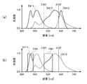
- FIG. 11 is a view similar to FIG. 5 and shows the spectral intensity distribution of the irradiation light L emitted from the light source device 204 in each observation mode.
- FIG. 11B also shows the spectral transmission characteristic T461 of the wavelength limiting filter 461.
- the first to third light source units 411 to 413 are all driven to emit light.
- the wavelength limiting filter 461 is removed from the optical path of the irradiation light L.
- the spectral intensity distribution D411 of the first light source unit 411 has peaks at wavelengths of about 415 nm and 470 nm. These two wavelengths are the peak wavelengths of purple LED light and blue fluorescence, respectively.
- the height of the peak at the wavelength of about 415 nm is set to be higher than the height of the peak at the wavelength of about 470 nm.
- the spectral intensity distribution D412 of the second light source unit 412 has peaks at about 450 nm and 600 nm. These two wavelengths are the peak wavelengths of blue LED light and yellow fluorescence, respectively.
- the spectral intensity distribution D413 of the third light source unit 413 has a steep intensity distribution having a peak wavelength of about 650 nm.
- the cutoff wavelengths ⁇ 431 and ⁇ 432 of the dichroic mirrors 431 and 432 are indicated by dotted lines. Cutoff wavelengths ⁇ 431 and ⁇ 432 are 520 nm and 630 nm, respectively. Any of the dichroic mirrors 431 and 432 transmits light having a wavelength band shorter than the cutoff wavelength, and reflects light having a wavelength band equal to or greater than the cutoff wavelength. By the dichroic mirrors 431 and 432, the optical paths of the light emitted from the light source units 411 to 413 are combined.
- the blue LED light having a peak wavelength of about 450 nm is shorter than the cutoff wavelength ⁇ 431, and thus is included in the light whose optical path is synthesized by the dichroic mirror 431. I can't.
- the light paths emitted from the respective light source units 411 to 413 are synthesized by the dichroic mirrors 431 and 432, so that the light source device 204 can emit light from the ultraviolet region (part of near ultraviolet) to the red region.
- Irradiation light L (normal light) having a wide wavelength band is emitted.
- the spectral intensity distribution of the irradiation light L (normal light) is the sum of the areas indicated by the solid lines in the spectral intensity distributions D411 to D413 shown in FIG.
- the first light source unit 411 and the second light source unit 412 are driven to emit light, and the third light source unit 413 is not driven to emit light.
- the wavelength limiting filter 461 is inserted on the optical path of the irradiation light L.
- the biological tissue in the body cavity imaged by the electronic endoscope system 1 is generally reddish due to blood.
- red light when red light is irradiated onto the living tissue in the special observation mode, the entire captured image is reddish, and it is difficult to obtain an effect of enhancing the superficial blood vessels.
- the red LED third light source unit 413 is not driven to emit light in the special observation mode, it is possible to prevent the enhancement effect of the superficial blood vessels from being reduced.
- the second light source unit 412 has a yellow phosphor, but the present invention is not limited to this.
- the second light source unit 412 may include a green phosphor having a peak wavelength near 550 nm, instead of the yellow phosphor.
- an endoscope light source device according to a fifth embodiment of the present invention will be described.
- the light source device according to the fifth embodiment is also used in the electronic endoscope system 1 in the same manner as the light source device 201 according to the first embodiment.
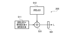
- FIG. 12 is a block diagram conceptually showing only a light source unit, a dichroic mirror, and a wavelength limiting filter in the light source device 205 according to the fifth embodiment.
- the light source device 205 includes a first light source unit 511, a second light source unit 512, a dichroic mirror 531, and a wavelength limiting filter 561.
- the light source units 511 and 512 are individually controlled to emit light by first and second light source driving circuits (not shown).
- the first light source unit 511 has a purple LED that emits light in a purple wavelength band (for example, a wavelength of 395 to 435 nm), and a green phosphor and a blue phosphor that are mounted on the light emitting surface of the purple LED. ing.
- the green phosphor is excited by the purple LED light emitted from the purple LED, and emits fluorescence in the green wavelength band (for example, the wavelength is 510 to 630 nm).
- the blue phosphor is excited by the violet LED light emitted from the violet LED, and emits fluorescence in a blue wavelength band (for example, the wavelength is 430 to 550 nm).
- the second light source unit 512 is a red LED that emits light in a red wavelength band (for example, a wavelength of 620 to 680 nm).
- the dichroic mirror 531 combines the optical path of the light emitted from the first light source unit 511 and the optical path of the light emitted from the second light source unit 512.
- the light whose optical path is synthesized by the dichroic mirror 531 is emitted toward the wavelength limiting filter 561 as the irradiation light L.
- the spectral transmission characteristic of the wavelength limiting filter 561 is the same as the spectral transmission characteristic of the wavelength limiting filter 161 of the first embodiment.
- the wavelength limiting filter 561 is removed from the optical path of the irradiation light L. In this case, the irradiation light L is emitted from the light source device 205 without the wavelength being limited by the wavelength limiting filter 561.
- the wavelength limiting filter 561 is inserted in the optical path of the irradiation light L. In this case, of the irradiation light L, only light having a wavelength near 415 nm and a wavelength near 550 nm is emitted from the light source device 205.
- FIG. 13 is a view similar to FIG. 5 and shows the spectral intensity distribution of the irradiation light L emitted from the light source device 205 in each observation mode.
- FIG. 13B also shows the spectral transmission characteristic T561 of the wavelength limiting filter 561.
- the first and second light source units 511 and 512 are driven to emit light.
- the wavelength limiting filter 561 is removed from the optical path of the irradiation light L.
- the spectral intensity distribution D511 of light emitted from the first light source unit 511 has peaks at wavelengths of about 415 nm, about 470 nm, and about 550 nm. These three wavelengths are respectively the peak wavelengths of the purple LED light emitted from the purple LED, the fluorescence emitted by the blue phosphor, and the fluorescence emitted by the green phosphor.
- the spectral intensity distribution D512 of light emitted from the second light source unit 512 has an intensity distribution with a peak wavelength of about 650 nm.
- the cutoff wavelength ⁇ 531 of the dichroic mirror 531 is indicated by a dotted line.
- the cutoff wavelength ⁇ 531 is 620 nm.
- the dichroic mirror 531 transmits light in a wavelength band shorter than the cutoff wavelength, and reflects light in a wavelength band equal to or greater than the cutoff wavelength.
- the optical paths of the light emitted from the first light source unit 511 and the second light source unit 512 are combined and emitted as irradiation light L.
- irradiation light L normal light
- the electronic endoscope system 1 when the electronic endoscope system 1 is in the special observation mode, only the first light source unit 511 is driven to emit light, and the second light source unit 512 is not driven to emit light. At this time, the wavelength limiting filter 561 is inserted on the optical path of the irradiation light L. As a result, the ratio of light in the vicinity of a wavelength of 415 nm, which is the peak of the absorbance of hemoglobin, in the irradiation light L (special light) is relatively high (that is, narrow-band light), and a captured image in which the surface blood vessels are emphasized is obtained. Obtainable.
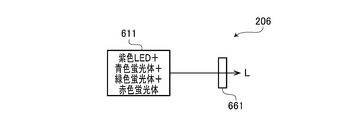
- the light source unit 611 includes a purple LED, a blue phosphor, a green phosphor, and a red phosphor that emit light in a purple wavelength band (for example, a wavelength of 395 to 435 nm).
- the blue phosphor is excited by the violet LED light emitted from the violet LED, and emits fluorescence in a blue wavelength band (for example, the wavelength is 430 to 550 nm).
- the green phosphor is excited by the purple LED light emitted from the purple LED, and emits fluorescence in the green wavelength band (for example, the wavelength is 510 to 630 nm).
- the red phosphor is excited by the violet LED light emitted from the violet LED, and emits fluorescence in a red wavelength band (for example, the wavelength is 550 to 750 nm).
- the light emitted from the light source unit 611 is emitted toward the wavelength limiting filter 661 as the irradiation light L.
- the spectral transmission characteristic of the wavelength limiting filter 661 is the same as the spectral transmission characteristic of the wavelength limiting filter 161 of the first embodiment.
- the wavelength limiting filter 661 is removed from the optical path of the irradiation light L.
- the irradiation light L is emitted from the light source device 206 without being limited in wavelength by the wavelength limiting filter 661.
- the wavelength limiting filter 661 is inserted into the optical path of the irradiation light L. In this case, of the irradiation light L, only light having a wavelength near 415 nm and a wavelength near 550 nm is emitted from the light source device 205.
- FIG. 15 is a view similar to FIG. 5 and shows the spectral intensity distribution of the irradiation light L emitted from the light source device 206 in each observation mode.
- FIG. 15B also shows the spectral transmission characteristic T661 of the wavelength limiting filter 661.
- the light source unit 611 is driven to emit light after the wavelength limiting filter 661 is removed from the optical path of the irradiation light L.
- the spectral intensity distribution D611 of light emitted from the light source unit 611 has peaks at wavelengths of about 415 nm, about 470 nm, about 550 nm, and about 650 nm. These four wavelengths are the peak wavelengths of the purple LED light emitted from the purple LED, the fluorescence emitted by the blue phosphor, the fluorescence emitted by the green phosphor, and the fluorescence emitted by the red phosphor.
- the light emitted from the light source unit 611 is irradiated to the subject as irradiation light L (normal light). Thereby, a normal color photographed image can be obtained.
- the light source unit 611 is driven to emit light after the wavelength limiting filter 661 is inserted in the optical path of the irradiation light L.
- the ratio of light in the vicinity of a wavelength of 415 nm, which is the peak of the absorbance of hemoglobin, in the irradiation light L (special light) is relatively high (that is, narrow-band light), and a captured image in which the surface blood vessels are emphasized is obtained.
- Embodiments of the present invention are not limited to those described above, and various modifications are possible within the scope of the technical idea of the present invention.
- the embodiment of the present invention also includes contents appropriately combined with embodiments or the like clearly shown in the specification or obvious embodiments.
- an LED is assumed as the solid state light emitting device.
- the present invention is not limited to this, and it is also possible to employ LD (Laser Diode) as a solid state light emitting device.
- FIG. 16 shows spectral transmission characteristics of the wavelength limiting filter according to the modification of the third embodiment.
- two wavelength limiting filters (wavelength limiting filters 361A and 361B) are provided.
- FIG. 16A shows the spectral transmission characteristic T361A of the wavelength limiting filter 361A
- FIG. 16B shows the spectral transmission characteristic T361B of the wavelength limiting filter 361B.
- the wavelength limiting filter 361A has a transmittance of about 1 (100%) for light in the vicinity of a wavelength of 415 nm (more specifically, 390 nm to 430 nm), and other wavelength bands.
- the transmittance for light is approximately 0 (0%).
- the wavelength limiting filter 361B has a transmittance of about 1 (100%) for light in the vicinity of a wavelength of 550 nm (more specifically, 520 nm to 580 nm), and other wavelength bands.
- the transmittance for light is approximately 0 (0%).
- the wavelength limiting filters 361A and 361B are supported so as to be insertable / removable with respect to the optical path of the irradiation light L by an insertion / extraction mechanism similar to the filter insertion / extraction mechanism 171.
- there are three observation modes normal observation mode, first special observation mode, and second special observation mode).
- the wavelength limiting filter 361A is inserted into the optical path of the irradiation light L, and the wavelength limiting filter 361B is not inserted into the optical path of the irradiation light L. In this case, only the light having a wavelength of about 415 nm out of the irradiation light L is emitted from the light source device 203.
- the wavelength limiting filter 361B is inserted into the optical path of the irradiation light L, and the wavelength limiting filter 361A is not inserted into the optical path of the irradiation light L. In this case, only the light having a wavelength of around 550 nm out of the irradiation light L is emitted from the light source device 203.
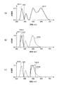
- FIG. 17 is a view similar to FIG. 5 and shows the spectral intensity distribution of the irradiation light L emitted from the light source device 203 in each observation mode of this modification.
- FIG. 17A shows the spectral intensity distribution of the irradiation light L (normal light) in the normal observation mode
- FIG. 17B shows the spectral intensity distribution of the irradiation light L (special light) in the first special observation mode
- FIG. 17C shows the spectral intensity distribution of the irradiation light L (special light) in the second special observation mode.
- FIGS. 17B and 17C also show the spectral transmission characteristic T361A of the wavelength limiting filter 361A and the spectral transmission characteristic T361B of the wavelength limiting filter 361B, respectively.
- the operation in the normal observation mode is the same as that of the third embodiment described with reference to FIGS. Therefore, in the normal observation mode, the irradiation light L (normal light) having the same spectral characteristics as in FIG. 9A, that is, the irradiation light L having a wide wavelength band from the ultraviolet region (part of the near ultraviolet) to the red region. It is injected. By irradiating the subject with the irradiation light L (normal light), a normal color photographed image can be obtained.
- both the first light source unit 311 and the second light source unit 312 are driven to emit light after the red phosphor 312b is removed from the optical path.
- the wavelength limiting filter 361A is inserted on the optical path of the irradiation light L, and the wavelength limiting filter 361B is extracted on the optical path of the irradiation light L.
- the intensity in the vicinity of the wavelength of 415 nm which is the absorption peak of hemogbin, is relatively higher than the intensity in other wavelength bands in the irradiation light L (special light). (That is, narrowband light having a peak only in the vicinity of a wavelength of 415 nm), and a captured image in which the surface blood vessels are emphasized can be obtained.
- the red phosphor 312b is removed from the optical path, and both the first light source unit 311 and the second light source unit 312 are driven to emit light.
- the wavelength limiting filter 361B is inserted on the optical path of the irradiation light L, and the wavelength limiting filter 361A is extracted on the optical path of the irradiation light L.
- the intensity around the wavelength of 550 nm which is the absorption peak of hemogbin, is relatively higher than the intensity in other wavelength bands in the irradiation light L (special light). (That is, narrowband light having a peak only in the vicinity of a wavelength of 550 nm), and a captured image in which the middle blood vessel is emphasized can be obtained.
- the present modification it is possible to obtain a photographed image that mainly emphasizes the surface blood vessels in the first special observation mode, and obtain a photographed image that mainly emphasizes the middle-layer blood vessels in the second special observation mode. Can do. That is, in this modification, by switching between the first special observation mode and the second special observation mode, blood vessels in a desired layer region (surface layer in the first special observation mode and middle layer in the second special observation mode) are focused. It is possible to observe an image that has been emphasized.
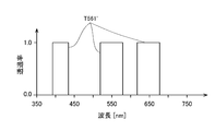
- FIG. 18 shows the spectral transmission characteristic T561 'of the wavelength limiting filter 561' according to the modification of the fifth embodiment.
- the wavelength limiting filter 561 ′ according to this modification includes a wavelength near 415 nm (more specifically, 390 nm to 430 nm), a wavelength near 550 nm (more specifically, 520 nm to 580 nm), and a wavelength near 650 nm ( More specifically, the transmittance with respect to light of 620 nm to 680 nm is approximately 1 (100%), and the transmittance with respect to light in other wavelength bands is approximately 0 (0%).
- the wavelength limiting filter 561 ' is removed from the optical path of the irradiation light L.
- the irradiation light L is emitted from the light source device 205 without being limited in wavelength by the wavelength limiting filter 561 ′.
- the wavelength limiting filter 561 ' is inserted in the optical path of the irradiation light L.
- the irradiation light L only light having a wavelength of about 415 nm, a wavelength of about 550 nm, and a wavelength of about 650 nm is emitted from the light source device 205.
- FIG. 19 is a view similar to FIG. 5 and shows the spectral intensity distribution of the irradiation light L emitted from the light source device 205 in each observation mode of this modification.
- FIG. 19B also shows the spectral transmission characteristic T561 'of the wavelength limiting filter 561'.
- irradiation light L normal light
- irradiation light L normal light
- a normal color photographed image can be obtained.
- the first and second light source units 511 and 512 are driven to emit light.
- the wavelength limiting filter 561 is inserted on the optical path of the irradiation light L.
- the ratio of light in the vicinity of wavelengths of 415 nm and 650 nm, which is the absorption peak of hemoglobin, of the irradiation light L (special light) is relatively high (that is, narrow-band light), and mainly the superficial blood vessels and deep layers. A captured image in which blood vessels are emphasized can be obtained.
- a configuration in which a narrow-band observation image in which a blood vessel is emphasized using special light (narrow-band light) is generated and displayed is employed.
- a configuration is adopted in which biological information (specifically, oxygen saturation) of a subject is quantitatively analyzed and imaged based on a plurality of images taken with light having different wavelength ranges. Also good.
- FIG. 20 shows a block diagram of a light source device 207 according to another embodiment. Another embodiment will be described based on the second embodiment for convenience.
- the light source device 207 has a configuration in which the wavelength limiting filter 261 is replaced with a rotary turret 400 and a filter rotating mechanism 430 is added to the light source device 202 according to the second embodiment.
- the rotary turret 400 is supported by a filter insertion / extraction mechanism 171 so as to be insertable / removable with respect to the optical path of the irradiation light L.
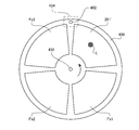
- FIG. 21 is a diagram showing a configuration of the rotary turret 400.
- a motor shaft 432 of a DC motor constituting the filter rotation mechanism 430 is press-fitted into the bearing hole formed at the center of the rotary turret 400.
- the rotary turret 400 is supported by a filter rotating mechanism 430 so as to be rotatable around a motor shaft 432.
- the filter rotation mechanism 430 since a well-known structure is employ
- the rotary turret 400 has four openings arranged in the circumferential direction. Each aperture is provided with a filter having a different spectral characteristic. Specifically, a wavelength limiting filter 261 ', a first oxygen saturation observation filter Fs1, a second oxygen saturation observation filter Fs2, and a narrow band observation filter Fs3 are arranged. In another embodiment, by using the rotary turret 400, in addition to the normal observation image in the normal observation mode and the narrow band observation image in the special observation mode, the oxygen saturation distribution image in the oxygen saturation observation mode is displayed. Is possible.
- FIG. 22 shows an absorption spectrum of hemoglobin near 550 nm.
- Hemoglobin has a strong absorption band called a Q band derived from porphyrin near 550 nm.
- the absorption spectrum of hemoglobin varies depending on the oxygen saturation (the ratio of oxygenated hemoglobin in the total hemoglobin).
- the solid line waveform in FIG. 22 shows an absorption spectrum when the oxygen saturation is 100% (that is, oxygenated hemoglobin HbO), and the long broken line waveform is when the oxygen saturation is 0% (that is, reduction).
- the absorption spectrum of hemoglobin Hb is shown.
- the short dashed line shows the absorption spectrum of hemoglobin (a mixture of oxygenated hemoglobin and reduced hemoglobin) at intermediate oxygen saturation (10, 20, 30,... 90%).
- oxygenated hemoglobin and reduced hemoglobin have different peak wavelengths. Specifically, oxygenated hemoglobin has an absorption peak P1 near a wavelength of 542 nm and an absorption peak P3 near a wavelength of 578 nm. On the other hand, reduced hemoglobin has an absorption peak P2 near 558 nm.
- FIG. 22 shows a two-component absorption spectrum in which the sum of the concentrations of the components (oxygenated hemoglobin and deoxyhemoglobin) is constant. Therefore, the absorption is constant regardless of the concentration of each component (that is, oxygen saturation). Iso-absorption points E1, E2, E3, E4 appear.
- wavelength region sandwiched between the equal absorption points E1 and E2 is referred to as “wavelength region R1”
- the wavelength region sandwiched between the equal absorption points E2 and E3 is referred to as “wavelength region R2”
- wavelength region R3 A wavelength region sandwiched between the absorption points E3 and E4 is referred to as a “wavelength region R3”.
- a wavelength region sandwiched between the isosbestic points E1 and E4 (that is, a combination of the wavelength regions R1, R2, and R3) is referred to as a “wavelength region R0”.
- the wavelength range R0 is 528 nm to 584 nm.
- the wavelength range R2 is 546 nm to 570 nm.
- the absorption monotonously increases or decreases with respect to the oxygen saturation between adjacent isosbestic points. Further, between adjacent isosbestic points, the absorption of hemoglobin changes almost linearly with respect to the oxygen saturation.
- the absorption A R1, A R3 of hemoglobin in the wavelength range R1, R3 is linearly and monotonously increases with respect to the concentration of oxygenated hemoglobin (oxygen saturation)
- the oxygen saturation can be calculated from the value of the index X.
- the first oxygen saturation observation filter Fs1 is an optical bandpass filter that selectively transmits light in the 550 nm band. As shown in FIG. 22, the first oxygen saturation observation filter Fs1 transmits light in the wavelength region from the equiabsorption points E1 to E4 (that is, the wavelength region R0) with low loss, and other wavelengths. It has spectral characteristics that shield the light in the area.
- the second oxygen saturation observation filter Fs2 transmits the light in the wavelength region (that is, the wavelength region R2) from the isosbestic points E2 to E3 with low loss and blocks the light in the other wavelength regions. have.
- the wavelength limiting filter 261 ′ has the same spectral transmission characteristics as the wavelength limiting filter 261.
- the narrowband observation filter Fs3 has a spectral transmission characteristic that transmits only light in the 650 nm band (630 to 650 nm) having a high absorbance with respect to a specific living tissue (mainly deep blood vessels).
- the rotary turret 400 has four wavelengths (first oxygen saturation observation filter Fs1, second oxygen saturation observation filter Fs2, narrowband observation filter Fs3, and wavelength limitation filter) that limit the wavelength.
- Four filters 261 ′) are arranged. That is, the rotary turret 400 is provided with a plurality (four) of wavelength limiting filters having different spectral transmission characteristics.
- both the first light source unit 211 and the second light source unit 212 with the rotary turret 400 removed from the optical path of the irradiation light L and the blue phosphor 211b inserted in the optical path. Is driven to emit light. That is, the operation in the normal observation mode is the same as that of the second embodiment described with reference to FIGS. Therefore, in the normal observation mode, irradiation light L (normal light) having the same spectral characteristics as in FIG. 7A is emitted. By irradiating the subject with the irradiation light L (normal light), a normal color photographed image can be obtained.
- the rotary turret 400 is inserted into the optical path of the irradiation light L and the blue phosphor 211b is removed from the optical path, and the first light source unit 211 and the second light source unit 212 are connected. Both are driven to emit light. At this time, the rotary turret 400 stops in a state where the wavelength limiting filter 261 ′ is located on the optical path of the irradiation light L. That is, the operation in the special observation mode is also the same as that in the second embodiment described with reference to FIGS. Therefore, in the special observation mode, the irradiation light L (normal light) having the same spectral characteristics as in FIG. 7B is emitted.
- each of the wavelength limiting filter 261 ′, the first oxygen saturation observation filter Fs1, the second oxygen saturation observation filter Fs2, and the narrowband observation filter Fs3 is irradiated with light L.
- the rotary turret 400 is rotationally driven at a period of one rotation in four frames so that the frames are sequentially inserted into the optical path at a timing synchronized with the frame rate (synchronized with the imaging period).
- a through hole 402 is formed in the rotary turret 400.
- the system controller 21 detects and adjusts the rotational phase of the rotary turret 400 based on the detection timing of the through hole 402 by the photo interrupter 434 constituting the filter rotating mechanism 430. Thereby, the rotary turret 400 is rotationally driven at a constant speed (a period of one rotation in four frames) during the oxygen saturation observation mode.
- the subject is filtered in accordance with the wavelength limiting filter 261 ′, the first oxygen saturation observation filter Fs1, the second oxygen saturation observation filter Fs2, and the narrowband observation filter Fs3.
- Irradiation light L is sequentially irradiated. Therefore, the post-stage signal processing circuit 28 is filtered by the wavelength limiting filter 261 ′, the first oxygen saturation observation filter Fs1, the second oxygen saturation observation filter Fs2, and the narrowband observation filter Fs3.
- the image signals corresponding to the irradiated light L are sequentially input.
- the post-stage signal processing circuit 28 uses the above equation (1) to apply the image signal corresponding to the first oxygen saturation observation filter Fs1 input from the image memory 27 and the second oxygen saturation observation filter Fs2.
- An index X is calculated from the corresponding image signal.
- a non-volatile memory (not shown) provided in the post-stage signal processing circuit 28 stores a numerical table showing a quantitative relationship between the oxygen saturation of hemoglobin and the value of the index X, which are experimentally acquired in advance.
- the post-stage signal processing circuit 28 refers to this numerical table and obtains the oxygen saturation SatO 2 (x, y) corresponding to the value of the index X calculated using the above equation (1).
- the post-stage signal processing circuit 28 uses the data obtained by multiplying the acquired oxygen saturation SatO 2 (x, y) by a predetermined constant as the pixel value of each pixel (x, y) (oxygen saturation distribution image data). ) Is generated.
- the post-stage signal processing circuit 28 generates narrowband observation image data (the image is the same as that in the special observation mode although the rate is reduced) using the image signal corresponding to the wavelength limiting filter 261 ′ input from the image memory 27. To do.
- the post-stage signal processing circuit 28 generates narrowband observation image data using an image signal corresponding to the narrowband observation filter Fs3 input from the image memory 27.
- the post-stage signal processing circuit 28 converts the oxygen saturation distribution image data into a predetermined video format signal.
- the converted video format signal is output to the monitor 300. Thereby, the oxygen saturation distribution image is displayed on the display screen of the monitor 300.
- the irradiation light L from each of the first oxygen saturation observation filter Fs1 and the second oxygen saturation observation filter Fs2 is generated using fluorescence emitted by the green phosphor. Yes.
- the calculation accuracy of the oxygen saturation is improved, a more suitable oxygen saturation distribution image can be obtained to assist the diagnosis of the lesioned part.
- oxygen the irradiation light L from each of the first oxygen saturation observation filter Fs1 and the second oxygen saturation observation filter Fs2 is generated using fluorescence emitted by the yellow phosphor, oxygen The effect of improving the calculation accuracy of the saturation can be obtained.
- the post-stage signal processing circuit 28 may convert the narrowband observation image data into a predetermined video format signal in addition to the oxygen saturation distribution image data.
- a narrow-band observation image is displayed on the display screen of the monitor 300 in addition to the oxygen saturation distribution image.
Landscapes
- Health & Medical Sciences (AREA)
- Life Sciences & Earth Sciences (AREA)
- Surgery (AREA)
- Biomedical Technology (AREA)
- Medical Informatics (AREA)
- Optics & Photonics (AREA)
- Pathology (AREA)
- Radiology & Medical Imaging (AREA)
- Biophysics (AREA)
- Engineering & Computer Science (AREA)
- Physics & Mathematics (AREA)
- Heart & Thoracic Surgery (AREA)
- Nuclear Medicine, Radiotherapy & Molecular Imaging (AREA)
- Molecular Biology (AREA)
- Animal Behavior & Ethology (AREA)
- General Health & Medical Sciences (AREA)
- Public Health (AREA)
- Veterinary Medicine (AREA)
- Endoscopes (AREA)
- Instruments For Viewing The Inside Of Hollow Bodies (AREA)
Abstract
Ce dispositif de source lumineuse pour endoscope comprend : une unité de source lumineuse qui comporte un premier élément électroluminescent à semi-conducteurs et un premier et un second corps fluorescent, et qui projette une lumière d'irradiation comprenant une lumière dans une première bande de longueur d'onde projetée par le premier élément électroluminescent à semi-conducteurs, une première lumière fluorescente émise par le premier corps fluorescent, et une seconde lumière fluorescente émise par le second corps fluorescent; au moins un filtre limiteur de longueur d'onde qui transmet uniquement la lumière dans une bande de longueurs d'onde spécifique provenant de la lumière d'irradiation; et un moyen d'insertion/retrait de filtre qui soutient le filtre limiteur de longueur d'onde de sorte que le filtre peut être inséré et retiré par rapport au chemin de la lumière d'irradiation. Lorsque le filtre limiteur de longueur d'onde est inséré dans le chemin de la lumière, la lumière d'irradiation est envoyée à l'endoscope en lumière filtrée qui comprend la lumière dans la première bande de longueur d'onde et la seconde lumière fluorescente. Lorsque le filtre limiteur de longueur d'onde est retiré du chemin de la lumière, la lumière d'irradiation est envoyée à l'endoscope sans filtrage.
Priority Applications (2)
| Application Number | Priority Date | Filing Date | Title |
|---|---|---|---|
| JP2018500242A JP6686127B2 (ja) | 2016-02-19 | 2017-02-20 | 内視鏡用光源装置及び内視鏡システム |
| CN201790000593.9U CN209091322U (zh) | 2016-02-19 | 2017-02-20 | 内窥镜用光源装置及内窥镜系统 |
Applications Claiming Priority (2)
| Application Number | Priority Date | Filing Date | Title |
|---|---|---|---|
| PCT/JP2016/054810 WO2017141415A1 (fr) | 2016-02-19 | 2016-02-19 | Dispositif de source de lumière destiné à un endoscope |
| JPPCT/JP2016/054810 | 2016-02-19 |
Publications (1)
| Publication Number | Publication Date |
|---|---|
| WO2017142095A1 true WO2017142095A1 (fr) | 2017-08-24 |
Family
ID=59624937
Family Applications (2)
| Application Number | Title | Priority Date | Filing Date |
|---|---|---|---|
| PCT/JP2016/054810 Ceased WO2017141415A1 (fr) | 2016-02-19 | 2016-02-19 | Dispositif de source de lumière destiné à un endoscope |
| PCT/JP2017/006122 Ceased WO2017142095A1 (fr) | 2016-02-19 | 2017-02-20 | Dispositif de source lumineuse pour endoscope et système endoscopique |
Family Applications Before (1)
| Application Number | Title | Priority Date | Filing Date |
|---|---|---|---|
| PCT/JP2016/054810 Ceased WO2017141415A1 (fr) | 2016-02-19 | 2016-02-19 | Dispositif de source de lumière destiné à un endoscope |
Country Status (3)
| Country | Link |
|---|---|
| JP (1) | JP6686127B2 (fr) |
| CN (1) | CN209091322U (fr) |
| WO (2) | WO2017141415A1 (fr) |
Cited By (1)
| Publication number | Priority date | Publication date | Assignee | Title |
|---|---|---|---|---|
| JPWO2019044802A1 (ja) * | 2017-08-28 | 2020-03-26 | Hoya株式会社 | 内視鏡用光源装置及び内視鏡システム |
Families Citing this family (2)
| Publication number | Priority date | Publication date | Assignee | Title |
|---|---|---|---|---|
| WO2022258591A1 (fr) * | 2021-06-10 | 2022-12-15 | Signify Holding B.V. | Source de lumière blanche sans source de lumière à l'état solide rouge |
| CN119791570A (zh) * | 2024-12-30 | 2025-04-11 | 深圳市星辰海医疗科技有限公司 | 一种内窥镜成像系统及其控制方法 |
Citations (4)
| Publication number | Priority date | Publication date | Assignee | Title |
|---|---|---|---|---|
| JP2006296636A (ja) * | 2005-04-19 | 2006-11-02 | Olympus Medical Systems Corp | 内視鏡装置 |
| JP2014144144A (ja) * | 2013-01-29 | 2014-08-14 | Olympus Corp | 光源装置及び被検体観察装置並びに光源制御方法 |
| JP2014171511A (ja) * | 2013-03-06 | 2014-09-22 | Olympus Corp | 被検体観察システム及びその方法 |
| JP2015066131A (ja) * | 2013-09-27 | 2015-04-13 | 富士フイルム株式会社 | 内視鏡システム及び光源装置 |
Family Cites Families (3)
| Publication number | Priority date | Publication date | Assignee | Title |
|---|---|---|---|---|
| WO2012121304A1 (fr) * | 2011-03-08 | 2012-09-13 | 三菱化学株式会社 | Dispositif électroluminescent et dispositif d'éclairage équipé dudit dispositif électroluminescent |
| JP6021391B2 (ja) * | 2012-04-05 | 2016-11-09 | オリンパス株式会社 | 内視鏡 |
| JP5690790B2 (ja) * | 2012-09-21 | 2015-03-25 | 富士フイルム株式会社 | 内視鏡システム及び内視鏡システムの作動方法 |
-
2016
- 2016-02-19 WO PCT/JP2016/054810 patent/WO2017141415A1/fr not_active Ceased
-
2017
- 2017-02-20 CN CN201790000593.9U patent/CN209091322U/zh active Active
- 2017-02-20 WO PCT/JP2017/006122 patent/WO2017142095A1/fr not_active Ceased
- 2017-02-20 JP JP2018500242A patent/JP6686127B2/ja active Active
Patent Citations (4)
| Publication number | Priority date | Publication date | Assignee | Title |
|---|---|---|---|---|
| JP2006296636A (ja) * | 2005-04-19 | 2006-11-02 | Olympus Medical Systems Corp | 内視鏡装置 |
| JP2014144144A (ja) * | 2013-01-29 | 2014-08-14 | Olympus Corp | 光源装置及び被検体観察装置並びに光源制御方法 |
| JP2014171511A (ja) * | 2013-03-06 | 2014-09-22 | Olympus Corp | 被検体観察システム及びその方法 |
| JP2015066131A (ja) * | 2013-09-27 | 2015-04-13 | 富士フイルム株式会社 | 内視鏡システム及び光源装置 |
Cited By (1)
| Publication number | Priority date | Publication date | Assignee | Title |
|---|---|---|---|---|
| JPWO2019044802A1 (ja) * | 2017-08-28 | 2020-03-26 | Hoya株式会社 | 内視鏡用光源装置及び内視鏡システム |
Also Published As
| Publication number | Publication date |
|---|---|
| JP6686127B2 (ja) | 2020-04-22 |
| JPWO2017142095A1 (ja) | 2018-12-20 |
| WO2017141415A1 (fr) | 2017-08-24 |
| CN209091322U (zh) | 2019-07-12 |
Similar Documents
| Publication | Publication Date | Title |
|---|---|---|
| JP6876810B2 (ja) | 内視鏡用光源装置及び内視鏡システム | |
| JP6878647B2 (ja) | 内視鏡用光源装置及び内視鏡システム | |
| JP5216429B2 (ja) | 光源装置および内視鏡装置 | |
| US9943230B2 (en) | Endoscope system, processor device of endoscope system, and image processing method | |
| JP5229723B2 (ja) | 蛍光画像取得装置 | |
| JP5110702B2 (ja) | 蛍光画像取得装置 | |
| EP1258221A2 (fr) | Système d'endoscope utilisant la lumière normale et la fluorescence | |
| JP2016049370A (ja) | 電子内視鏡システム | |
| JP6686127B2 (ja) | 内視鏡用光源装置及び内視鏡システム | |
| WO2018043293A1 (fr) | Endoscope électronique et système d'endoscope électronique | |
| JP6695416B2 (ja) | 内視鏡用光源装置及び内視鏡システム | |
| JP6277068B2 (ja) | 内視鏡用光源装置及び内視鏡システム | |
| WO2016132940A1 (fr) | Dispositif source de lumière |
Legal Events
| Date | Code | Title | Description |
|---|---|---|---|
| 121 | Ep: the epo has been informed by wipo that ep was designated in this application |
Ref document number: 17753349 Country of ref document: EP Kind code of ref document: A1 |
|
| WWE | Wipo information: entry into national phase |
Ref document number: 2018500242 Country of ref document: JP |
|
| NENP | Non-entry into the national phase |
Ref country code: DE |
|
| 122 | Ep: pct application non-entry in european phase |
Ref document number: 17753349 Country of ref document: EP Kind code of ref document: A1 |