WO2017195638A1 - Procédé de purification d'un anticorps ou d'un fragment d'anticorps comportant une région variable de chaîne κ - Google Patents
Procédé de purification d'un anticorps ou d'un fragment d'anticorps comportant une région variable de chaîne κ Download PDFInfo
- Publication number
- WO2017195638A1 WO2017195638A1 PCT/JP2017/016812 JP2017016812W WO2017195638A1 WO 2017195638 A1 WO2017195638 A1 WO 2017195638A1 JP 2017016812 W JP2017016812 W JP 2017016812W WO 2017195638 A1 WO2017195638 A1 WO 2017195638A1
- Authority
- WO
- WIPO (PCT)
- Prior art keywords
- antibody
- separation matrix
- antibody fragment
- affinity separation
- buffer
- Prior art date
- Legal status (The legal status is an assumption and is not a legal conclusion. Google has not performed a legal analysis and makes no representation as to the accuracy of the status listed.)
- Ceased
Links
Images


Classifications
-
- C—CHEMISTRY; METALLURGY
- C07—ORGANIC CHEMISTRY
- C07K—PEPTIDES
- C07K16/00—Immunoglobulins [IG], e.g. monoclonal or polyclonal antibodies
-
- B—PERFORMING OPERATIONS; TRANSPORTING
- B01—PHYSICAL OR CHEMICAL PROCESSES OR APPARATUS IN GENERAL
- B01D—SEPARATION
- B01D15/00—Separating processes involving the treatment of liquids with solid sorbents; Apparatus therefor
- B01D15/08—Selective adsorption, e.g. chromatography
- B01D15/26—Selective adsorption, e.g. chromatography characterised by the separation mechanism
- B01D15/38—Selective adsorption, e.g. chromatography characterised by the separation mechanism involving specific interaction not covered by one or more of groups B01D15/265 and B01D15/30 - B01D15/36, e.g. affinity, ligand exchange or chiral chromatography
- B01D15/3804—Affinity chromatography
-
- C—CHEMISTRY; METALLURGY
- C07—ORGANIC CHEMISTRY
- C07K—PEPTIDES
- C07K1/00—General methods for the preparation of peptides, i.e. processes for the organic chemical preparation of peptides or proteins of any length
- C07K1/14—Extraction; Separation; Purification
- C07K1/16—Extraction; Separation; Purification by chromatography
- C07K1/22—Affinity chromatography or related techniques based upon selective absorption processes
-
- C—CHEMISTRY; METALLURGY
- C07—ORGANIC CHEMISTRY
- C07K—PEPTIDES
- C07K1/00—General methods for the preparation of peptides, i.e. processes for the organic chemical preparation of peptides or proteins of any length
- C07K1/14—Extraction; Separation; Purification
- C07K1/36—Extraction; Separation; Purification by a combination of two or more processes of different types
-
- C—CHEMISTRY; METALLURGY
- C07—ORGANIC CHEMISTRY
- C07K—PEPTIDES
- C07K16/00—Immunoglobulins [IG], e.g. monoclonal or polyclonal antibodies
- C07K16/06—Immunoglobulins [IG], e.g. monoclonal or polyclonal antibodies from serum
- C07K16/065—Purification, fragmentation
-
- C—CHEMISTRY; METALLURGY
- C07—ORGANIC CHEMISTRY
- C07K—PEPTIDES
- C07K16/00—Immunoglobulins [IG], e.g. monoclonal or polyclonal antibodies
- C07K16/12—Immunoglobulins [IG], e.g. monoclonal or polyclonal antibodies against material from bacteria
- C07K16/1267—Gram-positive bacteria
- C07K16/1275—Streptococcus (G)
-
- C—CHEMISTRY; METALLURGY
- C07—ORGANIC CHEMISTRY
- C07K—PEPTIDES
- C07K2317/00—Immunoglobulins specific features
- C07K2317/50—Immunoglobulins specific features characterized by immunoglobulin fragments
- C07K2317/515—Complete light chain, i.e. VL + CL
-
- C—CHEMISTRY; METALLURGY
- C07—ORGANIC CHEMISTRY
- C07K—PEPTIDES
- C07K2317/00—Immunoglobulins specific features
- C07K2317/50—Immunoglobulins specific features characterized by immunoglobulin fragments
- C07K2317/55—Fab or Fab'
-
- C—CHEMISTRY; METALLURGY
- C07—ORGANIC CHEMISTRY
- C07K—PEPTIDES
- C07K2317/00—Immunoglobulins specific features
- C07K2317/60—Immunoglobulins specific features characterized by non-natural combinations of immunoglobulin fragments
- C07K2317/62—Immunoglobulins specific features characterized by non-natural combinations of immunoglobulin fragments comprising only variable region components
- C07K2317/622—Single chain antibody (scFv)
-
- C—CHEMISTRY; METALLURGY
- C07—ORGANIC CHEMISTRY
- C07K—PEPTIDES
- C07K2317/00—Immunoglobulins specific features
- C07K2317/90—Immunoglobulins specific features characterized by (pharmaco)kinetic aspects or by stability of the immunoglobulin
- C07K2317/92—Affinity (KD), association rate (Ka), dissociation rate (Kd) or EC50 value
Definitions
- the present invention relates to a method capable of successfully purifying an antibody or an antibody fragment containing a ⁇ chain variable region without causing damage.
- Protein A affinity separation matrix (hereinafter referred to as “SpA”) is used to capture and purify antibody drugs from animal cell cultures at a high purity at a time.
- an antibody when an antibody is purified using an affinity separation matrix, the antibody is selectively bound to the ligand of the affinity separation matrix, washed to remove impurities, and then separated from the affinity separation matrix. It has been broken.
- fractions of monomeric monoclonal antibodies are collected from an SpA affinity chromatography column to form an SpA product pool, the pH of the product pool is about 3.5 to about 4.5, and antibody aggregation Citrate and acetate are exemplified as the buffer for eluting the carrier, which describes a method for suppressing the above.
- Patent Documents 2 and 3 describe that an aqueous solution such as citrate or acetate is used as a washing solution for washing SpA on which the antibody is adsorbed, although it is not for eluting the antibody. ing.
- Monoclonal antibodies are basically developed as antibody drugs and are produced in large quantities using recombinant cultured cell technology.
- “Monoclonal antibody” refers to an antibody obtained from a clone derived from a single antibody-producing cell.
- Most antibody drugs currently on the market are immunoglobulin G (IgG) subclass in terms of molecular structure.
- antibody drugs composed of fragment antibodies, which are antibody derivatives having a molecular structure obtained by fragmenting immunoglobulin have been actively developed, and clinical development of various fragment antibody drugs is progressing (Non-patent Document 3).
- SpA affinity separation matrix is used for the initial purification step in the antibody drug manufacturing process.
- SpA is basically a protein that specifically binds to the Fc region of IgG. Therefore, a fragment antibody that does not contain an Fc region cannot be captured using the SpA affinity separation matrix. Therefore, from the viewpoint of developing a platform for antibody drug purification process, there is a great industrial need for an affinity separation matrix capable of capturing a fragment antibody that does not contain the Fc region of IgG.
- Non-Patent Document 4 A plurality of peptides that bind to regions other than the Fc region of IgG are already known (Non-Patent Document 4). Among these, from the viewpoint of the variety of fragment antibody formats that can be bound and the ability to bind to IgM and IgA, peptides that can bind to the variable region that is the antigen binding domain are most preferred.
- protein L is sometimes abbreviated as “PpL”).
- PpL is a protein containing a plurality of ⁇ chain variable region binding domains (hereinafter, the ⁇ chain variable region may be abbreviated as “VL- ⁇ ”), and the amino acid sequences of individual VL- ⁇ binding domains are different. .
- the number of VL- ⁇ binding domains and individual amino acid sequences differ depending on the type of strain. For example, the number of VL- ⁇ binding domains contained in PpL of the Peptostreptococcus magnus 312 strain is 5, and the VL- ⁇ binding domains contained in the PpL of the Peptostreptococcus magnus strain 3316 are five. Is four (Non-Patent Documents 5 to 7, Patent Documents 4 and 5). Among these nine VL- ⁇ binding domains, there are no domains having the same amino acid sequence.
- the present invention can make the pH of the eluate relatively high, so that the antibody fragment containing the antibody or ⁇ chain variable region does not damage the antibody fragment containing the antibody or ⁇ chain variable region and has a sharp elution peak.
- An object of the present invention is to provide a method capable of purifying the water well.
- the inventors of the present invention have made extensive studies to solve the above problems.
- an affinity separation matrix containing PpL to purify an antibody or a VL- ⁇ -containing peptide
- an acetate buffer is used as the eluent, and the antibody has a sharp elution peak even if the pH of the eluate is relatively high.
- the present invention has been completed by finding that the antibody fragments can be purified well. Hereinafter, the present invention will be described.
- a method for purifying an antibody or an antibody fragment containing a ⁇ chain variable region By contacting a liquid sample containing at least one of the antibody and antibody fragment with an affinity separation matrix in which protein L, a domain thereof, or a variant thereof is immobilized on an insoluble carrier as a ligand, the antibody and / or Adsorbing antibody fragments to an affinity separation matrix; Washing the affinity separation matrix to remove impurities other than the antibody and / or antibody fragment; and Separating the antibody and / or antibody fragment from the affinity separation matrix adsorbed with the antibody and / or antibody fragment using an acetate buffer.
- the pH of the eluate can be made relatively high. it can. Moreover, since the elution peak of the antibody or antibody fragment becomes sharp and the amount of the eluted fraction is reduced, further purification, concentration, and drying of the antibody or antibody fragment from the eluted fraction are easy. As a result, the purification cost of the antibody or VL- ⁇ -containing antibody fragment can be suppressed.
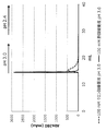
- FIG. 1 shows a case where a commercially available affinity separation matrix on which protein L is immobilized is used to purify a Fab fragment containing VL- ⁇ , and an acetic acid buffer solution or a citrate buffer solution having a pH of 3.0 is used as an eluent. Elution profile. A pH 2.4 citrate buffer is also used to dissociate the remaining Fab fragments in the matrix.
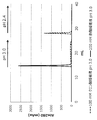
- FIG. 2 shows a case where a pH-3.0 acetate buffer or a citrate buffer is used as an eluent when purifying a Fab fragment containing VL- ⁇ using a prototype affinity separation matrix on which protein L is immobilized. Elution profile. A pH 2.4 citrate buffer is also used to dissociate the remaining Fab fragments in the matrix.
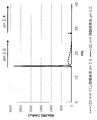
- FIG. 3 shows the case of using an acetate buffer or citrate buffer at pH 3.0 as an eluent when purifying Fab fragments containing VL- ⁇ using a prototype affinity separation matrix to which mutant protein L is immobilized. This is an elution profile.
- a pH 2.4 citrate buffer is also used to dissociate the remaining Fab fragments in the matrix.
- FIG. 4 shows a case where a commercially available affinity separation matrix on which protein L is immobilized is used to purify a Fab fragment containing VL- ⁇ , and an acetate buffer or a citrate buffer having a pH of 3.5 is used as an eluent. It is the figure which compared the elution profile.
- FIG. 5 shows a comparison of elution profiles when using a commercially available affinity separation matrix on which protein A is immobilized, and using an acetate buffer or citrate buffer having a pH of 3.0 as the eluent when purifying IgG. It is.
- a pH 2.4 citrate buffer is also used to dissociate the remaining IgG in the matrix.
- FIG. 6 is a diagram comparing elution profiles when using a commercially available affinity separation matrix on which protein A is immobilized and purifying IgG, using an acetic acid buffer solution or a citrate buffer solution at pH 3.5 as an eluent. It is.
- a pH 2.4 citrate buffer is also used to dissociate the remaining IgG in the matrix.
- the method of the present invention is a method for purifying antibodies and antibody fragments containing VL- ⁇ well without damaging them using an affinity separation matrix in which protein L, its domain, or a variant thereof is immobilized.
- the method of the present invention will be described step by step.
- Step 1 Adsorption step In this step, an affinity separation in which a liquid sample containing at least one of an antibody and a VL- ⁇ -containing antibody fragment is immobilized on an insoluble carrier as a protein L, its domain, or a variant thereof is used as a ligand.
- the antibody and / or antibody fragment is adsorbed to the affinity separation matrix by contacting with the matrix.
- Immunoglobulin (Ig) is a glycoprotein produced by B cells of lymphocytes and has a function of recognizing and binding molecules such as specific proteins.
- An immunoglobulin has a function of specifically binding to a specific molecule called an antigen and a function of detoxifying or removing a factor having the antigen in cooperation with other biomolecules or cells.
- Immunoglobulin is generally called “antibody”, which is a name that focuses on such a function.
- All immunoglobulins basically have the same molecular structure, and have a “Y” -shaped four-chain structure as a basic structure.
- the four-chain structure is composed of two polypeptide chains each called a light chain and a heavy chain.
- Immunoglobulin G is a monomeric immunoglobulin and is composed of two ⁇ chains and two light chains, and has two antigen-binding sites.
- the place corresponding to the vertical bar of the lower half of the “Y” of immunoglobulin is called the Fc region, and the “V” of the upper half is called the Fab region.
- the Fc region has an effector function that induces a reaction after the antibody binds to the antigen, and the Fab region has a function of binding to the antigen.
- the heavy chain Fab region and the Fc region are connected by a hinge part, and the proteolytic enzyme papain contained in papaya decomposes this hinge part and cleaves it into two Fab regions and one Fc region.
- the portion near the tip of the “Y” in the Fab region is called a variable region (V region) because various changes in the amino acid sequence are seen so that it can bind to various antigens.
- the variable region of the light chain is called the VL region, and the variable region of the heavy chain is called the VH region.
- the Fab region and the Fc region other than the V region are regions with relatively little change, and are called constant regions (C regions).
- the constant region of the light chain is referred to as the CL region, and the constant region of the heavy chain is referred to as the CH region.
- the CH region is further divided into three, CH1 to CH3.
- the heavy chain Fab region consists of a VH region and CH1, and the heavy chain Fc region consists of CH2 and CH3.
- the hinge part is located between CH1 and CH2.
- PpL binds to a variable region (VL- ⁇ ) in which the light chain is a ⁇ chain (Non-Patent Documents 5 to 7).
- the ligand of the affinity separation matrix according to the present invention is based on the sequence of protein L (PpL) and binds to the ⁇ chain variable region (VL- ⁇ ) of immunoglobulin.
- the antibody to be bound by the affinity separation matrix according to the present invention is not limited as long as it contains VL- ⁇ , and may be IgG containing the Fab region and the Fc region without deficiency, such as IgM, IgD, and IgA. Other Igs may also be used, or they may be derivatives of immunoglobulin molecules that have been modified by protein engineering.
- the antibody fragment to which the affinity separation matrix used in the present invention binds is not particularly limited as long as it is an antibody fragment having VL- ⁇ .
- Fab fragments fragmented only in the Fab region of immunoglobulin G, scFv and diabody consisting of only the variable region of immunoglobulin G, and partial domains of human immunoglobulin G as immunoglobulin G domains of other species examples thereof include chimeric immunoglobulin G fused by replacement, immunoglobulin G obtained by molecular modification of the sugar chain of the Fc region, and scFv fragment covalently bound to a drug.
- the liquid sample is not particularly limited as long as it contains at least one of the antibody to be purified and the antibody fragment containing VL- ⁇ , but the antibody and / or the antibody fragment containing VL- ⁇ is dissolved in an aqueous solvent.
- liquid samples include serum samples containing antibodies and / or VL- ⁇ -containing antibody fragments, bacterial cultures or homogenate supernatants containing VL- ⁇ -containing antibody fragments, and homogenates of monoclonal antibody-producing hybridomas.
- the pH of the liquid sample is preferably in the vicinity of neutrality of about 6 or more and 8 or less.
- the solvent of the body sample may be water alone, or may contain a water-miscible organic solvent such as C 1-4 alcohol as long as water is the main component, and the pH is 6 or more. A buffer solution of about 8 or less may be used.
- the affinity separation matrix used in the present invention is a protein L, its domain, or a variant thereof immobilized on an insoluble carrier as a ligand.
- protein L, a domain thereof, or a variant thereof may be collectively referred to as “VL- ⁇ binding peptide”.
- peptide includes all molecules having a polypeptide structure, and includes not only so-called proteins, but also fragments and those in which other peptides are linked by peptide bonds.
- a “domain” is a unit of protein conformation, consisting of a sequence of tens to hundreds of amino acid residues, sufficient to express some physicochemical or biochemical function.
- Protein L domain in the present invention refers to a protein showing affinity for VL- ⁇ .
- the “mutant” of protein L or domain is a protein or peptide in which at least one substitution, addition or deletion is introduced at the amino acid level with respect to the wild type protein L or domain sequence.
- the affinity to VL- ⁇ is at least maintained and preferably improved.
- the number of mutations is preferably 10 or less, more preferably 5 or less.
- Protein L is a protein derived from the cell wall of anaerobic gram-positive cocci belonging to the genus Peptostreptococcus.
- PpL derived from Peptostreptococcus magnus is preferable, and two types of PpL derived from Peptostreptococcus magnus 312 and Peptostreptococcus magnus 3316 are preferable, but are not limited thereto.
- PpL contains a plurality of VL- ⁇ binding domains consisting of 70 to 80 residues in a protein.
- the number of VL- ⁇ binding domains contained in PpL312 is five, and the number of VL- ⁇ binding domains contained in PpL3316 is four.
- the VL- ⁇ binding domain of PpL312 is called B1-5 domain in order from the N terminus, and the VL- ⁇ binding domain of PpL3316 is called C1-4 domain in order from the N terminus (Non-patent Documents 5-6).
- Non-patent Document 7 Studies have shown that about 20 residues at the N-terminus of the VL- ⁇ -binding domain of PpL do not have a specific secondary structure. Even when the N-terminus is deleted, VL- ⁇ binding It retains a three-dimensional structure as a sex domain and exhibits VL- ⁇ binding.
- the VL- ⁇ binding peptide according to the present invention also includes, as one embodiment, two or more, preferably three or more, more preferably 4 VL- ⁇ binding peptides that are monomers or single domains. It may be a multimer of multiple domains linked by one or more, more preferably five or more. The upper limit of the number of domains to be linked includes 10 or less, preferably 8 or less, more preferably 6 or less. These multimers may be homodimers such as homodimers and homotrimers that are linked to a single VL- ⁇ -binding peptide, or are linked to multiple types of VL- ⁇ -binding peptides. Heteromultimers such as heterodimers and heterotrimers may be used.
- a method of linking the monomeric VL- ⁇ binding peptide includes a method of linking with one or a plurality of amino acid residues, but is not limited to this method.
- the number of amino acid residues to be linked is not particularly limited, but is preferably 20 residues or less, and more preferably 15 residues or less.
- the ligand of the affinity separation matrix of the present invention includes a VL- ⁇ binding peptide or a multimer in which two or more VL- ⁇ binding domains are linked as one constituent component.
- a fusion peptide characterized in that it is fused with another peptide having a different function.
- fusion peptides include, but are not limited to, peptides fused with albumin or GST (glutathione S-transferase).
- a nucleic acid such as a DNA aptamer, a drug such as an antibiotic, and a polymer such as PEG (polyethylene glycol) are fused
- the present invention can be used if the affinity separation matrix obtained in the present invention is useful. Included in the invention.
- the VL- ⁇ binding peptide used in the present invention can be prepared by a conventional method. That is, a DNA encoding the amino acid sequence of a desired VL- ⁇ binding peptide or a fragment thereof is chemically synthesized, and the DNA encoding the VL- ⁇ binding peptide is amplified by PCR and incorporated into a vector such as a plasmid. The obtained vector is cultured after infecting Escherichia coli or the like, and a desired VL- ⁇ binding peptide may be purified from the cultured cells or culture solution by chromatography or the like.
- the affinity separation matrix used in the present invention is obtained by immobilizing the above VL- ⁇ binding peptide on an insoluble carrier.
- the “insoluble carrier” used in the present invention is insoluble in an aqueous solvent that is a solvent of a liquid sample containing a VL- ⁇ binding peptide, and specifically binds to the ligand by supporting the ligand. Those that can be used to purify antibodies and antibody fragments.
- insoluble carrier used in the present invention examples include inorganic carriers such as glass beads and silica gel; synthetic polymers such as crosslinked polyvinyl alcohol, crosslinked polyacrylate, crosslinked polyacrylamide, and crosslinked polystyrene; crystalline cellulose, crosslinked cellulose, crosslinked agarose, crosslinked Examples thereof include organic carriers composed of polysaccharides such as dextran; and organic-organic and organic-inorganic composite carriers obtained by combining these.
- GCL2000 a porous cellulose gel
- Sephacryl S-1000 in which allyl dextran and methylene bisacrylamide are covalently crosslinked
- Toyopearl an acrylate carrier
- Sepharose CL4B an agarose crosslinking carrier
- Cellufine which is a cellulosic crosslinking carrier.
- the water-insoluble carrier in the present invention is not limited to these exemplified carriers.
- the insoluble carrier used in the present invention preferably has a large surface area in view of the purpose and method of use of the affinity separation matrix, and is preferably a porous material having a large number of pores of an appropriate size.
- the form of the carrier can be any of beads, monoliths, fibers, membranes (including hollow fibers), and any form can be selected.
- a conventional method may be used as a method for immobilizing a VL- ⁇ binding peptide, which is a ligand, on an insoluble carrier.
- the reactive groups present on the surface of the insoluble carrier are used for immobilization.
- reactive groups such as amino groups, hydroxyl groups, and carboxy groups exist on the surface of general insoluble carriers, and these are activated, substituted with other reactive groups, or reacted with them.
- a linker group having a functional group may be introduced. For example, epichlorohydrin, diglycidyl ether, 1,4-bis (2,3-epoxypropoxy) butane etc.
- the linker group is not particularly limited.
- An ester group (—C ( ⁇ O) —O— or —O—C ( ⁇ O) —), an amide group (—C ( ⁇ O) —NH— or —NH—C ( ⁇ O) —), Sulfoxide group (—S ( ⁇ O) —), sulfonyl group (—S ( ⁇ O) 2 —), sulfonylamide group (—NH—S ( ⁇ O) 2 — and —S ( ⁇ O) 2 —NH— ), And a group in which
- a spacer molecule composed of a plurality of atoms may be introduced between the ligand and the carrier, or the ligand may be directly immobilized on the carrier.
- the VL- ⁇ binding peptide according to the present invention may be chemically modified for immobilization.
- the liquid sample and the affinity separation matrix are contacted to selectively bind the antibody and / or antibody fragment containing VL- ⁇ to the VL- ⁇ binding peptide as a ligand.
- the specific embodiment is not particularly limited, and the liquid sample and the affinity separation matrix may only be mixed.
- the affinity separation matrix according to the present invention is packed in a column and affinity is used.
- a liquid sample is passed through the affinity column, and the antibody and / or antibody fragment is selectively adsorbed to the VL- ⁇ binding peptide.
- the conditions for this step may be appropriately adjusted within a range in which the antibody and / or antibody fragment contained in the liquid sample is sufficiently adsorbed to the affinity separation matrix.
- the temperature in this step may be adjusted to 4 ° C. or higher and 40 ° C. or lower.
- the temperature is 4 ° C. or higher, the liquid sample having an appropriate fluidity can be passed through the matrix without freezing the liquid sample.
- the temperature is 40 ° C. or lower, the antibody and / or antibody fragment contained in the liquid sample can be purified with a low possibility of undergoing heat denaturation.
- the said temperature can be easily adjusted by utilizing a column jacket etc., when a chromatography system is used.
- Step 2 Washing step of affinity separation matrix
- the affinity separation matrix on which the antibody and / or antibody fragment is adsorbed and retained in Step 1 is washed to remove impurities other than the antibody and / or antibody fragment.
- the antibody and / or antibody fragment is adsorbed to the affinity separation matrix.
- washing solution used for washing the affinity separation matrix in this step a washing solution that does not interfere with the interaction between the antibody and / or antibody fragment and the VL- ⁇ binding peptide is used.
- a washing solution that does not interfere with the interaction between the antibody and / or antibody fragment and the VL- ⁇ binding peptide
- water or a buffer solution having a pH of 6 or more and 8 or less can be used as the cleaning solution.
- What is necessary is just to adjust the usage-amount of a washing
- Step 3 Separation of antibody and / or antibody fragment
- the antibody and / or antibody fragment is separated from the affinity separation matrix adsorbed with the antibody and / or antibody fragment using an acetate buffer as an eluent. To do.
- the purified antibody and / or antibody fragment can be obtained.
- an acetate buffer is used as an eluent for eluting the antibody and / or antibody fragment containing VL- ⁇ .
- the acetate buffer can efficiently elute the antibody and / or antibody fragment even when the pH is relatively high, and can reduce damage to the antibody and / or antibody fragment due to the eluate.
- an acetate buffer as an eluent, the above antibody and / or antibody fragment is favorably dissociated from the VL- ⁇ binding peptide.
- the above antibody and / or antibody fragment The elution peak of becomes sharp. As a result, the amount of the solution containing the antibody and / or antibody fragment is reduced, and further purification and drying of the antibody and / or antibody fragment is facilitated.
- the acetic acid buffer is obtained, for example, by mixing an aqueous solution of a salt of acetic acid and a weak base such as an aqueous sodium acetate solution and an aqueous acetic acid solution at a ratio that provides a desired pH.
- the pH of the acetate buffer is preferably 2.5 or more and 4.0 or less. If the said pH is 2.5 or more, the chemical change of the said antibody and / or antibody fragment, etc. can be suppressed more reliably. On the other hand, if the pH is 4.0 or less, the elution of the antibody and / or antibody fragment can be made more reliable.
- the pH is preferably 2.8 or more, more preferably 3.0 or more, more preferably 3.8 or less, and even more preferably 3.5 or less.
- the ion concentration of the acetate buffer also affects the elution of the antibody and / or antibody fragment.
- concentration of acetate ions in the acetate buffer is preferably 10 mM or more and 500 mM or less.
- the acetate ion concentration is 10 mM or more, the effect of sharpening the elution peak of the antibody and / or antibody fragment is easily obtained.
- the acetate ion concentration is 500 mM or less, pH adjustment after obtaining the above-obtained antibody and / or antibody fragment elution fraction becomes easier.
- the acetate ion concentration is more preferably 50 mM or more, still more preferably 80 mM or more, more preferably 400 mM or less or 300 mM or less, still more preferably 200 mM or less, and particularly preferably 150 mM or less.
- the conditions of this step may be appropriately adjusted within a range in which the antibody and / or antibody fragment adsorbed on the affinity separation matrix is sufficiently separated and eluted.
- the temperature in this step may be adjusted to 4 ° C. or higher and 40 ° C. or lower.
- the eluate or the like can be passed through the matrix with appropriate fluidity without freezing.
- the temperature is 40 ° C. or lower, the antibody and / or antibody fragment can be eluted with a low possibility of undergoing heat denaturation.
- the elution peak of the antibody and / or antibody fragment becomes sharp, and the amount of acetate buffer used as the eluate can be reduced.
- the amount of the acetate buffer used may be adjusted as appropriate. For example, it can be set to 2 times or more and 20 times or less the volume of the affinity separation matrix. If the said ratio is 2 times volume or more, the said antibody and / or antibody fragment can be eluted more reliably. On the other hand, when the ratio is 20 times or less, the amount of the fraction containing the antibody and / or antibody fragment can be reduced, and further purification is facilitated.
- the reference volume of the affinity separation matrix is the volume of the affinity separation matrix in a gel state that is in a suspended state and is tapped or allowed to stand until the volume does not decrease.
- Step 4 Post-treatment step In step 3 above, an acetate buffer solution of the antibody and / or antibody fragment is obtained.
- the antibody and / or antibody fragment may be further purified by salting out, chromatography, recrystallization or the like, or may be dried by freeze drying, spray drying, thin film drying or the like.
- Step 5 Regeneration step of affinity separation matrix
- the affinity separation matrix from which the antibody and / or antibody fragment has been separated in Step 3 above is regenerated by washing with an alkaline aqueous solution.
- this step does not necessarily have to be performed after the above step 3, and may be performed once in 3 times, once in 5 times, or once in 10 times. Absent. That is, this step is not necessarily performed in a state where the performance of the affinity separation matrix such as the binding capacity is maintained, and the frequency and conditions of the execution are also determined depending on the liquid sample containing the antibody and / or antibody fragment to be purified. Different.
- the “alkaline aqueous solution” used for the regeneration of the affinity separation matrix is an aqueous solution showing alkalinity to such an extent that the purpose of washing or sterilization can be achieved. More specifically, a sodium hydroxide aqueous solution of 0.01 M or more and 1.0 M or less or 0.01 N or more and 1.0 N or less is applicable, but is not limited thereto.
- the lower limit of the concentration is preferably 0.01M, more preferably 0.02M, and even more preferably 0.05M.
- the upper limit of the concentration of sodium hydroxide is preferably 1.0M, more preferably 0.5M, even more preferably 0.3M, still more preferably 0.2M, and even more preferably 0.1M.
- the alkaline aqueous solution is not necessarily a sodium hydroxide aqueous solution, but the pH is preferably 12 or more and 14 or less. Regarding the lower limit of pH, 12.0 or more is preferable, and 12.5 or more is more preferable. Regarding the upper limit of the pH, it is preferably 14 or less, more preferably 13.5 or less, and even more preferably 13.0 or less.
- the time for treating the affinity separation matrix that has undergone Step 3 with an alkaline aqueous solution is not particularly limited and may be adjusted as appropriate because the damage to the peptide varies depending on the concentration of the alkaline aqueous solution and the temperature during the treatment.
- the concentration of sodium hydroxide is 0.05M and the temperature at the time of immersion is room temperature
- the lower limit of the time for immersion in the alkaline aqueous solution is preferably 1 hour, more preferably 2 hours, more preferably 4 hours.
- the time is more preferable, and 20 hours is more preferable, but there is no particular limitation as long as the affinity separation matrix can be regenerated.
- the affinity separation matrix regenerated through this step can be used again in the above steps 1 to 3.
- Example 1 Preparation of commercially available affinity separation matrix As an affinity separation matrix capable of adsorbing antibody fragments containing the ⁇ chain variable region (VL- ⁇ ), HI Healthcare's “HiTrap Protein L 1 mL-gel” was obtained and a chromatography system Installed. Note that “1 mL-gel” means that the volume of the affinity separation matrix in the gel state in which the affinity separation matrix in the suspended state is tapped or allowed to stand until the volume does not decrease is 1 mL.
- a base sequence encoding the peptide is designed, and an artificial synthetic gene of DNA having a PstI recognition site at the 5 ′ end and an XbaI recognition site at the 3 ′ end is supplied by Eurofin Genomics Total synthesis by outsourcing to
- the expression plasmid after this subcloning was digested with restriction enzymes PstI and XbaI (Takara Bio), and the obtained DNA fragment was ligated to Brevibacillus expression vector pNCMO2 (Takara Bio) digested with the same restriction enzymes, Expression plasmids in which DNA encoding the amino acid sequence of the VL- ⁇ binding peptide was inserted into the Brevibacillus expression vector pNCMO2 were prepared.
- the ligation reaction was performed using a ligation reagent ("Ligation high" manufactured by TOYOBO) according to the protocol attached to the product, and Escherichia coli JM109 strain (Takara Bio) was used for plasmid preparation. .
- a ligation reagent ("Ligation high” manufactured by TOYOBO) according to the protocol attached to the product, and Escherichia coli JM109 strain (Takara Bio) was used for plasmid preparation. .
- Each expression plasmid DNA base sequence was confirmed using a DNA sequencer 3130xl Genetic Analyzer (Applied Biosystems). BigDye Terminator v. 1.1 Using a Cycling Sequencing Kit (Applied Biosystems) according to the attached protocol, each plasmid DNA was subjected to a sequencing PCR reaction, and the sequencing product was converted into a plasmid purification kit (Applied Biosystems, “BigDye XT Terminator Kit”). )) According to
- Brevibacillus choshinensis strain SP3 (Takara Bio Inc.) was transformed with the obtained plasmid, and a gene recombinant that secreted and produced the VL- ⁇ binding peptide was bred.
- This gene recombinant was added to 30 mL of A medium (polypeptone 3.0%, yeast extract 0.5%, glucose 3%, magnesium sulfate 0.01%, iron sulfate 0.001%, containing 60 ⁇ g / mL neomycin.
- the cells were cultured with shaking in manganese chloride (0.001%, zinc chloride 0.0001%) at 30 ° C. for 3 days. After the culture, the cells were separated by centrifuging the culture solution at 15,000 rpm and 25 ° C. for 5 minutes.
- VL- ⁇ binding peptide was purified by cation exchange chromatography using a cation exchange carrier (“UnoSphere S” manufactured by Bio-Rad). UnoSphere S was packed into a column (“Tricorn 10/200” manufactured by GE Healthcare Bioscience) and used.
- cation exchange buffer A 50 mM CH 3 COOH—CH 3 COONa, pH 4 0.0
- cation exchange buffer B 50 mM CH 3 COOH—CH 3 COONa, 1M
- the VL- ⁇ binding peptide was purified by anion exchange chromatography using an anion exchange carrier (“Nuvia Q” manufactured by Bio-Rad). Nuvia Q was packed into a column (“Tricorn 10/200” manufactured by GE Healthcare Bioscience) and used. Specifically, the fractionated VL- ⁇ binding peptide solution is dialyzed against anion exchange buffer A (50 mM Tris-HCl, pH 8.0) and equilibrated with anion exchange buffer A. The anion exchange buffer A and the anion exchange buffer B (50 mM Tris-HCl, 1.0 M NaCl, pH 8.0) were used after adding to the Nuvia Q column and washing with anion exchange buffer A.
- anion exchange buffer A 50 mM Tris-HCl, 1.0 M NaCl, pH 8.0
- VL- ⁇ -binding peptide eluted in the middle was fractionated with a salt concentration gradient.
- the separated VL- ⁇ binding peptide was dialyzed against ultrapure water, and an aqueous solution containing only the VL- ⁇ binding peptide was used as a final purified sample.
- protein purification by chromatography using the above-described column was performed using a chromatography system (“AKTA york 25 system” manufactured by GE Healthcare Bioscience).
- the prepared VL- ⁇ binding peptide was immobilized on a commercially available agarose carrier.
- N- [ ⁇ -Maleimidocaic acid] hydrazide.TFA (EMCH, Thermo Fisher Scientific) solution adjusted with a coupling buffer and adjusted to a concentration of 10 mM is added to a centrifuge tube containing a carrier, and the solution is mixed with 1 Reacted for hours. Thereafter, the carrier is transferred to a glass filter, 10 mL of washing buffer A (0.5 M ethanolamine, 0.5 M sodium chloride, pH 7.2), 10 mL of coupling buffer, and 10 mL of washing buffer A in this order. Was washed and allowed to stand at 25 ° C. for 15 minutes. Further, the carrier was washed with a coupling buffer (10 mL). The maleimide was provided to the support by the above operations.
- washing buffer A 0.5 M ethanolamine, 0.5 M sodium chloride, pH 7.2
- the carrier provided with maleimide was transferred to a centrifuge tube, a VL- ⁇ binding peptide solution was further added, and the carrier was reacted at 25 ° C. for 2 hours. Thereafter, the reacted carrier was transferred to a glass filter, and washed with 7 mL of coupling buffer to recover the unreacted VL- ⁇ binding peptide. Thereafter, 10 mL of washing buffer B (50 mM L-cysteine, 100 mM NaH 2 PO 4 -Na 2 HPO 4 , 0.5 M sodium chloride, pH 7.2), 10 mL of coupling buffer, and 10 mL of washing buffer B After washing the carrier in order, it was allowed to stand at 25 ° C. for 15 minutes.
- washing buffer B 50 mM L-cysteine, 100 mM NaH 2 PO 4 -Na 2 HPO 4 , 0.5 M sodium chloride, pH 7.2
- the absorbance at 280 nm of the collected unreacted VL- ⁇ binding peptide was measured with a spectrometer, and the amount of unreacted VL- ⁇ binding peptide was calculated from the extinction coefficient calculated from the amino acid sequence.
- the amount of immobilized VL- ⁇ -binding peptide was calculated from the difference between the charged amount of VL- ⁇ -binding peptide and the amount of unreacted VL- ⁇ -binding peptide quantified, and the ligand density was calculated from the volume of the carrier.
- the ligand density of prototype A was 10 mg / mL-gel.
- VL- ⁇ binding domain variants having the amino acid sequence of SEQ ID NO: 3 were linked with the interdomain connecting peptide contained in SEQ ID NO: 1, and a carrier-immobilizing active group was added to the C-terminus.
- a VL- ⁇ binding peptide was designed and prepared in the same manner as described above to prepare an affinity separation matrix (prototype B) immobilized on a carrier.
- the ligand density of prototype B was 12.5 mg / mL-gel.
- a 1 mL-gel portion of each of the prototype A and B affinity separation matrices was packed into a commercially available column (“Tricorn 5/50” manufactured by GE Healthcare).
- IgG-derived Fab fragment Fab fragment (IgG-Fab) was selected as a VL- ⁇ -containing peptide.
- a humanized monoclonal IgG preparation having VL- ⁇ was used as a raw material, and this was fragmented into a Fab fragment and an Fc fragment with papain, and only the Fab fragment was separated and purified.
- a preparation method of Fab derived from an anti-IgE monoclonal antibody (generic name “omalizumab”) is shown, but basically other monoclonal Fabs can be prepared by the same method.
- a humanized monoclonal IgG preparation (in the case of an anti-IgE monoclonal antibody, “Zolea” manufactured by Novartis Pharma) was added to a papain digestion buffer (0.1 M AcOH-AcONa, 2 mM EDTA, 1 mM cysteine, The solution was dissolved in pH 5.5), papain-immobilized agarose (“Papain Agarose from papalatex” manufactured by SIGMA) was added, and the mixture was incubated at 37 ° C. for about 8 hours while mixing with a rotator.
- a papain digestion buffer 0.1 M AcOH-AcONa, 2 mM EDTA, 1 mM cysteine
- papain-immobilized agarose (“Papain Agarose from papalatex” manufactured by SIGMA) was added, and the mixture was incubated at 37 ° C. for about 8 hours while mixing with a rotator.
- the Fab solution was recovered as a flow-through fraction by affinity chromatography using KANEK KanCapA TM column (Kaneka).
- the fractionated Fab solution was purified by gel filtration chromatography using a Superdex 75 10/300 GL column (standard buffer was used for equilibration and separation) to obtain a Fab solution. Chromatographic protein purification was performed using the AKTA york 25 system.
- Fab was eluted with a 100 mM acetate buffer with a linear pH gradient from pH 5.0 to pH 3.0.
- acetate buffer A 100 mM acetate buffer, pH 5.0
- acetate buffer B 100 mM acetate buffer
- the elution pH was evaluated from the Fab elution position in the step of increasing the concentration of the buffer solution (pH 3.0) linearly from 0% to 100%.
- the Fab elution pH when using a citrate buffer as a reference was adjusted to 20 CV after equilibrating the column with 5 CV citrate buffer A (100 mM citrate buffer, pH 5.0) as described above.
- prototype A As shown in Table 1, in any of the commercially available products, prototype A, and prototype B, 0.6 or more in the case of using the acetate buffer compared to the case of using the citrate buffer, It was found that the Fab elution pH was significantly increased. Since both the commercial product, prototype A and prototype B have the same tendency, the effect of improving the Fab elution pH by the acetate buffer is effective for the affinity separation matrix having both protein L and protein L variants as ligands. It is believed that there is.
- Example 2 (1) Elution experiment using pH 3.0 eluate An experiment was carried out in which Fab was adsorbed on an affinity separation matrix capable of adsorbing antibody fragments containing VL- ⁇ and then eluted.
- an affinity separation matrix capable of adsorbing antibody fragments containing VL- ⁇ as in Example 1 above, commercially available products (“HiTrap Protein L” manufactured by GE Healthcare) and prototype A and prototype B columns were used as AKTA school. 25. The following experiment was conducted at 25 ° C.
- FIG. 1 shows a chromatographic chart when an acetate buffer solution and a citrate buffer solution are used as eluents in a commercially available affinity separation matrix.
- citrate buffer When citrate buffer is used, Fab is almost completely eluted with 7.6 CV eluate, whereas when acetate buffer is used, Fab is almost completely eluted with 1.2 CV eluate. Eluted.
- Fig. 2 shows the results of the affinity separation matrix of prototype A.
- citrate buffer was used, Fab was not completely eluted even when the eluate was flowed at 10 CV, and Fab was eluted in the strongly washed fraction.
- Fab was eluted almost completely by flowing 1.5 CV of the eluate.
- Fig. 3 shows the result of the affinity separation matrix of prototype B.
- citrate buffer was used, Fab was not almost completely eluted even when the eluate was flowed at 10 CV, and Fab was eluted in the strongly washed fraction.
- Fab was almost completely eluted by flowing 1.2 CV of the eluate.
- Comparative Example 1 As a comparative experimental example, an antibody elution test was conducted using an acetate buffer solution and a citrate buffer solution as an eluent in another affinity separation matrix.
- affinity separation matrix a commercially available Protein A carrier (“HiTrap MabSelect SuRe” manufactured by GE Healthcare) (1 mL) generally used in antibody purification was used. Since Protein A carrier hardly adsorbs Fab, IgG was used as an antibody for the test.
- the aforementioned humanized monoclonal IgG preparation (anti-IgE monoclonal antibody, “Zolea” manufactured by Novartis Pharma) was used.
- Example 1 an antibody elution test was conducted in the same manner as in Example 1 (4) except that the above commercially available Protein A carrier was used instead of the Protein L carrier. Specifically, an AKTA york 25 system (GE Healthcare) was used as a chromatographic system, and a column packed with a commercially available Protein A carrier was connected. 1 mg of humanized monoclonal IgG was added to a column equilibrated with an equilibration buffer (20 mM NaH 2 PO 4 -Na 2 HPO 4 , 150 mM NaCl, pH 7.4).
- an equilibration buffer (20 mM NaH 2 PO 4 -Na 2 HPO 4 , 150 mM NaCl, pH 7.4
- IgG was eluted with a 100 mM acetate buffer with a pH linear gradient from pH 5.0 to pH 3.0. More specifically, after equilibrating the column with 5 CV of acetate buffer A (100 mM acetate buffer, pH 5.0), when passing 20 CV of buffer solution, acetate buffer B (100 mM acetate buffer). The elution pH was evaluated from the IgG elution position in the step of increasing the concentration of the buffer solution (pH 3.0) linearly from 0% to 100%.
- the IgG elution pH when using a reference citrate buffer was 20 CV after equilibrating the column with 5 CV citrate buffer A (100 mM citrate buffer, pH 5.0) as described above. Elution pH from the IgG elution position in the step of linearly increasing the concentration of citrate buffer B (100 mM citrate buffer, pH 2.4) from 0% to 100%. Evaluated. All the above operations were performed at a flow rate of 0.3 mL / min. The results of elution pH of the obtained humanized monoclonal IgG are summarized in Table 2.
- FIG. 5 shows a chromatograph chart in the case where an acetate buffer and a citrate buffer are used as eluents in a commercially available Protein A carrier. As shown in the results of FIG. 5, the elution curves were almost completely the same when citrate buffer was used and when acetate buffer was used. In either case, the elution solution was 1.2 CV. IgG was almost completely eluted, and there was no difference in the amount of eluate due to the difference between acetate buffer and citrate buffer.
- Comparative Example 3 After IgG was adsorbed on a commercially available Protein A carrier, an experiment was conducted to elute IgG. The pH of the eluate was changed from that in Comparative Example 2 above. A column packed with a commercially available Protein A carrier was connected to AKTA van 25. The following experiment was conducted at 25 ° C. As in Comparative Example 2, 3 mg of humanized monoclonal IgG was added to a column equilibrated with an equilibration buffer (20 mM NaH 2 PO 4 -Na 2 HPO 4 , 150 mM NaCl, pH 7.4).
- an equilibration buffer (20 mM NaH 2 PO 4 -Na 2 HPO 4 , 150 mM NaCl, pH 7.4
- the equilibration buffer was allowed to flow through the column at 10 CV (column volume), and then 100 mM acetate buffer (pH 3.5) was allowed to flow at 10 CV as the eluent. Further, 3 CV of equilibration buffer was flowed, and then 10 CV of strong washing solution (100 mM citrate buffer, pH 2.4) was flowed to elute IgG remaining on the column. Finally, 5 CV of equilibration buffer was flowed to complete the experiment. As a reference, an experiment using 100 mM citrate buffer pH 3.5 as an eluent was also conducted. FIG.
- FIG. 6 shows a chromatographic chart when an acetate buffer and a citrate buffer are used as eluents in a commercially available Protein A carrier.
- acetate buffer was used, IgG was almost completely eluted with 1.3 CV eluate, and when citrate buffer was used, IgG was almost completely eluted with 1.9 CV.
- citrate buffer was used, IgG was almost completely eluted with 1.9 CV.
- the amount of eluate was different between the acetate buffer and the citrate buffer, but the difference was small.
- the elution fraction amount is 0.6 CV when the acetate buffer is used as the eluate compared to the case where the citrate buffer is used as the eluate.
- the amount of elution fraction was reduced by only 32%.
Landscapes
- Chemical & Material Sciences (AREA)
- Organic Chemistry (AREA)
- Health & Medical Sciences (AREA)
- General Health & Medical Sciences (AREA)
- Biochemistry (AREA)
- Biophysics (AREA)
- Life Sciences & Earth Sciences (AREA)
- Genetics & Genomics (AREA)
- Medicinal Chemistry (AREA)
- Molecular Biology (AREA)
- Proteomics, Peptides & Aminoacids (AREA)
- Analytical Chemistry (AREA)
- Immunology (AREA)
- Chemical Kinetics & Catalysis (AREA)
- Peptides Or Proteins (AREA)
Abstract
La présente invention concerne un procédé apte à purifier de manière favorable un anticorps ou un fragment d'anticorps qui comporte une région variable de chaîne κ, tout en produisant un pic d'élution net sans endommager l'anticorps ou le fragment d'anticorps comportant la région variable de chaîne κ, comme il est possible de maintenir le pH de l'éluat comparativement élevé. Ce procédé de purification d'un anticorps ou d'un fragment d'anticorps qui comporte une région variable de chaîne κ est caractérisé en ce qu'il implique : une étape d'adsorption de l'anticorps et/ou du fragment d'anticorps au niveau d'une matrice de séparation par affinité par mise en contact d'un échantillon liquide qui comporte l'anticorps et/ou le fragment d'anticorps avec une matrice de séparation par affinité dans laquelle une protéine (L), un domaine associé, ou un variant associé est apposé(e) au niveau d'un support insoluble comme ligand ; une étape de nettoyage de la matrice de séparation par affinité et d'élimination des impuretés autres que l'anticorps et/ou le fragment d'anticorps de la matrice de séparation ; et une étape de séparation de l'anticorps et/ou du fragment d'anticorps de la matrice de séparation par affinité au niveau de laquelle l'anticorps et/ou le fragment d'anticorps est adsorbé, à l'aide d'un tampon acétate.
Priority Applications (1)
| Application Number | Priority Date | Filing Date | Title |
|---|---|---|---|
| US16/183,308 US10844112B2 (en) | 2016-05-09 | 2018-11-07 | Method for purifying antibody or antibody fragment containing κ-chain variable region |
Applications Claiming Priority (2)
| Application Number | Priority Date | Filing Date | Title |
|---|---|---|---|
| JP2016-094178 | 2016-05-09 | ||
| JP2016094178 | 2016-05-09 |
Related Child Applications (1)
| Application Number | Title | Priority Date | Filing Date |
|---|---|---|---|
| US16/183,308 Continuation US10844112B2 (en) | 2016-05-09 | 2018-11-07 | Method for purifying antibody or antibody fragment containing κ-chain variable region |
Publications (1)
| Publication Number | Publication Date |
|---|---|
| WO2017195638A1 true WO2017195638A1 (fr) | 2017-11-16 |
Family
ID=60266999
Family Applications (1)
| Application Number | Title | Priority Date | Filing Date |
|---|---|---|---|
| PCT/JP2017/016812 Ceased WO2017195638A1 (fr) | 2016-05-09 | 2017-04-27 | Procédé de purification d'un anticorps ou d'un fragment d'anticorps comportant une région variable de chaîne κ |
Country Status (2)
| Country | Link |
|---|---|
| US (1) | US10844112B2 (fr) |
| WO (1) | WO2017195638A1 (fr) |
Families Citing this family (3)
| Publication number | Priority date | Publication date | Assignee | Title |
|---|---|---|---|---|
| JP2026503790A (ja) | 2023-02-06 | 2026-01-29 | サイティバ・バイオプロセス・アールアンドディ・アクチボラグ | 溶出条件の最適化 |
| CN121712792A (zh) | 2023-08-14 | 2026-03-20 | 思拓凡生物工艺研发有限公司 | Vh3结合分离基质和其使用方法 |
| WO2025131519A1 (fr) | 2023-12-21 | 2025-06-26 | Cytiva Bioprocess R&D Ab | Séparation d'anticorps avec une matrice de séparation de liaison vh3 |
Citations (2)
| Publication number | Priority date | Publication date | Assignee | Title |
|---|---|---|---|---|
| WO2000015803A1 (fr) * | 1998-09-14 | 2000-03-23 | Affitech As | Proteine de liaison de l'immunoglobuline |
| WO2014141150A1 (fr) * | 2013-03-15 | 2014-09-18 | Glaxosmithkline Intellectual Property (No.2) Limited | Procédés de purification d'anticorps |
Family Cites Families (32)
| Publication number | Priority date | Publication date | Assignee | Title |
|---|---|---|---|---|
| SE8505922D0 (sv) | 1985-12-13 | 1985-12-13 | Kabigen Ab | Construction of an igg binding protein to facilitate downstream processing using protein engineering |
| SE9201331D0 (sv) | 1992-04-28 | 1992-04-28 | Hightech Receptor C O Active | Protein l och hybridproteiner daerav |
| DE69329877T2 (de) | 1992-05-07 | 2001-04-26 | Affitech A/S, Oslo | Von L Protein abgeleitete Immunoglobulin-bindende Proteine und deren anwendungen |
| SE9503925D0 (sv) | 1995-11-07 | 1995-11-07 | Pharmacia Biotech Ab | Separationsmedium för IgG |
| GB9823071D0 (en) | 1998-10-21 | 1998-12-16 | Affibody Technology Ab | A method |
| CA2473144C (fr) | 2002-02-05 | 2013-05-28 | Genentech, Inc. | Purification de proteines |
| US7709209B2 (en) | 2002-03-25 | 2010-05-04 | Ge Healthcare Bio-Sciences Ab | Protein ligands |
| SE0200943D0 (sv) | 2002-03-25 | 2002-03-25 | Amersham Biosciences Ab | Mutant protein |
| EP3225625A1 (fr) | 2002-09-11 | 2017-10-04 | Chugai Seiyaku Kabushiki Kaisha | Procédé de purification de protéines |
| AU2003903317A0 (en) | 2003-06-27 | 2003-07-10 | Proteome Systems Intellectual Property Pty Ltd | Method of isolating a protein |
| GB0323238D0 (en) | 2003-10-03 | 2003-11-05 | Domantis Ltd | Synthetic LG binding domains of protein L |
| WO2005113584A1 (fr) | 2004-04-29 | 2005-12-01 | Board Of Regents, University Of Texas System | Methodes et compositions contenant des domaines liant les ig de la proteine l pour le ciblage specifique de cellule |
| CN1319988C (zh) | 2004-10-14 | 2007-06-06 | 上海润龙生物科技有限公司 | 一种高亲和力免疫球蛋白结合分子及其制备方法 |
| WO2006065208A1 (fr) | 2004-12-14 | 2006-06-22 | Ge Healthcare Bio-Sciences Ab | Purification d'immunoglobulines |
| US8728828B2 (en) | 2004-12-22 | 2014-05-20 | Ge Healthcare Bio-Sciences Ab | Purification of immunoglobulins |
| JP2006304633A (ja) | 2005-04-26 | 2006-11-09 | Apro Life Science Institute Inc | イムノグロブリン結合タンパク質 |
| US20080230478A1 (en) | 2005-05-24 | 2008-09-25 | Ge Healthcare Bio-Sciences Ab | Regeneration Of A Chromatography Matrix |
| CN101287503A (zh) * | 2005-08-03 | 2008-10-15 | Rq生物科技有限公司 | 用于诊断IgA和IgM介导的肾脏疾病的方法和组合物 |
| WO2007097361A1 (fr) | 2006-02-21 | 2007-08-30 | Protenova Co., Ltd. | Ligand presentant une affinite pour les immunoglobulines |
| JP4179517B2 (ja) | 2006-02-21 | 2008-11-12 | プロテノバ株式会社 | イムノグロブリン親和性リガンド |
| SG2013061528A (en) | 2008-08-14 | 2015-03-30 | Merck Sharp & Dohme | Methods for purifying antibodies using protein a affinity chromatography |
| WO2011118699A1 (fr) | 2010-03-24 | 2011-09-29 | 株式会社カネカ | Protéine capable de se lier spécifiquement à une immunoglobuline, et ligand ayant une affinité de liaison pour l'immunoglobuline |
| CN107915769A (zh) | 2011-03-29 | 2018-04-17 | 葛兰素史密斯克莱有限责任公司 | 用于蛋白纯化的缓冲液体系 |
| JPWO2015041218A1 (ja) | 2013-09-17 | 2017-03-02 | 株式会社カネカ | 新規抗体精製方法及びそれから得られる抗体(NovelAntibodyPurificationMethodandAntibodyobtainedtherefrom)、並びに陽イオン交換基を用いた新規抗体精製法及びそれから得られる抗体(NovelAntibodyPurificationmethodusingCationExchangerandAntibodyobtainedtherefrom) |
| SG11201602995VA (en) * | 2013-11-25 | 2016-05-30 | Seattle Genetics Inc | Preparing antibodies from cho cell cultures for conjugation |
| WO2015190458A1 (fr) | 2014-06-09 | 2015-12-17 | 株式会社カネカ | Procédé de production d'une protéine recombinante à l'aide de bactéries recombinantes du genre brevibacillus |
| US10435732B2 (en) | 2014-06-09 | 2019-10-08 | Kaneka Corporation | Method for producing recombinant proteins using recombinant brevibacillus |
| US20170334947A1 (en) | 2014-08-28 | 2017-11-23 | Kaneka Corporation | Affinity separation matrix for fab region-containing peptide |
| JP6369807B2 (ja) | 2014-10-21 | 2018-08-08 | 株式会社プロテイン・エクスプレス | プロテインl変異体 |
| JP7204086B2 (ja) | 2014-12-17 | 2023-01-16 | サイティバ・バイオプロセス・アールアンドディ・アクチボラグ | 改変κ軽鎖結合ポリペプチド |
| WO2016121701A1 (fr) | 2015-01-26 | 2016-08-04 | 株式会社カネカ | Matrice pour séparation par affinité destinée à la purification de protéines contenant la région variable de la chaîne kappa des immunoglobulines |
| GB201503578D0 (en) | 2015-03-03 | 2015-04-15 | Ge Healthcare Bio Sciences Ab | Sanitization method for affinity chromatography matrices |
-
2017
- 2017-04-27 WO PCT/JP2017/016812 patent/WO2017195638A1/fr not_active Ceased
-
2018
- 2018-11-07 US US16/183,308 patent/US10844112B2/en active Active
Patent Citations (2)
| Publication number | Priority date | Publication date | Assignee | Title |
|---|---|---|---|---|
| WO2000015803A1 (fr) * | 1998-09-14 | 2000-03-23 | Affitech As | Proteine de liaison de l'immunoglobuline |
| WO2014141150A1 (fr) * | 2013-03-15 | 2014-09-18 | Glaxosmithkline Intellectual Property (No.2) Limited | Procédés de purification d'anticorps |
Non-Patent Citations (1)
| Title |
|---|
| GE HEALTHCARE BIO- SCIENCE AB, August 2014 (2014-08-01), pages 1, Retrieved from the Internet <URL:https://www.gelifesciences.com/gehcls-images/GELS/Related%20Content/Files/1346418936586/litdoc29010008_20161015023003.pdf> [retrieved on 20170713] * |
Also Published As
| Publication number | Publication date |
|---|---|
| US10844112B2 (en) | 2020-11-24 |
| US20190211082A1 (en) | 2019-07-11 |
Similar Documents
| Publication | Publication Date | Title |
|---|---|---|
| CN103269761B (zh) | 亲和色谱基质 | |
| EP2831096B1 (fr) | Matrice de chromatographie par affinité | |
| CN102895960B (zh) | 包括基于金黄色葡萄球菌a蛋白的新配体的层析基质 | |
| US8859726B2 (en) | Affinity chromatography matrix | |
| CN103269762B (zh) | 亲和色谱基质 | |
| CN104640983A (zh) | 亲和分离基质用蛋白质配体 | |
| WO2015041218A1 (fr) | Nouveau procédé de purification d'anticorps et anticorps obtenu au moyen de ce procédé, nouveau procédé de purification d'anticorps à l'aide d'un échangeur de cations et anticorps obtenu au moyen de ce procédé | |
| JP6663360B2 (ja) | 変異型免疫グロブリンκ鎖可変領域結合性ペプチド | |
| CN107849088A (zh) | 抗体样蛋白质的纯化方法 | |
| US20190134606A1 (en) | Method for producing affinity separation matrix, and affinity separation matrix | |
| WO2016031902A1 (fr) | MATRICE DE SÉPARATION PAR AFFINITÉ POUR UN PEPTIDE CONTENANT UNE RÉGION Fab | |
| WO2016121701A1 (fr) | Matrice pour séparation par affinité destinée à la purification de protéines contenant la région variable de la chaîne kappa des immunoglobulines | |
| US10844112B2 (en) | Method for purifying antibody or antibody fragment containing κ-chain variable region | |
| US20190153072A1 (en) | Method for producing antibody fragment | |
| JP7620456B2 (ja) | κ鎖を含む抗体の製造方法 | |
| US20180215796A1 (en) | ANTIBODY-BINDING PROTEIN HAVING REDUCED ANTIBODY BINDING CAPACITY IN ACIDIC pH REGION | |
| WO2020066270A1 (fr) | PROCÉDÉ DE PRODUCTION D'UN ANTICORPS CONTENANT UNE RÉGION VARIABLE DE CHAÎNE κ ET/OU D'UN FRAGMENT D'ANTICORPS | |
| WO2016136910A1 (fr) | Peptide modifié se liant à la région fab | |
| US20180215795A1 (en) | ANTIBODY-BINDING PROTEIN HAVING REDUCED ANTIBODY-BINDING CAPACITY IN ACIDIC pH REGIONS | |
| WO2017191747A1 (fr) | Procédé de production d'une protéine comprenant une région variable de chaîne κ | |
| US20180215835A1 (en) | ANTIBODY-BINDING PROTEIN HAVING REDUCED ANTIBODY-BINDING CAPACITY IN ACIDIC pH REGION |
Legal Events
| Date | Code | Title | Description |
|---|---|---|---|
| NENP | Non-entry into the national phase |
Ref country code: DE |
|
| 121 | Ep: the epo has been informed by wipo that ep was designated in this application |
Ref document number: 17795999 Country of ref document: EP Kind code of ref document: A1 |
|
| 122 | Ep: pct application non-entry in european phase |
Ref document number: 17795999 Country of ref document: EP Kind code of ref document: A1 |
|
| NENP | Non-entry into the national phase |
Ref country code: JP |