WO2018131663A1 - Imaging method of ampa receptors in brain of primate organism, program, diagnostic agent, companion diagnostic agent, drug, screening method, input terminal, server and system - Google Patents
Imaging method of ampa receptors in brain of primate organism, program, diagnostic agent, companion diagnostic agent, drug, screening method, input terminal, server and system Download PDFInfo
- Publication number
- WO2018131663A1 WO2018131663A1 PCT/JP2018/000532 JP2018000532W WO2018131663A1 WO 2018131663 A1 WO2018131663 A1 WO 2018131663A1 JP 2018000532 W JP2018000532 W JP 2018000532W WO 2018131663 A1 WO2018131663 A1 WO 2018131663A1
- Authority
- WO
- WIPO (PCT)
- Prior art keywords
- brain
- data
- ampa receptor
- primate
- ampa
- Prior art date
- Legal status (The legal status is an assumption and is not a legal conclusion. Google has not performed a legal analysis and makes no representation as to the accuracy of the status listed.)
- Ceased
Links
- 0 *N(*)*C*C1=CCC(*C=C)C=C1 Chemical compound *N(*)*C*C1=CCC(*C=C)C=C1 0.000 description 2
- OQLLEHHZQQBUNU-UHFFFAOYSA-N NC(COc(c(F)cc(SCCN(C(C1=C2C=CCC1)=O)C2=O)c1)c1F)=O Chemical compound NC(COc(c(F)cc(SCCN(C(C1=C2C=CCC1)=O)C2=O)c1)c1F)=O OQLLEHHZQQBUNU-UHFFFAOYSA-N 0.000 description 1
- NOBYLSUZDAFFKP-UHFFFAOYSA-N NCCSc(cc1F)cc(F)c1OCC(N)=O Chemical compound NCCSc(cc1F)cc(F)c1OCC(N)=O NOBYLSUZDAFFKP-UHFFFAOYSA-N 0.000 description 1
Images






Classifications
-
- A—HUMAN NECESSITIES
- A61—MEDICAL OR VETERINARY SCIENCE; HYGIENE
- A61K—PREPARATIONS FOR MEDICAL, DENTAL OR TOILETRY PURPOSES
- A61K51/00—Preparations containing radioactive substances for use in therapy or testing in vivo
- A61K51/02—Preparations containing radioactive substances for use in therapy or testing in vivo characterised by the carrier, i.e. characterised by the agent or material covalently linked or complexing the radioactive nucleus
- A61K51/04—Organic compounds
- A61K51/0474—Organic compounds complexes or complex-forming compounds, i.e. wherein a radioactive metal (e.g. 111In3+) is complexed or chelated by, e.g. a N2S2, N3S, NS3, N4 chelating group
-
- A—HUMAN NECESSITIES
- A61—MEDICAL OR VETERINARY SCIENCE; HYGIENE
- A61K—PREPARATIONS FOR MEDICAL, DENTAL OR TOILETRY PURPOSES
- A61K51/00—Preparations containing radioactive substances for use in therapy or testing in vivo
- A61K51/02—Preparations containing radioactive substances for use in therapy or testing in vivo characterised by the carrier, i.e. characterised by the agent or material covalently linked or complexing the radioactive nucleus
- A61K51/04—Organic compounds
- A61K51/0404—Lipids, e.g. triglycerides; Polycationic carriers
- A61K51/0406—Amines, polyamines, e.g. spermine, spermidine, amino acids, (bis)guanidines
-
- A—HUMAN NECESSITIES
- A61—MEDICAL OR VETERINARY SCIENCE; HYGIENE
- A61K—PREPARATIONS FOR MEDICAL, DENTAL OR TOILETRY PURPOSES
- A61K51/00—Preparations containing radioactive substances for use in therapy or testing in vivo
- A61K51/02—Preparations containing radioactive substances for use in therapy or testing in vivo characterised by the carrier, i.e. characterised by the agent or material covalently linked or complexing the radioactive nucleus
- A61K51/04—Organic compounds
-
- A—HUMAN NECESSITIES
- A61—MEDICAL OR VETERINARY SCIENCE; HYGIENE
- A61P—SPECIFIC THERAPEUTIC ACTIVITY OF CHEMICAL COMPOUNDS OR MEDICINAL PREPARATIONS
- A61P25/00—Drugs for disorders of the nervous system
-
- A—HUMAN NECESSITIES
- A61—MEDICAL OR VETERINARY SCIENCE; HYGIENE
- A61P—SPECIFIC THERAPEUTIC ACTIVITY OF CHEMICAL COMPOUNDS OR MEDICINAL PREPARATIONS
- A61P25/00—Drugs for disorders of the nervous system
- A61P25/18—Antipsychotics, i.e. neuroleptics; Drugs for mania or schizophrenia
-
- G—PHYSICS
- G01—MEASURING; TESTING
- G01T—MEASUREMENT OF NUCLEAR OR X-RADIATION
- G01T1/00—Measuring X-radiation, gamma radiation, corpuscular radiation, or cosmic radiation
- G01T1/16—Measuring radiation intensity
- G01T1/161—Applications in the field of nuclear medicine, e.g. in vivo counting
-
- A—HUMAN NECESSITIES
- A61—MEDICAL OR VETERINARY SCIENCE; HYGIENE
- A61K—PREPARATIONS FOR MEDICAL, DENTAL OR TOILETRY PURPOSES
- A61K2121/00—Preparations for use in therapy
-
- A—HUMAN NECESSITIES
- A61—MEDICAL OR VETERINARY SCIENCE; HYGIENE
- A61K—PREPARATIONS FOR MEDICAL, DENTAL OR TOILETRY PURPOSES
- A61K2123/00—Preparations for testing in vivo
Definitions
- the present invention relates to a technique for imaging AMPA receptors in the brain of primate organisms.
- AMPA receptors are widely distributed in the central nervous system and are known to be involved in learning, memory, neurodegeneration, and cell death.
- Patent Documents 1 to 3 research on treatment of psychiatric / neurological diseases targeting AMPA receptors has been underway.
- Patent Documents 1 to 3 In order to investigate the relationship between AMPA receptors and these diseases, it is required to evaluate the expression level and distribution of AMPA receptors in the brain.
- AMPA receptor analysis techniques include microscopic observation at the synapse and spine level (antibody staining using an electron microscope, fluorescence observation using a two-photon microscope, and functional observation at a synapse using an electrophysiological technique).
- relatively macroscopic observations such as single molecule tracking method using quantum dot method and immunostaining method using slice are known.
- Patent Document 4 describes an example in which an AMPA receptor in a monkey living brain is imaged using a substance having an affinity for an AMPA receptor.
- the decrease in the probe-derived radiation detection value depending on the dose of the unlabeled competitor is not actually confirmed, and only radiation derived from the probe present in the brain can be detected at a certain time. That is, it has not been proved that radiation derived from a probe bound to an AMPA receptor can be detected and the AMPA receptor can really be imaged based on the detected radiation.
- the present invention has been made in view of the above circumstances, and an object of the present invention is to provide a technique for imaging the brain AMPA receptor of a primate organism and its application.
- a substance which is administered to a primate organism and selectively binds to the AMPA receptor in the brain of the primate organism and has a radioactive label is transferred into the brain to bind to the AMPA receptor in the brain, Detecting radiation emitted from the substance bound to the brain AMPA receptor to obtain data on the distribution and / or expression level of the AMPA receptor in the brain.
- AMPA receptor imaging method
- the detection is performed after the first time has passed after the substance has entered the brain and after the second time longer than the first time, The method according to (1) or (2), wherein data relating to the distribution and / or amount of the AMPA receptor in the brain is obtained based on each detected value.
- the substance is a compound of formula (I), or a pharmaceutically acceptable salt or solvate thereof.
- a and Z are each independently CO, SO or SO 2 ;
- X and Y are each independently S or O;
- R 1 to R 4 are each independently hydrogen, alkyl, alkenyl, alkynyl or halo;
- R 5 is independently for each occurrence alkyl, alkenyl, alkynyl or halo;
- n is an integer from 0 to 4;
- One or more atoms are radioisotopes of the atoms.
- a diagnostic agent for a disease associated with a primate brain AMPA receptor comprising a substance that selectively binds to a brain AMPA receptor in a primate living body and has a radioactive label, or the above-mentioned disease
- a companion diagnostic for the treatment or prevention of cancer comprising a substance that selectively binds to a brain AMPA receptor in a primate living body and has a radioactive label, or the above-mentioned disease
- a medicament for the treatment or prevention of diseases associated with cerebral AMPA receptors in the primate body A pharmaceutical comprising a substance that selectively binds to a brain AMPA receptor in a primate living body as an active ingredient, and is administered in a dosage plan based on the data obtained by any of the methods (1) to (4) .
- a screening method for a therapeutic or prophylactic agent for a disease associated with an AMPA receptor in the brain of a primate body A method comprising a step of selecting the candidate substance based on the difference in the data obtained by the method according to any one of (1) to (4) before and after administration of the candidate substance to the primate organism.
- An input terminal that transmits information on the distribution and / or expression level of AMPA receptors in the brain of a primate organism to a server.
- (11) a database storing data in which the distribution and / or expression level of AMPA receptors in the primate brain and the disease state associated with the AMPA receptors in the primate brain are associated; Means for querying the input information regarding the distribution and / or expression level of AMPA receptors in the brain of the subject's living body and the data, and transmitting information regarding the disease state of the subject to an output terminal; Server to provide.
- One embodiment of the present invention is a method for imaging AMPA receptors in the brain of a primate organism.
- This method comprises a step of transferring a substance having a radiolabel and selectively binding to a primate living brain AMPA receptor, which is administered to the primate living body, to the brain and binding to the brain AMPA receptor.
- a substance having a radiolabel is used that selectively binds to the AMPA receptor in the brain of the primate organism as in the examples described later, the substance is released from the substance bound to the AMPA receptor in the brain of the primate organism.
- the present inventors discovered for the first time that detected radiation can be detected, and established a methodology according to this embodiment.
- this embodiment based on this novel discovery is a process of acquiring data related to the distribution and / or expression level of the brain AMPA receptor by detecting the radiation emitted from the substance bound to the brain AMPA receptor.
- the distribution and / or expression level of the AMPA receptor in the entire brain of the primate organism can be grasped, and the brain AMPA receptor in the primate organism can be imaged.
- Imaging by radiation detection is not particularly limited, but includes molecular imaging such as positron emission tomography (PETRON), multiphoton imaging method, two-photon imaging method, near infrared fluorescence imaging method, autoradiography, and Single photon emission tomography (SPECT) may be used. Among these, PET imaging is preferable.
- PET imaging is preferable.
- Primates are not particularly limited, but may be humans and monkeys. It is considered that there are some differences between humans and monkeys in terms of substance metabolism, permeability of the blood brain barrier, and the amount and distribution of AMPA receptors in the brain. In this regard, although the examples described later relate to human data, the present inventors have confirmed that monkeys can be similarly imaged (data not shown).
- bonded with the AMPA receptor in a brain can be detected more frequently, and the imaging accuracy of an AMPA receptor increases.
- the required time is not particularly limited, and may be set in advance based on the substance used and statistics (typically, 10 minutes or more, 20 minutes or more, 30 minutes or more, 40 minutes or more after administration of the substance). Or may be set to be 45 minutes or longer.), Or may be set for each target living body. Specifically, the required time is calculated by applying (i) a mathematical analysis model, or (ii) the difference between the region where the target protein is present and the region where the target protein is not present is It may be the maximum time zone in which the variation is reduced, and (iii) the time zone in which the difference between the disease and the healthy person can be detected most clearly.
- the required time is not particularly limited, but may be set to be 110 minutes or less, 100 minutes or less, 90 minutes or less, 80 minutes or less, 70 minutes or less, or 60 minutes or less after administration of the substance.
- the detection is performed after the first time has passed after the substance has entered the brain and after the second time longer than the first time, and the brain AMPA is determined based on the detected values. It is preferred to obtain data relating to receptor distribution and / or quantity. More preferably, the radiation dose derived from the above-mentioned substance taken into each brain region is detected continuously or discontinuously between the first time and the second time, and the average value is calculated. Based on the disparity between brain regions, data regarding the distribution and / or amount of AMPA receptors in the brain can be obtained. Alternatively, a region in which the radiation detection value has changed (decreased) with a relatively small width between the lapse of the first time and the lapse of the second time is likely to correspond to the AMPA receptor. For this reason, the imaging accuracy of an AMPA receptor can be increased by using data based on a difference in detected values. If the first time and the second time are excessively long, the absolute amount of radiation is attenuated, and detection with high accuracy may be difficult.
- the first time and the second time are not particularly limited, and may be set in advance based on a substance to be used and statistics, or may be set for each target living body.
- the first time may typically be set to be 10 minutes or more, 20 minutes or more, 30 minutes or more, 40 minutes or more, or 45 minutes or more after administration of the substance, 110 minutes or less, 100 minutes or less. It may be set to be less than or equal to 90 minutes, less than or equal to 90 minutes, less than or equal to 80 minutes, less than or equal to 70 minutes, or less than or equal to 60 minutes, and may be determined in the same manner as the required time described above.
- the second time may be set to be typically 30 minutes to 150 minutes (specifically 60 minutes or less) after administration of the substance.
- the above substance is not particularly limited, but for example, after parenteral administration, intravenous administration, or intraperitoneal administration, it passes through the blood-brain barrier and moves to the brain. For this reason, it is necessary that the substance has a characteristic capable of passing through the blood-brain barrier. From this viewpoint, it is preferable that the substance has required blood solubility as well as required low molecular weight and fat solubility.
- the said substance may be a single substance, or the state carried
- the substance may be contained in a pharmaceutically acceptable carrier.
- the pharmaceutically acceptable carrier is not particularly limited, and examples thereof include sterile water, saline, physiological saline or phosphate buffered saline (PBS), sodium chloride injection, Ringer's injection, isotonic dextrose injection, Examples include sterile water injection solution, dextrose, and lactated Ringer's injection solution.
- the dose of the above substance may be appropriately set depending on the type of substance used; age, weight, health condition, sex and diet content of the subject to be administered; number of administrations, administration route, and the like.
- the administration of the substance is not particularly limited.
- the said substance is not specifically limited,
- the compound of following formula (I), or its pharmaceutically acceptable salt or solvate may be sufficient.
- a and Z are each independently CO, SO or SO 2 ;
- X and Y are each independently S or O;
- R 1 to R 4 are each independently hydrogen, alkyl, alkenyl, alkynyl or halo;
- R 5 is independently for each occurrence alkyl, alkenyl, alkynyl or halo;
- n is an integer from 0 to 4;
- One or more atoms are radioisotopes of the atoms.
- the radioisotope is selected from the group consisting of 15 O, 13 N, 11 C, 18 F and the like, but is not particularly limited. From the viewpoint of half-life, the radioisotope is preferably 11 C or 18 F.
- R 1 to R 4 is a group containing a radioisotope (eg, [ 11 C] alkyl (preferably 11 CH 3 ), [ 11 C ] Alkenyl, or [ 11 C] alkynyl, or 18 F).
- R 2 is preferably alkyl, more preferably both R 3 and R 4 are hydrogen, or each of R 3 and R 4 is independently alkyl.
- A is SO 2 , Z is CO, X is S, Y is O, R 2 is alkyl, R 1 is hydrogen, alkyl or halo. , R 1 is alkyl or halo, R 1 is in the para position, one of R 3 and R 4 is hydrogen, the other is alkyl, and R 5 is halo, especially fluoro, , R 5 is present in both ortho positions relative to the Y group (ie, both meta positions relative to the X group), n is 2 and one of R1-R4 contains a radioisotope Groups such as [ 11 C] alkyl (preferably 11 CH 3 ), [ 11 C] alkenyl, or [ 11 C] alkynyl, or 18 F are preferred.
- A is SO 2 , Z is CO, X is S, Y is O, R 2 is alkyl and R 1 is When hydrogen, alkyl or halo and R 1 is alkyl or halo, R 1 is in the para position, one of R 3 and R 4 is hydrogen, the other is alkyl, and R 5 is Halo, especially fluoro, wherein R 5 is present in both ortho positions relative to the Y group (ie both meta positions relative to the X group), n is 2 and R 1 to R 4 More preferably, one group contains a radioisotope (eg, [ 11 C] alkyl (preferably 11 CH 3 ), [ 11 C] alkenyl, or [ 11 C] alkynyl, or 18 F).
- a radioisotope eg, [ 11 C] alkyl (preferably 11 CH 3 ), [ 11 C] alkenyl, or [ 11 C] alkynyl, or 18 F).
- radioactive isotopes include the following compounds:
- alkyl means a monovalent group resulting from the loss of one hydrogen atom of an aliphatic saturated hydrocarbon.
- Alkyl is, for example, 1 to 15 (C 1 -C 15 ) carbon atoms, typically 1 to 10 (C 1 -C 10 ), 1 to 8 (C 1 -C 8 ), 1 Up to 6 (C 1 -C 6 ), 1 to 5 (C 1 -C 5 ), 1 to 4 (C 1 -C 4 ), 1 to 3 (C 1 -C 3 ), 1 to 2 Having (C 1 -C 2 ) or 2 to 6 (C 2 -C 6 ) carbon atoms.
- Alkyl may be linear or branched.
- alkyl examples include, but are not limited to, methyl, ethyl, propyl, isopropyl, 2-methyl-1-propyl, 2-methyl-2-propyl, 2-methyl-1-butyl, 3-methyl-1-butyl 2-methyl-3-butyl, 2,2-dimethyl-1-propyl, 2-methyl-1-pentyl, 3-methyl-1-pentyl, 4-methyl-1-pentyl, 2-methyl-2-pentyl 3-methyl-2-pentyl, 4-methyl-2-pentyl, 2,2-dimethyl-1-butyl, 3,3-dimethyl-1-butyl, 2-ethyl-1-butyl, n-butyl, isobutyl , T-butyl, pentyl, isopentyl, neopentyl, and hexyl.
- the alkyl may be further substituted with a suitable substituent.
- alkenyl refers to an aliphatic unsaturated hydrocarbon group having at least one double bond.
- Alkenyl is, for example, 2 to 15 (C 2 -C 15 ) carbon atoms, typically 2 to 10 (C 2 -C 10 ), 2 to 8 (C 2 -C 8 ), 2 ⁇ 6 (C 2 -C 6 ), 2 to 5 (C 2 -C 5 ), 2 to 4 (C 2 -C 4 ), 2 to 3 (C 2 -C 3 ), 3 to 6 (C 3 -C 6 ), 3-8 (C 3 -C 8 ), 4-6 (C 4 -C 6 ), 4-7 (C 4 -C 7 ), or 4-8 It has (C 4 -C 8 ) carbon atoms.
- Alkenyl may be linear or branched.
- Alkenyl may be further substituted with a suitable substituent.
- alkynyl refers to an aliphatic unsaturated hydrocarbon group having at least one triple bond.
- Alkynyl is, for example, 2 to 15 (C 2 -C 15 ) carbon atoms, typically 2 to 10 (C 2 -C 10 ), 2 to 8 (C 2 -C 8 ), 2 ⁇ 6 (C 2 -C 6 ), 2 to 5 (C 2 -C 5 ), 2 to 4 (C 2 -C 4 ), 2 to 3 (C 2 -C 3 ), 3 to 6 (C 3 -C 6 ), 3-8 (C 3 -C 8 ), 4-6 (C 4 -C 6 ), 4-7 (C 4 -C 7 ), or 4-8 It has (C 4 -C 8 ) carbon atoms.
- Alkynyl may be linear or branched.
- alkynyl include, but are not limited to, ethynyl (-C ⁇ CH), - C ⁇ CH (CH 3), - C ⁇ C (CH 2 CH 3), - CH 2 C ⁇ CH, -CH 2 C ⁇ C (CH 3 ), —CH 2 C ⁇ C (CH 2 CH 3 ), and the like.
- Alkynyl may be further substituted with a suitable substituent.
- halogen or “halo” means fluoro (—F), chloro (—Cl), bromo (—Br), and iodo (—I).
- pharmaceutically acceptable salt refers to a salt that is not harmful to mammals, particularly humans.
- Pharmaceutically acceptable salts can be formed with nontoxic acids or bases, including inorganic acids or bases, or organic acids or bases.
- examples of pharmaceutically acceptable salts include metal salts formed from aluminum, calcium, lithium, magnesium, potassium, sodium and zinc, or lysine, N, N ′′ -dibenzylethylenediamine, chloroprocaine, choline Organic salts formed from diethanolamine, ethylenediamine, meglumine (N-methylglucamine) and procaine.
- Pharmaceutically acceptable salts include acid addition salts and base addition salts.
- solvate means a solvent-containing compound formed by the association of one or more solvent molecules with the compound of the present invention.
- Solvates include, for example, monosolvates, disolvates, trisolvates, and tetrasolvates. Solvates include hydrates.
- a compound of formula (I) or a pharmaceutically acceptable salt or solvate thereof wherein R 2 is alkyl, alkenyl or alkynyl includes, for example, a compound of formula (II) below or a pharmaceutically acceptable salt or solvate thereof object: (Wherein, A, X, Y, Z , R 1, R 3, R 4, R 5, and n are the same as defined in the compounds of formula (I).)
- R 3 and R 4 in formula (I) and formula (II) are both hydrogen.
- R 2 is [ 11 C] alkyl, [ 11 C] alkenyl, or [ 11 C] alkynyl, preferably R 2 is [ 11 C] alkyl, in particular 11 CH 3 . .
- X 1 is I.
- a specific example of a compound of formula (II) is 2- [2,6-difluoro-4-( ⁇ 2-[(phenylsulfonyl) amino] ethyl ⁇ thio) phenoxy] acetamide (PEPA).
- the reaction can be carried out in a polar aprotic solvent such as dimethylformamide (DMF), tetrahydrofuran, acetonitrile, acetone or dimethyl sulfoxide.
- a polar aprotic solvent such as dimethylformamide (DMF), tetrahydrofuran, acetonitrile, acetone or dimethyl sulfoxide.
- the reaction is preferably performed under basic conditions using a base such as NaOH.
- the reaction temperature is preferably room temperature to reflux temperature, particularly preferably 60 to 100 ° C., more preferably 80 ° C.
- the reaction time is 1 to 10 minutes, in particular 5 minutes.
- PET probes usually have to be manufactured in a short time and in high yield due to the short half-life of radioisotopes. Since the reaction proceeds quantitatively in a short time, it is suitable for the production of a PET probe.
- the inventors have found that the reaction of the compound of formula (II) with X 1 -R 2 occurs quantitatively at the NH group adjacent to the A group of the compound of formula (II). Therefore, even if R 3 and R 4 are hydrogen, only the NH group can be converted to an NR 2 group without using a protecting group.
- a compound of formula (II) or a pharmaceutically acceptable salt or solvate thereof produces a compound of formula (I) or a pharmaceutically acceptable salt or solvate thereof wherein R 2 is alkyl, alkenyl or alkynyl. It can be used as an intermediate for Also, the compound of formula (II) or a pharmaceutically acceptable salt or solvate thereof is a radiolabeled formula wherein R 2 is [ 11 C] alkyl, [ 11 C] alkenyl, or [ 11 C] alkynyl. It can be used as an intermediate for producing a compound of (I) or a pharmaceutically acceptable salt or solvate thereof.
- a compound of formula (I), wherein R 1 is alkyl, alkenyl, or alkynyl, or a pharmaceutically acceptable salt or solvate thereof is, for example, a compound of formula (III) below, or a pharmaceutically acceptable salt thereof: Or solvate: Wherein A, X, Y, Z, R 2 , R 3 , R 4 , R 5 , and n are the same as defined above, and each R a is independently alkyl, alkenyl, Or is alkynyl) can be prepared by reacting with X 1 -R 1 , wherein R 1 is the same as defined above and X 1 is a halogen. In one embodiment, R a is all n-butyl.
- R 1 is [ 11 C] alkyl, [ 11 C] alkenyl, or [ 11 C] alkynyl, preferably R 1 is [ 11 C] alkyl, in particular 11 CH 3 . .
- X 1 is I.
- the reaction can be performed in the presence of a palladium catalyst, a phosphine ligand, a carbonate and a copper halide.
- the palladium catalyst include tris (dibenzylideneacetone) dipalladium.
- the phosphine ligand include tri (o-tolyl) phosphine and (di-tert-butyl) methylphosphine.
- the carbonate include K 2 CO 3 .
- Examples of the copper halide include CuCl.
- the reaction can be carried out in a polar aprotic solvent such as dimethylformamide (DMF), tetrahydrofuran, acetonitrile, acetone or dimethyl sulfoxide.
- the reaction temperature is preferably room temperature to reflux temperature, particularly preferably 60 to 100 ° C., more preferably 80 ° C.
- the reaction time is 1 to 10 minutes, in particular 5 minutes.
- PET probes usually have to be manufactured in a short time and in high yield due to the short half-life of radioisotopes. Since the reaction proceeds quantitatively in a short time, it is suitable for the production of a PET probe.
- a compound of formula (III) or a pharmaceutically acceptable salt or solvate thereof is a compound of formula (I) or a pharmaceutically acceptable salt or solvate thereof wherein R 1 is alkyl, alkenyl, or alkynyl. It can be used as an intermediate for manufacturing.
- a compound of formula (III) or a pharmaceutically acceptable salt or solvate thereof is a radiolabeled formula wherein R 1 is [ 11 C] alkyl, [ 11 C] alkenyl, or [ 11 C] alkynyl. It can be used as an intermediate for producing a compound of (I) or a pharmaceutically acceptable salt or solvate thereof.
- the compound of formula (I) or a pharmaceutically acceptable salt or solvate thereof can also be produced by the methods shown in the following examples.
- One embodiment of the present invention is a program for causing a computer to execute the above-described imaging method. Specifically, the computer controls the imaging apparatus according to the program to image the AMPA receptor in the brain of the primate organism.
- One embodiment of the present invention is a diagnostic agent for a disease associated with cerebral AMPA receptors in the primate body, or a companion diagnostic agent for treatment or prevention of a disease.
- the companion diagnostic agent for treatment is a diagnostic agent for determining whether treatment is expected when it is found that the brain AMPA receptor is a related disease.
- the companion diagnostic agent for prevention is found to be a disease related to the brain AMPA receptor, it is assumed that the disease state (prognosis) in the future or further progression is suppressed. It is a diagnostic agent to judge whether prevention is possible.
- an AMPA receptor function activator can be recommended for subjects whose decrease in expression level of AMPA receptor is known, and AMPA receptor can be recommended for subjects whose increase in expression amount of AMPA receptor is known.
- the administration of antagonists can be recommended. For example, even in a group of diseases clinically diagnosed with depression according to the current disease classification (DSM-V, ICD-10, etc.), there are cases in which increased expression of AMPA receptors is observed, and there is no change or decrease. There are some cases. In that case, even if the clinical diagnosis is depression, tailor-made diagnosis and treatment such as administration of an antagonist is performed for depression with increased AMPA receptor.
- the type of AMPA receptor function activator / AMPA receptor antagonist Prevention in cases where the expression level or distribution of the AMPA receptor correlates with the signs of symptom seizures, etc. To administer the drug automatically).
- one embodiment of the present invention is a medicament for treating or preventing a disease associated with a primate brain AMPA receptor, which selectively binds to a brain AMPA receptor in a primate organism.
- the present invention also relates to a medicine that is administered in a dosage schedule based on data on the distribution and / or expression level of AMPA receptors in the brain obtained by the above-described imaging method using a substance as an active ingredient.
- the AMPA receptor antagonist which is a medicine according to one embodiment (which may be used together with a companion diagnostic agent), is not particularly limited, but peranpanel hydrate (Eisai Co., Ltd.), talampane (Teva Co.) for epilepsy disease. Is mentioned.
- a and Z are each independently CO, SO, or SO 2 , and these groups are expected to show an interaction with the AMPA receptor.
- a and Z are each independently CO or SO 2 , more preferably, A is SO 2 and Z is CO.
- X and Y are each independently S or O, preferably X is S and Y is O.
- R 1 to R 4 are each independently hydrogen, alkyl, alkenyl, alkynyl or halo.
- R 1 -R 4 are hydrogen, ie, at least one of R 1 -R 4 is other than hydrogen.
- R 2 is alkyl.
- R 1 is alkyl or halo.
- R 1 can be present in any of the ortho, meta, or para positions.
- R 1 is in the para position.
- one of R 3 and R 4 is hydrogen and the other is alkyl.
- R 1 , R 3 and R 4 are each independently hydrogen, alkyl, alkenyl, alkynyl or halo and R 2 is alkyl, alkenyl or alkynyl.
- R 5 is each independently alkyl, alkenyl, alkynyl or halo for each occurrence.
- R 5 is halo, particularly preferably fluoro. More preferably, R 5 is present in both ortho positions relative to the Y group (ie both meta positions relative to the X group).
- n is an integer of 0-4. Preferably n is 2.
- the combinations of substituents in the compound of formula (I) are as follows: A and Z are each independently CO, SO or SO 2 , X and Y are each independently S or O R 1 , R 3 and R 4 are each independently hydrogen, alkyl, alkenyl, alkynyl or halo, R 2 is alkyl, alkenyl or alkynyl and R 5 is each independently for each occurrence, A combination wherein alkyl, alkenyl, alkynyl or halo, and n is an integer of 0 to 4 is preferable.
- the combinations of substituents in the compound of formula (I) include: A is SO 2 , Z is CO, X is S, Y is O, and R 2 is When it is alkyl, R 1 is hydrogen, alkyl or halo and R 1 is alkyl or halo, R 1 is in the para position, one of R 3 and R 4 is hydrogen and the other is alkyl R 5 is each independently alkyl, alkenyl, alkynyl or halo, and n is preferably a combination of 0 to 4 integers.
- the combinations of substituents in the compound of formula (I) include: A is SO 2 , Z is CO, X is S, Y is O, and R 2 is When alkyl, R 1 is hydrogen, alkyl or halo and R 1 is alkyl or halo, R 1 is in the para position, one of R 3 and R 4 is hydrogen and the other is A combination in which R 5 is halo, in particular fluoro, R 5 is in both ortho positions with respect to the Y group (ie both meta positions with respect to the X group) and n is 2 Is preferred.
- the combinations of substituents in the compound of formula (I) include: A is SO 2 , Z is CO, X is S, Y is O, and R 2 is When it is alkyl, R 1 is hydrogen, alkyl or halo and R 1 is alkyl or halo, R 1 is in the para position, R 3 and R 4 are both hydrogen, and R 5 is Each independently represents a combination of alkyl, alkenyl, alkynyl or halo, and n is an integer of 0 to 4.
- the AMPA receptor function activator / AMPA receptor antagonist can be administered orally or parenterally.
- Oral administration agents can be solid preparations such as powders, granules, capsules and tablets, or liquid preparations such as syrups and elixirs.
- parenteral administration agents injections (vein, muscle, etc.), rectal administration agents, external preparations for skin, and inhalants can be used. These preparations are produced according to a conventional method by adding a pharmaceutically acceptable production aid to the active ingredient. Furthermore, it is possible to obtain a sustained-release preparation by a known technique.
- Diseases associated with primate brain AMPA receptors may be, but are not limited to, psychiatric or neurological diseases, for example, (1) Depression, major depression, bipolar depression, mood disorders, affective disorders, recurrent depression, postpartum depression, stress disorder, depressive symptoms, mania, anxiety, generalized anxiety disorder, anxiety syndrome, Panic disorder, phobia, social phobia, social anxiety disorder, obsessive-compulsive disorder, post-traumatic stress syndrome, post-traumatic stress disorder, taurette syndrome, autism, fragile X syndrome, Rett syndrome, adjustment disorder, bipolar Disorder, neurosis, schizophrenia, chronic fatigue syndrome, anxiety, obsessive-compulsive disorder, panic disorder, epilepsy, irritability, attention deficit hyperactivity disorder, psychotic major depression, refractory major depression , Psychiatric disorders such as treatment-resistant depression (2) Alzheimer's disease, Alzheimer-type senile dementia, Parkinson's disease, Huntington's chorea, multiple cerebral infarction dementia, frontotemporal dementia, Parkinson's fronto
- One embodiment of the present invention is a medicament for the treatment or prevention of diseases associated with cerebral AMPA receptors in the primate body.
- the above diseases are not particularly limited, but are selected from the group consisting of epilepsy, depression, schizophrenia, cerebral ischemia, Parkinson's disease, Alzheimer's disease, autism, attention hyperactivity disorder (ADHD) and multiple sclerosis It may be one or more.
- the drug according to this embodiment can include a pharmaceutically acceptable carrier.
- the pharmaceutically acceptable carrier is not particularly limited.
- One embodiment of the present invention is a screening method for a therapeutic or prophylactic agent for a disease associated with a primate brain AMPA receptor. This method includes a step of selecting candidate substances based on the difference in data regarding the distribution and / or expression level of AMPA receptors in the brain obtained by the above-described imaging method before and after administration of the candidate substance to a primate organism. .
- the candidate substance can be selected as an AMPA receptor function activator / AMPA receptor antagonist.
- the above-mentioned diseases that have conventionally been grouped are finely classified according to the expression level or distribution of brain AMPA receptors, and appropriate therapeutic or preventive drugs are prepared for each classification.
- the selected candidate substance actually has a therapeutic or prophylactic effect for a disease associated with a primate brain AMPA receptor (animal experiment, human test, etc.). Further, selection may be made based on the fact that the above-mentioned effect is actually provided.
- a system includes an input terminal, a server, and an output terminal.
- the input terminal transmits information on the distribution and / or expression level of AMPA receptors in the brain of the primate organism to the server. This information can be acquired by the imaging method of the present invention described above.
- the server stores data in which the distribution and / or expression level of AMPA receptors in the primate brain and the disease state associated with the AMPA receptors in the primate brain are associated with each other.
- the server inquires the input information on the distribution and / or expression level of AMPA receptors in the brain of the subject's living body and the data in the database, and generates information on the disease state of the subject (information creation unit) And means for transmitting this information to the output terminal.
- a system including this server can accurately present the state of the disease of the subject.
- the disease state includes, for example, the presence or absence of disease, seriousness, and seizure possibility.
- the information creation unit can select data that is the same as or similar to the distribution and / or expression level of the AMPA receptor in the brain of the subject from the database, and generate information related to the disease state.
- the server includes a distribution and / or expression level of a primate brain AMPA receptor, a disease state associated with the primate brain AMPA receptor, and a previously administered primate body.
- a database that stores data associated with the type, dose, and / or usage of a drug for the treatment or prevention of a disease associated with a brain AMPA receptor in the body.
- the server inquires the input information regarding the distribution and / or expression level of AMPA receptors in the brain of the subject's living body, and information on the recommended type, dose and / or usage of the drug to the subject. (Information creation unit), and further includes means for transmitting this information to the output terminal.
- a system including this server can recommend an appropriate medicine type, dose and / or usage depending on the subject. The specific embodiment may be the same as described above for the companion diagnostic agent and the medicine.
- the database of the server is fed back with information about the prevention or treatment outcome of the subject by administration according to the recommended type, dose and / or usage of the recommended medicine, and the subject's living brain. It is associated with information regarding the distribution and / or expression level of the internal AMPA receptor. This updates the database and further increases the recommended accuracy.
- the output terminal is an output terminal that outputs information transmitted from the server.
- the system 1000 (not shown) of the present invention includes an input terminal 200, a server 300, and an output terminal 500.
- the input terminal 200 has a function of transmitting information related to the distribution and / or expression level of AMPA receptors in the brain of the primate organism to the server. Specifically, an imaging data generation unit 210, a metadata generation unit 220, a synthesis unit 250, and a transmission unit 223 are provided.
- the input terminal 200 receives an output transmitted from the molecular imaging apparatus 100 such as PET, multiphoton imaging method, two-photon imaging method, near infrared fluorescence imaging method, autoradiography, SPECT, etc. as a receiving terminal (not shown). ) To the imaging data generation unit 210.
- the input data is data acquired by the imaging method of the present invention.
- the molecular imaging apparatus 100 will be described as PET.
- the imaging data generation unit 210 is connected to the molecular imaging apparatus 100 outside the system 1000, receives image data continuously or intermittently, performs a two-stage data conversion process, and receives the brain AMPA receptor.
- the data relating to the distribution and / or expression level is generated (the generated data is hereinafter referred to as imaging data).
- the first data conversion is data conversion for converting the direct output of the molecular imaging apparatus 100 into coordinates.
- the direct output of the molecular imaging apparatus 100 is, for example, continuous luminance (gradation) data in the order of the scan times of the helical scan.
- the imaging data generation unit 210 converts continuous data in the order of scan times into absolute coordinates or relative coordinates with a predetermined position in the brain as the origin. Then, data on the brain coordinates and brightness (gradation) is generated. As an example, it is expressed in the form of (Xi, Yj, Zk, B1) (X, Y, Z are three-dimensional coordinates of an arbitrary starting point, B is luminance (gradation), i, j, k, l is an integer). Since this data is four-dimensional data, it can be visualized as information giving brightness (gradation) to the intracerebral space by software capable of handling a three-dimensional space such as a three-dimensional CAD. Specifically, the luminance (gradation) in the brain can be expressed as a distribution, and two-dimensional segmentation is also possible.
- the second data conversion is data conversion in which data relating to luminance (gradation) among the data obtained by the first data conversion is converted into the expression level of the AMPA receptor by a predetermined calculation.
- the data used for the calculation at this time includes, for example, the name of the PET drug related to the imaging method of the present invention, its dose, the first time and the second time of the present invention, the time required from the completion of the synthesis to the administration (after the PET drug synthesis) The time required for the decay of radioactivity).
- AMPA receptor specific adsorption property is converted into the expression level of the AMPA receptor, and data relating to the expression level of the AMPA receptor is generated.
- it is expressed in the form of (Xi, Yj, Zk, Al) (X, Y, Z are three-dimensional coordinates of an arbitrary origin, A is the expression level of AMPA receptor, i, j, k , L is an integer).
- This data can express the AMPA receptor expression level in the brain as a distribution, and can also be cut out two-dimensionally.
- the metadata generation unit 220 generates additional data related to the data acquired from the molecular imaging apparatus 100.
- the data includes, for example, (1) data on the subject (data S), (2) data on the verification site (data R), (3) data on the imaging conditions (data Z), (4) date and time (data T), (5) Consists of data relating to terminal identification number data (data N).
- data on the subject (Data S) Examples of data on the subject (data S) include the subject's attribute (classification), identification number, and in the case of a human, for example, a patient identification code, gender, age, weight, disease name, and drug described in the medical record Histories of taking, etc.
- the data (data R) related to the collation part is related to a specific part in the brain that is collated by the server 300 (the information creating unit 310, the collation unit 350), and may be one or plural. For example, it is a code that designates a brain region (region X) of interest when making a diagnosis.
- the region X is, for example, the frontal lobe, dentate cortex, hippocampus, amygdala, nucleated nucleus, cerebellum, and bridge.
- Data related to imaging conditions examples include the device name of the molecular imaging apparatus 100 or a predetermined device code, the name of the PET drug used for imaging, the nuclide, the dose, the administration method, the synthesis date and time of the PET drug, The date and time of shipment (or the time required from synthesis or shipment to administration) and the first time and the second time in the imaging method of the present invention are selected.
- Date and time related data The date / time data (data T) is date / time data acquired for specifying and recording the generation time of metadata, and may be acquired from a clock memory (not shown) provided in the input terminal 200. Alternatively, it may be acquired from a clock 900 provided outside the input terminal 200.
- the date and time may be Coordinated Universal Time (UTC), standard time of a predetermined country based on UTC, or time of an Internet clock.
- UTC Coordinated Universal Time
- data N is a unique identification number assigned to each input terminal 200, and is preferably a number for which an authentication relationship with the server 300 is set in advance.
- the synthesizing unit 250 concatenates the imaging data generated by the imaging data generation unit 210 and the metadata generated by the metadata generation unit 220 to generate concatenated data.
- the entire data is packaged in a format that can be transmitted to the outside. Specifically, after concatenating the top of the metadata to the end of the imaging data, a header indicating the beginning of the data at the top of the entire data, and a footer indicating the end of the data at the end Combine to form a data package. If necessary, a synchronization signal that triggers data transmission / reception or an error correction code may be added. Further, a part or the whole of the data package may be encrypted as necessary.
- the transmission unit 223 transmits the data package to the server 300, and an existing communication terminal can be used as it is. Further, since the server 300 described later is not only installed in the vicinity of the input terminal 200 but may be installed at a long distance, the transmission unit 223 may be an intranet or Internet compatible communication terminal. .
- the server 300 includes a reception unit 332 (first reception unit 332), a separation unit 333, an information creation unit 310, a transmission unit 335 (first transmission unit 335), and a database 400 (first database 400). Prepare.
- the receiving unit 332 receives a data package from the input terminal 200, and an existing communication terminal can be used as it is.
- headers, footers, synchronization signals, etc., which have been assigned for transmission are removed. If the data is encrypted, the data is combined. As a result of these processes, the linked data in which the imaging data and the metadata are linked is restored.
- the separation unit 333 separates the imaging data and the metadata from the connection data. Then, data S, data R, data Z, data T, and data N are further separated from the metadata. Further, the separation unit 333 selects and transmits data S, data R, data Z, data T, and data N to the database 400 (first database 400) (here, as an example, the data R is transmitted). ). Further, the separation unit 333 sends the imaging data and part or all of the metadata to the information creation unit 310.
- the separation unit 333 further identifies the region X based on the data R, and then separates the imaging data of the region X from the imaging data.
- the separated imaging data of the region X is output to the information creation unit 310 (particularly the matching unit 350).
- the information creation unit 310 includes a collation unit 350 and an instruction unit 360. And the collation part 350 and the instruction
- the collation unit 350 collates the imaging data of the region X received from the separation unit 333 and the reference data received from the database 400 (first database 400).
- Reference data received from the database 400 (first database 400) is imaging data for the region X prepared in advance for reference in diagnosis, as will be described later.
- An example of the reference data is the AMPA receptor expression level in region X for a healthy subject or model subject.
- the collating unit 350 compares the imaging data for the region X with the reference data for the region X, and determines the magnitude of the AMPA receptor expression level.
- the AMPA receptor expression level input from the input terminal 200 to the verification unit 350 and the AMPA receptor expression level as reference data originating from the database 400 (first database 400) are expressed as 2 Compare and judge over stages.
- the first stage is to determine whether the expression level of the imaging data is larger than the expression level of the reference data
- the second stage is that the expression level of the imaging data is smaller than or equal to the expression level of the reference data
- the expression level of the imaging data can be determined in three levels, which are larger, smaller, and equal to the expression level of the reference data.
- the instruction unit 360 presents instructions according to the above three-stage determination. Specifically, data A is output when the expression level of the reference data is larger than the expression level of the imaging data, and data B is output when the expression level of the reference data is smaller than the expression level of the imaging data. If the imaging data expression level and the reference data expression level are the same, data C is output. These data A to C are stored in an internal memory (not shown) of the server 300 and are read out.
- data A is “AMPA receptor expression level is larger than reference”
- data B is “AMPA receptor expression level is lower than reference”
- data C is “AMPA receptor expression level is lower than reference”.
- the expression level of the receptor is the same level as the standard ”.
- Another example is the presentation of diagnosis results for diseases, etc., depending on the level of AMPA receptor expression.
- Another example is the presentation of a drug to be administered, such as a therapeutic drug, depending on the amount of AMPA receptor expressed.
- data A is “AMPA receptor antagonist prescription”
- data B is “AMPA receptor promoter prescription”
- data C is not indicated or “prescription” "No" or “Need to follow up”.
- the transmission unit 335 (first transmission unit 335) combines the data output from the information creation unit 310 (referred to as instruction data) and a part or all of the metadata separated by the separation unit 335, and the data package To the output terminal 500.
- the combined metadata is at least (1) data relating to the subject, and may be, for example, a subject identification number.
- the data package may include a header and a footer, or may include a synchronization signal and an error correction code. Further, the entire data package may be encrypted as necessary.
- the transmission unit (first transmission unit 335) can use an existing communication terminal as it is, and may be an intranet or Internet compatible communication terminal.
- the database 400 includes a selection unit 410 (first selection unit 410) and a reference data storage 450.
- the selection unit 410 (first selection unit 410) and the reference data storage 450 are connected in series.
- the selection unit 410 (first selection unit 410) generates a request command according to data sent from the separation unit 333 outside the database 400 (first database 400), and outputs the request command to the reference data storage 450. For example, when the data R is received from the separation unit 333, the reference data related to the described reference part is searched and a command to be output is generated.
- the reference data storage 450 stores data in which the distribution and / or expression level of the primate brain AMPA receptor is associated with the disease state associated with the primate brain AMPA receptor. ing.
- the storage form may be a read only memory (ROM) or a rewritable random access memory (RAM). Since the data is preferably updated based on the latest medical knowledge and medical information, a random access memory (RAM) is preferable.
- RAM random access memory
- the data stored in the reference data storage 450 is at least imaging data to be referred to when diagnosing a disease state associated with an AMPA receptor (hereinafter referred to as reference data).
- An example of the reference data is the AMPA receptor expression level for a healthy subject or a model subject. Since the brain regions to be referred to differ depending on the disease, the reference data is stored for each brain region. For example, the frontal lobe, the dentate cortex, the hippocampus, the amygdala, the nucleus, the cerebellum, the bridge, etc., and corresponds to the data R in the metadata. Accordingly, when the region X is designated as the referenced data, the reference data of the corresponding part is stored so that it can be output.
- the reference data storage 450 transmits corresponding reference data to the matching unit 350 in accordance with the request command generated by the selection unit 410 (first selection unit 410).
- the reference data is not limited to a healthy subject or model subject, and may be an AMPA receptor expression level for a typical example of a disease.
- the disease is, for example, depression, stress disorder, Alzheimer's disease, Parkinson's disease, age-related memory disorder, intrinsic sleep disorder, etc., and corresponds to data S in the metadata. Therefore, when the disease name is designated as the data to be referred to, it is stored so that typical reference data of the corresponding disease can be output.
- the output terminal 500 has a function of receiving and displaying data from the server 300. Specifically, the output terminal 500 includes a receiving unit 553, a separating unit 520, an instruction display unit 540, and a metadata display. Part 530.
- the receiving unit 553 receives a data package transmitted from the server 300 to the output terminal 500, and an existing communication terminal can be used as it is.
- the data package is encrypted, the data is decrypted, and when a header, footer, synchronization signal, and error correction code are attached, they are removed to generate a data string.
- the separation unit 520 separates instruction data and metadata from the data. Then, the separation unit 520 transmits the instruction data to the instruction display unit 540 and transmits the metadata to the metadata display unit 530.
- the instruction display unit 540 is a liquid crystal display, for example, and displays the instruction data in a form that is easy to visually recognize, such as characters and images.
- the metadata display unit 530 is a liquid crystal display, and displays metadata in a form that is easy to visually recognize, such as characters and images.
- the instruction display unit 540 and the metadata display unit 530 are different display devices (liquid crystal display or the like). However, even if a single liquid crystal display displays a display area separately, picture-in-picture (PinP) May be superimposed and displayed. Moreover, it is a single touch sensor built-in liquid crystal display, and the display of 2 screens may be switched by flick operation.
- a system 1000 (not shown) of the present invention includes an input terminal 200, a server 300, and an output terminal 500. These are connected by communication lines that can transmit and receive each other, but do not always need to be connected, and may be connected at any time by a command issued by any device as necessary. . Also, the installation location of the input terminal 200, the server 300, and the output terminal 500 is not limited as long as the connection can be established by a communication line, and each device is installed remotely. May be. A plurality of input terminals 200 and a plurality of output terminals 500 may be connected to one server 300.
- the server 300 includes an ID generation unit 340 and a second transmission unit 323, and the input terminal 200 includes a reception unit 232 and an ID authentication unit 240. It is intended that mutual authentication is established by the ID generation unit 340 inside the server 300 and the ID authentication unit 240 inside the input terminal 200.
- the ID generation unit 340 transmits the data N to the ID generation unit 340 when the data from the first reception unit 323 is input to the separation unit 333 and the data N is generated from the metadata.
- the data N is the identification number of the input terminal 200.
- the ID generation unit 340 encodes the data N using a predetermined function and outputs the encoded data N to the second transmission unit 323. Subsequently, the second transmission unit 323 transmits the encoded data (referred to as data NN) to the input terminal 200 (reception unit 232). The receiving unit 232 of the input terminal 200 receives the data NN and outputs the data NN to the ID authentication unit 240. Next, the ID authentication unit 240 performs decryption using a predetermined function and performs authentication. Specifically, the data NN is decrypted to generate data N, which is collated with an identification number (data N) that the input terminal 200 has.
- data NN is decrypted to generate data N, which is collated with an identification number (data N) that the input terminal 200 has.
- the encoding function and the decoding function are determined in advance.
- an encoding function is defined as f (x)
- a decoding function is defined as f ⁇ 1 (x) that is an inverse function of the encoding function.
- FIG. 3 is a diagram showing a preferred embodiment of the system 1000 of the present invention, and is the same as the configuration of FIG. 1 except that a second database 600 is provided.
- the second database 600 includes a second selection unit 610 and an instruction data storage 650.
- the second selection unit 610 and the instruction data storage 650 are connected in series.
- the second selection unit 610 generates a request command according to the data sent from the separation unit 333, and outputs the request command to the instruction data storage 650. For example, when the data R is received from the separation unit 333, the instruction data related to the described part is searched and a command to be output is generated.
- the instruction data storage 650 includes the distribution and / or expression level of the primate brain AMPA receptor, the state of the disease associated with the primate brain AMPA receptor, and the recommended drug for the subject. Data associated with information on type, dose and / or usage is stored. Similar to the first database 400, the storage may be a read-only memory (ROM) or a rewritable random access memory (RAM), but a random access memory (RAM) is desirable.
- ROM read-only memory
- RAM random access memory
- the data stored in the instruction data storage 650 is at least information on the type, dose, and / or usage of medicine recommended for the subject (hereinafter referred to as instruction data).
- instruction data is a drug recommended for a disease, with usage and dose. Since the recommended medicine differs for each disease, the instruction data is stored for each disease.
- the instruction data storage 650 transmits corresponding instruction data to the instruction unit 360 in response to the request command generated by the second selection unit 610.
- the instruction data storage 650 transmits corresponding instruction data to the instruction unit 360 in response to the request command generated by the second selection unit 610.
- the second selection unit can request data from the separation unit 333.
- presenting a drug when presenting a usage and a dose, data S is requested, and information on the sex, age, weight, and medication history of the subject is obtained. Then, taking into account information on these subjects, the usage and dose are determined by a predetermined algorithm, and output as additional data.
- the reference data storage 450 and the instruction data storage 650 are both updated and recommended by constantly feeding back information on the treatment and prevention results of subjects by administration according to the recommended type, dosage and / or usage of the recommended medicine. Accuracy can be increased.
- K 2 CO 3 8.40 g, 60.7 mmol
- Methyl bromoacetate 5.80 g, 38.5 mmol
- Chlorosulfonic acid (17.2 g, 24.7 mmol) was added dropwise to an DCM solution of (2,6-difluoro-phenoxy) -acetic acid methyl ester (2) (5.00 g, 24.7 mmol) in an ice bath.
- the reaction solution was heated to 45 ° C. and stirred for 1.5 hours. After completion of the reaction, the reaction mixture was quenched with 50 mL ice water, the organic layer was separated and washed with water (300 mL ⁇ 3).
- N- (2-bromo-ethyl) -benzenesulfonamide (9) (1.30 g, 4.90 mmol) and the reaction solution was stirred at room temperature overnight. After completion of the reaction, the reaction solution was poured into 30 mL of 2N HCl and extracted with EtOAc (50 mL ⁇ 3). The organic layer was washed with water (50 mL ⁇ 3) and brine (100 mL ⁇ 2), dried over Na 2 SO 4 , filtered and then concentrated in vacuo to give a residue.
- reaction solution was stirred at room temperature for 1 hour. After completion of the reaction, the reaction solution was diluted with 20 ml of water and extracted with EtOAc (30 mL ⁇ 3). The organic layer was washed with water (30 mL ⁇ 3) and brine (20 mL ⁇ 2), dried over Na 2 SO 4 , filtered and then concentrated under vacuum to give compound (6) as a yellow oil. (285 mg, 92%).
- K-1 A mixture of [4- (2-benzenesulfonylamino-ethylsulfanyl) -2,6-difluoro-phenoxy] -acetic acid methyl ester (5) (200 mg, 0.48 mmol) and 10 mL of 4N MeOH / NH 3 was stirred at room temperature. For 18 hours. After completion of the reaction, the reaction mixture was concentrated under vacuum to obtain a residue.
- benzenesulfonyl chloride (7) (3.00 g, 17.0 mmol)
- 2-bromoethylamine hydrobromide (8) (3.80 g, 18.7 mmol)
- DIPEA 4 .80 g, 37.4 mmol
- the reaction solution was then stirred at the same temperature for 1.5 hours. After completion of the reaction, the reaction solution was diluted with 20 mL of water and extracted with EtOAc (30 mL ⁇ 3).
- 1 H-NMR 400 MHz, CDCl 3 ): ⁇ 6.30 (s, 1H), 7.66-7.68 (m, 2H).
- triphenylphosphine 3.4 g, 13.1 mmol
- DMF 0.1 mL
- DCM 3 mL
- DCM 4 mL
- the solution was added dropwise.
- the reaction mixture was stirred at 25 ° C. for 2 hours.
- the organic layer was dried over sodium sulfate and the solvent was removed to give crude compound (11) as a yellow oil.
- 2- (2-bromo-ethyl) -isoindole-1,3-dione (13.2 g, 51.8 mmol)
- K 2 CO 3. (23.8 g, 172.4 mmol) was added.
- a methylamine alcohol solution (10 mL) of compound (13) (0.5 g, 1.2 mmol) was stirred at 100 ° C. for 30 minutes. Thereafter, the mixture was concentrated to obtain a crude compound (14) as a yellow oil (1 g).
- Compound (1) 39.0 g, 0.30 mol
- K 2 CO 3 (62.0 g, 0.45 mol)
- Compound (18) (50.1 g, 0.30 mol)
- acetone 200 mL
- a solution of compound (19) (50 g, 0.23 mol) in DCM (180 mL) at 35 ° C. was added ClSO 3 H (106 mL, 1.38 mol).
- the reaction mixture was heated to reflux temperature and stirred for about 1.5 hours. Then poured into ice. The organic layer was separated, dried over anhydrous sodium sulfate and concentrated to give compound 20 (37 g, 50%).
- a mixture of compound (20) (25.0 g, 0.08 mol), SnCl 2 (63.3 g, 0.28 mol), concentrated HCl (46.6 mL, 0.56 mol), and MeOH (333 mL) was added to the reflux temperature. And stirred for about 1.5 hours. Thereafter, the reaction mixture was poured onto ice and extracted with toluene. The organic layer was washed 3 times with 12% HCl, dried over anhydrous sodium sulfate and concentrated.
- Compound (23) (1.35 g, 11.0 mmol) was added to a solution of compound (22) (2.54 g, 10.0 mmol) in DCM (40 mL) followed by TEA (1.52 g, 15.0 mmol). Was added.
- the reaction mixture was then stirred at room temperature for about 3 hours and diluted with water.
- the solution was extracted with DCM (80 mL ⁇ 3). The organic layer was washed with brine, dried over anhydrous sodium sulfate and concentrated.
- a mixture of compound (21) (1.25 g, 5.36 mmol), K 2 CO 3 (905 mg, 6.55 mmol), compound (24) (1.88 g, 5.50 mmol), and acetone (50 mL) Stir for about 16 hours at room temperature.
- the reaction mixture was poured into 3% HCl and extracted with ethyl acetate (90 mL ⁇ 3). The organic layer was dried over anhydrous sodium sulfate and concentrated.
- a mixture of compound (25) (3.00 g, 6.06 mmol) and 2M NH 3 / MeOH (150 mL, 300 mmol) was stirred at room temperature for about 16 hours. The resulting precipitate was collected by filtration to obtain compound (26) (2.3 g, 80%).
- CapcellPak UG-80 (10X250) (Shiseido, Japan) was used for the column, separation was performed at a flow rate of 5.0 ml / min, and detection was performed using UV254 nm and RI. The RI peak portion in the vicinity of about 8 minutes was collected and concentrated using an evaporator under the addition of Tween 80 (final concentration 0.8%) and 2.5 mg ascorbic acid. The residue was dissolved by adding 2.5 ml of physiological saline.
- Rats were 2 to 3 weeks old adult male SD rats (Charles River, Japan). A 200 ⁇ m-thick acute brain section containing the striatum was prepared, placed on a cell strainer, and allowed to stand in oxygenated ACSF (artificial cerebrospinal fluid) for 60 minutes.
- ACSF artificial cerebrospinal fluid
- AMPA receptor binding compounds K-2 and K-4 are dissolved in 100% DMSO (Nacalai Tesque, Japan) to a concentration of 2.1 mM and diluted with physiological saline immediately before administration. Rats were intravenously administered at a dose of 5 ⁇ l / body weight (g) (15% DMSO, 320 ⁇ M) to give 1 mg / kg to 1.5 mg / kg.
- the neck skin was incised approximately 3 cm, the subcutaneous fat was opened, and the jugular vein was exposed.
- the cannula tube was removed from the needle, the tip of the cannula tube was inserted 2.5 cm into the jugular vein, and fixed near the insertion part with a suture.
- the other side of the cannula tube was exposed from the back of the rat to the outside of the body, a stopper was attached to prevent backflow, and the incised skin was sutured. After the operation, the rats were returned to their home cages.
- PET imaging was performed using microPET (Focus 220; Siemens Medical Solution). PET imaging experiment using rats: After anesthetizing rats in an anesthesia box filled with isoflurane (DS Pharma Animal Health, Japan), anesthesia was maintained at 1.5% isoflurane concentration (air 2L / min). The vein was secured from the tail vein with a 24G Surfflow indwelling needle (Terumo, Japan). After fixing the rat on the PET imaging table, radiographic imaging for attenuation correction was performed before imaging, and then radiolabeled K-2 (about 40 MBq) was administered. During PET imaging, the body temperature was maintained at 37 ⁇ 0.5 ° C.
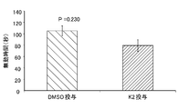
- the time during which the limbs of the rat were not moved was measured as the immobility time in 2 to 10 minutes (0 to 2 minutes was the environment adaptation time of the rat) of the captured movie. .
- the immobility time was compared between rats administered with 15% DMSO and rats administered with K-2 and K-4 dissolved in 15% DMSO.
- Example 1 PET imaging in humans
- PET imaging was performed on two subjects.
- a supine position was taken on the imaging table, and a venous path was secured with a 22 gauge surf flow needle on the forearm or back of the hand.
- After performing absorption correction CT imaging for about 1 minute about 370 MBq of [ 11 C] K-2 was intravenously administered as a PET drug over 1 minute. Thereafter, imaging was performed for 120 minutes.
- a PET / CT apparatus, aquidu (Toshiba Medical Systems) was used for imaging.
- the collected list data reconstructed a dynamic image by the OS-EM method.
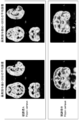
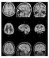
- FIG. 11 shows an image relating to 30 minutes from 0 minutes to 30 minutes after PET drug administration (referred to as the first half image), and from 120 minutes after PET drug administration (an example of the first time) to 120 minutes (an example of the second time). ) For 60 minutes until ().
- the obtained image is a high-resolution and high-contrast image in which the brain AMPA receptor-binding region can be clearly distinguished as compared with the AMPA receptor-binding non-binding region, and thus diagnosis of diseases related to AMPA receptor binding. It can be said that it is suitable for.
- the first half image from 0 minutes to 30 minutes after PET drug administration, both PET drug bound to brain AMPA receptor and PET drug not bound to brain AMPA receptor are detected mixed.
- the overall brightness is high but the resolution is low (coarse), it can be said that there is room for improvement in order to clearly determine the AMPA receptor binding region in the brain. In principle, it is possible to acquire images continuously from 0 minute to 120 minutes after PET drug administration.
- the continuous image is an additive image that includes the first half image with a low resolution and the second half image with a high resolution, and the first half image with a higher radiation dose has a higher contribution to the luminance, and as a whole, It becomes a low resolution image and requires some skill for diagnosis.
- an example of the setting method of the first time for obtaining a high-resolution and multi-gradation image is a time when the PET drug that is not bound to the brain AMPA receptor is markedly washed out or later, It may be after the time when the total amount of radiation detection in the entire brain becomes the maximum value.
- the difference between the first half image and the second half image has been confirmed in monkeys as well as human examples (data not shown).
- Example 2 PET imaging in humans
- PET imaging was performed on healthy men in their 20s (Y5, Y14, Y16).
- a supine position was taken on the imaging table, and a venous path was secured with a 22 gauge surf flow needle on the forearm or back of the hand.
- CT imaging for about 1 minute
- about 370 MBq [ 11 C] K-2 was intravenously administered over 1 minute.
- imaging was performed for 120 minutes.
- a PET / CT apparatus, aquidu (Toshiba Medical Systems) was used for imaging.
- the collected list data reconstructed a dynamic image by the OS-EM method.
- VOIs containing multiple regions such as frontal lobe, dentate cortex, hippocampus, amygdala, nucleus, cerebellum, and pons were created on the template MRI image using PMOD image analysis software (PMOD technologies). It was integrated and analyzed. The calculated value used for quantification was% SUV (% of standardized uptake value). In addition, based on the results of Example 1, the% SUV for three healthy subjects is the average value for 70 minutes from 50 minutes after PET drug administration (an example of the first time) to 120 minutes after (an example of the second time). Was calculated and used for analysis.
- the brain of the primate living body is considered.
- a substance that selectively binds to an internal AMPA receptor and has a radiolabel is used as an active ingredient of a diagnostic agent for a disease associated with an AMPA receptor in a primate brain or a companion diagnostic agent for treatment or prevention of the disease. It was suggested that it can be used.
- the results of the imaging method of the present invention can be used in drug administration plans for the treatment or prevention of diseases related to cerebral AMPA receptors in the primate and screening methods for treatment or prevention drugs. It was done.
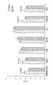
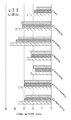
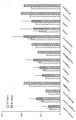
- Example 3 In Example 2, the average value of 10 minutes (referred to as the previous period) from 50 minutes (an example of the first time) to 60 minutes (an example of the second time) as a% SUV for 6 healthy subjects, And the average value for 30 minutes (referred to as the latter period) from 60 minutes (one example of the first time) to 90 minutes (one example of the second time) was calculated and used for the analysis. The result is shown in FIG.
- the vertical axis in FIG. 14 represents the relative value of% SUV of each brain region when the% SUV of the occipital lobe is 1.
- the occipital lobe has a relatively large blood flow component and is suitable as a reference site.
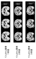
- Example 4 (AMPA-PET for epilepsy patients whose epilepsy focus is confined to the medial temporal lobe-PET imaging in patients with medial temporal lobe epilepsy) PET imaging was performed on 3 patients with medial temporal lobe epilepsy. A supine position was taken on the imaging table, and a venous path was secured with a 22 gauge surf flow needle on the forearm or back of the hand. After absorption correction CT imaging for about 1 minute, about 370 MBq [ 11 C] K-2 was intravenously administered over 1 minute. Thereafter, imaging was performed for 90 minutes. A PET / CT apparatus, aquidu (Toshiba Medical Systems) was used for imaging. The collected list data reconstructed a dynamic image by the OS-EM method.
- aquidu Toshiba Medical Systems
- the reconstructed image was analyzed by integrating the PET image onto the standardized MRI image using PMOD image analysis software (PMOD technologies).
- PMOD image analysis software PMOD technologies.
- a SUVR (standardized uptake value ratio) image was used as the reconstructed image.
- the SUVR image was created by dividing the 20-minute addition average image from 30 minutes to 50 minutes after PET drug administration by the average value of the amount of radioactivity accumulated in the corpus callosum as the reference region.
- Example 5 Subtraction image analysis for epilepsy patients
- PET imaging was performed on 3 patients with medial temporal lobe epilepsy.
- a supine position was taken on the imaging table, and a venous path was secured with a 22 gauge surf flow needle on the forearm or back of the hand.
- After absorption correction CT imaging for about 1 minute about 370 MBq [ 11 C] K-2 was intravenously administered over 1 minute. Thereafter, imaging was performed for 90 minutes.
- a PET / CT apparatus, aquidu (Toshiba Medical Systems) was used for imaging.
- the collected list data reconstructed a dynamic image by the OS-EM method.
- Reconstructed images were analyzed by integrating PET images onto standardized MRI images using PMOD image analysis software (PMOD technologies).
- a subtraction image was used as a reconstructed image and created as follows. 30 minutes after PET drug administration (one example of the first time) to 50 minutes after one minute (one example of the second time) 20-minute average images (images containing a lot of specific binding to AMPA receptors) and 1.5 minutes to An average image (an image containing a lot of blood flow components) for 1 minute until 2.5 minutes later was calculated.
- an SUVR image was calculated by dividing the average amount of radioactivity accumulated in the entire brain and using the entire brain as a reference region.
- a subtraction image was created by subtracting the 1-minute SUVR image from 1.5 to 2.5 minutes from the 20-minute SUVR image 30 to 50 minutes after PET drug administration, and calculating the difference.
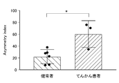
- Example 6 (Asymmetric index comparison for healthy subjects and patients with medial temporal lobe epilepsy) PET imaging was performed on 6 healthy subjects and 3 medial temporal lobe epilepsy patients. A supine position was taken on the imaging table, and a venous path was secured with a 22 gauge surf flow needle on the forearm or back of the hand. After absorption correction CT imaging for about 1 minute, about 370 MBq of [11C] K-2 was intravenously administered over 1 minute. Thereafter, imaging was performed for 90 minutes. A PET / CT apparatus, aquidu (Toshiba Medical Systems) was used for imaging. The collected list data reconstructed a dynamic image by the OS-EM method.
- aquidu Toshiba Medical Systems
- the reconstructed image is obtained by using PMOD image analysis software (PMOD technologies) VOIs including a plurality of regions such as frontal lobe, temporal lobe, occipital lobe, parietal lobe, hippocampus, amygdala, putamen, corpus callosum, cerebellum, and bridge.
- PMOD image analysis software PMOD technologies
- VOIs including a plurality of regions such as frontal lobe, temporal lobe, occipital lobe, parietal lobe, hippocampus, amygdala, putamen, corpus callosum, cerebellum, and bridge.
- a subtraction image was used as a reconstructed image and created as follows. Average image for 20 minutes from 30 minutes (example of first time) to 50 minutes (example of second time) after PET drug administration and average image for 1 minute from 1.5 minutes to 2.5 minutes (blood) An image containing a lot of flow components) was calculated.
- an SUVR image was calculated by dividing the average amount of radioactivity accumulated in the entire brain and using the entire brain as a reference region.
- a subtraction image was created by subtracting the 1-minute SUVR image from 1.5 to 2.5 minutes from the 20-minute SUVR image 30 to 50 minutes after PET drug administration, and calculating the difference.
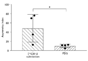
- image values were calculated using the VOIs of the left and right temporal lobes, and asymmetry index (AI) was obtained. AI was calculated from the following formula.
- AI 200 ⁇ (left temporal lobe VOI value ⁇ right temporal lobe VOI value) / (left temporal lobe VOI value + right temporal lobe VOI value)
- Example 7 (Comparison between K-2 imaging and FDG imaging for patients with medial temporal lobe epilepsy) PET imaging using 18F-FDG (fluorodeoxyglucose) was performed on three patients with medial temporal lobe epilepsy.
- 18F-FDG fluorodeoxyglucose
- a venous passage was secured with a 22 gauge surf flow needle on the forearm or back of the hand.
- About 185 MBq of 18F-FDG was administered intravenously over 1 minute. Then, after leaving still for 60 minutes, it imaged for 20 minutes (after performing CT imaging for absorption correction for about 1 minute).
- the collected list data was reconstructed into a dynamic image by the OS-EM method.
- the reconstructed image is obtained by using PMOD image analysis software (PMOD technologies) VOIs including a plurality of regions such as frontal lobe, temporal lobe, occipital lobe, parietal lobe, hippocampus, amygdala, putamen, corpus callosum, cerebellum, and bridge.
- PMOD image analysis software PMOD technologies
- VOIs including a plurality of regions such as frontal lobe, temporal lobe, occipital lobe, parietal lobe, hippocampus, amygdala, putamen, corpus callosum, cerebellum, and bridge.
- a SUVR standardized uptake value ratio
- the [ 11 C] K-2 subtraction image As shown in FIG. 18, when the AI calculated from the [ 11 C] K-2 subtraction image and the FDG SUVR image of the patient with medial temporal lobe epilepsy were compared, the [ 11 C] K-2 subtraction image The calculated AI showed a significantly high value. That is, it can be seen that by measuring the AI using a [ 11 C] K-2 subtraction image, it can be detected as a focal point with higher sensitivity than FDG.
- Example 8 (Depression status of depressed patients and AMPA-PET comparison in remission) PET imaging was performed on a depressed patient and a depressed patient in remission of the patient. A supine position was taken on the imaging table, and a venous path was secured with a 22 gauge surf flow needle on the forearm or back of the hand. After absorption correction CT imaging for about 1 minute, about 370 MBq [ 11 C] K-2 was intravenously administered over 1 minute. Thereafter, imaging was performed for 60 minutes. A PET / CT apparatus, aquidu (Toshiba Medical Systems) was used for imaging. The collected list data reconstructed a dynamic image by the OS-EM method.
- aquidu Toshiba Medical Systems
- the reconstructed image is obtained by using PMOD image analysis software (PMOD technology) VOIs including a plurality of regions such as frontal lobe, temporal lobe, occipital lobe, parietal lobe, hippocampus, amygdala, putamen, corpus callosum, cerebellum, and bridge.
- PMOD image analysis software PMOD technology
- VOIs including a plurality of regions such as frontal lobe, temporal lobe, occipital lobe, parietal lobe, hippocampus, amygdala, putamen, corpus callosum, cerebellum, and bridge.
- a SUVR standardized uptake value ratio
- Example 8 shows that the therapeutic effect of a disease and the recovery status until healing can be known from an imaging image, and it is possible to formulate a pharmaceutical administration plan for treatment or prevention. It can be said. Moreover, it can be said that it is possible to screen for therapeutic or preventive drugs for the above-mentioned diseases.
- a PET drug it is shown that a compound of the present invention, particularly a compound in which R 2 is alkyl, for example, K-2 is preferable to FDG.
- AMPA ⁇ -amino-3-hydroxy-5-methyl-4-isoxazole-propionic acid
- DIPEA diisopropylethylamine
- DCM dichloromethane
- EtOAc ethyl acetate
- FDG fluorodeoxyglucose
- PE petroleum ether
- PEPA 2- [2,6 -Difluoro-4-( ⁇ 2-[(phenylsulfonyl) amino] ethyl ⁇ thio) phenoxy] acetamide
- PET Positron tomography
- TEA Tetraethylammonium
- TMS Tetramethylsilane
- MeOH Methanol
- EtOH Ethanol
- DMF N, N- Dimethylformamide
- MeI Methyl iodide
- WSC ⁇ HCl Water-soluble carbodiimide hydrochloride
- DMSO Dimethyl sulfoxide
- ACFS Artificial cerebrospinal fluid
- Imaging apparatus 200 Input terminal 210: Imaging data generation unit 220: Metadata generation unit 300: Server 310: Information generation unit 350: Verification unit 360: Instruction unit 400: Database (first database) 500: Output terminal 600: Second database 900: Clock 1000: System
Landscapes
- Health & Medical Sciences (AREA)
- Life Sciences & Earth Sciences (AREA)
- General Health & Medical Sciences (AREA)
- Medicinal Chemistry (AREA)
- Public Health (AREA)
- Engineering & Computer Science (AREA)
- Pharmacology & Pharmacy (AREA)
- Chemical & Material Sciences (AREA)
- Animal Behavior & Ethology (AREA)
- Veterinary Medicine (AREA)
- Bioinformatics & Cheminformatics (AREA)
- Physics & Mathematics (AREA)
- Nuclear Medicine, Radiotherapy & Molecular Imaging (AREA)
- Biomedical Technology (AREA)
- Optics & Photonics (AREA)
- Organic Chemistry (AREA)
- General Chemical & Material Sciences (AREA)
- Neurology (AREA)
- Neurosurgery (AREA)
- Chemical Kinetics & Catalysis (AREA)
- Epidemiology (AREA)
- Proteomics, Peptides & Aminoacids (AREA)
- Molecular Biology (AREA)
- Psychiatry (AREA)
- Biophysics (AREA)
- Medical Informatics (AREA)
- General Physics & Mathematics (AREA)
- High Energy & Nuclear Physics (AREA)
- Spectroscopy & Molecular Physics (AREA)
- Medicines Containing Antibodies Or Antigens For Use As Internal Diagnostic Agents (AREA)
- Nuclear Medicine (AREA)
- Medicines That Contain Protein Lipid Enzymes And Other Medicines (AREA)
- Organic Low-Molecular-Weight Compounds And Preparation Thereof (AREA)
Abstract
Description
本発明は、霊長類生体の脳内AMPA受容体をイメージングする技術に関する。 The present invention relates to a technique for imaging AMPA receptors in the brain of primate organisms.
AMPA受容体は中神経系に広く分布し、学習、記憶、神経変性、及び細胞死などに関与することが知られている。近年、AMPA受容体を標的とした、精神・神経疾患の治療に関する研究が進められている(特許文献1~3)。AMPA受容体とこれらの疾患との関係を調査するためには、脳内におけるAMPA受容体の発現量及び分布を評価することが求められる。
AMPA receptors are widely distributed in the central nervous system and are known to be involved in learning, memory, neurodegeneration, and cell death. In recent years, research on treatment of psychiatric / neurological diseases targeting AMPA receptors has been underway (
従来、AMPA受容体の解析技術としては、シナプス、スパインレベルでのミクロスコピックな観察(電子顕微鏡を用いた抗体染色、2光子顕微鏡を用いた蛍光観察、電気生理学的手法を用いたシナプスにおける機能観察、quantum dot法を用いたsingle molecule tracking 法等)、および、スライスを用いた免疫染色法等の比較的マクロスコピックな観察が知られている。 Conventionally, AMPA receptor analysis techniques include microscopic observation at the synapse and spine level (antibody staining using an electron microscope, fluorescence observation using a two-photon microscope, and functional observation at a synapse using an electrophysiological technique). In addition, relatively macroscopic observations such as single molecule tracking method using quantum dot method and immunostaining method using slice are known.
このうち、2光子顕微鏡を用いたin vivoイメージング以外では、生体での解析が不可能である。他方、2光子顕微鏡を用いたin vivoイメージングでは、スパイン、樹状突起レベルのミクロスコピックな観察が生体で可能だが、逆に脳全体における観察ができない。また、二光子顕微鏡を用いたin vivoイメージング法では、蛍光タンパク質のタグをつけたAMPA受容体の観察しかできず、内在性のAMPA受容体の観察ができない。蛍光タンパク質のタグをつけたAMPA受容体は遺伝子導入法により人為的に発現させる必要があり、ヒト以外の霊長類においては費用面で大きな問題を生じ、ましてやヒトにおいては侵襲性の問題から事実上実施できない。 Of these, analysis in vivo is impossible except for in vivo imaging using a two-photon microscope. On the other hand, in vivo imaging using a two-photon microscope allows microscopic observation at the spine and dendritic level in a living body, but conversely, observation in the entire brain is not possible. Furthermore, in vivo imaging using a two-photon microscope can only observe AMPA receptors tagged with fluorescent proteins, and cannot observe endogenous AMPA receptors. AMPA receptors tagged with fluorescent proteins must be artificially expressed by gene transfer methods, which can cause significant cost problems for non-human primates and even humans because of invasive problems. Cannot be implemented.
また、特許文献4には、AMPA受容体との親和性を有する物質を用いて、サル生体脳のAMPA受容体のイメージングを行おうとした例が記載されている。しかし、非標識の競合物質の投与量に依存したプローブ由来放射線検知値の低下が、実際には確認されておらず、ある時点で脳に存在するプローブに由来した放射線を検知できたにとどまる。つまり、AMPA受容体に結合したプローブ由来の放射線を検知し、それに基づき本当にAMPA受容体をイメージング化できることは証明できていない。
本発明は、以上の実情に鑑みてなされたものであり、霊長類生体の脳内AMPA受容体をイメージング化する技術及びその応用の提供を目的とする。 The present invention has been made in view of the above circumstances, and an object of the present invention is to provide a technique for imaging the brain AMPA receptor of a primate organism and its application.
(1) 霊長類生体に投与された、霊長類生体の脳内AMPA受容体に選択的に結合しかつ放射性標識を有する物質を、脳内に移行させて前記脳内AMPA受容体に結合させ、
前記脳内AMPA受容体に結合した前記物質から放出される放射線を検知することで、前記脳内AMPA受容体の分布及び/又は発現量に関するデータを取得する工程を有する、霊長類生体の脳内AMPA受容体のイメージング方法。
(1) A substance which is administered to a primate organism and selectively binds to the AMPA receptor in the brain of the primate organism and has a radioactive label is transferred into the brain to bind to the AMPA receptor in the brain,
Detecting radiation emitted from the substance bound to the brain AMPA receptor to obtain data on the distribution and / or expression level of the AMPA receptor in the brain. AMPA receptor imaging method.
(2) 前記物質が脳内に移行した後に所要時間を空けることで、前記脳内AMPA受容体に結合していない前記物質の脳外排出を促した後、前記検知を行う(1)記載の方法。 (2) The detection according to (1), wherein the detection is performed after encouraging the extravasation of the substance that is not bound to the AMPA receptor in the brain by allowing a necessary time after the substance has entered the brain. Method.
(3) 前記物質が脳内に移行した後に第1時間経過後と、第1時間よりも長い第2時間経過後とにそれぞれ前記検出を行い、
それぞれの検出値に基づき、前記脳内AMPA受容体の分布及び/又は量に関するデータを取得する(1)又は(2)記載の方法。
(3) The detection is performed after the first time has passed after the substance has entered the brain and after the second time longer than the first time,
The method according to (1) or (2), wherein data relating to the distribution and / or amount of the AMPA receptor in the brain is obtained based on each detected value.
(4) 前記物質は、式(I)の化合物、又はその医薬として許容し得る塩若しくは溶媒和物
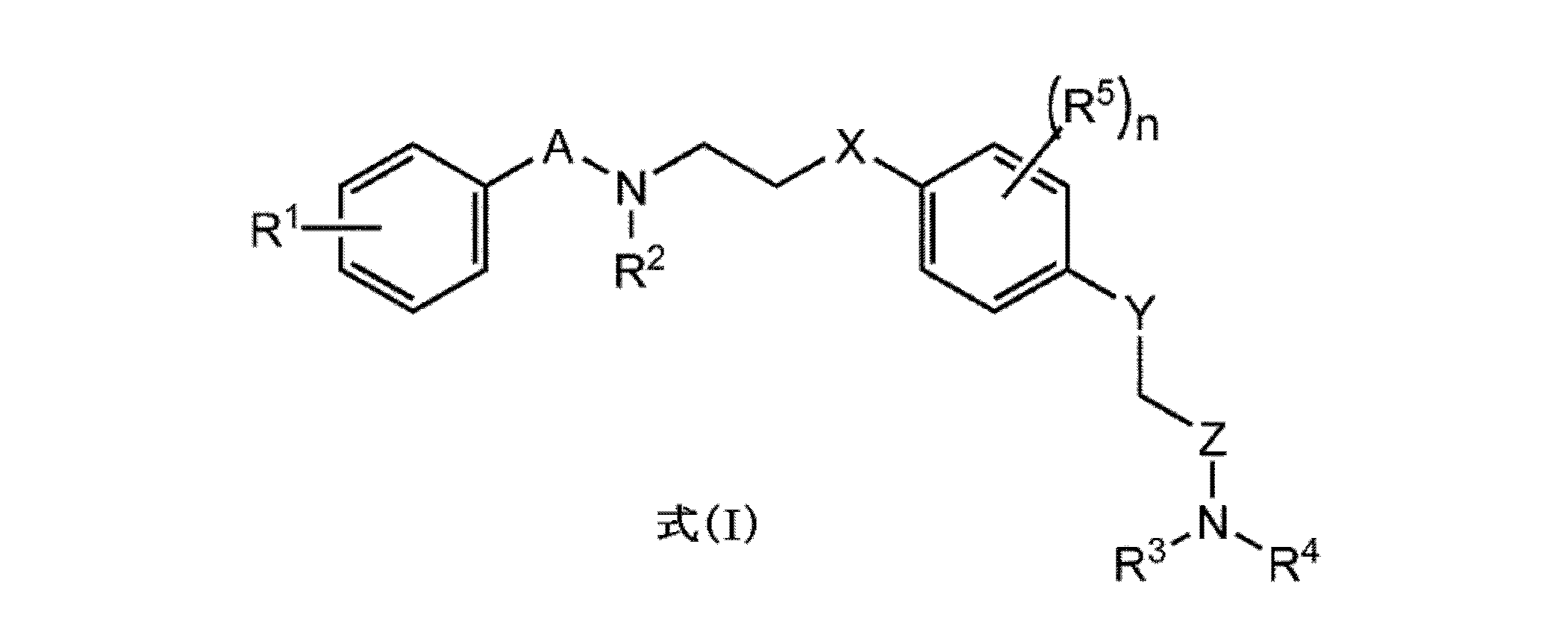
A及びZは、それぞれ独立に、CO、SO又はSO2であり;
X及びYは、それぞれ独立に、S又はOであり;
R1~R4は、それぞれ独立に、水素、アルキル、アルケニル、アルキニル又はハロであり;
R5は、出現ごとにそれぞれ独立に、アルキル、アルケニル、アルキニル又はハロであり;
nは、0~4の整数であり;
1個又はそれ以上の原子が該原子の放射性同位体である。)
を含む(1)から(3)いずれか記載の方法。
(4) The substance is a compound of formula (I), or a pharmaceutically acceptable salt or solvate thereof.
A and Z are each independently CO, SO or SO 2 ;
X and Y are each independently S or O;
R 1 to R 4 are each independently hydrogen, alkyl, alkenyl, alkynyl or halo;
R 5 is independently for each occurrence alkyl, alkenyl, alkynyl or halo;
n is an integer from 0 to 4;
One or more atoms are radioisotopes of the atoms. )
The method according to any one of (1) to (3).
(5) (1)から(4)いずれか記載の方法をコンピュータに実行させるためのプログラム。 (5) A program for causing a computer to execute the method described in any one of (1) to (4).
(6) 霊長類生体の脳内AMPA受容体に選択的に結合しかつ放射性標識を有する物質を有効成分とする、霊長類体の脳内AMPA受容体が関連する疾患の診断薬、又は前記疾患の治療又は予防のためのコンパニオン診断薬。 (6) A diagnostic agent for a disease associated with a primate brain AMPA receptor, comprising a substance that selectively binds to a brain AMPA receptor in a primate living body and has a radioactive label, or the above-mentioned disease A companion diagnostic for the treatment or prevention of cancer.
(7) 霊長類体の脳内AMPA受容体が関連する疾患の治療又は予防のための医薬であって、
霊長類生体の脳内AMPA受容体に選択的に結合する物質を有効成分とし、(1)から(4)いずれか記載の方法で得られる前記データに基づく投与計画で投与されるものである医薬。
(7) A medicament for the treatment or prevention of diseases associated with cerebral AMPA receptors in the primate body,
A pharmaceutical comprising a substance that selectively binds to a brain AMPA receptor in a primate living body as an active ingredient, and is administered in a dosage plan based on the data obtained by any of the methods (1) to (4) .
(8) 前記疾患は、精神疾患又は神経疾患である(6)又は(7)記載の薬。 (8) The drug according to (6) or (7), wherein the disease is a mental disease or a neurological disease.
(9) 霊長類体の脳内AMPA受容体が関連する疾患の治療又は予防薬のスクリーニング方法であって、
前記霊長類生体への候補物質の投与前後における、(1)から(4)いずれか記載の方法で得られる前記データの差異に基づき、前記候補物質を選抜する工程を有する方法。
(9) A screening method for a therapeutic or prophylactic agent for a disease associated with an AMPA receptor in the brain of a primate body,
A method comprising a step of selecting the candidate substance based on the difference in the data obtained by the method according to any one of (1) to (4) before and after administration of the candidate substance to the primate organism.
(10) 霊長類生体の脳内AMPA受容体の分布及び/又は発現量に関する情報をサーバへと送信する入力端末。 (10) An input terminal that transmits information on the distribution and / or expression level of AMPA receptors in the brain of a primate organism to a server.
(11) 霊長類体の脳内AMPA受容体の分布及び/又は発現量と、霊長類体の脳内AMPA受容体が関連する疾患の状態と、が関連づけられたデータを格納するデータベースと、
入力された被験者の生体の脳内AMPA受容体の分布及び/又は発現量に関する情報と、前記データとを照会し、前記被験者の前記疾患の状態に関する情報を出力端末へと送信する手段と、を備えるサーバ。
(11) a database storing data in which the distribution and / or expression level of AMPA receptors in the primate brain and the disease state associated with the AMPA receptors in the primate brain are associated;
Means for querying the input information regarding the distribution and / or expression level of AMPA receptors in the brain of the subject's living body and the data, and transmitting information regarding the disease state of the subject to an output terminal; Server to provide.
(12) 霊長類体の脳内AMPA受容体の分布及び/又は発現量と、霊長類体の脳内AMPA受容体が関連する疾患の状態と、既に投与された霊長類体の生体の脳内AMPA受容体が関連する疾患の治療又は予防のための医薬の種類、用量及び/又は用法と、が関連づけられたデータを格納するデータベースと、
入力された被験者の生体の脳内AMPA受容体の分布及び/又は発現量に関する情報と、前記データとを照会し、前記被験者への推奨される医薬の種類、用量及び/又は用法に関する情報を出力端末へと送信するする手段と、を備えるサーバ。
(12) Distribution and / or expression level of AMPA receptor in brain of primate body, disease state related to AMPA receptor in brain of primate body, and brain in living body of primate body already administered A database storing data associated with the type, dose and / or usage of a medicament for the treatment or prevention of a disease associated with an AMPA receptor;
Queries the inputted information on the distribution and / or expression level of AMPA receptors in the brain of the subject's living body and the data, and outputs information on the recommended type, dose and / or usage of the drug to the subject Means for transmitting to a terminal.
(13) (10)記載の入力端末と、
(11)又は(12)記載のサーバと、
(11)又は(12)記載のサーバから送信された前記情報を出力する出力端末と、を備えるシステム。
(13) The input terminal according to (10),
The server according to (11) or (12);
A system comprising: an output terminal that outputs the information transmitted from the server according to (11) or (12).
本発明によれば、霊長類生体の脳内AMPA受容体をイメージング化する技術及びその応用を提供することができる。 According to the present invention, it is possible to provide a technique for imaging an AMPA receptor in the brain of a primate organism and its application.
以下、本発明の実施形態を説明するが、これらに本発明が限定されるものではない。 Hereinafter, embodiments of the present invention will be described, but the present invention is not limited thereto.
(イメージング方法)
本発明の一実施形態は、霊長類生体の脳内AMPA受容体のイメージング方法である。この方法は、霊長類生体に投与された、霊長類生体の脳内AMPA受容体に選択的に結合しかつ放射性標識を有する物質を、脳内に移行させて脳内AMPA受容体に結合させる工程を有する。後述の実施例のように、霊長類生体の脳内AMPA受容体に選択的に結合しかつ放射性標識を有する物質を用いたときに、霊長類生体の脳内AMPA受容体に結合した物質から放出される放射線を検知できることを、本発明者らは初めて発見し、本実施形態に係る方法論を確立した。
(Imaging method)
One embodiment of the present invention is a method for imaging AMPA receptors in the brain of a primate organism. This method comprises a step of transferring a substance having a radiolabel and selectively binding to a primate living brain AMPA receptor, which is administered to the primate living body, to the brain and binding to the brain AMPA receptor. Have When a substance having a radiolabel is used that selectively binds to the AMPA receptor in the brain of the primate organism as in the examples described later, the substance is released from the substance bound to the AMPA receptor in the brain of the primate organism. The present inventors discovered for the first time that detected radiation can be detected, and established a methodology according to this embodiment.
つまり、この新規発見に基づく本実施形態は、脳内AMPA受容体に結合した物質から放出される放射線を検知することで、脳内AMPA受容体の分布及び/又は発現量に関するデータを取得する工程を有する。これにより、霊長類生体の脳全体におけるAMPA受容体の分布及び/又は発現量が把握され、霊長類生体の脳内AMPA受容体をイメージング化することができる。 That is, this embodiment based on this novel discovery is a process of acquiring data related to the distribution and / or expression level of the brain AMPA receptor by detecting the radiation emitted from the substance bound to the brain AMPA receptor. Have Thereby, the distribution and / or expression level of the AMPA receptor in the entire brain of the primate organism can be grasped, and the brain AMPA receptor in the primate organism can be imaged.
放射線検知によるイメージングは、特に限定されないが、分子イメージング、例えば、ポジトロン断層撮影法(Positron Emission Tomography, PET)、多光子イメージング法、二光子イメージング法、近赤外蛍光イメージング法、オートラジオグラフィー、及び単一光子放射断層撮影法(Single photon emission computed tomography, SPECT)などであってよい。中でもPETイメージングが好ましい。 Imaging by radiation detection is not particularly limited, but includes molecular imaging such as positron emission tomography (PETRON), multiphoton imaging method, two-photon imaging method, near infrared fluorescence imaging method, autoradiography, and Single photon emission tomography (SPECT) may be used. Among these, PET imaging is preferable.
霊長類は、特に限定されないが、ヒト、サルであってよい。ヒト及びサルの間では、物質代謝性、血液脳関門の通過性、脳内AMPA受容体の量、分布について、若干の差異があると考えられる。この点、後述の実施例はヒトでのデータに関するが、本発明者らはサルでも同様にイメージング化できることを確認している(データは示さず)。 Primates are not particularly limited, but may be humans and monkeys. It is considered that there are some differences between humans and monkeys in terms of substance metabolism, permeability of the blood brain barrier, and the amount and distribution of AMPA receptors in the brain. In this regard, although the examples described later relate to human data, the present inventors have confirmed that monkeys can be similarly imaged (data not shown).
一実施形態において、上記物質が脳内に移行した後に所要時間を空けることで、脳内AMPA受容体に結合していない上記物質の脳外排出を促した後、放射線検知を行うことが好ましい。これにより、脳内AMPA受容体に結合した上記物質に由来する放射線をより高頻度に検知することができ、AMPA受容体のイメージング化精度が増す。 In one embodiment, it is preferable to perform radiation detection after encouraging excretion of the substance not bound to the brain AMPA receptor by suspending the time required after the substance has moved into the brain. Thereby, the radiation derived from the said substance couple | bonded with the AMPA receptor in a brain can be detected more frequently, and the imaging accuracy of an AMPA receptor increases.
上記の所要時間は、特に限定されず、用いる物質や統計に基づき予め設定されてもよく(典型的には、上記物質の投与後から10分以上、20分以上、30分以上、40分以上、又は45分以上になるように設定されてよい。)、対象となる生体ごとに設定されてもよい。具体的に、所要時間は、(i)数理解析モデルに当てはめて算出する、または(ii)ターゲットタンパクが存在する領域と存在しない領域の比率が一定になった時点以降で、その差が最大かつばらつきが少なくなる最大の時間帯、(iii)疾患と健常者との差が最も明確に検出できる時間帯、であり得る。 The required time is not particularly limited, and may be set in advance based on the substance used and statistics (typically, 10 minutes or more, 20 minutes or more, 30 minutes or more, 40 minutes or more after administration of the substance). Or may be set to be 45 minutes or longer.), Or may be set for each target living body. Specifically, the required time is calculated by applying (i) a mathematical analysis model, or (ii) the difference between the region where the target protein is present and the region where the target protein is not present is It may be the maximum time zone in which the variation is reduced, and (iii) the time zone in which the difference between the disease and the healthy person can be detected most clearly.
他方、上記の所要時間を過剰に長くすると、放射線の絶対量が減衰し、高精度での検知が困難になり得る。そこで、上記所要時間は特に限定されないが、例えば上記物質の投与後から110分以下、100分以下、90分以下、80分以下、70分以下、又は60分以下になるよう設定されてよい。 On the other hand, if the above required time is excessively long, the absolute amount of radiation is attenuated, and detection with high accuracy may be difficult. Thus, the required time is not particularly limited, but may be set to be 110 minutes or less, 100 minutes or less, 90 minutes or less, 80 minutes or less, 70 minutes or less, or 60 minutes or less after administration of the substance.
一実施形態において、上記物質が脳内に移行した後に第1時間経過後と、第1時間よりも長い第2時間経過後とにそれぞれ前記検出を行い、それぞれの検出値に基づき、脳内AMPA受容体の分布及び/又は量に関するデータを取得することが好ましい。より好ましくは、第1時間と第2時間との間に連続又は不連続で、各脳領域に取り込まれている上記物質由来の放射線量を検出し、その平均値を算出し、各平均値の脳領域間格差に基づき、脳内AMPA受容体の分布及び/又は量に関するデータを取得することができる。あるいは、第1時間経過後から第2時間経過後までの間で、相対的に小さい幅で放射線検知値が変化(低下)した領域はAMPA受容体に対応する可能性が高い。このため、検出値の差異に基づくデータを利用することで、AMPA受容体のイメージング化精度を増すことができる。なお、上記の第1時間及び第2時間を過剰に長くすると、放射線の絶対量が減衰し、高精度での検知が困難になり得る。 In one embodiment, the detection is performed after the first time has passed after the substance has entered the brain and after the second time longer than the first time, and the brain AMPA is determined based on the detected values. It is preferred to obtain data relating to receptor distribution and / or quantity. More preferably, the radiation dose derived from the above-mentioned substance taken into each brain region is detected continuously or discontinuously between the first time and the second time, and the average value is calculated. Based on the disparity between brain regions, data regarding the distribution and / or amount of AMPA receptors in the brain can be obtained. Alternatively, a region in which the radiation detection value has changed (decreased) with a relatively small width between the lapse of the first time and the lapse of the second time is likely to correspond to the AMPA receptor. For this reason, the imaging accuracy of an AMPA receptor can be increased by using data based on a difference in detected values. If the first time and the second time are excessively long, the absolute amount of radiation is attenuated, and detection with high accuracy may be difficult.
第1時間及び第2時間は、特に限定されず、用いる物質や統計に基づき予め設定されてもよく、対象となる生体ごとに設定されてもよい。第1時間は、典型的には、上記物質の投与後から10分以上、20分以上、30分以上、40分以上、又は45分以上になるように設定されてよく、110分以下、100分以下、90分以下、80分以下、70分以下、又は60分以下になるように設定されてよく、上記した所要時間と同様に決められてよい。第2時間は、典型的には上記物質の投与後から30分以上150分以下(具体的には60分以下)になるように設定されてよい。 The first time and the second time are not particularly limited, and may be set in advance based on a substance to be used and statistics, or may be set for each target living body. The first time may typically be set to be 10 minutes or more, 20 minutes or more, 30 minutes or more, 40 minutes or more, or 45 minutes or more after administration of the substance, 110 minutes or less, 100 minutes or less. It may be set to be less than or equal to 90 minutes, less than or equal to 90 minutes, less than or equal to 80 minutes, less than or equal to 70 minutes, or less than or equal to 60 minutes, and may be determined in the same manner as the required time described above. The second time may be set to be typically 30 minutes to 150 minutes (specifically 60 minutes or less) after administration of the substance.
上記物質は、特に制限されないが、例えば、非経口投与、静脈内投与、又は腹腔内投与された後、血液脳関門を通過して脳へ移行する。このため、上記物質は、血液脳関門を通過可能な特性を有する必要があり、その観点で、所要の低分子量、脂溶性とともに、所要の血液溶解性を備えることが好ましい。なお、上記物質は、単一物質であってもよく、あるいは、DDS(ドラッグデリバリーシステム)に担持された状態でもよい。 The above substance is not particularly limited, but for example, after parenteral administration, intravenous administration, or intraperitoneal administration, it passes through the blood-brain barrier and moves to the brain. For this reason, it is necessary that the substance has a characteristic capable of passing through the blood-brain barrier. From this viewpoint, it is preferable that the substance has required blood solubility as well as required low molecular weight and fat solubility. In addition, the said substance may be a single substance, or the state carried | supported by DDS (drug delivery system).
また、上記物質は医薬として許容し得る担体中に含まれてもよい。医薬として許容し得る担体は、特に制限されないが、例えば、滅菌水、食塩水、生理食塩水又はリン酸緩衝食塩水(PBS)、塩化ナトリウム注射液、リンゲル注射液、等張性デキストロース注射液、無菌水注射液、デキストロース、及び乳酸リンゲル注射液などがある。 In addition, the substance may be contained in a pharmaceutically acceptable carrier. The pharmaceutically acceptable carrier is not particularly limited, and examples thereof include sterile water, saline, physiological saline or phosphate buffered saline (PBS), sodium chloride injection, Ringer's injection, isotonic dextrose injection, Examples include sterile water injection solution, dextrose, and lactated Ringer's injection solution.
上記物質の投与量は、使用される物質の種類;投与される対象の年齢、体重、健康状態、性別及び食事内容;投与の回数、及び投与経路等によって、適宜設定されてよい。上記物質の投与は、特に限定されない。 The dose of the above substance may be appropriately set depending on the type of substance used; age, weight, health condition, sex and diet content of the subject to be administered; number of administrations, administration route, and the like. The administration of the substance is not particularly limited.
上記物質は、特に限定されないが、例えば下記式(I)の化合物、又はその医薬として許容し得る塩若しくは溶媒和物であってもよい。

式中、
A及びZは、それぞれ独立に、CO、SO又はSO2であり;
X及びYは、それぞれ独立に、S又はOであり;
R1~R4は、それぞれ独立に、水素、アルキル、アルケニル、アルキニル又はハロであり;
R5は、出現ごとにそれぞれ独立に、アルキル、アルケニル、アルキニル又はハロであり;
nは、0~4の整数であり;
1個又はそれ以上の原子が該原子の放射性同位体である。
Where
A and Z are each independently CO, SO or SO 2 ;
X and Y are each independently S or O;
R 1 to R 4 are each independently hydrogen, alkyl, alkenyl, alkynyl or halo;
R 5 is independently for each occurrence alkyl, alkenyl, alkynyl or halo;
n is an integer from 0 to 4;
One or more atoms are radioisotopes of the atoms.
式(I)の化合物において、放射性同位体は、15O、13N、11C、及び18Fなどからなる群から選択されるが、特に限定されない。半減期の観点から、放射性同位体は、11C又は18Fが好ましい。 In the compound of formula (I), the radioisotope is selected from the group consisting of 15 O, 13 N, 11 C, 18 F and the like, but is not particularly limited. From the viewpoint of half-life, the radioisotope is preferably 11 C or 18 F.
好ましくは、R1~R4の1、2、3又は4個、好ましくは、1個が、放射性同位体を含む基(例えば、[11C]アルキル(好ましくは11CH3)、[11C]アルケニル、又は[11C]アルキニル、或いは、18F)である。具体的には、R2がアルキルであることが好ましく、さらにR3及びR4の双方が水素、またはR3及びR4の各々が独立してアルキルであることが好ましい。 Preferably, 1 , 2, 3 or 4, preferably 1 of R 1 to R 4 is a group containing a radioisotope (eg, [ 11 C] alkyl (preferably 11 CH 3 ), [ 11 C ] Alkenyl, or [ 11 C] alkynyl, or 18 F). Specifically, R 2 is preferably alkyl, more preferably both R 3 and R 4 are hydrogen, or each of R 3 and R 4 is independently alkyl.
式(I)の化合物として、AがSO2であり、ZがCOであり、XがSであり、YがOであり、R2がアルキルであり、R1が水素、アルキル又はハロであり、R1がアルキル又はハロである場合、R1はパラ位に存在し、R3及びR4のうち一方が、水素であり、他方がアルキルであり、R5がハロ、特にフルオロであって、R5はY基に対して両方のオルト位(すなわち、X基に対して両方のメタ位)に存在し、nが2であって、R1~R4の1個が、放射性同位体を含む基(例えば、[11C]アルキル(好ましくは11CH3)、[11C]アルケニル、又は[11C]アルキニル、或いは、18F)が好ましい。 As a compound of formula (I), A is SO 2 , Z is CO, X is S, Y is O, R 2 is alkyl, R 1 is hydrogen, alkyl or halo. , R 1 is alkyl or halo, R 1 is in the para position, one of R 3 and R 4 is hydrogen, the other is alkyl, and R 5 is halo, especially fluoro, , R 5 is present in both ortho positions relative to the Y group (ie, both meta positions relative to the X group), n is 2 and one of R1-R4 contains a radioisotope Groups such as [ 11 C] alkyl (preferably 11 CH 3 ), [ 11 C] alkenyl, or [ 11 C] alkynyl, or 18 F are preferred.
さらに他の実施態様において、式(I)の化合物として、AがSO2であり、ZがCOであり、XがSであり、YがOであり、R2がアルキルであり、R1が水素、アルキル又はハロであり、R1がアルキル又はハロである場合、R1はパラ位に存在し、R3及びR4のうち一方が、水素であり、他方がアルキルであり、R5がハロ、特にフルオロであって、R5はY基に対して両方のオルト位(すなわち、X基に対して両方のメタ位)に存在し、nが2であって、R1~R4の1個が、放射性同位体を含む基(例えば、[11C]アルキル(好ましくは11CH3)、[11C]アルケニル、又は[11C]アルキニル、或いは、18F)がより好ましい。 In yet another embodiment, as a compound of formula (I), A is SO 2 , Z is CO, X is S, Y is O, R 2 is alkyl and R 1 is When hydrogen, alkyl or halo and R 1 is alkyl or halo, R 1 is in the para position, one of R 3 and R 4 is hydrogen, the other is alkyl, and R 5 is Halo, especially fluoro, wherein R 5 is present in both ortho positions relative to the Y group (ie both meta positions relative to the X group), n is 2 and R 1 to R 4 More preferably, one group contains a radioisotope (eg, [ 11 C] alkyl (preferably 11 CH 3 ), [ 11 C] alkenyl, or [ 11 C] alkynyl, or 18 F).
放射性同位体を含む化合物の具体的な例を挙げると、以下の化合物がある:
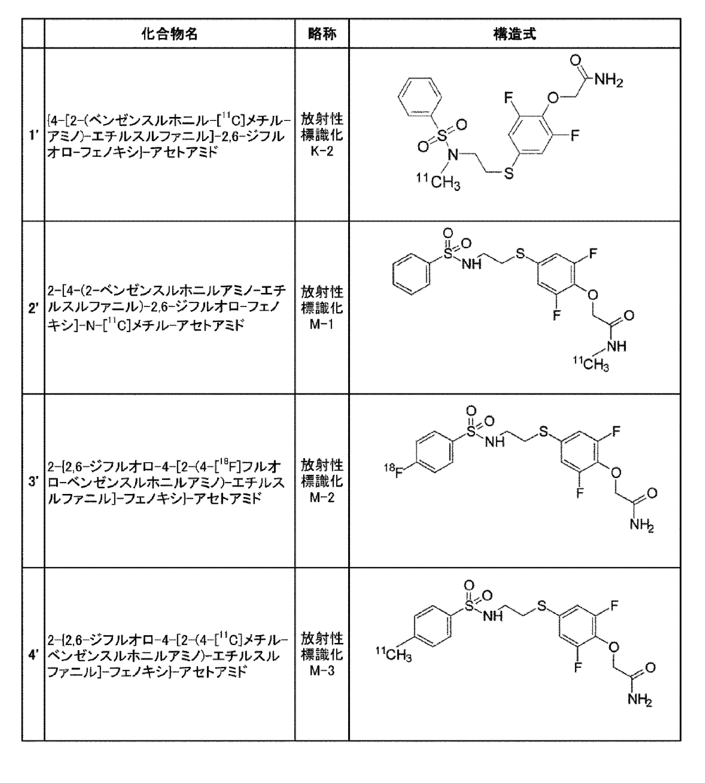
(定義)
用語「アルキル」とは、脂肪族飽和炭化水素の水素原子1個が失われて生じる1価の基を意味する。アルキルは、例えば、1~15個(C1-C15)の炭素原子、典型的には、1~10個(C1-C10)、1~8個(C1-C8)、1~6個(C1-C6)、1~5個(C1-C5)、1~4個(C1-C4)、1~3個(C1-C3)、1~2個(C1-C2)、又は2~6個(C2-C6)の炭素原子を有する。アルキルは、直鎖若しくは分枝状であってもよい。アルキルの例を挙げると、限定されないが、メチル、エチル、プロピル、イソプロピル、2-メチル-1-プロピル、2-メチル-2-プロピル、2-メチル-1-ブチル、3-メチル-1-ブチル、2-メチル-3-ブチル、2,2-ジメチル-1-プロピル、2-メチル-1-ペンチル、3-メチル-1-ペンチル、4-メチル-1-ペンチル、2-メチル-2-ペンチル、3-メチル-2-ペンチル、4-メチル-2-ペンチル、2,2-ジメチル-1-ブチル、3,3-ジメチル-1-ブチル、2-エチル-1-ブチル、n-ブチル、イソブチル、t-ブチル、ペンチル、イソペンチル、ネオペンチル、及びヘキシルなどがある。アルキルは、さらに適当な置換基によって置換されてもよい。
(Definition)
The term “alkyl” means a monovalent group resulting from the loss of one hydrogen atom of an aliphatic saturated hydrocarbon. Alkyl is, for example, 1 to 15 (C 1 -C 15 ) carbon atoms, typically 1 to 10 (C 1 -C 10 ), 1 to 8 (C 1 -C 8 ), 1 Up to 6 (C 1 -C 6 ), 1 to 5 (C 1 -C 5 ), 1 to 4 (C 1 -C 4 ), 1 to 3 (C 1 -C 3 ), 1 to 2 Having (C 1 -C 2 ) or 2 to 6 (C 2 -C 6 ) carbon atoms. Alkyl may be linear or branched. Examples of alkyl include, but are not limited to, methyl, ethyl, propyl, isopropyl, 2-methyl-1-propyl, 2-methyl-2-propyl, 2-methyl-1-butyl, 3-methyl-1-butyl 2-methyl-3-butyl, 2,2-dimethyl-1-propyl, 2-methyl-1-pentyl, 3-methyl-1-pentyl, 4-methyl-1-pentyl, 2-methyl-2-pentyl 3-methyl-2-pentyl, 4-methyl-2-pentyl, 2,2-dimethyl-1-butyl, 3,3-dimethyl-1-butyl, 2-ethyl-1-butyl, n-butyl, isobutyl , T-butyl, pentyl, isopentyl, neopentyl, and hexyl. The alkyl may be further substituted with a suitable substituent.
用語「アルケニル」とは、少なくとも1つの二重結合を持つ脂肪族不飽和炭化水素基を意味する。アルケニルは、例えば、2~15個(C2-C15)の炭素原子、典型的には、2~10個(C2-C10)、2~8個(C2-C8)、2~6個(C2-C6)、2~5個(C2-C5)、2~4個(C2-C4)、2~3個(C2-C3)、3~6個(C3-C6)、3~8個(C3-C8)、4~6個(C4-C6)、4~7個(C4-C7)、又は4~8個(C4-C8)の炭素原子を有する。アルケニルは、直鎖若しくは分枝状であってもよい。アルケニルの例を挙げると、限定されないが、具体的には、ビニル(-CH=CH2)、アリル(-CH2CH=CH2)、-CH=CH(CH3)、-CH=C(CH3)2、-C(CH3)=CH2、-C(CH3)=CH(CH3)、-C(CH2CH3)=CH2、1,3-ブタジエニル(-CH=CH-CH=CH2)、及びヘプタ-1,6-ジエン-4-イル(-CH2-(CH2CH=CH2)2)などがある。アルケニルは、さらに適当な置換基によって置換されてもよい。 The term “alkenyl” refers to an aliphatic unsaturated hydrocarbon group having at least one double bond. Alkenyl is, for example, 2 to 15 (C 2 -C 15 ) carbon atoms, typically 2 to 10 (C 2 -C 10 ), 2 to 8 (C 2 -C 8 ), 2 ˜6 (C 2 -C 6 ), 2 to 5 (C 2 -C 5 ), 2 to 4 (C 2 -C 4 ), 2 to 3 (C 2 -C 3 ), 3 to 6 (C 3 -C 6 ), 3-8 (C 3 -C 8 ), 4-6 (C 4 -C 6 ), 4-7 (C 4 -C 7 ), or 4-8 It has (C 4 -C 8 ) carbon atoms. Alkenyl may be linear or branched. Examples of alkenyl include, but are not limited to, vinyl (—CH═CH 2 ), allyl (—CH 2 CH═CH 2 ), —CH═CH (CH 3 ), —CH═C ( CH 3 ) 2 , —C (CH 3 ) ═CH 2 , —C (CH 3 ) ═CH (CH 3 ), —C (CH 2 CH 3 ) ═CH 2 , 1,3-butadienyl (—CH═CH -CH = CH 2 ), and hepta-1,6-dien-4-yl (-CH 2- (CH 2 CH = CH 2 ) 2 ). Alkenyl may be further substituted with a suitable substituent.
用語「アルキニル」とは、少なくとも1つの三重結合を持つ脂肪族不飽和炭化水素基を意味する。アルキニルは、例えば、2~15個(C2-C15)の炭素原子、典型的には、2~10個(C2-C10)、2~8個(C2-C8)、2~6個(C2-C6)、2~5個(C2-C5)、2~4個(C2-C4)、2~3個(C2-C3)、3~6個(C3-C6)、3~8個(C3-C8)、4~6個(C4-C6)、4~7個(C4-C7)、又は4~8個(C4-C8)の炭素原子を有する。アルキニルは、直鎖若しくは分枝状であってもよい。アルキニルの例を挙げると、限定されないが、エチニル(-C≡CH)、-C≡CH(CH3)、-C≡C(CH2CH3)、-CH2C≡CH、-CH2C≡C(CH3)、及び-CH2C≡C(CH2CH3)などがある。アルキニルは、さらに適当な置換基によって置換されてもよい。
The term “alkynyl” refers to an aliphatic unsaturated hydrocarbon group having at least one triple bond. Alkynyl is, for example, 2 to 15 (C 2 -C 15 ) carbon atoms, typically 2 to 10 (C 2 -C 10 ), 2 to 8 (C 2 -C 8 ), 2 ˜6 (C 2 -C 6 ), 2 to 5 (C 2 -C 5 ), 2 to 4 (C 2 -C 4 ), 2 to 3 (C 2 -C 3 ), 3 to 6 (C 3 -C 6 ), 3-8 (C 3 -C 8 ), 4-6 (C 4 -C 6 ), 4-7 (C 4 -C 7 ), or 4-8 It has (C 4 -C 8 ) carbon atoms. Alkynyl may be linear or branched. Examples of alkynyl include, but are not limited to, ethynyl (-C≡CH), - C≡CH (CH 3), - C≡C (
用語「ハロゲン」又は「ハロ」は、フルオロ(-F)、クロロ(-Cl)、ブロモ(-Br)、及びヨード(-I)を意味する。 The term “halogen” or “halo” means fluoro (—F), chloro (—Cl), bromo (—Br), and iodo (—I).
用語「医薬として許容し得る塩」とは、哺乳動物、特にヒトに対して有害でない塩を指す。医薬として許容し得る塩は、無機酸若しくは無機塩基、又は有機酸若しくは有機塩基を含む、無毒性の酸又は塩基を用いて形成することができる。医薬として許容し得る塩の例を挙げると、アルミニウム、カルシウム、リチウム、マグネシウム、カリウム、ナトリウム及び亜鉛などから形成される金属塩、又はリジン、N,N’’-ジベンジルエチレンジアミン、クロロプロカイン、コリン、ジエタノールアミン、エチレンジアミン、メグルミン(N-メチルグルカミン)及びプロカインなどから形成される有機塩などがある。また、医薬として許容し得る塩は、酸付加塩及び塩基付加塩を包含する。 The term “pharmaceutically acceptable salt” refers to a salt that is not harmful to mammals, particularly humans. Pharmaceutically acceptable salts can be formed with nontoxic acids or bases, including inorganic acids or bases, or organic acids or bases. Examples of pharmaceutically acceptable salts include metal salts formed from aluminum, calcium, lithium, magnesium, potassium, sodium and zinc, or lysine, N, N ″ -dibenzylethylenediamine, chloroprocaine, choline Organic salts formed from diethanolamine, ethylenediamine, meglumine (N-methylglucamine) and procaine. Pharmaceutically acceptable salts include acid addition salts and base addition salts.
用語「溶媒和物」とは、本発明化合物に対する1つ又は複数の溶媒分子の会合により形成される含溶媒化合物を意味する。溶媒和物は、例えば、一溶媒和物、二溶媒和物、三溶媒和物、及び四溶媒和物を含む。また、溶媒和物は、水和物を含む。 The term “solvate” means a solvent-containing compound formed by the association of one or more solvent molecules with the compound of the present invention. Solvates include, for example, monosolvates, disolvates, trisolvates, and tetrasolvates. Solvates include hydrates.
(製造方法及び中間体)
(合成例1)
R2がアルキル、アルケニル又はアルキニルである式(I)の化合物又はその医薬として許容し得る塩若しくは溶媒和物は、例えば、下記式(II)の化合物又はその医薬として許容し得る塩若しくは溶媒和物:

(Synthesis Example 1)
A compound of formula (I) or a pharmaceutically acceptable salt or solvate thereof wherein R 2 is alkyl, alkenyl or alkynyl includes, for example, a compound of formula (II) below or a pharmaceutically acceptable salt or solvate thereof object:
該反応は、ジメチルホルムアミド(DMF)、テトラヒドロフラン、アセトニトリル、アセトン又はジメチルスルホキシドなどの極性非プロトン性溶媒中で行うことができる。また、該反応は、NaOHなどの塩基を用いて、塩基性条件下で行うことが好ましい。反応温度は、室温~還流温度、特に、60~100℃が好ましく、より好ましくは80℃である。反応時間は、1分~10分、特に5分である。 The reaction can be carried out in a polar aprotic solvent such as dimethylformamide (DMF), tetrahydrofuran, acetonitrile, acetone or dimethyl sulfoxide. The reaction is preferably performed under basic conditions using a base such as NaOH. The reaction temperature is preferably room temperature to reflux temperature, particularly preferably 60 to 100 ° C., more preferably 80 ° C. The reaction time is 1 to 10 minutes, in particular 5 minutes.
PETプローブは、通常、放射性同位体の短い半減期のため、短い時間でかつ高収率で製造しなければならない。該反応は、短い時間で定量的に進行するため、PETプローブの製造に適している。 PET probes usually have to be manufactured in a short time and in high yield due to the short half-life of radioisotopes. Since the reaction proceeds quantitatively in a short time, it is suitable for the production of a PET probe.
本発明者らは、式(II)の化合物とX1-R2との反応が、式(II)の化合物のA基に隣接するNH基で定量的に起こることを見出した。したがって、たとえR3及びR4が水素であったとしても、保護基を使用せずに該NH基のみをN-R2基に変換することができる。 The inventors have found that the reaction of the compound of formula (II) with X 1 -R 2 occurs quantitatively at the NH group adjacent to the A group of the compound of formula (II). Therefore, even if R 3 and R 4 are hydrogen, only the NH group can be converted to an NR 2 group without using a protecting group.
式(II)の化合物又はその医薬として許容し得る塩若しくは溶媒和物は、R2がアルキル、アルケニル又はアルキニルである式(I)の化合物又はその医薬として許容し得る塩若しくは溶媒和物を製造するための中間体として使用することができる。また、式(II)の化合物又はその医薬として許容し得る塩若しくは溶媒和物は、R2が[11C]アルキル、[11C]アルケニル、又は[11C]アルキニルである放射性標識された式(I)の化合物又はその医薬として許容し得る塩若しくは溶媒和物を製造するための中間体として使用することができる。 A compound of formula (II) or a pharmaceutically acceptable salt or solvate thereof produces a compound of formula (I) or a pharmaceutically acceptable salt or solvate thereof wherein R 2 is alkyl, alkenyl or alkynyl. It can be used as an intermediate for Also, the compound of formula (II) or a pharmaceutically acceptable salt or solvate thereof is a radiolabeled formula wherein R 2 is [ 11 C] alkyl, [ 11 C] alkenyl, or [ 11 C] alkynyl. It can be used as an intermediate for producing a compound of (I) or a pharmaceutically acceptable salt or solvate thereof.
(合成例2)
R1がアルキル、アルケニル、又はアルキニルである式(I)の化合物、又はその医薬として許容し得る塩若しくは溶媒和物は、例えば、下記式(III)の化合物、又はその医薬として許容し得る塩若しくは溶媒和物:

A compound of formula (I), wherein R 1 is alkyl, alkenyl, or alkynyl, or a pharmaceutically acceptable salt or solvate thereof is, for example, a compound of formula (III) below, or a pharmaceutically acceptable salt thereof: Or solvate:
式(III)の化合物の具体例は、下記である。

該反応は、パラジウム触媒、ホスフィンリガンド、炭酸塩及びハロゲン化銅の存在下で行うことができる。該パラジウム触媒は、例えば、トリス(ジベンジリデンアセトン)ジパラジウムなどがある。また、該ホスフィンリガンドは、例えば、トリ(o-トリル)ホスフィン又は(ジ-tert-ブチル)メチルホスフィンなどがある。該炭酸塩は、K2CO3などがある。該ハロゲン化銅は、CuClなどがある。該反応は、ジメチルホルムアミド(DMF)、テトラヒドロフラン、アセトニトリル、アセトン又はジメチルスルホキシドなどの極性非プロトン性溶媒中で行うことができる。反応温度は、室温~還流温度、特に、60~100℃が好ましく、より好ましくは80℃である。反応時間は、1分~10分、特に5分である。 The reaction can be performed in the presence of a palladium catalyst, a phosphine ligand, a carbonate and a copper halide. Examples of the palladium catalyst include tris (dibenzylideneacetone) dipalladium. Examples of the phosphine ligand include tri (o-tolyl) phosphine and (di-tert-butyl) methylphosphine. Examples of the carbonate include K 2 CO 3 . Examples of the copper halide include CuCl. The reaction can be carried out in a polar aprotic solvent such as dimethylformamide (DMF), tetrahydrofuran, acetonitrile, acetone or dimethyl sulfoxide. The reaction temperature is preferably room temperature to reflux temperature, particularly preferably 60 to 100 ° C., more preferably 80 ° C. The reaction time is 1 to 10 minutes, in particular 5 minutes.
PETプローブは、通常、放射性同位体の短い半減期のため、短い時間でかつ高収率で製造しなければならない。該反応は、短い時間で定量的に進行するため、PETプローブの製造に適している。 PET probes usually have to be manufactured in a short time and in high yield due to the short half-life of radioisotopes. Since the reaction proceeds quantitatively in a short time, it is suitable for the production of a PET probe.
式(III)の化合物又はその医薬として許容し得る塩若しくは溶媒和物は、R1がアルキル、アルケニル、又はアルキニルである式(I)の化合物又はその医薬として許容し得る塩若しくは溶媒和物を製造するための中間体として使用することができる。また、式(III)の化合物又はその医薬として許容し得る塩若しくは溶媒和物は、R1が[11C]アルキル、[11C]アルケニル、又は[11C]アルキニルである放射性標識された式(I)の化合物又はその医薬として許容し得る塩若しくは溶媒和物を製造するための中間体として使用することができる。 A compound of formula (III) or a pharmaceutically acceptable salt or solvate thereof is a compound of formula (I) or a pharmaceutically acceptable salt or solvate thereof wherein R 1 is alkyl, alkenyl, or alkynyl. It can be used as an intermediate for manufacturing. A compound of formula (III) or a pharmaceutically acceptable salt or solvate thereof is a radiolabeled formula wherein R 1 is [ 11 C] alkyl, [ 11 C] alkenyl, or [ 11 C] alkynyl. It can be used as an intermediate for producing a compound of (I) or a pharmaceutically acceptable salt or solvate thereof.
式(I)の化合物又はその医薬として許容し得る塩若しくは溶媒和物は、下記実施例に示す方法によっても製造することができる。 The compound of formula (I) or a pharmaceutically acceptable salt or solvate thereof can also be produced by the methods shown in the following examples.
(プログラム)
本発明の一実施形態は、前述のイメージング方法をコンピュータに実行させるためのプログラムである。具体的に、プログラムによってコンピュータは、イメージング装置を制御し、霊長類生体の脳内AMPA受容体をイメージングする。
(program)
One embodiment of the present invention is a program for causing a computer to execute the above-described imaging method. Specifically, the computer controls the imaging apparatus according to the program to image the AMPA receptor in the brain of the primate organism.
(診断薬、コンパニオン診断薬、医薬)
本発明の一実施形態は、霊長類体の脳内AMPA受容体が関連する疾患の診断薬、又は疾患の治療又は予防のためのコンパニオン診断薬である。なお、ここで治療のためのコンパニオン診断薬とは、脳内AMPA受容体が関連する疾患であることが判明した場合に、治療が見込めるかどうかを判断するため診断薬である。また、ここで予防のためのコンパニオン診断薬とは、脳内AMPA受容体が関連する疾患であることが判明した場合に、今後の疾患病勢(予後)を推測する、もしくはそれ以上の進行を抑制する予防が見込めるかどうかを判断するため診断薬である。
(Diagnostics, companion diagnostics, pharmaceuticals)
One embodiment of the present invention is a diagnostic agent for a disease associated with cerebral AMPA receptors in the primate body, or a companion diagnostic agent for treatment or prevention of a disease. Here, the companion diagnostic agent for treatment is a diagnostic agent for determining whether treatment is expected when it is found that the brain AMPA receptor is a related disease. In addition, when the companion diagnostic agent for prevention is found to be a disease related to the brain AMPA receptor, it is assumed that the disease state (prognosis) in the future or further progression is suppressed. It is a diagnostic agent to judge whether prevention is possible.
上記診断薬を用いて、霊長類生体の対象から得られる脳内AMPA受容体の分布及び/又は発現量に関するデータを、上記疾患と、脳内AMPA受容体の分布及び/又は発現量との相関性に照会することで、対象の上記疾患に関する診断(具体的には、上記疾患の罹患有無、重篤性、発作可能性等)が可能である。 Correlation between the above-mentioned disease and the distribution and / or expression level of the AMPA receptor in the brain using the above diagnostic agent and the data on the distribution and / or expression level of the brain AMPA receptor obtained from the subject of the primate organism By referring to sex, it is possible to make a diagnosis (specifically, the presence or absence of the disease, severity, seizure possibility, etc.) of the target disease.
また、上記コンパニオン診断薬を用いて、霊長類生体の対象から得られる脳内AMPA受容体の分布及び/又は発現量に関するデータを、上記疾患と、脳内AMPA受容体の分布及び/又は発現量との相関性に照会することで、対象の上記疾患状態が把握されるので、これに基づき、上記疾患の予防/治療計画(投与する予防/治療薬の種類や組合せ、用量、用法など)を策定することができる。 Further, using the above-mentioned companion diagnostic agent, data on the distribution and / or expression level of brain AMPA receptor obtained from a subject of a primate organism, the distribution of the above disease, and / or the distribution and / or expression level of AMPA receptor in the brain By referring to the correlation with the above, the above-mentioned disease state of the subject is grasped, and based on this, the prevention / treatment plan for the above-mentioned disease (type / combination of preventive / therapeutic drugs to be administered, dosage, usage, etc.) is determined. Can be formulated.
例えば、AMPA受容体の発現量の減少が把握された対象には、AMPA受容体機能活性化薬の投与を推奨でき、AMPA受容体の発現量の増加が把握された対象には、AMPA受容体拮抗薬の投与を推奨できる。たとえば、現在の疾患分類(DSM-VやICD-10など)により臨床的にうつ病と診断された疾患群においても、AMPA受容体の発現量の増加を認める症例もあれば、変化なしないし減少する症例もある。その場合は、臨床診断上はうつ病であっても、AMPA受容体増加型うつ病には拮抗薬を投与するなど、テーラーメイド型の診断・治療が行われることとなる。また、AMPA受容体の発現量の減少/増加幅や、分布異常(健常者を正常としたとき)の類型や度合いに応じて、AMPA受容体機能活性化薬/AMPA受容体拮抗薬の種類(生体内代謝時間の違いや有効血中濃度の違い)、投与量、投与頻度、投与タイミング(例えば、症状の発作の予兆に相関するAMPA受容体の発現量又は分布が把握された場合に、予防的に薬剤を投与する)を設定することができる。 For example, administration of an AMPA receptor function activator can be recommended for subjects whose decrease in expression level of AMPA receptor is known, and AMPA receptor can be recommended for subjects whose increase in expression amount of AMPA receptor is known. The administration of antagonists can be recommended. For example, even in a group of diseases clinically diagnosed with depression according to the current disease classification (DSM-V, ICD-10, etc.), there are cases in which increased expression of AMPA receptors is observed, and there is no change or decrease. There are some cases. In that case, even if the clinical diagnosis is depression, tailor-made diagnosis and treatment such as administration of an antagonist is performed for depression with increased AMPA receptor. Depending on the decrease / increase in the expression level of AMPA receptor and the type and degree of distribution abnormality (when normal is normal), the type of AMPA receptor function activator / AMPA receptor antagonist ( Prevention in cases where the expression level or distribution of the AMPA receptor correlates with the signs of symptom seizures, etc. To administer the drug automatically).
つまり、本発明の一実施形態は、霊長類体の脳内AMPA受容体が関連する疾患の治療又は予防のための医薬であって、霊長類生体の脳内AMPA受容体に選択的に結合する物質を有効成分とし、上記したイメージング方法で得られる脳内AMPA受容体の分布及び/又は発現量に関するデータに基づく投与計画で投与される医薬にも関する。 That is, one embodiment of the present invention is a medicament for treating or preventing a disease associated with a primate brain AMPA receptor, which selectively binds to a brain AMPA receptor in a primate organism. The present invention also relates to a medicine that is administered in a dosage schedule based on data on the distribution and / or expression level of AMPA receptors in the brain obtained by the above-described imaging method using a substance as an active ingredient.
一実施形態に係る医薬(コンパニオン診断薬とともに用いられてもよい)であるAMPA受容体拮抗薬としては、特に限定されないが、てんかん病に対するペランパネル水和物(エーザイ社)、talampanel(テバ社)が挙げられる。 The AMPA receptor antagonist, which is a medicine according to one embodiment (which may be used together with a companion diagnostic agent), is not particularly limited, but peranpanel hydrate (Eisai Co., Ltd.), talampane (Teva Co.) for epilepsy disease. Is mentioned.
一実施形態に係る医薬(コンパニオン診断薬とともに用いられてもよいAMPA受容体機能活性化薬)としては、特に限定されないが、式(I)の化合物、又はその医薬として許容し得る塩若しくは溶媒和物であってよい。
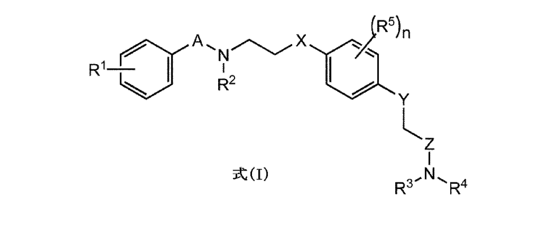
X及びYは、それぞれ独立に、S又はOであり、好ましくは、XがSであり、かつYがOである。
R1~R4は、それぞれ独立に、水素、アルキル、アルケニル、アルキニル又はハロである。一実施態様において、R1~R4の全てが水素となることはない、すなわち、R1~R4の少なくとも1つは水素以外である。一実施態様において、R2はアルキルである。他の実施態様において、R1はアルキル又はハロである。R1は、オルト位、メタ位、又はパラ位のいずれかに存在することができる。好ましくは、R1は、パラ位に存在する。さらに他の実施態様において、R3及びR4のうちの一方が、水素であり、他方がアルキルである。最も好ましくは、R1、R3及びR4は、それぞれ独立に、水素、アルキル、アルケニル、アルキニル又はハロであり、R2は、アルキル、アルケニル又はアルキニルである。
R5は、出現ごとにそれぞれ独立に、アルキル、アルケニル、アルキニル又はハロである。好ましくは、R5は、ハロであり、特に好ましくは、フルオロである。さらに好ましくは、R5は、Y基に対して両方のオルト位(すなわち、X基に対して両方のメタ位)に存在する。
nは、0~4の整数である。好ましくは、nは2である。
Although it does not specifically limit as a pharmaceutical (AMPA receptor function activator which may be used with a companion diagnostic agent) which concerns on one Embodiment, The pharmacologically acceptable salt or solvation thereof. It can be a thing.
X and Y are each independently S or O, preferably X is S and Y is O.
R 1 to R 4 are each independently hydrogen, alkyl, alkenyl, alkynyl or halo. In one embodiment, not all of R 1 -R 4 are hydrogen, ie, at least one of R 1 -R 4 is other than hydrogen. In one embodiment, R 2 is alkyl. In other embodiments, R 1 is alkyl or halo. R 1 can be present in any of the ortho, meta, or para positions. Preferably R 1 is in the para position. In yet another embodiment, one of R 3 and R 4 is hydrogen and the other is alkyl. Most preferably, R 1 , R 3 and R 4 are each independently hydrogen, alkyl, alkenyl, alkynyl or halo and R 2 is alkyl, alkenyl or alkynyl.
R 5 is each independently alkyl, alkenyl, alkynyl or halo for each occurrence. Preferably R 5 is halo, particularly preferably fluoro. More preferably, R 5 is present in both ortho positions relative to the Y group (ie both meta positions relative to the X group).
n is an integer of 0-4. Preferably n is 2.
さらに他の実施態様において、式(I)の化合物における各置換基の組み合わせとしては、A及びZがそれぞれ独立に、CO、SO又はSO2であり、X及びYがそれぞれ独立に、S又はOであり、R1、R3及びR4がそれぞれ独立に、水素、アルキル、アルケニル、アルキニル又はハロであり、R2がアルキル、アルケニル、又はアルキニルであり、R5が出現ごとにそれぞれ独立に、アルキル、アルケニル、アルキニル又はハロであり、nが0~4の整数である組み合わせが好ましい。 In yet another embodiment, the combinations of substituents in the compound of formula (I) are as follows: A and Z are each independently CO, SO or SO 2 , X and Y are each independently S or O R 1 , R 3 and R 4 are each independently hydrogen, alkyl, alkenyl, alkynyl or halo, R 2 is alkyl, alkenyl or alkynyl and R 5 is each independently for each occurrence, A combination wherein alkyl, alkenyl, alkynyl or halo, and n is an integer of 0 to 4 is preferable.
さらに他の実施態様において、式(I)の化合物における各置換基の組み合わせとしては、AがSO2であり、ZがCOであり、XがSであり、YがOであり、R2がアルキルであり、R1が水素、アルキル又はハロであり、R1がアルキル又はハロである場合、R1はパラ位に存在し、R3及びR4のうち一方が水素であり、他方がアルキルであり、R5は、出現ごとにそれぞれ独立に、アルキル、アルケニル、アルキニル又はハロであり、nは、0~4の整数である組み合わせが好ましい。 In yet another embodiment, the combinations of substituents in the compound of formula (I) include: A is SO 2 , Z is CO, X is S, Y is O, and R 2 is When it is alkyl, R 1 is hydrogen, alkyl or halo and R 1 is alkyl or halo, R 1 is in the para position, one of R 3 and R 4 is hydrogen and the other is alkyl R 5 is each independently alkyl, alkenyl, alkynyl or halo, and n is preferably a combination of 0 to 4 integers.
さらに他の実施態様において、式(I)の化合物における各置換基の組み合わせとしては、AがSO2であり、ZがCOであり、XがSであり、YがOであり、R2がアルキルであり、R1が水素、アルキル又はハロであり、R1がアルキル又はハロである場合、R1はパラ位に存在し、R3及びR4のうち一方が、水素であり、他方がアルキルであり、R5がハロ、特にフルオロであって、R5はY基に対して両方のオルト位(すなわち、X基に対して両方のメタ位)に存在し、nが2である組み合わせが好ましい。 In yet another embodiment, the combinations of substituents in the compound of formula (I) include: A is SO 2 , Z is CO, X is S, Y is O, and R 2 is When alkyl, R 1 is hydrogen, alkyl or halo and R 1 is alkyl or halo, R 1 is in the para position, one of R 3 and R 4 is hydrogen and the other is A combination in which R 5 is halo, in particular fluoro, R 5 is in both ortho positions with respect to the Y group (ie both meta positions with respect to the X group) and n is 2 Is preferred.
さらに他の実施態様において、式(I)の化合物における各置換基の組み合わせとしては、AがSO2であり、ZがCOであり、XがSであり、YがOであり、R2がアルキルであり、R1が水素、アルキル又はハロであり、R1がアルキル又はハロである場合、R1はパラ位に存在し、R3及びR4が共に水素であり、R5は、出現ごとにそれぞれ独立に、アルキル、アルケニル、アルキニル又はハロであり、nは、0~4の整数である組み合わせが好ましい。 In yet another embodiment, the combinations of substituents in the compound of formula (I) include: A is SO 2 , Z is CO, X is S, Y is O, and R 2 is When it is alkyl, R 1 is hydrogen, alkyl or halo and R 1 is alkyl or halo, R 1 is in the para position, R 3 and R 4 are both hydrogen, and R 5 is Each independently represents a combination of alkyl, alkenyl, alkynyl or halo, and n is an integer of 0 to 4.
式(I)の化合物の具体的な例を挙げると、以下の化合物がある:

製造方法及び中間体は、前述と同様である。 The production method and intermediate are the same as described above.
AMPA受容体機能活性化薬/AMPA受容体拮抗薬は、経口的あるいは非経口的に投与することができる。経口投与剤としては、散剤、顆粒剤、カプセル剤、錠剤などの固形製剤あるいはシロップ剤、エリキシル剤などの液状製剤とすることができる。また、非経口投与剤として注射剤(静脈、筋肉など)、直腸投与剤、皮膚外用剤、吸入剤とすることができる。これらの製剤は有効成分に薬学的に認容である製造助剤を加えることにより常法に従って製造される。更に公知の技術により持続性製剤とすることも可能である。 The AMPA receptor function activator / AMPA receptor antagonist can be administered orally or parenterally. Oral administration agents can be solid preparations such as powders, granules, capsules and tablets, or liquid preparations such as syrups and elixirs. Further, as parenteral administration agents, injections (vein, muscle, etc.), rectal administration agents, external preparations for skin, and inhalants can be used. These preparations are produced according to a conventional method by adding a pharmaceutically acceptable production aid to the active ingredient. Furthermore, it is possible to obtain a sustained-release preparation by a known technique.
霊長類体の脳内AMPA受容体が関連する疾患は、特に限定されないが精神疾患又は神経疾患であってよく、例えば、
(1)うつ病、大うつ病、双極性うつ病、気分変調障害、情動障害、再発性うつ病、産後うつ病、ストレス性障害、うつ症状、躁病、不安、全般性不安障害、不安症候群、パニック障害、恐怖症、社会性恐怖症、社会性不安障害、強迫性障害、心的外傷後ストレス症候群、外傷後ストレス障害、タウレット症候群、自閉症、脆弱X症候群、レット症候群、適応障害、双極性障害、神経症、統合失調症、慢性疲労症候群、不安神経症、強迫神経症、恐慌性障害、てんかん、神経過敏症、注意欠陥多動性障害、精神病性大うつ病、難治性大うつ病、治療抵抗性うつ病などの精神疾患
(2)アルツハイマー病、アルツハイマー型老人性認知症、パーキンソン病、ハンチントン舞踏病、多発脳梗塞性認知症、前頭側頭認知症、パーキンソン型前頭側頭認知症、進行性核上麻痺、ピック症候群、ニーマン-ピック症候群、大脳皮質基底核変性症、ダウン症、欠陥性認知症、レヴィー小体認知症、筋委縮性脊髄側索硬化症、運動神経原性疾患、クロイツフェルト・ヤコブ病、脳性麻痺、進行性核上麻痺、多発性硬化症などの神経変性疾患
(3)加齢性記憶障害、老人性認知症などの加齢に伴う認知・記憶障害
(4)内在因性睡眠障害、外在因性睡眠障害、概日リズム障害、睡眠時随伴症、内科又は精神科障害に伴う睡眠障害、ストレス性不眠症、不眠症、不眠性神経症、睡眠時無呼吸症候群などの睡眠障害
(5)麻酔薬、外傷性疾患、又は神経変性疾患などに起因する呼吸抑制
(6)外傷性脳損傷、脳卒中、神経性食欲不振、摂食障害、神経性無食欲症、過食症、その他の摂食障害、アルコール依存症、アルコール乱用、アルコール性健忘症、アルコール妄想症、アルコール嗜好性、アルコール離脱、アルコール性精神病、アルコール中毒、アルコール性嫉妬、アルコール性躁病、アルコール依存性精神障害、アルコール精神病、薬物嗜好、薬物恐怖症、薬物狂、薬物離脱、偏頭痛、ストレス性頭痛、緊張性頭痛、糖尿病性ニューロパシー、肥満、糖尿病、筋肉痙攣、メニエール病、自律神経失調症、脱毛症、緑内障、難聴、高血圧、心臓病、頻脈、うっ血性心不全、過呼吸、気管支喘息、無呼吸、乳幼児突然死症候群、炎症性疾患、アレルギー疾患、インポテンス、更年期障害、不妊症、癌、HIV感染による免疫不全症候群、脳脊髄膜炎、末端肥大症、失禁、メタボリック・シンドローム、骨粗しょう症、消化性潰瘍、過敏性腸症候群、炎症性腸疾患、潰瘍性大腸炎、クローン病、ストレス性胃腸障害、神経性嘔吐、消化性潰瘍、下痢、便秘、術後イレウス
などが挙げられる。
Diseases associated with primate brain AMPA receptors may be, but are not limited to, psychiatric or neurological diseases, for example,
(1) Depression, major depression, bipolar depression, mood disorders, affective disorders, recurrent depression, postpartum depression, stress disorder, depressive symptoms, mania, anxiety, generalized anxiety disorder, anxiety syndrome, Panic disorder, phobia, social phobia, social anxiety disorder, obsessive-compulsive disorder, post-traumatic stress syndrome, post-traumatic stress disorder, taurette syndrome, autism, fragile X syndrome, Rett syndrome, adjustment disorder, bipolar Disorder, neurosis, schizophrenia, chronic fatigue syndrome, anxiety, obsessive-compulsive disorder, panic disorder, epilepsy, irritability, attention deficit hyperactivity disorder, psychotic major depression, refractory major depression , Psychiatric disorders such as treatment-resistant depression (2) Alzheimer's disease, Alzheimer-type senile dementia, Parkinson's disease, Huntington's chorea, multiple cerebral infarction dementia, frontotemporal dementia, Parkinson's frontotemporal Dementia, progressive supranuclear palsy, Pick syndrome, Niemann-Pick syndrome, basal ganglia degeneration, Down syndrome, defective dementia, Lewy body dementia, amyotrophic spinal sclerosis, motor neurogenicity Diseases, Creutzfeldt-Jakob disease, cerebral palsy, progressive supranuclear palsy, multiple sclerosis and other neurodegenerative diseases (3) age-related memory impairment, cognitive / memory impairment associated with aging such as senile dementia ( 4) Endogenous sleep disorder, extrinsic sleep disorder, circadian rhythm disorder, parasomnia, sleep disorder associated with internal medicine or psychiatric disorder, stress insomnia, insomnia, insomnia neuropathy, sleep Sleep disorders such as apnea syndrome (5) Respiratory depression caused by anesthetics, traumatic diseases, or neurodegenerative diseases (6) Traumatic brain injury, stroke, anorexia nervosa, eating disorders, anorexia nervosa , Bulimia, other eating disorders, alcohol Alcohol abuse, Alcohol amnesia, Alcohol delusion, Alcoholism, Alcohol withdrawal, Alcohol psychosis, Alcoholism, Alcoholism, Alcoholism, Alcoholism, Alcoholism, Alcoholism, Drug preference, Drug Phobia, drug madness, drug withdrawal, migraine, stress headache, tension headache, diabetic neuropathy, obesity, diabetes, muscle spasms, Meniere's disease, autonomic ataxia, alopecia, glaucoma, hearing loss, hypertension, heart disease , Tachycardia, congestive heart failure, hyperventilation, bronchial asthma, apnea, sudden infant death syndrome, inflammatory disease, allergic disease, impotence, menopause, infertility, cancer, immunodeficiency syndrome due to HIV infection, encephalomyelitis , Acromegaly, incontinence, metabolic syndrome, osteoporosis, peptic ulcer, irritable bowel symptoms Group, inflammatory bowel disease, ulcerative colitis, Crohn's disease, stress gastrointestinal disorder, neurogenic vomiting, peptic ulcer, diarrhea, constipation, postoperative ileus and the like.
本発明の一実施形態は、霊長類体の脳内AMPA受容体が関連する疾患の治療又は予防のための医薬である。 One embodiment of the present invention is a medicament for the treatment or prevention of diseases associated with cerebral AMPA receptors in the primate body.
上記疾患は、特に限定されないが、癲癇、うつ病、統合失調症、脳虚血、パーキンソン病、アルツハイマー病、自閉症、注意多動性障害(ADHD)及び多発性硬化症からなる群より選ばれる1種以上であってよい。 The above diseases are not particularly limited, but are selected from the group consisting of epilepsy, depression, schizophrenia, cerebral ischemia, Parkinson's disease, Alzheimer's disease, autism, attention hyperactivity disorder (ADHD) and multiple sclerosis It may be one or more.
本実施形態に係る薬は、医薬として許容し得る担体を含むことができる。
医薬として許容し得る担体としては、特に制限されないが、例えば、滅菌水、食塩水、生理食塩水又はリン酸緩衝食塩水(PBS)、塩化ナトリウム注射液、リンゲル注射液、等張性デキストロース注射液、無菌水注射液、デキストロース、及び乳酸リンゲル注射液などがある。
The drug according to this embodiment can include a pharmaceutically acceptable carrier.
The pharmaceutically acceptable carrier is not particularly limited. For example, sterile water, saline, physiological saline or phosphate buffered saline (PBS), sodium chloride injection, Ringer's injection, isotonic dextrose injection , Sterile water injection, dextrose, and lactated Ringer's injection.
(スクリーニング方法)
本発明の一実施形態は、霊長類体の脳内AMPA受容体が関連する疾患の治療又は予防薬のスクリーニング方法である。この方法は、霊長類生体への候補物質の投与前後における、上記したイメージング方法で得られる脳内AMPA受容体の分布及び/又は発現量に関するデータの差異に基づき、候補物質を選抜する工程を有する。
(Screening method)
One embodiment of the present invention is a screening method for a therapeutic or prophylactic agent for a disease associated with a primate brain AMPA receptor. This method includes a step of selecting candidate substances based on the difference in data regarding the distribution and / or expression level of AMPA receptors in the brain obtained by the above-described imaging method before and after administration of the candidate substance to a primate organism. .
具体的には、霊長類生体への候補物質の投与前後における、脳内AMPA受容体の発現量の増加/減少幅や、分布異常(健常者を正常としたとき)の類型や度合いの変化に基づき、候補物質をAMPA受容体機能活性化薬/AMPA受容体拮抗薬として選抜することができる。これにより、従来は一括りにされてきた上記疾患を脳内AMPA受容体の発現量又は分布によって細かく分類し、分類ごとに適切な治療又は予防薬を作製することが期待できる。 Specifically, the increase / decrease in the expression level of AMPA receptor in the brain before and after the administration of the candidate substance to the primate organism, and the change in the type and degree of distribution abnormality (when normal subjects are normal) Based on this, the candidate substance can be selected as an AMPA receptor function activator / AMPA receptor antagonist. As a result, it is expected that the above-mentioned diseases that have conventionally been grouped are finely classified according to the expression level or distribution of brain AMPA receptors, and appropriate therapeutic or preventive drugs are prepared for each classification.
一実施形態において、選抜された候補物質が、霊長類体の脳内AMPA受容体が関連する疾患の治療又は予防効果を実際に有するか否かを確認(動物実験、ヒトでの試験等)し、上記効果を実際に有することに基づいてさらに選抜してもよい。 In one embodiment, it is confirmed whether the selected candidate substance actually has a therapeutic or prophylactic effect for a disease associated with a primate brain AMPA receptor (animal experiment, human test, etc.). Further, selection may be made based on the fact that the above-mentioned effect is actually provided.
(システム)
本発明の一実施形態に係るシステムは、入力端末と、サーバと、出力端末と、を備える。
(system)
A system according to an embodiment of the present invention includes an input terminal, a server, and an output terminal.
入力端末は、霊長類生体の脳内AMPA受容体の分布及び/又は発現量に関する情報をサーバへと送信する。この情報は、上記した本発明のイメージング方法で取得することができる。 The input terminal transmits information on the distribution and / or expression level of AMPA receptors in the brain of the primate organism to the server. This information can be acquired by the imaging method of the present invention described above.
一実施形態におけるサーバは、霊長類体の脳内AMPA受容体の分布及び/又は発現量と、霊長類体の脳内AMPA受容体が関連する疾患の状態と、が関連づけられたデータを格納するデータベースを備える。サーバは、入力された被験者の生体の脳内AMPA受容体の分布及び/又は発現量に関する情報と、データベース内のデータとを照会し、被験者の疾患の状態に関する情報を生成し(情報作成部)、この情報を出力端末へと送信する手段をさらに備える。このサーバを備えるシステムは、被験者の上記疾患の状態を正確に提示することができる。疾患の状態とは、例えば疾患の罹患有無、重篤性、発作可能性を包含する。 In one embodiment, the server stores data in which the distribution and / or expression level of AMPA receptors in the primate brain and the disease state associated with the AMPA receptors in the primate brain are associated with each other. Provide a database. The server inquires the input information on the distribution and / or expression level of AMPA receptors in the brain of the subject's living body and the data in the database, and generates information on the disease state of the subject (information creation unit) And means for transmitting this information to the output terminal. A system including this server can accurately present the state of the disease of the subject. The disease state includes, for example, the presence or absence of disease, seriousness, and seizure possibility.
例えば、上記の情報作成部は、データベースから、被験者の脳内AMPA受容体の分布及び/又は発現量に同一又は類似するデータを選別し、疾患の状態に関する情報を生成することができる。 For example, the information creation unit can select data that is the same as or similar to the distribution and / or expression level of the AMPA receptor in the brain of the subject from the database, and generate information related to the disease state.
別の実施形態におけるサーバは、霊長類体の脳内AMPA受容体の分布及び/又は発現量と、霊長類体の脳内AMPA受容体が関連する疾患の状態と、既に投与された霊長類体の生体の脳内AMPA受容体が関連する疾患の治療又は予防のための医薬の種類、用量及び/又は用法と、が関連づけられたデータを格納するデータベースを備える。サーバは、入力された被験者の生体の脳内AMPA受容体の分布及び/又は発現量に関する情報と、前記データとを照会し、被験者への推奨される医薬の種類、用量及び/又は用法に関する情報を生成し(情報作成部)、この情報を出力端末へと送信する手段をさらに備える。このサーバを備えるシステムは、被験者に応じた適切な医薬の種類、用量及び/又は用法を推奨することができる。具体的な態様は、コンパニオン診断薬及び医薬について上記したのと同様であってよい。 In another embodiment, the server includes a distribution and / or expression level of a primate brain AMPA receptor, a disease state associated with the primate brain AMPA receptor, and a previously administered primate body. A database that stores data associated with the type, dose, and / or usage of a drug for the treatment or prevention of a disease associated with a brain AMPA receptor in the body. The server inquires the input information regarding the distribution and / or expression level of AMPA receptors in the brain of the subject's living body, and information on the recommended type, dose and / or usage of the drug to the subject. (Information creation unit), and further includes means for transmitting this information to the output terminal. A system including this server can recommend an appropriate medicine type, dose and / or usage depending on the subject. The specific embodiment may be the same as described above for the companion diagnostic agent and the medicine.
一実施形態において、サーバのデータベースは、推奨された医薬の種類、用量及び/又は用法に従った投与による被験者の予防又は治療成果に関する情報がフィードバックされ、既に入力されている当該被験者の生体の脳内AMPA受容体の分布及び/又は発現量に関する情報と関連付けられる。これにより、データベースがアップデートされ、推奨精度がさらに高まる。 In one embodiment, the database of the server is fed back with information about the prevention or treatment outcome of the subject by administration according to the recommended type, dose and / or usage of the recommended medicine, and the subject's living brain. It is associated with information regarding the distribution and / or expression level of the internal AMPA receptor. This updates the database and further increases the recommended accuracy.
出力端末は、上記のサーバから送信された情報を出力する出力端末である。 The output terminal is an output terminal that outputs information transmitted from the server.
本発明の実施形態に係るシステムの具体的な構成について、図1を用いて説明する。
図1に示すように、本発明のシステム1000(図示せず)は、入力端末200と、サーバ300と、出力端末500と、を備える。
A specific configuration of the system according to the embodiment of the present invention will be described with reference to FIG.
As shown in FIG. 1, the system 1000 (not shown) of the present invention includes an
<1>入力端末200
入力端末200は、霊長類生体の脳内AMPA受容体の分布及び/又は発現量に関する情報をサーバへと送信する機能を有する。具体的には、イメージングデータ生成部210と、メタデータ生成部220と、合成部250と、送信部223と、を備える。そして、入力端末200は、PET、多光子イメージング法、二光子イメージング法、近赤外蛍光イメージング法、オートラジオグラフィー、SPECT等の分子イメージング装置100から送信される出力が、受信端末(図示せず)を経由して、イメージングデータ生成部210に入力される。入力されるデータは、本発明のイメージング方法によって取得したデータである。以降、分子イメージング装置100は、PETとして説明する。
<1>
The
イメージングデータ生成部210は、システム1000の外部にある分子イメージング装置100と接続されて、画像データを連続的、または間欠的に受信し、2段階のデータ変換処理を行って、脳内AMPA受容体の分布及び/又は発現量に関するデータを生成する(生成したデータは、以降、イメージングデータと称する)。
第1のデータ変換は、分子イメージング装置100の直接の出力を、座標変換するデータ変換である。分子イメージング装置100のデータ生成方式によるが、分子イメージング装置100の直接出力は、例えば、ヘリカルスキャンしたスキャン時刻順の連続した輝度(階調)データである。イメージングデータ生成部210は、スキャン時刻順の連続データを、絶対座標、または脳内の所定位置を原点とした相対座標に変換する。そして脳内座標と輝度(階調)に関するデータを生成する。一例としては、(Xi、Yj、Zk、Bl)の様式で表現される(X、Y、Zは、任意起点の三次元座標、Bは輝度(階調)を表し、i、j、k、lは整数)。このデータは、四次元データであるので、三次元CAD等、三次元空間を取り扱うことのできるソフトウエアによって、脳内空間に輝度(階調)を与えた情報として、可視化ができるものである。具体的には、脳内の輝度(階調)を分布として表現し得るものであり、二次元切り出しも可能なものである。
The imaging
The first data conversion is data conversion for converting the direct output of the
第2のデータ変換は、第1のデータ変換によって得られたデータのうち、輝度(階調)に関するデータを、所定の演算により、AMPA受容体の発現量に変換するデータ変換である。この時演算に用いるデータは、例えば、本発明のイメージング方法に係るPET薬剤名、その投与量、本発明の第1時間、及び第2時間、合成完了から投与までの所要時間(PET薬剤合成後の放射能の減衰に係る時間)、である。そして、本発明のPET薬剤の脳内移行性、AMPA受容体特異吸着性、核種に依存する合成後からの放射線減衰曲線、測定時間(第1時間、及び第2時間)等から、輝度(階調)をAMPA受容体の発現量に変換し、AMPA受容体の発現量に関するデータを生成する。一例としては、(Xi、Yj、Zk、Al)の様式で表現される(X、Y、Zは、任意起点の三次元座標、AはAMPA受容体の発現量を表し、i、j、k、lは整数)。このデータは、脳内のAMPA受容体発現量を分布として表現し得るものであり、二次元切り出しも可能なものである。 The second data conversion is data conversion in which data relating to luminance (gradation) among the data obtained by the first data conversion is converted into the expression level of the AMPA receptor by a predetermined calculation. The data used for the calculation at this time includes, for example, the name of the PET drug related to the imaging method of the present invention, its dose, the first time and the second time of the present invention, the time required from the completion of the synthesis to the administration (after the PET drug synthesis) The time required for the decay of radioactivity). Then, from the intracerebral transitivity of the PET drug of the present invention, AMPA receptor specific adsorption property, radiation decay curve after synthesis depending on the nuclide, measurement time (first time and second time), etc., the luminance (floor) Is converted into the expression level of the AMPA receptor, and data relating to the expression level of the AMPA receptor is generated. As an example, it is expressed in the form of (Xi, Yj, Zk, Al) (X, Y, Z are three-dimensional coordinates of an arbitrary origin, A is the expression level of AMPA receptor, i, j, k , L is an integer). This data can express the AMPA receptor expression level in the brain as a distribution, and can also be cut out two-dimensionally.
メタデータ生成部220は、分子イメージング装置100より取得したデータに関連する付加的なデータを生成する。データは、例えば、(1)被験体に関するデータ(データS)、(2)照合部位に関するデータ(データR)、(3)イメージング条件に関するデータ(データZ)、(4)日時(データT)、(5)端末識別番号データ(データN)、に関するデータからなる。
(1)被験体に関するデータ(データS)
被験体に関するデータ(データS)の一例としては、被験体の属性(分類)、識別番号、ヒトの場合には、例えばカルテに記載された患者識別コード、性別、年齢、体重、疾患名、薬の服用履歴、などが挙げられる。
(2)照合部位に関するデータ(データR)
照合部位に関するデータ(データR)とは、サーバ300(情報作成部310、照合部350)で照合を行う脳内の特定部位に関するものであり、1つでも複数でもよい。例えば、診断を行う際に、注目する脳内部位(領域X)を指定するコードである。領域Xとは、例えば、前頭葉、歯状皮質、海馬、扁桃体、被核、小脳、橋である。
(3)イメージング条件に関するデータ(データZ)
イメージング条件に関するデータ(データZ)の一例としては、分子イメージング装置100の機器名、またはあらかじめ定めた機器コード、イメージングに用いたPET薬剤名、核種、投与量、投与方法、PET薬剤の合成日時または出荷日時(あるいは合成または出荷から投与までの所要時間)、本発明のイメージング方法における第1時間及び第2時間、から選ばれる。
(4)日時に関するデータ(データT)
日時に関するデータ(データT)は、メタデータの生成時刻を特定、記録するために取得する日時データであり、入力端末200の内部に設けたクロックメモリー(図示せず)から取得してもよいし、入力端末200の外部に設けたクロック900から取得してもよい。日時は、協定世界時(UTC)や、UTCを基準とした所定の国の標準時刻、インターネット時計の時刻であってよい。
(5)端末識別番号データ(データN)
端末識別番号データ(データN)は、入力端末200ごとに個別に付与されたユニークな識別番号であり、事前にサーバ300と、認証関係を設定した番号であることが望ましい。
The
(1) Data on the subject (Data S)
Examples of data on the subject (data S) include the subject's attribute (classification), identification number, and in the case of a human, for example, a patient identification code, gender, age, weight, disease name, and drug described in the medical record Histories of taking, etc.
(2) Data related to verification part (data R)
The data (data R) related to the collation part is related to a specific part in the brain that is collated by the server 300 (the
(3) Data related to imaging conditions (Data Z)
Examples of data relating to imaging conditions (data Z) include the device name of the
(4) Date and time related data (Data T)
The date / time data (data T) is date / time data acquired for specifying and recording the generation time of metadata, and may be acquired from a clock memory (not shown) provided in the
(5) Terminal identification number data (data N)
The terminal identification number data (data N) is a unique identification number assigned to each
合成部250は、イメージングデータ生成部210において生成したイメージングデータ、及びメタデータ生成部220で生成したメタデータを連結し、連結データを生成する。続いて、外部へ送信可能な様式に、データ全体をパッケージ化する。具体的には、上記イメージングデータの最後尾に、上記メタデータの先頭部を連結したうえで、全体の先頭部にデータの先頭であることを示すヘッダー、最後尾にデータの終了を示すフッターを結合して、データパッケージを構成する。なお、必要に応じて、データ送受信のトリガーとなる同期信号や、エラー訂正符号を付与してもよい。また、必要に応じて、データパッケージの一部または全体を暗号化してもよい。
The synthesizing
送信部223は、上記データパッケージをサーバ300へ送信するものであり、既存の通信端末をそのまま用いることができる。また、後述するサーバ300は、入力端末200の近傍に設置するのみならず、遠距離に設置しても構わないものであるから、送信部223は、イントラネット、インターネット対応の通信端末であってよい。
The
<2>サーバ300
サーバ300は、受信部332(第1受信部332)と、分離部333と、情報作成部310と、送信部335(第1送信部335)と、データベース400(第1データベース400)と、を備える。
<2>
The
ここで、受信部332(第1受信部332)は、入力端末200からのデータパッケージを受信するものであり、既存の通信端末をそのまま用いることができる。ここで伝送のために付与されていたヘッダー、フッター、同期信号などを除去する。また、暗号化されていた場合には、データを複合する。これら処理の結果、イメージングデータとメタデータとが連結した連結データを復元する。
Here, the receiving unit 332 (first receiving unit 332) receives a data package from the
分離部333は、上記連結データから、イメージングデータとメタデータとを分離する。そして、さらにメタデータから、データS、データR、データZ、データT、データN、を分離する。
分離部333は、さらに、データベース400(第1データベース400)に対して、データS、データR、データZ、データT、データNから選択して送信する(ここでは一例として、データRを送信する)。分離部333は、さらに、情報作成部310に対して、イメージングデータと、メタデータの一部または全部を送出する。
The
Further, the
分離部333は、さらに、データRに基づいて、領域Xを特定したうえで、上記イメージングデータから、領域Xのイメージングデータを分離する。分離した領域Xのイメージングデータは、情報作成部310(特に照合部350)に出力される。
The
[情報作成部310]
情報作成部310は、照合部350と、指示部360と、を備える。そして、照合部350と、指示部360とは、直列に連結されている。
[Information creation unit 310]
The
照合部350では、分離部333から受け取った領域Xのイメージングデータと、データベース400(第1データベース400)から受信したレファランスデータの照合を行う。データベース400(第1データベース400)から受信するレファランスデータについては、後述するが、診断にあたり参照すべく、あらかじめ用意された領域Xについてのイメージングデータである。レファランスデータの一例としては、健常な被験体またはモデル被験体について、領域XのAMPA受容体発現量である。
The
照合部350では、図2に示すように、領域Xについてのイメージングデータと、同じく領域Xについてのレファランスデータを比較し、AMPA受容体発現量の大小を判定する。具体的には、入力端末200を起源として照合部350に入力されたAMPA受容体発現量と、データベース400(第1データベース400)を起源とするレファランスデータとしてのAMPA受容体発現量の大小を2段階にわたって比較、判定する。第1段階は、イメージングデータの発現量が、レファランスデータの発現量よりも大きいか否かの判定であり、第2段階は、イメージングデータの発現量が、レファランスデータの発現量よりも小さいか等しい時に、イメージングデータの発現量が、レファランスデータの発現量よりも小さいか否かを判定する。この2段階の比較により、イメージングデータの発現量が、レファランスデータの発現量よりも大きい、小さい、等しい、の3段階に判定できる。
As shown in FIG. 2, the collating
続いて、指示部360では、上記3段階の判定に応じた指示を提示する。具体的には、イメージングデータの発現量よりもレファランスデータの発現量が大の場合に、データAを出力し、イメージングデータの発現量よりもレファランスデータの発現量が小の場合、データBを出力し、イメージングデータの発現量とレファランスデータの発現量が同じである場合、データCを出力する。これらデータA~Cは、サーバ300の内部メモリ(図示せず)に格納されており、読み出される。
Subsequently, the
これらデータA~Cの一例としては、データAは、「AMPA受容体の発現量が基準より大」、データBは、「AMPA受容体の発現量が基準より小」、データCは、「AMPA受容体の発現量が基準と同レベル」、である。 As an example of these data A to C, data A is “AMPA receptor expression level is larger than reference”, data B is “AMPA receptor expression level is lower than reference”, and data C is “AMPA receptor expression level is lower than reference”. The expression level of the receptor is the same level as the standard ”.
また、別の一例としては、AMPA受容体の発現量の大小に係り、疾患等の診断結果の提示である。また、別の一例としては、AMPA受容体の発現量の大小に係り、投与する薬剤、例えば治療薬の提示である。具体的な例としては、データAは、「AMPA受容体拮抗薬の処方」であり、データBは、「AMPA受容体促進薬の処方」であり、データCは、無表示、あるいは、「処方せず」、あるいは、「要経過観察」である。
このようにして、情報作成部310内では、入力データとレファランスデータとの照合を行い、照合結果に応じた指示を決定する。決定した結果は、情報作成部310の外部にある送信部335(第1送信部335)へ出力する。
Another example is the presentation of diagnosis results for diseases, etc., depending on the level of AMPA receptor expression. Another example is the presentation of a drug to be administered, such as a therapeutic drug, depending on the amount of AMPA receptor expressed. As a specific example, data A is “AMPA receptor antagonist prescription”, data B is “AMPA receptor promoter prescription”, and data C is not indicated or “prescription” "No" or "Need to follow up".
In this way, in the
送信部335(第1送信部335)は、情報作成部310から出力されたデータ(指示データと呼ぶ)と、分離部335において分離されたメタデータの一部、またはすべてを結合し、データパッケージとして、出力端末500へ送信する。結合されるメタデータは、少なくとも(1)被験体に関するデータであり、例えば被験体の認識番号であり得る。
なお、データパッケージには、ヘッダー、フッターを含んでもよいし、同期信号、エラー訂正符号を含んでもよい。また、必要に応じて、データパッケージ全体を暗号化してもよい。なお送信部(第1送信部335)は、既存の通信端末をそのまま用いることができ、イントラネット、インターネット対応の通信端末であってよい。
The transmission unit 335 (first transmission unit 335) combines the data output from the information creation unit 310 (referred to as instruction data) and a part or all of the metadata separated by the
The data package may include a header and a footer, or may include a synchronization signal and an error correction code. Further, the entire data package may be encrypted as necessary. The transmission unit (first transmission unit 335) can use an existing communication terminal as it is, and may be an intranet or Internet compatible communication terminal.
[データベース400(第1データベース400)]
データベース400(第1データベース400)は、選択部410(第1選択部410)と、レファランスデータストレージ450、とを備える。選択部410(第1選択部410)と、レファランスデータストレージ450とは、直列に連結されている。
[Database 400 (first database 400)]
The database 400 (first database 400) includes a selection unit 410 (first selection unit 410) and a
選択部410(第1選択部410)は、データベース400(第1データベース400)の外部にある分離部333から送られるデータに応じて、リクエストコマンドを生成し、レファランスデータストレージ450へ出力する。例えば、分離部333から、データRを受信した場合には、記述されている参照部位に関するレファランスデータを検索し、出力するコマンドを生成する。
The selection unit 410 (first selection unit 410) generates a request command according to data sent from the
レファランスデータストレージ450には、霊長類体の脳内AMPA受容体の分布及び/又は発現量と、霊長類体の脳内AMPA受容体が関連する疾患の状態と、が関連づけられたデータが保存されている。ストレージの形態は、リードオンリーメモリー(ROM)であっても、書き換え可能なランダムアクセスセスメモリー(RAM)であってもよい。データは、最新の医学知識や医療情報に基づいて更新されるのが望ましいから、ランダムアクセスセスメモリー(RAM)が望ましい。また被験体、特にヒトに関しては、疾患の進行、あるいは回復の状況に応じて、繰り返しPETイメージングを行うことが想定されるから、被験体ごとのイメージングデータも都度記録あるいは上書き記録できるランダムアクセスセスメモリー(RAM)が、やはり望ましい。
The
レファランスデータストレージ450に保存されているデータは、少なくとも、AMPA受容体が関連する疾患の状態を診断するにあたり、参照すべきイメージングデータである(以降、レファランスデータと呼ぶ)。レファランスデータの一例としては、健常な被験体またはモデル被験体について、AMPA受容体発現量である。疾患ごとに参照すべき脳内部位は異なるから、レファランスデータは脳内部位ごとに保存されている。例えば、前頭葉、歯状皮質、海馬、扁桃体、被核、小脳、橋などであり、メタデータのうち、データRと対応している。従って、参照したデータとして、領域Xが指定されると、対応する部位のレファランスデータを出力することができるように保存されている。
The data stored in the
そして、レファランスデータストレージ450は、選択部410(第1選択部410)の生成するリクエストコマンドに応じて、対応するレファランスデータを、照合部350へ送信する。
Then, the
なお、レファランスデータは、健常な被験体またはモデル被験体に限るものではなく、疾患の典型例についてのAMPA受容体発現量であってもよい。疾患は、例えばうつ病、ストレス性障害、アルツハイマー病、パーキンソン病、加齢性記憶障害、内在因性睡眠障害などであり、メタデータのうち、データSと対応している。従って、参照するデータとして、疾患名が指定されると、対応する疾患の典型的なレファランスデータを出力することができるように保存されている。 The reference data is not limited to a healthy subject or model subject, and may be an AMPA receptor expression level for a typical example of a disease. The disease is, for example, depression, stress disorder, Alzheimer's disease, Parkinson's disease, age-related memory disorder, intrinsic sleep disorder, etc., and corresponds to data S in the metadata. Therefore, when the disease name is designated as the data to be referred to, it is stored so that typical reference data of the corresponding disease can be output.
<3>出力端末500
出力端末500は、サーバ300からのデータを受信し、表示する機能を有し、出力端末500は、具体的には、受信部553と、分離部520と、指示表示部540と、メタデータ表示部530と、を備える。
<3> Output terminal 500
The output terminal 500 has a function of receiving and displaying data from the
受信部553は、先述のサーバ300から出力端末500に対して送信されるデータパッケージを受信するものであり、既存の通信端末をそのまま用いることができる。データパッケージが暗号化されている場合には、データを復号化し、ヘッダー、フッター、同期信号、エラー訂正符号が付されていた場合には、これらを除去して、データ列を生成する。
The receiving
分離部520は、上記データから、指示データとメタデータとを分離する。そして分離部520は、指示データを指示表示部540へ送信し、メタデータをメタデータ表示部530へ送信する。指示表示部540は、例えば液晶ディスプレイであり、指示データを文字、画像など、視認しやすい形態で表示する。またメタデータ表示部530は、液晶ディスプレイであり、メタデータを文字、画像など、視認しやすい形態で表示する。指示表示部540と、メタデータ表示部530とは、互いに異なる表示装置(液晶ディスプレイ等)であるが、単一の液晶ディスプレイについて、表示領域を分けて表示するものでも、ピクチャーインピクチャー(PinP)として重畳表示するものであってもよい。また、単一のタッチセンサー内蔵液晶ディスプレイであり、フリック操作によって2画面の表示を切り替えるものであってもよい。
The
以上、入力端末200、サーバ300(情報作成部310、データベース400を含む)、出力端末500について説明してきた。本発明のシステム1000(図示せず)は、入力端末200と、サーバ300と、出力端末500と、を備えるものである。これらは互いに送受信可能な通信回線によって接続されるものであるが、常時接続する必要はなく、必要に応じ、いずれかの機器が発するコマンドにより、任意の時刻に接続されるものであってもよい。また、入力端末200と、サーバ300と、出力端末500の設置場所は、通信回線によって接続が樹立できる環境でならば、なんら制約があるものではなく、各々の機器が、遠隔地に離れて設置されていてもよい。また1つのサーバ300に対して、複数の入力端末200、複数の出力端末500が接続されていてもよい。
The
次に、本発明のシステム1000の好ましい構成について、同じく図1を用いて説明する。上述の最も基本的な構成に加え、サーバ300の内部にID生成部340と第2送信部323を、入力端末200の内部に受信部232とID認証部240を有している。サーバ300内部のID生成部340と、入力端末200内部のID認証部240により、相互認証を樹立することを意図している。
具体的には、ID生成部340は、分離部333に第1受信部323からのデータが入力され、メタデータからデータNを生成した時点で、ID生成部340にデータNを送信する。上述したように、データNは入力端末200の識別番号である。ID生成部340は、データNを、あらかじめ定めた関数によって符号化し、第2送信部323に出力する。続いて、第2送信部323は入力端末200(受信部232)に対して、符号化したデータ(データNNと呼ぶ)を送信する。
入力端末200の受信部232は、データNNを受信し、ID認証部240へデータNN出力する。次に、ID認証部240は、あらかじめ定めた関数によって復号化し、認証を行う。具体的には、データNNは復号化によって、データNが生成され、入力端末200が有する識別番号(データN)と照合する。照合の結果、両者が一致すれば認証関係が樹立したとみなし、入力端末200とサーバ300は、通信を継続し、一致しない場合は、通信を中止する、または認証のリトライを限定した回数行う。
なお、ここで、符号化の関数と、復号化の関数は、あらかじめ定めておくものである。典型例としては、符号化関数をf(x)、復号化関数を、符号化関数の逆関数であるf-1(x)として定める。
Next, a preferable configuration of the system 1000 of the present invention will be described with reference to FIG. In addition to the most basic configuration described above, the
Specifically, the
The receiving
Here, the encoding function and the decoding function are determined in advance. As a typical example, an encoding function is defined as f (x), and a decoding function is defined as f −1 (x) that is an inverse function of the encoding function.
次に本発明のシステムの、さらに好ましい構成について、図3を用いて説明する。図3は、本発明のシステム1000の好ましい態様を示す図であり、第2データベース600が設けられている他は、図1の構成と同じである。 Next, a more preferable configuration of the system of the present invention will be described with reference to FIG. FIG. 3 is a diagram showing a preferred embodiment of the system 1000 of the present invention, and is the same as the configuration of FIG. 1 except that a second database 600 is provided.
[第2データベース600]
第2データベース600は、第2選択部610と、指示データストレージ650を備える。第2選択部610と、指示データストレージ650とは、直列に連結されている。
[Second database 600]
The second database 600 includes a
第2選択部610は、分離部333から送られるデータに応じて、リクエストコマンドを生成し、指示データストレージ650へ出力する。例えば、分離部333から、データRを受信した場合には、記述されている部位に関する指示データを検索し、出力するコマンドを生成する。
The
指示データストレージ650には、霊長類体の脳内AMPA受容体の分布及び/又は発現量と、霊長類体の脳内AMPA受容体が関連する疾患の状態と、被験体への推奨される医薬の種類、用量及び/又は用法に関する情報が関連づけられたデータが保存されている。第1データベース400と同様、ストレージの形態は、リードオンリーメモリー(ROM)であっても、書き換え可能なランダムアクセスセスメモリー(RAM)であってもよいが、ランダムアクセスセスメモリー(RAM)が望ましい。
The
指示データストレージ650に保存されているデータは、少なくとも、被験体への推奨される医薬の種類、用量及び/又は用法に関する情報である(以降、指示データと呼ぶ)。指示データの一例としては、疾患に関して推奨する医薬であり、用法や用量を伴う。疾患ごとに推奨する医薬は異なるから、指示データは、疾患ごとに保存されている。
The data stored in the
そして、指示データストレージ650は、第2選択部610の生成するリクエストコマンドに応じて、対応する指示データを、指示部360へ送信する。照合部350で判断したAMPA受容体の発現量の大小に係り、例えば、上述のデータAとして、「AMPA受容体拮抗薬AAの処方」であり、データBとして、「AMPA受容体促進薬BBの処方」であり、データCとして、「処方せず」であり、これらデータA~Cがセットで指示部360に出力される。この時、AMPA受容体の発現量の大小だけではなく、指示にあたり加味すべき情報が必要な場合には、第2選択部は、分離部333に対してデータを要求することができる。例えば、薬の提示にあたり、用法、用量を提示する場合には、データSを要求し、被験体の性別、年齢、体重、薬の服用履歴の情報を得る。そして、これら被験体に関する情報を加味し、所定のアルゴリズムによって、用法、用量を決定し、追加データとして出力する。
The
なお、照合部350に第1データベース400を接続し、指示部360に第2データベース600を接続することによって、診断及び処方に関して提示する情報のバリエーションは拡大し、精度の高い情報を提供することができる。また、推奨された医薬の種類、用量及び/又は用法に従った投与による被験者の治療や予防成果に関する情報を、常時フィードバックすることにより、レファランスデータストレージ450、指示データストレージ650が共にアップデートされ、推奨精度を高めることができる。
By connecting the first database 400 to the
(合成例1)
(K-1及びK-2の合成)
下記スキームに従って、2-[2,6-ジフルオロ-4-({2-[(フェニルスルホニル)アミノ]エチル}チオ)フェノキシ]アセトアミド(K-1,PEPA)及び{4-[2-(ベンゼンスルホニル-メチル-アミノ)-エチルスルファニル]-2,6-ジフルオロ-フェノキシ}-アセトアミド(K-2)を合成した。
各化合物の1H NMRスペクトルは、TMSを内部標準として使用し、Bruker Avance III 400MHz又はVarian Mercury plus-300MHzで記録した。

(Synthesis of K-1 and K-2)
According to the following scheme, 2- [2,6-difluoro-4-({2-[(phenylsulfonyl) amino] ethyl} thio) phenoxy] acetamide (K-1, PEPA) and {4- [2- (benzenesulfonyl) -Methyl-amino) -ethylsulfanyl] -2,6-difluoro-phenoxy} -acetamide (K-2) was synthesized.
1 H NMR spectra of each compound were recorded on a Bruker Avance III 400 MHz or Varian Mercury plus-300 MHz using TMS as an internal standard.
工程(i):(2,6-ジフルオロ-フェノキシ)-酢酸メチルエステル(2)の合成

1H NMR(300MHz,CDCl3):δ 3.78(s,3H),4.74(s,2H),6.86-6.99(m,3H).
Step (i): Synthesis of (2,6-difluoro-phenoxy) -acetic acid methyl ester (2)
1 H NMR (300 MHz, CDCl 3 ): δ 3.78 (s, 3H), 4.74 (s, 2H), 6.86-6.99 (m, 3H).
工程(ii):(4-クロロスルホニル-2,6-ジフルオロ-フェノキシ)-酢酸メチルエステル(3)の合成

1H NMR(300MHz,CDCl3): δ 3.81 (s,3H),4.96(s,2H),7.61(s,1H),7.64(s,1H).
Step (ii): Synthesis of (4-chlorosulfonyl-2,6-difluoro-phenoxy) -acetic acid methyl ester (3)
1 H NMR (300 MHz, CDCl 3 ): δ 3.81 (s, 3H), 4.96 (s, 2H), 7.61 (s, 1H), 7.64 (s, 1H).
工程(iii):(2,6-ジフルオロ-4-メルカプト-フェノキシ)-酢酸メチルエステル(4)の合成

1H NMR(300MHz,CDCl3): δ 3.52(s,1H),3.77(s,3H),4.71(s,2H),6.83(s,1H),6.86(s,1H).
Step (iii): Synthesis of (2,6-difluoro-4-mercapto-phenoxy) -acetic acid methyl ester (4)
1 H NMR (300 MHz, CDCl 3 ): δ 3.52 (s, 1H), 3.77 (s, 3H), 4.71 (s, 2H), 6.83 (s, 1H), 6.86 (S, 1H).
工程(iv):[4-(2-ベンゼンスルホニルアミノ-エチルスルファニル)-2,6-ジフルオロ-フェノキシ]-酢酸メチルエステル(5)の合成
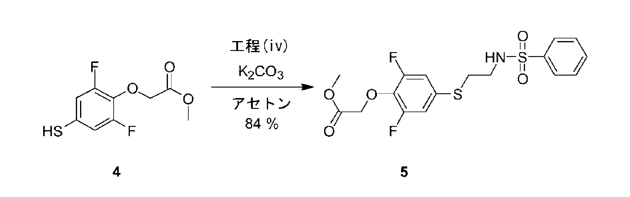
1HNMR(300MHz,CDCl3): δ 2.95(t,J=6.6Hz,2H),3.12(q,J=6.3Hz,2H),3.78(s,3H),4.72(s,2H),5.20(t,J=6.0Hz,1H),6.76-6.83(m,2H),7.47-7.60(m,3H),7.82-7.84(m,2H).
Step (iv): Synthesis of [4- (2-Benzenesulfonylamino-ethylsulfanyl) -2,6-difluoro-phenoxy] -acetic acid methyl ester (5)
1 HNMR (300 MHz, CDCl 3 ): δ 2.95 (t, J = 6.6 Hz, 2H), 3.12 (q, J = 6.3 Hz, 2H), 3.78 (s, 3H), 4 .72 (s, 2H), 5.20 (t, J = 6.0 Hz, 1H), 6.76-6.83 (m, 2H), 7.47-7.60 (m, 3H), 7 .82-7.84 (m, 2H).
工程(v):{4-[2-(ベンゼンスルホニル-メチル-アミノ)-エチルスルファニル]-2,6-ジフルオロ-フェノキシ}-酢酸メチルエステル(6)の合成

1HNMR(300MHz,CDCl3): δ 2.81(s,3H),3.04-3.09(m,2H),3.19-3.24(m,2H),3.79(s,3H),4.74(s,2H),6.90-6.94(m,2H),7.50-7.60(m,3H),7.74-7.77(m,2H).
Step (v): Synthesis of {4- [2- (benzenesulfonyl-methyl-amino) -ethylsulfanyl] -2,6-difluoro-phenoxy} -acetic acid methyl ester (6)
1 HNMR (300 MHz, CDCl 3 ): δ 2.81 (s, 3H), 3.04-3.09 (m, 2H), 3.19-3.24 (m, 2H), 3.79 (s , 3H), 4.74 (s, 2H), 6.90-6.94 (m, 2H), 7.50-7.60 (m, 3H), 7.74-7.77 (m, 2H) ).
工程(vi):{4-[2-(ベンゼンスルホニル-メチル-アミノ)-エチルスルファニル]-2,6-ジフルオロ-フェノキシ}-アセトアミド(K-2)の合成
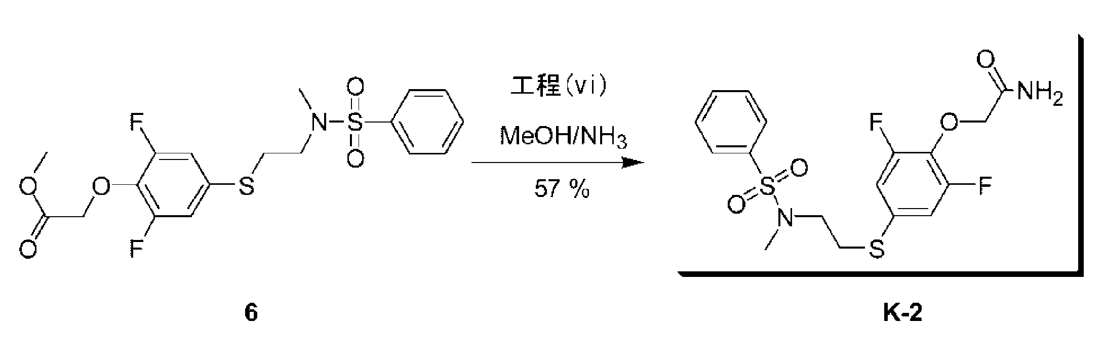
1HNMR(300MHz,CDCl3): δ 2.82(s,3H),3.08-3.13(m,2H),3.20-3.26(m,2H),4.58(s,2H),6.93-6.99(m,2H),7.50-7.63(m,3H),7.75-7.78(m,2H).
Step (vi): Synthesis of {4- [2- (benzenesulfonyl-methyl-amino) -ethylsulfanyl] -2,6-difluoro-phenoxy} -acetamide (K-2)
1 HNMR (300 MHz, CDCl 3): δ 2.82 (s, 3H), 3.08-3.13 (m, 2H), 3.20-3.26 (m, 2H), 4.58 (s, 2H), 6.93-6.99 (m, 2H), 7.50-7.63 (m, 3H), 7.75-7.78 (m, 2H).
{4-[2-(ベンゼンスルホニル-メチル-アミノ)-エチルスルファニル]-2,6-ジフルオロ-フェノキシ}-N,N-ジメチル-アセトアミド(K-4)の合成
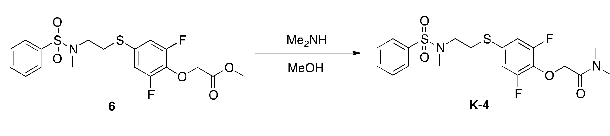
1H NMR(CDCl3,400MHz):δ2.82(s,3H),2.98(s,3H),3.05-3.09(m,5H),3.19-3.22(m,2H),4.80(s,2H),6.89-6.92(m,2H),7.53-7.55(m,2H),7.58-7.60(m,1H),7.75-7.77(m,2H).
LCMS[移動相:55%水(0.05%ギ酸)及び45%アセトニトリル(0.05%ギ酸)で6.5分間分析]純度>95%,保持時間=3.445min;MS Calcd.:444.5;MS Found:445.0[M+1]+.
Synthesis of {4- [2- (benzenesulfonyl-methyl-amino) -ethylsulfanyl] -2,6-difluoro-phenoxy} -N, N-dimethyl-acetamide (K-4)
1 H NMR (CDCl 3 , 400 MHz): δ 2.82 (s, 3H), 2.98 (s, 3H), 3.05-3.09 (m, 5H), 3.19-3.22 (m , 2H), 4.80 (s, 2H), 6.89-6.92 (m, 2H), 7.53-7.55 (m, 2H), 7.58-7.60 (m, 1H) ), 7.75-7.77 (m, 2H).
LCMS [mobile phase: analyzed with 55% water (0.05% formic acid) and 45% acetonitrile (0.05% formic acid) for 6.5 minutes] purity> 95%, retention time = 3.445 min; MS Calcd. : 444.5; MS Found: 445.0 [M + 1] + .
工程(vii):2-[2,6-ジフルオロ-4-({2-[(フェニルスルホニル)アミノ]エチル}チオ)フェノキシ]アセトアミド(K-1)の合成
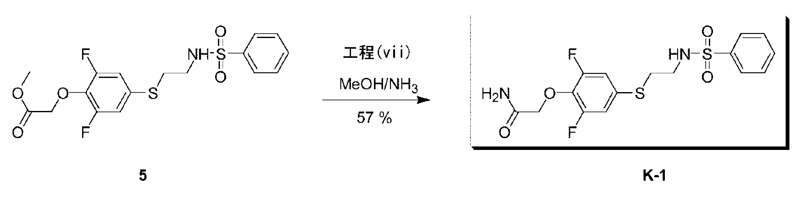
1HNMR(300MHz,CDCl3+D2O): δ 2.97-3.02(m,2H),3.11-3.16(m,2H),4.56(s,2H),6.82-6.90(m,2H),7.48-7.61(m,3H),7.82-7.87(m,2H).
Step (vii): Synthesis of 2- [2,6-difluoro-4-({2-[(phenylsulfonyl) amino] ethyl} thio) phenoxy] acetamide (K-1)
1 HNMR (300 MHz, CDCl 3 + D 2 O): δ 2.97-3.02 (m, 2H), 3.11-3.16 (m, 2H), 4.56 (s, 2H), 6. 82-6.90 (m, 2H), 7.48-7.61 (m, 3H), 7.82-7.87 (m, 2H).
{4-[2-(ベンゼンスルホニル-アミノ)-エチルスルファニル]-2,6-ジフルオロ-フェノキシ}-N,N-ジメチル-アセトアミド(K-5)の合成

水を加えて反応を停止し、酢酸エチルで生成物を抽出した後、この酢酸エチル溶液に飽和炭酸水素ナトリウム水溶液を加え、水層に生成物を抽出し、再度、塩酸を加えて酸性とした後に、再び酢酸エチルで抽出し、飽和食塩水で洗浄した後、無水硫酸ナトリウムを加えて水分を除去した。この溶液を減圧留去し、得られた残渣をジクロロメタン溶液で溶解し、この溶液にジメチルアミン・塩酸塩(94.5mg)、WSC・HCl(150.8mg)、ジイソプロピルエチルアミン(265μL)を加え、室温で2時間攪拌した。これに塩酸を加えて反応を停止し、酢酸エチルで生成物を抽出した。これを飽和炭酸水素ナトリウム水溶液、飽和食塩水で洗浄し、溶媒を減圧留去した。残渣をシリカゲルクロマトグラフィーにより精製し、目的とするK-5を白色固体(148.1mg,84%)として得た。
Synthesis of {4- [2- (benzenesulfonyl-amino) -ethylsulfanyl] -2,6-difluoro-phenoxy} -N, N-dimethyl-acetamide (K-5)
The reaction was stopped by adding water, and the product was extracted with ethyl acetate. Then, a saturated aqueous sodium hydrogen carbonate solution was added to the ethyl acetate solution, and the product was extracted into the aqueous layer. Thereafter, the mixture was extracted again with ethyl acetate, washed with saturated saline, and then anhydrous sodium sulfate was added to remove water. This solution was distilled off under reduced pressure, and the resulting residue was dissolved in a dichloromethane solution. Dimethylamine hydrochloride (94.5 mg), WSC · HCl (150.8 mg), diisopropylethylamine (265 μL) were added to the solution, Stir at room temperature for 2 hours. Hydrochloric acid was added thereto to stop the reaction, and the product was extracted with ethyl acetate. This was washed with a saturated aqueous sodium hydrogen carbonate solution and saturated brine, and the solvent was distilled off under reduced pressure. The residue was purified by silica gel chromatography to obtain the target K-5 as a white solid (148.1 mg, 84%).
工程(viii):N-(2-ブロモ-エチル)-ベンゼンスルホンアミド(9)の合成

1HNMR(300MHz,CDCl3): δ 3.36-3.39(m,4H),5.09(s,1H),7.50-7.63(s,3H),7.87-7.89(s,2H).
Step (viii): Synthesis of N- (2-bromo-ethyl) -benzenesulfonamide (9)
1 HNMR (300 MHz, CDCl 3 ): δ 3.36-3.39 (m, 4H), 5.09 (s, 1H), 7.50-7.63 (s, 3H), 7.87-7 .89 (s, 2H).
(合成例2)
(M-1、M-2及びM-3の合成)
下記スキームに従って、2-[4-(2-ベンゼンスルホニルアミノ-エチルスルファニル)-2,6-ジフルオロ-フェノキシ]-N-メチル-アセトアミド(M-1)、2-{2,6-ジフルオロ-4-[2-(4-フルオロ-ベンゼンスルホニルアミノ)-エチルスルファニル]-フェノキシ}-アセトアミド(M-2)、及び2-{2,6-ジフルオロ-4-[2-(4-メチル-ベンゼンスルホニルアミノ)-エチルスルファニル]-フェノキシ}-アセトアミド(M-3)を合成した。
各化合物の1H NMRスペクトルは、TMSを内部標準として使用し、Varian Mercury plus-400MHzで記録した。LCMSは、下記のものを使用した:Agilent1200A,カラム:C18;カラムサイズ:4.6*50分;移動相:B(ACN),A(0.05%NH3の水);勾配(B%):合成例に示すとおり。

(Synthesis of M-1, M-2 and M-3)
According to the following scheme, 2- [4- (2-benzenesulfonylamino-ethylsulfanyl) -2,6-difluoro-phenoxy] -N-methyl-acetamide (M-1), 2- {2,6-difluoro-4 -[2- (4-Fluoro-benzenesulfonylamino) -ethylsulfanyl] -phenoxy} -acetamide (M-2) and 2- {2,6-difluoro-4- [2- (4-methyl-benzenesulfonyl) Amino) -ethylsulfanyl] -phenoxy} -acetamide (M-3) was synthesized.
1 H NMR spectra of each compound were recorded at Varian Mercury plus-400 MHz using TMS as an internal standard. LCMS used the following: Agilent 1200A, column: C18; column size: 4.6 * 50 min; mobile phase: B (ACN), A (0.05% NH 3 in water); gradient (B% ): As shown in the synthesis example.
工程(i):3,5-ジフルオロ-4-ヒドロキシ-ベンゼンスルホニルクロリド(10)の合成

1H-NMR(400MHz,CDCl3): δ 6.30(s,1H),7.66-7.68(m,2H).
Step (i): Synthesis of 3,5-difluoro-4-hydroxy-benzenesulfonyl chloride (10)
1 H-NMR (400 MHz, CDCl 3 ): δ 6.30 (s, 1H), 7.66-7.68 (m, 2H).
工程(ii):2,6-ジフルオロ-4-メルカプト-フェノール(11)の合成

工程(iii):2-[2-(3,5-ジフルオロ-4-ヒドロキシ-フェニルスルファニル)-エチル]-イソインドール-1,3-ジオン(12)の合成
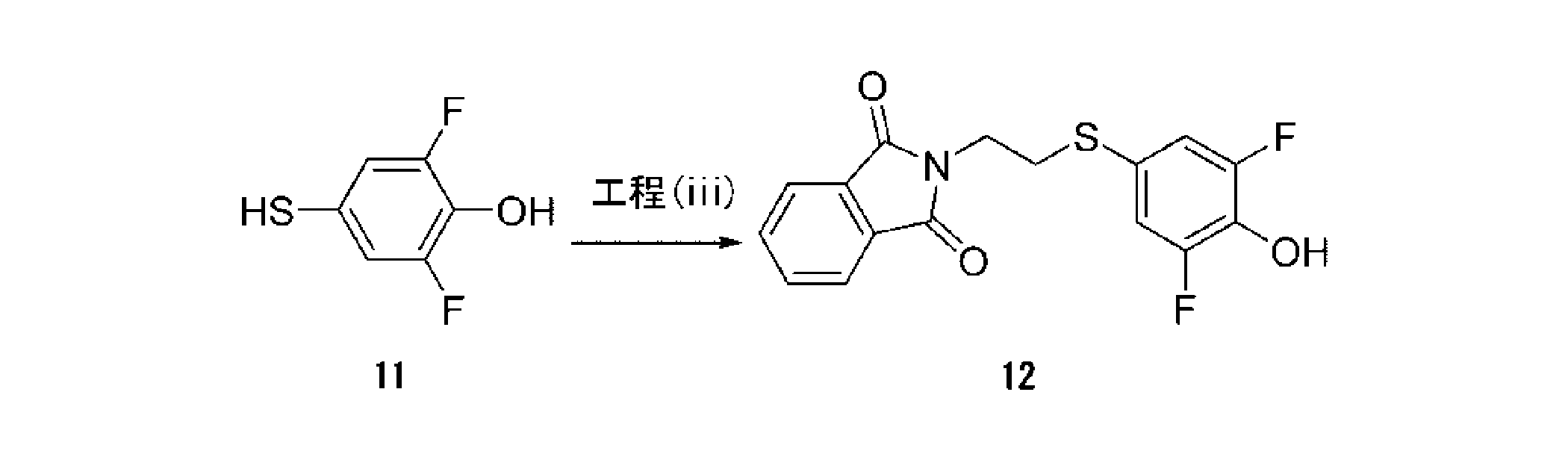
1H-NMR(400MHz,DMSO_d6): δ 3.20-3.23(t,2H),3.75-3.79(t,2H),7.08-7.10(d,2H),7.84(s,4H).
Step (iii): Synthesis of 2- [2- (3,5-difluoro-4-hydroxy-phenylsulfanyl) -ethyl] -isoindole-1,3-dione (12)
1 H-NMR (400 MHz, DMSO_d6): δ 3.20-3.23 (t, 2H), 3.75-3.79 (t, 2H), 7.08-7.10 (d, 2H), 7.84 (s, 4H).
工程(iv):{4-[2-(1,3-ジオキソ-1,3-ジヒドロ-イソインドール-2-イル)-エチルスルファニル]-2,6-ジフルオロ-フェノキシ}-酢酸エチルエステル(13)の合成
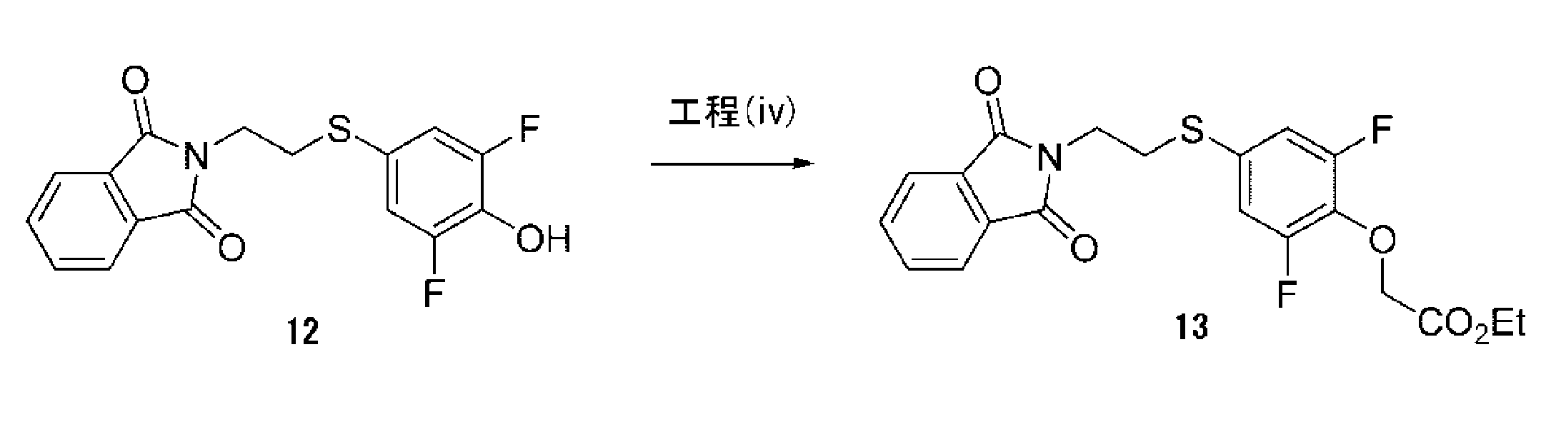
1H-NMR(400MHz,CDCl3): δ 1.21-1.24(t,3H),3.11-3.14(t,2H),3.84-3.88(t,2H),4.18-4.20(d,2H),4.61(s,2H),6.91-6.94(d,2H),7.66-7.68(m,2H),7.77-7.79(m,2H).
Step (iv): {4- [2- (1,3-dioxo-1,3-dihydro-isoindol-2-yl) -ethylsulfanyl] -2,6-difluoro-phenoxy} -acetic acid ethyl ester (13 ) Synthesis
1 H-NMR (400 MHz, CDCl 3 ): δ 1.21-1.24 (t, 3H), 3.11-3.14 (t, 2H), 3.84-3.88 (t, 2H) 4.18-4.20 (d, 2H), 4.61 (s, 2H), 6.91-6.94 (d, 2H), 7.66-7.68 (m, 2H), 7 .77-7.79 (m, 2H).
工程(v):2-{4-[2-(1,3-ジオキソ-1,3-ジヒドロ-イソインドール-2-イル)-エチルスルファニル]-2,6-ジフルオロ-フェノキシ}-N-メチル-アセトアミド(14)の合成
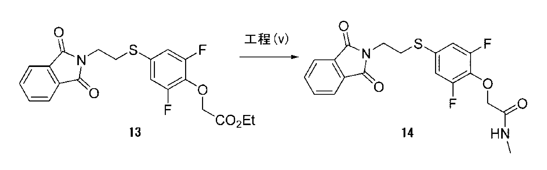
工程(vi):2-[4-(2-アミノ-エチルスルファニル)-2,6-ジフルオロ-フェノキシ]-N-メチル-アセトアミド(15)の合成

工程(vii):2-[4-(2-ベンゼンスルホニルアミノ-エチルスルファニル)-2,6-ジフルオロ-フェノキシ]-N-メチル-アセトアミド(M-1)の合成

1H-NMR(400MHz,DMSO_d6):δ 2.65-2.66(d,3H),2.91-2.94(t,2H),2.01-3.04(t,2H),4.50(s,2H),7.10-7.12(d,2H),7.57-7.65(m,3H),7.76-7.78(d,2H),7.92-7.95(t,1H),8.05(s,1H).
MS: m/z 417(M+1)+
LCMS[移動相:90%水(0.1%NH4OH)及び10%CH3CNから5%水(0.1%NH4OH)及び95%CH3CN、6.0分、最終的にこれらの条件下0.5分]純度97.4%,Rt=3.341分;MS Calcd.:416;MS Found:417([M+1]+).
Step (vii): Synthesis of 2- [4- (2-benzenesulfonylamino-ethylsulfanyl) -2,6-difluoro-phenoxy] -N-methyl-acetamide (M-1)
1 H-NMR (400 MHz, DMSO_d6): δ 2.65-2.66 (d, 3H), 2.91-2.94 (t, 2H), 2.01-3.04 (t, 2H), 4.50 (s, 2H), 7.10-7.12 (d, 2H), 7.57-7.65 (m, 3H), 7.76-7.78 (d, 2H), 7. 92-7.95 (t, 1H), 8.05 (s, 1H).
MS: m / z 417 (M + 1) +
LCMS [mobile phase: 90% water (0.1% NH 4 OH) and 10% CH 3 CN to 5% water (0.1% NH 4 OH) and 95% CH 3 CN, 6.0 min, final For 0.5 minutes under these conditions] purity 97.4%, Rt = 3.341 minutes; MS Calcd. : 416; MS Found: 417 ([M + 1] + ).
工程(viii):2-{4-[2-(1,3-ジオキソ-1,3-ジヒドロ-イソインドール-2-イル)-エチルスルファニル]-2,6-ジフルオロ-フェノキシ}-アセトアミド(16)の合成
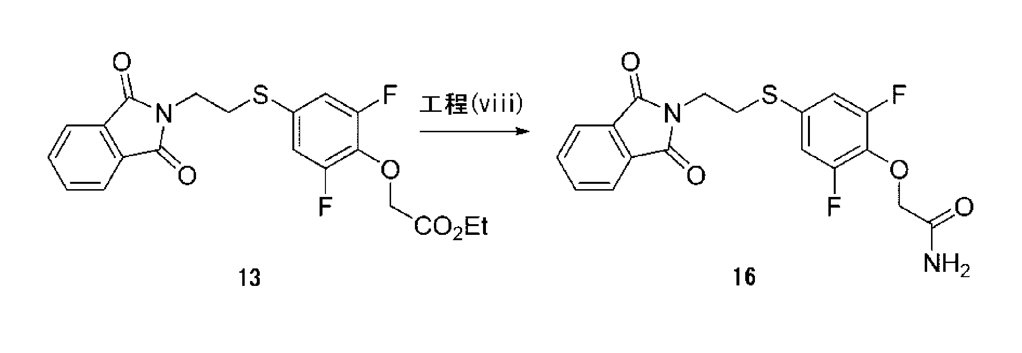
工程(ix):2-[4-(2-アミノ-エチルスルファニル)-2,6-ジフルオロ-フェノキシ]-アセトアミド(17)の合成

工程(x):2-{2,6-ジフルオロ-4-[2-(4-フルオロ-ベンゼンスルホニルアミノ)-エチルスルファニル]-フェノキシ}-アセトアミド(M-2)の合成
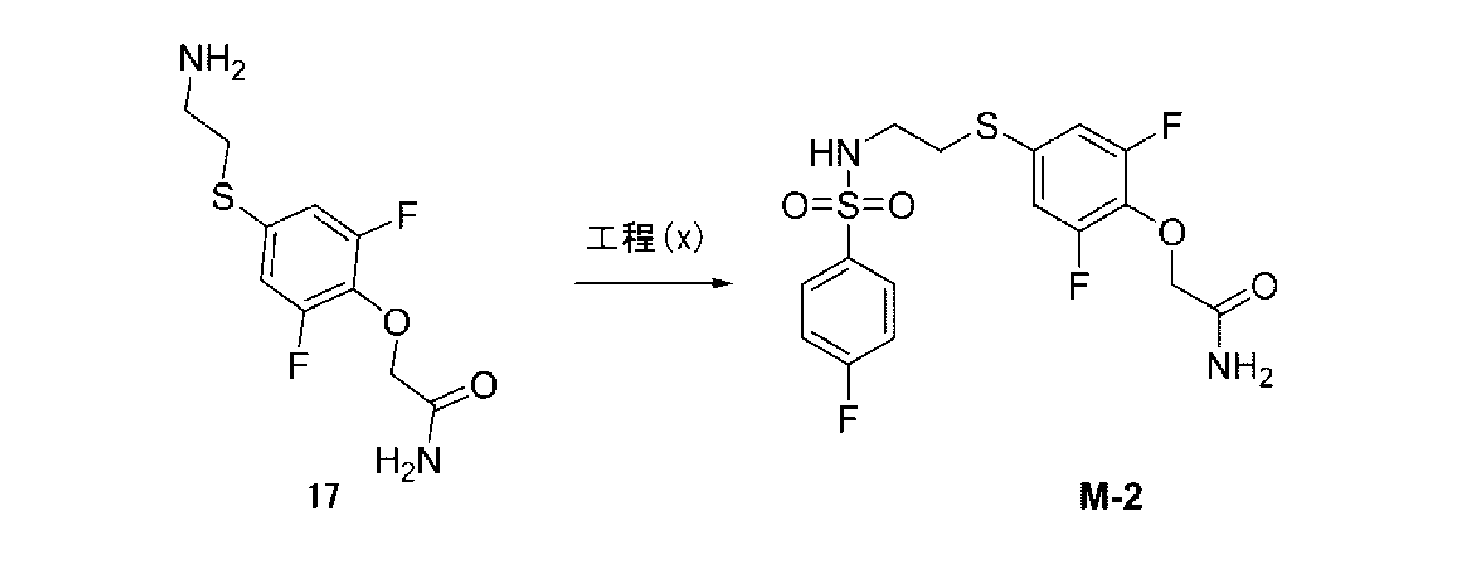
1H-NMR(400MHz,DMSO_d6):δ 2.92-2.95(t,2H),3.01-3.04(t,2H),4.45(s,2H),7.09-7.11(d,2H),7.40-7.44(m,3H),7.47(s,1H),7.81-7.85(m,2H),7.95-7.98(t,1H).
MS: m/z 421(M+1)+
LCMS [移動相: 90%水(0.1%NH4OH)及び10%CH3CNから5%水(0.1%NH4OH)及び95%CH3CN、6分、最終的にこれらの条件下0.5分]純度95.1%,Rt=3.284分;MS Calcd.: 420;MS Found:421([M+1]+).
Step (x): Synthesis of 2- {2,6-difluoro-4- [2- (4-fluoro-benzenesulfonylamino) -ethylsulfanyl] -phenoxy} -acetamide (M-2)
1 H-NMR (400 MHz, DMSO_d6): δ 2.92-2.95 (t, 2H), 3.01-3.04 (t, 2H), 4.45 (s, 2H), 7.09- 7.11 (d, 2H), 7.40-7.44 (m, 3H), 7.47 (s, 1H), 7.81-7.85 (m, 2H), 7.95-7. 98 (t, 1H).
MS: m / z 421 (M + 1) +
LCMS [mobile phase: 90% water (0.1% NH 4 OH) and 10% CH 3 CN to 5% water (0.1% NH 4 OH) and 95% CH 3 CN, 6 min, finally these For 0.5 minutes] purity 95.1%, Rt = 3.284 minutes; MS Calcd. : 420; MS Found: 421 ([M + 1] + ).
工程(xi):2-{2,6-ジフルオロ-4-[2-(4-メチル-ベンゼンスルホニルアミノ)-エチルスルファニル]-フェノキシ}-アセトアミド(M-3)の合成
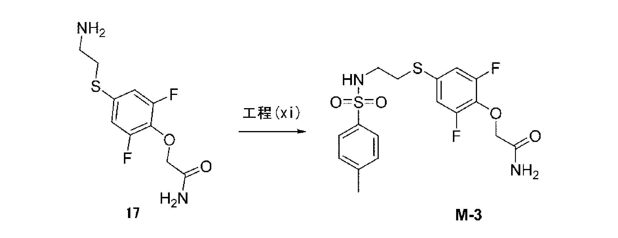
1H-NMR(400MHz,DMSO_d6): δ 2.38(s,3H),2.88-2.91(t,2H),2.99-3.02(t,2H),4.49(s,2H),7.08-7.10(d,2H),7.37-7.48(m,4H),7.64-7.66(d,2H),7.81-7.84(t,1H).
MS:m/z 417(M+1)+
LCMS[移動相:90%水(0.1%NH4OH)及び10%CH3CNから5%水(0.1%NH4OH)及び95%CH3CN、6.0分、最終的にこれらの条件下0.5分]純度96.6%,Rt=3.365分;MS Calcd.:416;MS Found:417([M+1]+).
Step (xi): Synthesis of 2- {2,6-difluoro-4- [2- (4-methyl-benzenesulfonylamino) -ethylsulfanyl] -phenoxy} -acetamide (M-3)
1 H-NMR (400 MHz, DMSO_d6): δ 2.38 (s, 3H), 2.88-2.91 (t, 2H), 2.99-3.02 (t, 2H), 4.49 ( s, 2H), 7.08-7.10 (d, 2H), 7.37-7.48 (m, 4H), 7.64-7.66 (d, 2H), 7.81-7. 84 (t, 1H).
MS: m / z 417 (M + 1) +
LCMS [mobile phase: 90% water (0.1% NH 4 OH) and 10% CH 3 CN to 5% water (0.1% NH 4 OH) and 95% CH 3 CN, 6.0 min, final For 0.5 minutes under these conditions] purity 96.6%, Rt = 3.365 minutes; MS Calcd. : 416; MS Found: 417 ([M + 1] + ).
(合成例3)
(M-3preの合成)
下記スキームに従って、2-(2,6-ジフルオロ-4-((2-(4-(トリブチルスタンニル)フェニルスルホンアミド)エチル)チオ)フェノキシ)アセトアミド(M-3pre)を合成した。
各化合物の1H NMRスペクトルは、TMSを内部標準として使用し、Bruker Avance III 400MHz及びBruker Fourier 300MHzで記録した。LCMSは、下記のものを使用した:四重極質量分析計、Agilent LC/MSD1200シリーズ(カラム:ODS2000(50×4.6mm,5μm)ES (+)又は(-)イオン化モードで操作;T=30℃;流速=1.5mL/分;検出波長:254nm。
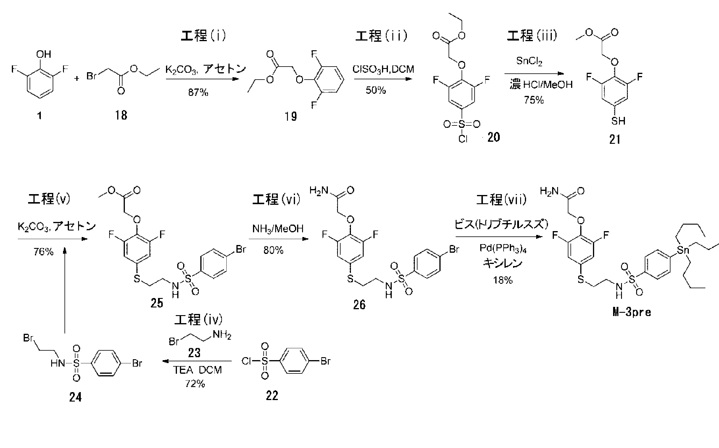
(Synthesis of M-3pre)
2- (2,6-Difluoro-4-((2- (4- (tributylstannyl) phenylsulfonamido) ethyl) thio) phenoxy) acetamide (M-3pre) was synthesized according to the following scheme.
1 H NMR spectra of each compound were recorded on Bruker Avance III 400 MHz and
工程(i):エチル 2-(2,6-ジフルオロフェノキシ)アセテート(19)の合成

1H NMR(CDCl3,300MHz):δ 1.19(t,J=7.2Hz,3H),4.17(q,J=7.2Hz,2H),4.82(s,2H),7.06-7.13(m,3H).
Step (i): Synthesis of ethyl 2- (2,6-difluorophenoxy) acetate (19)
1 H NMR (CDCl 3 , 300 MHz): δ 1.19 (t, J = 7.2 Hz, 3H), 4.17 (q, J = 7.2 Hz, 2H), 4.82 (s, 2H), 7.06-7.13 (m, 3H).
工程(ii):エチル 2-(4-(クロロスルホニル)-2,6-ジフルオロフェノキシ)アセテート(20)の合成
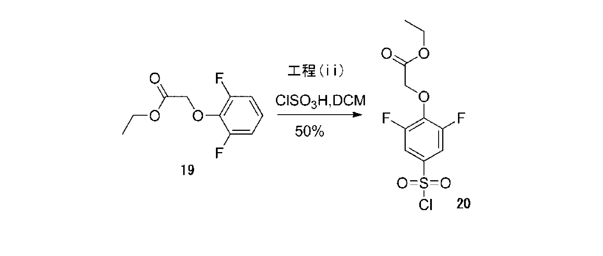
1H NMR(CDCl3,300MHz): δ 1.18(t,J=6.9Hz,3H),4.16(q,J=6.9Hz,2H),4.83(s,2H),7.18-7.21(m,2H).
Step (ii): Synthesis of ethyl 2- (4- (chlorosulfonyl) -2,6-difluorophenoxy) acetate (20)
1 H NMR (CDCl 3 , 300 MHz): δ 1.18 (t, J = 6.9 Hz, 3H), 4.16 (q, J = 6.9 Hz, 2H), 4.83 (s, 2H), 7.18-7.21 (m, 2H).
工程(iii):メチル 2-(2,6-ジフルオロ-4-メルカプトフェノキシ)アセテート(21)の合成

1H NMR (CDCl3,300MHz):δ 3.52(s,1H),3.79(s,3H),4.72(s,2H),6.88(d,J=6.3Hz,2H).
Step (iii): Synthesis of methyl 2- (2,6-difluoro-4-mercaptophenoxy) acetate (21)
1 H NMR (CDCl 3 , 300 MHz): δ 3.52 (s, 1H), 3.79 (s, 3H), 4.72 (s, 2H), 6.88 (d, J = 6.3 Hz, 2H).
工程(iv):4-ブロモ-N-(2-ブロモエチル)ベンゼンスルホンアミド(24)の合成
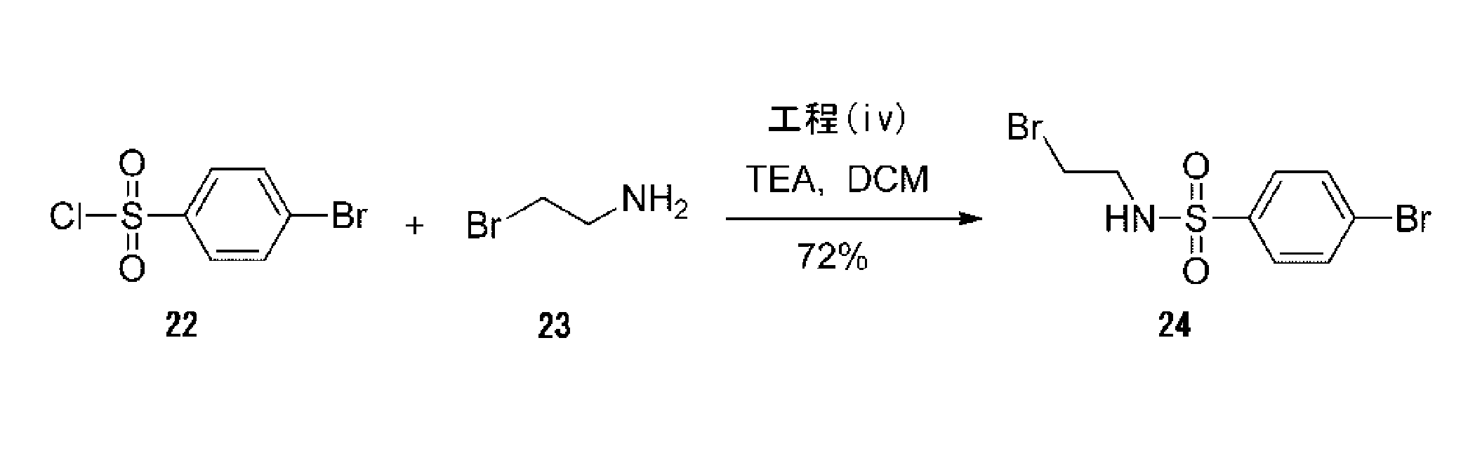
1H NMR(DMSO-d6,300MHz): δ 3.12-3.16(m,2H),3.43(t,J=3.6Hz,2H),7.69-7.73(m,2H),7.79-7.82(m,2H),8.13(t,J=3.9Hz,1H).
Step (iv): Synthesis of 4-bromo-N- (2-bromoethyl) benzenesulfonamide (24)
1 H NMR (DMSO-d 6 , 300 MHz): δ 3.12-3.16 (m, 2H), 3.43 (t, J = 3.6 Hz, 2H), 7.69-7.73 (m , 2H), 7.79-7.82 (m, 2H), 8.13 (t, J = 3.9 Hz, 1H).
工程(v):メチル 2-(4-((2-(4-ブロモフェニルスルホンアミド)エチル)チオ)-2,6-ジフルオロフェノキシ)アセテート(25)の合成

1H NMR(CDCl3,300MHz):δ 2.94-2.98(m,2H),3.08-3.14(m,2H),3.77(s,3H),4.73(s,2H),5.33(t,J=6.0Hz,1H),6.78-6.84(m,2H),7.61-7.70(m,4H).
Step (v): Synthesis of methyl 2- (4-((2- (4-bromophenylsulfonamido) ethyl) thio) -2,6-difluorophenoxy) acetate (25)
1 H NMR (CDCl 3 , 300 MHz): δ 2.94-2.98 (m, 2H), 3.08-3.14 (m, 2H), 3.77 (s, 3H), 4.73 ( s, 2H), 5.33 (t, J = 6.0 Hz, 1H), 6.78-6.84 (m, 2H), 7.61-7.70 (m, 4H).
工程(vi):2-(4-((2-(4-ブロモフェニルスルホンアミド)エチル)チオ)-2,6-ジフルオロフェノキシ)アセトアミド(26)の合成

1H NMR(DMSO-d6,400MHz):δ 2.93-2.96 (m,2H),3.00-3.03(m,2H),4.48(s,2H),7.10(d,J=9.2Hz,2H),7.40-7.45(m,2H),7.70(d,J=8.4Hz,2H),7.80(d,J=8.4Hz,2H),8.01(brs,1H).
Step (vi): Synthesis of 2- (4-((2- (4-bromophenylsulfonamido) ethyl) thio) -2,6-difluorophenoxy) acetamide (26)
1 H NMR (DMSO-d 6 , 400 MHz): δ 2.93-2.96 (m, 2H), 3.00-3.03 (m, 2H), 4.48 (s, 2H), 7. 10 (d, J = 9.2 Hz, 2H), 7.40-7.45 (m, 2H), 7.70 (d, J = 8.4 Hz, 2H), 7.80 (d, J = 8 .4 Hz, 2H), 8.01 (brs, 1H).
工程(vii):2-(2,6-ジフルオロ-4-((2-(4-(トリブチルスタンニル)フェニルスルホンアミド)エチル)チオ)フェノキシ)アセトアミド(M-3pre)の合成

1H NMR(CD3OD,300MHz): δ 0.94(t,J=7.2Hz,9H),1.12-1.17(m,5H),1.29-1.39(m,8H),1.52-1.60(m,5H),2.98-3.06(m,4H),4.55(s,2H),7.01(d,J=9.0Hz,2H),7.68(d,J=8.1Hz,2H),7.77(d,J=8.1Hz,2H);LCMS[移動相:30%水(0.02%NH4OAc)及び70%CH3CNから5%水(0.02%NH4OAc)及び95%CH3CN、6分、最終的にこれらの条件下0.5分]純度>95%,Rt=4.259分;MS Calcd.:692;MS Found:693([M+H]+).
Step (vii): Synthesis of 2- (2,6-difluoro-4-((2- (4- (tributylstannyl) phenylsulfonamido) ethyl) thio) phenoxy) acetamide (M-3pre)
1 H NMR (CD 3 OD, 300 MHz): δ 0.94 (t, J = 7.2 Hz, 9H), 1.12-1.17 (m, 5H), 1.29-1.39 (m, 8H), 1.52-1.60 (m, 5H), 2.98-3.06 (m, 4H), 4.55 (s, 2H), 7.01 (d, J = 9.0 Hz, 2H), 7.68 (d, J = 8.1 Hz, 2H), 7.77 (d, J = 8.1 Hz, 2H); LCMS [mobile phase: 30% water (0.02% NH 4 OAc) And 70% CH 3 CN to 5% water (0.02% NH 4 OAc) and 95% CH 3 CN, 6 minutes, finally 0.5 minutes under these conditions] purity> 95%, Rt = 4. 259 min; MS Calcd. : 692; MS Found: 693 ([M + H] + ).
(合成例4)
(放射性標識化K-2の合成)
放射性標識化K-2を下記のように合成した。

(Synthesis of radiolabeled K-2)
Radiolabeled K-2 was synthesized as follows.
HPLCを用いて、放射性標識化K-2と非標識K-2との比較を行った。HPLC分析は、カラムにCapcellPak UG-80(4.6X250)(資生堂、日本)を用い、流速1.0ml/分で展開し、検出はUV254nmとRIを用いて行った。非標識K-2(UV検出)と放射性標識化K-2(RI検出)は、保持時間8分のところで同一のピークを示した。これは、2つが同一物質であり、放射性標識化K-2が製造できたことを示している。 Comparison of radiolabeled K-2 and unlabeled K-2 was performed using HPLC. HPLC analysis was performed using CapcellPak UG-80 (4.6X250) (Shiseido, Japan) at a flow rate of 1.0 ml / min, and detection was performed using UV254 nm and RI. Unlabeled K-2 (UV detection) and radiolabeled K-2 (RI detection) showed the same peak at a retention time of 8 minutes. This indicates that the two were the same substance and radiolabeled K-2 could be produced.
反応させたヨウ化メチルは、100%PEPAのスルホンアミドに結合することがわかり、放射性標識化K-2の合成は、極めて簡素であり、かつ高い収率を示すことがわかった。 The reacted methyl iodide was found to bind to the sulfonamide of 100% PEPA, and the synthesis of radiolabeled K-2 was found to be extremely simple and yield high yields.
(合成例5)
(放射性標識化M-3の合成)
1mLのガラスバイアルに、Pd2(dba)3(1.74mg)、塩化第一銅(1.7mg)、炭酸カリウム(2.25mg)を計り取り、その混合物に、P(o-tol)3(1.7mg)のDMF(300μL)溶液を窒素雰囲気下で加えた。室温下で約5分程度撹拌してから、本溶液を標識用反応容器に移した。[11C]CH3Iを冷却下で捕集し、放射能が飽和した後に、原料のトリブチルスズ体(preM-3)(1.6mg)のDMF溶液(300μL)を加え、80℃で約5分間反応させた。反応混合物をPTFEフィルターを通して固形物を除去してからHPLC分離を行い、約7分付近のRIピーク部分を分取、濃縮、調剤化した。
Pd2(dba)3:トリス(ジベンジリデンアセトン)ジパラジウム
P(o-tolyl)3:トリ(o-トリル)ホスフィン
(Synthesis Example 5)
(Synthesis of radiolabeled M-3)
Pd 2 (dba) 3 (1.74 mg), cuprous chloride (1.7 mg), potassium carbonate (2.25 mg) are weighed into a 1 mL glass vial, and P (o-tol) 3 is added to the mixture. A solution of (1.7 mg) in DMF (300 μL) was added under a nitrogen atmosphere. After stirring for about 5 minutes at room temperature, this solution was transferred to a labeling reaction vessel. [ 11 C] CH 3 I was collected under cooling, and after the radioactivity was saturated, a DMF solution (300 μL) of a starting material tributyltin (preM-3) (1.6 mg) was added and about 5 at 80 ° C. Reacted for 1 minute. Solids were removed from the reaction mixture through a PTFE filter, followed by HPLC separation, and the RI peak portion in the vicinity of about 7 minutes was separated, concentrated and formulated.
Pd 2 (dba) 3 : Tris (dibenzylideneacetone) dipalladium P (o-tolyl) 3 : Tri (o-tolyl) phosphine
(参考例1)
(in vitro autoradiography法)
ラットは、2~3週齢のオス成体SDラット(チャールズリバー、日本)を使用した。線条体を含む厚さ200μmの急性脳切片を作成し、セルストレイナー上に置き、酸素化ACSF(人工脳脊髄液)中で60分間静置した。
(Reference Example 1)
(In vitro autoradiography method)
Rats were 2 to 3 weeks old adult male SD rats (Charles River, Japan). A 200 μm-thick acute brain section containing the striatum was prepared, placed on a cell strainer, and allowed to stand in oxygenated ACSF (artificial cerebrospinal fluid) for 60 minutes.
100μM PEPA(1%DMSO含有ACSF)を12ml分調整し、そこに20nMになるよう[11C]K-2を加え、60分間静置した。反応後、ACSFで10秒間洗浄を3回行い、最後に蒸留水で10秒間洗浄を行った。反応後の脳切片より大きめにセルストレイナーを切除し、イメージングプレートに2時間感光させた。感光後のイメージングプレートはTyphoon(GEヘルスケア・ジャパン)で撮像した(分解能25pixel)。 100 μM PEPA (ACSF containing 1% DMSO) was adjusted for 12 ml, and [ 11 C] K-2 was added thereto to 20 nM, and the mixture was allowed to stand for 60 minutes. After the reaction, washing was performed 3 times with ACSF for 10 seconds, and finally, washing with distilled water was performed for 10 seconds. The cell strainer was excised larger than the brain section after the reaction, and exposed to an imaging plate for 2 hours. The imaging plate after exposure was imaged with Typhoon (GE Healthcare Japan) (resolution: 25 pixels).
(生物学的実施例)
(AMPA受容体結合化合物の調製及び投与)
強制水泳試験では、AMPA受容体結合化合物K-2およびK-4を、100%DMSO(ナカライテスク、日本)に2.1mMの濃度となるように溶解し、投与直前に生理食塩水で希釈し(15%DMSO、320μM)、5μl/体重(g)の投与量にて、1mg/kgないし1.5mg/kgとなるようにラットに経静脈的に投与した。
(Biological Example)
(Preparation and administration of AMPA receptor binding compounds)
In the forced swimming test, AMPA receptor binding compounds K-2 and K-4 are dissolved in 100% DMSO (Nacalai Tesque, Japan) to a concentration of 2.1 mM and diluted with physiological saline immediately before administration. Rats were intravenously administered at a dose of 5 μl / body weight (g) (15% DMSO, 320 μM) to give 1 mg / kg to 1.5 mg / kg.
(動物実験)
すべての動物実験は、横浜市立大学の動物実験委員会の審議及び承認を受けた(承認番号:F-A-15-051)。
ラットは、6~10週齢のオス成体Wistarラット及びうつ病モデルラットであるWistar kyoto ラット(WKYラット)(チャールズリバー、日本)を使用した。
反復してK-2投与を行う経路を確保するため、実験を実施する1週間前に頚静脈カニューレ留置手術を行った。頚静脈カニューレ留置手術は、イソフルラン(DSファーマ アニマルヘルス、日本)でラットを入眠させた後、1.5%のイソフルラン濃度(空気2L/分)で麻酔を維持して行った。頚部の皮膚を約3cm切開し、皮下脂肪を開創し、頚静脈を露出させた。カニューレチューブを装着した針で頚静脈を貫通させた後、カニューレチューブを針から取り外し、カニューレチューブの尖端を頚静脈内に2.5cm挿入し、縫合糸で挿入部付近へ固定した。カニューレチューブの反対側は、ラット背部より体外へ露出させ、逆流を防ぐために栓を装着し、切開した皮膚を縫合した。手術終了後、ラットはホームケージに戻した。
(Animal experimentation)
All animal experiments were reviewed and approved by the Animal Experiment Committee of Yokohama City University (approval number: F-A-15-051).
Rats were 6-10 week old male Wistar rats and depression model rats, Wistar kyoto rats (WKY rats) (Charles River, Japan).
In order to secure a route for repeated K-2 administration, a jugular vein cannula operation was performed one week before the experiment. The jugular cannula indwelling operation was carried out with the anesthesia maintained at 1.5% isoflurane concentration (air 2 L / min) after the rats were asleep with isoflurane (DS Pharma Animal Health, Japan). The neck skin was incised approximately 3 cm, the subcutaneous fat was opened, and the jugular vein was exposed. After penetrating the jugular vein with a needle equipped with a cannula tube, the cannula tube was removed from the needle, the tip of the cannula tube was inserted 2.5 cm into the jugular vein, and fixed near the insertion part with a suture. The other side of the cannula tube was exposed from the back of the rat to the outside of the body, a stopper was attached to prevent backflow, and the incised skin was sutured. After the operation, the rats were returned to their home cages.
(ラットを用いたインビボPET撮像)
PET撮像は、microPET(Focus 220;Siemens Medical Solution)を用いて行った。
ラットを用いたPET撮像実験:イソフルラン(DSファーマ アニマルヘルス、日本)を充填した麻酔箱の中でラットを入眠させた後、1.5%のイソフルラン濃度(空気2L/分)で麻酔を維持し、尾静脈より24Gサーフロー留置針(テルモ、日本)で静脈路を確保した。ラットをPET撮影台に固定した後、撮像の前に減弱補正のための放射撮像を行い、その後放射性標識したK-2(約40MBq)を投与した。PET撮像中は、体温をフィードバック型加温盤(BWT-100A;バイオリサーチセンター、日本)を用いて37±0.5℃に維持した。撮像後、静脈路を抜去し、イソフルラン投与を中止した後に、ラットをPET台より外し、ホームケージに戻した。ラットは、撮像後1週間、撮像した部屋にて飼育し、その後、通常のラット集団飼育室に戻した。
加算(Summation)画像を構成するとともに、0.5mmハニング(Hanning)フィルターで裏写りを取り除いてダイナミック画像を再構成した。再構成画像は、PMOD画像分析ソフトウェア(PMOD technologies)を用いて、海馬、内側前頭前皮質、側坐核、線条体、視床、小脳などの複数の領域を含むVOIsを、鋳型MRI画像上に作成したものと統合して解析した。定量に用いた算出値は%SUV(% of standardized uptake value)であり、以下の式で求めた;
%SUV = VOIで囲われた各組織の放射線量(kBq/cc)/投与した放射線量(MBq)×体重(kg)×100
(In vivo PET imaging using rats)
PET imaging was performed using microPET (
PET imaging experiment using rats: After anesthetizing rats in an anesthesia box filled with isoflurane (DS Pharma Animal Health, Japan), anesthesia was maintained at 1.5% isoflurane concentration (air 2L / min). The vein was secured from the tail vein with a 24G Surfflow indwelling needle (Terumo, Japan). After fixing the rat on the PET imaging table, radiographic imaging for attenuation correction was performed before imaging, and then radiolabeled K-2 (about 40 MBq) was administered. During PET imaging, the body temperature was maintained at 37 ± 0.5 ° C. using a feedback heating board (BWT-100A; Bioresearch Center, Japan). After imaging, the venous tract was removed, and after administration of isoflurane was stopped, the rat was removed from the PET table and returned to the home cage. Rats were housed in the imaged room for one week after imaging, and then returned to the normal rat group breeding room.
A summation image was constructed and a show-through was removed with a 0.5 mm Hanning filter to reconstruct a dynamic image. The reconstructed image is obtained by using the PMOD image analysis software (PMOD technologies) and VOIs including a plurality of regions such as hippocampus, medial prefrontal cortex, nucleus accumbens, striatum, thalamus, and cerebellum on template MRI image. It was analyzed by integrating with the created one. The calculated value used for quantification was% SUV (% of standardized uptake value), which was determined by the following formula;
% SUV = radiation dose (kBq / cc) of each tissue surrounded by VOI / radiation dose (MBq) × body weight (kg) × 100
(ラットを用いた強制水泳試験)
強制水泳試験は、薬剤投与前日の投与前実験、薬剤投与1日目の急性投与実験、薬剤投与7日目の慢性投与実験を行った。
強制水泳試験(投与前実験、急性投与実験、慢性投与実験)では、ラットを実験の実施する部屋へ馴化させるため、1時間静置した。投与前実験は、馴化後すぐに強制水泳試験を行った。急性投与実験は、強制水泳試験を開始する30分前に15%DMSO又は15%DMSOに溶解したK-2およびK-4を経静脈的に投与した。慢性投与実験は、15%DMSO又は15%DMSOに溶解したK-2およびK-4の経静脈的投与を7日間行い、投与7日目の投与30分後に強制水泳試験を行った。
強制水泳試験は、直径20cm泳高さ50cmの円筒ケージを用いて、水温26±0.5℃の水道水を水位40cmまで注水して行った。この円筒ケージ内にラットを投入し、10分間泳ぐ様子をデジタルビデオカメラ(HC-V750、パナソニック、日本)を用いて撮影した。撮影後、ペーパータオルでラットの水滴を拭き取り、ホームケージへ戻した。
解析は、急性投与実験及び慢性投与実験において、撮影した動画の2~10分間(0~2分はラットの環境適応時間とした)における、ラットの四肢を動かさない時間を無動時間として計測した。15%DMSOを投与したラットと15%DMSOに溶解したK-2およびK-4を投与したラットで無動時間を比較した。
(Forced swimming test using rats)
In the forced swimming test, a pre-administration experiment on the day before drug administration, an acute administration experiment on the first day of drug administration, and a chronic administration experiment on the seventh day of drug administration were performed.
In the forced swimming test (pre-dose experiment, acute administration experiment, chronic administration experiment), the rat was allowed to stand for 1 hour in order to acclimate to the room where the experiment was conducted. In the pre-dose experiment, a forced swimming test was conducted immediately after acclimatization. In the acute administration experiment, K-2 and K-4 dissolved in 15% DMSO or 15% DMSO were intravenously administered 30 minutes before the start of the forced swimming test. In the chronic administration experiment, intravenous administration of K-2 and K-4 dissolved in 15% DMSO or 15% DMSO was performed for 7 days, and a forced swimming test was performed 30 minutes after administration on the 7th day.
The forced swimming test was performed by pouring tap water having a water temperature of 26 ± 0.5 ° C. to a water level of 40 cm using a cylindrical cage having a diameter of 20 cm and a height of 50 cm. A rat was put into this cylindrical cage and a 10-minute swim was photographed using a digital video camera (HC-V750, Panasonic, Japan). After photographing, the water droplets of the rat were wiped off with a paper towel and returned to the home cage.
In the analysis for acute administration and chronic administration, the time during which the limbs of the rat were not moved was measured as the immobility time in 2 to 10 minutes (0 to 2 minutes was the environment adaptation time of the rat) of the captured movie. . The immobility time was compared between rats administered with 15% DMSO and rats administered with K-2 and K-4 dissolved in 15% DMSO.
(実験結果)
(AMPA受容体結合性試験結果)
in vitro autoradiography法によって、K-2はAMPA受容体に結合親和性を示すことが確認され、そのKd値は47.9nMであった(図4)。
電気生理学的検証によって、K-2及びK-4はAMPA受容体に結合親和性を示すことが確認された(図5)。
(Experimental result)
(AMPA receptor binding test results)
In vitro autoradiography confirmed that K-2 showed binding affinity to the AMPA receptor, and its Kd value was 47.9 nM (FIG. 4).
Electrophysiological validation confirmed that K-2 and K-4 show binding affinity for AMPA receptors (FIG. 5).
(AMPA受容体標識化合物K-2を用いたラットでのPET撮像)
放射性標識化K-2([11C]K-2)をWistarラットとWKYラットに投与し、インビボでのPET撮像を行った(図6,7)。次に、これら画像に対して、PMOD画像分析ソフトウェアを用いて、解析を行った。その結果、WKYラットの内側前頭前皮質、線条体、大脳皮質、視床において放射性標識化K-2の有意に低い脳内への取り込みを示し(図8)、これらの部位においてAMPA受容体の集積量の低下が示唆された。これらの結果より、うつ病モデルラットであるWKYラットでは、特定の脳領域において、AMPA受容体の発現量が低下していることがわかった。
(PET imaging in rats using AMPA receptor labeled compound K-2)
Radiolabeled K-2 ([ 11 C] K-2) was administered to Wistar and WKY rats and PET imaging was performed in vivo (FIGS. 6 and 7). Next, these images were analyzed using PMOD image analysis software. As a result, WKY rats showed significantly lower incorporation of radiolabeled K-2 into the brain in the medial prefrontal cortex, striatum, cerebral cortex, and thalamus (FIG. 8). The decrease in accumulation amount was suggested. From these results, it was found that the expression level of AMPA receptor was decreased in a specific brain region in WKY rats, which are depression model rats.
(AMPA受容体結合化合物K-2を用いたラットでの強制水泳試験)
急性投与実験では、15%DMSOを投与したラットとK-2を投与したラット間で無動時間の変化は認められなかった(図9)。慢性投与実験では、K-2投与ラットでは無動時間の有意な低下が認められ、その効果は1mg/kgよりも1.5mg/kgで有意に高かった(図10)。また同様にK-4投与ラットでも無動時間の有意な低下が認められ、1mg/kg投与間で比較するとK-4はK-2に比して有意に無働時間を低下させた(図10)。この結果より、うつ病モデルラットであるWKYラットにおけるK-2およびK-4慢性投与は抗うつ効果を示すことが示唆された。
AMPA受容体に結合親和性を示すK-2やK-4といった化合物と、AMPA受容体の関連が知られているうつ病との関連(Jingli Zhangら、Rev.Neurosci.2013;24(5):499-505、Simon E Wardら、British Journal оf Pharmacology(2010),160,181-190)が確認されたことから、上記の抗うつ効果はAMPA受容体機能活性化に起因すると示唆される。このため、K-2及びK-4、並びにこれらに類似する本発明の化合物は、うつ病に限らずAMPA受容体が関連する疾患の治療に有用であることが示唆される。
これらは、哺乳類生体の脳内AMPA受容体のイメージング結果に基づき、脳内AMPA受容体が関連する疾患の診断、上記疾患の治療又は予防のためのコンパニオン診断、上記疾患の治療又は予防のための医薬の投与計画の策定、上記疾患の治療又は予防薬のスクリーニングが可能であることを示唆する。
(Forced swimming test in rats using AMPA receptor binding compound K-2)
In the acute administration experiment, no change in the immobility time was observed between the rat administered with 15% DMSO and the rat administered with K-2 (FIG. 9). In chronic administration experiments, a significant decrease in immobility time was observed in K-2 treated rats, and the effect was significantly higher at 1.5 mg / kg than at 1 mg / kg (FIG. 10). Similarly, a significant decrease in immobility time was also observed in rats treated with K-4, and K-4 significantly decreased inactivity time compared with K-2 when compared between 1 mg / kg doses (Fig. 10). From these results, it was suggested that chronic administration of K-2 and K-4 in WKY rats, which are depression model rats, shows an antidepressant effect.
Association between compounds such as K-2 and K-4, which show binding affinity for AMPA receptor, and depression with known association of AMPA receptor (Jingli Zhang et al., Rev. Neurosci. 2013; 24 (5) : 499-505, Simon E Ward et al., British Journal of Pharmacology (2010), 160, 181-190), the above-mentioned antidepressant effect is suggested to be due to activation of AMPA receptor function. For this reason, it is suggested that K-2 and K-4, and the compounds of the present invention similar thereto are useful not only for depression but also for the treatment of diseases associated with AMPA receptors.
These are based on the imaging results of AMPA receptors in the brain of mammalian organisms, for diagnosis of diseases related to AMPA receptors in the brain, for companion diagnosis for treatment or prevention of the above diseases, for treatment or prevention of the above diseases This suggests that it is possible to formulate a drug administration plan and to screen for drugs for treating or preventing the above diseases.
(実施例1)
(ヒトでのPET撮像)
2名の被験者を対象としてPET撮像を行った。撮像台上で臥位をとり、前腕ないし手背に22ゲージサーフロー針で静脈路を確保した。約1分間の吸収補正用CT撮像を行った後、PET薬剤として、約370MBqの[11C]K-2を1分間かけて静脈投与した。その後、120分間の撮像を行った。撮像にはPET/CT装置aquiduo(東芝メディカルシステムズ)を用いた。収集されたリストデータはOS-EM法でダイナミック画像を再構成した。
Example 1
(PET imaging in humans)
PET imaging was performed on two subjects. A supine position was taken on the imaging table, and a venous path was secured with a 22 gauge surf flow needle on the forearm or back of the hand. After performing absorption correction CT imaging for about 1 minute, about 370 MBq of [ 11 C] K-2 was intravenously administered as a PET drug over 1 minute. Thereafter, imaging was performed for 120 minutes. A PET / CT apparatus, aquidu (Toshiba Medical Systems) was used for imaging. The collected list data reconstructed a dynamic image by the OS-EM method.
再構成画像は、PMOD画像分析ソフトウェア(PMOD technologies)を用いて、前頭葉、歯状皮質、海馬、扁桃体、被核、小脳、橋などの複数の領域を含むVOIsを、鋳型MRI画像上に作成したものと統合して解析した。
図11は、PET薬剤投与0分後から30分後までの30分間に関する画像(前半画像という)、及びPET薬剤投与60分後(第1時間の一例)から120分後(第2時間の一例)までの60分間に関する画像(後半画像という)である。
両被験者ともに、前半画像では[11C]K-2の海馬集積に左右差が若干確認されるものの、明確には確認しにくかったが、後半画像(右側)では海馬集積に左右差が確認され、被験者Aでは右海馬に、被験者Bでは左海馬に高い集積を認めた。この結果は、PET薬剤投与の30分後~60分後の間に、脳内AMPA受容体に結合していないPET薬剤による非特異的な集積が顕著に洗い出されたことを示す。したがって、PET薬剤として[11C]K-2を使う場合には、特に限定されないが、少なくとも投与後30分、特に60分程度経過した後に検知及び検出を行うことが好ましいことが分かる。
Reconstructed images were created on template MRI images by using PMOD image analysis software (PMOD technologies) to create VOIs containing multiple regions such as frontal lobe, dentate cortex, hippocampus, amygdala, nuclei, cerebellum, and pons. It was integrated and analyzed.
FIG. 11 shows an image relating to 30 minutes from 0 minutes to 30 minutes after PET drug administration (referred to as the first half image), and from 120 minutes after PET drug administration (an example of the first time) to 120 minutes (an example of the second time). ) For 60 minutes until ().
For both subjects, the left-right difference was slightly confirmed in [ 11 C] K-2 hippocampal accumulation in the first half image, but it was difficult to confirm clearly, but in the second half image (right side), left-right difference was confirmed in hippocampal accumulation. Subject A showed high accumulation in the right hippocampus and Subject B showed high accumulation in the left hippocampus. This result shows that non-specific accumulation by PET drug not bound to brain AMPA receptor was markedly washed out between 30 minutes and 60 minutes after administration of PET drug. Therefore, when [ 11 C] K-2 is used as a PET drug, it is not particularly limited, but it is understood that detection and detection are preferably performed at least 30 minutes after administration, particularly after about 60 minutes have elapsed.
得られる画像は、脳内AMPA受容体結合領域が、AMPA受容体結合非結合領域と比べて、明瞭に区別できる高解像かつコントラストの高い画像であり、従ってAMPA受容体結合に関わる疾患の診断にふさわしいものであるといえる。なお、PET薬剤投与0分後から30分後までの前半画像では、脳内AMPA受容体に結合したPET薬剤と、脳内AMPA受容体に結合していないPET薬剤の両方が混合して検出されており、全体的に輝度は高いものの、解像度の低い(粗い)画像となるため、脳内AMPA受容体結合領域を明確に判断するには、改善の余地のある画像であるといえる。なお、PET薬剤投与0分後から120分後まで連続して画像を取得することは、原理上は可能である。しかしその連続画像は、低解像度な前半画像と高解像度な後半画像を含んだ加算画像であり、さらには放射線量の高い前半画像の方が、画像への輝度の寄与が高いため、全体としては低解像度な画像となってしまい、診断には多少のスキルを要する。
また、高解像度かつ多階調の画像を得るための第1時間の設定方法の一例は、脳内AMPA受容体に結合していないPET薬剤が顕著に洗い出される時間かそれ以降であるから、少なくとも脳内全体の放射線検出量総量が最大値となる時間の以降であり得る。
なお、上記前半画像と後半画像の相違については、ヒトの例以外に、サルでも確認している(データ示さず)。
The obtained image is a high-resolution and high-contrast image in which the brain AMPA receptor-binding region can be clearly distinguished as compared with the AMPA receptor-binding non-binding region, and thus diagnosis of diseases related to AMPA receptor binding. It can be said that it is suitable for. In the first half image from 0 minutes to 30 minutes after PET drug administration, both PET drug bound to brain AMPA receptor and PET drug not bound to brain AMPA receptor are detected mixed. Although the overall brightness is high but the resolution is low (coarse), it can be said that there is room for improvement in order to clearly determine the AMPA receptor binding region in the brain. In principle, it is possible to acquire images continuously from 0 minute to 120 minutes after PET drug administration. However, the continuous image is an additive image that includes the first half image with a low resolution and the second half image with a high resolution, and the first half image with a higher radiation dose has a higher contribution to the luminance, and as a whole, It becomes a low resolution image and requires some skill for diagnosis.
In addition, since an example of the setting method of the first time for obtaining a high-resolution and multi-gradation image is a time when the PET drug that is not bound to the brain AMPA receptor is markedly washed out or later, It may be after the time when the total amount of radiation detection in the entire brain becomes the maximum value.
In addition, the difference between the first half image and the second half image has been confirmed in monkeys as well as human examples (data not shown).
(実施例2)
(ヒトでのPET撮像)
20代健常男性(Y5、Y14、Y16)を対象としてPET撮像を行った。撮像台上で臥位をとり、前腕ないし手背に22ゲージサーフロー針で静脈路を確保した。約1分間の吸収補正用CT撮像を行った後、約370MBqの[11C]K-2を1分間かけて静脈投与した。その後、120分間の撮像を行った。撮像にはPET/CT装置aquiduo(東芝メディカルシステムズ)を用いた。収集されたリストデータはOS-EM法でダイナミック画像を再構成した。
(Example 2)
(PET imaging in humans)
PET imaging was performed on healthy men in their 20s (Y5, Y14, Y16). A supine position was taken on the imaging table, and a venous path was secured with a 22 gauge surf flow needle on the forearm or back of the hand. After absorption correction CT imaging for about 1 minute, about 370 MBq [ 11 C] K-2 was intravenously administered over 1 minute. Thereafter, imaging was performed for 120 minutes. A PET / CT apparatus, aquidu (Toshiba Medical Systems) was used for imaging. The collected list data reconstructed a dynamic image by the OS-EM method.
再構成画像は、PMOD画像分析ソフトウェア(PMOD technologies)を用いて、前頭葉、歯状皮質、海馬、扁桃体、被核、小脳、橋などの複数の領域を含むVOIsを、鋳型MRI画像上に作成したものと統合して解析した。定量に用いた算出値は%SUV(% of standardized uptake value)を使用した。また、実施例1の結果を踏まえ、健常者3名分の%SUVはPET薬剤投与50分後(第1時間の一例)から120分後(第2時間の一例)までの70分間の平均値を算出し、解析に用いた。 For reconstructed images, VOIs containing multiple regions such as frontal lobe, dentate cortex, hippocampus, amygdala, nucleus, cerebellum, and pons were created on the template MRI image using PMOD image analysis software (PMOD technologies). It was integrated and analyzed. The calculated value used for quantification was% SUV (% of standardized uptake value). In addition, based on the results of Example 1, the% SUV for three healthy subjects is the average value for 70 minutes from 50 minutes after PET drug administration (an example of the first time) to 120 minutes after (an example of the second time). Was calculated and used for analysis.
図12及び13に示されるように、脳内領域間の検出値格差について個体差が少ないことが分かる。特に図12から、脳への取り込みが高く、脳領域間格差も明確で、かつAMPA受容体が存在しない白質にはほとんどシグナルがないことが分かる。つまり、ヒト生体の脳内AMPA受容体のイメージングを適切に行えることが分かった。 12 and 13, it can be seen that there are few individual differences in the detected value difference between the brain regions. In particular, it can be seen from FIG. 12 that white matter that has high uptake into the brain, clear disparity between brain regions, and no AMPA receptor has little signal. That is, it was found that imaging of AMPA receptors in the brain of a human living body can be performed appropriately.
また、哺乳類の脳内AMPA受容体が関連する疾患と、哺乳類生体のAMPA受容体の発現量及び局在との相関性を示す図6及び7の結果をあわせて考慮すると、霊長類生体の脳内AMPA受容体に選択的に結合しかつ放射性標識を有する物質が、霊長類体の脳内AMPA受容体が関連する疾患の診断薬又は疾患の治療又は予防のためのコンパニオン診断薬の有効成分として使用できることが示唆された。同様に、霊長類体の脳内AMPA受容体が関連する疾患の治療又は予防のための医薬の投与計画や、治療又は予防薬のスクリーニング方法において、本発明のイメージング方法による結果が使用できることも示唆された。 In addition, considering the results of FIGS. 6 and 7 showing the correlation between the diseases related to the AMPA receptor in the mammalian brain and the expression level and localization of the AMPA receptor in the mammalian living body, the brain of the primate living body is considered. A substance that selectively binds to an internal AMPA receptor and has a radiolabel is used as an active ingredient of a diagnostic agent for a disease associated with an AMPA receptor in a primate brain or a companion diagnostic agent for treatment or prevention of the disease. It was suggested that it can be used. Similarly, it is also suggested that the results of the imaging method of the present invention can be used in drug administration plans for the treatment or prevention of diseases related to cerebral AMPA receptors in the primate and screening methods for treatment or prevention drugs. It was done.
(実施例3)
実施例2において、健常者6名分の%SUVとしてPET薬剤投与50分後(第1時間の一例)から60分後(第2時間の一例)までの10分間(前期という)の平均値、及びPET薬剤投与60分後(第1時間の一例)から90分後(第2時間の一例)までの30分間(後期という)の平均値を算出し、解析に用いた。この結果を図14に示す。なお、図14の縦軸は、後頭葉の%SUVを1としたときの、各脳領域の%SUVの相対値である。後頭葉は比較的血流成分が多く、参照部位として適している。
(Example 3)
In Example 2, the average value of 10 minutes (referred to as the previous period) from 50 minutes (an example of the first time) to 60 minutes (an example of the second time) as a% SUV for 6 healthy subjects, And the average value for 30 minutes (referred to as the latter period) from 60 minutes (one example of the first time) to 90 minutes (one example of the second time) was calculated and used for the analysis. The result is shown in FIG. The vertical axis in FIG. 14 represents the relative value of% SUV of each brain region when the% SUV of the occipital lobe is 1. The occipital lobe has a relatively large blood flow component and is suitable as a reference site.
後期では放射線量が著しく減衰したため、健常者間での値に大きなばらつきが生じ、標準偏差が拡大した。値のばらつきが拡大すると、統計解析上の有意差を検出するのが困難になりやすい。したがって、PET薬剤として約370MBqの[11C]K-2を使う場合には、特に限定されないが、投与後60分以内に検知及び検出を行うことが好ましいことが分かる。 In later periods, radiation doses were significantly attenuated, resulting in large variations in values among healthy subjects, and an increase in standard deviation. When the variation of values increases, it becomes difficult to detect a significant difference in statistical analysis. Therefore, when using about 370 MBq of [ 11 C] K-2 as a PET drug, it is understood that detection and detection are preferably performed within 60 minutes after administration, although not particularly limited.
(実施例4)
(てんかん焦点が、内側側頭葉に限局しているてんかん患者のAMPA-PET-内側側頭葉てんかん患者でのPET撮像)
3例の内側側頭葉てんかん患者を対象としてPET撮像を行った。撮像台上で臥位をとり、前腕ないし手背に22ゲージサーフロー針で静脈路を確保した。約1分間の吸収補正用CT撮像を行った後、約370MBqの[11C]K-2を1分間かけて静脈投与した。その後、90分間の撮像を行った。撮像にはPET/CT装置aquiduo(東芝メディカルシステムズ)を用いた。収集されたリストデータはOS-EM法でダイナミック画像を再構成した。
Example 4
(AMPA-PET for epilepsy patients whose epilepsy focus is confined to the medial temporal lobe-PET imaging in patients with medial temporal lobe epilepsy)
PET imaging was performed on 3 patients with medial temporal lobe epilepsy. A supine position was taken on the imaging table, and a venous path was secured with a 22 gauge surf flow needle on the forearm or back of the hand. After absorption correction CT imaging for about 1 minute, about 370 MBq [ 11 C] K-2 was intravenously administered over 1 minute. Thereafter, imaging was performed for 90 minutes. A PET / CT apparatus, aquidu (Toshiba Medical Systems) was used for imaging. The collected list data reconstructed a dynamic image by the OS-EM method.
再構成画像は、PMOD画像分析ソフトウェア(PMOD technologies)を用いて、PET画像を標準化MRI画像上に統合して解析した。再構成画像には、SUVR(standardized uptake value ratio)画像を用いた。SUVR画像はPET薬剤投与30分~50分後までの20分間の加算平均画像から参照領域である脳梁に集積する同時間の放射能量の平均値で割ることで作成した。 The reconstructed image was analyzed by integrating the PET image onto the standardized MRI image using PMOD image analysis software (PMOD technologies). As the reconstructed image, a SUVR (standardized uptake value ratio) image was used. The SUVR image was created by dividing the 20-minute addition average image from 30 minutes to 50 minutes after PET drug administration by the average value of the amount of radioactivity accumulated in the corpus callosum as the reference region.
図15に示されるように、焦点側において内側側頭葉に[11C]K-2の高い集積を認めた(白い矢印)。この結果は、PET薬剤投与30分後(第1時間の一例)から50分後(第2時間の一例)のSUVR画像を用いることで、内側側頭葉の焦点部位に限局的にAMPA受容体が集積していることが分かる。 As shown in FIG. 15, high accumulation of [ 11 C] K-2 was observed in the medial temporal lobe on the focal side (white arrow). This result shows that an AMPA receptor is localized at the focal site of the medial temporal lobe by using an SUVR image from 30 minutes (example of first time) to 50 minutes (example of second time) after PET drug administration. It can be seen that is accumulated.
(実施例5)
(てんかん患者を対象としたsubtraction画像解析)
3例の内側側頭葉てんかん患者を対象としてPET撮像を行った。撮像台上で臥位をとり、前腕ないし手背に22ゲージサーフロー針で静脈路を確保した。約1分間の吸収補正用CT撮像を行った後、約370MBqの[11C]K-2を1分間かけて静脈投与した。その後、90分間の撮像を行った。撮像にはPET/CT装置aquiduo(東芝メディカルシステムズ)を用いた。収集されたリストデータはOS-EM法でダイナミック画像を再構成した。
(Example 5)
(Subtraction image analysis for epilepsy patients)
PET imaging was performed on 3 patients with medial temporal lobe epilepsy. A supine position was taken on the imaging table, and a venous path was secured with a 22 gauge surf flow needle on the forearm or back of the hand. After absorption correction CT imaging for about 1 minute, about 370 MBq [ 11 C] K-2 was intravenously administered over 1 minute. Thereafter, imaging was performed for 90 minutes. A PET / CT apparatus, aquidu (Toshiba Medical Systems) was used for imaging. The collected list data reconstructed a dynamic image by the OS-EM method.
再構成画像は、PMOD画像分析ソフトウェア(PMOD technologies)を用いて、PET画像を標準化MRI画像上に統合して解析した。再構成画像には、subtraction画像を用い、以下のように作成した。
PET薬剤投与30分後(第1時間の一例)~50分後(第2時間の一例)の20分間の平均画像(AMPA受容体への特異的結合を多く含む画像)と1.5分~2.5分後までの1分間の平均画像(血流成分を多く含む画像)を算出した。それぞれの時間帯で、脳全体に集積する放射能量の平均値で割り、脳全体を参照領域としたSUVR画像を算出した。PET薬剤投与30分~50分後の20分間のSUVR画像から1.5分~2.5分後の1分間のSUVR画像を引いて、差を算出することでsubtraction画像を作成した。
Reconstructed images were analyzed by integrating PET images onto standardized MRI images using PMOD image analysis software (PMOD technologies). A subtraction image was used as a reconstructed image and created as follows.
30 minutes after PET drug administration (one example of the first time) to 50 minutes after one minute (one example of the second time) 20-minute average images (images containing a lot of specific binding to AMPA receptors) and 1.5 minutes to An average image (an image containing a lot of blood flow components) for 1 minute until 2.5 minutes later was calculated. In each time period, an SUVR image was calculated by dividing the average amount of radioactivity accumulated in the entire brain and using the entire brain as a reference region. A subtraction image was created by subtracting the 1-minute SUVR image from 1.5 to 2.5 minutes from the 20-
図16に示されるように、焦点側において内側側頭葉に[11C]K-2の高い集積を認めた(白い矢印)。この結果は、subtraction画像を用いることで、内側側頭葉の焦点部位に限局的にAMPA受容体が集積していることが分かる。 As shown in FIG. 16, high accumulation of [ 11 C] K-2 was observed in the medial temporal lobe on the focal side (white arrow). This result shows that AMPA receptors are locally accumulated at the focal site of the medial temporal lobe by using a subtraction image.
(実施例6)
(健常者及び内側側頭葉てんかん患者を対象としたAsymmetry Index比較)
6例の健常者と3例の内側側頭葉てんかん患者を対象としてPET撮像を行った。撮像台上で臥位をとり、前腕ないし手背に22ゲージサーフロー針で静脈路を確保した。約1分間の吸収補正用CT撮像を行った後、約370MBqの[11C]K-2を1分間かけて静脈投与した。その後、90分間の撮像を行った。撮像にはPET/CT装置aquiduo(東芝メディカルシステムズ)を用いた。収集されたリストデータはOS-EM法でダイナミック画像を再構成した。
(Example 6)
(Asymmetric index comparison for healthy subjects and patients with medial temporal lobe epilepsy)
PET imaging was performed on 6 healthy subjects and 3 medial temporal lobe epilepsy patients. A supine position was taken on the imaging table, and a venous path was secured with a 22 gauge surf flow needle on the forearm or back of the hand. After absorption correction CT imaging for about 1 minute, about 370 MBq of [11C] K-2 was intravenously administered over 1 minute. Thereafter, imaging was performed for 90 minutes. A PET / CT apparatus, aquidu (Toshiba Medical Systems) was used for imaging. The collected list data reconstructed a dynamic image by the OS-EM method.
再構成画像は、PMOD画像分析ソフトウェア(PMOD technologies)を用いて、前頭葉、側頭葉、後頭葉、頭頂葉、海馬、扁桃体、被殻、脳梁、小脳、橋などの複数の領域を含むVOIsを、標準化MRI画像上に作成したものと統合して解析した。再構成画像には、subtraction画像を用い、以下のように作成した。
PET薬剤投与30分後(第1時間の一例)~50分後(第2時間の一例)の20分間の平均画像と1.5分~2.5分後までの1分間の平均画像(血流成分を多く含む画像)を算出した。それぞれの時間帯で、脳全体に集積する放射能量の平均値で割り、脳全体を参照領域としたSUVR画像を算出した。PET薬剤投与30分~50分後の20分間のSUVR画像から1.5分~2.5分後の1分間のSUVR画像を引いて、差を算出することでsubtraction画像を作成した。
健常者と内側側頭葉てんかん患者のsubtraction画像において、左右の側頭葉のVOIを用いて画像値を算出し、Asymmetry inex(AI)を求めた。AIは以下の計算式より算出した。
健常者;AI=200×(左側頭葉VOI値-右側頭葉VOI値)/(左側頭葉VOI値+右側頭葉VOI値)
内側側頭葉てんかん患者;AI=200×(焦点側側頭葉のVOIで囲まれたsubtraction画像値-非焦点側側頭葉VOIで囲まれたsubtraction画像値)/(焦点側側頭葉VOIで囲まれたsubtraction画像値+非焦点側側頭葉VOIで囲まれたsubtraction画像値)
The reconstructed image is obtained by using PMOD image analysis software (PMOD technologies) VOIs including a plurality of regions such as frontal lobe, temporal lobe, occipital lobe, parietal lobe, hippocampus, amygdala, putamen, corpus callosum, cerebellum, and bridge. Were integrated and analyzed with those created on standardized MRI images. A subtraction image was used as a reconstructed image and created as follows.
Average image for 20 minutes from 30 minutes (example of first time) to 50 minutes (example of second time) after PET drug administration and average image for 1 minute from 1.5 minutes to 2.5 minutes (blood) An image containing a lot of flow components) was calculated. In each time period, an SUVR image was calculated by dividing the average amount of radioactivity accumulated in the entire brain and using the entire brain as a reference region. A subtraction image was created by subtracting the 1-minute SUVR image from 1.5 to 2.5 minutes from the 20-
In the subtraction images of healthy subjects and patients with medial temporal lobe epilepsy, image values were calculated using the VOIs of the left and right temporal lobes, and asymmetry index (AI) was obtained. AI was calculated from the following formula.
Healthy person; AI = 200 × (left temporal lobe VOI value−right temporal lobe VOI value) / (left temporal lobe VOI value + right temporal lobe VOI value)
Medial temporal lobe epilepsy patient: AI = 200 × (subtraction image value surrounded by VOI of focal temporal temporal lobe−subtraction image value surrounded by non-focal temporal temporal VOI) / (focus temporal temporal lobe VOI) Subtraction image value surrounded by + subtraction image value surrounded by non-focal temporal temporal lobe VOI)
図17に示されるように、健常者と内側側頭葉てんかん患者におけるAIを比較したところ、内側側頭葉てんかん患者において有意に高いAIを示した。AIを用いることで、内側側頭葉てんかん患者において焦点側の内側側頭葉にAMPA受容体が高集積していることが分かる。 As shown in FIG. 17, when AI was compared between healthy subjects and medial temporal lobe epilepsy patients, AI was significantly higher in medial temporal lobe epilepsy patients. By using AI, it can be seen that AMPA receptors are highly accumulated in the focal inner temporal lobe in patients with medial temporal lobe epilepsy.
(実施例7)
(内側側頭葉てんかん患者を対象としたK-2撮像とFDG撮像の比較)
3例の内側側頭葉てんかん患者を対象として、18F-FDG(フルオロデオキシグルコース)を用いたPET撮像を行った。前室で、前腕ないし手背に22ゲージサーフロー針で静脈路を確保した。約185MBqの18F-FDGを1分間かけて静脈投与した。その後、60分間静置した後、20分間の撮像を行った(約1分間の吸収補正用CT撮像を行った後)。収集されたリストデータはOS-EM法でダイナミック画像に再構成した。
(Example 7)
(Comparison between K-2 imaging and FDG imaging for patients with medial temporal lobe epilepsy)
PET imaging using 18F-FDG (fluorodeoxyglucose) was performed on three patients with medial temporal lobe epilepsy. In the anterior chamber, a venous passage was secured with a 22 gauge surf flow needle on the forearm or back of the hand. About 185 MBq of 18F-FDG was administered intravenously over 1 minute. Then, after leaving still for 60 minutes, it imaged for 20 minutes (after performing CT imaging for absorption correction for about 1 minute). The collected list data was reconstructed into a dynamic image by the OS-EM method.
再構成画像は、PMOD画像分析ソフトウェア(PMOD technologies)を用いて、前頭葉、側頭葉、後頭葉、頭頂葉、海馬、扁桃体、被殻、脳梁、小脳、橋などの複数の領域を含むVOIsを、標準化MRI画像上に作成したものと統合して解析した。再構成画像には、SUVR(standardized uptake value ratio)画像を用いた。SUVR画像はPET薬剤投与後全時間の加算平均画像から参照領域である脳梁に集積する放射能量の平均値で割ることで作成した。
内側側頭葉てんかん患者の[11C]K-2のsubtraction画像(実施例6において、3例の内側側頭葉てんかん患者を対象として取得した画像)、及びFDGのSUVR画像からAsymmetry Index(AI)を求めた。AIは、以下の計算式より算出した。
FDG画像;AI=200×(焦点側側頭葉のVOIで囲まれた放射能量(MBq/cc))-非焦点側側頭葉のVOIで囲まれた放射能量(MBq/cc))/(焦点側側頭葉のVOIで囲まれた放射能量(MBq/cc)+非焦点側側頭葉のVOIで囲まれた放射能量(MBq/cc))
[11C]K-2subtraction画像;AI=200×(焦点側側頭葉のVOIで囲まれたsubtraction画像値-非焦点側側頭葉VOIで囲まれたsubtraction画像値)/(焦点側側頭葉VOIで囲まれたsubtraction画像値+非焦点側側頭葉VOIで囲まれたsubtraction画像値)
The reconstructed image is obtained by using PMOD image analysis software (PMOD technologies) VOIs including a plurality of regions such as frontal lobe, temporal lobe, occipital lobe, parietal lobe, hippocampus, amygdala, putamen, corpus callosum, cerebellum, and bridge. Were integrated and analyzed with those created on standardized MRI images. As the reconstructed image, a SUVR (standardized uptake value ratio) image was used. The SUVR image was created by dividing the average value of the radioactivity accumulated in the corpus callosum, which is the reference region, from the addition average image for the entire time after the PET drug administration.
[ 11 C] K-2 subtraction images of patients with medial temporal lobe epilepsy (images obtained in Example 6 for 3 medial temporal lobe epilepsy patients) and FDG's SUVR images, an Asymmetry Index (AI ) AI was calculated from the following calculation formula.
FDG image; AI = 200 × (radioactivity surrounded by VOI of focal temporal temporal lobe (MBq / cc)) − radioactivity surrounded by VOI of non-focal temporal temporal lobe (MBq / cc)) / ( Radioactivity surrounded by VOI in focal temporal lobe (MBq / cc) + radioactivity surrounded by VOI in non-focal temporal lobe (MBq / cc))
[ 11 C] K-2 subtraction image; AI = 200 × (subtraction image value surrounded by VOI of focal side temporal lobe−subtraction image value surrounded by non-focal side temporal lobe VOI) / (focus side temporal) (Subtraction image value surrounded by leaf VOI + subtraction image value surrounded by non-focal temporal temporal VOI)
図18に示されるように、内側側頭葉てんかん患者の[11C]K-2のsubtraction画像及びFDGのSUVR画像から算出したAIを比較したところ、[11C]K-2のsubtraction画像から算出したAIにおいて有意に高い値を示した。つまり、[11C]K-2のsubtraction画像を用いてAIを測定することで、FDGに比して焦点部位として高い感度で検出できることが分かる。 As shown in FIG. 18, when the AI calculated from the [ 11 C] K-2 subtraction image and the FDG SUVR image of the patient with medial temporal lobe epilepsy were compared, the [ 11 C] K-2 subtraction image The calculated AI showed a significantly high value. That is, it can be seen that by measuring the AI using a [ 11 C] K-2 subtraction image, it can be detected as a focal point with higher sensitivity than FDG.
(実施例8)
(うつ病患者のうつ病状態と、寛解期のAMPA-PET比較)
うつ病患者と、同患者の寛解期のうつ病患者を対象としてPET撮像を行った。撮像台上で臥位をとり、前腕ないし手背に22ゲージサーフロー針で静脈路を確保した。約1分間の吸収補正用CT撮像を行った後、約370MBqの[11C]K-2を1分間かけて静脈投与した。その後、60分間の撮像を行った。撮像にはPET/CT装置aquiduo(東芝メディカルシステムズ)を用いた。収集されたリストデータはOS-EM法でダイナミック画像を再構成した。
(Example 8)
(Depression status of depressed patients and AMPA-PET comparison in remission)
PET imaging was performed on a depressed patient and a depressed patient in remission of the patient. A supine position was taken on the imaging table, and a venous path was secured with a 22 gauge surf flow needle on the forearm or back of the hand. After absorption correction CT imaging for about 1 minute, about 370 MBq [ 11 C] K-2 was intravenously administered over 1 minute. Thereafter, imaging was performed for 60 minutes. A PET / CT apparatus, aquidu (Toshiba Medical Systems) was used for imaging. The collected list data reconstructed a dynamic image by the OS-EM method.
再構成画像は、PMOD画像分析ソフトウェア(PMOD technologies)を用いて、前頭葉、側頭葉、後頭葉、頭頂葉、海馬、扁桃体、被殻、脳梁、小脳、橋などの複数の領域を含むVOIsを、標準化MRI画像上に作成したものと統合して解析した。再構成画像には、SUVR(standardized uptake value ratio)画像を用いた。SUVR画像はPET薬剤投与30分~50分後までの20分間の加算平均画像から参照領域である脳梁に集積する同時間の放射能量の平均値で割ることで作成した。 The reconstructed image is obtained by using PMOD image analysis software (PMOD technology) VOIs including a plurality of regions such as frontal lobe, temporal lobe, occipital lobe, parietal lobe, hippocampus, amygdala, putamen, corpus callosum, cerebellum, and bridge. Were integrated and analyzed with those created on standardized MRI images. As the reconstructed image, a SUVR (standardized uptake value ratio) image was used. The SUVR image was created by dividing the 20-minute addition average image from 30 minutes to 50 minutes after PET drug administration by the average value of the amount of radioactivity accumulated in the corpus callosum as the reference region.
図19に示されるように、うつ病患者では複数の脳領域において[11C]K-2の低集積を認めた。この結果は、PET薬剤投与30~50分後のSUVR画像を用いることで、うつ病患者におけるAMPA受容体密度の低下を明確にすることができた。一方で寛解期のうつ病患者では、うつ病患者に比べ、ほとんどの脳領域において[11C]K-2の高集積を認め、健常者と同等のレベルにまでAMPA受容体密度が回復していることがわかった。この結果は、PET薬剤投与30分後(第1時間の一例)~50分後(第2時間の一例)のSUVR画像を用いることで、うつ病患者における臨床症状による変化をAMPA受容体密度の違いから検出することができた。
As shown in FIG. 19, in depression patients, [ 11 C] K-2 low accumulation was observed in multiple brain regions. As a result, it was possible to clarify a decrease in AMPA receptor density in depressed patients by using an
以上の実施例1~8は、霊長類、特にヒトについて、生体の脳内AMPA受容体のイメージングを行い、その画像を用いることにより、脳内AMPA受容体が関連する疾患の診断が可能であること、及び上記疾患の治療又は予防のためのコンパニオン診断が可能であることを示している。また、特に実施例8は、疾患の治療効果や、治癒までの回復状況を、イメージング画像から知ることができることを示すものであり、治療又は予防のための医薬の投与計画の策定が可能であるといえる。また、上記疾患の治療又は予防薬のスクリーニングが可能であることを、示唆しているといえる。
またPET薬剤としては、FDGよりも、本発明の化合物、特にK-2を一例とするR2がアルキルである化合物が好ましいことを示している。
In the above Examples 1 to 8, imaging of AMPA receptors in the brain of a living body is performed for primates, particularly humans, and by using the images, it is possible to diagnose diseases related to AMPA receptors in the brain. And that companion diagnosis for treatment or prevention of the above-mentioned diseases is possible. In particular, Example 8 shows that the therapeutic effect of a disease and the recovery status until healing can be known from an imaging image, and it is possible to formulate a pharmaceutical administration plan for treatment or prevention. It can be said. Moreover, it can be said that it is possible to screen for therapeutic or preventive drugs for the above-mentioned diseases.
In addition, as a PET drug, it is shown that a compound of the present invention, particularly a compound in which R 2 is alkyl, for example, K-2 is preferable to FDG.
(略号の説明)
AMPA:α-アミノ-3-ヒドロキシ-5-メチル-4-イソキサゾール-プロピオン酸
DIPEA:ジイソプロピルエチルアミン
DCM:ジクロロメタン
EA、EtOAc:酢酸エチル
FDG:フルオロデオキシグルコース
PE:石油エーテル
PEPA:2-[2,6-ジフルオロ-4-({2-[(フェニルスルホニル)アミノ]エチル}チオ)フェノキシ]アセトアミド
PET:ポジトロン断層撮影法
TEA:テトラエチルアンモニウム
TMS:テトラメチルシラン
MeOH:メタノール
EtOH:エタノール
DMF:N,N-ジメチルホルムアミド
MeI:ヨウ化メチル
WSC・HCl:水溶性カルボジイミド塩酸塩
DMSO:ジメチルスルホキシド
ACFS:人工脳脊髄液
(Explanation of abbreviations)
AMPA: α-amino-3-hydroxy-5-methyl-4-isoxazole-propionic acid DIPEA: diisopropylethylamine DCM: dichloromethane EA, EtOAc: ethyl acetate FDG: fluorodeoxyglucose PE: petroleum ether PEPA: 2- [2,6 -Difluoro-4-({2-[(phenylsulfonyl) amino] ethyl} thio) phenoxy] acetamide PET: Positron tomography TEA: Tetraethylammonium TMS: Tetramethylsilane MeOH: Methanol EtOH: Ethanol DMF: N, N- Dimethylformamide MeI: Methyl iodide WSC · HCl: Water-soluble carbodiimide hydrochloride DMSO: Dimethyl sulfoxide ACFS: Artificial cerebrospinal fluid
100:分子イメージング装置
200:入力端末
210:イメージングデータ生成部
220:メタデータ生成部
300:サーバ
310:情報作成部
350:照合部
360:指示部
400:データベース(第1データベース)
500:出力端末
600:第2データベース
900:クロック
1000:システム
100: Molecular imaging apparatus 200: Input terminal 210: Imaging data generation unit 220: Metadata generation unit 300: Server 310: Information generation unit 350: Verification unit 360: Instruction unit 400: Database (first database)
500: Output terminal 600: Second database 900: Clock 1000: System
Claims (13)
前記脳内AMPA受容体に結合した前記物質から放出される放射線を検知することで、前記脳内AMPA受容体の分布及び/又は発現量に関するデータを取得する工程を有する、霊長類生体の脳内AMPA受容体のイメージング方法。 A substance that is administered to a primate organism and selectively binds to the AMPA receptor in the brain of the primate organism and has a radioactive label, is transferred into the brain to bind to the AMPA receptor in the brain;
Detecting radiation emitted from the substance bound to the brain AMPA receptor to obtain data on the distribution and / or expression level of the AMPA receptor in the brain. AMPA receptor imaging method.
それぞれの検出値に基づき、前記脳内AMPA受容体の分布及び/又は量に関するデータを取得する請求項1又は2記載の方法。 Performing the detection after a first time after the substance has entered the brain and after a second time longer than the first time,
The method according to claim 1 or 2, wherein data relating to the distribution and / or amount of the AMPA receptor in the brain is obtained based on each detected value.

A及びZは、それぞれ独立に、CO、SO又はSO2であり;
X及びYは、それぞれ独立に、S又はOであり;
R1~R4は、それぞれ独立に、水素、アルキル、アルケニル、アルキニル又はハロであり;
R5は、出現ごとにそれぞれ独立に、アルキル、アルケニル、アルキニル又はハロであり;
nは、0~4の整数であり;
1個又はそれ以上の原子が該原子の放射性同位体である。)
を含む請求項1から3いずれか記載の方法。 Said substance is a compound of formula (I) or a pharmaceutically acceptable salt or solvate thereof
A and Z are each independently CO, SO or SO 2 ;
X and Y are each independently S or O;
R 1 to R 4 are each independently hydrogen, alkyl, alkenyl, alkynyl or halo;
R 5 is independently for each occurrence alkyl, alkenyl, alkynyl or halo;
n is an integer from 0 to 4;
One or more atoms are radioisotopes of the atoms. )
The method according to claim 1, comprising:
霊長類生体の脳内AMPA受容体に選択的に結合する物質を有効成分とし、請求項1から4いずれか記載の方法で得られる前記データに基づく投与計画で投与されるものである医薬。 A medicament for the treatment or prevention of diseases associated with cerebral AMPA receptors in the primate body,
A pharmaceutical agent comprising, as an active ingredient, a substance that selectively binds to an AMPA receptor in the brain of a primate living body, and administered according to a dosage plan based on the data obtained by the method according to any one of claims 1 to 4.
前記霊長類生体への候補物質の投与前後における、請求項1から4いずれか記載の方法で得られる前記データの差異に基づき、前記候補物質を選抜する工程を有する方法。 A method for screening a therapeutic or prophylactic agent for a disease associated with cerebral AMPA receptor in a primate body, comprising:
The method which has the process of selecting the said candidate substance based on the difference of the said data obtained by the method of any one of Claim 1 to 4 before and after administration of the candidate substance to the said primate body.
入力された被験者の生体の脳内AMPA受容体の分布及び/又は発現量に関する情報と、前記データとを照会し、前記被験者の前記疾患の状態に関する情報を出力端末へと送信する手段と、を備えるサーバ。 A database storing data in which the distribution and / or expression level of AMPA receptors in the primate brain and the disease state associated with the AMPA receptors in the primate brain are associated;
Means for querying the input information regarding the distribution and / or expression level of AMPA receptors in the brain of the subject's living body and the data, and transmitting information regarding the disease state of the subject to an output terminal; Server to provide.
入力された被験者の生体の脳内AMPA受容体の分布及び/又は発現量に関する情報と、前記データとを照会し、前記被験者への推奨される医薬の種類、用量及び/又は用法に関する情報を出力端末へと送信するする手段と、を備えるサーバ。 Distribution and / or expression level of cerebral AMPA receptor in primate body, disease state associated with cerebral AMPA receptor in primate body, and brain AMPA receptor in living primate body A database storing data associated with the type, dose and / or usage of a medicament for the treatment or prevention of a disease associated with
Queries the inputted information on the distribution and / or expression level of AMPA receptors in the brain of the subject's living body and the data, and outputs information on the recommended type, dose and / or usage of the drug to the subject Means for transmitting to a terminal.
請求項11又は12記載のサーバと、
請求項11又は12記載のサーバから送信された前記情報を出力する出力端末と、を備えるシステム。 An input terminal according to claim 10;
A server according to claim 11 or 12, and
An output terminal that outputs the information transmitted from the server according to claim 11 or 12.
Priority Applications (7)
| Application Number | Priority Date | Filing Date | Title |
|---|---|---|---|
| US16/476,717 US20200384134A1 (en) | 2017-01-11 | 2018-01-11 | Imaging method of ampa receptors in brain of primate organism, program, diagnostic agent, companion diagnostic agent, drug, screening method, input terminal, server and system |
| CN201880014627.9A CN110461369A (en) | 2017-01-11 | 2018-01-11 | Imaging method, program, diagnostic drug, accompanying diagnostic drug, medicine, screening method, input terminal, server, and system of primate organism's brain AMPA receptor |
| EP18738938.2A EP3569255A4 (en) | 2017-01-11 | 2018-01-11 | IMAGING METHOD OF AMPA RECEPTORS IN THE BRAIN OF PRIMATE ORGANISMS, PROGRAM, DIAGNOSTIC, COMPAGNON DIAGNOSTIC, MEDICINAL PRODUCTS, SCREENING METHOD, INPUT DEVICE, SERVER AND SYSTEM |
| KR1020197022967A KR20190104575A (en) | 2017-01-11 | 2018-01-11 | Imaging methods, programs, diagnostics, companion diagnostics, medicines, screening methods, input terminals, servers, and systems of APMPA receptors in the brain of primates |
| CA3053581A CA3053581A1 (en) | 2017-01-11 | 2018-01-11 | Imaging method of ampa receptors in brain of primate organism, program, diagnostic agent, companion diagnostic agent, drug, screening method, input terminal, server and system |
| JP2018561421A JPWO2018131663A1 (en) | 2017-01-11 | 2018-01-11 | Imaging method, program, diagnostic agent, companion diagnostic agent, drug, screening method, input terminal, server and system for AMPA receptor in brain of primate organism |
| RU2019124658A RU2019124658A (en) | 2017-01-11 | 2018-01-11 | METHOD FOR VISUALIZATION OF AMPA RECEPTORS IN THE BRAIN OF A PRIMATE ORGANISM, PROGRAM, DIAGNOSTIC AGENT, ASSOCIATED DIAGNOSTIC AGENT, DRUG AND DRUG WITH SCREENING TREATMENT |
Applications Claiming Priority (4)
| Application Number | Priority Date | Filing Date | Title |
|---|---|---|---|
| JP2017002960 | 2017-01-11 | ||
| JP2017-002960 | 2017-01-11 | ||
| JP2017059301A JP6241974B1 (en) | 2017-01-11 | 2017-03-24 | Imaging method, program and screening method for AMPA receptor in brain of primate organism |
| JP2017-059301 | 2017-03-24 |
Publications (1)
| Publication Number | Publication Date |
|---|---|
| WO2018131663A1 true WO2018131663A1 (en) | 2018-07-19 |
Family
ID=60570420
Family Applications (1)
| Application Number | Title | Priority Date | Filing Date |
|---|---|---|---|
| PCT/JP2018/000532 Ceased WO2018131663A1 (en) | 2017-01-11 | 2018-01-11 | Imaging method of ampa receptors in brain of primate organism, program, diagnostic agent, companion diagnostic agent, drug, screening method, input terminal, server and system |
Country Status (8)
| Country | Link |
|---|---|
| US (1) | US20200384134A1 (en) |
| EP (1) | EP3569255A4 (en) |
| JP (2) | JP6241974B1 (en) |
| KR (1) | KR20190104575A (en) |
| CN (1) | CN110461369A (en) |
| CA (1) | CA3053581A1 (en) |
| RU (1) | RU2019124658A (en) |
| WO (1) | WO2018131663A1 (en) |
Cited By (1)
| Publication number | Priority date | Publication date | Assignee | Title |
|---|---|---|---|---|
| JPWO2022138943A1 (en) * | 2020-12-25 | 2022-06-30 |
Families Citing this family (1)
| Publication number | Priority date | Publication date | Assignee | Title |
|---|---|---|---|---|
| EP3821815A4 (en) * | 2018-07-13 | 2021-12-29 | Life Science Institute, Inc. | Mental/nervous system disorder estimation system, estimation program, and estimation method |
Citations (10)
| Publication number | Priority date | Publication date | Assignee | Title |
|---|---|---|---|---|
| WO1996025926A1 (en) * | 1995-02-21 | 1996-08-29 | Nippon Suisan Kaisha, Ltd. | Glutamic acid receptor agonist |
| JP2003159220A (en) * | 2001-11-28 | 2003-06-03 | New Industry Research Organization | Compiling program of images for medical use and compiling method therefor as well as compiling unit of images for medical use |
| JP2006525292A (en) | 2003-05-05 | 2006-11-09 | レ ラボラトワール セルヴィエ | Thiadiazine derivatives and their use as positive AMPA receptor modulators |
| JP2010202525A (en) | 2009-02-27 | 2010-09-16 | Tomoyuki Nishizaki | Protein phosphatase-1 inhibitor |
| WO2011002096A1 (en) * | 2009-07-03 | 2011-01-06 | 独立行政法人理化学研究所 | Labeling compound for pet |
| JP2011520195A (en) * | 2008-05-09 | 2011-07-14 | コーニンクレッカ フィリップス エレクトロニクス エヌ ヴィ | Method and system for personalized guideline-based therapy augmented by imaging information |
| JP2012207021A (en) | 2000-06-12 | 2012-10-25 | Eisai R & D Management Co Ltd | 1,2-dihydropyridine compound, method for producing the same and use thereof |
| WO2015066456A1 (en) * | 2013-10-31 | 2015-05-07 | Bristol-Myers Squibb Company | Radioligands for imaging the lpa-1 receptor |
| JP2016522786A (en) | 2013-04-04 | 2016-08-04 | エーザイ・アール・アンド・ディー・マネジメント株式会社 | [11C] and [18F] labeled 1,3-diphenyl-5- (pyrimidin-2-yl) -pyridin-2 (1H) -one derivatives and their use for PET imaging of AMPA receptors |
| WO2017006931A1 (en) * | 2015-07-06 | 2017-01-12 | 公立大学法人横浜市立大学 | Novel compound that specifically binds to ampa receptor |
-
2017
- 2017-03-24 JP JP2017059301A patent/JP6241974B1/en active Active
-
2018
- 2018-01-11 CA CA3053581A patent/CA3053581A1/en not_active Abandoned
- 2018-01-11 US US16/476,717 patent/US20200384134A1/en not_active Abandoned
- 2018-01-11 KR KR1020197022967A patent/KR20190104575A/en not_active Withdrawn
- 2018-01-11 RU RU2019124658A patent/RU2019124658A/en not_active Application Discontinuation
- 2018-01-11 CN CN201880014627.9A patent/CN110461369A/en active Pending
- 2018-01-11 WO PCT/JP2018/000532 patent/WO2018131663A1/en not_active Ceased
- 2018-01-11 JP JP2018561421A patent/JPWO2018131663A1/en active Pending
- 2018-01-11 EP EP18738938.2A patent/EP3569255A4/en active Pending
Patent Citations (10)
| Publication number | Priority date | Publication date | Assignee | Title |
|---|---|---|---|---|
| WO1996025926A1 (en) * | 1995-02-21 | 1996-08-29 | Nippon Suisan Kaisha, Ltd. | Glutamic acid receptor agonist |
| JP2012207021A (en) | 2000-06-12 | 2012-10-25 | Eisai R & D Management Co Ltd | 1,2-dihydropyridine compound, method for producing the same and use thereof |
| JP2003159220A (en) * | 2001-11-28 | 2003-06-03 | New Industry Research Organization | Compiling program of images for medical use and compiling method therefor as well as compiling unit of images for medical use |
| JP2006525292A (en) | 2003-05-05 | 2006-11-09 | レ ラボラトワール セルヴィエ | Thiadiazine derivatives and their use as positive AMPA receptor modulators |
| JP2011520195A (en) * | 2008-05-09 | 2011-07-14 | コーニンクレッカ フィリップス エレクトロニクス エヌ ヴィ | Method and system for personalized guideline-based therapy augmented by imaging information |
| JP2010202525A (en) | 2009-02-27 | 2010-09-16 | Tomoyuki Nishizaki | Protein phosphatase-1 inhibitor |
| WO2011002096A1 (en) * | 2009-07-03 | 2011-01-06 | 独立行政法人理化学研究所 | Labeling compound for pet |
| JP2016522786A (en) | 2013-04-04 | 2016-08-04 | エーザイ・アール・アンド・ディー・マネジメント株式会社 | [11C] and [18F] labeled 1,3-diphenyl-5- (pyrimidin-2-yl) -pyridin-2 (1H) -one derivatives and their use for PET imaging of AMPA receptors |
| WO2015066456A1 (en) * | 2013-10-31 | 2015-05-07 | Bristol-Myers Squibb Company | Radioligands for imaging the lpa-1 receptor |
| WO2017006931A1 (en) * | 2015-07-06 | 2017-01-12 | 公立大学法人横浜市立大学 | Novel compound that specifically binds to ampa receptor |
Non-Patent Citations (6)
| Title |
|---|
| AHMED, A. H. ET AL.: "Molecular Mechanism of Flop Selectivity and Subsite Recognition for an AMPA Receptor Allosteric Modulator: Structures of GluA2 and GluA3 in Complexes with PEPA", BIOCHEMISTRY, vol. 49, no. 13, 2010, pages 2843 - 2850, XP055345667, ISSN: 0006-2960 * |
| COSTES, N. ET AL.: "A 18F-MPPF PET Normative Database of 5-HT1A Receptor Binding in Men and Women Over Aging", SOCIETY OF NUCLEAR MEDICINE, vol. 46, no. 12, 2005, pages 1980 - 1989, XP055514963, ISSN: 2159-662X * |
| JINGLI ZHANG ET AL., REV. NEUROSCI., vol. 24, no. 5, 2013, pages 499 - 505 |
| OI, N. ET AL.: "Development of Novel PET Probes for Central 2-Amino-3-(3-hydroxy-5-methyl-4-isoxazolyl) propionic Acid Receptors", JOURNAL OF MEDICINAL CHEMISTRY, vol. 58, 12 November 2015 (2015-11-12), pages 8444 - 8462, XP055514959, ISSN: 0022-2623 * |
| See also references of EP3569255A4 * |
| SIMON E WARD ET AL., BRITISH JOURNAL OF PHARMACOLOGY, vol. 160, 2010, pages 181 - 190 |
Cited By (2)
| Publication number | Priority date | Publication date | Assignee | Title |
|---|---|---|---|---|
| JPWO2022138943A1 (en) * | 2020-12-25 | 2022-06-30 | ||
| WO2022138943A1 (en) * | 2020-12-25 | 2022-06-30 | 公立大学法人横浜市立大学 | Production method for non-human model animal with mental disorder or neurological disorder, said non-human model animal, and method for screening for prophylactic or therapeutic agent for said mental disorder or neurological disorder |
Also Published As
| Publication number | Publication date |
|---|---|
| EP3569255A1 (en) | 2019-11-20 |
| EP3569255A4 (en) | 2020-10-28 |
| JP6241974B1 (en) | 2017-12-06 |
| CA3053581A1 (en) | 2018-07-19 |
| JP2018111682A (en) | 2018-07-19 |
| JPWO2018131663A1 (en) | 2020-04-09 |
| US20200384134A1 (en) | 2020-12-10 |
| KR20190104575A (en) | 2019-09-10 |
| RU2019124658A (en) | 2021-02-12 |
| CN110461369A (en) | 2019-11-15 |
Similar Documents
| Publication | Publication Date | Title |
|---|---|---|
| Kumar et al. | Synthesis and in vivo evaluation of a novel 5-HT1A receptor agonist radioligand [O-methyl-11C] 2-(4-(4-(2-methoxyphenyl) piperazin-1-yl) butyl)-4-methyl-1, 2, 4-triazine-3, 5 (2 H, 4 H) dione in nonhuman primates | |
| JP2010241788A (en) | Pharmaceutical composition for diagnosis or treatment of degenerative brain disease | |
| TW201247226A (en) | Novel compound having affinity for amyloid | |
| TWI859303B (en) | Receptor agonistic activity | |
| JP4467307B2 (en) | Pyridylsulfone derivatives as 5-HT receptor antagonists | |
| JP2022523106A (en) | Solid form of factors that promote spine formation | |
| TWI675820B (en) | Novel compounds that specifically bind to AMPA receptors | |
| CN107188900B (en) | Ligand compound of α7 nicotinic acetylcholine receptor and its application | |
| JP6241974B1 (en) | Imaging method, program and screening method for AMPA receptor in brain of primate organism | |
| JP6253668B2 (en) | Biphenyl-ethyl-pyrrolidine derivatives as histamine H3 receptor modulators for the treatment of cognitive impairment | |
| JP2003527377A (en) | 5-substituted allyl pyrimidine | |
| JP7008299B2 (en) | Imaging and in vitro diagnostics for intractable neurological diseases | |
| JP7219091B2 (en) | Drugs for non-motor symptoms associated with Parkinson's disease | |
| JP2018531978A (en) | New compounds for tau imaging | |
| CN116003402B (en) | A tricyclic compound, its intermediate, preparation method and application | |
| US20250340562A1 (en) | Chromane imaging ligands | |
| RU2512529C2 (en) | Radiolabelled inhibitors of glicine 1 transporters | |
| EP3569229A1 (en) | Prophylactic and/or therapeutic agent for diseases associated with ampa receptors | |
| CN106220590A (en) | Tetrahydroxy furfuryl group piperazine compounds, its preparation method and application in conjunction with sigma 1 receptor | |
| US20110098326A1 (en) | 2-fluorothiazole derivatives useful as imaging agents; methods of synthesis, and methods of use | |
| JPH05310711A (en) | Spin label compound | |
| TW201639840A (en) | A radioactive iodine labeled pyrido [1, 2-a] benzimidazole derivative compound |
Legal Events
| Date | Code | Title | Description |
|---|---|---|---|
| 121 | Ep: the epo has been informed by wipo that ep was designated in this application |
Ref document number: 18738938 Country of ref document: EP Kind code of ref document: A1 |
|
| DPE1 | Request for preliminary examination filed after expiration of 19th month from priority date (pct application filed from 20040101) | ||
| ENP | Entry into the national phase |
Ref document number: 2018561421 Country of ref document: JP Kind code of ref document: A |
|
| NENP | Non-entry into the national phase |
Ref country code: DE |
|
| ENP | Entry into the national phase |
Ref document number: 20197022967 Country of ref document: KR Kind code of ref document: A |
|
| ENP | Entry into the national phase |
Ref document number: 3053581 Country of ref document: CA |
|
| ENP | Entry into the national phase |
Ref document number: 2018738938 Country of ref document: EP Effective date: 20190812 |