WO2019235367A1 - Dispositif de prédiction d'image tomographique et procédé de prédiction d'image tomographique - Google Patents
Dispositif de prédiction d'image tomographique et procédé de prédiction d'image tomographique Download PDFInfo
- Publication number
- WO2019235367A1 WO2019235367A1 PCT/JP2019/021637 JP2019021637W WO2019235367A1 WO 2019235367 A1 WO2019235367 A1 WO 2019235367A1 JP 2019021637 W JP2019021637 W JP 2019021637W WO 2019235367 A1 WO2019235367 A1 WO 2019235367A1
- Authority
- WO
- WIPO (PCT)
- Prior art keywords
- tomographic image
- brain
- prediction
- input
- image
- Prior art date
- Legal status (The legal status is an assumption and is not a legal conclusion. Google has not performed a legal analysis and makes no representation as to the accuracy of the status listed.)
- Ceased
Links
Images





Classifications
-
- A—HUMAN NECESSITIES
- A61—MEDICAL OR VETERINARY SCIENCE; HYGIENE
- A61B—DIAGNOSIS; SURGERY; IDENTIFICATION
- A61B5/00—Measuring for diagnostic purposes; Identification of persons
- A61B5/0033—Features or image-related aspects of imaging apparatus, e.g. for MRI, optical tomography or impedance tomography apparatus; Arrangements of imaging apparatus in a room
- A61B5/004—Features or image-related aspects of imaging apparatus, e.g. for MRI, optical tomography or impedance tomography apparatus; Arrangements of imaging apparatus in a room adapted for image acquisition of a particular organ or body part
- A61B5/0042—Features or image-related aspects of imaging apparatus, e.g. for MRI, optical tomography or impedance tomography apparatus; Arrangements of imaging apparatus in a room adapted for image acquisition of a particular organ or body part for the brain
-
- A—HUMAN NECESSITIES
- A61—MEDICAL OR VETERINARY SCIENCE; HYGIENE
- A61B—DIAGNOSIS; SURGERY; IDENTIFICATION
- A61B5/00—Measuring for diagnostic purposes; Identification of persons
- A61B5/40—Detecting, measuring or recording for evaluating the nervous system
- A61B5/4058—Detecting, measuring or recording for evaluating the nervous system for evaluating the central nervous system
- A61B5/4064—Evaluating the brain
-
- A—HUMAN NECESSITIES
- A61—MEDICAL OR VETERINARY SCIENCE; HYGIENE
- A61B—DIAGNOSIS; SURGERY; IDENTIFICATION
- A61B5/00—Measuring for diagnostic purposes; Identification of persons
- A61B5/40—Detecting, measuring or recording for evaluating the nervous system
- A61B5/4076—Diagnosing or monitoring particular conditions of the nervous system
- A61B5/4088—Diagnosing of monitoring cognitive diseases, e.g. Alzheimer, prion diseases or dementia
-
- A—HUMAN NECESSITIES
- A61—MEDICAL OR VETERINARY SCIENCE; HYGIENE
- A61B—DIAGNOSIS; SURGERY; IDENTIFICATION
- A61B5/00—Measuring for diagnostic purposes; Identification of persons
- A61B5/72—Signal processing specially adapted for physiological signals or for diagnostic purposes
- A61B5/7235—Details of waveform analysis
- A61B5/7264—Classification of physiological signals or data, e.g. using neural networks, statistical classifiers, expert systems or fuzzy systems
- A61B5/7267—Classification of physiological signals or data, e.g. using neural networks, statistical classifiers, expert systems or fuzzy systems involving training the classification device
-
- A—HUMAN NECESSITIES
- A61—MEDICAL OR VETERINARY SCIENCE; HYGIENE
- A61B—DIAGNOSIS; SURGERY; IDENTIFICATION
- A61B5/00—Measuring for diagnostic purposes; Identification of persons
- A61B5/72—Signal processing specially adapted for physiological signals or for diagnostic purposes
- A61B5/7271—Specific aspects of physiological measurement analysis
- A61B5/7275—Determining trends in physiological measurement data; Predicting development of a medical condition based on physiological measurements, e.g. determining a risk factor
-
- A—HUMAN NECESSITIES
- A61—MEDICAL OR VETERINARY SCIENCE; HYGIENE
- A61B—DIAGNOSIS; SURGERY; IDENTIFICATION
- A61B6/00—Apparatus or devices for radiation diagnosis; Apparatus or devices for radiation diagnosis combined with radiation therapy equipment
- A61B6/02—Arrangements for diagnosis sequentially in different planes; Stereoscopic radiation diagnosis
- A61B6/03—Computed tomography [CT]
- A61B6/037—Emission tomography
-
- A—HUMAN NECESSITIES
- A61—MEDICAL OR VETERINARY SCIENCE; HYGIENE
- A61B—DIAGNOSIS; SURGERY; IDENTIFICATION
- A61B6/00—Apparatus or devices for radiation diagnosis; Apparatus or devices for radiation diagnosis combined with radiation therapy equipment
- A61B6/50—Apparatus or devices for radiation diagnosis; Apparatus or devices for radiation diagnosis combined with radiation therapy equipment specially adapted for specific body parts; specially adapted for specific clinical applications
- A61B6/501—Apparatus or devices for radiation diagnosis; Apparatus or devices for radiation diagnosis combined with radiation therapy equipment specially adapted for specific body parts; specially adapted for specific clinical applications for diagnosis of the head, e.g. neuroimaging or craniography
-
- A—HUMAN NECESSITIES
- A61—MEDICAL OR VETERINARY SCIENCE; HYGIENE
- A61B—DIAGNOSIS; SURGERY; IDENTIFICATION
- A61B6/00—Apparatus or devices for radiation diagnosis; Apparatus or devices for radiation diagnosis combined with radiation therapy equipment
- A61B6/52—Devices using data or image processing specially adapted for radiation diagnosis
- A61B6/5211—Devices using data or image processing specially adapted for radiation diagnosis involving processing of medical diagnostic data
- A61B6/5217—Devices using data or image processing specially adapted for radiation diagnosis involving processing of medical diagnostic data extracting a diagnostic or physiological parameter from medical diagnostic data
-
- G—PHYSICS
- G06—COMPUTING OR CALCULATING; COUNTING
- G06N—COMPUTING ARRANGEMENTS BASED ON SPECIFIC COMPUTATIONAL MODELS
- G06N3/00—Computing arrangements based on biological models
- G06N3/02—Neural networks
- G06N3/04—Architecture, e.g. interconnection topology
- G06N3/042—Knowledge-based neural networks; Logical representations of neural networks
-
- G—PHYSICS
- G06—COMPUTING OR CALCULATING; COUNTING
- G06N—COMPUTING ARRANGEMENTS BASED ON SPECIFIC COMPUTATIONAL MODELS
- G06N3/00—Computing arrangements based on biological models
- G06N3/02—Neural networks
- G06N3/04—Architecture, e.g. interconnection topology
- G06N3/0464—Convolutional networks [CNN, ConvNet]
-
- G—PHYSICS
- G06—COMPUTING OR CALCULATING; COUNTING
- G06N—COMPUTING ARRANGEMENTS BASED ON SPECIFIC COMPUTATIONAL MODELS
- G06N3/00—Computing arrangements based on biological models
- G06N3/02—Neural networks
- G06N3/08—Learning methods
-
- G—PHYSICS
- G06—COMPUTING OR CALCULATING; COUNTING
- G06N—COMPUTING ARRANGEMENTS BASED ON SPECIFIC COMPUTATIONAL MODELS
- G06N3/00—Computing arrangements based on biological models
- G06N3/02—Neural networks
- G06N3/08—Learning methods
- G06N3/09—Supervised learning
-
- G—PHYSICS
- G06—COMPUTING OR CALCULATING; COUNTING
- G06T—IMAGE DATA PROCESSING OR GENERATION, IN GENERAL
- G06T7/00—Image analysis
- G06T7/0002—Inspection of images, e.g. flaw detection
- G06T7/0012—Biomedical image inspection
-
- G—PHYSICS
- G16—INFORMATION AND COMMUNICATION TECHNOLOGY [ICT] SPECIALLY ADAPTED FOR SPECIFIC APPLICATION FIELDS
- G16H—HEALTHCARE INFORMATICS, i.e. INFORMATION AND COMMUNICATION TECHNOLOGY [ICT] SPECIALLY ADAPTED FOR THE HANDLING OR PROCESSING OF MEDICAL OR HEALTHCARE DATA
- G16H30/00—ICT specially adapted for the handling or processing of medical images
- G16H30/40—ICT specially adapted for the handling or processing of medical images for processing medical images, e.g. editing
-
- G—PHYSICS
- G16—INFORMATION AND COMMUNICATION TECHNOLOGY [ICT] SPECIALLY ADAPTED FOR SPECIFIC APPLICATION FIELDS
- G16H—HEALTHCARE INFORMATICS, i.e. INFORMATION AND COMMUNICATION TECHNOLOGY [ICT] SPECIALLY ADAPTED FOR THE HANDLING OR PROCESSING OF MEDICAL OR HEALTHCARE DATA
- G16H50/00—ICT specially adapted for medical diagnosis, medical simulation or medical data mining; ICT specially adapted for detecting, monitoring or modelling epidemics or pandemics
- G16H50/20—ICT specially adapted for medical diagnosis, medical simulation or medical data mining; ICT specially adapted for detecting, monitoring or modelling epidemics or pandemics for computer-aided diagnosis, e.g. based on medical expert systems
-
- A—HUMAN NECESSITIES
- A61—MEDICAL OR VETERINARY SCIENCE; HYGIENE
- A61B—DIAGNOSIS; SURGERY; IDENTIFICATION
- A61B2576/00—Medical imaging apparatus involving image processing or analysis
- A61B2576/02—Medical imaging apparatus involving image processing or analysis specially adapted for a particular organ or body part
- A61B2576/026—Medical imaging apparatus involving image processing or analysis specially adapted for a particular organ or body part for the brain
-
- A—HUMAN NECESSITIES
- A61—MEDICAL OR VETERINARY SCIENCE; HYGIENE
- A61B—DIAGNOSIS; SURGERY; IDENTIFICATION
- A61B6/00—Apparatus or devices for radiation diagnosis; Apparatus or devices for radiation diagnosis combined with radiation therapy equipment
- A61B6/46—Arrangements for interfacing with the operator or the patient
- A61B6/461—Displaying means of special interest
- A61B6/463—Displaying means of special interest characterised by displaying multiple images or images and diagnostic data on one display
-
- G—PHYSICS
- G06—COMPUTING OR CALCULATING; COUNTING
- G06N—COMPUTING ARRANGEMENTS BASED ON SPECIFIC COMPUTATIONAL MODELS
- G06N3/00—Computing arrangements based on biological models
- G06N3/02—Neural networks
- G06N3/04—Architecture, e.g. interconnection topology
- G06N3/045—Combinations of networks
-
- G—PHYSICS
- G06—COMPUTING OR CALCULATING; COUNTING
- G06T—IMAGE DATA PROCESSING OR GENERATION, IN GENERAL
- G06T2207/00—Indexing scheme for image analysis or image enhancement
- G06T2207/10—Image acquisition modality
- G06T2207/10072—Tomographic images
- G06T2207/10088—Magnetic resonance imaging [MRI]
-
- G—PHYSICS
- G06—COMPUTING OR CALCULATING; COUNTING
- G06T—IMAGE DATA PROCESSING OR GENERATION, IN GENERAL
- G06T2207/00—Indexing scheme for image analysis or image enhancement
- G06T2207/10—Image acquisition modality
- G06T2207/10072—Tomographic images
- G06T2207/10104—Positron emission tomography [PET]
-
- G—PHYSICS
- G06—COMPUTING OR CALCULATING; COUNTING
- G06T—IMAGE DATA PROCESSING OR GENERATION, IN GENERAL
- G06T2207/00—Indexing scheme for image analysis or image enhancement
- G06T2207/10—Image acquisition modality
- G06T2207/10072—Tomographic images
- G06T2207/10108—Single photon emission computed tomography [SPECT]
-
- G—PHYSICS
- G06—COMPUTING OR CALCULATING; COUNTING
- G06T—IMAGE DATA PROCESSING OR GENERATION, IN GENERAL
- G06T2207/00—Indexing scheme for image analysis or image enhancement
- G06T2207/20—Special algorithmic details
- G06T2207/20081—Training; Learning
-
- G—PHYSICS
- G06—COMPUTING OR CALCULATING; COUNTING
- G06T—IMAGE DATA PROCESSING OR GENERATION, IN GENERAL
- G06T2207/00—Indexing scheme for image analysis or image enhancement
- G06T2207/20—Special algorithmic details
- G06T2207/20084—Artificial neural networks [ANN]
-
- G—PHYSICS
- G06—COMPUTING OR CALCULATING; COUNTING
- G06T—IMAGE DATA PROCESSING OR GENERATION, IN GENERAL
- G06T2207/00—Indexing scheme for image analysis or image enhancement
- G06T2207/30—Subject of image; Context of image processing
- G06T2207/30004—Biomedical image processing
- G06T2207/30016—Brain
Definitions
- the present disclosure relates to an apparatus and a method for predicting a tomographic image.
- Patent Document 1 discloses a technique for diagnosing the state of a subject's brain from a tomographic image of the subject's brain using a neural network.
- Patent Document 1 diagnoses the current degree of dementia from a tomographic image of a subject's current brain. If the current degree of dementia of a subject can be diagnosed, it is useful for the care or treatment of the subject. However, just knowing a subject's current degree of dementia is not sufficient to develop a care, treatment or prevention policy for the subject from now to the future.
- the object of the present invention is to provide a tomographic image prediction apparatus and a tomographic image prediction method that are useful for planning a future treatment policy for a subject.
- the embodiment of the present invention is a tomographic image prediction apparatus.
- the tomographic image prediction apparatus includes (1) an input unit that inputs a tomographic image of a subject's brain, and (2) when the tomographic image is acquired by a deep neural network based on the tomographic image of the brain input to the input unit.
- a prediction unit that predicts a later tomographic image of the brain of the subject, and (3) an output unit that outputs a result of prediction by the prediction unit.
- the embodiment of the present invention is a tomographic image prediction method.
- the tomographic image prediction method includes (1) an input step of inputting a tomographic image of a subject's brain, and (2) a time when the tomographic image is acquired by a deep neural network based on the tomographic image of the brain input in the input step. A prediction step of predicting a later tomographic image of the brain of the subject, and (3) an output step of outputting a prediction result in the prediction step.
- a more effective policy can be established for the care, treatment, or prevention of the subject from the present to the future.
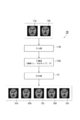
- FIG. 1 is a diagram showing the configuration of the tomographic image prediction apparatus 1.
- FIG. 2 is a diagram illustrating a configuration of the tomographic image prediction apparatus 1 in which the deep neural network has been learned.
- FIG. 3 is a diagram illustrating a configuration of the tomographic image prediction apparatus 1A.
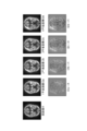
- FIG. 4 is a diagram illustrating an example of a tomographic image (input image I1) input to the input unit 11, output images I2a to I2d output from the output unit 13, and a difference image output from the output unit 13. .
- FIG. 5 is a diagram showing a comparison between an actual tomographic image and a predicted tomographic image.
- FIG. 6 is a diagram showing a comparison between tomographic images predicted in the worst scenario and the best scenario.
- FIG. 7 is a diagram showing a configuration of the tomographic image prediction apparatus 1B.
- FIG. 8 is a diagram illustrating a configuration of the tomographic image prediction apparatus 1C.
- FIG. 1 is a diagram showing a configuration of the tomographic image prediction apparatus 1.
- the tomographic image prediction apparatus 1 inputs a tomographic image of the subject's brain as an input image I1, predicts the tomographic image of the subject's brain after the acquisition of the tomographic image by a deep neural network, and the predicted tomographic image.
- the image is output as an output image I2.
- the tomographic image prediction apparatus 1 can learn a deep neural network using the tomographic image database 15.
- the tomographic image prediction apparatus 1 is configured using, for example, a computer and includes an input unit 11, a prediction unit (processing unit) 12, an output unit 13, and a learning unit 14.
- the input unit 11 inputs a tomographic image of the subject's brain as an input image I1.
- the tomographic image is, for example, a PET image acquired by a PET (Positron Emission Tomography) apparatus, a SPECT image acquired by a SPECT (Single Photon Emission Computed Tomography) apparatus, and an MRI image acquired by an MRI (Magnetic Resonance Imaging) apparatus. Any of these may be used.
- the tomographic image may be a two-dimensional tomographic image of one slice or a plurality of slices, or may be a three-dimensional tomographic image.
- the prediction unit 12 uses a deep neural network to obtain a tomographic image of the subject's brain after the tomographic image is acquired. Etc.
- the prediction unit 12 may also be configured to predict the brain health of the subject based on the predicted brain tomographic image of the subject.
- the brain health status is, for example, the degree of dementia, and can be expressed as a numerical Z score, or it can be expressed as a risk level (necessary examination, attention required, follow-up observation, etc.). .
- the deep neural network may be a convolutional neural network.
- a convolution layer for extracting a feature amount and a pooling layer for compressing the feature amount are alternately provided.
- the prediction unit 12 may perform processing in the deep neural network by a CPU (Central Processing Unit), but is preferably performed by a DSP (Digital Signal Processor) or GPU (Graphics Processing Unit) capable of higher speed processing. It is.
- DSP Digital Signal Processor
- GPU Graphics Processing Unit
- the output unit 13 outputs the result of prediction by the prediction unit 12.
- the output unit 13 outputs the tomographic image of the brain of the subject predicted by the prediction unit 12 as the output image I2.
- the output unit 13 also outputs the brain health of the subject predicted by the prediction unit 12.
- the output unit 13 may include a display that displays an image.
- the learning unit 14 uses the tomographic image database 15 to learn the deep neural network of the prediction unit 12.
- the tomographic image database 15 stores brain tomographic image data acquired at a plurality of time points for a plurality of subjects.
- the tomographic image database 15 stores, for example, the tomographic image data of the brain of each subject acquired at intervals of approximately one year.
- the learning unit 14 uses a tomographic image acquired in a certain year of each subject as an input image I1, and acquires the output image I2 corresponding to the input image I1 and the next year of the subject (or a year after a certain number of years have elapsed). Based on the obtained tomographic image, the deep neural network of the prediction unit 12 is learned. Such learning of a deep neural network is called deep learning.
- the tomographic image prediction method using such a tomographic image prediction apparatus 1 includes an input step by the input unit 11, a prediction step by the prediction unit 12, an output step by the output unit 13, and a learning step by the learning unit 14.
- a tomographic image of the subject's brain is input as the input image I1.
- the prediction step based on the tomographic image of the brain (input image I1) input in the input step, the tomographic image of the subject's brain after the acquisition of the tomographic image is predicted by the deep neural network. Moreover, it is good also as a structure which also estimates the healthy state of the test subject's brain based on the predicted tomographic image of the test subject's brain in the prediction step.
- the tomographic image of the brain of the subject predicted in the prediction step is output as an output image I2, and the health state of the brain of the subject predicted in the prediction step is also output.
- the deep neural network is learned using the tomographic image database 15.
- the input, prediction, and output steps can be repeated thereafter, so the learning step is performed each time the input, prediction, and output steps are performed. There is no need to do.
- the learning unit 14 is not necessary if the deep neural network has already been learned. However, even when the deep neural network has already been learned, there may be a learning step and learning unit 14 when further learning is performed in order to enable prediction with higher accuracy.
- FIG. 2 is a diagram showing a configuration of the tomographic image prediction apparatus 1 in which the deep neural network has already been learned.
- the tomographic image prediction apparatus 1 inputs a tomographic image of the subject's brain as an input image I1, and the tomographic image of the subject's brain after the acquisition of the tomographic image (for example, one year later, after a certain number of years has elapsed).
- the image can be predicted by the deep neural network and output as the output image I2, and the health condition of the subject's brain can also be predicted based on the predicted tomographic image of the subject's brain. Since the tomographic image of the subject's future brain (and brain health) can be predicted, a more effective policy can be made for the care, treatment or prevention of the subject from the present to the future.
- the following measures can be taken.
- the symptoms can be improved by brain surgical treatment.
- the abnormality is a thyroid hormone abnormality
- symptoms can be improved by medical treatment.
- the progression of symptoms can be delayed by administration of a drug.
- the tomographic image prediction apparatus 1 and the tomographic image prediction method are not limited to a mode in which one input image I1 is input and one output image I2 is output, and various modes are possible. Hereinafter, another aspect of the tomographic image prediction apparatus will be described.
- FIG. 3 is a diagram showing a configuration of the tomographic image prediction apparatus 1A.
- the predicting unit 12A predicts brain tomographic images at a plurality of time points after the acquisition of the brain tomographic image (input image I1) input to the input unit 11.
- the output unit 13 may individually display the brain tomographic images (output images I2a to I2e) at each of the plurality of time points predicted by the prediction unit 12A, and preferably, the brain at each of the plurality of time points. You may display a moving image by displaying a tomographic image in order.
- the output image I2a is a predicted image after one year has elapsed since the acquisition of the input image I1.
- the output image I2b is a predicted image after two years have elapsed since the acquisition of the input image I1.
- the output image I2c is a predicted image after three years have elapsed since the acquisition of the input image I1.
- the output image I2d is a predicted image four years after the input image I1 is acquired.
- the output image I2e is a predicted image after five years from the acquisition of the input image I1.
- the prediction unit 12A predicts an output image I2a from the input image I1, predicts an output image I2b from the output image I2a, predicts an output image I2c from the output image I2b, and predicts an output image I2d from the output image I2c.
- the output image I2e may be predicted from the output image I2d.
- the deep neural network may be learned so that the output images I2a to I2e can be predicted from the input image I1.
- FIG. 4 is a diagram illustrating an example of a tomographic image (input image I1) input to the input unit 11, output images I2a to I2d output from the output unit 13, and a difference image output from the output unit 13. .
- the output unit 13 includes a brain tomographic image (output images I2a to I2d and the like) predicted by the prediction unit 12A and a brain tomographic image (input image I1) input to the input unit 11. It is also possible to obtain a difference image (I2a-I1, I2b-I1, I2c-I1, I2d-I1, etc.) representing the difference between and output this difference image.
- a difference image I2a-I1, I2b-I1, I2c-I1, I2d-I1, etc.
- FIG. 5 is a diagram showing a comparison between an actual tomographic image and a predicted tomographic image.
- a deep neural network was trained using a tomographic image database storing 470 sets of PET image data, with a set of PET images at a certain time point and a PET image one year later.
- This figure shows the actual PET images from the first year to the fifth year of the brain of a particular subject, and the predicted PET images from the first year to the fifth year of that particular subject's brain. Is also shown.
- This figure also shows a difference image representing the difference between the actual PET image of the fifth year and the PET image of the first year, and between the predicted PET image of the fifth year and the PET image of the first year. A difference image representing the difference is also shown.
- the portion where the activity was reduced was common between the actual PET image of a certain year and the predicted PET image of the same year. Moreover, the site
- FIG. 6 is a diagram showing a comparison between tomographic images predicted in the worst scenario and the best scenario.
- the prediction unit 12A may be configured to predict a brain tomographic image after the acquisition of the brain tomographic image input to the input unit 11 for each of the worst scenario and the best scenario.
- the deep neural network learns using the tomographic image databases having different temporal change speeds among the tomographic image databases.
- a deep neural network is trained using a database of tomographic images that change rapidly over time (eg, tomographic images of subjects close to Alzheimer's dementia), it is possible to predict tomographic images of the future brain in the worst scenario. .
- a deep neural network is trained using a database of tomographic images that change slowly over time (for example, tomographic images of healthy subjects)
- a future brain tomographic image can be predicted in the best scenario.
- various policies for care, treatment, or prevention from the present to the future of the subject can be established.
- FIG. 7 is a diagram showing a configuration of the tomographic image prediction apparatus 1B.
- the input unit 11B inputs a plurality of tomographic images (input images I1a and I1b) of the subject's brain.
- the prediction unit 12B predicts a tomographic image of the brain after the time of acquisition of the tomographic images based on the plurality of tomographic images of the brain of the subject input to the input unit 11B.
- the deep neural network is trained so that the output images I2a to I2e can be predicted from a plurality of tomographic images (input images I1a and I1b) of the subject's brain.
- the tomographic images of the subject's brain are tomographic images of the subject's brain acquired at each of a plurality of time points (for example, a tomographic image acquired in a certain year and a tomographic image acquired in the following year). is there.
- the plurality of tomographic images of the subject's brain are tomographic images (for example, PET images, SPECT images, and MRI images) of the subject's brain acquired by each of a plurality of types of tomographic image acquisition devices.
- prediction accuracy can be improved by predicting a future brain tomographic image based on a plurality of tomographic images of the subject's brain.
- FIG. 8 is a diagram showing a configuration of the tomographic image prediction apparatus 1C.
- the input unit 11C not only inputs a tomographic image (input image I1) of the subject's brain, but also inputs other information related to the subject.
- the prediction unit 12C predicts a tomographic image of the brain after the acquisition of the tomographic image based on the tomographic image of the brain of the subject input to the input unit 11C and other information related to the subject.
- the deep neural network is trained so that the output images I2a to I2e can be predicted from the tomographic image of the subject's brain and other information related to the subject.
- Other information regarding the subject is information that may be related to the health condition of the brain, for example, information such as age, sex, genetic information, medical history, lifestyle habits, and the like.
- the prediction accuracy can be improved by predicting the future brain tomographic image based on the tomographic image of the brain of the subject and other information relating to the subject.
- the present invention is not limited to the above embodiment and configuration examples, and various modifications are possible. For example, you may combine the arbitrary aspects of the various aspects demonstrated so far.
- a medical institution that includes a tomographic image acquisition apparatus (PET apparatus, SPECT apparatus, MRI apparatus) to acquire a tomographic image of the subject's brain, and an institution that includes a tomographic image prediction apparatus to predict a tomographic image of the subject's future brain ( (Hereinafter referred to as “prediction organization”) may be separate.
- the tomographic image of the subject's brain acquired in the medical institution is sent to the prediction institution via the communication line between the medical institution and the prediction institution, and the future brain tomographic image predicted in the prediction institution is the medical institution.
- a doctor or the like can make an effective policy for care, treatment, or prevention from the present to the future of the subject based on a future brain tomographic image sent from the prediction institution.
- the tomographic image prediction apparatus includes (1) an input unit that inputs a tomographic image of a subject's brain, and (2) a tomographic image of the brain based on the tomographic image of the brain input to the input unit.
- a prediction unit that predicts a tomographic image of the brain of the subject after the image is acquired, and (3) an output unit that outputs a result of prediction by the prediction unit.
- the above-described tomographic image prediction apparatus may further include a learning unit that learns a deep neural network using a database of brain tomographic images acquired at a plurality of time points for a plurality of subjects.
- the prediction unit may be configured to predict the health state of the subject's brain based on the predicted tomographic image of the subject's brain.
- the prediction unit may be configured to predict a brain tomographic image at each of a plurality of time points after the acquisition of the brain tomographic image input to the input unit.
- the output unit may display a brain tomographic image at each of a plurality of time points predicted by the prediction unit as a moving image.
- the output unit obtains a difference image representing a difference between the tomographic image of the brain predicted by the prediction unit and the tomographic image of the brain input to the input unit, and calculates the difference image. It may be configured to output.
- the predicting unit learns using a tomographic image database having a different temporal change speed from a database of brain tomographic images acquired at a plurality of time points for a plurality of subjects.
- a configuration in which a tomographic image of the brain after the acquisition of the tomographic image of the brain input to the input unit is predicted by a neural network may be employed.
- the prediction unit may be configured to predict a tomographic image of the brain after the acquisition of the tomographic images based on the tomographic images of the brain of the subject acquired at each of a plurality of time points. good.
- the prediction unit may be configured to predict a tomographic image of the brain after the time of acquisition of the tomographic images based on the tomographic images of the brain of the subject acquired by each of the plurality of types of tomographic image acquisition devices.
- the predicting unit may be configured to predict a tomographic image of the brain after the acquisition of the tomographic image based on the tomographic image of the brain input to the input unit and other information about the subject.
- the tomographic image prediction method includes (1) an input step of inputting a tomographic image of a subject's brain, and (2) a tomographic image of the brain by a deep neural network based on the tomographic image of the brain input in the input step.
- a prediction step of predicting a tomographic image of the subject's brain after the image acquisition time and (3) an output step of outputting a prediction result in the prediction step are provided.
- the above-described tomographic image prediction method may further include a learning step of learning a deep neural network using a brain tomographic image database acquired at a plurality of time points for a plurality of subjects.
- the prediction step may be configured to predict the health state of the subject's brain based on the predicted tomographic image of the subject's brain.
- the prediction step may be configured to predict a brain tomographic image at each of a plurality of time points after the acquisition of the brain tomographic image input in the input step.
- the output step may display a brain tomographic image at each of a plurality of time points predicted in the prediction step as a moving image.
- the output step obtains a difference image representing a difference between the brain tomographic image predicted in the prediction step and the brain tomographic image input in the input step, It may be configured to output.
- the step of predicting is a deep layer learned using a database of tomographic images having different speeds of temporal change from a database of tomographic images of the brain acquired at a plurality of time points for a plurality of subjects.
- a configuration in which a tomographic image of the brain after the acquisition of the tomographic image of the brain input in the input step is predicted by a neural network may be adopted.
- the prediction step may be configured to predict a tomographic image of the brain after the acquisition of the tomographic images based on the tomographic images of the subject's brain acquired at each of a plurality of time points. good.
- the prediction step may be configured to predict a tomographic image of the brain after the acquisition of the tomographic images based on the tomographic images of the brain of the subject acquired by each of the plurality of types of tomographic image acquisition devices.
- the prediction step may be configured to predict a tomographic image of the brain after the tomographic image is acquired based on the tomographic image of the brain input in the input step and other information about the subject.
- the present invention can be used as a tomographic image predicting apparatus and a tomographic image predicting method useful for planning a future treatment policy for a subject.
Landscapes
- Health & Medical Sciences (AREA)
- Engineering & Computer Science (AREA)
- Life Sciences & Earth Sciences (AREA)
- Medical Informatics (AREA)
- Physics & Mathematics (AREA)
- General Health & Medical Sciences (AREA)
- Biomedical Technology (AREA)
- Public Health (AREA)
- Molecular Biology (AREA)
- Biophysics (AREA)
- Pathology (AREA)
- Nuclear Medicine, Radiotherapy & Molecular Imaging (AREA)
- Radiology & Medical Imaging (AREA)
- Heart & Thoracic Surgery (AREA)
- Veterinary Medicine (AREA)
- Surgery (AREA)
- Animal Behavior & Ethology (AREA)
- Artificial Intelligence (AREA)
- Theoretical Computer Science (AREA)
- Neurology (AREA)
- Physiology (AREA)
- Data Mining & Analysis (AREA)
- Computer Vision & Pattern Recognition (AREA)
- Mathematical Physics (AREA)
- Evolutionary Computation (AREA)
- High Energy & Nuclear Physics (AREA)
- Epidemiology (AREA)
- Primary Health Care (AREA)
- Optics & Photonics (AREA)
- General Physics & Mathematics (AREA)
- Psychiatry (AREA)
- Computational Linguistics (AREA)
- Computing Systems (AREA)
- Software Systems (AREA)
- General Engineering & Computer Science (AREA)
- Signal Processing (AREA)
- Neurosurgery (AREA)
- Databases & Information Systems (AREA)
- Oral & Maxillofacial Surgery (AREA)
- Fuzzy Systems (AREA)
Abstract
La présente invention concerne un dispositif de prédiction d'image tomographique 1 qui comprend une unité d'entrée 11, une unite de prédiction 12, une unité de sortie 13 et une unité d'apprentissage 14. Le dispositif de prédiction d'image tomographique 1 : reçoit une entrée d'une image tomographique du cerveau d'un sujet en tant qu'image d'entrée I1 ; prédit, au moyen d'un réseau neuronal profond de l'unité de prédiction 12, une image tomographique du cerveau du sujet à un temps postérieur à l'acquisition de ladite image tomographique ; et délivre en sortie l'image tomographique prédite en tant qu'image de sortie I2. Le dispositif de prédiction d'image tomographique 1 permet l'apprentissage du réseau neuronal profond au moyen d'une base de données d'images tomographiques 15. Grâce à cette configuration, un dispositif de prédiction d'image tomographique et un procédé de prédiction d'image tomographique qui sont utiles pour planifier une cure de traitement future ou similaire pour le sujet sont réalisés.
Priority Applications (4)
| Application Number | Priority Date | Filing Date | Title |
|---|---|---|---|
| US15/734,318 US20210196125A1 (en) | 2018-06-04 | 2019-05-30 | Tomographic image prediction device and tomographic image prediction method |
| CN201980037277.2A CN112219138A (zh) | 2018-06-04 | 2019-05-30 | 断层图像预测装置及断层图像预测方法 |
| EP19815564.0A EP3805807A4 (fr) | 2018-06-04 | 2019-05-30 | Dispositif de prédiction d'image tomographique et procédé de prédiction d'image tomographique |
| KR1020207032270A KR20210018214A (ko) | 2018-06-04 | 2019-05-30 | 단층 화상 예측 장치 및 단층 화상 예측 방법 |
Applications Claiming Priority (2)
| Application Number | Priority Date | Filing Date | Title |
|---|---|---|---|
| JP2018106908A JP7114347B2 (ja) | 2018-06-04 | 2018-06-04 | 断層画像予測装置および断層画像予測方法 |
| JP2018-106908 | 2018-06-04 |
Publications (1)
| Publication Number | Publication Date |
|---|---|
| WO2019235367A1 true WO2019235367A1 (fr) | 2019-12-12 |
Family
ID=68769302
Family Applications (1)
| Application Number | Title | Priority Date | Filing Date |
|---|---|---|---|
| PCT/JP2019/021637 Ceased WO2019235367A1 (fr) | 2018-06-04 | 2019-05-30 | Dispositif de prédiction d'image tomographique et procédé de prédiction d'image tomographique |
Country Status (7)
| Country | Link |
|---|---|
| US (1) | US20210196125A1 (fr) |
| EP (1) | EP3805807A4 (fr) |
| JP (1) | JP7114347B2 (fr) |
| KR (1) | KR20210018214A (fr) |
| CN (1) | CN112219138A (fr) |
| TW (1) | TWI827610B (fr) |
| WO (1) | WO2019235367A1 (fr) |
Families Citing this family (8)
| Publication number | Priority date | Publication date | Assignee | Title |
|---|---|---|---|---|
| JP6679017B1 (ja) * | 2019-12-20 | 2020-04-15 | 株式会社Splink | 認知症リスクの提示システムおよび方法 |
| KR102582293B1 (ko) * | 2020-09-09 | 2023-09-25 | 사회복지법인 삼성생명공익재단 | 내이기관의 내림프수종 비율 측정 방법 및 그 장치 |
| JP7777398B2 (ja) * | 2021-04-05 | 2025-11-28 | 浜松ホトニクス株式会社 | 断層画像推測装置および断層画像推測方法 |
| JP7116445B1 (ja) * | 2021-10-01 | 2022-08-10 | 株式会社CogSmart | 認知症予防支援装置、認知症予防支援プログラムおよび認知症予防支援方法 |
| JP7820792B2 (ja) * | 2021-10-26 | 2026-02-26 | 株式会社Splink | コンピュータプログラム、情報処理装置、情報処理方法及び学習モデル生成方法 |
| WO2023153839A1 (fr) * | 2022-02-09 | 2023-08-17 | 사회복지법인 삼성생명공익재단 | Procédé de calcul d'informations de démence et dispositif d'analyse utilisant une irm bidimensionnelle |
| CN115661152B (zh) * | 2022-12-27 | 2023-04-07 | 四川大学华西医院 | 基于模型预测的目标发展情况分析方法 |
| JPWO2025126343A1 (fr) * | 2023-12-12 | 2025-06-19 |
Citations (5)
| Publication number | Priority date | Publication date | Assignee | Title |
|---|---|---|---|---|
| WO2008056638A1 (fr) | 2006-11-06 | 2008-05-15 | Fujifilm Ri Pharma Co., Ltd. | Procédé d'aide au diagnostic à partir d'une image du cerveau, logiciel, et procédé d'enregistrement |
| JP2008157640A (ja) * | 2006-12-20 | 2008-07-10 | Fujifilm Ri Pharma Co Ltd | 脳画像データに関する時系列データの解析方法、プログラムおよび記録媒体 |
| US20130116540A1 (en) * | 2010-03-15 | 2013-05-09 | Shi-Jiang Li | Systems and methods for detection and prediction of brain disorders based on neural network interaction |
| WO2017106645A1 (fr) * | 2015-12-18 | 2017-06-22 | The Regents Of The University Of California | Interprétation et quantification de caractéristique d'urgence sur une tomographie assistée par ordinateur de tête |
| US20180144466A1 (en) * | 2016-11-23 | 2018-05-24 | General Electric Company | Deep learning medical systems and methods for image acquisition |
Family Cites Families (17)
| Publication number | Priority date | Publication date | Assignee | Title |
|---|---|---|---|---|
| US6577700B1 (en) * | 2001-06-22 | 2003-06-10 | Liang-Shih Fan | Neural network based multi-criteria optimization image reconstruction technique for imaging two- and three-phase flow systems using electrical capacitance tomography |
| US9492114B2 (en) * | 2004-06-18 | 2016-11-15 | Banner Health Systems, Inc. | Accelerated evaluation of treatments to prevent clinical onset of alzheimer's disease |
| US7929737B2 (en) * | 2005-09-29 | 2011-04-19 | General Electric Company | Method and system for automatically generating a disease severity index |
| US20080298657A1 (en) * | 2005-11-23 | 2008-12-04 | Junji Shiraishi | Computer-Aided Method for Detection of Interval Changes in Successive Whole-Body Bone Scans and Related Computer Program Program Product and System |
| US9563950B2 (en) * | 2013-03-20 | 2017-02-07 | Cornell University | Methods and tools for analyzing brain images |
| US9700219B2 (en) * | 2013-10-17 | 2017-07-11 | Siemens Healthcare Gmbh | Method and system for machine learning based assessment of fractional flow reserve |
| WO2015134665A1 (fr) * | 2014-03-04 | 2015-09-11 | SignalSense, Inc. | Classification de données à l'aide d'enregistrements neuronaux d'apprentissage profond raffinés de façon incrémentale via des entrées d'experts |
| US10546233B1 (en) * | 2014-07-23 | 2020-01-28 | Hrl Laboratories, Llc | Method and system to predict and interpret conceptual knowledge in the brain |
| CN107636659B (zh) * | 2015-05-11 | 2021-10-12 | 西门子保健有限责任公司 | 使用深度神经网络检测医学图像中的地标的方法和系统 |
| WO2017006764A1 (fr) * | 2015-07-08 | 2017-01-12 | 株式会社日立製作所 | Dispositif de calcul d'image, procédé de calcul d'image, et tomographe |
| CN105718952B (zh) * | 2016-01-22 | 2018-10-30 | 武汉科恩斯医疗科技有限公司 | 使用深度学习网络对断层医学影像进行病灶分类的系统 |
| WO2017223560A1 (fr) * | 2016-06-24 | 2017-12-28 | Rensselaer Polytechnic Institute | Reconstruction d'images tomographiques par apprentissage machine |
| CN109690554B (zh) * | 2016-07-21 | 2023-12-05 | 西门子保健有限责任公司 | 用于基于人工智能的医学图像分割的方法和系统 |
| CN107451609A (zh) * | 2017-07-24 | 2017-12-08 | 上海交通大学 | 基于深度卷积神经网络的肺结节图像识别系统 |
| CN107616796B (zh) * | 2017-08-31 | 2020-09-11 | 北京医拍智能科技有限公司 | 基于深度神经网络的肺结节良恶性检测方法及装置 |
| US11918333B2 (en) * | 2017-12-29 | 2024-03-05 | Analytics For Life Inc. | Method and system to assess disease using phase space tomography and machine learning |
| CN112292691B (zh) * | 2018-06-18 | 2024-06-04 | 谷歌有限责任公司 | 用于使用深度学习提高癌症检测的方法与系统 |
-
2018
- 2018-06-04 JP JP2018106908A patent/JP7114347B2/ja active Active
-
2019
- 2019-05-30 US US15/734,318 patent/US20210196125A1/en not_active Abandoned
- 2019-05-30 CN CN201980037277.2A patent/CN112219138A/zh active Pending
- 2019-05-30 EP EP19815564.0A patent/EP3805807A4/fr not_active Withdrawn
- 2019-05-30 KR KR1020207032270A patent/KR20210018214A/ko not_active Ceased
- 2019-05-30 WO PCT/JP2019/021637 patent/WO2019235367A1/fr not_active Ceased
- 2019-06-04 TW TW108119283A patent/TWI827610B/zh active
Patent Citations (5)
| Publication number | Priority date | Publication date | Assignee | Title |
|---|---|---|---|---|
| WO2008056638A1 (fr) | 2006-11-06 | 2008-05-15 | Fujifilm Ri Pharma Co., Ltd. | Procédé d'aide au diagnostic à partir d'une image du cerveau, logiciel, et procédé d'enregistrement |
| JP2008157640A (ja) * | 2006-12-20 | 2008-07-10 | Fujifilm Ri Pharma Co Ltd | 脳画像データに関する時系列データの解析方法、プログラムおよび記録媒体 |
| US20130116540A1 (en) * | 2010-03-15 | 2013-05-09 | Shi-Jiang Li | Systems and methods for detection and prediction of brain disorders based on neural network interaction |
| WO2017106645A1 (fr) * | 2015-12-18 | 2017-06-22 | The Regents Of The University Of California | Interprétation et quantification de caractéristique d'urgence sur une tomographie assistée par ordinateur de tête |
| US20180144466A1 (en) * | 2016-11-23 | 2018-05-24 | General Electric Company | Deep learning medical systems and methods for image acquisition |
Non-Patent Citations (1)
| Title |
|---|
| See also references of EP3805807A4 |
Also Published As
| Publication number | Publication date |
|---|---|
| US20210196125A1 (en) | 2021-07-01 |
| TW202011895A (zh) | 2020-04-01 |
| JP2019211307A (ja) | 2019-12-12 |
| JP7114347B2 (ja) | 2022-08-08 |
| KR20210018214A (ko) | 2021-02-17 |
| EP3805807A4 (fr) | 2022-02-23 |
| TWI827610B (zh) | 2024-01-01 |
| CN112219138A (zh) | 2021-01-12 |
| EP3805807A1 (fr) | 2021-04-14 |
Similar Documents
| Publication | Publication Date | Title |
|---|---|---|
| WO2019235367A1 (fr) | Dispositif de prédiction d'image tomographique et procédé de prédiction d'image tomographique | |
| US20240274249A1 (en) | Diagnosis support system, information processing method, and program | |
| Popescu et al. | Local brain-age: a U-net model | |
| CN108784655B (zh) | 针对医疗患者的快速评估和后果分析 | |
| EP3596697B1 (fr) | Analyse d'image médicale généralisable à l'aide de réseaux neuronaux de segmentation et de classification | |
| US9265441B2 (en) | Assessment of traumatic brain injury | |
| US11730420B2 (en) | Maternal-fetal sepsis indicator | |
| JP7600138B2 (ja) | 病状によって影響を受ける可能性がある被験者を識別するための方法およびシステム | |
| JP7339270B2 (ja) | 医用画像処理装置、方法およびプログラム | |
| EP3918987B1 (fr) | Modèle entraîné | |
| US12026872B2 (en) | Diagnosis assisting system, diagnosis assisting method and diagnosis assisting program | |
| Garbarino et al. | Differences in topological progression profile among neurodegenerative diseases from imaging data | |
| JP2019101485A (ja) | 情報処理方法、情報処理装置、情報処理システム及びプログラム | |
| Combès et al. | A clinically-compatible workflow for computer-aided assessment of brain disease activity in multiple sclerosis patients | |
| JP2021104140A (ja) | 医用情報処理装置、医用情報処理方法、及び医用情報処理プログラム | |
| WO2021245733A1 (fr) | Dispositif d'analyse d'image cérébrale, procédé de commande et support lisible par ordinateur | |
| JP7852212B2 (ja) | 診断支援装置、コンピュータプログラム及び医療機器の作動方法 | |
| CN114450752A (zh) | 用于利用深度学习模型的计算机辅助诊断的方法和系统 | |
| JP7064430B2 (ja) | 優先度判定装置、方法およびプログラム | |
| US20200160517A1 (en) | Priority judgement device, method, and program | |
| US20180004901A1 (en) | Systems and methods for holistic analysis of medical conditions | |
| JP7313165B2 (ja) | アルツハイマー病生存分析装置及びアルツハイマー病生存分析プログラム | |
| Powell et al. | Mild traumatic brain injury history is associated with lower brain network resilience in soldiers | |
| KR20220082869A (ko) | 진단 툴 | |
| Bourque et al. | Discovering Hidden Links: Harnessing Similarity Network Fusion to Reveal Common Clusters in Healthy Aging, Mild Cognitive Impairment, and Dementia |
Legal Events
| Date | Code | Title | Description |
|---|---|---|---|
| 121 | Ep: the epo has been informed by wipo that ep was designated in this application |
Ref document number: 19815564 Country of ref document: EP Kind code of ref document: A1 |
|
| ENP | Entry into the national phase |
Ref document number: 20207032270 Country of ref document: KR Kind code of ref document: A |
|
| NENP | Non-entry into the national phase |
Ref country code: DE |
|
| ENP | Entry into the national phase |
Ref document number: 2019815564 Country of ref document: EP Effective date: 20210111 |