WO2020071665A2 - 세포의 텔로미어를 신장시키는 조성물 및 그 제조방법 - Google Patents
세포의 텔로미어를 신장시키는 조성물 및 그 제조방법Info
- Publication number
- WO2020071665A2 WO2020071665A2 PCT/KR2019/012150 KR2019012150W WO2020071665A2 WO 2020071665 A2 WO2020071665 A2 WO 2020071665A2 KR 2019012150 W KR2019012150 W KR 2019012150W WO 2020071665 A2 WO2020071665 A2 WO 2020071665A2
- Authority
- WO
- WIPO (PCT)
- Prior art keywords
- cells
- exosomes
- telomeres
- medium
- stimulation
- Prior art date
- Legal status (The legal status is an assumption and is not a legal conclusion. Google has not performed a legal analysis and makes no representation as to the accuracy of the status listed.)
- Ceased
Links
Images


Classifications
-
- C—CHEMISTRY; METALLURGY
- C12—BIOCHEMISTRY; BEER; SPIRITS; WINE; VINEGAR; MICROBIOLOGY; ENZYMOLOGY; MUTATION OR GENETIC ENGINEERING
- C12N—MICROORGANISMS OR ENZYMES; COMPOSITIONS THEREOF; PROPAGATING, PRESERVING, OR MAINTAINING MICROORGANISMS; MUTATION OR GENETIC ENGINEERING; CULTURE MEDIA
- C12N5/00—Undifferentiated human, animal or plant cells, e.g. cell lines; Tissues; Cultivation or maintenance thereof; Culture media therefor
- C12N5/06—Animal cells or tissues; Human cells or tissues
- C12N5/0602—Vertebrate cells
- C12N5/0603—Embryonic cells ; Embryoid bodies
-
- C—CHEMISTRY; METALLURGY
- C12—BIOCHEMISTRY; BEER; SPIRITS; WINE; VINEGAR; MICROBIOLOGY; ENZYMOLOGY; MUTATION OR GENETIC ENGINEERING
- C12N—MICROORGANISMS OR ENZYMES; COMPOSITIONS THEREOF; PROPAGATING, PRESERVING, OR MAINTAINING MICROORGANISMS; MUTATION OR GENETIC ENGINEERING; CULTURE MEDIA
- C12N13/00—Treatment of microorganisms or enzymes with electrical or wave energy, e.g. magnetism, sonic waves
-
- A—HUMAN NECESSITIES
- A61—MEDICAL OR VETERINARY SCIENCE; HYGIENE
- A61P—SPECIFIC THERAPEUTIC ACTIVITY OF CHEMICAL COMPOUNDS OR MEDICINAL PREPARATIONS
- A61P39/00—General protective or antinoxious agents
- A61P39/06—Free radical scavengers or antioxidants
-
- C—CHEMISTRY; METALLURGY
- C07—ORGANIC CHEMISTRY
- C07K—PEPTIDES
- C07K14/00—Peptides having more than 20 amino acids; Gastrins; Somatostatins; Melanotropins; Derivatives thereof
- C07K14/435—Peptides having more than 20 amino acids; Gastrins; Somatostatins; Melanotropins; Derivatives thereof from animals; from humans
- C07K14/46—Peptides having more than 20 amino acids; Gastrins; Somatostatins; Melanotropins; Derivatives thereof from animals; from humans from vertebrates
- C07K14/47—Peptides having more than 20 amino acids; Gastrins; Somatostatins; Melanotropins; Derivatives thereof from animals; from humans from vertebrates from mammals
- C07K14/4701—Peptides having more than 20 amino acids; Gastrins; Somatostatins; Melanotropins; Derivatives thereof from animals; from humans from vertebrates from mammals not used
- C07K14/4702—Regulators; Modulating activity
- C07K14/4705—Regulators; Modulating activity stimulating, promoting or activating activity
-
- C—CHEMISTRY; METALLURGY
- C12—BIOCHEMISTRY; BEER; SPIRITS; WINE; VINEGAR; MICROBIOLOGY; ENZYMOLOGY; MUTATION OR GENETIC ENGINEERING
- C12N—MICROORGANISMS OR ENZYMES; COMPOSITIONS THEREOF; PROPAGATING, PRESERVING, OR MAINTAINING MICROORGANISMS; MUTATION OR GENETIC ENGINEERING; CULTURE MEDIA
- C12N5/00—Undifferentiated human, animal or plant cells, e.g. cell lines; Tissues; Cultivation or maintenance thereof; Culture media therefor
- C12N5/06—Animal cells or tissues; Human cells or tissues
- C12N5/0602—Vertebrate cells
- C12N5/0608—Germ cells
- C12N5/0611—Primordial germ cells, e.g. embryonic germ cells [EG]
-
- C—CHEMISTRY; METALLURGY
- C12—BIOCHEMISTRY; BEER; SPIRITS; WINE; VINEGAR; MICROBIOLOGY; ENZYMOLOGY; MUTATION OR GENETIC ENGINEERING
- C12N—MICROORGANISMS OR ENZYMES; COMPOSITIONS THEREOF; PROPAGATING, PRESERVING, OR MAINTAINING MICROORGANISMS; MUTATION OR GENETIC ENGINEERING; CULTURE MEDIA
- C12N5/00—Undifferentiated human, animal or plant cells, e.g. cell lines; Tissues; Cultivation or maintenance thereof; Culture media therefor
- C12N5/06—Animal cells or tissues; Human cells or tissues
- C12N5/0602—Vertebrate cells
- C12N5/0618—Cells of the nervous system
- C12N5/0623—Stem cells
-
- C—CHEMISTRY; METALLURGY
- C12—BIOCHEMISTRY; BEER; SPIRITS; WINE; VINEGAR; MICROBIOLOGY; ENZYMOLOGY; MUTATION OR GENETIC ENGINEERING
- C12N—MICROORGANISMS OR ENZYMES; COMPOSITIONS THEREOF; PROPAGATING, PRESERVING, OR MAINTAINING MICROORGANISMS; MUTATION OR GENETIC ENGINEERING; CULTURE MEDIA
- C12N9/00—Enzymes; Proenzymes; Compositions thereof; Processes for preparing, activating, inhibiting, separating or purifying enzymes
- C12N9/10—Transferases (2.)
- C12N9/12—Transferases (2.) transferring phosphorus containing groups, e.g. kinases (2.7)
- C12N9/1241—Nucleotidyltransferases (2.7.7)
-
- C—CHEMISTRY; METALLURGY
- C12—BIOCHEMISTRY; BEER; SPIRITS; WINE; VINEGAR; MICROBIOLOGY; ENZYMOLOGY; MUTATION OR GENETIC ENGINEERING
- C12N—MICROORGANISMS OR ENZYMES; COMPOSITIONS THEREOF; PROPAGATING, PRESERVING, OR MAINTAINING MICROORGANISMS; MUTATION OR GENETIC ENGINEERING; CULTURE MEDIA
- C12N2521/00—Culture process characterised by the use of hydrostatic pressure, flow or shear forces
- C12N2521/10—Sound, e.g. ultrasounds
-
- C—CHEMISTRY; METALLURGY
- C12—BIOCHEMISTRY; BEER; SPIRITS; WINE; VINEGAR; MICROBIOLOGY; ENZYMOLOGY; MUTATION OR GENETIC ENGINEERING
- C12N—MICROORGANISMS OR ENZYMES; COMPOSITIONS THEREOF; PROPAGATING, PRESERVING, OR MAINTAINING MICROORGANISMS; MUTATION OR GENETIC ENGINEERING; CULTURE MEDIA
- C12N2523/00—Culture process characterised by temperature
-
- C—CHEMISTRY; METALLURGY
- C12—BIOCHEMISTRY; BEER; SPIRITS; WINE; VINEGAR; MICROBIOLOGY; ENZYMOLOGY; MUTATION OR GENETIC ENGINEERING
- C12N—MICROORGANISMS OR ENZYMES; COMPOSITIONS THEREOF; PROPAGATING, PRESERVING, OR MAINTAINING MICROORGANISMS; MUTATION OR GENETIC ENGINEERING; CULTURE MEDIA
- C12N2529/00—Culture process characterised by the use of electromagnetic stimulation
- C12N2529/10—Stimulation by light
Definitions
- the present invention relates to an exosome that extends telomeres of cells, a composition comprising the same, and a method for preparing the same, and more specifically, TERT, TERF2, DKC1, TERF2IP, RFC1, RAD50, TERF1, PINX1, TNKS1BP1, ACD, NBN, HSPA1L, PARP1, PTGES3, SMG6, BLM, XRCC5, XRCC6, ERCC4, PRKDC, TEP1 and ⁇ -Catenin, characterized in that it contains at least one RNA or protein, exosomes that extend telomeres of cells, including them
- the present invention relates to a method for inducing exosomes to extend telomeres from cells by providing physical stimulation directly or indirectly to the composition and cells.
- Telomere is a DNA repeating structure at both ends of the chromosome (TTAGGG in humans). Telomeres form a protective cap by combining with a shelterin complex, which regulates the amplification capacity of cells and prevents binding between chromosomes and loss of genetic information during cell division. Because DNA polymerase cannot amplify all 3 'ends, telomeres are shortened by 30 to 200 bp for every cell division. When the telomeres become shorter than the threshold and close to the coding DNA and the telomer loop structure cannot be maintained, the exposed telomeres are recognized by the p53 or p16INK4a signaling system, and cell division stops and ages and dies.
- telomeres can be directly stretched by the reverse transcriptase telomerase, and the human telomerase complex is an RNA molecule that acts as a template for telomerase synthesis, TERC, a catalytic subunit, TERT, DKC1 and TEP1, etc. It is composed of an auxiliary protein. Normally, there is little telomerase activity of somatic cells, and high activity is detected only in stem cells and progenitor cells that require active division.
- telomeres When telomeres are shortened and the division of multiple cells is stopped, various problems may appear, and various symptoms, particularly aging and degeneration, may appear. Diseases that cause regeneration of bone marrow cells, such as dyskeratosis congenita and aplastic anemia, which are deficient in blood cells, are telomerase or mutations in the shelterine complex-related gene that protects telomeres. It has been discovered earlier that it appears to be due to a malfunction of the telomere retention function. Meanwhile, research on the possibility of delaying aging by increasing the length of telomeres has been convincing, as studies have recently shown that telomeres of patients with aging-related diseases such as hypertension, metabolic syndrome, diabetes, and dementia are shorter than normal. have.
- telomeres are a risk factor for cancer, including numerous cancer cells in mice that constantly overexpress genes that can increase the length of telomeres.
- telomere kidney activity can be activated only for a limited time, symptoms related to various aging and degeneration can be alleviated without the risk of cancer, and these technologies are not only telomer abnormalities caused by the genetic defects described above, but also other regenerative medicine. It can be expected to be useful in the field.
- U.S. Patent Publication No. 2018-0280413 discloses a pharmaceutical composition comprising a compound for enhancing telomerase activity, for which a process of synthesizing and purifying the compound is required, and the composition can be obtained through chemical synthesis Not only must the starting material necessary for the synthesis of silver be purified, but various by-products harmful to the environment are generated in the synthesis process, and the compound has a risk of producing by-products in the metabolic process in vivo.
- RNA encoding telomerase which also requires synthesis and purification processes, and because RNA is hydrophilic, additional carriers are required for delivery into cells. Therefore, there is a need for a composition for telomer elongation that can be manufactured more simply, has fewer hazards, and is more efficient.
- the present invention has been devised to solve the above-mentioned problems, and the present invention according to an embodiment of the present invention has been devised to solve the above-described problems, and the exosomes according to an embodiment of the present invention are TERT, TERF2, DKC1.
- RNAs or proteins selected from TERF2IP, RFC1, RAD50, TERF1, PINX1, TNKS1BP1, ACD, NBN, HSPA1L, PARP1, PTGES3, SMG6, BLM, XRCC5, XRCC6, ERCC4, PRKDC, TEP1 and ⁇ -Catenin It is characterized by.
- another embodiment of the present invention provides a composition for stretching telomeres of cells comprising the exosome.
- a method for preparing an exosome to stretch telomeres of cells includes providing physical stimulation directly or indirectly to the cells; Culturing the mixture of cells and media for a certain period of time; And isolating the exosomes from the mixture; wherein, providing the stimulus directly is to apply physical stimulation to the medium containing the cells, and providing the stimulation indirectly is to the medium not containing the cells. Characterized in that the medium and the cells are mixed after physical stimulation is applied.
- exosomes that extend the telomeres of cells are TERT, TERF2, DKC1, TERF2IP, RFC1, RAD50, TERF1, PINX1, TNKS1BP1, ACD, NBN , HSPA1L, PARP1, PTGES3, SMG6, BLM, XRCC5, XRCC6, ERCC4, PRKDC, TEP1 and ⁇ -Catenin.
- the exosome may be characterized by being induced by providing physical stimulation to the cells.
- the shape of the physical stimulus may be any one selected from ultrasound, heat and light.
- the physical stimulation is provided directly or indirectly to the cell, and providing the direct stimulation applies physical stimulation to the medium containing the cells, and providing the indirect stimulation provides physical stimulation to the medium not containing the cells. After adding the may be to mix the medium and the cells.
- the process of providing a physical stimulus capable of inducing the exosome is to provide a physical stimulus to the mixture after mixing the cell and medium, or to provide a physical stimulus to the medium and then to mix the medium and cell, or to physically apply the cell.
- the cell and medium are mixed, or after providing the cell with a physical stimulus, the cell and medium are mixed and then providing the mixture with a physical stimulus, or after providing the medium with a physical stimulus, the medium and the cell
- the physical stimulation is ultrasonic stimulation
- the direct ultrasonic stimulation is characterized in that the intensity is 0.1 to 3 W / cm 2
- the frequency is 20 kHz to 20 MHz
- the duration is 0.1 seconds to 20 minutes
- the stimulus may be characterized in that the intensity is 1 to 20 W / cm 2
- the frequency is 20 kHz to 20 MHz
- the duration is 0.1 second to 20 minutes.
- the physical stimulus is a thermal stimulus, and the thermal stimulus is applied in a manner of exposing the cells to a temperature condition of 40 to 50 ° C for 1 to 10 minutes and then exposing the cell to a temperature condition of 0 to 4 ° C for 5 to 10 seconds. can do.
- the light stimulation may be characterized by irradiating and applying a pulsed beam having a wavelength of 300 to 900 nm for 1 to 10 seconds, selected from laser or light-emitting diode light.
- the exosome may be characterized in that the total RNA is contained in 150 ng or more in 8 exosome 1X10 particles.
- the exosomes may be characterized in that the total protein is contained in 1 ⁇ g or more of 8 ⁇ m 1X10 particles.
- the telomerase activity of the cells may be increased.
- the ⁇ -galactosidase activity of the cells may be reduced.
- composition for stretching telomeres of cells includes the aforementioned exosomes.
- the composition may include the exosomes at a concentration of 10 6 to 10 14 pcs / ml.
- a method of manufacturing an exosome to stretch a cell's telomeres comprises providing physical stimulation directly or indirectly to the cell; Culturing the mixture of cells and media for a certain period of time; And isolating the exosomes from the mixture; wherein, providing the stimulus directly is to apply physical stimulation to the medium containing the cells, and providing the stimulation indirectly is to the medium not containing the cells. After the physical stimulation is applied, the medium is mixed with the cells.
- the shape of the physical stimulus may be any one selected from ultrasound, heat and light.
- the physical stimulation is ultrasonic stimulation
- the direct ultrasonic stimulation is characterized in that the intensity is 0.1 to 3 W / cm 2
- the frequency is 20 kHz to 20 MHz
- the duration is 0.1 seconds to 20 minutes
- the stimulus may be characterized in that the intensity is 1 to 20 W / cm 2
- the frequency is 20 kHz to 20 MHz
- the duration is 0.1 second to 20 minutes.
- the physical stimulus is a thermal stimulus, and the thermal stimulus is applied in a manner of exposing the cells to a temperature condition of 40 to 50 ° C for 1 to 10 minutes and then exposing the cell to a temperature condition of 0 to 4 ° C for 5 to 10 seconds. can do.
- the physical stimulation is light stimulation, and the light stimulation is characterized by applying a pulsed beam having a wavelength of 300 to 900 nm selected from laser or light-emitting diode light for 1 to 10 seconds. Can be done with
- Each of the cells may be selected from the group consisting of stem cells, progenitor cells, fibroblasts, keratinocytes, or tissue cells in an organ derived from a mammal.
- the medium may be selected from culture medium or differentiation-inducing medium.
- Incubation of the mixture may be characterized in that it proceeds for 1 hour to 10 days.
- the step of separating the exosomes may be characterized by using at least one of ultracentrifugation, density gradient, filtration, size exclusion chromatography, immunoaffinity separation, precipitation, and separation by microfluid.
- the step of separating the exosomes comprises: centrifuging the mixture after the culture to obtain a supernatant; Filtering the supernatant with a filter to obtain a filtrate; And concentrating the filtrate.
- the isolated exosomes are TERT, TERF2, DKC1, TERF2IP, RFC1, RAD50, TERF1, PINX1, TNKS1BP1, ACD, NBN, HSPA1L, PARP1, PTGES3, SMG6, compared to the exosomes secreted from the cells prior to the preparation method.
- One or more gene expression levels among BLM, XRCC5, XRCC6, ERCC4, PRKDC, TEP1 and ⁇ -Catenin may be characterized.
- the isolated exosome may be characterized in that the telomerase activity of the cell is increased upon treatment to the cell.
- ⁇ -galactosidase activity of the cells may be reduced.
- a method of manufacturing an exosome that extends the telomeres of the cell may be characterized in that the cell mobility of the cell is increased when the isolated exosome is treated with the cell.
- a method of manufacturing an exosome that extends the telomeres of the cells may be characterized in that regeneration of the tissue is promoted when the isolated exosome is treated with tissue.
- a method of manufacturing an exosome that stretches the telomeres of the cells may be characterized in that healing of the wound is promoted when the isolated exosome is treated on the wound.
- a method of manufacturing an exosome that extends the telomeres of the cells may be characterized in that healing of the scar is promoted when the isolated exosome is treated with a scar.
- a method of manufacturing an exosome that extends the telomeres of the cell may be characterized by having an anti-aging effect on the cell when the isolated exosome is treated with the cell.
- the composition for stretching telomeres of cells according to an embodiment of the present invention is simpler to prepare than the composition for stretching the telomeres described above. That is, the compounds in the existing composition are chemically synthesized, which has no specificity and has to go through a long and complicated process.
- the method of elongating telomeres of cells according to an embodiment of the present invention can promote the expression of various factors related to telomere elongation and the production of exosomes containing the factors by simply treating the cells with physical stimuli, This process consists of separating the secreted exosomes from the culture medium of the cells, and the process is simple.
- the hydrophilic nucleic acid is required to additionally load the nucleic acid into the carrier, since it is difficult to flow into the cell before the effect is exhibited without the carrier capable of delivering into the cell.
- the method for producing exosomes telomere kidney-related factors are obtained in a form surrounded by a lipid membrane, and thus, there is no need to proceed with additional loading.
- the composition for elongating telomeres of cells according to an embodiment of the present invention is superior in efficiency and application method compared to the previously disclosed composition. That is, the compound that enhances the above-described telomerase activity appears to have to inject a high concentration of the compound in the body in order to exhibit a sufficient effect, whereas the exosome that extends the telomeres of cells according to an embodiment of the present invention has a small amount. , It can be effective by applying in a non-invasive method such as local application.
- the exosomes for extending the telomeres of cells induces to secrete a large amount of exosomes containing a large amount of telomeres-related factors, in the aspect of producing exosomes of cells containing useful substances The yield is high.
- exosomes that extend the telomeres of cells according to one embodiment of the present invention have high safety. That is, the compound may have additional risks such as toxicity in metabolic processes, and in particular, the existing technology described above is a high level of compound per mg of the target animal's unit weight (kg), and wastes that are burdensome to the environment in the course of chemical synthesis. This is produced, but the method according to the invention is relatively less of this burden.
- the exosomes for extending the telomeres of cells according to an embodiment of the present invention are not only for extending the telomeres, but also induce cell division and have anti-aging and tissue regeneration effects. It can be expected that it is possible to improve and prevent various diseases and symptoms directly related to aging and tissue regeneration, as well as problems caused by the shortened telomere length.
- Example 1 is an analysis data of the telomer length change according to the cell type after direct ultrasonic stimulation treatment and culture according to Example 1 of the present invention.
- Figure 2 is an ultrasonic stimulation treatment according to Example 1 of the present invention, heat stimulation treatment according to Example 2, light-emitting diode light stimulation treatment according to Example 3 and telomer length change analysis data according to each physical stimulation treatment after each culture.
- Figure 3 is an indirect ultrasonic stimulation treatment according to Example 5 of the present invention and telomere length change analysis data after culture.
- Figure 4 is an ultrasound stimulation treatment according to Example 1 of the present invention and telomere length change analysis data according to the type of medium after culture.
- telomer length change is an analysis data of the telomer length change according to the number of times of ultrasonic stimulation treatment after culture and ultrasonic stimulation treatment according to Example 1 of the present invention.
- TERT gene expression change is an analysis data of the TERT gene expression change according to the cell type after ultrasonic stimulation treatment and culture according to Example 1 of the present invention.
- Example 7 is an analysis data of the TERT gene expression change after ultrasonic stimulation treatment and culture according to Example 1 of the present invention for CB-HDF and Adipo-MSC.
- Example 8 is an analysis data of ⁇ -Catenin gene expression after ultrasonic stimulation treatment and culture according to Example 1 of the present invention for CB-HDF and Adipo-MSC.
- Figure 9 is an analysis data of the telomerase activity after ultrasonic stimulation treatment and culture according to Example 1 of the present invention for CB-HDF and Adipo-MSC.
- Figure 11 is a cell fluorescence staining data of TERT and Ki67 after ultrasonic stimulation treatment and culture according to Example 1 of the present invention for CB-HDF.
- Example 12 is an aging-related ⁇ -galactosidase activity analysis data after ultrasonic stimulation treatment and culture according to Example 1 of the present invention for CB-HDF.
- Example 13 is an analysis of exosome marker expression in CB-HDF sonicated according to Example 5 of the present invention.
- Example 16 is a total RNA and total protein amount analysis data in exosomes prepared according to Example 5 of the present invention.
- Figure 17 is Gene ontology analysis data for RNA in exosomes prepared according to Example 5 of the present invention.
- Figure 18 is RNA-seq analysis data for RNA in exosomes prepared according to Example 5 of the present invention.
- Figure 21 is a telomere length change analysis data according to cell processing of exosomes prepared according to Example 5 of the present invention.
- FIG. 22 is an analysis data of telomer length change by cell type according to cell treatment of exosomes prepared according to Example 5 of the present invention.
- Figure 23 shows the telomeres qFISH analysis data in the cells after cell treatment of the exosomes prepared according to Example 5 of the present invention.
- Figure 24 is a cell treatment of the exosomes prepared according to Example 5 of the present invention, Tel-seq analysis data in the cells.
- Figure 25 is Gene ontology analysis data for the RNA in the cell after cell processing of the exosomes prepared according to Example 5 of the present invention.
- 26 shows RNA-seq analysis data for RNA in a cell after cell treatment of exosomes prepared according to Example 5 of the present invention.
- FIG. 27 shows TERT RNA and protein expression analysis data in corresponding cells after cell treatment of exosomes prepared according to Example 5 of the present invention.
- Figure 28 is a cell division marker analysis data in the cells after cell processing of the exosomes prepared according to Example 5 of the present invention.
- 29 is a cell division marker analysis data by exosome concentration in the cell after cell treatment of the exosomes prepared according to Example 5 of the present invention.
- Example 30 is a cell treatment of exosomes prepared according to Example 5 of the present invention, ⁇ -galactosidase activity analysis data in the cells.
- FIG. 31 is an analysis data of telomer length and TERT RNA expression by exosome concentration at a corresponding site after application of mouse ears of exosomes prepared according to Example 5 of the present invention.
- FIG. 32 is telomer FISH analysis data by exosome treatment time of the corresponding site after application of mouse ears of exosomes prepared according to Example 5 of the present invention.
- RNA and protein expression analysis data of the cell proliferation-related gene by exosome treatment time of the site after application of the mouse ear of the exosome prepared according to Example 5 of the present invention is RNA and protein expression analysis data of the cell proliferation-related gene by exosome treatment time of the site after application of the mouse ear of the exosome prepared according to Example 5 of the present invention.
- FIG. 34 shows TERT immunofluorescence analysis data by exosome treatment time of the corresponding site after application of mouse ears of exosomes prepared according to Example 5 of the present invention.
- 35 is wound healing assay data by cell processing concentration of exosomes prepared according to Example 5 of the present invention.
- FIG. 38 is telomer-related analysis data when treating cells derived from a patient with premature ejaculation of exosomes prepared according to Example 5 of the present invention.
- Figure 39 is a cell proliferation-related analysis data when processing cells derived from a patient with premature exosomes prepared according to Example 5 of the present invention.
- Figure 40 is ⁇ -galactosidase activity analysis data when processing cells derived from patients with premature exosomes prepared according to Example 5 of the present invention.
- each gene used under the designation of a gene in the present invention is an officially known gene name, a commonly used name, or a name of a product of the gene, such as a protein in the case of a gene encoding a protein.
- the inventors of the present invention provide physical stimulation capable of promoting environmental inflow to a mixture of cells and culture medium in Korean Patent No. 10-1855967, and cultivate the mixture provided with the physical stimulation for a certain period of time to recover the cells. It has been disclosed that reprogrammed cells can be obtained if the cells can be programmed and treated with exosomes obtained from the mixture. Here, it was found that the direction of reprogramming appears differently depending on the composition of the medium upon physical stimulation regardless of the type of the cell subjected to physical stimulation or the cell to which the exosome is treated. In the process of analyzing the effect of the cell reprogramming method later, it was confirmed that the expression of the gene group associated with telomere kidney is increased by applying physical stimulation to the cell.
- telomeres can be stretched by physical stimulation, regardless of the type of cells tested, as well as the composition of the medium, unlike the disclosed invention. I was able to conclude. At this time, it was confirmed that not only the expression of factors related to telomere kidney is increased, but also the secretion of exosomes containing these factors is greatly increased, and this was disclosed as a new invention.
- the exosomes that extend the telomeres of cells are TERT, TERF2, DKC1, TERF2IP, RFC1, RAD50, TERF1, PINX1, TNKS1BP1, ACD, NBN, HSPA1L, PARP1, PTGES3, SMG6, BLM, XRCC5 , XRCC6, ERCC4, PRKDC, TEP1 and ⁇ -Catenin is characterized by including one or more RNA or protein selected from the gene. Most preferably, the exosome may include RNA or protein of all the genes.
- TERT Telomerase reverse transcriptase
- TERF1 telomeric repeat binding factor 1
- TERF2 telomeric repeat binding factor 2
- DKC1 (Dyskerin pseudouridine synthase 1), RFC1 (Replication factor C subunit 1), TNKS1BP1 (Tankyrase 1 binding protein 1), NBN (Nibrin), HSPA1L (Heat shock protein family A member 1 like), PARP1 (Poly ADP-ribose polymerase) 1), PTGES3 (Prostaglandin E synthase 3), SMG6 (Smg6 homolog, Nonsense mediated mRNA decay factor), XRCC5 (X-ray repair cross complementing 5), XRCC6 (X-ray repair cross complementing 6), stabilize and maintain telomeres It is said to be necessary.
- TERF2IP TERF2 interacting protein
- RAD50 RAD50 Homolog, Double strand break repair protein
- PINX1 PIN2 / TERF1-interacting telomerase inhibitor 1
- Adrenocortical Dysplasia protein homolog (ACD), Excision repair cross-complementation group 4 (ERCC4), Protein kinase, DNA-activated, catalytic subunit (PRKDC) are required for telomer length control and protection, and Bloom syndrome protein (BLM) DNA
- ACD Adrenocortical Dysplasia protein homolog
- ERCC4 Excision repair cross-complementation group 4
- PRKDC Protein kinase
- PRKDC DNA-activated, catalytic subunit
- BBM Bloom syndrome protein
- ⁇ -Catenin is a transcription regulator that promotes TERT expression. All of the above factors have been reported to have a telomeric elongation and stabilizing effect.
- the exosomes for extending telomeres according to one aspect of the present invention comprising the gene product as described above can be used for various effects to be described later.
- the exosome may be characterized by being induced by providing physical stimulation to the cell.
- the shape of the physical stimulus may be selected from low frequency, ultrasonic, high frequency, heat, light, electric wave, radio wave, stretch, and compression, and preferably may be any one selected from ultrasound, heat, and light. .
- the physical stimulation is provided directly or indirectly to the cell, and providing the direct stimulation applies physical stimulation to the medium containing the cells, and providing the indirect stimulation provides physical stimulation to the medium not containing the cells. After adding the may be to mix the medium and the cells.
- the process of providing a physical stimulus capable of inducing the exosome is to provide a physical stimulus to the mixture after mixing the cell and medium, or to provide a physical stimulus to the medium and then to mix the medium and cell, or to physically apply the cell.
- the cell and medium are mixed, or after providing the cell with a physical stimulus, the cell and medium are mixed and then providing the mixture with a physical stimulus, or after providing the medium with a physical stimulus, the medium and the cell
- the provision of such physical stimulation may be performed one or more times in a direct or indirect manner to a cell or a combination of one or more of the above methods, and the telomeric elongation effect of exosomes may increase proportionally as the number of times increases.
- the physical stimulation is ultrasonic stimulation
- the direct ultrasonic stimulation is characterized in that the intensity is 0.1 to 3 W / cm 2
- the frequency is 20 kHz to 20 MHz
- the duration is 0.1 seconds to 20 minutes
- the stimulus may be characterized in that the intensity is 1 to 20 W / cm 2
- the frequency is 20 kHz to 20 MHz
- the duration is 0.1 second to 20 minutes.
- the direct ultrasonic stimulation is characterized in that the intensity is 0.5 to 2 W / cm 2 , the frequency is 20 kHz to 2 MHz, the duration is 0.1 seconds to 10 minutes, the indirect ultrasonic stimulation, the intensity It may be characterized in that 2 to 10 W / cm 2 , the frequency is 20 kHz to 2 MHz, and the duration is 1 second to 15 minutes.
- the physical stimulus is a thermal stimulus, and the thermal stimulus is applied in a manner of exposing the cells to a temperature condition of 40 to 50 ° C for 1 to 10 minutes and then exposing the cell to a temperature condition of 0 to 4 ° C for 5 to 10 seconds. can do.
- the light stimulation may be applied by irradiating a pulsed beam having a wavelength of 300 to 900 nm for 1 to 10 seconds, more preferably 3 to 7 seconds, selected from laser or light-emitting diode light. It can be characterized as.
- the exosome may be characterized in that total RNA is contained in 100 ng or more, preferably 150 ng or more, and more preferably 180 ng or more in the 8 exosome 1X10 particles.
- the exosome may be characterized in that the total protein is contained in the exosome 1X10 8 particles of 0.5 ⁇ g or more, preferably 1 ⁇ g or more, and more preferably 1.2 ⁇ g or more.
- the telomerase activity of the cells may be increased. This increase in telomerase activity can lead to telomere length elongation and cell division promotion after treatment on cells.
- ⁇ -galactosidase activity of the cells may be reduced.
- ⁇ -galactosidase is an enzyme that catalyzes the hydrolysis of ⁇ -galactoside to monosaccharides, and lysosomal ⁇ -galactosidase is over-expressed and accumulated in senescent cells.
- the activity of ⁇ -galactosidase is high in aged cells, and when the cells are treated with exosomes that increase the telomeres of cells according to an embodiment of the present invention, the activity of the enzyme is shown to be lowered. , It can be determined that the exosome has an anti-aging effect.
- composition for stretching telomeres of cells includes the aforementioned exosomes.
- the exosomes include the gene products of various factors having the above-described telomere elongation and stabilization function, and the exosomes can move along the bloodstream and deliver the factors directly to the cells, thereby exhibiting a therapeutic effect.
- the containing composition can be used for therapeutic purposes in individuals in need of such gene activity.
- congenital dysphagia congenital dysphagia, aplastic anemia, Werner syndrome, Bloom syndrome, capillary dilating ataxia (Ataxia-telangiectasia), NBS (Nijmegen breakage syndrome), capillary dilating ataxia
- telomere maintenance such as Ataxia-telangiectasia-like syndrome and Pulmonary fibrosis.
- Diabetes, skin aging, dystrophy, hair loss, skin aging, etc. can be used for treatment or improvement.
- it can be used for rapid regeneration of various injuries such as burns, wounds, injuries, organ failure, organ loss, and ulcers.
- composition may be that the exosomes are contained at a concentration of at least 10 2 pcs / ml, preferably 10 6 to 10 14 pcs / ml.
- the composition may be characterized in that it can be administered by any one method selected from oral, intravenous, intramuscular, skin, epidural, and transdermal administration. Preferably, the composition can be administered by skin or transdermal.
- the composition may include diluents, preservatives, solubilizers, emulsifiers, adjuvants, buffers, and carriers, which may be pharmaceutically included in addition to the effective dosages of exosomes as described above.
- ingredients that can be included in the composition include buffers such as neutral buffered saline, phosphate buffered saline, carbohydrates such as glucose, mannose, sucrose, dextran, mannitol, amino acids such as polypeptides, glycine, antioxidants, EDTA, glutathione, etc. Chelating reagents, adjuvants such as aluminum hydroxide, and preservatives.
- a method of manufacturing an exosome to stretch a cell's telomeres comprises providing physical stimulation directly or indirectly to the cell; Culturing the mixture of cells and media for a certain period of time; And isolating the exosomes from the mixture; wherein, providing the stimulus directly is to apply physical stimulation to the medium containing the cells, and providing the stimulation indirectly is to the medium not containing the cells. After the physical stimulation is applied, the medium is mixed with the cells.
- the shape of the physical stimulus may be selected from low frequency, ultrasonic, high frequency, heat, light, electric wave, radio wave, stretch, compression, and preferably any one selected from ultrasound, heat, and light. You can.
- the provision of such physical stimulation may be performed one or more times in a direct or indirect manner to a cell or a combination of one or more of the above methods, and the telomeric elongation effect of exosomes may increase proportionally as the number of times increases.
- the physical stimulation is ultrasonic stimulation
- the direct ultrasonic stimulation is characterized in that the intensity is 0.1 to 3 W / cm 2
- the frequency is 20 kHz to 20 MHz
- the duration is 0.1 seconds to 20 minutes
- the stimulus may be characterized in that the intensity is 1 to 20 W / cm 2
- the frequency is 20 kHz to 20 MHz
- the duration is 0.1 second to 20 minutes.
- the direct ultrasonic stimulation is characterized in that the intensity is 0.5 to 2 W / cm 2 , the frequency is 20 kHz to 2 MHz, the duration is 0.1 seconds to 10 minutes, the indirect ultrasonic stimulation, the intensity It may be characterized in that 2 to 10 W / cm 2 , the frequency is 20 kHz to 2 MHz, and the duration is 1 second to 15 minutes.
- the physical stimulus is a thermal stimulus, and the thermal stimulus is applied in a manner of exposing the cells to a temperature condition of 40 to 50 ° C for 1 to 10 minutes and then exposing the cell to a temperature condition of 0 to 4 ° C for 5 to 10 seconds. can do.
- the physical stimulus is a light stimulus
- the light stimulus is a laser beam or a light-emitting diode (light-emitting diode) selected from among light, a pulsed beam having a wavelength of 300 to 900 nm for 1 to 10 seconds, more preferably 3 It may be characterized by being applied by irradiation for 7 seconds.
- a method for producing exosomes for extending telomeres of cells has been shown to induce secretion of exosomes capable of extending telomeres of each cell when applied to various cells.
- the cells may be selected from the group consisting of mammalian-derived stem cells, progenitor cells, fibroblasts, keratinocytes or tissue cells in organs.
- the cells may be either autologous, allogeneic or heterologous, and preferably from mammals if they are heterologous. It may be obtained.
- allogeneic most preferably autologous, can be used.
- Each of the cells may be selected from the group consisting of stem cells, progenitor cells, fibroblasts, keratinocytes, or tissue cells in an organ derived from a mammal.
- the medium may be selected from culture medium or differentiation-inducing medium.
- the 'culture medium' is a medium that is optimized for the survival of cells while maintaining uniformity of a specific cell, and is a medium used for the proliferation of uniform cells.
- 'differentiation-inducing medium' refers to a medium for inducing specific cells to cells having different differentiation capacity.
- Incubation of the mixture may be characterized in that it proceeds for 1 hour to 10 days, and preferably for 1 day to 5 days. It is necessary to cultivate this time.
- expression of various genes to be described later is increased. Synthesis and folding of bioactive materials such as transcription and protein synthesis, folding, etc. This is because it takes time to generate an exosome loaded with a bioactive substance having a renal effect of telomeres.
- the production of exosomes decreases over time after sonication, and an excessively long incubation time may not help in exosome production efficiency.
- the step of separating the exosomes may be characterized by using at least one of ultracentrifugation, density gradient, filtration, size exclusion chromatography, immunoaffinity separation, precipitation, and separation by microfluid.
- the step of separating the exosomes comprises: centrifuging the mixture after the culture to obtain a supernatant; Filtering the supernatant with a filter to obtain a filtrate; And concentrating the filtrate.
- the isolated exosomes are TERT, TERF2, DKC1, TERF2IP, RFC1, RAD50, TERF1, PINX1, TNKS1BP1, ACD, NBN, HSPA1L, PARP1, PTGES3, SMG6, compared to the exosomes secreted from the cells prior to the preparation method.
- One or more gene expression levels among BLM, XRCC5, XRCC6, ERCC4, PRKDC, TEP1 and ⁇ -Catenin may be characterized.
- the genes are known to have functions such as telomeria elongation, stabilization, protection, etc., and exosomes prepared by the above manufacturing method have been shown to promote telomeria elongation as well as cell growth and have anti-aging effects when treated with cells.
- the isolated exosome may be characterized in that the telomerase activity of the cell is increased upon treatment to the cell. This increase in telomerase activity can lead to telomere length elongation and cell division promotion after treatment on cells.
- the ⁇ -galactosidase activity of the cells may be reduced.
- the enzyme is over-expressed in senescent cells and accumulates in lysosomes, resulting in high activity.
- the activity of the enzyme decreases. Appeared.
- a method of manufacturing an exosome that extends the telomeres of the cell may be characterized in that the cell mobility of the cell is increased when the isolated exosome is treated with the cell.
- a method of manufacturing an exosome that extends the telomeres of the cells may be characterized in that regeneration of the tissue is promoted when the isolated exosome is treated with tissue.
- the exosome may increase cell mobility and cell proliferation, and may increase expression of a number of cell cycles, proliferation, and metabolism-related genes in addition to telomer-related genes. This regeneration can cover both physical and chemical damage.
- a method of manufacturing an exosome that stretches the telomeres of the cells may be characterized in that healing of the wound is promoted when the isolated exosome is treated on the wound.
- a method of manufacturing an exosome that extends the telomeres of the cells may be characterized in that healing of the scar is promoted when the isolated exosome is treated with a scar.
- a method of manufacturing an exosome that extends the telomeres of the cell may be characterized by having an anti-aging effect on the cell when the isolated exosome is treated with the cell.
- the cells were treated with the exosomes prepared according to the manufacturing method according to an embodiment of the present invention, it was found that not only the ⁇ -galactosidase activity of the cells could be reduced, but also the mobility and proliferation of the cells increased. This effect appears not only in normal cells, but also in cells of patients with premature ejaculation, which have problems with telomere kidney, such exosomes can be used to delay and prevent aging or to accelerate accelerated aging such as premature ejaculation.
- Example 1 Direct stimulation of telomeres in cells by ultrasonic stimulation
- Example 2 Inducing telomere elongation of cells by direct thermal stimulation
- 1x10 6 cells in 1 ml of hES medium were placed in a 1.5 ml tube and exposed to 50 ° C for 5 seconds, followed by exposure to 0 ° C for 10 seconds, and cultured in the culture dish for at least 1 day with the same type of medium.
- Example 4 Inducing telomere kidney in cells by indirect ultrasonic stimulation
- Example 5 Preparation of exosomes that can induce telomere elongation of cells using ultrasonic stimulation
- Cells were cultured by adding exosomes prepared according to Example 5 to the medium at 1 ⁇ 10 9 particles (pieces) / ml.
- Example 1 To compare the effects of Example 1 according to cell type, 1x10 6 CB-HDF (cell bio), Adipo-MSC (Prof. Hwang Dong-yeon's laboratory), HDFa (Invitrogen) in 1 ml of DMEM medium (fibroblast culture medium), HFF (College of Medical Sciences), Hef (College of Medical Sciences), GFP-HDF (GFP-HNDF, Angio-proteomie), Skin Fibroblast (via 60-year-old stroke patient sample, via IRB), HDP (Cell Bio), L132 (ATCC), h-PreAdipo (ATCC), CB-HDF x / Entr and MSC x / Entr were each subjected to ultrasonic stimulation according to Example 1, followed by incubation for 0, 1 and 2 days, and telomer length analysis (
- CB-HDF x / Entr and MSC x / Entr are ultrasonic treatments of research by the inventors of the present invention (Lee et al
- telomere length was analyzed according to the qPCR method using the kit (Absolute Human Telomere Length Quantification qPCR Assay Kit, Cat No. 8918, ScienCell TM ). As a result, as shown in Figure 1 and Table 1, regardless of the type of cell, it was shown that the telomeres were elongated.
- each physical stimulus was added according to Examples 1 to 3 using human embryonic stem cell culture medium and CB-HDF, and then cultured for 0, 1 and 2 days, Telomere length was analyzed according to the qPCR method. As a result, as shown in Fig. 2, it was shown that the ultrasound, heat and light stimulation all stretch the telomeres.
- Example 1 To compare the effect of Example 1 according to the medium composition, 1 ml of human embryonic stem cell culture medium, neural stem cell culture medium, primary germ cell culture medium, 1x10 6 CB-HDF in DMEM medium, respectively After ultrasonic stimulation was applied, the cells were cultured for 0, 1 and 2 days, and the telomere length was analyzed according to the qPCR method. As a result, it was shown that telomeres were all stretched regardless of the medium composition as shown in FIG. 4.
- ultrasonic stimulation was added to 1x10 6 CB-HDF in 1 ml of DMEM medium, and cultured for 1, 3 and 6 days, The ultrasonic stimulation was applied once for the first time or at intervals of 2 days, and the telomere length was analyzed according to the qPCR method.
- the length of telomeres increased regardless of the number of ultrasonic treatments, and the higher the number of ultrasonic treatments, the greater the increase in the length of telomeres compared to the control group without ultrasonic treatment.
- the expression level of TERT increased significantly on the first day of culture after sonication, and then decreased again on the second day. As it has a risk, it can be expected to improve aging-related symptoms caused by shortened telomeres while lowering this risk through temporary TERT expression.
- Example 1 According to Example 1 to 1x10 6 CB-HDF and Adipo-MSC in 1 ml of DMEM medium, to determine how the telomere elongation method according to Example 1 affects the ⁇ -Catenin gene, a transcriptional activator of TERT Ultrasonic stimulation was applied and cultured for 0, 1 and 2 days, and the ⁇ -Catenin gene expression change was analyzed according to the qPCR method. As a result, it was found that the amount of ⁇ -Catenin expression increased in both cell types tested as shown in FIG. 8.
- telomere activity was analyzed by Sciencell's Telomerase Activity Quantification qPCR kit (TAQ). As a result, telomerase activity was increased in both cell types tested as shown in FIG. 9.
- telomere elongation method 1 s10 6 CB-HDF and Adipo-MSC in 1 ml of DMEM medium were subjected to ultrasonic stimulation and subjected to ultrasonic stimulation for 1 day After incubation, FISH was performed using a TelG telomere probe (TTAGGGTTAGGGTTAGGG), and the fluorescent signal was analyzed by confocal microscopy. At this time, it was stained with Hoechst 33342. As a result, it was shown that the amount of telomeres increased in both cell types tested as shown in FIG. 10.
- telomere elongation method 1 s10 6 CB-HDF in 1 ml of DMEM medium was subjected to ultrasonic stimulation according to Example 1 and cultured for 1 day. Subsequently, fluorescent staining was performed using anti-Ki67 and anti-TERT antibodies, and this was analyzed with a confocal microscope. At this time, the cells were counterstained with DAPI. As a result, as shown in FIG. 11, not only did telomeres increase in all TERT proteins upon sonication, but also the expression of the cell division marker Ki67 protein increased.
- Exosomes were prepared according to Example 5, and exosome production efficiency was analyzed.
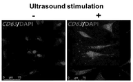
- 24 hours after sonication as a result of analyzing the expression of HDF exosome marker (CD63) by immunofluorescence staining (at this time, DAPI was used as a control stain), it was confirmed that it was significantly increased compared to the exosome-treated group.
- Figure 13 The marker expression was analyzed using a Nanosight LM10 microscope to see if the expression was actually linked to secretion of exosomes.
- a large amount of exosomes having a diameter of 30-200 nm was secreted in the ultrasound treatment group, especially the total number of exosomes. 9.02 ⁇ 0.47X10 8 pcs./ml compared to the group without ultrasound (1.5 ⁇ 0.43X10 8 pcs / ml), it was confirmed that the increase was more than 5 times (Figs. 14 and 15).
- Example 5 it was analyzed whether there is a change in the total amount of RNA and protein in secretion-induced exosomes by treating with ultrasound.
- Total RNA was extracted from exosomes using total TRIzol (RNAi, Takara, Otsu, Japan), and then quantified using Agilent 2100 Bioanalyzer (Agilent, Santa Clara, CA, USA) .Exo secreted from 1x10 6 cells It was confirmed that the total amount of RNA in the inside was 1003.5 ⁇ 167.6 ng when prepared by ultrasonic treatment as in Example 5, and increased 5 times or more compared to the case where the ultrasonic treatment was not performed (186.3 ⁇ 17.3 ng).
- the total amount of RNA compared to the same number of exosomes (1x10 8 exosomes) was 222.4 ⁇ 37.1 ng, indicating a level similar to that of the non-ultrasonic treatment (241.5 ⁇ 22.5 ng) (FIG. 16 and Table 3).
- the total protein amount in the exosomes secreted from 1x10 6 cells was 6.11 ⁇ 0.058 ⁇ g when prepared by ultrasonic treatment as in Example 5, when not treated (0.15 ⁇ 0.009 It was confirmed that the increase was about 40 times compared to ⁇ g).
- the total number of proteins compared to the same number of exosomes (1x10 8 exosomes) was 1.35 ⁇ 0.013 ⁇ g, which was increased by 6 times or more compared to the case without ultrasonic treatment (0.20 ⁇ 0.012 ⁇ g) (FIG. 16 and Table 3). .
- Total RNA ng
- Total protein ug
- RNA-seq was performed to identify RNA in exosomes prepared according to Example 5, and Gene ontology analysis was performed.
- a number of genes related to telomer maintenance and composition, cell cycle and proliferation, DNA repair and amplification existed (FIG. 17-18), and in particular, telomerase activity-related genes in exosomes secreted from experimental group cells as shown in Table 4 And increased telomere expression of genes related to maintenance and protection.
- telomere increase As a result of analyzing the cell proliferation, telomere increase, and anti-aging-related RNA in exosomes with qPCR, Cyclin-D1 and PCNA related to cell proliferation, Ctnnb1, TERT and STAT3 related to telomere increase, TEP1, MMP1 related to anti-aging, It was confirmed that MMP2, MMP9, Elastin, Keratin, HYAL1, Laminin, Filaggrin, Invducrin, etc. increased significantly compared to the control group, which is a level that is difficult to detect (FIG. 19).
- the exosome prepared according to Example 5 contains RNA and protein related to a large amount of telomeres, cell proliferation and anti-aging.
- telomer length was significantly increased as the concentration of the exosome treatment increased as compared to the cell (con) treated before the exosome (con) and the group cultured for 2 days without treatment with the exosome (non) (FIG. 21).
- telomeres were significantly expanded in all four types of cells tested, and telomerase activity was also high (FIG. 22).
- exosomes prepared according to the method of the present invention were treated with cells. It was confirmed that the telomere kidney effect can be obtained regardless of the cell type.
- telomere repeat sequence TTAGGG sequence
- Cy3 labeled PNA telomere probe was complemented with Cy3 labeled PNA telomere probe and stained with Hoechst33342.
- Colcemid was treated with the medium for 48 hours to fix the medium in the mid-splitting state in which chromosome in the cell nucleus was generated, and then the chromosome in the cell nucleus was attached to the slide glass using a low osmotic medium and a fixative solution, followed by hybridizstion buffer.
- PNA probe mixed solution (warm 90 ° in advance) is added to the chromosome, incubated at 85 ° C for 10 minutes, cooled for 1 hour at RT, washed with 2xSSC buffer, stained with Hoechst 33342, a nuclear staining reagent, mounted, and mounted on a Confocal laser Telomere fluorescence expression was compared to chromosome at a magnification of 1000 times or more under a microscope.
- telomere ends were analyzed through whole genome sequencing.
- the frequency of repetitive sequences of telomere ends was analyzed through whole genome sequencing.
- the TTAGGG sequence appeared more frequently than the control group not treated with exosomes in the cell group cultured for 2 days in the culture medium containing the exosomes at a concentration of 1 ⁇ 10 9 cells / ml (FIG. 24a).
- Telomeres having a complex structure may appear as various sequences when sequencing, and as a result of further analysis of the frequency of the possible repeat sequences, all possible sequences were high in the experimental group (FIG. 24B and Table 5).
- RNA-seq was performed to identify RNA in cells treated with exosomes according to Example 6, and Gene ontology analysis was performed. As a result, it was confirmed that a large number of genes related to telomere elongation, telomerase activity, cell cycle and proliferation, metabolism, etc. exist (FIGS. 25-26). In particular, as shown in Table 3, the expression of genes related to telomerase activity and telomerase maintenance and protection increased in exosomes secreted from cells of the experimental group.
- telomere length of the exosome-coated ear increased with time, and especially, the higher the exosome application amount, the greater the telomere length increase, and the exosome-treated group had a relatively increased expression of TERT. Appeared (Fig. 31).
- Fig. 31 As a result of performing immunofluorescence staining for telomer FISH and TERT on ear tissues treated with 9 exosomes 1x10 9 prepared according to Example 5, it was also confirmed that the fluorescence signal intensity and frequency of each increased with time. Were present ( Figures 32 and 34).
- Exosomes prepared according to the manufacturing method of the present invention have been revealed as described above as having effects such as telomere elongation and cell proliferation, and since these effects are highly related to cell regeneration, these exosomes regenerate skin and wound And how it affects the healing of scars.
- exosomes prepared according to the exosome preparation method of Example 5 were added to the medium, and the HDF was cultured to fill the plate, and then selected with a 20 ⁇ l yellow tip. After drawing the distance between cells, the wound healing assay was performed by incubating exosomes at a concentration of 0, 5, 10, 20 (X10 9 ) / ml.
- the higher the concentration of exosomes the more the empty space appeared to be filled relatively quickly (FIG. 35), and it was confirmed that the exosomes prepared according to the manufacturing method of the present invention increase cell migration.
- the exosomes were stained using Vybrant ⁇ iD Cell-Labeling Solution and treated on the skin, and after 2 weeks, the stained exosomes were analyzed with a fluorescence microscope, and the exosomes were generally damaged. It can be inferred that the above-described effect could be exhibited due to the overall penetration, and (Fig. 37).
- tissue regeneration and wound healing-related genes Col1 ⁇ 1, Col3 ⁇ 1, ELN, N-cadherin, and Cyclin-D1 at wound sites treated with exosomes at a concentration of 0, 5, 10, 20 (X10 9 ) dogs / ml Expression was analyzed by qRT-PCR, and as a result, in the experimental group, the expression of one or more of the genes was higher than that of the control group (FIG. 37). At this time, the gene with the highest increase in each concentration was different, and it was difficult to confirm the correlation between concentration and expression increase and decrease.
- the exosomes produced by the method of the present invention can have skin regeneration, wound and scar healing effects in vitro as well as in vitro.
- the exosomes according to the manufacturing method of the present invention induce various gene expressions related to anti-aging, and the exosomes were used to confirm whether these exosomes could have an effect even in cells with accelerated aging.
- fibroblasts derived from patients with premature ejaculation
- telomere length derived from patients with premature ejaculation
- TERT expression telomerase activity
- cell proliferation e.g., cell proliferation
- Ki67 expression aging-related ⁇ -galactosidase activity
- the cells used here are cells taken from the thigh skin of patients with Hutchinson-Gilford progeria syndrome (white / eight years old / male) (AG06297, Coriell Institute, Camden, USA).
- the exosomes were treated with 1x10 9 cells / ml and the cells were analyzed 14 days after the first treatment, and the first treatment group (ReprosomeX1), an experimental group using a medium containing exosomes of the above concentration continuously every time the medium was exchanged (ReprosomeX ⁇ , 4 times for 14 days).
- telomeres were elongated, telomerase activity increased, TERT gene expression increased (Fig. 38), cell proliferation increased, and Ki67 protein expression increased in both the experimental group treated with the first exosome and the experimental group treated continuously. It was increased (Fig. 39), ⁇ -galactosidase activity was lowered (Fig. 40), and it was confirmed that it has an overall anti-aging effect even with a single treatment against accelerated aging in the cells of premature ejaculation.
- the exosomes for extending the telomeres of cells according to the present invention and compositions comprising them induce not only telomeres elongation but also cell division and have anti-aging and tissue regeneration effects, as well as problems caused by shorter telomeres length and aging and tissues directly It can be used as a drug that can prevent, improve and treat various diseases and symptoms related to regeneration.
- the exosomes as described above and compositions containing them can also be effective by applying small amounts in a non-invasive way.
- the method for producing exosomes to extend the telomeres of cells according to the present invention can load various factors having the above-described effects into exosomes capable of penetrating into cells by only processing physical stimuli on the cells. In a way, it is possible to obtain a high yield of exosomes containing a high concentration of the active ingredient through this method.
Landscapes
- Health & Medical Sciences (AREA)
- Life Sciences & Earth Sciences (AREA)
- Chemical & Material Sciences (AREA)
- Organic Chemistry (AREA)
- Engineering & Computer Science (AREA)
- Zoology (AREA)
- Genetics & Genomics (AREA)
- Bioinformatics & Cheminformatics (AREA)
- Wood Science & Technology (AREA)
- Biomedical Technology (AREA)
- Biochemistry (AREA)
- General Health & Medical Sciences (AREA)
- Biotechnology (AREA)
- Microbiology (AREA)
- General Engineering & Computer Science (AREA)
- Medicinal Chemistry (AREA)
- Toxicology (AREA)
- Developmental Biology & Embryology (AREA)
- Molecular Biology (AREA)
- Biophysics (AREA)
- Proteomics, Peptides & Aminoacids (AREA)
- Gastroenterology & Hepatology (AREA)
- Cell Biology (AREA)
- Chemical Kinetics & Catalysis (AREA)
- General Chemical & Material Sciences (AREA)
- Nuclear Medicine, Radiotherapy & Molecular Imaging (AREA)
- Pharmacology & Pharmacy (AREA)
- Animal Behavior & Ethology (AREA)
- Public Health (AREA)
- Veterinary Medicine (AREA)
- Neurosurgery (AREA)
- Neurology (AREA)
- Gynecology & Obstetrics (AREA)
- Reproductive Health (AREA)
- Micro-Organisms Or Cultivation Processes Thereof (AREA)
- Medicines Containing Material From Animals Or Micro-Organisms (AREA)
- Medicines That Contain Protein Lipid Enzymes And Other Medicines (AREA)
Abstract
본 발명은 세포의 텔로미어를 신장시키는 엑소좀, 이를 포함하는 조성물 및 그 제조방법에 관한 것으로서, 보다 상세하게는 TERT, TERF2, DKC1, TERF2IP, RFC1, RAD50, TERF1, PINX1, TNKS1BP1, ACD, NBN, HSPA1L, PARP1, PTGES3, SMG6, BLM, XRCC5, XRCC6, ERCC4, PRKDC, TEP1 및 β-Catenin 중에서 선택된 유전자 하나 이상의 유전자 산물을 포함하는 것을 특징으로 하는, 세포의 텔로미어를 신장시키는 엑소좀, 이를 포함하는 조성물 및 세포에 직간접적으로 물리적 자극을 제공하여 상기 세포로부터 텔로미어를 신장시키는 엑소좀을 유도하는 방법을 개시한다. 이러한 엑소좀을 포함하는 조성물은 기존에 개시된 조성물에 비해 저농도로, 생체에 비침습적인 적용방법으로도 효과를 나타낼 수 있으며, 안전성이 높을 뿐 아니라, 단순히 텔로미어를 신장시키는 데에 그치지 않고, 세포 분열을 유도하고 항노화, 조직재생, 상처치유 및 흉터치유 등의 효과를 가질 수 있다. 또한, 본 발명의 텔로미어를 신장시키는 엑소좀의 제조방법은 기존의 텔로미어 신장용 조성물에 비해 제조가 간단하고, 수율이 높으며, 환경적인 부담이 줄어들 수 있는 효과를 가진다.
Description
본 발명은 세포의 텔로미어를 신장시키는 엑소좀, 이를 포함하는 조성물 및 그 제조방법에 관한 것으로서, 보다 상세하게는 TERT, TERF2, DKC1, TERF2IP, RFC1, RAD50, TERF1, PINX1, TNKS1BP1, ACD, NBN, HSPA1L, PARP1, PTGES3, SMG6, BLM, XRCC5, XRCC6, ERCC4, PRKDC, TEP1 및 β-Catenin 중에서 선택된 유전자 하나 이상의 RNA또는 단백질을 포함하는 것을 특징으로 하는, 세포의 텔로미어를 신장시키는 엑소좀, 이를 포함하는 조성물 및 세포에 직간접적으로 물리적 자극을 제공하여 상기 세포로부터 텔로미어를 신장시키는 엑소좀을 유도하는 방법에 관한 것이다.
텔로미어(telomere)는 염색체 양 끝의 DNA 반복 구조(인간의 경우 TTAGGG)이다. 텔로미어는 쉘터린(shelterin) 복합체와 결합하여 보호 캡을 형성하는데, 이는 세포의 증폭능을 조절하고 세포 분열 시 염색체 간의 결합과 유전 정보의 소실을 방지하는 역할을 한다. DNA 중합효소는 3' 말단을 모두 증폭할 수 없기 때문에 텔로미어는 매 세포분열마다 30 내지 200 bp만큼 짧아지게 된다. 텔로미어가 임계치보다 짧아져 코딩 DNA에 가까워지고 텔로미어의 루프(loop) 구조가 유지될 수 없게 되면, 노출된 텔로미어는 p53 또는 p16INK4a 신호전달체계에 의해 인식되어 세포 분열이 멈추고 노쇠하여 사멸하게 된다.
텔로미어는 역전사효소인 텔로머레이즈(telomerase)에 의해 직접적으로 신장될 수 있는데, 인간의 텔로머레이즈 복합체는 텔로미어 합성의 주형으로 작용하는 RNA 분자인 TERC, 촉매 서브유닛인 TERT와, DKC1 및 TEP1 등의 보조단백질로 구성되어 있다. 정상적인 경우 체세포의 텔로머레이즈 활성은 거의 없으며, 활발한 분열능이 필요한 줄기세포 및 전구세포에서만 높은 활성이 감지된다.
텔로미어가 짧아지면서 다수 세포의 분열이 중단되면 다양한 문제가 나타날 수 있으며, 특히 노화(aging)와 퇴화(degeneration)로 대표되는 다양한 증상이 나타날 수 있다. 피부조직이 퇴화되는 선천성 각하이상증(dyskeratosis congenita) 및 혈액 세포가 부족한 재생불량성 빈혈(aplastic anemia) 등 골수세포의 재생에 문제가 생기는 질병은 텔로머레이즈 또는 텔로미어를 보호하는 쉘터린 복합체 관련 유전자의 돌연변이 등 텔로미어 유지기능 이상으로 인해 나타나는 것으로 일찍이 밝혀졌다. 한편, 최근 고혈압, 대사증후군, 당뇨병, 치매 등 노화와 관련된 질환을 앓고 있는 환자들의 텔로미어가 정상인보다 짧다는 연구결과가 나오는 등 텔로미어의 길이를 늘려 노화를 늦출 수 있는 가능성에 대한 연구가 설득력을 얻고 있다. 또한, 일반적으로 텔로머레이즈의 활성이 거의 없는 것으로 알려진 체세포에서 텔로머레이즈 활성을 높임으로써 재생활동을 활성화할 수 있는 것으로 나타났는데, 특히 화상, 부상, 노화 및 질병에 따른 심근, 간, 각막, 피부, 혈관, 연골 및 뼈 등의 각종 조직, 면역세포, 혈액세포 등을 보충할 필요가 있는 경우 효과가 있는 것으로 보고된 바 있다. 그러나 텔로미어의 길이를 신장시킬 수 있는 유전자를 지속적으로 과발현시킨 쥐에서 암세포가 다수 발생하는 등 긴 텔로미어가 암 발병의 위험요소가 된다는 연구결과도 나오고 있다. 따라서 텔로미어 신장활성을 한시적으로만 활성화시킬 수 있다면, 암 발생의 위험 없이 각종 노화 및 퇴화와 관련된 증상을 완화할 수 있을 것이며, 이러한 기술은 전술한 유전적인 결함에 의한 텔로미어 이상은 물론 그 외 재생의학분야에서 유용할 것으로 예상할 수 있다.
상기와 같이 예상되는 다양한 효용에 따라, 텔로미어를 신장시키기 위한 조성물들이 고안된 바 있다. 미국 공개특허 제2018-0280413호는 텔로머레이즈 활성을 높이기 위한 화합물을 포함하는 약학조성물을 개시하고 있는데, 이를 위해서는 화합물을 합성 및 정제하는 과정이 필요하며, 화학합성을 통해 수득할 수 있는 상기 조성물은 합성에 필요한 시재료(starting material)가 정제된 상태여야 할 뿐 아니라 합성 과정에서 환경에 유해한 다양한 부산물이 생성되며, 화합물은 생체 내 대사 과정에서 부산물을 생산하는 등의 위험성을 가진다. 유럽 공개특허 제2959005호는 텔로머레이즈를 코딩하는 RNA를 개시하고 있는데, 이 또한 합성 및 정제 과정이 필요할 뿐 아니라, RNA는 친수성이므로 세포 내로 전달되기 위해서 전달체가 추가적으로 필요하다. 따라서 좀더 간단하게 제조할 수 있고 유해성이 적으며 효율이 좋은 텔로미어 신장용 조성물에 대한 요구가 존재한다.
본 발명은 전술한 문제를 해결하고자 안출된 것으로서, 본 발명의 일 실시예에 따른 본 발명은 전술한 문제를 해결하고자 안출된 것으로서, 본 발명의 일 실시예에 따른 엑소좀은 TERT, TERF2, DKC1, TERF2IP, RFC1, RAD50, TERF1, PINX1, TNKS1BP1, ACD, NBN, HSPA1L, PARP1, PTGES3, SMG6, BLM, XRCC5, XRCC6, ERCC4, PRKDC, TEP1 및 β-Catenin 중에서 선택된 유전자 하나 이상의 RNA 또는 단백질을 포함하는 것을 특징으로 한다.
또한, 본 발명의 다른 일 실시예는 상기 엑소좀을 포함하는 세포의 텔로미어 신장용 조성물을 제공한다.
또한, 본 발명의 또 다른 일 실시예에 따른 세포의 텔로미어를 신장시키는 엑소좀의 제조방법은 세포에 직간접적으로 물리적 자극을 제공하는 단계; 상기 세포 및 배지의 혼합물을 일정 시간 배양하는 단계; 및 상기 혼합물로부터 엑소좀을 분리하는 단계;를 포함하고, 상기 직접적으로 자극을 제공하는 것은 세포가 포함된 배지에 물리적 자극을 가하는 것이고, 상기 간접적으로 자극을 제공하는 것은 세포가 포함되지 않은 배지에 물리적 자극을 가한 후 상기 배지와 상기 세포를 혼합하는 것을 특징으로 한다.
본 발명이 이루고자 하는 기술적 과제는 이상에서 언급한 기술적 과제로 한정되지 않으며, 언급되지 않은 또 다른 기술적 과제들은 아래의 기재로부터 본 발명이 속하는 기술 분야에서 통상의 지식을 가진 자에게 명확하게 이해될 수 있을 것이다.
전술한 기술적 과제를 달성하기 위한 기술적 수단으로서, 본 발명의 일 측면에 따른, 세포의 텔로미어를 신장시키는 엑소좀은 TERT, TERF2, DKC1, TERF2IP, RFC1, RAD50, TERF1, PINX1, TNKS1BP1, ACD, NBN, HSPA1L, PARP1, PTGES3, SMG6, BLM, XRCC5, XRCC6, ERCC4, PRKDC, TEP1 및 β-Catenin 중에서 선택된 유전자 하나 이상의 RNA 또는 단백질을 포함하는 것을 특징으로 한다.
여기서, 상기 엑소좀은 세포에 물리적 자극을 제공하여 유도된 것을 특징으로 할 수 있다.
상기 물리적 자극의 형태는 초음파, 열 및 빛 중에서 선택되는 어느 하나일 수 있다.
상기 물리적 자극은 세포에 직간접적으로 제공되는 것이고, 상기 직접적으로 자극을 제공하는 것은 세포가 포함된 배지에 물리적 자극을 가하는 것이고, 상기 간접적으로 자극을 제공하는 것은 세포가 포함되지 않은 배지에 물리적 자극을 가한 후 상기 배지와 상기 세포를 혼합하는 것일 수 있다.
상기 엑소좀을 유도할 수 있는 물리적 자극을 제공하는 과정은 세포와 배지를 혼합한 후 상기 혼합물에 물리적 자극을 제공하거나, 배지에 물리적 자극을 제공한 후 상기 배지와 세포를 혼합하거나, 세포에 물리적 자극을 제공한 후 상기 세포와 배지를 혼합하거나, 세포에 물리적 자극을 제공한 후 상기 세포와 배지를 혼합한 다음 상기 혼합물에 물리적 자극을 제공하거나, 배지에 물리적 자극을 제공한 후 상기 배지와 세포를 혼합한 다음 상기 혼합물에 물리적 자극을 제공하거나, 세포와 배지에 각각 물리적 자극을 제공한 후 상기 세포 및 상기 배지를 혼합하거나, 세포와 배지에 각각 물리적 자극을 제공한 후 상기 세포 및 상기 배지를 혼합한 다음 상기 혼합물에 물리적 자극을 제공하는 방식 중에서 선택된 어느 하나 이상의 방식으로 진행되는 것을 특징으로 할 수 있다.
상기 물리적 자극은 초음파 자극이고, 상기 직접적인 초음파 자극은 강도가 0.1 내지 3 W/cm2이며, 주파수가 20 kHz 내지 20 MHz이고, 지속시간이 0.1초 내지 20분인 것을 특징으로 하며, 상기 간접적인 초음파 자극은, 강도가 1 내지 20 W/cm2이고, 주파수가 20 kHz 내지 20 MHz이며, 지속시간이 0.1초 내지 20분인 것을 특징으로 할 수 있다.
상기 물리적 자극은 열 자극이고, 상기 열 자극은 세포를 40 내지 50℃의 온도 조건에 1 내지 10분 노출시킨 후 0 내지 4℃의 온도 조건에 5 내지 10초간 노출하는 방식으로 가해지는 것을 특징으로 할 수 있다.
상기 빛 자극은 레이저 또는 발광 다이오드(light-emitting diode) 빛 중에서 선택된 어느 하나의, 파장 대역 300 내지 900 nm의 펄스형 빔을 1 내지 10초 동안 조사하여 가하는 것을 특징으로 할 수 있다.
상기 엑소좀은 엑소좀 1X10⁸ 개 입자 내에 총 RNA가 150 ng 이상 포함된 것을 특징으로 할 수 있다.
상기 엑소좀은 엑소좀 1X10⁸ 개 입자 내에 총 단백질이 1 μg 이상 포함된 것을 특징으로 할 수 있다.
상기 엑소좀을 세포에 처리시 상기 세포의 telomerase 활성이 높아지는 것을 특징으로 할 수 있다.
상기 엑소좀을 세포에 처리시 상기 세포의 β-galactosidase 활성이 감소되는 것을 특징으로 할 수 있다.
본 발명의 다른 일 측면에 따른 세포의 텔로미어 신장용 조성물은 전술한 엑소좀을 포함한다.
여기서, 상기 조성물은 상기 엑소좀이 106 내지 1014개/ml농도로 포함된 것일 수 있다.
본 발명의 또 다른 일측면에 따른 세포의 텔로미어를 신장시키는 엑소좀의 제조방법은 세포에 직간접적으로 물리적 자극을 제공하는 단계; 상기 세포 및 배지의 혼합물을 일정 시간 배양하는 단계; 및 상기 혼합물로부터 엑소좀을 분리하는 단계;를 포함하고, 상기 직접적으로 자극을 제공하는 것은 세포가 포함된 배지에 물리적 자극을 가하는 것이고, 상기 간접적으로 자극을 제공하는 것은 세포가 포함되지 않은 배지에 물리적 자극을 가한 후 상기 배지와 상기 세포를 혼합하는 것이다.
여기서, 상기 물리적 자극의 형태는 초음파, 열 및 빛 중에서 선택되는 어느 하나일 수 있다.
상기 세포에 직간접적으로 물리적 자극을 제공하는 과정은 세포와 배지를 혼합한 후 상기 혼합물에 물리적 자극을 제공하거나, 배지에 물리적 자극을 제공한 후 상기 배지와 세포를 혼합하거나, 세포에 물리적 자극을 제공한 후 상기 세포와 배지를 혼합하거나, 세포에 물리적 자극을 제공한 후 상기 세포와 배지를 혼합한 다음 상기 혼합물에 물리적 자극을 제공하거나, 배지에 물리적 자극을 제공한 후 상기 배지와 세포를 혼합한 다음 상기 혼합물에 물리적 자극을 제공하거나, 세포와 배지에 각각 물리적 자극을 제공한 후 상기 세포 및 상기 배지를 혼합하거나, 세포와 배지에 각각 물리적 자극을 제공한 후 상기 세포 및 상기 배지를 혼합한 다음 상기 혼합물에 물리적 자극을 제공하는 방식 중에서 선택된 어느 하나 이상의 방식으로 진행되는 것을 특징으로 할 수 있다.
상기 물리적 자극은 초음파 자극이고, 상기 직접적인 초음파 자극은 강도가 0.1 내지 3 W/cm2이고, 주파수가 20 kHz 내지 20 MHz이며, 지속시간이 0.1초 내지 20분인 것을 특징으로 하고, 상기 간접적인 초음파 자극은, 강도가 1 내지 20 W/cm2이고, 주파수가 20 kHz 내지 20 MHz이며, 지속시간이 0.1초 내지 20분인 것을 특징으로 할 수 있다.
상기 물리적 자극은 열 자극이고, 상기 열 자극은 세포를 40 내지 50℃의 온도 조건에 1 내지 10분 노출시킨 후 0 내지 4℃의 온도 조건에 5 내지 10초간 노출하는 방식으로 가해지는 것을 특징으로 할 수 있다.
상기 물리적 자극은 빛 자극이고, 상기 빛 자극은 레이저 또는 발광 다이오드(light-emitting diode) 빛 중에서 선택된 어느 하나의, 파장 대역 300 내지 900 nm의 펄스형 빔을 1 내지 10초 동안 조사하여 가하는 것을 특징으로 할 수 있다.
상기 세포는 각각 포유류 유래의 줄기세포, 전구세포, 섬유아세포, 각질세포 또는 기관 내 조직세포로 구성되는 군으로부터 선택되는 것을 특징으로 할 수 있다.
상기 배지는 배양 배지 또는 분화유도 배지 중에서 선택되는 것을 특징으로 할 수 있다.
상기 혼합물의 배양은 1시간 내지 10일 동안 진행되는 것을 특징으로 할 수 있다.
상기 엑소좀을 분리하는 단계는 초원심분리, 밀도구배, 여과, 크기 배제 크로마토그래피, 면역친화성분리, 침전, 미세유체에 의한 분리방법 중 하나 이상을 이용하는 것을 특징으로 할 수 있다.
상기 엑소좀을 분리하는 단계는 상기 배양 후의 혼합물을 원심분리하여 상층액을 수득하는 단계; 상기 상층액을 필터로 여과하여 여과액을 수득하는 단계; 및 상기 여과액을 농축하는 단계;를 포함할 수 있다.
상기 분리된 엑소좀은 상기 제조방법을 거치기 전의 세포에서 분비된 엑소좀에 비해 TERT, TERF2, DKC1, TERF2IP, RFC1, RAD50, TERF1, PINX1, TNKS1BP1, ACD, NBN, HSPA1L, PARP1, PTGES3, SMG6, BLM, XRCC5, XRCC6, ERCC4, PRKDC, TEP1 및 β-Catenin 중에서 하나 이상의 유전자 발현량이 높은 것을 특징으로 할 수 있다.
상기 분리된 엑소좀은 세포에 처리시 상기 세포의 telomerase 활성이 높아지는 것을 특징으로 할 수 있다.
상기 분리된 엑소좀을 세포에 처리시 상기 세포의 β-galactosidase 활성이 감소되는 것을 특징으로 할 수 있다.
상기 세포의 텔로미어를 신장시키는 엑소좀의 제조방법은, 상기 분리된 엑소좀을 세포에 처리시 상기 세포의 세포 이동성이 증가되는 것을 특징으로 할 수 있다.
상기 세포의 텔로미어를 신장시키는 엑소좀의 제조방법은, 상기 분리된 엑소좀을 조직에 처리시 상기 조직의 재생이 촉진되는 것을 특징으로 할 수 있다.
상기 세포의 텔로미어를 신장시키는 엑소좀의 제조방법은, 상기 분리된 엑소좀을 상처에 처리시 상기 상처의 치유가 촉진되는 것을 특징으로 할 수 있다.
상기 세포의 텔로미어를 신장시키는 엑소좀의 제조방법은, 상기 분리된 엑소좀을 흉터에 처리시 상기 흉터의 치유가 촉진되는 것을 특징으로 할 수 있다.
상기 세포의 텔로미어를 신장시키는 엑소좀의 제조방법은, 상기 분리된 엑소좀을 세포에 처리시 상기 세포에 항노화 효과를 가지는 것을 특징으로 할 수 있다.
상술한 과제 해결 수단은 단지 예시적인 것으로서, 본 발명을 제한하려는 의도로 해석되지 않아야 한다. 상술한 예시적인 실시예 외에도, 도면 및 발명의 상세한 설명에 추가적인 실시예가 존재할 수 있다.
본 발명의 일 실시예에 따른 세포의 텔로미어를 신장시키는 조성물은 전술한 텔로미어를 신장시키는 조성물에 비해 제조가 간단하다. 즉, 기존의 조성물 중 화합물은 화학적으로 합성되는데, 이는 특이성이 없고 길고 복잡한 공정을 거쳐야 한다. 이에 반해 본 발명의 일 실시예에 따른 세포의 텔로미어를 신장시키는 방법은 물리적 자극을 세포에 처리하는 것 만으로 텔로미어 신장과 관련된 다양한 인자의 발현 및 상기 인자를 포함하는 엑소좀 생산을 촉진할 수 있으며, 이렇게 분비된 엑소좀을 상기 세포를 배양한 배지로부터 분리하는 과정으로 구성되어 그 공정이 간단하다. 또한, 세포는 이중 지질막으로 둘러싸여 있으므로, 친수성의 핵산은 세포 내로 전달할 수 있는 전달체가 없이는, 효과를 나타내기 이전에 필요한 세포 내 유입이 어려워 전달체에 핵산을 로딩하는 단계가 추가로 필요한데 반해, 본 발명의 엑소좀 제조방법에 따르면 텔로미어 신장 관련 인자가 지질막으로 둘러싸인 형태로 수득되므로 추가적인 로딩을 진행할 필요가 없다.
또한, 본 발명의 일 실시예에 따른 세포의 텔로미어를 신장시키는 조성물은 기존에 개시된 조성물에 비해 효율과 적용방법 면에서 우수하다. 즉, 전술한 텔로머레이즈 활성을 높이는 화합물은 충분한 효과를 나타내기 위해서 높은 농도의 화합물을 체내 주입해야 하는 것으로 나타나는 데 반해 본 발명의 일 실시예에 따른 세포의 텔로미어를 신장시키는 엑소좀은 소량을, 국부도포 등 비침습적인 방법으로 적용하는 것으로 효과를 낼 수 있다.
또한, 본 발명의 일 실시예에 따른 세포의 텔로미어를 신장시키는 엑소좀은 텔로미어 신장과 관련된 인자를 다량 함유하는 엑소좀을 다량 분비할 수 있도록 유도하므로, 유용한 물질을 담은 세포의 엑소좀 생산 측면에서 수율이 높다.
또한, 본 발명의 일 실시예에 따른 세포의 텔로미어를 신장시키는 엑소좀은 안전성이 높다. 즉, 화합물은 대사과정에서 독성 등 추가적 위험이 존재할 수 있으며, 특히 전술한 기존 기술은 화합물의 처리량이 대상동물 단위무게(kg)당 mg 수준으로 상당히 높고 화학 합성 과정에서도 환경에 부담이 되는 폐기물 등이 생성되나, 본발명에 따른 방법은 상대적으로 이러한 부담이 적다.
추가로, 본 발명의 일 실시예에 따른 세포의 텔로미어를 신장시키는 엑소좀은 단순히 텔로미어를 신장시키는 데에 그치지 않고, 세포 분열을 유도하며 항노화 및 조직재생 효과를 가지는 것을 확인하였으며, 따라서 이를 통해 단순히 텔로미어 길이가 짧아져 생기는 문제뿐만 아니라 직접적으로 노화 및 조직재생과 관련된 다양한 질병과 증상까지 개선 및 예방할 수 있을 것으로 기대할 수 있다.
본 발명의 효과는 상기한 효과로 한정되는 것은 아니며, 본 발명의 상세한 설명 또는 특허청구범위에 기재된 발명의 구성으로부터 추론 가능한 모든 효과를 포함하는 것으로 이해되어야 한다.
도1은 본 발명의 실시예1에 따른 직접적 초음파 자극 처리 및 배양 후 세포 종류에 따른 텔로미어 길이 변화 분석 데이터.
도2는 본 발명의 실시예1에 따른 초음파 자극 처리, 실시예2에 따른 열 자극 처리, 실시예 3에 따른 발광 다이오드 빛 자극 처리 및 각 배양 후 각 물리적 자극 처리에 따른 텔로미어 길이 변화 분석 데이터.
도3은 본 발명의 실시예5에 따른 간접적 초음파 자극 처리 및 배양 후 텔로미어 길이 변화 분석 데이터.
도 4는 본 발명의 실시예1에 따른 초음파 자극 처리 및 배양 후 배지 종류에 따른 텔로미어 길이 변화 분석 데이터.
도 5은 본 발명의 실시예1에 따른 초음파 자극 처리 및 배양 후 초음파 자극 처리 횟수에 따른 텔로미어 길이 변화 분석 데이터.
도 6은 본 발명의 실시예1에 따른 초음파 자극 처리 및 배양 후 세포 종류에 따른 TERT 유전자 발현 변화 분석 데이터.
도7는 CB-HDF 및 Adipo-MSC에 대한 본 발명의 실시예1에 따른 초음파 자극 처리 및 배양 후 TERT 유전자 발현 변화 분석 데이터.
도8은 CB-HDF 및 Adipo-MSC에 대한 본 발명의 실시예1에 따른 초음파 자극 처리 및 배양 후 β-Catenin 유전자 발현 변화 분석 데이터.
도9은 CB-HDF 및 Adipo-MSC에 대한 본 발명의 실시예1에 따른 초음파 자극 처리 및 배양 후 텔로머레이즈 활성 변화 분석 데이터.
도10은 CB-HDF 및 Adipo-MSC에 대한 본 발명의 실시예1에 따른 초음파 자극 처리 및 배양 후 telomere FISH 데이터.
도11는 CB-HDF에 대한 본 발명의 실시예1에 따른 초음파 자극 처리 및 배양 후 TERT 및 Ki67의 세포형광염색 데이터.
도12은 CB-HDF에 대한 본 발명의 실시예1에 따른 초음파 자극 처리 및 배양 후 노화관련 β-galactosidase 활성 분석 데이터.
도13은 본 발명의 실시예5에 따라 초음파 처리된 CB-HDF에서의 엑소좀 마커 발현 분석 데이터.
도14는 본 발명의 실시예5에 따라 CB-HDF로부터 분비된 엑소좀의 입자 크기별 Nanosight 분석 데이터.
도15는 본 발명의 실시예5에 따라 CB-HDF로부터 분비된 엑소좀의 총 수에 대한 Nanosight 분석 데이터.
도16은 본 발명의 실시예5에 따라 제조된 엑소좀 내 총 RNA 및 총 단백질 양 분석 데이터.
도17은 본 발명의 실시예5에 따라 제조된 엑소좀 내 RNA에 대한 Gene ontology 분석 데이터.
도18은 본 발명의 실시예5에 따라 제조된 엑소좀 내 RNA에 대한 RNA-seq 분석 데이터.
도19는 본 발명의 실시예5에 따라 제조된 엑소좀 내 세포 증식, 텔로미어 증가 및 항노화 관련 일부 RNA에 대한 qPCR 분석 데이터.
도20은 본 발명의 실시예5에 따라 제조된 엑소좀 내 TERT 단백질에 대한 면역형광 분석 데이터.
도21은 본 발명의 실시예5에 따라 제조된 엑소좀의 세포 처리에 따른 텔로미어 길이 변화 분석 데이터.
도22는 본 발명의 실시예5에 따라 제조된 엑소좀의 세포 처리에 따른 세포 종류별 텔로미어 길이 변화 분석 데이터.
도23은 본 발명의 실시예5에 따라 제조된 엑소좀의 세포 처리 후 해당 세포 내 텔로미어 qFISH 분석 데이터.
도24는 본 발명의 실시예5에 따라 제조된 엑소좀의 세포 처리 후 해당 세포 내 Tel-seq 분석 데이터.
도25는 본 발명의 실시예5에 따라 제조된 엑소좀의 세포 처리 후 해당 세포 내 RNA에 대한 Gene ontology 분석 데이터.
도26은 본 발명의 실시예5에 따라 제조된 엑소좀의 세포 처리 후 해당 세포 내 RNA에 대한 RNA-seq 분석 데이터.
도27은 본 발명의 실시예5에 따라 제조된 엑소좀의 세포 처리 후 해당 세포 내 TERT RNA 및 단백질 발현 분석 데이터.
도28은 본 발명의 실시예5에 따라 제조된 엑소좀의 세포 처리 후 해당 세포 내 세포 분열 마커 분석 데이터.
도 29는 본 발명의 실시예5에 따라 제조된 엑소좀의 세포 처리 후 해당 세포 내 엑소좀 농도별 세포 분열 마커 분석 데이터.
도 30은 본 발명의 실시예5에 따라 제조된 엑소좀의 세포 처리 후 해당 세포 내 β-galactosidase 활성 분석 데이터.
도 31은 본 발명의 실시예5에 따라 제조된 엑소좀의 마우스 귀 도포 후 해당 부위의 엑소좀 농도별 텔로미어 길이 및 TERT RNA 발현량 분석 데이터.
도 32는 본 발명의 실시예5에 따라 제조된 엑소좀의 마우스 귀 도포 후 해당 부위의 엑소좀 처리 시간별 텔로미어 FISH 분석 데이터.
도 33은 본 발명의 실시예5에 따라 제조된 엑소좀의 마우스 귀 도포 후 해당 부위의 엑소좀 처리 시간별 세포 증식 관련 유전자의 RNA 및 단백질 발현 분석 데이터.
도 34는 본 발명의 실시예5에 따라 제조된 엑소좀의 마우스 귀 도포 후 해당 부위의 엑소좀 처리 시간별 TERT 면역형광 분석 데이터.
도 35는 본 발명의 실시예5에 따라 제조된 엑소좀의 세포 처리 농도별 wound healing assay 데이터.
도 36은 본 발명의 실시예5에 따라 제조된 엑소좀의 in vivo 처리 농도별 wound healing assay 데이터.
도 37은 본 발명의 실시예5에 따라 제조된 엑소좀의 마우스 피부 도포 시 조직 침투 분석 데이터.
도 38은 본 발명의 실시예5에 따라 제조된 엑소좀의 조로증 환자 유래 세포 처리 시 텔로미어 관련 분석 데이터.
도 39는 본 발명의 실시예5에 따라 제조된 엑소좀의 조로증 환자 유래 세포 처리 시 세포 증식 관련 분석 데이터.
도 40은 본 발명의 실시예5에 따라 제조된 엑소좀의 조로증 환자 유래 세포 처리 시 β-galactosidase 활성 분석 데이터.
이하, 본 발명을 더욱 상세하게 설명한다. 그러나 본 발명은 여러 가지 상이한 형태로 구현될 수 있으며 여기에서 설명하는 실시예에 의해 본 발명이 한정되지 않으며 본 발명은 후술할 청구범위의 의해 정의될 뿐이다.
덧붙여, 본 발명에서 사용한 용어는 단지 특정한 실시예를 설명하기 위해 사용된 것으로, 본 발명을 한정하려는 의도가 아니다. 단수의 표현은 문맥상 명백하게 다르게 뜻하지 않는 한, 복수의 표현을 포함한다. 본 발명의 명세서 전체에서 어떤 구성요소를 '포함'한다는 것은 특별히 반대되는 기재가 없는 한 다른 구성요소를 제외하는 것이 아니라 다른 구성요소를 더 포함할 수 있다는 것을 의미한다.
또한, 본 발명에서 유전자라는 지칭 하에 사용한 각 유전자의 고유 명칭은 공식적으로 알려진 유전자명, 관용적으로 사용되는 명칭 또는 그 유전자의 산물의 명칭, 예컨대 단백질을 코딩하는 유전자의 경우 그 단백질이다.
본 발명의 발명자들은 대한민국 등록특허 제10-1855967호에서 세포 및 배양 배지의 혼합물에 환경유입을 촉진할 수 있는 물리적 자극을 제공하고, 상기 물리적 자극을 제공받은 혼합물을 일정 시간 배양하여 상기 세포를 리프로그래밍할 수 있으며, 상기 혼합물로부터 수득된 엑소좀을 세포에 처리할 경우 리프로그래밍된 세포를 수득할 수 있다는 것을 개시한 바 있다. 여기서 리프로그래밍의 방향은 물리적 자극을 받는 세포 또는 엑소좀이 처리되는 세포의 종류와는 관계없이 물리적 자극시 배지의 조성에 따라 다르게 나타난다는 것을 밝혔다. 추후 상기 세포 리프로그래밍 방법의 효과를 분석하는 과정에서, 세포에 물리적 자극을 가함으로써 텔로미어 신장과 관련된 유전자군의 발현이 증가된다는 것을 확인하게 되었다. 이에 대한 추가 연구를 통해, 개시된 발명과는 다르게 실험한 세포의 종류뿐만 아니라 배지의 조성에까지 관계없이 물리적 자극에 의해 텔로미어를 신장할 수 있는 것을 확인하여 환경 유입보다도 물리적 자극 자체에 의해서 텔로미어 신장이 유도된다는 결론을 내릴 수 있었다. 이때, 텔로미어 신장과 관련된 인자들의 발현이 증가될 뿐 아니라 이들 인자들을 함유한 엑소좀의 분비 역시 크게 증가한다는 것을 확인하고, 이를 새로운 발명으로 개시하게 되었다.
본 발명의 일 측면에 따른, 세포의 텔로미어를 신장시키는 엑소좀은 TERT, TERF2, DKC1, TERF2IP, RFC1, RAD50, TERF1, PINX1, TNKS1BP1, ACD, NBN, HSPA1L, PARP1, PTGES3, SMG6, BLM, XRCC5, XRCC6, ERCC4, PRKDC, TEP1 및 β-Catenin 중에서 선택된 유전자 하나 이상의 RNA 또는 단백질을 포함하는 것을 특징으로 한다. 가장 바람직하게 상기 엑소좀은 상기 모든 유전자의 RNA 또는 단백질을 포함할 수 있다.
여기서, TERT(Telomerase reverse transcriptase)는 텔로머레이즈의 촉매활성을 가지는 서브유닛이고, TERF1(Telomeric repeat binding factor 1) 및 TERF2(Telomeric repeat binding factor 2)는 텔로미어 서열을 인식한다. DKC1(Dyskerin pseudouridine synthase 1), RFC1(Replication factor C subunit 1), TNKS1BP1(Tankyrase 1 binding protein 1), NBN(Nibrin), HSPA1L(Heat shock protein family A member 1 like), PARP1(Poly ADP-ribose polymerase 1), PTGES3(Prostaglandin E synthase 3), SMG6(Smg6 homolog, Nonsense mediated mRNA decay factor), XRCC5(X-ray repair cross complementing 5), XRCC6(X-ray repair cross complementing 6)는 텔로미어의 안정화와 유지에 필요한 것으로 알려져 있다. TERF2IP(TERF2 interacting protein), RAD50 (RAD50 Homolog, Double strand break repair protein)은 텔로미어 재조합을 억제하며, PINX1(PIN2/TERF1-interacting telomerase inhibitor 1)는 TERF1 및 TERT의 인(nucleolus) 내 축적을 매개하고 TRF1가 텔로머레이즈에 결합하는 것을 도우며 S기에 텔로머레이즈 활성을 저해하는 것으로 보고되었다. ACD(Adrenocortical Dysplasia protein homolog), ERCC4(Excision repair cross-complementation group 4), PRKDC(Protein kinase, DNA-activated, catalytic subunit)는 텔로미어의 길이조절 및 보호를 위해 필요하며, BLM(Bloom syndrome protein) DNA합성시 텔로미어 증폭을 돕고, TEP1(Telomerase associated protein 1)은 텔로머레이즈 복합체의 일부를 형성한다. β-Catenin은 TERT발현을 촉진하는 전사조절인자이다. 상기 인자는 모두 텔로미어의 신장 및 안정화 효과를 가지는 것으로 보고된 바 있다. 상기와 같은 유전자 산물을 포함하는 본 발명의 일측면에 따른 텔로미어를 신장시키는 엑소좀은 후술할 다양한 효과를 위해 이용될 수 있다.
상기 엑소좀은 세포에 물리적 자극을 제공하여 유도된 것을 특징으로 할 수 있다.
상기 물리적 자극의 형태는 저주파, 초음파, 고주파, 열, 빛, 전기파, 전파, 당김(stretch), 압축(compression) 중에서 선택될 수 있고, 바람직하게 초음파, 열 및 빛 중에서 선택되는 어느 하나일 수 있다.
상기 물리적 자극은 세포에 직간접적으로 제공되는 것이고, 상기 직접적으로 자극을 제공하는 것은 세포가 포함된 배지에 물리적 자극을 가하는 것이고, 상기 간접적으로 자극을 제공하는 것은 세포가 포함되지 않은 배지에 물리적 자극을 가한 후 상기 배지와 상기 세포를 혼합하는 것일 수 있다.
상기 엑소좀을 유도할 수 있는 물리적 자극을 제공하는 과정은 세포와 배지를 혼합한 후 상기 혼합물에 물리적 자극을 제공하거나, 배지에 물리적 자극을 제공한 후 상기 배지와 세포를 혼합하거나, 세포에 물리적 자극을 제공한 후 상기 세포와 배지를 혼합하거나, 세포에 물리적 자극을 제공한 후 상기 세포와 배지를 혼합한 다음 상기 혼합물에 물리적 자극을 제공하거나, 배지에 물리적 자극을 제공한 후 상기 배지와 세포를 혼합한 다음 상기 혼합물에 물리적 자극을 제공하거나, 세포와 배지에 각각 물리적 자극을 제공한 후 상기 세포 및 상기 배지를 혼합하거나, 세포와 배지에 각각 물리적 자극을 제공한 후 상기 세포 및 상기 배지를 혼합한 다음 상기 혼합물에 물리적 자극을 제공하는 방식 중에서 선택된 어느 하나 이상의 방식으로 진행되는 것을 특징으로 할 수 있다. 이러한 물리적 자극의 제공은 세포에 직간접적인 방법으로 1회 이상 또는 상기 방식 중에서 하나 이상을 선택한 조합으로 진행될 수 있고, 횟수가 증가함에 따라 엑소좀의 텔로미어 신장 효과가 비례적으로 증가할 수 있다. 이렇게 1회 이상 물리적 자극을 제공할 때에는 각 회차 사이에 세포가 회복할 수 있도록 시간 간격을 두는 것이 바람직하며, 상기 시간 간격은 1일 이상, 좀더 바람직하게 2일 이상으로 둘 수 있다.
상기 물리적 자극은 초음파 자극이고, 상기 직접적인 초음파 자극은 강도가 0.1 내지 3 W/cm2이며, 주파수가 20 kHz 내지 20 MHz이고, 지속시간이 0.1초 내지 20분인 것을 특징으로 하며, 상기 간접적인 초음파 자극은, 강도가 1 내지 20 W/cm2이고, 주파수가 20 kHz 내지 20 MHz이며, 지속시간이 0.1초 내지 20분인 것을 특징으로 할 수 있다. 보다 바람직하게, 상기 직접적인 초음파 자극은 강도가 0.5 내지 2 W/cm2이며, 주파수가 20 kHz 내지 2 MHz이고, 지속시간이 0.1초 내지 10분인 것을 특징으로 하며, 상기 간접적인 초음파 자극은, 강도가 2내지 10 W/cm2이고, 주파수가 20 kHz 내지 2 MHz이며, 지속시간이 1초 내지 15분인 것을 특징으로 할 수 있다.
상기 물리적 자극은 열 자극이고, 상기 열 자극은 세포를 40 내지 50℃의 온도 조건에 1 내지 10분 노출시킨 후 0 내지 4℃의 온도 조건에 5 내지 10초간 노출하는 방식으로 가해지는 것을 특징으로 할 수 있다.
상기 빛 자극은 레이저 또는 발광 다이오드(light-emitting diode) 빛 중에서 선택된 어느 하나의, 파장 대역 300 내지 900 nm의 펄스형 빔을 1 내지 10초 동안, 보다 바람직하게 3 내지 7초 동안 조사하여 가하는 것을 특징으로 할 수 있다.
상기 엑소좀은 엑소좀 1X10⁸ 개 입자 내에 총 RNA가 100 ng 이상, 바람직하게 150 ng 이상, 더 바람직하게 180 ng 이상 포함된 것을 특징으로 할 수 있다.
상기 엑소좀은 엑소좀 1X10⁸ 개 입자 내에 총 단백질이 0.5 μg 이상, 바람직하게 1 μg 이상, 더 바람직하게 1.2 μg 이상 포함된 것을 특징으로 할 수 있다.
상기 엑소좀을 세포에 처리시 상기 세포의 telomerase 활성이 높아지는 것을 특징으로 할 수 있다. 이러한 텔로머레이즈 활성의 증가는 세포에 처리 후 텔로미어 길이 신장 및 세포 분열 촉진으로 연결될 수 있다.
상기 엑소좀을 세포에 처리시 상기 세포의 β-galactosidase 활성이 감소되는 것을 특징으로 할 수 있다. β-galactosidase는 베타갈락토사이드(β-galactoside)의 단당류로의 가수분해를 촉매하는 효소인데, 리소좀 β-galactosidase는 노화된 세포(senescent cell)에서 과발현되어 축적된다. 그 결과 노화된 세포에서는 β-galactosidase의 활성이 높게 나타나는 것으로 보고된 바 있는데, 본 발명의 일 실시예에 따른 세포의 텔로미어를 신장시키는 엑소좀을 세포에 처리하면 상기 효소의 활성이 낮아지는 것으로 나타나, 상기 엑소좀은 항노화 효과를 가지는 것으로 판단할 수 있다.
본 발명의 다른 일 측면에 따른 세포의 텔로미어 신장용 조성물은 전술한 엑소좀을 포함한다.
상기 엑소좀은 전술한 텔로미어 신장 및 안정화 기능을 하는 각종 인자의 유전자 산물을 포함하고 있고, 엑소좀은 혈류를 따라 이동하여 세포에 직접 상기 인자들을 전달하여 치료효과를 나타낼 수 있으므로, 이러한 엑소좀을 포함하는 조성물은 이러한 유전자의 활성이 필요한 개체에 치료 목적으로 사용될 수 있다. 예컨대, 우선 선천성 각하이상증, 재생불량성 빈혈, 베르너증후군(Werner syndrome), 블룸증후군(Bloom syndrome), 모세혈관확장성운동실조(Ataxia-telangiectasia), NBS(Nijmegen breakage syndrome), 모세혈관확장성 운동실조유사증후군(Ataxia-telangiectasia-like syndrome), 폐섬유증(Pulmonary fibrosis) 등 텔로미어 유지 이상으로 발생하는 것으로 알려진 질병의 치료 또는 개선을 위해 사용될 수 있다. 그 외에도 세포의 노화와 소실로 나타나는 각종 질병과 증상, 예컨대 치매, 운동실조, 연골 및 뼈의 퇴화 등의 각종 퇴행성 질환, 크론병, 만성폐색성폐질환 등을 포함하는 자가면역질환, 고혈압, 대사증후군, 당뇨병, 피부노화, 색소이상, 탈모, 피부노화 등의 치료 또는 개선에 사용될 수 있다. 추가적으로, 화상, 상처, 부상, 장기부전(organ failure), 장기소실, 궤양 등 각종 손상에 대한 빠른 재생을 위해 사용될 수 있다.
여기서, 상기 조성물은 상기 엑소좀이 최소한 102개/ml농도, 바람직하게 106 내지 1014개/ml농도로 포함된 것일 수 있다.
상기 조성물은 경구, 정맥, 근육, 피부, 경막, 경피 투여 중에서 선택된 어느 한 방법으로 투여될 수 있는 것을 특징으로 할 수 있다. 바람직하게, 상기 조성물은 피부 또는 경피 투여될 수 있다. 상기 조성물은 전술한 바와 같은 유효투여량의 엑소좀 외에도 약학적으로 포함될 수 있는 희석제, 방부제, 용해제, 유화제, 어쥬반트(adjuvant), 완충제 및 담체를 포함할 수 있다. 상기 조성물에 포함될 수 있는 성분으로는 중성완충식염수, 인산완충식염수 등의 완충액, 글루코스, 만노스, 수크로즈, 덱스트란, 만니톨 등의 탄수화물, 폴리펩티드, 글리신 등의 아미노산, 항산화제, EDTA, 글루타티온 등의 킬레이트 시약, 알루미늄 하이드록시드 등의 어쥬반트 및 보존제 등이 있다.
상기 투여경로의 선택 및 각 투여경로에 따른 추가성분 및 그 조성은 본 발명의 분야의 통상의 기술자에게 알려져 있는 바이므로, 이에 대한 자세한 설명은 생략한다.
본 발명의 또 다른 일측면에 따른 세포의 텔로미어를 신장시키는 엑소좀의 제조방법은 세포에 직간접적으로 물리적 자극을 제공하는 단계; 상기 세포 및 배지의 혼합물을 일정 시간 배양하는 단계; 및 상기 혼합물로부터 엑소좀을 분리하는 단계;를 포함하고, 상기 직접적으로 자극을 제공하는 것은 세포가 포함된 배지에 물리적 자극을 가하는 것이고, 상기 간접적으로 자극을 제공하는 것은 세포가 포함되지 않은 배지에 물리적 자극을 가한 후 상기 배지와 상기 세포를 혼합하는 것이다.
여기서, 상기 물리적 자극의 형태는 저주파, 초음파, 고주파, 열, 빛, 전기파, 전파, 당김(stretch), 압축(compression) 중에서 선택될 수 있고, 바람직하게 초음파, 열 및 빛 중에서 선택되는 어느 하나일 수 있다.
상기 세포에 직간접적으로 물리적 자극을 제공하는 과정은 세포와 배지를 혼합한 후 상기 혼합물에 물리적 자극을 제공하거나, 배지에 물리적 자극을 제공한 후 상기 배지와 세포를 혼합하거나, 세포에 물리적 자극을 제공한 후 상기 세포와 배지를 혼합하거나, 세포에 물리적 자극을 제공한 후 상기 세포와 배지를 혼합한 다음 상기 혼합물에 물리적 자극을 제공하거나, 배지에 물리적 자극을 제공한 후 상기 배지와 세포를 혼합한 다음 상기 혼합물에 물리적 자극을 제공하거나, 세포와 배지에 각각 물리적 자극을 제공한 후 상기 세포 및 상기 배지를 혼합하거나, 세포와 배지에 각각 물리적 자극을 제공한 후 상기 세포 및 상기 배지를 혼합한 다음 상기 혼합물에 물리적 자극을 제공하는 방식 중에서 선택된 어느 하나 이상의 방식으로 진행되는 것을 특징으로 할 수 있다. 이러한 물리적 자극의 제공은 세포에 직간접적인 방법으로 1회 이상 또는 상기 방식 중에서 하나 이상을 선택한 조합으로 진행될 수 있고, 횟수가 증가함에 따라 엑소좀의 텔로미어 신장 효과가 비례적으로 증가할 수 있다. 이렇게 1회 이상 물리적 자극을 제공할 때에는 각 회차 사이에 세포가 회복할 수 있도록 시간 간격을 두는 것이 바람직하며, 상기 시간 간격은 1일 이상, 좀더 바람직하게 2일 이상으로 둘 수 있다.
상기 물리적 자극은 초음파 자극이고, 상기 직접적인 초음파 자극은 강도가 0.1 내지 3 W/cm2이고, 주파수가 20 kHz 내지 20 MHz이며, 지속시간이 0.1초 내지 20분인 것을 특징으로 하고, 상기 간접적인 초음파 자극은, 강도가 1 내지 20 W/cm2이고, 주파수가 20 kHz 내지 20 MHz이며, 지속시간이 0.1초 내지 20분인 것을 특징으로 할 수 있다. 보다 바람직하게, 상기 직접적인 초음파 자극은 강도가 0.5 내지 2 W/cm2이며, 주파수가 20 kHz 내지 2 MHz이고, 지속시간이 0.1초 내지 10분인 것을 특징으로 하며, 상기 간접적인 초음파 자극은, 강도가 2내지 10 W/cm2이고, 주파수가 20 kHz 내지 2 MHz이며, 지속시간이 1초 내지 15분인 것을 특징으로 할 수 있다.
상기 물리적 자극은 열 자극이고, 상기 열 자극은 세포를 40 내지 50℃의 온도 조건에 1 내지 10분 노출시킨 후 0 내지 4℃의 온도 조건에 5 내지 10초간 노출하는 방식으로 가해지는 것을 특징으로 할 수 있다.
상기 물리적 자극은 빛 자극이고, 상기 빛 자극은 레이저 또는 발광 다이오드(light-emitting diode) 빛 중에서 선택된 어느 하나의, 파장 대역 300 내지 900 nm의 펄스형 빔을 1 내지 10초 동안, 보다 바람직하게 3 내지 7초 동안 조사하여 가하는 것을 특징으로 할 수 있다.
본 발명의 일 실시예에 따른 세포의 텔로미어를 신장시키는 엑소좀의 제조방법은 다양한 세포에 적용했을 때 모두 각 세포의 텔로미어를 신장시킬 수 있는 엑소좀 분비를 유도하는 것으로 나타났다. 상기 세포는 포유류 유래의 줄기세포, 전구세포, 섬유아세포, 각질세포 또는 기관 내 조직세포로 구성되는 군으로부터 선택되는 것을 특징으로 할 수 있다. 상기 텔로미어를 신장시키는 엑소좀을 생체 적용 목적으로 사용할 경우, 상기 세포는 자가유래(autologous), 동종유래(allogeneic) 또는 이종유래(heterologous) 중 어느 하나일 수 있고, 이종유래인 경우 바람직하게 포유류로부터 얻어진 것일 수 있다. 면역거부반응에 대한 가능성을 줄이기 위해, 바람직하게는 동종유래, 가장 바람직하게는 자가유래의 것을 사용할 수 있다. 상기 세포는 각각 포유류 유래의 줄기세포, 전구세포, 섬유아세포, 각질세포 또는 기관 내 조직세포로 구성되는 군으로부터 선택되는 것을 특징으로 할 수 있다.
상기 배지는 배양 배지 또는 분화유도 배지 중에서 선택되는 것을 특징으로 할 수 있다. 여기서, '배양 배지'는 특정 세포의 균일성을 유지하면서 그 세포의 생존에 최적화되어 있는 배지로, 균일한 세포의 증식을 위해 사용되는 배지이다. 반면, '분화유도 배지'는 특정 세포를 다른 분화능을 가지는 세포로 유도하기 위한 배지를 말한다.
상기 혼합물의 배양은 1시간 내지 10일 동안 진행되는 것을 특징으로 할 수 있고, 바람직하게 1일 내지 5일 동안 진행될 수 있다. 이렇게 배양하는 시간이 필요한 것은, 전술한 물리적 자극을 세포에 가하면 후술할 다양한 유전자의 발현이 증가되는데, '유전자 발현'은 전사와 단백질 합성 등 생활성 물질의 합성과 접힘(folding), 상기 물질의 세포 내 필요한 위치로의 이동을 포함하며, 따라서 실제로 텔로미어의 신장 효과를 가지는 생활성 물질이 로딩된 엑소좀이 생성되기까지는 시간이 소요되기 때문이다. 또한, 초음파 처리 후 시간이 지남에 따라 엑소좀의 생성이 감소하는 것으로 나타나 지나치게 긴 배양시간은 엑소좀 제조 효율에 도움이 되지 않을 수 있다.
상기 엑소좀을 분리하는 단계는 초원심분리, 밀도구배, 여과, 크기 배제 크로마토그래피, 면역친화성분리, 침전, 미세유체에 의한 분리방법 중 하나 이상을 이용하는 것을 특징으로 할 수 있다.
상기 엑소좀을 분리하는 단계는 상기 배양 후의 혼합물을 원심분리하여 상층액을 수득하는 단계; 상기 상층액을 필터로 여과하여 여과액을 수득하는 단계; 및 상기 여과액을 농축하는 단계;를 포함할 수 있다.
상기 분리된 엑소좀은 상기 제조방법을 거치기 전의 세포에서 분비된 엑소좀에 비해 TERT, TERF2, DKC1, TERF2IP, RFC1, RAD50, TERF1, PINX1, TNKS1BP1, ACD, NBN, HSPA1L, PARP1, PTGES3, SMG6, BLM, XRCC5, XRCC6, ERCC4, PRKDC, TEP1 및 β-Catenin 중에서 하나 이상의 유전자 발현량이 높은 것을 특징으로 할 수 있다. 상기 유전자들은 텔로미어 신장, 안정화, 보호 등과 같은 기능을 하는 것으로 알려져 있으며, 상기 제조방법에 의해 제조된 엑소좀은 세포에 처리시 텔로미어 신장뿐만 아니라 세포 성장을 촉진하고 항노화 효과를 가지는 것으로 나타났다.
상기 분리된 엑소좀은 세포에 처리시 상기 세포의 telomerase 활성이 높아지는 것을 특징으로 할 수 있다. 이러한 텔로머레이즈 활성의 증가는 세포에 처리 후 텔로미어 길이 신장 및 세포 분열 촉진으로 연결될 수 있다.
상기 분리된 엑소좀을 세포에 처리시 상기 세포의 β-galactosidase활성이 감소되는 것을 특징으로 할 수 있다. 전술한 바와 같이 상기 효소는 노화된 세포에서 과발현되어 리소좀에 축적되어 활성이 높게 나타나는데, 본 발명의 일 실시예에 따른 제조방법에 따라 제조된 엑소좀을 세포에 처리시 상기 효소의 활성이 낮아지는 것으로 나타났다.
상기 세포의 텔로미어를 신장시키는 엑소좀의 제조방법은, 상기 분리된 엑소좀을 세포에 처리시 상기 세포의 세포 이동성이 증가되는 것을 특징으로 할 수 있다.
상기 세포의 텔로미어를 신장시키는 엑소좀의 제조방법은, 상기 분리된 엑소좀을 조직에 처리시 상기 조직의 재생이 촉진되는 것을 특징으로 할 수 있다. 후술할 바와 같이, 상기 엑소좀은 세포의 이동성 및 세포의 증식을 증가시킬 수 있으며, 텔로미어 관련 유전자 외에도 다수의 세포 주기, 증식, 대사 관련 유전자 발현을 증가시킬 수 있다. 이러한 재생은 물리적인 손상 뿐만 아니라 화학적인 손상까지 모두 대상으로 할 수 있다.
상기 세포의 텔로미어를 신장시키는 엑소좀의 제조방법은, 상기 분리된 엑소좀을 상처에 처리시 상기 상처의 치유가 촉진되는 것을 특징으로 할 수 있다.
상기 세포의 텔로미어를 신장시키는 엑소좀의 제조방법은, 상기 분리된 엑소좀을 흉터에 처리시 상기 흉터의 치유가 촉진되는 것을 특징으로 할 수 있다.
상기 세포의 텔로미어를 신장시키는 엑소좀의 제조방법은, 상기 분리된 엑소좀을 세포에 처리시 상기 세포에 항노화 효과를 가지는 것을 특징으로 할 수 있다. 전술한 바와 같이, 본 발명의 일 실시예에 따른 제조방법에 따라 제조된 엑소좀을 세포에 처리시 세포의 β-galactosidase 활성이 감소시킬 수 있었을 뿐 아니라 세포의 이동성 및 증식 역시 증가하는 것으로 나타났다. 이러한 효과는 정상세포 뿐 아니라 텔로미어 신장에 문제가 있는 조로증 환자의 세포에서도 나타나, 상기와 같은 엑소좀은 노화를 지연 및 방지하거나 조로증 등과 같이 가속화된 노화를 치료하는 데에 이용될 수 있다.
이하, 본 발명이 속하는 기술 분야에서 통상의 지식을 가진 자가 용이하게 실시할 수 있도록 본 발명의 실시예에 대하여 상세히 설명한다. 그러나 본 발명은 여러 가지 상이한 형태로 구현될 수 있으며 여기에서 설명하는 실시예에 한정되지 않는다.
본 발명의 모든 실시예 및 실험예 상의 모든 세포 배양은 37℃, 5% CO2 조건에서 이루어졌다.
실시예1. 직접적인 초음파 자극에 의한 세포의 텔로미어 신장 유도
1 ml의 배지 내 1x10⁶ 개 세포에 직접적으로 1 W/cm2, 5초간 (20KHz) 초음파 자극을 가한 세포를 culture dish에서 동일한 종류의 배지와 함께 1일 이상 배양하였다.
실시예2. 직접적인 열 자극에 의한 세포의 텔로미어 신장 유도
1 ml의 hES 배지 내 1x10⁶ 개 세포를 1.5 ml tube에 넣어 50℃에 5초간 노출 후 0℃에 10초간 노출시키고, culture dish에서 동일한 종류의 배지와 함께 1일 이상 배양하였다.
실시예3. 직접적인 발광 다이오드 빛 자극에 의한 세포의 텔로미어 신장 유도
1 ml의 hES 배지 내 1x10⁶ 개 세포에 직접적으로 808 nm의 발광 다이오드 레이저 빛 자극(1 W, 500 Hz/sec)을 5초간 가한 세포를 culture dish에서 동일한 종류의 배지와 함께 1일 이상 배양하였다.
실시예4. 간접적인 초음파 자극에 의한 세포의 텔로미어 신장 유도
hES 배지에 0, 3, 5, 10 W/cm2의 초음파를 10분간 처리한 후, 이를 초음파 처리되지 않은 세포와 혼합하여 culture dish에서 1일 이상 배양하였다.
실시예5. 초음파 자극을 이용한, 세포의 텔로미어 신장을 유도할 수 있는 엑소좀의 제조
1 ml의 배지 내 1x10⁶ 개 세포에 직접적으로 1W, 5초간 (20KHz) 초음파 자극을 가한 세포를 culture dish에서 동일한 종류의 배지와 함께 1-2일 배양하였다. 다음으로, 배양액을 회수하여 3000 rpm으로 5분간 원심분리하여 세포나 세포 사멸체 등을 제거하고 상층액만을 회수하여 0.2 μm 필터로 여과하여 0.2 μm 크기 이하 성분만 포함된 여과액을 모으고, 100 kDa 필터에 상기 여과액을 넣고 10000xg로 30분간 원심분리하여 분자량 100 kDa 이하의 성분을 제거하여 농축된 엑소좀을 수득하였다. 상기 농축된 엑소좀이 포함된 100 kDa 필터에 PBS를 넣고 10000xg로 5분간 washing하는 과정을 2회 반복하여 상기 엑소좀 농축액 내 배지 성분을 제거하여 엑소좀을 수득하였다.
실시예6. 세포의 텔로미어 신장을 유도할 수 있는 엑소좀을 이용한 세포의 텔로미어 신장
실시예 5에 따라 제조된 엑소좀(reprosome)을 1X10⁹ particle(개)/ml로 배지에 첨가하여 세포를 배양하였다.
실험예1. 세포 종류에 따른 텔로미어 신장 효과 분석
세포 종류에 따른 실시예1의 효과를 비교하기 위해 1 ml의 DMEM 배지(섬유아세포 배양배지) 내 1x10⁶ 개의 CB-HDF(세포바이오), Adipo-MSC(황동연 교수 실험실), HDFa(Invitrogen), HFF(차의과학대학), Hef(차의과학대학), GFP-HDF(GFP-HNDF, Angio-proteomie), Skin Fibroblast(60세 뇌졸중 환자샘플, IRB를 통해), HDP(세포바이오), L132(ATCC), h-PreAdipo(ATCC), CB-HDF x/Entr 및 MSC x/Entr 각각에 대하여 실시예1에 따라 초음파 자극을 가한 후 0, 1 및 2일간 배양하고, 텔로미어 길이를 분석하였다(여기서 CB-HDF x/Entr 및 MSC x/Entr는 본 발명의 발명자에 의한 연구(Lee et al., An ultra-effective method of generating extramultipotent cells from human fibroblasts by ultrasound, Biomaterials, 2017.)의 초음파 처리를 통한 다능성 세포 유도 방법을 적용하여 각각 CB-HDF 및 MSC로부터 인간배아줄기세포 배양배지에서 유도된 다능성 세포이다). 이때, 텔로미어의 길이는 키트를 사용하여 qPCR 방법에 따라 분석하였다(Absolute Human Telomere Length Quantification qPCR Assay Kit, Cat No. 8918, ScienCellTM). 그 결과, 도1 및 표1과 같이 세포의 종류에 관계없이 모두 텔로미어가 신장된 것으로 나타났다.
| 세포 종류 | 대조군 대비 텔로미어 길이 변화량 | |
| 절대값(kb) | 상대값(배) | |
| CB-HDF | 12.04 | 4.47 |
| MSC | 1.65 | 2.28 |
| HDFa | 2.36 | 1.79 |
| HFF | 2.00 | 2.14 |
| Hef | 3.22 | 3.80 |
| GFP-HDF | 2.36 | 1.79 |
| Skin Fibroblast | 0.57 | 1.34 |
| HDP | 4.05 | 2.40 |
| L132 | 1.30 | 1.54 |
| h-PreAdipo | 3.15 | 2.44 |
| CB-HDF x/Entr | 0.85 | 1.26 |
| MSC x/Entr | 1.82 | 2.93 |
실험예2. 물리적 자극의 종류에 따른 텔로미어 신장 효과 분석
물리적 자극의 종류에 따른 텔로미어의 신장 효과를 분석하기 위해, 인간배아줄기세포 배양배지와 CB-HDF를 이용하여 실시예 1 내지 3에 따라 각 물리적 자극을 가한 후 0, 1 및 2일간 배양하고, 텔로미어 길이를 qPCR 방법에 따라 분석하였다. 그 결과 도2와 같이, 초음파, 열 및 빛 자극 모두 텔로미어를 신장시키는 것으로 나타났다.
실험예3. 간접적 초음파 자극에 의한 텔로미어 신장 효과 분석
직접적 물리적 자극이 아니라 간접적 물리적 자극에 의해서도 텔로미어가 신장되는지 확인하기 위해, 실시예2에 따라 CB-HDF에 간접적 초음파 자극을 가한 후 1일간 배양하고 텔로미어 길이를 qPCR 방법에 따라 분석하였다. 그 결과 도3과 같이, 간접적 초음파의 경우 사용된 초음파의 강도에 비례적으로 텔로미어 신장 효과가 큰 것으로 나타났다.
실험예4. 배지조성에 따른 초음파 처리 후 텔로미어 신장 효과 분석
배지조성에 따른 실시예1의 효과를 비교하기 위해, 1 ml의 인간배아줄기세포 배양배지, 신경줄기세포 배양배지, 1차 생식세포 배양배지, DMEM 배지 각각 내 1x10⁶ 개 CB-HDF에 실시예1에 따라 초음파 자극을 가한 후 0, 1 및 2일간 배양하고, 텔로미어 길이를 qPCR 방법에 따라 분석하였다. 그 결과, 도4와 같이 배지조성과 관계없이 모두 텔로미어가 신장된 것으로 나타났다.
실험예5. 초음파 처리 횟수에 따른 텔로미어 신장 효과 분석
실시예1에 따른 초음파 처리를 반복시에 차이가 나타나는지 확인하기 위해, 1 ml의 DMEM배지 내 1x10⁶ 개 CB-HDF에 실시예1에 따라 초음파 자극을 가하고 1, 3 및 6일 배양하였는데, 이때 초음파 자극은 최초 1회 가하거나 2일 간격으로 가하였고, 텔로미어 길이를 qPCR 방법에 따라 분석하였다. 그 결과, 도5와 같이 초음파 처리 횟수에 관계없이 모두 텔로미어 길이가 증가하였으며, 초음파 처리 횟수가 높을수록 초음파를 처리하지 않은 대조군 대비 텔로미어 길이의 증가폭이 큰 것으로 나타났다.
실험예6. 세포 종류에 따른 초음파 처리 후 TERT 유전자 발현 분석
세포 종류에 따라 실시예1에 따른 TERT 유전자 발현량 변화에 차이가 나타나는지 확인하기 위해, 1 ml의 DMEM배지 내 1x10⁶ 개 CB-HDF, Adipo-MSC, HDFa HFF Hef, GFP-HDF, Skin Fibroblast, HDP, L132, h-PreAdipo, CB-HDF x/Entr 및 MSC x/Entr에 각각 실시예1에 따라 초음파 자극을 가하고 0, 1 및 2일 배양하고, TERT 유전자 발현 변화를 qPCR 방법에 따라 분석하였다. 그 결과, 도6 및 표2와 같이 세포 종류에 관계없이 모두 TERT 발현량이 증가한 것으로 나타났다. 또한, 도7과 같이 CB-HDF 및 Adipo-MSC를 이용한 경우, TERT의 발현량이 초음파 처리 후 배양 1일차에 크게 증가했다가 2일차에는 다시 감소한 것으로 나타났는데, TERT의 지속적인 발현은 암 등의 발생 위험을 가지고 있는 바, 한시적인 TERT 발현을 통해 이러한 위험을 낮추면서 짧아진 텔로미어에 의해 일어나는 노화 관련 증상에 대한 개선을 기대할 수 있다.
| 세포 종류 | 대조군 대비 TERT 유전자 발현 변화량(배) |
| CB-HDF | 5.4 |
| MSC | 19.46 |
| HDFa | 21.61 |
| HFF | 16.81 |
| Hef | 3.50 |
| GFP-HDF | 3.05 |
| Skin Fibroblast | 1.53 |
| HDP | 2.66 |
| L132 | 6.88 |
| h-PreAdipo | 11.92 |
| CB-HDF x/Entr | 3.08 |
| MSC x/Entr | 22.20 |
실험예7. 초음파 처리에 의한 세포 내 β-Catenin 유전자 발현 변화 분석
실시예1에 따른 텔로미어 신장 방법이 TERT의 전사활성인자인 β-Catenin 유전자에 어떤 영향을 미치는지 확인하기 위해, 1 ml의 DMEM배지 내 1x10⁶ 개 CB-HDF 및 Adipo-MSC에 실시예1에 따라 초음파 자극을 가하고 0, 1 및 2일간 배양하고, β-Catenin 유전자 발현 변화를 qPCR 방법에 따라 분석하였다. 그 결과, 도8과 같이 실험한 두 세포 종류 모두 β-Catenin 발현량이 증가한 것으로 나타났다.
실험예8. 초음파 처리에 의한 세포 내 텔로머레이즈 활성 변화 분석
실시예1에 따른 텔로미어 신장 방법이 텔로머레이즈 활성에 어떤 영향을 미치는지 확인하기 위해, 1 ml의 DMEM배지 내 1x10⁶ 개 CB-HDF 및 Adipo-MSC에 실시예1에 따라 초음파 자극을 가하고 0, 1 및 2일간 배양한 후, 텔로머레이즈 활성을 Sciencell의 Telomerase Activity Quantification qPCR kit(TAQ)으로 분석하였다. 그 결과, 도9와 같이 실험한 두 세포 종류 모두 텔로머레이즈 활성이 증가한 것으로 나타났다.
실험예9. 초음파 처리된 세포의 텔로미어 FISH
실시예1에 따른 텔로미어 신장 방법에 의한 텔로미어의 증가를 또 다른 방법으로 확인하기 위해, 1 ml의 DMEM배지 내 1x10⁶ 개 CB-HDF 및 Adipo-MSC에 실시예1에 따라 초음파 자극을 가하고 1일간 배양한 후, TelG telomere probe(TTAGGGTTAGGGTTAGGG)를 이용하여 FISH를 수행하고, 형광시그널을 공초점현미경으로 분석하였다. 이때, Hoechst 33342로 대조염색하였다. 그 결과, 도10과 같이 실험한 두 세포 종류 모두 텔로미어 양이 증가한 것으로 나타났다.
실험예10. 초음파 처리된 세포의 Ki67 및 TERT 세포면역형광염색
실시예1에 따른 텔로미어 신장 방법에 의한 TERT 유전자 발현이 단백질 발현 증가로 이어지는지 확인하기 위해, 1 ml의 DMEM배지 내 1x10⁶ 개 CB-HDF에 실시예1에 따라 초음파 자극을 가하고 1일간 배양한 후, 항 Ki67 및 항TERT항체를 이용하여 형광염색하고, 이를 공초점현미경으로 분석하였다. 이때, 동 세포는DAPI로 대조염색하였다. 그 결과, 도11과 같이 초음파 처리시 TERT 단백질 모두 텔로미어가 증가하였을 뿐 아니라, 추가적으로 세포 분열 마커인 Ki67 단백질의 발현 또한 증가한 것으로 나타났다.
실험예11. 초음파 처리된 세포의 노화 관련 β-galactosidase 활성 분석
실시예1에 따른 텔로미어 신장 방법에 의한 전술한 변화가 세포 노화에 어떤 영향을 미치는지 확인하기 위해, 1 ml의 DMEM배지 내 1x10⁶ 개 CB-HDF에 실시예1에 따라 초음파 자극을 가하고 1일 및 3일간 배양한 후, 세포 내 노화와 상관관계가 있는 것으로 알려진 β-galactosidase의 활성에 대한 시험을 X-gal을 이용하여 수행하고, 이를 위상차현미경으로 분석하였다. 이때, 초음파 처리는 최초 1회 또는 매 2일마다 진행하였다. 그 결과, 도12와 같이 β-galactosidase 활성이 낮아진 것을 확인할 수 있었으며, 따라서 본 발명에 따른 텔로미어 신장 방법은 항노화효과가 있는 것으로 나타났다.
실험예12. 초음파 처리를 이용한 제조방법에 따른 엑소좀 생산효율 및 상기 엑소좀 내 성분 분석
실시예 5에 따라 엑소좀을 제조하고, 엑소좀 생산 효율을 분석해보았다. 우선 초음파 처리 24시간 후 HDF의 엑소좀 마커(CD63) 발현을 면역형광염색으로 분석한 결과(이때, 대조염색은 DAPI를 이용하였다), 엑소좀을 처리하지 않은 군에 비해 크게 증가한 것을 확인할 수 있었다(도13). 상기 마커의 발현이 실제 엑소좀 분비로 연결되었는지 알아보기 위해 Nanosight LM10 현미경을 이용하여 분석하였으며, 그 결과 초음파 처리군에서 특히 30-200 nm 직경을 가지는 엑소좀이 다량 분비되었고, 특히 총 엑소좀 수가 9.02±0.47X10⁸ 개/ml로 초음파를 처리하지 않은 군(1.5±0.43X10⁸ 개/ml)에 비해 5배 이상 증가한 것을 확인할 수 있었다(도14 및 15).
다음으로, 실시예 5와 같이 초음파를 처리하여 분비 유도된 엑소좀 내 RNA 및 단백질의 총량에 변화가 있는지 분석해보았다. 총 RNA는 엑소좀으로부터 총 RNA를 TRIzol (RNAi, Takara, Otsu, Japan)을 이용하여 추출 후 Agilent 2100 Bioanalyzer (Agilent, Santa Clara, CA, USA)로 정량한 결과, 1x10⁶개 세포로부터 분비된 엑소좀 내 총 RNA 양은 실시예 5와 같이 초음파를 처리하여 제조한 경우 1003.5±167.6 ng으로, 초음파 처리하지 않은 경우(186.3±17.3 ng)에 비해 5배 이상 증가한 것을 확인할 수 있었다. 이때, 같은 엑소좀 수(1x10⁸ 개 엑소좀) 대비 총 RNA 양은 222.4±37.1 ng으로, 초음파 처리하지 않은 경우(241.5±22.5 ng)와 유사한 수준을 나타내었다(도16 및 표3). BCA 방법으로 총 단백질을 정량한 결과, 1x10⁶개 세포로부터 분비된 엑소좀 내 총 단백질 양은 실시예 5와 같이 초음파를 처리하여 제조한 경우 6.11±0.058 μg으로, 초음파 처리하지 않은 경우(0.15±0.009 μg)에 비해 약 40배 증가한 것을 확인할 수 있었다. 이때, 같은 엑소좀 수(1x10⁸ 개 엑소좀) 대비 총 단백질 양은 1.35±0.013 μg으로, 초음파 처리하지 않은 경우(0.20±0.012 μg)에 비해 6배 이상 증가된 것으로 나타났다(도16 및 표3).
| Total RNA (ng) | Total protein (ug) | |||
| Sample name | HDF-exo | Reprosome | HDF-exo | Reprosome |
| Average | 241.5356 | 222.4102 | 0.197332 | 1.354832 |
| SEM | 22.4809 | 37.14242 | 0.011881 | 0.012999 |
실험예13. 초음파 처리에 의해 세포로부터 분비 유도된 엑소좀 내 RNA 및 단백질 분석
실시예 5에 따라 제조된 엑소좀 내 RNA를 동정하기 위해 RNA-seq을 진행하고, Gene ontology 분석을 수행하였다. 그 결과, 텔로미어 유지 및 조성, 세포 주기 및 증식, DNA 수리 및 증폭과 관련된 유전자가 다수 존재하였으며(도 17-18), 특히 표4와 같이 실험군 세포에서 분비된 엑소좀에서는 텔로머레이즈 활성 관련 유전자와 텔로미어 유지 및 보호와 관련된 유전자의 발현이 증가한 것으로 나타났다.
| 분류 | 유전자 | Read count | |
| HDF.Exo | x.Exo | ||
| 텔로머레이즈 활성 관련 유전자 | TNKS1BP1 | 0 | 17 |
| PINX1 | 0 | 15 | |
| TERF1 | 0 | 7 | |
| RAD50 | 0 | 6 | |
| RFC1 | 0 | 5 | |
| TERF2IP | 0 | 5 | |
| DKC1 | 0 | 3 | |
| TERF2 | 0 | 3 | |
| TERT | 0 | 3 | |
| 텔로미어 유지 및 보호 관련 유전자 | TEP1 | 0 | 94 |
| PRKDC | 0 | 33 | |
| ERCC4 | 0 | 23 | |
| XRCC5 | 0 | 19 | |
| XRCC6 | 0 | 18 | |
| TNKS1BP1 | 0 | 17 | |
| PINX1 | 0 | 15 | |
| BLM | 0 | 11 | |
| SMG6 | 0 | 11 | |
| PTGES3 | 0 | 10 | |
| PARP1 | 0 | 7 | |
| TERF1 | 0 | 7 | |
| RAD50 | 0 | 6 | |
| RFC1 | 0 | 5 | |
| TERF2IP | 0 | 5 | |
| HSPA1L | 0 | 4 | |
| NBN | 0 | 4 | |
| ACD | 0 | 3 | |
| DKC1 | 0 | 3 | |
| TERF2 | 0 | 3 | |
| TERT | 0 | 3 | |
또한, 엑소좀 내 세포 증식, 텔로미어 증가 및 항노화 관련 RNA를 qPCR로 분석한 결과, 세포 증식과 관련된 Cyclin-D1 및 PCNA, 텔로미어 증가와 관련된 Ctnnb1, TERT 및 STAT3, 항노화와 관련된 TEP1, MMP1, MMP2, MMP9, Elastin, Keratin, HYAL1, Laminin, Filaggrin, Invducrin 등의 RNA가 검출하기 힘든 수준인 대조군에 비해 크게 증가한 것을 확인할 수 있었다(도19).
초음파 자극에 의해 분비된 엑소좀 내 TERT 단백질을 확인하기 위해 엑소좀 마커와 TERT 단백질에 대한 면역형광염색을 진행한 결과, 두 시그널이 겹치는 부분을 다수 확인할 수 있었다(도20). 이를 상기 엑소좀 내 RNA 분석결과와 종합해 볼 때, 실시예 5에 따라 제조된 엑소좀에는 다량의 텔로미어 증가, 세포 증식 및 항노화 등과 관련된 RNA 및 단백질이 포함되어 있는 것으로 보인다.
실험예14. 초음파 처리에 의해 분비 유도된 엑소좀 처리된 세포의 텔로미어 분석
실시예 5에 따라 제조된 엑소좀으로 세포의 텔로미어 신장에 어떤 영향을 미치는지 확인하기 위해 세포를 상기 엑소좀을 포함한 배양액에서 배양하고, 2일 후 텔로미어 길이를 qPCR 방법에 따라 분석하였다. 그 결과, 엑소좀을 처리하기 전(con) 세포 및 엑소좀을 처리하지 않고 2일 배양한 군(non)에 비해 엑소좀 처리 농도가 높아짐에 따라 유의미하게 텔로미어 길이가 길어진 것으로 나타났다(도21).
다음으로, 상기 엑소좀의 텔로미어 신장 효과가 세포에 따라 다르게 나타나는지 확인하기 위해 HDF, HEK, HDP 및 Adipo-MSC을 실시예 6에 따라 상기 엑소좀을 1X10⁹ 개/ml 농도로 포함한 배양액에서 각각 0, 1, 2일간 배양하고 텔로미어 길이 및 텔로머레이즈 활성을 분석하였다. 그 결과, 실험한 4종류의 세포에서 모두 유의미하게 텔로미어가 신장되고 텔로머레이즈 활성 또한 높은 것으로 나타났으며(도22), 종합해보면 본 발명의 제조방법에 따라 제조된 엑소좀은 세포에 처리시 세포 종류에 상관없이 텔로미어 신장 효과를 가질 수 있음을 확인할 수 있었다.
텔로미어 신장을 추가적으로 검증하기 위해 텔로미어의 반복서열인 TTAGGG sequence에 상보적이며 Cy3로 label된 PNA telomere probe 및 Hoechst33342로 염색하였다. 이때, 실험전 전처리로 Colcemid 를 배지에 48시간 동안 처리하여 세포 핵내 chromosome이 생성되는 분열 중기 상태로 고정하고, 저삼투압 배지와 고정액 등을 이용하여 세포 핵내 chromosome을 슬라이드 글라스에 붙여준 다음, hybridizstion buffer와 PNA probe 가 섞인 용액(미리 90도로 가온)을 chromosome에 첨가하여 85도에서 10분간 배양후 RT에서 서서히 1시간동안 냉각 후 2xSSC buffer로 washing하여 핵 염색 시약인 Hoechst 33342로 염색후 mounting 하여 Confocal laser 현미경으로 1000배 이상의 배율로 chromosome 대비 Telomere 형광 발현을 분석하였다. 그 결과, 도23a와 같이 상기 엑소좀을 1X10⁹ 개/ml 농도로 포함한 배양액에서 2일간 배양한 세포군에서 엑소좀을 처리하지 않은 대조군에 비해 Cy3 형광이 좀더 강하게 나타나는 빈도가 높다는 것을 알 수 있었다. 다음으로, 실험군과 대조군에서 염색체(Hoechst33342) 대비 텔로미어 형광강도 차이를 비교한 결과(도23b) 및 이렇게 분석된 수치의 평균으로부터 텔로미어 길이를 분석한 텔로미어 qFISH 결과(도23c), 텔로미어가 유의미하게 신장되었음을 확인할 수 있었다.
다음으로, whole genome sequencing을 통해 텔로미어 말단의 반복적인 서열의 빈도를 분석해보았다. 우선 TTAGGG 서열 빈도를 분석한 결과, 상기 엑소좀을 1X10⁹ 개/ml 농도로 포함한 배양액에서 2일간 배양한 세포군에서 엑소좀을 처리하지 않은 대조군에 비해 TTAGGG 서열이 더 자주 나타나는 것을 확인할 수 있었다(도24a). 복잡한 구조를 띄고 있는 텔로미어는 시퀀싱 시 다양한 서열로 나타날 수 있는데, 그 가능 반복 서열들의 빈도를 추가로 분석한 결과, 모든 가능 서열이 실험군에서 높게 나타났다(도24b 및 표5).
| Sample | Repeat sequence | Total Repeat Counts |
| Non-Treated | GTATGG | 28595288 |
| GTGTAG | 31109759 | |
| AGTGGT | 56157875 | |
| GTGGAT | 53677254 | |
| GATGGT | 50041670 | |
| TTAGGG | 36690395 | |
| TGTGAG | 76312707 | |
| GTTGGA | 47035230 | |
| AGGTTG | 61574758 | |
| GGGATT | 79803631 | |
| Reprosome | GTATGG | 36398934 |
| GTGTAG | 40032083 | |
| AGTGGT | 72108868 | |
| GTGGAT | 68187857 | |
| GATGGT | 66658272 | |
| TTAGGG | 45160936 | |
| TGTGAG | 91292791 | |
| GTTGGA | 63576514 | |
| AGGTTG | 76579716 | |
| GGGATT | 95902062 |
실험예15. 초음파 처리에 의해 분비 유도된 엑소좀 처리된 세포의 유전자 발현 분석
실시예 6에 따라 엑소좀 처리된 세포 내 RNA를 동정하기 위해 RNA-seq을 진행하고, Gene ontology 분석을 수행하였다. 그 결과, 텔로미어 신장, 텔로머레이즈 활성, 세포 주기 및 증식, 대사 등과 관련된 유전자가 다수 존재하는 것을 확인할 수 있었다(도 25-26). 특히 표3과 같이 실험군 세포에서 분비된 엑소좀에서는 텔로머레이즈 활성 관련 유전자와 텔로미어 유지 및 보호와 관련된 유전자의 발현이 증가한 것으로 나타났다.
다음으로, 실시예 6에 따라 엑소좀 처리된 세포 내 TERT 발현을 확인하기 위해 엑소좀 처리 0, 1, 2일 후 각각 qPCR 및 면역형광염색을 진행한 결과, TERT RNA 및 단백질 양이 엑소좀 처리 시간이 오래 될수록 증가하는 것으로 나타났다(도27).
실험예16. 초음파 유도 엑소좀이 처리된 세포의 Ki67 세포면역형광염색
초음파 유도된 엑소좀이 처리된 세포의 유전자 발현 변화가 세포 증식에는 어떤 영향을 미치는지 확인하기 위해, 실시예 6에 따라 엑소좀을 포함한 배지에서 HDF를 0, 1, 2일간 배양한 후, 항 Ki67 항체를 이용하여 형광염색하고, 이를 공초점현미경으로 분석하였다(이때, 동 세포는 Hoechst로 대조염색하였다). 그 결과, 도 28과 같이 엑소좀 공배양 기간이 증가함에 따라 세포 분열 마커인 Ki67 단백질의 발현이 증가한 것으로 나타났다.
추가로, 상기 엑소좀의 농도가 세포 증식에 어떤 영향을 미치는지 확인하기 위해, 각각 0, 1, 2, 4(X109 개/ml)의 엑소좀이 포함된 배지에서 HDF를 2일간 배양한 후, 항 Ki67 항체를 이용하여 형광염색하고, 이를 공초점현미경으로 분석하였다(이때, 동 세포는 Hoechst로 대조염색하였다). 그 결과, 도 29 및 표6과 같이 실험한 엑소좀 범위 모두에서 대조군에 비해 세포 분열 마커인 Ki67 단백질을 발현하는 세포 수가 유의미하게 증가한 것으로 나타났다.
| % Ki67-positive cells | Concentration of exosomes (X109 particles/ml) | |||
| 0 | 1 | 2 | 4 | |
| Mean | 11.76 | 51.38 | 51.35 | 43.48 |
| SEM | 1.436 | 0.768 | 0.9572 | 1.232 |
실험예17. 초음파 유도 엑소좀이 처리된 세포의 노화 관련 β-galactosidase 활성 분석
초음파 유도된 엑소좀이 처리된 세포의 유전자 발현 변화가 세포 노화에 어떤 영향을 미치는지 확인하기 위해, 실시예 6에 따라 엑소좀을 포함한 배지에서 HDF를 0, 1, 2일간 배양한 후, 세포 내 노화와 상관관계가 있는 것으로 알려진 β-galactosidase의 활성에 대한 시험을 X-gal을 이용하여 수행하고, 이를 위상차현미경으로 분석하였다. 그 결과, 도30과 같이 β-galactosidase 활성이 낮아진 것으로 나타났으며, 따라서 본 발명에 따라 제조된 엑소좀은 항노화효과를 가질 수 있음을 확인할 수 있었다.
실험예18. 초음파 유도된 엑소좀의 생체 적용에 의한 텔로미어 신장 효과 및 TERT 유전자 발현 변화 분석
초음파 유도된 엑소좀을 동물에 도포하였을 때 텔로미어 길이에 어떤 효과를 나타내는지 알아보기 위하여, 10주령 ICR mouse에 한쪽 귀에 실시예 5에 따라 제조된 엑소좀1x10⁹개 및 1x10¹⁰개를 주 3회 바르고 각각 1주일과 2주일째 귀 샘플을 회수하여 텔로미어 길이, TERT RNA 발현량을 분석하였다. 이때, 대조군으로는 상기 엑소좀을 희석한 D-PBS를 도포한 반대편 귀를 이용하였으며, 이를 도31에서 0으로 표시하였다. 실험 결과, 시간이 지남에 따라 엑소좀을 바른 귀의 텔로미어 길이가 증가하고, 특히, 엑소좀 도포량이 높을수록 텔로미어 길이의 증가폭이 컸으며, 엑소좀을 처리한 군에서는 TERT의 발현이 상대적으로 증가한 것으로 나타났다(도31). 실시예 5에 따라 제조된 엑소좀 1x10⁹개를 처리한 귀 조직에 텔로미어 FISH와 TERT에 대한 면역형광염색을 실시한 결과 역시 시간이 감에 따라 각각에 대한 형광신호강도 및 빈도가 높아졌음을 확인할 수 있었다(도32 및 34). 추가적으로 세포 증식과 관련된 유전자인 PCNA, Cyclin D1 및 N-cadherin의 RNA 발현을 qPCR로 확인해 본 결과, 상기 세 유전자의 RNA 발현량이 증가한 것으로 나타났고, Ki67에 대한 면역형광염색 결과 역시 시간이 감에 따라 형광신호강도 및 빈도가 높아졌음을 확인할 수 있었다(도33).
실험예19. 초음파 유도된 엑소좀의 적용에 따른 피부재생, 상처 및 흉터 치유 효과 분석
본 발명의 제조방법에 따라 제조된 엑소좀은 텔로미어 신장 및 세포 증식 등의 효과를 내는 것으로 전술한 바와 같이 밝혔으며, 이러한 효과는 세포의 재생과 연관성이 높으므로, 이러한 엑소좀이 피부재생, 상처 및 흉터의 치유에 어떤 영향을 미치는지 확인해보았다. 우선, 세포의 이동에는 어떤 영향을 끼치는지 알아보기 위해, 실시예 5의 엑소좀 제조방법에 따라 제조한 엑소좀을 배지에 추가하여 HDF를 플레이트에 가득 차도록 배양한 다음, 20 ㎕ yellow tip으로 선을 그어 세포 간 거리를 떨어뜨린 후 0, 5, 10, 20(X109) 개/ml의 농도로 엑소좀을 처리하여 배양하는 wound healing assay를 진행하였다. 그 결과, 엑소좀 농도가 높을수록 빈 공간이 상대적으로 빠르게 채워지는 것으로 나타나(도 35), 본 발명의 제조방법에 따라 제조된 엑소좀은 세포의 이동을 증가시키는 것을 확인할 수 있었다.
다음으로, 상기 엑소좀의 효과를 in vivo에서 확인하기 위해 6주된 C57 마우스의 피부에 punch로 6 mm의 상처를 내고, 상기 엑소좀을 각각 0, 5, 10, 20 (X109) 개/ml 농도로 PBS에 희석하여 상처 부위에 주2회 발라주었다. 그 결과, 1주 후 엑소좀 처리 마우스의 경우 외부로 드러난 상처(opening)의 크기가 처리하지 않은 마우스에 비해 확연히 줄어든 것으로 나타났다. 또한 1주 및 2주 후 각각 상처부위 조직 단면을 H&E 염색으로 확인한 결과, 엑소좀을 처리하지 않은 마우스에 비해 처리한 경우 상처의 크기가 작아 빠르게 피부 재생, 상처 및 흉터 치유가 일어난 것을 확인할 수 있었는데, 특히 상처 부위에 10 및 20 (X109) 개/ml 농도의 엑소좀을 처리한 경우 2주차에 피부 조직이 완전히 회복되어 피부조직을 구성하는 표피와 진피층, 지방층, 근육층이 명확하게 구분할 수 있도록 재생되었고, 진피층과 지방층에 상처 부위와 주변 부위 간 차이 없이 피부 부속조직인 모낭이 자리잡은 것으로 나타났다(도 36).
추가로, 상기 엑소좀을 Vybrant섵iD Cell-Labeling Solution을 이용하여 염색하여 피부에 처리하고 2주 후 상기 염색된 엑소좀이 처리된 부위를 형광현미경으로 분석한 결과, 상기 엑소좀이 상처 부위 전반에 침투되었고, 이러한 전반적인 침투에 의해 상기와 같은 효과가 나타날 수 있었음을 유추해볼 수 있다(도37). 다음으로, 0, 5, 10, 20(X109) 개/ml의 농도로 엑소좀을 처리한 상처 부위에서 조직 재생 및 상처 치유 관련 유전자인 Col1α1, Col3α1, ELN, N-cadherin 및 Cyclin-D1의 발현을 qRT-PCR로 분석하였고, 그 결과 실험군의 경우 대조군에 비해 상기 유전자 중 하나 이상의 발현이 대체로 높게 나타났다(도 37). 이때, 각 농도별로 증가량이 가장 많은 유전자는 달랐고, 농도와 발현 증감의 상관관계는 확인하기 어려웠다.
종합해보면, 본 발명의 제조방법에 의해 제조된 엑소좀은 in vitro에서뿐만 아니라 in vivo에서도 피부 재생, 상처 및 흉터 치유 효과를 가질 수 있음을 확인해볼 수 있었다.
실험예20. 초음파 유도된 엑소좀의 조로증 환자 유래 세포 적용에 따른 효과 분석
전술한 바와 같이 본 발명의 제조방법에 따른 엑소좀은 항노화와 관련된 다양한 유전자 발현을 유도하는 것으로 나타났는데, 이러한 엑소좀이 노화가 가속화된 세포에서도 효과를 가질 수 있는지 확인해보기 위해 상기 엑소좀을 조로증 환자 유래의 섬유아세포에 처리한 후 텔로미어 길이, 텔로머레이즈 활성, TERT 발현, 세포 증식, Ki67 발현 및 노화 관련 β-galactosidase 활성을 확인해보았다. 여기서 사용된 세포는 허친슨-길포드 조로증후군(Hutchinson-Gilford progeria syndrome) 환자(백인/8세/남)의 허벅지 피부로부터 채취된 세포이다(AG06297, Coriell Institute, Camden, USA). 엑소좀은 1x10⁹ 개/ml로 처리하고 최초 처리 14일 후에 세포를 분석하였으며, 최초 1회 처리한 실험군(ReprosomeX1), 배지 교환시마다 지속적으로 상기 농도의 엑소좀을 넣은 배지를 사용한 실험군(ReprosomeX∞, 14일동안 4회 처리)으로 나누어 실험하였다.
그 결과, 엑소좀을 최초 1회 처리한 실험군 및 지속적으로 처리한 실험군 모두에서 텔로미어가 신장되고 텔로머레이즈 활성이 증가하였으며 TERT 유전자 발현이 증가하고(도38), 세포 증식이 높아지고 Ki67 단백질 발현이 증가하였으며(도39), β-galactosidase 활성이 낮아져(도40), 조로증 환자 세포에서 가속화된 노화에 대하여 1회 처리만으로도 전반적인 항노화 효과를 가진다는 것을 확인할 수 있었다.
전술한 본 발명에 대한 설명은 예시를 위한 것이며, 본 발명이 속하는 기술분야의 통상의 지식을 가진 자는 본 발명의 기술적 사상이나 필수적인 특징을 변경하지 않고서 다른 구체적인 형태로 쉽게 변형이 가능하다는 것을 이해할 수 있을 것이다. 그러므로 이상에서 기술한 실시예들은 모든 면에서 예시적인 것이며 한정적이 아닌 것으로 이해해야만 한다. 예를 들어, 단일형으로 설명되어 있는 각 구성 요소는 분산되어 실시될 수도 있으며, 마찬가지로 분산된 것으로 설명되어 있는 구성 요소들도 결합된 형태로 실시될 수 있다.
본 발명의 범위는 후술하는 특허청구범위에 의하여 나타내어지며, 특허청구범위의 의미 및 범위 그리고 그 균등 개념으로부터 도출되는 모든 변경 또는 변형된 형태가 본 발명의 범위에 포함되는 것으로 해석되어야 한다.
본 발명에 따른 세포의 텔로미어를 신장시키는 엑소좀 및 이를 포함하는 조성물은 텔로미어 신장 뿐 아니라 세포 분열을 유도하며 항노화 및 조직재생 효과를 가져, 텔로미어 길이가 짧아져 생기는 문제 뿐만 아니라 직접적으로 노화 및 조직재생과 관련된 다양한 질병과 증상을 예방, 개선 및 치료할 수 있는 약물로서 이용될 수 있다. 상기와 같은 엑소좀 및 이를 포함하는 조성물은 소량을 비침습적인 방법으로 적용하는 것으로도 효과를 낼 수 있다.
또한, 본 발명에 따른 세포의 텔로미어를 신장시키는 엑소좀의 제조방법은 상기와 같은 효과를 가지는 다양한 인자를, 물리적 자극을 세포에 처리하는 것 만으로도 세포 내 침투가 가능한 형태의 엑소좀에 로딩할 수 있는 방법으로, 이러한 방법을 통해 높은 농도의 유효성분을 담은 엑소좀을 고수율로 수득할 수 있다.
Claims (33)
- TERT, TERF2, DKC1, TERF2IP, RFC1, RAD50, TERF1, PINX1, TNKS1BP1, ACD, NBN, HSPA1L, PARP1, PTGES3, SMG6, BLM, XRCC5, XRCC6, ERCC4, PRKDC, TEP1 및 β-Catenin 중에서 선택된 유전자 하나 이상의 RNA 또는 단백질을 포함하는 것을 특징으로 하는, 세포의 텔로미어를 신장시키는 엑소좀.
- 제1항에 있어서,상기 엑소좀은 세포에 물리적 자극을 제공하여 유도된 것을 특징으로 하는, 세포의 텔로미어를 신장시키는 엑소좀.
- 제2항에 있어서,상기 물리적 자극의 형태는 초음파, 열 및 빛 중에서 선택되는 어느 하나인, 세포의 텔로미어를 신장시키는 엑소좀.
- 제2항에 있어서,상기 물리적 자극은 세포에 직간접적으로 제공되는 것이고,상기 직접적으로 자극을 제공하는 것은 세포가 포함된 배지에 물리적 자극을 가하는 것이고, 상기 간접적으로 자극을 제공하는 것은 세포가 포함되지 않은 배지에 물리적 자극을 가한 후 상기 배지와 상기 세포를 혼합하는 것인, 세포의 텔로미어를 신장시키는 엑소좀.
- 제2항에 있어서,상기 엑소좀을 유도할 수 있는 물리적 자극을 제공하는 과정은세포와 배지를 혼합한 후 상기 혼합물에 물리적 자극을 제공하거나,배지에 물리적 자극을 제공한 후 상기 배지와 세포를 혼합하거나,세포에 물리적 자극을 제공한 후 상기 세포와 배지를 혼합하거나,세포에 물리적 자극을 제공한 후 상기 세포와 배지를 혼합한 다음 상기 혼합물에 물리적 자극을 제공하거나,배지에 물리적 자극을 제공한 후 상기 배지와 세포를 혼합한 다음 상기 혼합물에 물리적 자극을 제공하거나,세포와 배지에 각각 물리적 자극을 제공한 후 상기 세포 및 상기 배지를 혼합하거나,세포와 배지에 각각 물리적 자극을 제공한 후 상기 세포 및 상기 배지를 혼합한 다음 상기 혼합물에 물리적 자극을 제공하는 방식중에서 선택된 어느 하나의 방식으로 진행되는 것을 특징으로 하는, 세포의 텔로미어를 신장시키는 엑소좀.
- 제2항에 있어서,상기 물리적 자극은 초음파 자극이고,상기 직접적인 초음파 자극은 강도가 0.1 내지 3 W/cm2이며, 주파수가 20 kHz 내지 20 MHz이고, 지속시간이 0.1초 내지 20분인 것을 특징으로 하며,상기 간접적인 초음파 자극은, 강도가 1 내지 20 W/cm2이고, 주파수가 20 kHz 내지 20 MHz이며, 지속시간이 0.1초 내지 20분인 것을 특징으로 하는, 세포의 텔로미어를 신장시키는 엑소좀.
- 제2항에 있어서,상기 물리적 자극은 열 자극이고,상기 열 자극은 세포를 40 내지 50℃의 온도 조건에 1 내지 10분 노출시킨 후 0 내지 4℃의 온도 조건에 5 내지 10초간 노출하는 방식으로 가해지는 것을 특징으로 하는, 세포의 텔로미어를 신장시키는 엑소좀.
- 제2항에 있어서,상기 물리적 자극은 빛 자극이고,상기 빛 자극은 레이저 또는 발광 다이오드(light-emitting diode) 빛 중에서 선택된 어느 하나의, 파장 대역 300 내지 900 nm의 펄스형 빔을 1 내지 10초 동안 조사하여 가하는 것을 특징으로 하는, 세포의 텔로미어를 신장시키는 엑소좀.
- 제1항에 있어서,상기 엑소좀은 엑소좀 1X10⁸ 개 입자 내에 총 RNA가 150 ng 이상 포함된 것을 특징으로 하는, 세포의 텔로미어를 신장시키는 엑소좀.
- 제1항에 있어서,상기 엑소좀은 엑소좀 1X10⁸ 개 입자 내에 총 단백질이 1 μg 이상 포함된 것을 특징으로 하는, 세포의 텔로미어를 신장시키는 엑소좀.
- 제1항에 있어서,상기 엑소좀을 세포에 처리시 상기 세포의 telomerase 활성이 높아지는 것을 특징으로 하는, 세포의 텔로미어를 신장시키는 엑소좀.
- 제1항에 있어서,상기 엑소좀을 세포에 처리시 상기 세포의 β-galactosidase 활성이 감소되는 것을 특징으로 하는, 세포의 텔로미어를 신장시키는 엑소좀.
- 제1항의 엑소좀을 포함하는, 세포의 텔로미어 신장용 조성물.
- 제13항에 있어서,상기 조성물은 상기 엑소좀이 106 내지 1014개/ml농도로 포함된, 세포의 텔로미어 신장용 조성물.
- 세포에 직간접적으로 물리적 자극을 제공하는 단계;상기 세포 및 배지의 혼합물을 일정 시간 배양하는 단계; 및상기 혼합물로부터 엑소좀을 분리하는 단계;를 포함하고,상기 직접적으로 자극을 제공하는 것은 세포가 포함된 배지에 물리적 자극을 가하는 것이고, 상기 간접적으로 자극을 제공하는 것은 세포가 포함되지 않은 배지에 물리적 자극을 가한 후 상기 배지와 상기 세포를 혼합하는 것인, 세포의 텔로미어를 신장시키는 엑소좀의 제조방법.
- 제15항에 있어서,상기 물리적 자극의 형태는 초음파, 열 및 빛 중에서 선택되는 어느 하나인, 세포의 텔로미어를 신장시키는 엑소좀의 제조방법.
- 제15항에 있어서,상기 세포에 직간접적으로 물리적 자극을 제공하는 과정은세포와 배지를 혼합한 후 상기 혼합물에 물리적 자극을 제공하거나,배지에 물리적 자극을 제공한 후 상기 배지와 세포를 혼합하거나,세포에 물리적 자극을 제공한 후 상기 세포와 배지를 혼합하거나,세포에 물리적 자극을 제공한 후 상기 세포와 배지를 혼합한 다음 상기 혼합물에 물리적 자극을 제공하거나,배지에 물리적 자극을 제공한 후 상기 배지와 세포를 혼합한 다음 상기 혼합물에 물리적 자극을 제공하거나,세포와 배지에 각각 물리적 자극을 제공한 후 상기 세포 및 상기 배지를 혼합하거나,세포와 배지에 각각 물리적 자극을 제공한 후 상기 세포 및 상기 배지를 혼합한 다음 상기 혼합물에 물리적 자극을 제공하는 방식중에서 선택된 어느 하나의 방식으로 진행되는 것을 특징으로 하는, 세포의 텔로미어를 신장시키는 엑소좀의 제조방법.
- 제15항에 있어서,상기 물리적 자극은 초음파 자극이고,상기 직접적인 초음파 자극은 강도가 0.1 내지 3 W/cm2이고, 주파수가 20 kHz 내지 20 MHz이며, 지속시간이 0.1초 내지 20분인 것을 특징으로 하고,상기 간접적인 초음파 자극은, 강도가 1 내지 20 W/cm2이고, 주파수가 20 kHz 내지 20 MHz이며, 지속시간이 0.1초 내지 20분인 것을 특징으로 하는, 세포의 텔로미어를 신장시키는 엑소좀의 제조방법.
- 제15항에 있어서,상기 물리적 자극은 열 자극이고,상기 열 자극은 세포를 40 내지 50℃의 온도 조건에 1 내지 10분 노출시킨 후 0 내지 4℃의 온도 조건에 5 내지 10초간 노출하는 방식으로 가해지는 것을 특징으로 하는, 세포의 텔로미어를 신장시키는 엑소좀의 제조방법.
- 제15항에 있어서,상기 물리적 자극은 빛 자극이고,상기 빛 자극은 레이저 또는 발광 다이오드(light-emitting diode) 빛 중에서 선택된 어느 하나의, 파장 대역 300 내지 900 nm의 펄스형 빔을 1 내지 10초 동안 조사하여 가하는 것을 특징으로 하는, 세포의 텔로미어를 신장시키는 엑소좀의 제조방법.
- 제15항에 있어서상기 세포는 각각 포유류 유래의 줄기세포, 전구세포, 섬유아세포, 각질세포 또는 기관 내 조직세포로 구성되는 군으로부터 선택되는 것을 특징으로 하는, 세포의 텔로미어를 신장시키는 엑소좀의 제조방법.
- 제15항에 있어서상기 배지는 배양 배지 또는 분화유도 배지 중에서 선택되는 것을 특징으로 하는, 세포의 텔로미어를 신장시키는 엑소좀의 제조방법.
- 제15항에 있어서상기 혼합물의 배양은 1시간 내지 10일 동안 진행되는 것을 특징으로 하는, 세포의 텔로미어를 신장시키는 엑소좀의 제조방법.
- 제15항에 있어서상기 엑소좀을 분리하는 단계는 초원심분리, 밀도구배, 여과, 크기 배제 크로마토그래피, 면역친화성분리, 침전, 미세유체에 의한 분리방법 중 하나 이상을 이용하는 것을 특징으로 하는, 세포의 텔로미어를 신장시키는 엑소좀의 제조방법.
- 제15항에 있어서,상기 엑소좀을 분리하는 단계는상기 배양 후의 혼합물을 원심분리하여 상층액을 수득하는 단계;상기 상층액을 필터로 여과하여 여과액을 수득하는 단계; 및상기 여과액을 농축하는 단계;를 포함하는, 세포의 텔로미어를 신장시키는 엑소좀의 제조방법.
- 제15항에 있어서,상기 분리된 엑소좀은 상기 제조방법을 거치기 전의 세포에서 분비된 엑소좀에 비해 TERT, TERF2, DKC1, TERF2IP, RFC1, RAD50, TERF1, PINX1, TNKS1BP1, ACD, NBN, HSPA1L, PARP1, PTGES3, SMG6, BLM, XRCC5, XRCC6, ERCC4, PRKDC, TEP1 및 β-Catenin 중에서 하나 이상의 유전자 발현량이 높은 것을 특징으로 하는, 세포의 텔로미어를 신장시키는 엑소좀의 제조방법.
- 제15항에 있어서,상기 분리된 엑소좀은 세포에 처리시 상기 세포의 telomerase 활성이 높아지는 것을 특징으로 하는, 세포의 텔로미어를 신장시키는 엑소좀의 제조방법.
- 제15항에 있어서,상기 분리된 엑소좀을 세포에 처리시 상기 세포의 β-galactosidase 활성이 감소되는 것을 특징으로 하는, 세포의 텔로미어를 신장시키는 엑소좀의 제조방법.
- 제15항에 있어서,상기 분리된 엑소좀을 세포에 처리시 상기 세포의 세포 이동성이 증가되는 것을 특징으로 하는, 세포의 텔로미어를 신장시키는 엑소좀의 제조방법.
- 제15항에 있어서,상기 분리된 엑소좀을 조직에 처리시 상기 조직의 재생이 촉진되는 것을 특징으로 하는, 세포의 텔로미어를 신장시키는 엑소좀의 제조방법.
- 제15항에 있어서,상기 분리된 엑소좀을 상처에 처리시 상기 상처의 치유가 촉진되는 것을 특징으로 하는, 세포의 텔로미어를 신장시키는 엑소좀의 제조방법.
- 제15항에 있어서,상기 분리된 엑소좀을 흉터에 처리시 상기 흉터의 치유가 촉진되는 것을 특징으로 하는, 세포의 텔로미어를 신장시키는 엑소좀의 제조방법.
- 제15항에 있어서,상기 분리된 엑소좀을 세포에 처리시 상기 세포에 항노화 효과를 가지는 것을 특징으로 하는, 세포의 텔로미어를 신장시키는 엑소좀의 제조방법.
Priority Applications (3)
| Application Number | Priority Date | Filing Date | Title |
|---|---|---|---|
| JP2021516687A JP7320302B2 (ja) | 2018-10-02 | 2019-09-19 | 細胞のテロメアを伸長させる組成物およびその製造方法 |
| EP19869268.3A EP3862429B1 (en) | 2018-10-02 | 2019-09-19 | Composition for extending telomere of cell and preparation method therefor |
| US17/277,587 US20220073901A1 (en) | 2018-10-02 | 2019-09-19 | Composition for extending telomere of cell and preparation method therefor |
Applications Claiming Priority (4)
| Application Number | Priority Date | Filing Date | Title |
|---|---|---|---|
| KR10-2018-0117265 | 2018-10-02 | ||
| KR20180117265 | 2018-10-02 | ||
| KR10-2019-0115381 | 2019-09-19 | ||
| KR1020190115381A KR102228136B1 (ko) | 2018-10-02 | 2019-09-19 | 세포의 텔로미어를 신장시키는 조성물 및 그 제조방법 |
Publications (2)
| Publication Number | Publication Date |
|---|---|
| WO2020071665A2 true WO2020071665A2 (ko) | 2020-04-09 |
| WO2020071665A3 WO2020071665A3 (ko) | 2020-05-22 |
Family
ID=70055663
Family Applications (1)
| Application Number | Title | Priority Date | Filing Date |
|---|---|---|---|
| PCT/KR2019/012150 Ceased WO2020071665A2 (ko) | 2018-10-02 | 2019-09-19 | 세포의 텔로미어를 신장시키는 조성물 및 그 제조방법 |
Country Status (4)
| Country | Link |
|---|---|
| US (1) | US20220073901A1 (ko) |
| JP (1) | JP7320302B2 (ko) |
| CN (1) | CN110982780A (ko) |
| WO (1) | WO2020071665A2 (ko) |
Families Citing this family (4)
| Publication number | Priority date | Publication date | Assignee | Title |
|---|---|---|---|---|
| KR102176845B1 (ko) * | 2018-10-02 | 2020-11-10 | 주식회사 스템온 | 유도된 엑소좀을 포함하는 피부 재생 및 상처 치유용 조성물 |
| CN112553152B (zh) * | 2020-10-27 | 2024-09-17 | 重庆市铂而斐细胞生物技术有限公司 | 一种快速提高脂肪间充质干细胞外泌体产量的方法 |
| CN116333982A (zh) * | 2023-03-23 | 2023-06-27 | 深圳市第三人民医院(深圳市肝病研究所) | 一种用于皮肤舒敏、抗衰与修复的细胞活性因子制剂组合物及其制备方法与应用 |
| CN120425013A (zh) * | 2025-05-13 | 2025-08-05 | 温州聚恩辈实业有限公司 | 一种筛选保护皮肤细胞端粒的活性物的方法 |
Citations (3)
| Publication number | Priority date | Publication date | Assignee | Title |
|---|---|---|---|---|
| EP2959005A1 (en) | 2013-02-22 | 2015-12-30 | The Board of Trustees of The Leland Stanford Junior University | Compounds, compositions, methods, and kits relating to telomere extension |
| KR101855967B1 (ko) | 2016-03-11 | 2018-05-10 | 가톨릭관동대학교산학협력단 | 물리적 자극에 의한 환경유입을 이용한 세포 리프로그래밍 방법 |
| US20180280413A1 (en) | 2009-05-18 | 2018-10-04 | Telomerase Activation Sciences, Inc. | Compositions and Methods For Increasing Telomerase Activity |
Family Cites Families (6)
| Publication number | Priority date | Publication date | Assignee | Title |
|---|---|---|---|---|
| WO2006066247A2 (en) * | 2004-12-17 | 2006-06-22 | Exvivo Technologies | Methods and compositions for extending telomere length and increasing cell lifespan |
| WO2011053257A2 (en) * | 2009-11-02 | 2011-05-05 | Agency For Science, Technology And Research (A*Star) | Methods of monitoring cellular states |
| US20150174166A1 (en) * | 2013-12-20 | 2015-06-25 | Advanced ReGen Medical Technologies, LLC | Compositions for Cellular Restoration and Methods of Making and Using Same |
| CA2855657A1 (en) * | 2014-07-03 | 2016-01-03 | Dan Grebenisan | System and method for increasing the length of telomeres |
| WO2017155166A1 (ko) * | 2016-03-11 | 2017-09-14 | 가톨릭관동대학교기술지주 주식회사 | 물리적 자극에 의한 환경유입을 이용한 세포 리프로그래밍 방법 |
| US11541078B2 (en) * | 2016-09-20 | 2023-01-03 | Cedars-Sinai Medical Center | Cardiosphere-derived cells and their extracellular vesicles to retard or reverse aging and age-related disorders |
-
2019
- 2019-09-19 WO PCT/KR2019/012150 patent/WO2020071665A2/ko not_active Ceased
- 2019-09-19 US US17/277,587 patent/US20220073901A1/en active Pending
- 2019-09-19 JP JP2021516687A patent/JP7320302B2/ja active Active
- 2019-10-08 CN CN201910951046.4A patent/CN110982780A/zh active Pending
Patent Citations (3)
| Publication number | Priority date | Publication date | Assignee | Title |
|---|---|---|---|---|
| US20180280413A1 (en) | 2009-05-18 | 2018-10-04 | Telomerase Activation Sciences, Inc. | Compositions and Methods For Increasing Telomerase Activity |
| EP2959005A1 (en) | 2013-02-22 | 2015-12-30 | The Board of Trustees of The Leland Stanford Junior University | Compounds, compositions, methods, and kits relating to telomere extension |
| KR101855967B1 (ko) | 2016-03-11 | 2018-05-10 | 가톨릭관동대학교산학협력단 | 물리적 자극에 의한 환경유입을 이용한 세포 리프로그래밍 방법 |
Non-Patent Citations (1)
| Title |
|---|
| LEE ET AL.: "An ultra-effective method of generating extramultipotent cells from human fibroblasts by ultrasound", BIOMATERIALS, 2017 |
Also Published As
| Publication number | Publication date |
|---|---|
| US20220073901A1 (en) | 2022-03-10 |
| JP2022501052A (ja) | 2022-01-06 |
| WO2020071665A3 (ko) | 2020-05-22 |
| CN110982780A (zh) | 2020-04-10 |
| JP7320302B2 (ja) | 2023-08-03 |
Similar Documents
| Publication | Publication Date | Title |
|---|---|---|
| WO2020071665A2 (ko) | 세포의 텔로미어를 신장시키는 조성물 및 그 제조방법 | |
| WO2018074758A2 (ko) | 면역질환 치료를 위한 고효능 줄기세포 선별방법 | |
| WO2018097540A9 (ko) | 무혈청면역세포배양용 배지첨가키트, 상기 키트를 이용한 면역세포배양방법, 상기 키트 또는 배양방법에 의해 얻어진 무혈청면역세포배양액 및 상기 배양액을 포함하는 화장료조성물 | |
| WO2015156649A1 (ko) | 섬유증 억제 활성을 가지는 펩티드 및 이를 포함하는 조성물 | |
| WO2017022962A1 (ko) | Ripk 억제제를 유효성분으로 포함하는 면역질환의 예방 또는 치료용 조성물 | |
| WO2021261891A1 (ko) | 박테리아 세포밖 소포체의 암 치료 효능 증진 방법 및 조성 | |
| WO2022154205A1 (ko) | 태아 유래 태초 면역글로불린을 포함하는 세포외소포의 제조방법 | |
| WO2021006670A1 (ko) | 혼합물 4f를 이용한 줄기세포 생물학적 활성 증가용 조성물 | |
| WO2019198995A1 (ko) | 엑소좀 기반의 면역세포의 교차분화 방법 | |
| WO2018056706A1 (ko) | 티오레독신 결합단백질 유래 펩타이드 또는 이를 암호화 하는 폴리뉴클레오타이드를 유효성분으로 함유하는 노화 줄기세포의 역노화용 조성물 및 이의 용도 | |
| WO2015026215A9 (ko) | 바이구아나이드 유도체 화합물을 유효성분으로 포함하는 면역질환 또는 염증질환의 예방 또는 치료용 약제학적 조성물 | |
| WO2022235085A1 (ko) | 3차원 스페로이드형 세포 응집체 유래 세포외소포를 포함하는 혈관신생촉진용 조성물 | |
| WO2015167243A1 (ko) | 면역질환 치료효과를 갖는 신규한 화합물 및 이의 용도 | |
| WO2020091463A1 (ko) | 분리된 미토콘드리아를 포함하는 건병증 예방 또는 치료용 약학 조성물 | |
| KR20200038180A (ko) | 세포의 텔로미어를 신장시키는 조성물 및 그 제조방법 | |
| WO2015023147A1 (ko) | mTOR/STAT3 신호억제제 처리된 면역조절능을 갖는 간엽 줄기세포 및 이를 포함하는 면역질환의 예방 또는 치료용 세포치료제 조성물 | |
| WO2022231083A1 (ko) | 미토콘드리아를 유효성분으로 포함하는 자가면역질환 예방 또는 치료용 조성물 | |
| WO2025127645A1 (ko) | 혼합 엑소좀을 유효성분으로 포함하는 조성물 및 이의 용도 | |
| WO2020071652A1 (ko) | 세포의 텔로미어를 신장시키는 방법 | |
| WO2015023165A1 (ko) | 염증조절복합체 및 stat3 신호분자 차단을 통한 면역조절능 최적화된 안정화 중간엽줄기세포 | |
| WO2020159191A1 (ko) | Mls-stat3를 포함하는 면역질환의 예방 또는 치료용 조성물 | |
| WO2016204545A1 (ko) | 골 형성 촉진 또는 골 흡수 억제용 펩타이드 및 이의 용도 | |
| WO2020149538A1 (ko) | 클로날 줄기세포를 포함하는 아토피 피부염 예방 또는 치료용 약학적 조성물 | |
| WO2015194710A1 (ko) | Stat3 억제제가 처리된 간엽줄기세포를 유효성분으로 포함하는 면역질환의 예방 또는 치료용 조성물 | |
| WO2024122713A1 (ko) | 피부 상태 개선 활성을 갖는 펩타이드 및 이의 용도 |
Legal Events
| Date | Code | Title | Description |
|---|---|---|---|
| 121 | Ep: the epo has been informed by wipo that ep was designated in this application |
Ref document number: 19869268 Country of ref document: EP Kind code of ref document: A2 |
|
| ENP | Entry into the national phase |
Ref document number: 2021516687 Country of ref document: JP Kind code of ref document: A |
|
| NENP | Non-entry into the national phase |
Ref country code: DE |
|
| ENP | Entry into the national phase |
Ref document number: 2019869268 Country of ref document: EP Effective date: 20210503 |