WO2020091020A1 - Procédé de production d'un organoïde intestinal dérivé de cellules souches pluripotentes - Google Patents
Procédé de production d'un organoïde intestinal dérivé de cellules souches pluripotentes Download PDFInfo
- Publication number
- WO2020091020A1 WO2020091020A1 PCT/JP2019/042938 JP2019042938W WO2020091020A1 WO 2020091020 A1 WO2020091020 A1 WO 2020091020A1 JP 2019042938 W JP2019042938 W JP 2019042938W WO 2020091020 A1 WO2020091020 A1 WO 2020091020A1
- Authority
- WO
- WIPO (PCT)
- Prior art keywords
- intestinal
- cells
- inhibitor
- culture
- growth factor
- Prior art date
- Legal status (The legal status is an assumption and is not a legal conclusion. Google has not performed a legal analysis and makes no representation as to the accuracy of the status listed.)
- Ceased
Links
Images







Classifications
-
- G—PHYSICS
- G01—MEASURING; TESTING
- G01N—INVESTIGATING OR ANALYSING MATERIALS BY DETERMINING THEIR CHEMICAL OR PHYSICAL PROPERTIES
- G01N33/00—Investigating or analysing materials by specific methods not covered by groups G01N1/00 - G01N31/00
- G01N33/48—Biological material, e.g. blood, urine; Haemocytometers
- G01N33/50—Chemical analysis of biological material, e.g. blood, urine; Testing involving biospecific ligand binding methods; Immunological testing
- G01N33/5005—Chemical analysis of biological material, e.g. blood, urine; Testing involving biospecific ligand binding methods; Immunological testing involving human or animal cells
- G01N33/5008—Chemical analysis of biological material, e.g. blood, urine; Testing involving biospecific ligand binding methods; Immunological testing involving human or animal cells for testing or evaluating the effect of chemical or biological compounds, e.g. drugs, cosmetics
- G01N33/5082—Supracellular entities, e.g. tissue, organisms
-
- A—HUMAN NECESSITIES
- A61—MEDICAL OR VETERINARY SCIENCE; HYGIENE
- A61L—METHODS OR APPARATUS FOR STERILISING MATERIALS OR OBJECTS IN GENERAL; DISINFECTION, STERILISATION OR DEODORISATION OF AIR; CHEMICAL ASPECTS OF BANDAGES, DRESSINGS, ABSORBENT PADS OR SURGICAL ARTICLES; MATERIALS FOR BANDAGES, DRESSINGS, ABSORBENT PADS OR SURGICAL ARTICLES
- A61L27/00—Materials for grafts or prostheses or for coating grafts or prostheses
- A61L27/36—Materials for grafts or prostheses or for coating grafts or prostheses containing ingredients of undetermined constitution or reaction products thereof, e.g. transplant tissue, natural bone, extracellular matrix
- A61L27/38—Materials for grafts or prostheses or for coating grafts or prostheses containing ingredients of undetermined constitution or reaction products thereof, e.g. transplant tissue, natural bone, extracellular matrix containing added animal cells
- A61L27/3804—Materials for grafts or prostheses or for coating grafts or prostheses containing ingredients of undetermined constitution or reaction products thereof, e.g. transplant tissue, natural bone, extracellular matrix containing added animal cells characterised by specific cells or progenitors thereof, e.g. fibroblasts, connective tissue cells, kidney cells
-
- C—CHEMISTRY; METALLURGY
- C12—BIOCHEMISTRY; BEER; SPIRITS; WINE; VINEGAR; MICROBIOLOGY; ENZYMOLOGY; MUTATION OR GENETIC ENGINEERING
- C12N—MICROORGANISMS OR ENZYMES; COMPOSITIONS THEREOF; PROPAGATING, PRESERVING, OR MAINTAINING MICROORGANISMS; MUTATION OR GENETIC ENGINEERING; CULTURE MEDIA
- C12N5/00—Undifferentiated human, animal or plant cells, e.g. cell lines; Tissues; Cultivation or maintenance thereof; Culture media therefor
- C12N5/06—Animal cells or tissues; Human cells or tissues
- C12N5/0602—Vertebrate cells
- C12N5/0679—Cells of the gastro-intestinal tract
-
- G—PHYSICS
- G01—MEASURING; TESTING
- G01N—INVESTIGATING OR ANALYSING MATERIALS BY DETERMINING THEIR CHEMICAL OR PHYSICAL PROPERTIES
- G01N33/00—Investigating or analysing materials by specific methods not covered by groups G01N1/00 - G01N31/00
- G01N33/48—Biological material, e.g. blood, urine; Haemocytometers
- G01N33/50—Chemical analysis of biological material, e.g. blood, urine; Testing involving biospecific ligand binding methods; Immunological testing
- G01N33/5005—Chemical analysis of biological material, e.g. blood, urine; Testing involving biospecific ligand binding methods; Immunological testing involving human or animal cells
- G01N33/5008—Chemical analysis of biological material, e.g. blood, urine; Testing involving biospecific ligand binding methods; Immunological testing involving human or animal cells for testing or evaluating the effect of chemical or biological compounds, e.g. drugs, cosmetics
- G01N33/5014—Chemical analysis of biological material, e.g. blood, urine; Testing involving biospecific ligand binding methods; Immunological testing involving human or animal cells for testing or evaluating the effect of chemical or biological compounds, e.g. drugs, cosmetics for testing toxicity
-
- G—PHYSICS
- G01—MEASURING; TESTING
- G01N—INVESTIGATING OR ANALYSING MATERIALS BY DETERMINING THEIR CHEMICAL OR PHYSICAL PROPERTIES
- G01N33/00—Investigating or analysing materials by specific methods not covered by groups G01N1/00 - G01N31/00
- G01N33/48—Biological material, e.g. blood, urine; Haemocytometers
- G01N33/50—Chemical analysis of biological material, e.g. blood, urine; Testing involving biospecific ligand binding methods; Immunological testing
- G01N33/5005—Chemical analysis of biological material, e.g. blood, urine; Testing involving biospecific ligand binding methods; Immunological testing involving human or animal cells
- G01N33/5008—Chemical analysis of biological material, e.g. blood, urine; Testing involving biospecific ligand binding methods; Immunological testing involving human or animal cells for testing or evaluating the effect of chemical or biological compounds, e.g. drugs, cosmetics
- G01N33/5044—Chemical analysis of biological material, e.g. blood, urine; Testing involving biospecific ligand binding methods; Immunological testing involving human or animal cells for testing or evaluating the effect of chemical or biological compounds, e.g. drugs, cosmetics involving specific cell types
-
- C—CHEMISTRY; METALLURGY
- C12—BIOCHEMISTRY; BEER; SPIRITS; WINE; VINEGAR; MICROBIOLOGY; ENZYMOLOGY; MUTATION OR GENETIC ENGINEERING
- C12N—MICROORGANISMS OR ENZYMES; COMPOSITIONS THEREOF; PROPAGATING, PRESERVING, OR MAINTAINING MICROORGANISMS; MUTATION OR GENETIC ENGINEERING; CULTURE MEDIA
- C12N2500/00—Specific components of cell culture medium
- C12N2500/30—Organic components
- C12N2500/32—Amino acids
-
- C—CHEMISTRY; METALLURGY
- C12—BIOCHEMISTRY; BEER; SPIRITS; WINE; VINEGAR; MICROBIOLOGY; ENZYMOLOGY; MUTATION OR GENETIC ENGINEERING
- C12N—MICROORGANISMS OR ENZYMES; COMPOSITIONS THEREOF; PROPAGATING, PRESERVING, OR MAINTAINING MICROORGANISMS; MUTATION OR GENETIC ENGINEERING; CULTURE MEDIA
- C12N2501/00—Active agents used in cell culture processes, e.g. differentation
- C12N2501/01—Modulators of cAMP or cGMP, e.g. non-hydrolysable analogs, phosphodiesterase inhibitors, cholera toxin
-
- C—CHEMISTRY; METALLURGY
- C12—BIOCHEMISTRY; BEER; SPIRITS; WINE; VINEGAR; MICROBIOLOGY; ENZYMOLOGY; MUTATION OR GENETIC ENGINEERING
- C12N—MICROORGANISMS OR ENZYMES; COMPOSITIONS THEREOF; PROPAGATING, PRESERVING, OR MAINTAINING MICROORGANISMS; MUTATION OR GENETIC ENGINEERING; CULTURE MEDIA
- C12N2501/00—Active agents used in cell culture processes, e.g. differentation
- C12N2501/10—Growth factors
- C12N2501/11—Epidermal growth factor [EGF]
-
- C—CHEMISTRY; METALLURGY
- C12—BIOCHEMISTRY; BEER; SPIRITS; WINE; VINEGAR; MICROBIOLOGY; ENZYMOLOGY; MUTATION OR GENETIC ENGINEERING
- C12N—MICROORGANISMS OR ENZYMES; COMPOSITIONS THEREOF; PROPAGATING, PRESERVING, OR MAINTAINING MICROORGANISMS; MUTATION OR GENETIC ENGINEERING; CULTURE MEDIA
- C12N2501/00—Active agents used in cell culture processes, e.g. differentation
- C12N2501/10—Growth factors
- C12N2501/115—Basic fibroblast growth factor (bFGF, FGF-2)
-
- C—CHEMISTRY; METALLURGY
- C12—BIOCHEMISTRY; BEER; SPIRITS; WINE; VINEGAR; MICROBIOLOGY; ENZYMOLOGY; MUTATION OR GENETIC ENGINEERING
- C12N—MICROORGANISMS OR ENZYMES; COMPOSITIONS THEREOF; PROPAGATING, PRESERVING, OR MAINTAINING MICROORGANISMS; MUTATION OR GENETIC ENGINEERING; CULTURE MEDIA
- C12N2501/00—Active agents used in cell culture processes, e.g. differentation
- C12N2501/10—Growth factors
- C12N2501/119—Other fibroblast growth factors, e.g. FGF-4, FGF-8, FGF-10
-
- C—CHEMISTRY; METALLURGY
- C12—BIOCHEMISTRY; BEER; SPIRITS; WINE; VINEGAR; MICROBIOLOGY; ENZYMOLOGY; MUTATION OR GENETIC ENGINEERING
- C12N—MICROORGANISMS OR ENZYMES; COMPOSITIONS THEREOF; PROPAGATING, PRESERVING, OR MAINTAINING MICROORGANISMS; MUTATION OR GENETIC ENGINEERING; CULTURE MEDIA
- C12N2501/00—Active agents used in cell culture processes, e.g. differentation
- C12N2501/10—Growth factors
- C12N2501/15—Transforming growth factor beta (TGF-β)
-
- C—CHEMISTRY; METALLURGY
- C12—BIOCHEMISTRY; BEER; SPIRITS; WINE; VINEGAR; MICROBIOLOGY; ENZYMOLOGY; MUTATION OR GENETIC ENGINEERING
- C12N—MICROORGANISMS OR ENZYMES; COMPOSITIONS THEREOF; PROPAGATING, PRESERVING, OR MAINTAINING MICROORGANISMS; MUTATION OR GENETIC ENGINEERING; CULTURE MEDIA
- C12N2501/00—Active agents used in cell culture processes, e.g. differentation
- C12N2501/10—Growth factors
- C12N2501/155—Bone morphogenic proteins [BMP]; Osteogenins; Osteogenic factor; Bone inducing factor
-
- C—CHEMISTRY; METALLURGY
- C12—BIOCHEMISTRY; BEER; SPIRITS; WINE; VINEGAR; MICROBIOLOGY; ENZYMOLOGY; MUTATION OR GENETIC ENGINEERING
- C12N—MICROORGANISMS OR ENZYMES; COMPOSITIONS THEREOF; PROPAGATING, PRESERVING, OR MAINTAINING MICROORGANISMS; MUTATION OR GENETIC ENGINEERING; CULTURE MEDIA
- C12N2501/00—Active agents used in cell culture processes, e.g. differentation
- C12N2501/10—Growth factors
- C12N2501/16—Activin; Inhibin; Mullerian inhibiting substance
-
- C—CHEMISTRY; METALLURGY
- C12—BIOCHEMISTRY; BEER; SPIRITS; WINE; VINEGAR; MICROBIOLOGY; ENZYMOLOGY; MUTATION OR GENETIC ENGINEERING
- C12N—MICROORGANISMS OR ENZYMES; COMPOSITIONS THEREOF; PROPAGATING, PRESERVING, OR MAINTAINING MICROORGANISMS; MUTATION OR GENETIC ENGINEERING; CULTURE MEDIA
- C12N2501/00—Active agents used in cell culture processes, e.g. differentation
- C12N2501/40—Regulators of development
- C12N2501/415—Wnt; Frizzeled
-
- C—CHEMISTRY; METALLURGY
- C12—BIOCHEMISTRY; BEER; SPIRITS; WINE; VINEGAR; MICROBIOLOGY; ENZYMOLOGY; MUTATION OR GENETIC ENGINEERING
- C12N—MICROORGANISMS OR ENZYMES; COMPOSITIONS THEREOF; PROPAGATING, PRESERVING, OR MAINTAINING MICROORGANISMS; MUTATION OR GENETIC ENGINEERING; CULTURE MEDIA
- C12N2501/00—Active agents used in cell culture processes, e.g. differentation
- C12N2501/70—Enzymes
- C12N2501/72—Transferases [EC 2.]
-
- C—CHEMISTRY; METALLURGY
- C12—BIOCHEMISTRY; BEER; SPIRITS; WINE; VINEGAR; MICROBIOLOGY; ENZYMOLOGY; MUTATION OR GENETIC ENGINEERING
- C12N—MICROORGANISMS OR ENZYMES; COMPOSITIONS THEREOF; PROPAGATING, PRESERVING, OR MAINTAINING MICROORGANISMS; MUTATION OR GENETIC ENGINEERING; CULTURE MEDIA
- C12N2501/00—Active agents used in cell culture processes, e.g. differentation
- C12N2501/70—Enzymes
- C12N2501/72—Transferases [EC 2.]
- C12N2501/727—Kinases (EC 2.7.)
-
- C—CHEMISTRY; METALLURGY
- C12—BIOCHEMISTRY; BEER; SPIRITS; WINE; VINEGAR; MICROBIOLOGY; ENZYMOLOGY; MUTATION OR GENETIC ENGINEERING
- C12N—MICROORGANISMS OR ENZYMES; COMPOSITIONS THEREOF; PROPAGATING, PRESERVING, OR MAINTAINING MICROORGANISMS; MUTATION OR GENETIC ENGINEERING; CULTURE MEDIA
- C12N2501/00—Active agents used in cell culture processes, e.g. differentation
- C12N2501/70—Enzymes
- C12N2501/73—Hydrolases (EC 3.)
- C12N2501/734—Proteases (EC 3.4.)
-
- C—CHEMISTRY; METALLURGY
- C12—BIOCHEMISTRY; BEER; SPIRITS; WINE; VINEGAR; MICROBIOLOGY; ENZYMOLOGY; MUTATION OR GENETIC ENGINEERING
- C12N—MICROORGANISMS OR ENZYMES; COMPOSITIONS THEREOF; PROPAGATING, PRESERVING, OR MAINTAINING MICROORGANISMS; MUTATION OR GENETIC ENGINEERING; CULTURE MEDIA
- C12N2506/00—Differentiation of animal cells from one lineage to another; Differentiation of pluripotent cells
- C12N2506/45—Differentiation of animal cells from one lineage to another; Differentiation of pluripotent cells from artificially induced pluripotent stem cells
-
- C—CHEMISTRY; METALLURGY
- C12—BIOCHEMISTRY; BEER; SPIRITS; WINE; VINEGAR; MICROBIOLOGY; ENZYMOLOGY; MUTATION OR GENETIC ENGINEERING
- C12N—MICROORGANISMS OR ENZYMES; COMPOSITIONS THEREOF; PROPAGATING, PRESERVING, OR MAINTAINING MICROORGANISMS; MUTATION OR GENETIC ENGINEERING; CULTURE MEDIA
- C12N2533/00—Supports or coatings for cell culture, characterised by material
- C12N2533/50—Proteins
- C12N2533/52—Fibronectin; Laminin
-
- C—CHEMISTRY; METALLURGY
- C12—BIOCHEMISTRY; BEER; SPIRITS; WINE; VINEGAR; MICROBIOLOGY; ENZYMOLOGY; MUTATION OR GENETIC ENGINEERING
- C12N—MICROORGANISMS OR ENZYMES; COMPOSITIONS THEREOF; PROPAGATING, PRESERVING, OR MAINTAINING MICROORGANISMS; MUTATION OR GENETIC ENGINEERING; CULTURE MEDIA
- C12N2533/00—Supports or coatings for cell culture, characterised by material
- C12N2533/70—Polysaccharides
-
- C—CHEMISTRY; METALLURGY
- C12—BIOCHEMISTRY; BEER; SPIRITS; WINE; VINEGAR; MICROBIOLOGY; ENZYMOLOGY; MUTATION OR GENETIC ENGINEERING
- C12N—MICROORGANISMS OR ENZYMES; COMPOSITIONS THEREOF; PROPAGATING, PRESERVING, OR MAINTAINING MICROORGANISMS; MUTATION OR GENETIC ENGINEERING; CULTURE MEDIA
- C12N2533/00—Supports or coatings for cell culture, characterised by material
- C12N2533/90—Substrates of biological origin, e.g. extracellular matrix, decellularised tissue
Definitions
- the present invention relates to a method for producing an intestinal organoid derived from pluripotent stem cells and its use.
- This application is based on Japanese Patent Application No. 2018-207108 filed on Nov. 2, 2018 and Japanese Patent Application No. 2019-0923336 filed on May 15, 2019. It claims priority and the entire content of the patent application is incorporated by reference.
- the small intestine is a very important organ in considering the pharmacokinetics of orally administered drugs.
- the use of primary small intestinal epithelial cells is desirable in order to comprehensively evaluate the pharmacokinetics (absorption, excretion, metabolism) of the drug in the human small intestine in the stage of non-clinical studies, but this is difficult to obtain.
- a three-dimensional tissue structure organoid
- Intestinal organoids hold great promise as important tools for the study and treatment of intestinal disorders.
- human tissue-derived intestinal organoids are experimentally used, but they are very invasive, and it is very difficult to obtain and stably supply them. Therefore, intestinal organoids derived from human induced pluripotent stem (iPS) cells, which have low invasiveness and can be used as a disease model, are attracting attention.
- iPS human induced pluripotent stem
- mammals other than humans are useful for disease models and biomedical research.
- cynomolgus monkeys which are used as experimental animals in drug discovery research, are very similar to humans, and the amino acid sequences of drug-metabolizing enzymes and transporters show 90-95% homology, and their substrate specificity is high. Is similar. Therefore, it is possible to predict humans more accurately by confirming the in vivo and in vitro correlations using cynomolgus monkeys in the non-clinical trial stage.
- Intestinal epithelial cells were produced from ES cells or iPS cells by two-dimensional (2D) culture (see, for example, Patent Documents 1 and 2), and intestinal organoids were produced by three-dimensional (3D) culture using Matrigel or the like. (For example, Patent Documents 3 and 4 and Non-Patent Documents 1 to 4) have been reported.
- the first object of the present invention is to prepare an intestinal organoid having characteristics closer to those of the small intestine of a living body from pluripotent stem cells, and a second object is to improve the efficiency of preparing the intestinal organoid.
- the present inventors have added original innovations to the culture conditions at the time of induction of differentiation (especially the combination of low molecular weight compounds and the presence or absence of passage operation), and verified the effectiveness or effect.
- a budding intestinal organoid which is said to have a structure close to that of the living intestine. That is, a condition for producing an intestinal organoid having characteristics closer to those of the small intestine was found.
- the usefulness of the prepared intestinal organoid was examined, it was revealed that it is highly practical and can be used and applied to drug screening systems and the like. All of the factors adopted for the new combination are low-molecular compounds that are inexpensive and have a small lot-to-lot difference. This feature not only reduces the production cost of intestinal organoids but also improves the quality and reliability of intestinal organoids. ..
- a method for producing an intestinal organoid from pluripotent stem cells which comprises the following steps (1) to (5): (1) a step of differentiating pluripotent stem cells into endoderm-like cells; (2) a step of differentiating the endoderm-like cells obtained in step (1) into intestinal stem cell-like cells; (3) A step of culturing the intestinal stem cell-like cells obtained in step (2) in the presence of epidermal growth factor, fibroblast growth factor, TGF ⁇ receptor inhibitor, GSK-3 ⁇ inhibitor and ROCK inhibitor; (4) A step of culturing the cells after the step (3) to form spheroids; (5) A step of differentiating the spheroid formed in step (4) to form an intestinal organoid, which comprises culturing in the presence of an epidermal growth factor, a BMP inhibitor and a Wnt signal activator.
- the fibroblast growth factor is FGF2, FGF4 or FGF10
- the TGF ⁇ receptor inhibitor is A-83-01
- the GSK-3 ⁇ inhibitor is CHIR99021, SB216763, CHIR98014, TWS119, Tideglusib, SB415286, BIO. , AZD2858, AZD1080, AR-A014418, TDZD-8, LY2090314, IM-12, Indirubin, Bikinin or 1-Azakenpaullone
- the ROCK inhibitor is Y-27632, any one of [1] to [5]
- step (4) In the culture of step (4), a plurality of spheroids are collected together by using a culture container in which a plurality of wells having a uniform shape and size are formed on a culture surface with low cell adhesion or non-cell adhesion.
- a liquid medium in which a material that forms a three-dimensional network structure is added to an aqueous solution is used for the culture in step (5), and the plurality of spheroids formed in step (4) are collectively subjected to suspension culture. 1] to the manufacturing method according to any one of [8].
- step (5) epidermal growth factor, BMP inhibitor and Wnt signal activator, MEK1 / 2 inhibitor, DNA methylation inhibitor, TGF ⁇ receptor inhibitor and ⁇ -secretase inhibitor
- MEK1 / 2 inhibitor DNA methylation inhibitor
- TGF ⁇ receptor inhibitor DNA methylation inhibitor
- ⁇ -secretase inhibitor The production method according to any one of [1] to [13], which is performed in the presence of [15]
- the culture of step (5) is performed in the presence of an MEK1 / 2 inhibitor, a DNA methylation inhibitor and a TGF ⁇ receptor inhibitor in addition to the epidermal growth factor, the BMP inhibitor and the Wnt signal activator.
- a method for producing an intestinal disease model which comprises inducing a pathological condition of intestinal disease in an intestinal organoid obtained by the method according to any one of [1] to [17].
- a method for producing an intestinal cell layer from pluripotent stem cells which comprises the following steps (1) to (6): (1) a step of differentiating pluripotent stem cells into endoderm-like cells; (2) a step of differentiating the endoderm-like cells obtained in step (1) into intestinal stem cell-like cells; (3) A step of culturing the intestinal stem cell-like cells obtained in step (2) in the presence of epidermal growth factor, fibroblast growth factor, TGF ⁇ receptor inhibitor, GSK-3 ⁇ inhibitor and ROCK inhibitor; (4) A step of culturing the cells after the step (3) to form spheroids; (5) A step of differentiating the spheroid formed in step (4) to form an intestinal organoid, which comprises culturing in the presence of an epidermal growth factor, a BMP inhibitor and a Wnt signal activator; (6) A step of plate-culturing the cells constituting the intestinal organoid formed in step (5) in the presence of an epidermal growth factor and a TGF ⁇ receptor inhibitor
- [34] A method for evaluating pharmacokinetics or toxicity of a test substance using the intestinal cell layer according to [33]. [35] The method according to [34], wherein the pharmacokinetics is metabolism, absorption, membrane permeability, drug interaction, induction of drug-metabolizing enzyme, or induction of drug transporter.
- A Control group (group in which cells were seeded on EZSPHERE (registered trademark) on day 7 of differentiation induction to form intestinal organoids).
- B Cells cultured on iMatrix511 on day 7 of differentiation induction and cultured in medium 5 until day 11. It was passaged every two days.
- C Cells cultured on iMatrix511 on day 7 of differentiation induction and cultured in medium 5 until day 14 of culture. It was passaged every two days.
- D Cells cultured using SB216763 (2 ⁇ mol / mL) instead of CHIR99021.
- E Cells cultured using SB415286 (10 ⁇ mol / mL) instead of CHIR99021.
- F Cells cultured using CHIR98014 (200 nmol / mL) instead of CHIR99021.
- G Cells cultured using (2'Z, 3'E) -6-Bromoindirubin-3'-oxime (1 ⁇ mol / mL) instead of CHIR99021.
- H Cells cultured using AZD1080 (2 ⁇ mol / mL) instead of CHIR99021.
- I Cells cultured using LY2090314 (4 ⁇ mol / mL) instead of CHIR99021. Scale bar 500 ⁇ m.
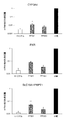
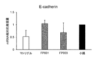
- the mRNA expression level in the adult small intestine was set to 1.
- DAPI Nuclear stain. Scale bar 50 ⁇ m.
- EZSPHERE mRNA expression analysis of cells immediately before seeding. Average S.D. (n 3).
- Control cells at day 7 of differentiation induction.
- Day 14 Cells seeded on iMatrix 511 silk on day 7 of differentiation induction and cultured until day 14 using medium 5.
- the subcultured group was subcultured every 2 days, seeded on iMatrix511 silk, and cultured using Medium 5. The non-passaged group was cultured with the medium 5 without passage.
- the control mRNA expression level was set to 1. Preparation of inflammatory bowel disease model by addition of TNF- ⁇ .
- A Protocol for preparing inflammatory bowel disease model.
- C Immunofluorescent staining of intestinal organoids with addition of TNF- ⁇ and infliximab.
- DAPI Nuclear stain. Scale bar 50 ⁇ m.
- FIG. 7 Preparation of fibrosis model by addition of TNF- ⁇ and TGF- ⁇ .
- A Fibrosis model preparation protocol.
- Intestinal Organoids Intestinal Organoids on Day 34 of Differentiation Induction, Flat Culture: Subculture of intestinal Organoids on iMatrix511 silk, and low molecular compounds (30 ⁇ mol / mL forskolin, 2.5 ⁇ mol / mL) were added to Medium 7 (see FIG. 9). Cells cultured for 7 days with addition of mL DAPT, 3 ⁇ mol / mL CHIR99021). Contol does not include low molecular weight compounds.
- (n 1) Formation of villus-like structures by gas-liquid culture and addition of low molecular weight compounds.
- AE Intestinal organoids were subcultured on iMatrix511silk, and low molecular compounds (2.5 ⁇ mol / mL DAPT (96 hours after seeding), 3 ⁇ mol / mL CHIR99021 (96 hours after seeding), 10 ⁇ mol / mL Y-27632 (24 hours after seeding)), and the factors to be examined (A: A83-01 not added, 30 ⁇ mol / mL forskolin. B: A83-01 added, forskolin not added. C: A83- 01 addition, 30 ⁇ mol / mL forskolin.
- D A8383-01 addition, 1 mmol / mL 8-Br-cAMP.
- E A8383 addition, 500 ⁇ mol / mL IBMX) were used for culture for 10 days. Scale bar 100 ⁇ m.
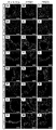
- G E-cad (epithelial cell adhesion factor) / CHGA (Chromogranin A).
- J)-(L) Ki67 (proliferative cell marker) / LGR5 (stem cell marker).
- M E-cad / Lyso (Lysozyme).
- P)-(R) Vim (Vimentin: fibroblast marker) / ⁇ -SMA (smooth muscle marker).
- S)-(U) Occludin (Tight Junction).
- V)-(X) ABCG2 / BCRP (Emission Transporter).
- DAPI Nuclear stain. Scale bar 50 ⁇ m.
- CYP3A4 Metabolic Activity of Human Intestinal Organoids Differentiated Using Polysaccharide Polymer. Data are presented as mean ⁇ SD (n 4). Control; ketoconazole-free group. ND; Not detected. ** P ⁇ 0.01, * P ⁇ 0.05 vs control group.
- the present invention relates to a method for producing an intestinal organoid from pluripotent stem cells (hereinafter, also referred to as “the production method of the present invention”).
- the production method of the present invention a method for producing an intestinal organoid from pluripotent stem cells (hereinafter, also referred to as “the production method of the present invention”).
- INDUSTRIAL APPLICABILITY According to the present invention, an intestinal organoid that is a three-dimensional tissue structure (mimicking intestinal tissue) that exhibits properties similar to those of a living organism is obtained.
- Pluripotent stem cells are capable of differentiating into all cells that compose the living body (pluripotency) and the ability to give rise to daughter cells that have the same differentiating potential as self through cell division (self-renewal ability). ) And cells that have both. Pluripotency can be evaluated by transplanting the cells to be evaluated into nude mice and testing for the presence or absence of teratoma formation including cells of the three germ layers (ectoderm, mesoderm, endoderm). it can.
- pluripotent stem cells examples include embryonic stem cells (ES cells), embryonic germ cells (EG cells), induced pluripotent stem cells (iPS cells), etc., which have both pluripotency and self-renewal ability It is not limited to this as long as it is a cell.
- ES cells or iPS cells are preferably used. More preferably, iPS cells are used.
- the pluripotent stem cells are preferably mammalian cells (eg, humans, chimpanzees, primates such as cynomolgus monkeys, rodents such as mice and rats), and particularly preferably human cells.
- ES cells can be established, for example, by culturing an early embryo before implantation, an inner cell mass that constitutes the early embryo, a single blastomere, etc. (Manipulating the Mouse Embryo A Laboratory Manual, Second Edition, Cold Spring Harbor Laboratory Press (1994); Thomson, J.A. et al., Science, 282, 1145-1147 (1998)).
- an early embryo produced by nuclear transfer of the somatic cell nucleus may be used (Wilmut et al. (Nature, 385, 810 (1997)), Cibelli et al. (Science, 280, 1256). (1998)), Akira Iritani et al.
- ES cells are available from the preservation organization or are commercially available.
- human ES cells are available from Kyoto University Institute for Frontier Medical Sciences (eg KhES-1, KhES-2 and KhES-3), WiCell Research Institute, ESI BIO and the like.
- ES cells can be established by culturing primordial germ cells in the presence of LIF, bFGF, SCF, etc. (Matsui et.al., Cell, 70, 841-847 (1992), Shamblott et al., Proc. Natl. Acad. Sci. USA, 95 (23), 13726-13731 (1998), Turnpenny et al., Stem Cells, 21 (5), 598-609, (2003)).
- iPS cells are cells that have pluripotency (multipotency) and proliferative capacity, which are created by reprogramming somatic cells by introducing reprogramming factors. Induced pluripotent stem cells have properties similar to ES cells.
- Somatic cells used for producing iPS cells are not particularly limited, and may be differentiated somatic cells or undifferentiated stem cells. The origin is not particularly limited, but preferably mammalian somatic cells (for example, humans, chimpanzees, cynomolgus monkeys and other primates, mice and rats and other rodents), and particularly preferably human somatic cells.
- the iPS cells can be produced by various methods reported so far. In addition, it is naturally envisaged to apply the iPS cell production method developed in the future.
- iPS cells derived from patients with intestinal diseases iPS cells prepared from somatic cells of patients
- the iPS cells are prepared from somatic cells (eg, skin, blood, mononuclear cells, etc.) collected from a patient.
- intestinal diseases include refractory inflammatory bowel disease (Crohn's disease, ulcerative colitis), polyps, colon cancer, drug-induced enteritis and the like.
- Disease-specific intestinal organoids are useful as models for intestinal pathology, and can be expected to contribute to drug evaluation systems and to elucidate the mechanisms of diseases (molecular mechanisms related to onset, pathogenesis, progression, etc.).
- the most basic method of iPS cell production is to introduce the transcription factors Oct3 / 4, Sox2, Klf4 and c-Myc into the cells using a virus (Takahashi K, Yamanaka S) : Cell 126 (4), 663-676, 2006; Takahashi, K, et al: Cell 131 (5), 861-72, 2007).
- a virus Teakahashi K, Yamanaka S
- Cell 126 (4), 663-676 Cell 126 (4), 663-676, 2006
- Takahashi, K, et al Cell 131 (5), 861-72, 2007
- human iPS cells there is a report of establishment by introducing four factors of Oct4, Sox2, Lin28 and Nonog (YuJ, et al: Science318 (5858), 1917-1920, 2007).
- 3 factors excluding c-Myc Neakagawa M, et al: Nat. Biotechnol.
- Transformation of iPS cells that is, cells that have undergone reprogramming (reprogramming), express pluripotent stem cell markers (undifferentiated markers) such as Fbxo15, Nanog, Oct / 4, Fgf-4, Esg-1 and Cript Can be selected as an index.
- pluripotent stem cell markers such as Fbxo15, Nanog, Oct / 4, Fgf-4, Esg-1 and Cript Can be selected as an index.
- the selected cells are collected as iPS cells.
- IPS cells can also be provided from, for example, Kyoto University National University or RIKEN BioResource Center.
- the terms “differentiate” and “induce” mean to act to differentiate along a specific cell lineage.
- pluripotent stem cells are differentiated into intestinal organoids.
- the production method of the present invention is roughly divided into five stages of culture steps, namely, (1) a step of differentiating pluripotent stem cells into endoderm-like cells (step (1)) and a step obtained in step (1).
- step (2) The step of differentiating the germ layer-like cells into the intestinal stem cell-like cells (step (2)) and the intestinal stem cell-like cells obtained in the step (2) are treated with epidermal growth factor, fibroblast growth factor, TGF ⁇ receptor inhibitor , A step of culturing in the presence of a GSK-3 ⁇ inhibitor and a ROCK inhibitor (step (3)), a step of culturing cells after the step (3) to form spheroids (step (4)), A step of differentiating the spheroid formed in (4) to form an intestinal organoid, the step comprising culturing in the presence of an epidermal growth factor, a BMP inhibitor and a Wnt signal activator (step (5)) including.
- epidermal growth factor fibroblast growth factor
- TGF ⁇ receptor inhibitor A step of culturing in the presence of a GSK-3 ⁇ inhibitor and a ROCK inhibitor (step (3))
- step (4) A step of culturing cells after the step (3) to form spheroids (step (4)
- pluripotent stem cells are cultured and differentiated into endoderm-like cells.
- pluripotent stem cells are cultured under conditions that induce differentiation into endoderm-like cells.
- Culture conditions are not particularly limited as long as pluripotent stem cells are differentiated into endoderm-like cells.
- it is cultured in a medium containing activin A according to a conventional method.
- the concentration of activin A in the medium is, for example, 10 ng / mL to 200 ng / mL, preferably 20 ng / mL to 150 ng / mL.
- serum or serum substitute Knockout serum replacement (KSR), etc.
- the serum is not limited to fetal bovine serum, and human serum, sheep serum and the like can be used.
- the added amount of serum or serum substitute is, for example, 0.1% (v / v) to 10% (v / v).
- An inhibitor of the Wnt / ⁇ -catenin signaling pathway eg, hexachlorophen, quercetin, Wnt ligand Wnt3a
- Wnt / ⁇ -catenin signaling pathway eg, hexachlorophen, quercetin, Wnt ligand Wnt3a
- the first stage culture is performed in a medium supplemented with a relatively low concentration of serum (eg, 0.1% (v / v) to 1% (v / v)), and the second stage culture is followed by the first stage culture. It is carried out in a medium having a higher serum concentration than that (for example, the serum concentration is 1% (v / v) to 10% (v / v)). It is preferable to adopt the two-step culture as described above because the growth of the undifferentiated cells is suppressed by the culture of the first step and the cells differentiated by the subsequent second step are grown.
- a relatively low concentration of serum eg, 0.1% (v / v) to 1% (v / v)
- the second stage culture is followed by the first stage culture. It is carried out in a medium having a higher serum concentration than that (for example, the serum concentration is 1% (v / v) to 10% (v / v)).
- the period (culture period) of step (1) is, for example, 1 to 10 days, preferably 2 to 7 days.
- the first-stage culture period is, for example, 1 to 7 days, preferably 2 to 5 days
- the second-stage culture period is, for example, 1 to 6 days. It is set to one day, preferably one to four days.
- the endoderm-like cells obtained in step (1) are cultured and differentiated into intestinal stem cell-like cells.
- endoderm cells are cultured under conditions that induce differentiation into intestinal stem cell-like cells. Culture conditions are not particularly limited as long as the endoderm-like cells are differentiated into intestinal stem cell-like cells.
- FGF4 fibroblast growth factor 4
- Wnt agonist eg Wnt3a, BML-284, 2-Amino-4-
- Human FGF4 eg, human recombinant FGF4
- FGF4 fibroblast growth factor 4
- FGF2 fibroblast growth factor 2
- GSK-3 inhibitor eg CHIR99021
- human FGF2 eg, human recombinant FGF2
- step (2) may be carried out after selecting endoderm-like cells from the cell population obtained through step (1).
- the endoderm-like cells can be selected, for example, by using a flow cytometer (cell sorter) with the cell surface marker as an index.
- FGF4 and Wnt agonist In the presence of FGF4 and Wnt agonist, is synonymous with the condition in which FGF4 and Wnt agonist are added to the medium. Therefore, in order to carry out culture in the presence of FGF4 and Wnt agonist, a medium containing FGF4 and Wnt agonist may be used.
- concentration of FGF4 added is 100 ng / mL to 5 ⁇ g / mL, preferably 300 ng / mL to 1 ⁇ g / mL.
- concentration of Wnt agonist added in the case of Wnt3a
- Wnt3a is 100 ng / mL to 5 ⁇ g / mL, preferably 300 ng / mL to 1 ⁇ g / mL.
- FGF2 and GSK-3 inhibitor in the presence of FGF2 and GSK-3 inhibitor, is synonymous with the condition in which FGF2 and GSK-3 inhibitor were added to the medium. Therefore, in order to carry out the culture in the presence of FGF2 and GSK-3 inhibitor, a medium containing FGF2 and GSK-3 inhibitor may be used.
- concentration of FGF2 added is 50 ng / mL to 2.5 ⁇ g / mL, preferably 150 ng / mL to 500 ng / mL.
- concentration of GSK-3 inhibitor added in the case of CHIR99021 is 600 nmol / L to 60 ⁇ mol / L, preferably 1 ⁇ mol / L to 20 ⁇ mol / L.
- the period (culture period) of step (2) is, for example, 2 to 10 days, preferably 3 to 7 days. If the culture period is too short, expected effects (increased differentiation efficiency, promotion of acquisition of functions as intestinal stem cells) cannot be sufficiently obtained. On the other hand, if the culture period is too long, the differentiation efficiency will decrease.
- intestinal stem cell marker As an index.
- intestinal stem cell markers are G protein-coupled receptor 5 (LGR5) containing leucine-rich repeats and ephrin B2 receptor (EphB2).
- Step (3) Induction of differentiation direction to intestinal tract>
- the intestinal stem cell-like cells obtained in step (2) are cultured in the presence of epidermal growth factor, fibroblast growth factor, TGF ⁇ receptor inhibitor, GSK-3 ⁇ inhibitor and ROCK inhibitor.
- the process can be understood as “the step of inducing the differentiation direction to the intestinal tract while maintaining the stem cell property”. This step is important for the morphology (showing a villus-crypt-like structure) and function of the finally obtained intestinal organoid (the intestinal marker and the drug transporter show gene expression close to that of the adult small intestine). This is one of the greatest features of the invention.
- the cell population obtained through the step (2) or a part thereof is subjected to the step (3) without selection.
- intestinal stem cell-like cells may be selected from the cell population obtained through step (2), and then step (3) may be performed.
- the intestinal stem cell-like cells may be selected, for example, by a flow cytometer (cell sorter) using the cell surface marker as an index.
- FGF2, FGF4 or FGF10 should be used as the fibroblast growth factor. Two or three of these FGF families may be used in combination.
- TGF ⁇ receptor inhibitor for example, A-83-01 can be used.
- GSK-3 ⁇ inhibitors include CHIR99021, SB216763, CHIR98014, TWS119, Tideglusib, SB415286, BIO, AZD2858, AZD1080, AR-A014418, TDZD-8, LY2090314, IM-12, Indirubin, Bikinin, 1-Azakenpaullone. it can.
- Y-27632 can be used as a ROCK inhibitor.
- concentration of epidermal growth factor added is 10 ng / mL to 500 ng / mL, preferably 50 ng / mL to 200 ng / mL.
- concentration of added fibroblast growth factor is 5 ng / mL to 200 ng / mL, preferably 20 ng / mL to 50 ng / mL, which is a TGF ⁇ receptor inhibitor.
- An example of the additive concentration of (in the case of A-83-01) is 0.1 ⁇ M to 5 ⁇ M, preferably 0.3 ⁇ M to 3 ⁇ M, and an example of the additive concentration of GSK-3 ⁇ inhibitor (in the case of CHIR99021) is 0.5 ⁇ M to 100 ⁇ M, preferably 1 ⁇ M to 30 ⁇ M, and an example of the concentration of the ROCK inhibitor added (in the case of Y-27632) is 1 ⁇ M to 50 ⁇ M, preferably 3 ⁇ M to 30 ⁇ M.
- serum substitutes should be used in combination. That is, preferably, the culture is performed under the condition that the above compound and serum substitute are added to the medium.
- the serum substitute is a composition used as a substitute for serum containing a differentiation inducing factor because it generally cultures iPS cells, ES cells and the like while maintaining their undifferentiated state.
- KSR Knockout serum replacement
- An example of added concentration of serum substitute is 5% (v / v) to 20% (v / v), preferably 5% (v / v) to 15% (v / v) is there.
- a culture container with a cell-adhesive culture surface is used for the culture in step (3).
- a basement membrane component eg, laminin, type IV collagen, entactin, vitronectin, fibronectin
- laminin 511 or its E8 fragment it is advisable to adopt a culture surface coated with laminin 511 or its E8 fragment.
- E8 fragment of laminin 511 a product (product name: iMatrix-511, manufacturing: Nippi Co., Ltd., sales: Matricsome Co., Ltd.) obtained by purifying the recombinant (recombinant human laminin 511-E8 protein) with high purity is commercially available.
- the culture container is not particularly limited, and for example, a dish, a flask, a multiwell plate, etc. can be used.
- the period (culture period) of the step (3) is, for example, 2 days to 14 days, preferably 3 days to 12 days. If the culture period is too short, the expected effect (ie, induction of differentiation direction necessary for forming a functional intestinal organoid having a villus-crypt-like structure) cannot be sufficiently obtained.
- the culture period can be set longer by repeating the subculture operation.
- the passage operation is performed in step (3). That is, subculture is performed in the middle of step (3).
- the passage procedure is considered to be particularly effective for purifying intestinal epithelial stem cell-like cells (improvement in purity and homogeneity) and more selectively inducing differentiation into the intestine. This has been clarified by chance by experiments shown in Examples described later, and is extremely important for understanding the specificity, value and significance of the present invention.
- the number of passage operations is, for example, 1 to 5 times, preferably 1 to 3 times, more preferably 1 or 2 times. If there are too many subculture operations, damage to cells will increase, leading to a decrease in production efficiency.
- the procedure of the subculture operation may follow a conventional method. For example, when the cells become confluent or sub-confluent, a part of the cells is collected and transferred to another culture container to continue the culture. A cell dissociation solution or the like may be used to recover the cells.
- the cell dissociation solution for example, trypsin-EDTA, collagenase IV, proteolytic enzymes such as metalloproteases, and the like can be used alone or in appropriate combination. Those having less cytotoxicity are preferable.
- a commercially available product such as dispase (Adia), TrypLE (Invitrogen) or accutase (MILLIPORE) is available.
- the cells that have undergone step (3) are cultured to form spheroids.
- Suspension culture is suitable for forming spheroids.
- a culture surface with low cell adhesion or non-cell adhesion for example, a culture surface to which low cell adhesion / cell non-adhesion is imparted by treatment / bonding with a polymer material or hydrogel
- a culture vessel is used to culture cells in a state away from the culture surface (that is, in a floating state).
- the culture container used for suspension culture is not particularly limited, and for example, a dish, a flask, a multiwell plate, a tube, a tray, a culture bag, or the like can be used.
- a culture vessel in which a plurality of wells having a uniform shape and size are formed on a culture surface having low cell adhesion or non-cell adhesion generally called a pattern plate.
- EZSPHERE registered trademark
- Elplasia etc. provided by Kuraray Co., Ltd.
- spheroids can be formed efficiently, and the intestinal organoid production efficiency is improved.
- cells / cell masses may be statically cultivated, or may be cultivated by shaking or shaking, as long as the non-adhesive state to the culture surface can be maintained.
- the suspension culture here is performed by static culture. Static culture has many advantages such as the fact that no special equipment is required, the impact or damage to cells is expected to be small, and the amount of culture solution can be reduced.
- the culture conditions are not particularly limited as long as spheroids can be formed.
- a BMP inhibitor and a Wnt signal activator that is, a medium containing these components is used. Suspension culture.
- the effect of promoting cell growth can be expected.
- the effect of suppressing the differentiation of stem cells and maintaining the stemness can be expected.
- the Wnt signal activator can be expected to have an effect of maintaining the proliferation and stemness of stem cells.
- Noggin for example, can be used as a BMP inhibitor. Further, R-spondin-1 can be used as a Wnt signal activator, for example.
- concentration of epidermal growth factor added is 10 ng / mL to 500 ng / mL, preferably 50 ng / mL to 200 ng / mL.
- concentration of BMP inhibitor added is 10 ng / mL to 500 ng / mL, preferably 50 ng / mL to 200 ng / mL.
- concentration of Wnt signal activator added is 10 ng / mL to 1000 ng / mL, preferably 50 ng / mL to 500 g / mL.
- the period (culture period) of the step (4) is, for example, 1 to 10 days, preferably 2 to 7 days. If the culture period is too short, a sufficient size of spheroid will not be formed. On the other hand, if the culture period is too long, the spheroids become larger than necessary, and the cells inside may undergo necrosis.
- spheroids having a diameter of about 100 ⁇ m to 200 ⁇ m are formed.
- a spheroid of the size can be formed by using a pattern plate in which wells having a diameter of 400 to 500 ⁇ m and a depth of 100 to 200 ⁇ m are uniformly formed.
- Step (5) Formation of intestinal organoid>
- the spheroid formed in step (4) is differentiated to form an intestinal organoid.
- active differentiation induction is not essential, and in one aspect (first aspect), the culture is performed under the same or similar medium conditions as in step (4). Specifically, it is cultured in the presence of an epidermal growth factor, a BMP inhibitor and a Wnt signal activator, that is, using a medium to which these components are added. Specific examples and addition concentrations of each compound are the same as in the case of the step (4).
- the period (culture period) of step (5) is, for example, 12 days to 36 days.
- the culture in step (5) is suspension culture.
- a liquid medium in which a material that forms a three-dimensional network structure is added to an aqueous solution, and to collect a plurality of spheroids formed in step (4) (that is, one spheroid).
- Suspension culture (with multiple spheroids coexisting in the culture vessel). That is, in a preferred embodiment, a part (however, 2 or more) or all of the spheroids formed in the step (4) are subjected to suspension culture using a characteristic liquid medium.
- viscous material a material that forms a three-dimensional network structure in an aqueous solution
- spheroids are trapped or trapped in the network structure, or the viscosity of the culture medium is increased to limit the movement of the spheroids. It is possible to prevent the association and aggregation of spheroids. Therefore, a plurality of spheroids can be collectively subjected to suspension culture, which enables efficient formation of intestinal organoids.
- polymer gel or polysaccharide can be used as the viscous material.
- polymer gels are collagen, polymer hydrogels, Matrigel TM (regular Matrigel, growth factor reduced (GFR) Matrigel with reduced growth factor content, etc.).
- polysaccharides are gellan gum, crystalline cellulose, nanocellulose, carboxycellulose, carboxymethylcellulose and the like. You may decide to use 2 or more types of materials together.
- a polymer compound having an anionic functional group is used as the viscous material.
- the anionic functional group include a carboxy group, a sulfo group, a phosphoric acid group and salts thereof, and a carboxy group or a salt thereof is preferable.
- the polymer compound used in the present invention those having one kind or two or more kinds selected from the group of the anionic functional groups can be used.
- Preferred specific examples of the polymer compound herein are not particularly limited, but include polysaccharides obtained by polymerizing 10 or more monosaccharides (for example, triose, tetrose, pentose, hexose, heptose, etc.), More preferably, an acidic polysaccharide having an anionic functional group is used.
- the acidic polysaccharide referred to here is not particularly limited as long as it has an anionic functional group in its structure, but for example, a polysaccharide having uronic acid (eg, glucuronic acid, iduronic acid, galacturonic acid, mannuronic acid).
- hyaluronic acid gellan gum, deacylated gellan gum, rhamsan gum, diutan gum, xanthan gum, carrageenan, xanthan gum, hexuronic acid, fucoidan, pectin, pectic acid, pectinic acid, heparan sulfate, heparin, heparitin sulfate, kerato.
- examples thereof include those composed of one or more selected from the group consisting of sulfuric acid, chondroitin sulfate, dermatan sulfate, rhamnan sulfate and salts thereof.
- the polysaccharide is preferably hyaluronic acid, deacylated gellan gum, diutan gum, xanthan gum, carrageenan or a salt thereof, and most preferably, deacylated, considering that the purpose can be achieved by using at a low concentration. It is gellan gum.
- the salt here include salts of alkali metals such as lithium, sodium and potassium, salts of alkaline earth metals such as calcium, barium and magnesium or salts of aluminum, zinc, copper, iron, ammonium, organic bases and amino acids. Is mentioned.
- the weight average molecular weight of the polymer compound is preferably 10,000 to 50,000,000,000, more preferably 100,000 to 20,000,000,000, further preferably 1,000,000,000. It is up to 10,000,000.
- the molecular weight can be measured in terms of pullulan by gel permeation chromatography (GPC).
- deacylated gellan gum may be phosphorylated.
- the phosphorylation can be performed by a known method.
- the above-mentioned polysaccharides can be used in combination of plural kinds (preferably two kinds).
- the type of combination of polysaccharides is not particularly limited as long as it can prevent the association and aggregation of spheroids, but preferably the combination contains at least deacylated gellan gum or a salt thereof.
- a suitable combination of polysaccharides includes deacylated gellan gum or a salt thereof, and polysaccharides other than deacylated gellan gum or a salt thereof (eg, xanthan gum, alginic acid, carrageenan, diutan gum, methylcellulose, locust bean gum or Their salts).
- Specific polysaccharide combinations include deacylated gellan gum and rhamsan gum, deacylated gellan gum and diutan gum, deacylated gellan gum and xanthan gum, deacylated gellan gum and carrageenan, deacylated gellan gum and xanthan gum, deacylated.
- Examples thereof include, but are not limited to, gellan gum and locust bean gum, deacylated gellan gum and ⁇ -carrageenan, deacylated gellan gum and sodium alginate, deacylated gellan gum and methylcellulose.
- More preferable specific examples of the viscous material used in the present invention include hyaluronic acid, deacylated gellan gum, diutan gum, carrageenan and xanthan gum, and salts thereof, and the most preferable example is deacylated gellan gum or a salt thereof.
- deacylated gellan gum commercially available products, for example, “KELCOGEL (registered trademark of CP Kelco) CG-LA” manufactured by Sansyo Co., Ltd., “Kelcogel (Cie Kelco) manufactured by Sanei Gen FFI Co., Ltd. “Registered trademark of the company” and the like can be used.
- polymer FP001 is a compounding ingredient of the three-dimensional culture medium FCeM (registered trademark) series manufactured by Nissan Chemical Industries, Ltd.
- polymer FP003 is a compounding ingredient of the same FCeM (registered trademark) Advance Preparation Kit.
- the amount of the viscous material used is not particularly limited as long as the expected effects described above can be exhibited, but for example, the viscous material is adjusted so that the viscosity of the medium is 5 mPas ⁇ s to 2000 mPas ⁇ s. Adjust the usage of. If the viscosity of the medium is too low, the effect of preventing spheroid association and aggregation cannot be obtained. On the other hand, if the viscosity of the medium is too high, the operability (handling) is affected (for example, the collection operation becomes complicated), and the supply of medium components to cells may be affected.
- Matrigel When Matrigel is used as a specific example of the amount of the viscous material used, it may be about 1% to 10% of the amount used in normal use (that is, use as a substrate for three-dimensional culture).
- deacylated gellan gum 0.001% to 1.0% (w / v), preferably 0.003% to 0.5% (w / v), more preferably 0.005% to 0.3% (w / v), and even more preferably 0.01% to 0.05% (w / v), and most preferably 0.01% to 0.03% (w / v) medium.
- the culture vessel used for suspension culture is not particularly limited, and for example, a dish, flask, multiwell plate, tube, tray, culture bag, etc. can be used.
- step (5) in addition to epidermal growth factor, BMP inhibitor and Wnt signal activator, MEK1 / 2 inhibitor, DNA methylation inhibitor, TGF ⁇ receptor inhibitor and ⁇ - Culturing in step (5) is carried out in the presence of a secretase inhibitor to actively induce differentiation.
- MEK1 / 2 inhibitor in addition to epidermal growth factor, BMP inhibitor and Wnt signal activator, MEK1 / 2 inhibitor, DNA methylation inhibitor, TGF ⁇ receptor inhibitor and ⁇ - Culturing in step (5) is carried out in the presence of a secretase inhibitor to actively induce differentiation.
- the point of using a combination of a MEK1 / 2 inhibitor, a DNA methylation inhibitor, a TGF ⁇ receptor inhibitor and a ⁇ -secretase inhibitor is particularly characteristic in the present invention, and it is possible to improve the efficiency of differentiation induction and maturation of intestinal organoids. Contribute to promotion.
- TGF ⁇ receptor inhibitor one that exhibits an inhibitory activity against one or more of the TGF- ⁇ receptors ALK4, ALK5 and ALK7 is preferably used.
- A-83-01, SB431542, SB-505124, SB525334, D4476, ALK5inhibitor, LY2157299, LY364947, GW788388, RepSox satisfy the condition.
- ⁇ -secretase inhibitors include N-[(3,5-difluorophenyl) acetyl] -L-alanyl-2-phenyl-1,1-dimethylethyl ester-glycine (DAPT), L-685,458, CompoundE (CAS 209986-17-4), (R) -Flurbiprofen, BMS299897, JLK6, LY-411575, R04929097, MK-0752, SCP0004, SCP0025, gamma-Secretase Inhibitor XI, gamma-Secretase Inhibitor VI, gamma-Secretase Inhibitor I , Gamma-Secretase Inhibitor VII, Semagacestat (LY450139), gamma-Secretase Inhibitor III, Compound 34, BMS-708163, Compound W, YO-01027 (Dibenzo
- An example of the concentration of MEK1 / 2 inhibitor added is 4 ⁇ M to 100 ⁇ M, preferably 10 to 40 ⁇ M.
- an example of the addition concentration of the DNA methylation inhibitor in the case of 5-aza-2'-deoxycytidine
- the addition concentration of the TGF ⁇ receptor inhibitor is Example (in the case of A-83-01) is 0.1 ⁇ M to 2.5 ⁇ M, preferably 0.2 ⁇ M to 1 ⁇ M
- an example of the concentration of ⁇ -secretase inhibitor added is 1 nM It is 20 ⁇ M, preferably 0.1 ⁇ M to 10 ⁇ M.
- the compounds exemplified that is, PD98059, 5-aza-2'-deoxycytidine, A-83-01, the addition concentration in the case of using a compound different from DAPT, the characteristics of the compound used, and exemplified Considering the difference in the characteristics of the compounds (in particular, the difference in the activity), those skilled in the art can set the concentration according to the above-mentioned concentration range. Whether or not the set concentration range is appropriate can be confirmed by preliminary experiments according to the examples described later.
- step (5) the following step (5-1) and step (5-2) are performed in this order.
- step (5) the combination of low-molecular compounds characteristic of step (5) (that is, the combination of MEK1 / 2 inhibitor, DNA methylation inhibitor, TGF ⁇ receptor inhibitor and ⁇ -secretase inhibitor) is combined with step (5). -2) will be used.
- This step is a preparatory step before promoting active differentiation induction by the combination of four kinds of low molecular weight compounds (MEK1 / 2 inhibitor, DNA methylation inhibitor, TGF ⁇ receptor inhibitor and ⁇ -secretase inhibitor) Can be positioned as By intervening this step, the growth of spheroids is promoted, which is advantageous for the construction of functional intestinal organoids.
- the culture in this step can be performed under the same conditions as in step (4) (that is, suspension culture in the presence of epidermal growth factor, BMP inhibitor and Wnt signal activator), but preferably in an aqueous solution.
- a liquid medium containing a material (viscous material) that forms a three-dimensional network structure and collectively culturing a plurality of spheroids formed in step (4) in suspension culture the operability and efficiency are improved. ..
- the period (culture period) of step (5-1) is, for example, 3 to 15 days, preferably 6 to 12 days. If the culture period is too short, the spheroids are small and cell death is likely to occur. On the other hand, if the culture period is too long, a sufficient effect cannot be obtained by the low molecular weight compound.
- ⁇ Step (5-2)> In this process, in the presence of MEK1 / 2 inhibitor, DNA methylation inhibitor, TGF ⁇ receptor inhibitor and ⁇ -secretase inhibitor, in addition to epidermal growth factor, BMP inhibitor and Wnt signal activator Culturing is performed to promote positive differentiation induction and form intestinal organoids. Since the culture conditions are the same as the culture conditions in the above step (5-1), the description thereof will be omitted.
- the period (culture period) of the step (5-2) is, for example, 3 to 21 days, preferably 9 to 18 days. If the culture period is too short, the function will not be sufficiently improved. On the other hand, if the culture period is too long, damage to the spheroids may cause cell death.
- MEK1 / 2 inhibitor DNA methylation inhibitor, TGF ⁇ receptor inhibitor and epidermal growth factor (EGF) are present (hereinafter, this condition is referred to as “first The condition (hereinafter referred to as “condition”) and the condition in which cAMP is supplied to cells (hereinafter, this condition is referred to as “second condition”) are cultivated in the step (5) to actively induce differentiation.
- the first condition is similar to that of the second mode, and therefore its description is omitted.
- the second condition that is, the condition that cAMP is supplied to cells, is synonymous with the condition that there is a compound that can be taken up into cells and that acts as cAMP when taken into cells.
- a medium to which a cAMP derivative that can be taken up into cells is added may be used.
- PKA activator as a cAMP derivative e.g., 8-Br-cAMP (8-Bromoadenosine-3 ′, 5′-cyclic monomonophosphate sodium salt, CAS number: 76939-46-3), 6-Bnz-cAMP (N6-Benzoyladenosine- 3 ', 5'-cyclic monophosphate, sodium, salt, salt, CASNumber: 1135306-29-4), cAMPS-Rp ((R) -Adenosine, cyclic 3', 5 '-(hydrogenphosphorothioate) triethylammonium salt, CASNumber: 151837- 09-1), cAMPS-Sp ((S) -Adenosine, cyclic3 ', 5'-(hydrogenphosphorothioate) triethylammonium salt, CASNumber: 1518
- concentration of the cAMP derivative added is 0.1 mM to 10 mM, preferably 0.2 mM to 5 mM, more preferably 0.5 mM to 2 mM.
- concentration when using a compound exemplified that is, a compound different from 8-Br-cAMP
- the difference between the characteristics of the compound used and the characteristics of the exemplified compound (8-Br-cAMP) particularly Considering (activity difference)
- those skilled in the art can set the concentration according to the above-mentioned concentration range. Whether or not the set concentration range is appropriate can be confirmed by a preliminary experiment.
- any of the following culture steps A to C is performed.
- ⁇ Culture process A> In the culturing step A, (a-1) MEK1 / 2 inhibitor, DNA methylation inhibitor, TGF ⁇ receptor inhibitor and epidermal growth factor are cultivated in the presence of (a-2) MEK1 / 2 inhibitor, DNA methylation inhibitor, TGF ⁇ receptor inhibitor and epidermal growth factor are present, and culture is performed under the condition that cAMP is supplied to the cells. That is, two-stage culturing that differs depending on the presence or absence of the condition of supplying cAMP to cells is performed. In this way, the effects of promoting differentiation into intestinal epithelial cells, maturation, and gain of function can be obtained.
- the period for culturing (a-1) is, for example, 1 to 5 days.
- the culture period of (a-2) is, for example, 3 to 15 days.
- the culture of (a-2) may be carried out under the condition that a cAMP degrading enzyme inhibitor is present in addition to the MEK1 / 2 inhibitor, DNA methylation inhibitor, TGF ⁇ receptor inhibitor and epidermal growth factor.
- a cAMP degrading enzyme inhibitor is present in addition to the MEK1 / 2 inhibitor, DNA methylation inhibitor, TGF ⁇ receptor inhibitor and epidermal growth factor.
- inhibition of cAMP degradation suppresses a decrease in intracellular cAMP concentration and promotes induction of differentiation into intestinal epithelium, particularly acquisition of a function as an intestinal epithelial cell. That is, the conditions are advantageous for preparing a more functional intestinal organoid.
- IBMX 3-isobutyl-1-methylxanthine
- MIX Theophylline
- Papaverine Pentoxifylline
- KS-505, 8-Methoxymethyl-IBMX Vinpocetine
- TCV-3B Lixazinone
- LY-186126 Cilosamide
- OPC3689 Cilosamide
- Bemoradan RWJ-22867
- Anergrelide BL4162A
- Indolidan LY195115
- Cilostazol OPC-13013
- Milrinone WIN47203
- Siguazodan SKF-94836
- 5-Methyl-imazodan CI 930
- SKF-95654 Pirilobendan
- UD-CG 115 BS Pirilobendan
- MDL 17043 Enoximone
- Imazodan CCL 914
- SKF-94120 Vesnarinone
- concentration of the cAMP-degrading enzyme inhibitor added is 0.05 mM to 5 mM, preferably 0.1 mM to 3 mM, and more preferably 0.2 mM to 1 mM.
- concentration to be added when the exemplified compound that is, a compound different from IBMX, is used, considering the characteristics of the compound used and the difference in the characteristics of the exemplified compound (IBMX) (especially the difference in activity).
- concentration to be added when the exemplified compound that is, a compound different from IBMX
- IBMX the difference in the characteristics of the exemplified compound
- Those skilled in the art can set the concentration according to the above concentration range. Whether or not the set concentration range is appropriate can be confirmed by a preliminary experiment.
- culture in the presence of MEK1 / 2 inhibitor, DNA methylation inhibitor, TGF ⁇ receptor inhibitor and epidermal growth factor (culture of (a-3)) will be performed. May be.
- the culture period is, for example, 1 to 10 days.
- ⁇ Culturing step B> In the culturing step B, (b-1) culturing in the presence of a MEK1 / 2 inhibitor, a DNA methylation inhibitor, a TGF ⁇ receptor inhibitor and an epidermal growth factor and under the condition that cAMP is supplied to the cells, Following the culturing, culturing is carried out in the presence of (b-2) MEK1 / 2 inhibitor, DNA methylation inhibitor, TGF ⁇ receptor inhibitor, epidermal growth factor and cAMP degrading enzyme inhibitor.
- the culture period of (b-1) is, for example, 3 to 15 days.
- the culture period of (b-2) is, for example, 3 to 15 days.
- the culture of (b-1) may be carried out under the condition that a cAMP degrading enzyme inhibitor is present in addition to the MEK1 / 2 inhibitor, DNA methylation inhibitor, TGF ⁇ receptor inhibitor and epidermal growth factor.
- a cAMP degrading enzyme inhibitor is present in addition to the MEK1 / 2 inhibitor, DNA methylation inhibitor, TGF ⁇ receptor inhibitor and epidermal growth factor.
- culture in the presence of MEK1 / 2 inhibitor, DNA methylation inhibitor, TGF ⁇ receptor inhibitor and epidermal growth factor (culture of (b-3)) will be performed. May be.
- the culture period is, for example, 1 to 10 days.
- ⁇ Culture process C> In the culturing step C, culturing is performed in the presence of (c-1) MEK1 / 2 inhibitor, DNA methylation inhibitor, TGF ⁇ receptor inhibitor and epidermal growth factor, and under the condition that cAMP is supplied to cells. ..
- the period of culture of (c-1) is, for example, 3 to 15 days.
- the culture of (c-1) is carried out under the condition that cAMP degrading enzyme inhibitor is present in addition to MEK1 / 2 inhibitor, DNA methylation inhibitor, TGF ⁇ receptor inhibitor and epidermal growth factor (cAMP is supplied to cells.
- the conditions may also be used together). By adopting the conditions, it is possible to suppress the decrease in intracellular cAMP concentration while supplying cAMP to the cells. Therefore, it can be expected that the condition is effective for maintaining intracellular cAMP at a high level, and efficient induction of differentiation into intestinal epithelial cells is promoted.
- culture in the presence of MEK1 / 2 inhibitor, DNA methylation inhibitor, TGF ⁇ receptor inhibitor and epidermal growth factor (culture of (c-2)) will be performed. May be.
- the culture period is, for example, 1 to 10 days.
- a MEK1 / 2 inhibitor, a DNA methylation inhibitor, a TGF ⁇ receptor inhibitor, an epidermal growth factor and a cAMP activator are used in combination in step (5).
- "Use of MEK1 / 2 inhibitor, DNA methylation inhibitor, TGF ⁇ receptor inhibitor, epidermal growth factor and cAMP activator in combination” means to carry out one or more cultures constituting step (5). , All of these compounds are required, and all of these compounds are required to be used at the same time, that is, it is required as an essential condition that culture is performed in a medium to which all of these compounds are added. Not a thing.
- the step (5) in the fourth aspect is constituted by one or more cultures (details will be described later).
- a medium to which an epidermal growth factor and a cAMP activator are added as essential components for example, a MEK1 / 2 inhibitor, a DNA methylation inhibitor, a TGF ⁇ receptor inhibitor and an epithelium Medium with growth factor added as an essential component, medium with epidermal growth factor added as an essential component, MEK1 / 2 inhibitor, DNA methylation inhibitor, TGF ⁇ receptor inhibitor, epidermal growth factor and cAMP activation
- a medium or the like to which a substance is added as an essential component is used.
- the MEK1 / 2 inhibitor, DNA methylation inhibitor, TGF ⁇ receptor inhibitor, and epidermal growth factor are the same as in Condition 1 of the above-mentioned third aspect, so description thereof will be omitted.
- the cAMP activator forskolin, indomethacin, NKH477 (colforsin daropate), cell-derived toxin protein (pertussis toxin, cholera toxin), PACAP-27, PACAP-38, SKF83822 and the like can be used.
- Forskolin exerts an adenylate cyclase activating effect and promotes intracellular cAMP synthesis.
- concentration added of the cAMP activator (for forskolin) is 1 ⁇ M to 200 ⁇ M, preferably 5 ⁇ M to 100 ⁇ M.
- addition concentration when using the exemplified compound that is, a compound different from forskolin
- those skilled in the art can set the concentration according to the above concentration range. Whether or not the set concentration range is appropriate can be confirmed by preliminary experiments according to the examples described later.
- the step (5) in the fourth aspect is added to the above-mentioned conditions, and the conditions under which cAMP is supplied to cells (referred to as “additional condition 1”) and the conditions under which a cAMP-degrading enzyme inhibitor exists (referred to as “additional condition 2”). ), Or under any of these conditions.
- the additional condition 1 is the same as the first condition of the third aspect.
- the additional condition 2 is the same as the condition that can be adopted in the culture steps A to C of the third aspect.
- any of the following culture steps A to D is performed.
- ⁇ Culture process A> In the culturing step A, (a-1) culturing in the presence of an epidermal growth factor and an intracellular cAMP synthesis promoter, and (a-2) MEK1 / 2 inhibitor and DNA methylation inhibition, which are performed after the culturing. Culture is performed in the presence of the agent, TGF ⁇ receptor inhibitor, and epidermal growth factor.
- the period of culturing (a-1) is, for example, 2 to 10 days, preferably 4 to 8 days, and the period of culturing (a-2) is, for example, 9 days to 29 days, preferably 7 days to 27 days. It is a day.
- the above corresponding explanations are incorporated.
- addition condition 1 The condition (a-1) in which cAMP is supplied to cells (referred to as “additional condition 1") and the condition in which a cAMP-degrading enzyme inhibitor is present (referred to as “additional condition 2”), or any of these You may decide to carry out on the conditions of. The same applies to the culture of (a-2). Details of the additional condition 1 and the additional condition 2 are as described above.
- ⁇ Culturing step B> In the culturing step B, (b-1) culturing in the presence of epidermal growth factor, and (b-2) MEK1 / 2 inhibitor, DNA methylation inhibitor, TGF ⁇ receptor inhibitor, which is performed after the culturing , Culturing in the presence of epidermal growth factor and intracellular cAMP synthesis promoter.
- the period of culturing (b-1) is, for example, 2 to 10 days, preferably 4 to 8 days
- the period of culturing (b-2) is, for example, 9 to 19 days, preferably 7 to 17 days. It is a day.
- culture in the presence of MEK1 / 2 inhibitor, DNA methylation inhibitor, TGF ⁇ receptor inhibitor and epidermal growth factor (culture of (b-3)) will be performed. May be.
- the culture period is, for example, 1 to 10 days.
- ⁇ Culture process C> In the culturing step C, (c-1) MEK1 / 2 inhibitor, DNA methylation inhibition, which is performed after the culture in the presence of (c-1) epidermal growth factor and intracellular cAMP synthesis promoter Culture is performed in the presence of an agent, a TGF ⁇ receptor inhibitor, an epidermal growth factor, and an intracellular cAMP synthesis promoter.
- the period of culturing (c-1) is, for example, 2 to 10 days, preferably 4 to 8 days, and the period of culturing (c-2) is, for example, 9 to 19 days, preferably 7 to 17 days. It is a day.
- the above corresponding explanations are incorporated.
- culture in the presence of MEK1 / 2 inhibitor, DNA methylation inhibitor, TGF ⁇ receptor inhibitor and epidermal growth factor (culture of (c-3)) will be performed. May be.
- the culture period is, for example, 1 to 10 days.
- culturing step D culturing is performed in the presence of (d-1) MEK1 / 2 inhibitor, DNA methylation inhibitor, TGF ⁇ receptor inhibitor, epidermal growth factor and intracellular cAMP synthesis promoter.
- This culturing step is particularly advantageous in that the culturing operation is simple, it is more effective for differentiation into intestinal epithelial cells, and a stable effect can be expected because it is a compound.
- the period of culture of (d-1) is, for example, 15 days to 25 days, preferably 17 days to 23 days. Incidentally, as for matters not particularly explained (compounds usable for each culture, addition concentration of each compound, etc.), the above corresponding explanations are incorporated.
- culture in the presence of MEK1 / 2 inhibitor, DNA methylation inhibitor, TGF ⁇ receptor inhibitor and epidermal growth factor (culture of (d-2)) will be performed. May be.
- the culture period is, for example, 1 to 10 days.
- Other culture conditions (culture temperature and the like) in each step constituting the present invention may be those generally adopted in the culture of animal cells. That is, it may be cultured, for example, in an environment of 37 ° C. and 5% CO 2 .
- the basic medium Iscove's modified Dulbecco's medium (IMDM) (GIBCO, etc.), Ham's F12 medium (HamF12) (SIGMA, Gibco, etc.), Dulbecco's modified Eagle medium (D-MEM) (Nacalai Tesque, Inc., Sigma, Gibco, etc.), Glasgow basal medium (Gibco, etc.), RPMI1640 medium, etc. can be used. You may decide to use 2 or more types of basic media together.
- IMDM Iscove's modified Dulbecco's medium
- HamF12 Ham's F12 medium
- D-MEM Dulbecco's modified Eagle medium
- Gibco, etc. Glasgow basal medium
- a basal medium suitable for culturing epithelial cells for example, a mixed medium of D-MEM and Ham's F12 medium, D-MEM.
- a basal medium suitable for culturing epithelial cells for example, a mixed medium of D-MEM and Ham's F12 medium, D-MEM.
- components that can be added to the medium include bovine serum albumin (BSA), antibiotics, 2-mercaptoethanol, PVA, non-essential amino acids (NEAA), insulin, transferrin, and selenium.
- Subculture may be carried out during the steps (1) and (2) constituting the present invention.
- a part of the cells is collected and transferred to another culture container to continue the culture.
- a cell dissociation solution or the like may be used to recover the cells.
- the cell dissociation solution for example, trypsin-EDTA, collagenase IV, proteolytic enzymes such as metalloproteases, and the like can be used alone or in appropriate combination. Those having less cytotoxicity are preferable.
- a commercially available product such as dispase (Adia), TrypLE (Invitrogen) or accutase (MILLIPORE) is available.
- the recovered cells may be treated with a cell strainer or the like so as to be in a dispersed (discrete) state and then subjected to subculture.
- the medium is exchanged as needed. For example, it is recommended to change the medium once every 24 hours to 3 days.
- step (3) it is preferable to perform subculture for step (3). It is also possible to carry out subculture (eg, 1 to 5 times) in step (5).
- the substituting operation in this case may follow the following procedure, for example. First, after collecting cell clumps (spheroids) and washing as necessary, dispersion / dissociation solution for spheroids (for example, EDTA from Thermo Fisher Scientific, Dispase II from Roche, and Gentle Cell Dissociation Reagent from STEMCELL Technologies). ). Then, after being dispersed and suspended by a pipetting operation or the like, seeding is carried out and cultivation is continued.
- dispersion / dissociation solution for spheroids for example, EDTA from Thermo Fisher Scientific, Dispase II from Roche, and Gentle Cell Dissociation Reagent from STEMCELL Technologies.
- ROCK inhibitor Rho-associated coiled-coil forming kinase / Rho-linked kinase
- an intestinal organoid having a structure close to that of a living intestinal tract that is, a budding intestinal organoid
- the intestinal organoid of the present invention is similar in function to the intestinal tract of a living body, and exerts a pharmacokinetic function characteristic of the intestinal tract, specifically a metabolic (absorption, excretion) function.
- Such functional intestinal organoids can be evaluated by the expression of various transporters (peptide transporter, efflux transporter, organic anion transporter, etc.) and the expression / activity of drug-metabolizing enzymes.
- the presence or absence and the degree of drug responsiveness are also useful as an index for evaluating the functionality of intestinal organoids.
- An example of a peptide transporter is SLC15A1 / PEPT1 (SLC (solute carrier) family member 15A1 / Peptide transporter 1)
- an example of an efflux transporter is ABCB1 / MDR1 (ATP-binding cassette transporter B1 / multidrug resistance protein 1)
- ABCC2 / MRP2 ATP-binding cassette transporter C2 / multidrug resistance-related protein 2
- ABCG2 / BCRP ATP-binding cassette transporter G2 / breast cancer resistance protein
- organic anion transporters are SLCO2B1 / OATP2B1 (SLC (Solute carrier) Organic anion transporter 2B1)
- examples of drug-metabolizing enzymes are CYP3A4 (cytochrome P4503A4) and CYP3A8 (cytochrome P4503A8).
- CYP3A drug metabolizing enzyme
- CYP3A8 in cynomolgus monkey
- a rifampicin or vitamin D receptor for example, expression induction of drug metabolizing enzyme CYP3A (for example, CYP3A4 in human, CYP3A8 in cynomolgus monkey) via a rifampicin or vitamin D receptor can be used as an index.
- CDX2 (Caudal-type homeobox 2), Chromogranin A, E-cad (E-cadherin; epithelial cadherin), LGR5 (G protein-coupled receptor including leucine-rich repeats), Lysozyme (lysozyme), MUC2 (mucin) 2 glycoprotein), OLFM4 (olfactmedin 4), Villin (vilin), Vim (Vimentin; vimentin), etc. are also useful for evaluating the structure or functionality of intestinal organoids.
- the present invention provides a method for producing an intestinal cell layer exhibiting similar characteristics to intestinal tissue of a living body (hereinafter, also referred to as “intestinal cell layer producing method of the present invention”) To do.
- the method for producing an intestinal cell layer of the present invention utilizes the intestinal organ guide obtained in the above aspect, and the intestinal organoid is subjected to flat culture (that is, transfer / conversion to a two-dimensional culture system) to form a cell layer. ..
- the intestinal cell layer production method of the present invention is characterized by the following steps (1) to (6).
- Step of differentiating pluripotent stem cells into endoderm-like cells (2) Step of differentiating endoderm-like cells obtained in step (1) into intestinal stem cell-like cells (3) Step (2) Culturing the obtained intestinal stem cell-like cells in the presence of epidermal growth factor, fibroblast growth factor, TGF ⁇ receptor inhibitor, GSK-3 ⁇ inhibitor and ROCK inhibitor (4) after step (3) Step of culturing cells to form spheroids (5) Differentiating the spheroids formed in step (4) to form intestinal organoids, which comprises epidermal growth factor, BMP inhibitor and Wnt signal activator (6) Step of culturing in the presence of the intestinal organoid-forming cells formed in the step (5) in the presence of an epidermal growth factor and a TGF ⁇ receptor inhibitor
- Steps (1) to (5) are the same as those in the first phase, so the description thereof is omitted.
- step (6) subsequent to step (5) cells constituting the intestinal organoid formed in step (5) are subjected to flat culture in the presence of epidermal growth factor and TGF ⁇ receptor inhibitor.
- cells are collected from the formed intestinal organoids, transferred to a two-dimensional culture system, and cultured under specific conditions.
- the cell population recovered from the intestinal organoid or a part thereof is subjected to flat culture without selection.
- a cell dissociation solution or the like may be used to recover cells from the intestinal organoid.
- cell dissociation solution for example, trypsin-EDTA, collagenase IV, proteolytic enzymes such as metalloproteases, and the like can be used alone or in appropriate combination. Those having less cytotoxicity are preferable.
- a cell dissociation solution for example, a commercially available product such as dispase (Adia), TrypLE (Invitrogen) or accutase (MILLIPORE) is available. In addition, it may be treated with a cell strainer or the like to enhance the dispersibility of cells.
- ROCK inhibitor Rho-associated coiled-coil forming kinase / Rho-binding kinase
- the culture condition of step (6),“ in the presence of epidermal growth factor and TGF ⁇ receptor inhibitor ” is synonymous with the condition in which these compounds were added to the medium. Therefore, in order to carry out the culture in the presence of the epidermal growth factor and the TGF ⁇ receptor inhibitor, a medium containing these compounds may be used.
- a TGF ⁇ receptor inhibitor for example, A-83-01 can be used.
- concentration of epidermal growth factor added is 10 ng / mL to 500 ng / mL, preferably 50 ng / mL to 200 ng / mL.
- concentration of the TGF ⁇ receptor inhibitor added in the case of A-83-01
- concentration of the addition concentration in the case of using the exemplified compound, that is, the compound different from A-83-01 should be taken into consideration.
- concentration according to the above concentration range Whether or not the set concentration range is appropriate can be confirmed by preliminary experiments according to the examples described later.
- the step (6) is cultured in the presence of a cAMP activator to enhance the function of the formed cell layer. More preferably, a ⁇ -secretase inhibitor and / or a GSK-3 ⁇ inhibitor is added together with the cAMP activator to further enhance the function of the formed cell layer.
- cAMP activator forskolin, indomethacin, NKH477 (colforsin daropate), cell-derived toxin protein (pertussis toxin, cholera toxin), PACAP-27, PACAP-38, SKF83822, etc. can be used.
- ⁇ -secretase inhibitors include N-[(3,5-difluorophenyl) acetyl] -L-alanyl-2-phenyl-1,1-dimethylethylester-glycine (DAPT), L-685,458, Compound (CAS) 209986-17-4, (R) -Flurbiprofen, BMS299897, JLK6, LY-411575, R04929097, MK-0752, SCP0004, SCP0025, gamma-Secretase Inhibitor XI, gamma-Secretase Inhibitor XVI, gamma-Secretase Inhibitor I, gamma-Secretase Inhibitor VII, Semagacestat (LY450139), gamma-Secretase Inhibitor III, Compound 34, BMS-708163, Compound W, YO-01027 (Di
- CHIR99021, SB216763, CHIR98014, TWS119, Tideglusib, SB415286, BIO, AZD2858, AZD1080, AR-A014418, TDZD-8, LY2090314, IM-12, Indirubin, Bikinin, 1-Azakenpaullone are exemplified as GSK-3 ⁇ inhibitors. be able to.
- concentration of cAMP activator added is 1 ⁇ M to 200 ⁇ M, preferably 5 ⁇ M to 100 ⁇ M.
- concentration of ⁇ -secretase inhibitor added in the case of DAPT
- concentration of GSK-3 ⁇ inhibitor added in the case of CHIR99021
- the addition concentration when using a compound different from the exemplified compounds that is, forskolin, DAPT, and CHIR99021
- the difference between the characteristics of the compound used and the characteristics of the exemplified compounds is considered.
- those skilled in the art can set the concentration according to the above concentration range. Whether or not the set concentration range is appropriate can be confirmed by preliminary experiments according to the examples described later.
- a culture container having a cell-adhesive culture surface is used for the culture in the step (6).
- a basement membrane component eg, laminin, type IV collagen, entactin, vitronectin, fibronectin
- laminin 511 or its E8 fragment it is advisable to adopt a culture surface coated with laminin 511 or its E8 fragment.
- E8 fragment of laminin 511 a product (product name: iMatrix-511, manufacturing: Nippi Co., Ltd., sales: Matricsome Co., Ltd.) obtained by purifying the recombinant (recombinant human laminin 511-E8 protein) with high purity is commercially available.
- the culture container is not particularly limited, and for example, a dish, a flask, a multiwell plate, etc. can be used.
- cells are cultured on a semipermeable membrane (porous membrane) to form a cell layer.
- a cell container is obtained by using a culture vessel equipped with an insert (for example, Transwell (registered trademark) provided by Corning Inc.) and inoculating cells in the insert and culturing.
- Transwell registered trademark
- cells are cultured on a semipermeable membrane (porous membrane) to form a cell layer.
- a cell container is obtained by using a culture vessel equipped with an insert (for example, Transwell (registered trademark) provided by Corning Inc.) and inoculating cells in the insert and culturing.
- Transwell registered trademark
- the method for constructing the evaluation system, the method for using the evaluation system, and the like will be described later (4. Use of intestinal cell layer).
- At least a part of the flat culture of (6) is gas-liquid culture.
- the cells are seeded on the semipermeable membrane, the cells adhere to the culture surface (that is, the semipermeable membrane), and then the gas-liquid culture is switched to.
- it is switched to gas-liquid culture after confirming that the cell layer has been formed.
- the timing of switching to gas-liquid culture is 3 hours to 3 days after seeding (eg, 6 hours, 8 hours, 12 hours, 24 hours, 36 hours, 48 hours, 60 hours).
- the time until the cells adhere to the culture surface and the time until the cell layer is formed may vary, so preliminary experiments It is advisable to determine the timing of switching by the method or to observe the cells after seeding and switch to gas-liquid culture at an appropriate timing.
- gas-liquid culture means that the entire cell surface (excluding the part that adheres to the culture surface and the part that contacts or adheres to other cells) does not contact the medium, but a part of the cell surface. Means culturing in the state of being in contact with the gas phase.
- a culture vessel equipped with an insert typically composed of an insert and a well plate serving as a saucer for the insert
- cells are seeded in the insert, and after the cells have adhered, If the culture solution in the insert is removed (preferably after the cell layer is formed), the gas phase exists on the upper surface side of the cell and the liquid phase (medium) exists on the lower surface side of the cell (that is, the side adhered to the insert).
- the gas-liquid culture can be started.
- the culture solution in the well is removed to form a state in which the liquid phase is present on the upper surface side of the cells and the gas phase is present on the lower surface side of the cells, and the gas-liquid culture is performed. You can go.
- the gas-liquid culture should be carried out in the presence of a factor that activates the cAMP signal in addition to the epidermal growth factor and TGF ⁇ receptor inhibitor, that is, using a medium to which these three types of substances have been added.
- a factor that activates the cAMP signal a cAMP derivative, a cAMP degrading enzyme inhibitor, or a cAMP activating substance can be used. Two or more of these substances may be used in combination.
- PKA activator as a cAMP derivative (e.g., 8-Br-cAMP (8-Bromoadenosine-3 ′, 5′-cyclic monomonophosphate sodium salt, CAS number: 76939-46-3), 6-Bnz-cAMP (N6-Benzoyladenosine- 3 ', 5'-cyclic monophosphate, sodium, salt, salt, CASNumber: 1135306-29-4), cAMPS-Rp ((R) -Adenosine, cyclic 3', 5 '-(hydrogenphosphorothioate) triethylammonium salt, CASNumber: 151837- 09-1), cAMPS-Sp ((S) -Adenosine, cyclic3 ', 5'-(hydrogenphosphorothioate) triethylammoniumsalt, CASNumber: 93602-66-5), Dibutyryl-cAMP (N6, O2'-Dibutyryl a
- concentration of cAMP derivative added is 0.1 mM to 10 mM, preferably 0.2 mM to 5 mM, and more preferably 0.5 mM to 2 mM.
- addition concentration when using the exemplified compound that is, a compound different from 8-Br-cAMP
- the difference between the characteristics of the compound used and the characteristics of the exemplified compound (8-Br-cAMP) particularly Considering (activity difference)
- those skilled in the art can set the concentration according to the above concentration range. Whether or not the set concentration range is appropriate can be confirmed by a preliminary experiment.
- IBMX 3-isobutyl-1-methylxanthine
- MIX Theophylline
- Papaverine Pentoxifylline
- KS-505, 8-Methoxymethyl-IBMX Vinpocetine
- TCV-3B Lixazinone
- LY-186126 Cilosamide
- OPC3689 Cilosamide
- Bemoradan RWJ-22867
- Anergrelide BL4162A
- Indolidan LY195115
- Cilostazol OPC-13013
- Milrinone WIN47203
- Siguazodan SKF-94836
- 5-Methyl-imazodan CI 930
- SKF-95654 Pirilobendan
- UD-CG 115 BS Pirilobendan
- MDL 17043 Enoximone
- Imazodan CCL 914
- SKF-94120 Vesnarinone
- concentration of the cAMP-degrading enzyme inhibitor added is 0.05 mM to 5 mM, preferably 0.1 mM to 3 mM, and more preferably 0.2 mM to 1 mM.
- concentration to be added when the exemplified compound that is, a compound different from IBMX, is used, considering the characteristics of the compound used and the difference in the characteristics of the exemplified compound (IBMX) (especially the difference in activity).
- concentration to be added when the exemplified compound that is, a compound different from IBMX
- IBMX the difference in the characteristics of the exemplified compound
- Those skilled in the art can set the concentration according to the above concentration range. Whether or not the set concentration range is appropriate can be confirmed by a preliminary experiment.
- cAMP activator forskolin, indomethacin, NKH477 (colforsin daropate), cell-derived toxin protein (pertussis toxin, cholera toxin), PACAP-27, PACAP-38, SKF83822, etc.
- concentration added of the cAMP activator is 1 ⁇ M to 200 ⁇ M, preferably 5 ⁇ M to 100 ⁇ M.
- addition concentration when using the exemplified compound that is, a compound different from forskolin, consideration should be given to the difference between the characteristics of the compound used and the characteristics of the exemplified compound (forskolin) (especially the difference in activity). Then, those skilled in the art can set the concentration according to the above concentration range. Whether or not the set concentration range is appropriate can be confirmed by preliminary experiments according to the examples described later.
- the gas-liquid culture is carried out, for example, for 3 to 21 days, preferably 4 to 18 days, more preferably 5 to 14 days. Although gas-liquid culture is typically performed under aerobic conditions, anaerobic conditions may be adopted.
- the period (culture period) of step (6) is, for example, 12 days to 36 days. Subculture may be performed during the step (6). For example, when the cells become confluent or sub-confluent, a part of the cells is collected and transferred to another culture container to continue the culture. A cell dissociation solution or the like may be used to recover the cells.
- the cell dissociation solution for example, trypsin-EDTA, collagenase IV, proteolytic enzymes such as metalloproteases, and the like can be used alone or in appropriate combination. Those having less cytotoxicity are preferable.
- a cell dissociation solution for example, a commercially available product such as dispase (Adia), TrypLE (Invitrogen) or accutase (MILLIPORE) is available.
- the recovered cells may be treated with a cell strainer or the like so as to be in a dispersed (discrete) state and then subjected to subculture.
- medium exchange is performed as necessary. For example, the medium may be exchanged once every 24 hours to 3 days.
- ROCK inhibitor Rho-associated coiled-coil forming kinase / Rho-binding kinase
- the basal medium is not particularly limited, but preferably a basal medium suitable for culturing epithelial cells (for example, a mixed medium of D-MEM and Ham's F12 medium, D-MEM) is used.
- a basal medium suitable for culturing epithelial cells for example, a mixed medium of D-MEM and Ham's F12 medium, D-MEM
- components that can be added to the medium include bovine serum albumin (BSA), antibiotics, 2-mercaptoethanol, PVA, non-essential amino acids (NEAA), insulin, transferrin, and selenium.
- the intestinal cell layer is similar to intestinal organoids in the intestinal tract (especially in the small intestine) for evaluation of pharmacokinetics (absorption, metabolism, etc.), toxicity, or inflammation. It is useful for the production of intestinal disease models, fibrosis models, etc., and since it is two-dimensional, it enables higher throughput analysis. Further, it can be expected to be used for the construction of a co-culture system with the intestinal bacterial flora and the assay using the same.
- the second aspect of the present invention relates to use of the intestinal organoids obtained by the production method of the present invention.
- Various assays are provided as a first use.
- the intestinal organoid of the present invention can be used in a model system of the intestine, particularly the small intestine, and is useful for evaluation of pharmacokinetics (absorption, metabolism, etc.) and toxicity in the intestine, especially the small intestine.
- the intestinal organoid of the present invention can be used for evaluating the pharmacokinetics and toxicity of compounds.
- the intestinal organoid of the present invention can be used to test the metabolism, absorbability, membrane permeability, drug interaction, induction of drug-metabolizing enzyme, induction of drug transporter, toxicity, etc. of a test substance. .. That is, the present invention is a method for evaluating the metabolism, absorption, membrane permeability, drug interaction, induction of a drug-metabolizing enzyme, induction of a drug transporter, toxicity, etc. of a test substance as one of the uses of the intestinal organoid. I will provide a.
- (I) a step of bringing a test substance into contact with the intestinal organoid obtained by the production method of the present invention, and (II) metabolism, absorbability, membrane permeability, drug interaction, drug metabolism of the test substance Inducing an enzyme, inducing a drug transporter, or measuring and evaluating toxicity is performed.
- “Contacting” in step (I) is typically performed by adding a test substance to the medium.
- the timing of addition of the test substance is not particularly limited. Therefore, after starting the culture in the medium not containing the test substance, the test substance may be added at a certain time point, or the culture may be started in advance in the medium containing the test substance.
- organic compounds or inorganic compounds of various molecular sizes can be used as the test substance.
- organic compounds include nucleic acids, peptides, proteins, lipids (simple lipids, complex lipids (phosphoglycerides, sphingolipids, glycosyl glycerides, cerebrosides, etc.), prostaglandins, isoprenoids, terpenes, steroids, polyphenols, catechins, vitamins (B1, B2, B3, B5, B6, B7, B9, B12, C, A, D, E etc.)
- Existing or candidate ingredients such as pharmaceuticals, nutritional foods, food additives, agricultural chemicals, cosmetics (cosmetics), etc. Is also one of the preferable test substances.
- test substance A plant extract, a cell extract, a culture supernatant, etc. may be used as the test substance. It may be possible to investigate the interaction between them, synergism, etc.
- the test substance may be derived from a natural product or may be a synthetic product. It is possible to build an efficient assay systems using techniques of combinatorial synthesis.
- the period of contact with the test substance can be set arbitrarily.
- the contact period is, for example, 10 minutes to 3 days, preferably 1 hour to 1 day.
- the contact may be divided into a plurality of times.
- step (I) the metabolism, absorption, membrane permeability, drug interaction, induction of drug-metabolizing enzyme, induction of drug transporter, or toxicity of the test substance is measured and evaluated (step (II)). ..
- the metabolism and the like can be measured and evaluated without a substantial time interval, or a fixed time (for example, 10 minutes to 5 hours)
- the metabolism and the like may be measured and evaluated after the elapse.
- the measurement of metabolism can be performed, for example, by detecting a metabolite.
- the expected metabolite is usually measured qualitatively or quantitatively using the culture solution after step (I) as a sample.
- a suitable measurement method may be selected according to the metabolite, and examples thereof include mass spectrometry, liquid chromatography, immunological methods (eg, fluorescence immunoassay (FIA method), enzyme immunoassay (EIA method)). ) Etc. can be adopted.
- a metabolite of a test substance when a metabolite of a test substance is detected, it is judged or evaluated as "metabolized.”
- the amount of metabolism of the test substance can be evaluated according to the amount of metabolites.
- the metabolic efficiency of the test substance may be calculated based on the detection result of the metabolite and the amount of the test substance used (typically, the amount added to the medium).
- cytochrome P450 CYP3A4 in humans, CYP3A8 in cynomolgus monkeys
- uridine diphosphate-glucuronosyltransferase especially UGT1A8, UGT1A10
- sulphosyltransferases especially SULT1A3
- expression of drug-metabolizing enzymes can be assessed at the mRNA or protein level. For example, when an increase in the mRNA level of a drug-metabolizing enzyme is observed, it can be determined that “the expression level at the gene level has increased”.
- the test substance for example, measure the residual amount of the test substance in the culture solution.
- the test substance is quantified using the culture medium after step (I) as a sample.
- the measurement method may be selected appropriately depending on the test substance. For example, mass spectrometry, liquid chromatography, immunological methods (for example, fluorescence immunoassay (FIA method), enzyme immunoassay (EIA method)) and the like can be adopted.
- FFA method fluorescence immunoassay
- EIA method enzyme immunoassay
- the absorption amount or absorption efficiency of the test substance can be judged and evaluated according to the degree of decrease.
- the absorption can also be evaluated by measuring the amount of the test substance taken into the cells.
- the intestinal organoids obtained by the production method of the present invention can reproduce the pathological condition of intestinal diseases. Then, as the second use of the intestinal organoid of the present invention, an intestinal disease model, a method for producing the same, an assay using the same, and the like are provided.
- an intestinal disease model for example, a pathological model of inflammatory bowel disease, a pathological model (fibrosis model) that reproduces fibrosis of a tissue that progresses due to inflammation of inflammatory bowel disease, cancer
- a disease state model or the like can be prepared.
- a pathological model of inflammatory bowel disease for example, in the presence of inflammatory cytokines (one or a combination of two or more) such as TNF- ⁇ , IFN- ⁇ , IL-1, IL-6, IL-17a.
- the intestinal organoid of the present invention may be cultured to induce inflammation or injury.
- the pathological condition model may be prepared by utilizing co-culture with immune cells.
- TGF- ⁇ may be used, or TNF- ⁇ and TNF- ⁇ may be used in combination to induce fibrosis.
- Intestinal disease model is useful for drug screening. That is, if an intestinal disease model is used, an in vitro evaluation system for screening a substance effective for intestinal disease (active ingredient of drug or lead compound) can be constructed. In this evaluation system, the action and effect of the test substance on the pathological condition reproduced by the intestinal disease model is investigated, and its effectiveness is evaluated.
- a transplant material containing an intestinal organoid is provided as a third use of the intestinal organoid obtained by the production method of the present invention.
- the transplant material of the present invention is applicable to the treatment of various intestinal diseases (for example, refractory inflammatory bowel disease). In particular, it is expected to be used as a material for the regeneration / reconstruction of damaged (including dysfunctional) intestinal tissue. That is, it can be expected to contribute to regenerative medicine.
- the graft material of the present invention can be used as a graft material as it is, or after being subjected to a treatment such as embedding Matrigel or collagen gel.
- DMSO dimethyl sulfoxide
- serum albumin for the purpose of protecting cells
- antibiotics, etc. for the purpose of preventing bacterial contamination
- various components for the purpose of cell activation, proliferation or induction of differentiation.
- Cytokines, growth factors, steroids, etc. may be included in the transplant material of the present invention.
- compositions e.g., carrier, excipient, disintegrant, buffer, emulsifier, suspension, soothing agent, stabilizer, preservative, preservative, physiological saline, etc.
- pharmaceutically acceptable ingredients e.g, carrier, excipient, disintegrant, buffer, emulsifier, suspension, soothing agent, stabilizer, preservative, preservative, physiological saline, etc.
- the transplant material of the present invention can also be used to construct an in vivo experimental system.
- a transplant material containing an intestinal organoid prepared using human pluripotent stem cells is transplanted to an experimental animal such as a mouse, rat, guinea pig, hamster, pig, cynomolgus monkey, rhesus monkey, or chimpanzee, and a humanized animal (human intestinal model) Can be produced.
- an experimental animal such as a mouse, rat, guinea pig, hamster, pig, cynomolgus monkey, rhesus monkey, or chimpanzee
- a humanized animal human intestinal model
- Such humanized animals are particularly useful for experiments such as pharmacokinetics and toxicity tests, and are expected to contribute to the study of the influence of the first-pass effect on oral drugs and drug-induced enteritis.
- the intestinal organoids produced using iPS cells derived from patients with intestinal diseases can be used as a model for intestinal pathological conditions in drug evaluation systems, and we aimed to elucidate the mechanism of onset, pathogenesis and / or progression of intestinal diseases. It can also be used for various experiments in research.
- a further aspect of the invention relates to uses of the intestinal cell layer.
- the intestinal cell layer is useful for evaluation of pharmacokinetics (absorption, metabolism, etc.) and toxicity in the intestinal tract (especially small intestine) as in the case of the intestinal tract organoid.
- the assay using the intestinal cell layer becomes a two-dimensional evaluation system and enables higher throughput analysis.
- the intestinal cell layer of the present invention can be used to test the metabolism, absorbability, membrane permeability, drug interaction, induction of drug-metabolizing enzyme, induction of drug transporter, toxicity, etc. of a test substance. it can. That is, the present invention provides a method for evaluating the metabolism, absorbability, membrane permeability, drug interaction, induction of drug-metabolizing enzyme, induction of drug transporter, toxicity, etc. of a test substance as the use of the intestinal cell layer (No. 1 aspect) is provided.
- a step of preparing an intestinal cell layer obtained by the method for producing an intestinal cell layer of the present invention (ii) a step of bringing a test substance into contact with the intestinal cell layer, and (iii) the intestinal tract Quantifying the test substance that has permeated the cell layer and measuring / evaluating the metabolism, absorption, membrane permeability, drug interaction, induction of drug-metabolizing enzyme, or induction of drug transporter, or toxicity of the test substance I do.
- step (i) typically, an intestinal cell layer formed by culturing cells collected from intestinal organoid on a semipermeable membrane (porous membrane) is prepared.
- step (ii) is the same as step (I) of the evaluation method using intestinal organoids, its description is omitted.
- step (iii) the test substance that has penetrated the cell layer is quantified.
- the test substance that has permeated the insert that is, the test substance that has moved into the upper or lower container through the cell layer.
- a measurement method such as mass spectrometry, liquid chromatography, an immunological method (for example, a fluorescent immunoassay method (FIA method), an enzyme immunoassay method (EIA method)) depending on the test substance.
- FIA method fluorescent immunoassay method
- EIA method enzyme immunoassay method
- the evaluation method of this aspect is particularly useful for evaluation of metabolism, absorption or membrane permeability of a test substance.
- the following steps are performed: (I) a step of contacting a test substance with the intestinal cell layer obtained by the method for producing an intestinal cell layer of the present invention; ) A method for evaluating and including metabolism or absorption of a test substance, drug interaction, induction of a drug-metabolizing enzyme, induction of a drug transporter, or measurement and evaluation of toxicity.
- step (I) and step (II) are the same as step (I) and step (II) of the evaluation method using an intestinal organoid, respectively, description thereof will be omitted.
- an intestinal disease model As a second use of the intestinal cell layer of the present invention, an intestinal disease model, a method for producing the same, an assay using the same, and the like are provided. That is, similar to the intestinal organoid, if the intestinal cell layer of the present invention is used, an intestinal disease model, for example, a pathological model of inflammatory bowel disease, a pathological model that reproduces fibrosis of a tissue that progresses by inflammation of inflammatory bowel disease ( A fibrosis model), a cancer pathological model, etc. can be created.
- the method of preparing each disease state model may be similar to that for intestinal organoids.
- the intestinal disease model prepared by using the intestinal cell layer of the present invention is useful for, for example, drug screening as in the case of intestinal organoids.
- iPS cells Human iPS cells (iPS-51: Windy) are human fetal lung fibroblast MRC-5 with octamer binding protein 3/4 (OCT3 / 4), sex determining region Y-box 2 (SOX2), kruppel-like factor 4 (KLF4), v-myc myelocytomatosis viral oncogene homolog (avian) (c-MYC) was introduced using a pantropic retrovirus vector, and then cloned human ES cell-like colonies, Granted by Dr. Akihiro Umezawa, National Center for Child Health and Development. Mouse embryonic fibroblasts (MEF) were used as feeder cells.
- MEF mouse embryonic fibroblasts
- Human iPS cells were seeded on MEF (6 ⁇ 10 5 cells / 100 mm dish) treated with mitomycin C, and were placed in a CO 2 incubator under 5% CO 2 /95% air 37 Incubated at ° C. Human iPS cells were passaged at a split ratio of 1: 2-1: 3 after 3-5 days of culture. For human iPS cells, the medium was exchanged 48 hours after thawing and daily thereafter.
- 100 ng / mL activin A 100 units / mL penicillin G, 100 ⁇ g / mL streptomycin, 2 mmol / L L-Glu in Roswell Park Memorial Institute (RPMI) medium for 1 day, 0.2% FBS, 100 ng / mL Activin A, 100 units / mL penicillin G, 100 ⁇ g / mL streptomycin, 2 mmol / L L-Glu in RPMI medium for 1 day, 2% FBS, 100 ng / mL activin A, 100 units / mL penicillin G, 100 It was differentiated into endoderm by culturing in RPMI medium containing ⁇ g / mL streptomycin and 2 mmol / L L-Glu for 1 day.
- RPMI Roswell Park Memorial Institute
- the cells were cultured in RPMI + glutamax medium containing 2% FBS, 500 ng / mL FGF4, 3 ⁇ mol / L CHIR99021, 100 units / mL penicillin G, 100 ⁇ g / mL streptomycin for 4 days to differentiate into intestinal stem cells.
- FGF4 and CHIR99021 Y-27632 (Rho-binding kinase inhibitor) was added at 10 ⁇ mol / L and treated at 37 ° C for 60 minutes in a CO 2 incubator under 5% CO 2 /95% air conditions.
- the cells were detached with 0.05% trypsin-EDTA, the cell mass was crushed with a 40 ⁇ m nylon mesh cell strainer, and the iMatrix 511 silk-coated 100 mm dish was diluted to 0.16 ⁇ g / mL with PBS (-). 2.0 ⁇ 10 6 cells were seeded.
- Cells are 1% glutamax, 10% KSR, 100 units / mL penicillin G, 100 ⁇ g / mL streptomycin, 100 ng / mL epidermal growth factor (EGF), 30 ng / mL FGF2, 0.5 ⁇ mol / L A-83-
- the cells were cultured in Advanced-DMEM / F12 containing 01, 3 ⁇ mol / L CHIR99021 and 10 ⁇ mol / L Y-27632. Then, every 2 days, the cells were detached with acutase up to twice, and 2.0 ⁇ 10 6 cells were seeded on a 100 mm dish coated with iMatrix 511 silk diluted to 0.16 ⁇ g / mL with PBS (-).
- Cells are 1% glutamax, 10% KSR, 100 units / mL penicillin G, 100 ⁇ g / mL streptomycin, 100 ng / mL epidermal growth factor (EGF), 30 ng / mL FGF2, 0.5 ⁇ mol / L A-83-
- the cells were cultured in Advanced-DMEM / F12 containing 01, 3 ⁇ mol / L CHIR99021 and 10 ⁇ mol / L Y-27632. After 2 or 3 days, the cells were similarly detached, and then 3.0 ⁇ 10 6 cells were seeded on 100 mm EZSPHERE (registered trademark).
- PD98059 5-aza-2'-deoxycytidine, A-83-01 and N-[(3,5-difluorophenyl) acetyl ] -L-alanyl-2-phenyl-1,1-dimethylethylester-glycine (DAPT) was also added to study the effect.
- DAPT N-[(3,5-difluorophenyl) acetyl ] -L-alanyl-2-phenyl-1,1-dimethylethylester-glycine
- the cells were detached with a solution (0.05% trypsin-EDTA), and the cell mass was crushed with a 40 ⁇ m nylon mesh cell strainer.
- Cells were seeded at a concentration of 3.0 ⁇ 10 5 cells / cm 2 on a Transwell (registered trademark) insert coated with iMatrix 511 silk or a multiwell plate. After seeding, the cells were treated with Y-27632 (10 ⁇ M) for 24 hours. Then, it was cultured in the medium 7 (see FIG. 9) for 6 to 14 days. During the culture, various compounds were added to search for optimum conditions.
- RNA total ribonucleic acid
- HE Hematoxylin-Eosin Staining and Alcian Blue Staining
- intestinal organoids were fixed with 4% paraformaldehyde and frozen-embedded with OCT compound. After preparing a frozen section with a thickness of 10 ⁇ m, it was attached to a slide glass, and HE staining was performed using Mayer's hematoxylin and eosin alcohol. For Alcian blue staining, Alcian blue at pH 2.5 was stained, and Nuclea Fast Red was used as a nuclear stain.
- ⁇ -SMA alpha-smooth muscle actin. Smooth muscle marker
- ABCB1 / P-gp ATP-binding cassette transporter B1 / multidrug resistance protein 1, P-glycoprotein.
- Efflux transporter ABCG2 / BCRP (ATP-binding cassette transporter G2 / breast cancer resistance protein.
- APOB apolipoprotein B.
- APOC3 apolipoprotein C3, which is mainly synthesized in the small intestine and plays an essential role in lipid absorption from the small intestine
- BRACHYURY encodes a transcriptional regulator with a novel DNA-binding domain called T-box and is important for the formation of posterior mesoderm
- Caspase-3 Caspase-3 (Cas)
- CD34 cell surface glycoprotein. Hematopoietic and vascular endothelial progenitor cell marker derived from mesodermal lineage) CDX2 (Caudal-type homeobox 2.
- ChromograninA (a specific protein present in secretory granules, a marker for intestinal endocrine cells)
- Collagen type 1 (colla-1) (main component of extracellular matrix, fibrous collagen)
- CYP3A4 major drug-metabolizing enzyme in the intestinal tract)
- E-cadherin (E-cad) (Epithelial cadherin, a group of glycoproteins present on the cell surface that regulates cell adhesion)
- Fibronectin (a large glycoprotein that functions as a cell adhesion molecule)
- FLK1 KDR activated kinase, early mesoderm marker
- HOXA13 gene that determines the anterior-posterior axis and segmentation of tissues in the early stages of embryonic development in animals; colon-specific marker
- ISX Intestine-specific homeobox, involved in intestinal metaplasia and cell proliferation
- LGR5 G protein-coupled receptor containing leu
- MUC2 mamucin 2 glycoprotein, goblet cell marker
- Occludin a major protein involved in the formation of tight junctions SATB2 (protein specifically expressed in colon and rectal epithelial cells)
- SLC15A1 / PEPT1 SLC (solute carrier) family member 15A1 / Peptide transporter 1.
- Villin main component of microvilli, a marker for absorbing epithelial cells
- Vimentin (Vim) (Vimentin. Intermediate filament unique to mesenchymal cells)
- ZO-1 main protein involved in the formation of tight junctions
- intestinal organoids are a cell population containing secretory cells (Fig. 3A, B).
- intestinal organoids are a cell population containing secretory cells (Fig. 3A, B).
- markers of various cells aborptive epithelial cells, intestinal stem cells, goblet cells, intestinal endocrine cells, Paneth cells, mesenchymal cells constituting the intestinal tract was observed (Fig. 3C). ). Therefore, it was suggested that the intestinal organoid is an intestinal tissue analog containing these cells.
- a novel intestinal organoid production method suppressed mesodermal differentiation induction.
- MRNA expression was analyzed for the state of each cell immediately before seeding in EZSPHERE at the time of differentiation induction.
- CDX2 which is a gut-specific transcription factor
- the expression of mesodermal markers BRACHYURY, CD34, and FLK1 was in medium 5. It was lowered by culturing with it, and its expression was further reduced by further passage (Fig. 6). From the above results, it is suggested that the differentiation direction may be selectively induced to the intestinal tract by performing the passage operation together with the medium 5.
- TNF- ⁇ was added for 96 hours from the 34th day after the end of differentiation induction (FIG. 7A).
- Addition of TNF- ⁇ decreased the expression of villin and MUC2 genes and increased TNF- ⁇ expression.
- the change in its expression was suppressed by the anti-TNF- ⁇ antibody, infliximab (Fig. 7B).
- the expression of tight junction proteins and apoptosis markers was confirmed by immunofluorescence staining. It was observed that the tight junction structure was destroyed by adding TNF- ⁇ .
- TNF- ⁇ intestinal organoids
- TGF- ⁇ intestinal organoids
- villus-like structure was not observed when A83-01, which is a TGF- ⁇ inhibitor, was not added (Fig. 13A). From these results, it was suggested that the cAMP signal and TGF- ⁇ signal are strongly involved in the construction of the tissue-like structure in the flat culture system.
- a model useful for higher throughput analysis could be prepared by transferring the prepared intestinal organoids to flat culture. Furthermore, by performing gas-liquid culture, we succeeded in constructing a highly functional evaluation system having a villous structure. This evaluation system has both convenience for social use and functions as an evaluation system, and is extremely useful in drug discovery research, food-related research, co-culture with intestinal bacterial flora, intestinal cell infection research, etc. Considered useful.
- suspension culture was performed using a medium using a polysaccharide polymer as a substitute for Matrigel when producing intestinal organoids. ..
- iPS cells Human iPS cells (iPS-51: Windy) are human fetal lung fibroblast MRC-5 with octamer binding protein 3/4 (OCT3 / 4), sex determining region Y-box 2 (SOX2), kruppel-like factor 4 (KLF4), v-myc myelocytomatosis viral oncogene homolog (avian) (c-MYC) was introduced using a pantropic retrovirus vector, and then cloned human ES cell-like colonies, Granted by Dr. Akihiro Umezawa, National Center for Child Health and Development. Mouse embryonic fibroblasts (MEF) were used as feeder cells.
- MEF mouse embryonic fibroblasts
- Human iPS cells were seeded on MEF (6 ⁇ 10 5 cells / 100 mm dish) treated with mitomycin C, and were placed in a CO 2 incubator under 5% CO 2 /95% air 37 Incubated at ° C. Human iPS cells were passaged at a split ratio of 1: 2-1: 3 after 3-5 days of culture. For human iPS cells, the medium was exchanged 48 hours after thawing and daily thereafter.
- RPMI Roswell Park Memorial Institute
- RPMI medium containing 100 ng / mL activin A, 100 units / mL penicillin G, 100 ⁇ g / mL streptomycin, 2 mmol / L L-Glu for 1 day, 0.2% FBS, 100 ng / mL activin A, 100 units / mL penicillin G, 100 ⁇ g / mL streptomycin, 2 mmol / L L-Glu in RPMI medium for 1 day, 2% FBS, 100 ng / mL activin A, 100 units / mL penicillin G, 100 ⁇ g / mL It was differentiated into endoderm by culturing in RPMI medium containing streptomycin and 2 mmol / L L-Glu for 1 day.
- the low molecular weight compounds we previously found PD98059 (20 ⁇ mol / L), 5-aza-2'-deoxycytidine (5 ⁇ mol / L), A-83-01 In addition to (0.5 ⁇ mol / L), 5 ⁇ mol / L N-[(3,5-difluorophenyl) acetyl] -L-alanyl-2-phenyl-1,1-dimethylethyl ester-glycine (DAPT) was added.
- DAPT N-[(3,5-difluorophenyl) acetyl] -L-alanyl-2-phenyl-1,1-dimethylethyl ester-glycine
- Drug-metabolizing enzyme inducer treatment was 2 mmol / L L-Glu, 2% B27 supplement, 1% N2 supplement, 100 units / mL penicillin G, 100 ⁇ g / mL streptomycin, 100 ng / mL EGF, 100 ng / mL Noggin. , 200 ng / mL R-spondin-1, 1 ⁇ mol of 1 ⁇ , 25-dihydroxyvitamin D 3 (VD3) in Advanced-DMEM / F12 containing growth factor-free Matrigel 3% or polysaccharide polymer (deacylated gellan gum) It was added by adding / L and cultured for 72 hours before recovery.
- the polysaccharide polymer FP001 or FP003 provided by Nissan Chemical Industries, Ltd. was used at a concentration of 0.015% (w / v) in the medium, and the effect on differentiation into intestinal organoids was examined.
- RNA total ribonucleic acid
- Hematoxylin-Eosin (HE) Staining After the induction of differentiation, the intestinal organoids were fixed with 4% paraformaldehyde and embedded frozen in OCT compound. After preparing a frozen section with a thickness of 10 ⁇ m, it was attached to a slide glass and stained using Mayer's hematoxylin and eosin alcohol.
- Rhodamine 123 Transport Experiment After completion of differentiation induction, intestinal organoids were incubated at 37 ° C. in HBSS containing rhodamine 123.
- HBSS is 137 mmol / L sodium chloride, 5.4 mmol / L potassium chloride, 0.81 mmol / L magnesium sulfate, 0.44 mmol / L potassium dihydrogen phosphate, 0.34 mmol / L disodium hydrogen phosphate, 1.3 mmol / L calcium chloride , PH 4.2 containing 4.2 mmol / L sodium hydrogen carbonate, 5.6 mmol / L D-glucose, 10 mmol / L HEPES was used.
- Rhodamine 123 was released outside the organoid by incubating in PBS warmed to 37 ° C for 4 hours. Then, the fluorescence intensity of the supernatant was measured using a Synergy HTX microplate reader. After the transport experiment was completed, protein quantification was performed and the fluorescence intensity was corrected by the amount of protein.
- Proliferating cell marker LGR5, Lysozyme, MUC2, Occludin, OLFM4, PXR (pregnane X receptor, nuclear receptor involved in transcriptional regulation of CYP3A4), Sucrase-isomaltase (transmembrane transmembrane) Type II glycoprotein (expressed in the brush border of intestinal epithelial cells), SLC15A1 / PEPT1, Villin, and Vim (Vimentin) were used.
- Villin aborptive epithelial cells
- sucrase-isomaltase brush border of epithelial cells
- MUC2 goblet cells
- LGR5 intestinal stem cells
- CYP3A4 Inducing Ability of Human Intestinal Organoids Induced to Differentiate Using Polysaccharide Polymer
- VD3 1 ⁇ , 25-dihydroxyvitamin D 3
- CYP3A4 mRNA expression was significantly induced in the VD3-added group as compared to the control group (FIG. 19A).
- the metabolic activity of midazolam was significantly increased (Fig. 19B), and induction of CYP3A4 at the protein level was observed. From this, it was suggested that the human intestinal organoids induced to differentiate using the polysaccharide polymer have the drug responsiveness of CYP3A4.
- CYP3A4 Metabolic Activity of Human Intestinal Organoids Induced to Differentiate Using Polysaccharide Polymer The metabolic activity of CYP3A4 was evaluated using midazolam which is a substrate of CYP3A4 and ketoconazole which is its inhibitor. Metabolic activity of midazolam was observed in human intestinal organ guide, and the activity was significantly inhibited by the addition of ketoconazole (FIG. 20). Therefore, it was suggested that the human intestinal organoids induced to differentiate using the polysaccharide polymer have the ability to metabolize CYP3A4.
- the present invention enables production of an intestinal organoid having a structure close to that of a living intestinal structure.
- the combination of factors used in the present invention is a low molecular weight compound which is inexpensive and has little difference between lots. This characteristic is extremely important in terms of practicality, and according to the present invention, the production cost of intestinal organoids is reduced, and the quality and reliability of intestinal organoids are improved.
- the budding intestinal organoids produced by the present invention can be expected to be applied to various uses. For example, in vitro evaluation system (evaluation of drug efficacy, toxicity, and pharmacokinetics), preparation of intestinal pathological model of intractable inflammatory bowel disease, elucidation of pathological mechanism using it, biological (human, experimental animal, etc.) It can be used for transplantation.
Landscapes
- Health & Medical Sciences (AREA)
- Engineering & Computer Science (AREA)
- Life Sciences & Earth Sciences (AREA)
- Biomedical Technology (AREA)
- Chemical & Material Sciences (AREA)
- Immunology (AREA)
- Cell Biology (AREA)
- Urology & Nephrology (AREA)
- Bioinformatics & Cheminformatics (AREA)
- Biotechnology (AREA)
- Molecular Biology (AREA)
- General Health & Medical Sciences (AREA)
- Hematology (AREA)
- Zoology (AREA)
- Biochemistry (AREA)
- Microbiology (AREA)
- Medicinal Chemistry (AREA)
- Toxicology (AREA)
- Wood Science & Technology (AREA)
- Organic Chemistry (AREA)
- Genetics & Genomics (AREA)
- Tropical Medicine & Parasitology (AREA)
- Food Science & Technology (AREA)
- Physics & Mathematics (AREA)
- Analytical Chemistry (AREA)
- General Physics & Mathematics (AREA)
- Pathology (AREA)
- General Engineering & Computer Science (AREA)
- Gastroenterology & Hepatology (AREA)
- Botany (AREA)
- Chemical Kinetics & Catalysis (AREA)
- Dermatology (AREA)
- Oral & Maxillofacial Surgery (AREA)
- Transplantation (AREA)
- Epidemiology (AREA)
- Animal Behavior & Ethology (AREA)
- Public Health (AREA)
- Veterinary Medicine (AREA)
- Micro-Organisms Or Cultivation Processes Thereof (AREA)
- Measuring Or Testing Involving Enzymes Or Micro-Organisms (AREA)
Abstract
La présente invention aborde le problème de la production d'un organoïde intestinal, possédant des caractéristiques plus proches de celles d'un intestin grêle d'un corps vivant, à partir de cellules souches pluripotentes. L'organoïde intestinal est produit à partir de cellules souches pluripotentes au moyen : (1) d'une étape de différenciation des cellules souches pluripotentes en cellules de type endoderme ; (2) d'une étape de différenciation des cellules de type endoderme obtenues à l'étape (1) en cellules de type cellules souches intestinales ; (3) d'une étape de culture des cellules de type cellules souches intestinales obtenues à l'étape (2) en présence d'un facteur de croissance épidermique, d'un facteur de croissance des fibroblastes, d'un inhibiteur du récepteur de TGFβ, d'un inhibiteur de GSK-3β et d'un inhibiteur de ROCK ; (4) d'une étape de formation d'un sphéroïde par culture des cellules après l'étape (3) ; (5) d'une étape de formation de l'organoïde intestinal par différenciation du sphéroïde formé à l'étape (4), l'organoïde intestinal étant produit par une étape comprenant une culture en présence d'un facteur de croissance épidermique, d'un inhibiteur de BMP et d'un activateur de signal Wnt. De plus, les cellules constituant l'organoïde intestinal formé à l'étape (5) sont soumises à une culture plane en présence d'un facteur de croissance épidermique et d'un inhibiteur du récepteur de TGFβ pour produire un système de culture plane. Une culture gaz-liquide est mise en œuvre pendant la culture plane pour construire un système d'évaluation hautement fonctionnel présenta une structure de villosités.
Priority Applications (3)
| Application Number | Priority Date | Filing Date | Title |
|---|---|---|---|
| EP19878058.7A EP3901250A4 (fr) | 2018-11-02 | 2019-10-31 | Procédé de production d'un organoïde intestinal dérivé de cellules souches pluripotentes |
| JP2020554968A JP7367992B2 (ja) | 2018-11-02 | 2019-10-31 | 多能性幹細胞由来腸管オルガノイドの作製法 |
| US17/290,446 US12134784B2 (en) | 2018-11-02 | 2019-10-31 | Method for preparing intestinal tract cell layer derived from pluripotent stem cell |
Applications Claiming Priority (4)
| Application Number | Priority Date | Filing Date | Title |
|---|---|---|---|
| JP2018-207108 | 2018-11-02 | ||
| JP2018207108 | 2018-11-02 | ||
| JP2019-092336 | 2019-05-15 | ||
| JP2019092336 | 2019-05-15 |
Publications (1)
| Publication Number | Publication Date |
|---|---|
| WO2020091020A1 true WO2020091020A1 (fr) | 2020-05-07 |
Family
ID=70463351
Family Applications (1)
| Application Number | Title | Priority Date | Filing Date |
|---|---|---|---|
| PCT/JP2019/042938 Ceased WO2020091020A1 (fr) | 2018-11-02 | 2019-10-31 | Procédé de production d'un organoïde intestinal dérivé de cellules souches pluripotentes |
Country Status (4)
| Country | Link |
|---|---|
| US (1) | US12134784B2 (fr) |
| EP (1) | EP3901250A4 (fr) |
| JP (1) | JP7367992B2 (fr) |
| WO (1) | WO2020091020A1 (fr) |
Cited By (7)
| Publication number | Priority date | Publication date | Assignee | Title |
|---|---|---|---|---|
| CN113201592A (zh) * | 2021-04-15 | 2021-08-03 | 南昌五元生物科技有限公司 | 一种鉴定肿瘤类器官的方法 |
| JP2021122208A (ja) * | 2020-02-03 | 2021-08-30 | 国立大学法人大阪大学 | 腸上皮様細胞及びその作製方法 |
| WO2022168908A1 (fr) * | 2021-02-04 | 2022-08-11 | 公立大学法人名古屋市立大学 | Procédé de production de cellules du tractus intestinal dérivées de cellules souches pluripotentes et ayant des structures de type crypt-villus, et leur utilisation |
| WO2023074648A1 (fr) | 2021-10-25 | 2023-05-04 | 富士フイルム株式会社 | Procédé de production de cellules endocrines intestinales, cellules endocrines intestinales dérivées de cellules souches pluripotentes, milieu ou kit de milieu et leur utilisation |
| WO2024053406A1 (fr) * | 2022-09-09 | 2024-03-14 | 国立大学法人大阪大学 | Cellules de type épithélium de l'intestin de petite taille et leur procédé de production |
| JP2024520019A (ja) * | 2022-03-30 | 2024-05-21 | オルガノイドサイエンシーズ・リミテッド | 腸オルガノイド製造用培地組成物 |
| WO2025018366A1 (fr) * | 2023-07-18 | 2025-01-23 | 公立大学法人名古屋市立大学 | Procédé de production de cellules épithéliales intestinales |
Families Citing this family (8)
| Publication number | Priority date | Publication date | Assignee | Title |
|---|---|---|---|---|
| CN114480274A (zh) * | 2021-12-30 | 2022-05-13 | 中国人民解放军海军军医大学第一附属医院 | 肠道类器官定向分化潘氏细胞的方法 |
| KR102879106B1 (ko) * | 2022-02-09 | 2025-11-05 | 한국생명공학연구원 | 3차원 장관 오가노이드 유래 장 줄기세포 집합체 배양을 위한 화학적 조성이 명확한 2차원 배양법 |
| CN115299434B (zh) * | 2022-08-09 | 2023-11-21 | 广州明迅生物科技有限责任公司 | 细胞冻存液、细胞冻存和复苏方法及其应用 |
| EP4549551A1 (fr) | 2023-10-31 | 2025-05-07 | Vilnius University | Procede de production de cellules epitheliales intestinales immortalisees |
| CN120555324B (zh) * | 2024-02-28 | 2026-03-24 | 广州国家实验室 | 一种构建肠类器官的试剂组合或试剂盒及其应用 |
| CN118374432B (zh) * | 2024-06-26 | 2024-10-01 | 淇嘉科技(苏州)有限公司 | 匀质化肠道类器官制备方法 |
| CN118374446B (zh) * | 2024-06-26 | 2024-10-01 | 淇嘉科技(苏州)有限公司 | 巨噬细胞包含型结肠类器官批量制备方法 |
| KR102942588B1 (ko) | 2025-02-20 | 2026-03-20 | 재단법인대구경북과학기술원 | 후두 오가노이드의 제조방법 및 이의 용도 |
Citations (9)
| Publication number | Priority date | Publication date | Assignee | Title |
|---|---|---|---|---|
| JP2006239169A (ja) | 2005-03-03 | 2006-09-14 | Miyako Takagi | 胚性幹細胞から分化誘導した腸管様細胞塊における壁内神経系の形成方法 |
| WO2012060315A1 (fr) | 2010-11-02 | 2012-05-10 | 国立大学法人 熊本大学 | Procédé de production de cellules intestinales |
| WO2014132933A1 (fr) | 2013-02-26 | 2014-09-04 | 公立大学法人名古屋市立大学 | Méthode d'induction d'une différenciation de cellules souches pluripotentes induites en cellules épithéliales intestinales |
| WO2016093222A1 (fr) | 2014-12-09 | 2016-06-16 | 公立大学法人横浜市立大学 | Cellules primitives d'endoderme d'intestin et leur procédé de production |
| WO2017205511A1 (fr) * | 2016-05-25 | 2017-11-30 | Salk Institute For Biological Studies | Compositions et procédés pour la production d'organoïdes et la modélisation de maladies |
| WO2018038042A1 (fr) * | 2016-08-24 | 2018-03-01 | 学校法人慶應義塾 | Organoïde 2d pour l'infection et la culture de virus de la diarrhée humaine, et utilisation dudit organoïde 2d |
| WO2018207714A1 (fr) * | 2017-05-09 | 2018-11-15 | 公立大学法人名古屋市立大学 | Procédé de production d'un organoïde intestinal dérivé de cellules souches pluripotentes |
| JP2018207108A (ja) | 2017-06-05 | 2018-12-27 | バーサム マテリアルズ ユーエス,リミティド ライアビリティ カンパニー | 半導体デバイスの製造中に窒化ケイ素を選択的に除去するためのエッチング溶液 |
| JP2019092336A (ja) | 2017-11-16 | 2019-06-13 | オムロン株式会社 | 電源装置及び制御装置 |
Family Cites Families (3)
| Publication number | Priority date | Publication date | Assignee | Title |
|---|---|---|---|---|
| SG10201801654RA (en) * | 2014-05-28 | 2018-04-27 | Childrens Hospital Med Ct | Methods and systems for converting precursor cells into gastric tissues through directed differentiation |
| JP6937036B2 (ja) * | 2016-03-08 | 2021-09-22 | 公立大学法人名古屋市立大学 | 人工多能性幹細胞の腸管上皮細胞への分化誘導 |
| KR20190105237A (ko) * | 2017-02-20 | 2019-09-16 | 고리츠다이가쿠호징 나고야시리츠다이가쿠 | 인공 다능성 줄기 세포 유래 장관 줄기 세포의 유지 배양 |
-
2019
- 2019-10-31 EP EP19878058.7A patent/EP3901250A4/fr active Pending
- 2019-10-31 US US17/290,446 patent/US12134784B2/en active Active
- 2019-10-31 WO PCT/JP2019/042938 patent/WO2020091020A1/fr not_active Ceased
- 2019-10-31 JP JP2020554968A patent/JP7367992B2/ja active Active
Patent Citations (9)
| Publication number | Priority date | Publication date | Assignee | Title |
|---|---|---|---|---|
| JP2006239169A (ja) | 2005-03-03 | 2006-09-14 | Miyako Takagi | 胚性幹細胞から分化誘導した腸管様細胞塊における壁内神経系の形成方法 |
| WO2012060315A1 (fr) | 2010-11-02 | 2012-05-10 | 国立大学法人 熊本大学 | Procédé de production de cellules intestinales |
| WO2014132933A1 (fr) | 2013-02-26 | 2014-09-04 | 公立大学法人名古屋市立大学 | Méthode d'induction d'une différenciation de cellules souches pluripotentes induites en cellules épithéliales intestinales |
| WO2016093222A1 (fr) | 2014-12-09 | 2016-06-16 | 公立大学法人横浜市立大学 | Cellules primitives d'endoderme d'intestin et leur procédé de production |
| WO2017205511A1 (fr) * | 2016-05-25 | 2017-11-30 | Salk Institute For Biological Studies | Compositions et procédés pour la production d'organoïdes et la modélisation de maladies |
| WO2018038042A1 (fr) * | 2016-08-24 | 2018-03-01 | 学校法人慶應義塾 | Organoïde 2d pour l'infection et la culture de virus de la diarrhée humaine, et utilisation dudit organoïde 2d |
| WO2018207714A1 (fr) * | 2017-05-09 | 2018-11-15 | 公立大学法人名古屋市立大学 | Procédé de production d'un organoïde intestinal dérivé de cellules souches pluripotentes |
| JP2018207108A (ja) | 2017-06-05 | 2018-12-27 | バーサム マテリアルズ ユーエス,リミティド ライアビリティ カンパニー | 半導体デバイスの製造中に窒化ケイ素を選択的に除去するためのエッチング溶液 |
| JP2019092336A (ja) | 2017-11-16 | 2019-06-13 | オムロン株式会社 | 電源装置及び制御装置 |
Non-Patent Citations (42)
| Title |
|---|
| "Manipulating the Mouse Embryo A Laboratory Manual", 1994, COLD SPRING HARBOR LABORATORY PRESS |
| AKIRA IRITANI ET AL., PROTEIN NUCLEIC ACID ENZYME, vol. 44, 1999, pages 892 |
| BAGUISI ET AL., NATURE BIOTECHNOLOGY, vol. 17, 1999, pages 456 |
| BIOCHEM. BIOPHYS. RES. COMMUN., vol. 391, no. 7, 2010, pages 38 - 42 |
| CAS, no. 1135306-29-4 |
| CHUNG Y. ET AL., CELL STEM CELL, vol. 2, 2008, pages 113 - 117 |
| CIBELLI ET AL., SCIENCE, vol. 280, 1998, pages 1256 - 1147 |
| DRUG METAB. PHARMACOKINETIC, vol. 29, no. 1, 2014, pages 44 - 51 |
| KABEYA T. ET AL.: "Cyclic AMP signaling promotes the differentiation of human induced pluripotent stem cells into intestinal epithelial cells", DRUG MATAB DISPOS, vol. 46, no. 10, 1 August 2018 (2018-08-01), pages 1411 - 1419, XP055629232, DOI: 10.1124/dmd.118.082123 * |
| KIM DKIM CHMOON JI ET AL., CELL STEM CELL, vol. 4, 2009, pages 472 - 476 |
| KIM ET AL., CELL STEM CELL, vol. 1, 2007, pages 346 - 352 |
| KIM JB ET AL., CELL, vol. 136, no. 3, 2009, pages 411 - 419 |
| KIM JB ET AL., NATURE, vol. 454, no. 7204, 2008, pages 646 - 650 |
| KLIMANSKAYA I. ET AL., NATURE, vol. 444, 2006, pages 481 - 485 |
| MATSUI ET AL., CELL, vol. 70, 1992, pages 841 - 847 |
| NAKAGAWA M ET AL., NAT. BIOTECHNOL., vol. 26, no. 11, 2008, pages 1269 - 1275 |
| NAKAJIMA ET AL., STEM CELLS, vol. 25, 2007, pages 983 - 985 |
| NATURE GENETICS, vol. 22, 1999, pages 127 |
| NATURE, vol. 470, 2011, pages 105 - 109 |
| ONOZATO D. ET AL.: "Efficient generation of cynomologous monkey induced pluripotent stem cell -derived intestinal organoids with pharmacokinetic functions", STEM CELLS AND DEVELOPMENT, vol. 27, no. 15, 10 May 2018 (2018-05-10), pages 1033 - 1045, XP055659656 * |
| ONOZATO D. ET AL.: "Generation of intestinal organoids suitable for pharmacokinetic studies from human induced pluripotent stem cells.", DRUG METAB DISPOS., vol. 46, no. 11, 3 April 2018 (2018-04-03), pages 1572 - 1580, XP055659658, DOI: 10.1124/dmd.118.080374 * |
| PROC. NATL. ACAD. SCI. USA, vol. 96, 1999, pages 14984 |
| REVAZOVA ET AL., CLONING STEM CELLS, vol. 10, 2008, pages 11 - 24 |
| REVAZOVA, CLONING STEM CELLS, vol. 9, 2007, pages 432 - 449 |
| RIDEOUT III ET AL., NATURE GENETICS, vol. 24, 2000, pages 109 |
| SHAMBLOTT ET AL., PROC. NATL. ACAD. SCI. USA, vol. 95, no. 23, 1998, pages 13726 - 13731 |
| SILVA J ET AL., PLOS. BIOL., vol. 6, no. 10, 2008, pages e253 |
| STADTFELD M ET AL., SCIENCE, vol. 322, no. 5903, 2008, pages 949 - 953 |
| STRELCHENKO N. ET AL., REPROD BIOMED ONLINE, vol. 9, 2004, pages 623 - 629 |
| TACHIBANA ET AL.: "Human Embryonic Stem Cells Derived by Somatic Cell Nuclear Transfer", CELL, vol. 153, 2013, pages 1228 - 1238, XP055295481, DOI: 10.1016/j.cell.2013.05.006 |
| TAKAHASHI K ET AL., CELL, vol. 131, no. 5, 2007, pages 861 - 72 |
| TAKAHASHI KYAMANAKA S, CELL, vol. 126, no. 4, 2006, pages 663 - 676 |
| TURNPENNY ET AL., STEM CELLS, vol. 21, no. 5, 2003, pages 598 - 609 |
| WAKAYAMA ET AL., NATURE, vol. 394, 1998, pages 369 |
| WASSARMAN, P.M. ET AL., METHODS IN ENZYMOLOGY, vol. 365, 2003 |
| WILMUT ET AL., NATURE, vol. 385, 1997, pages 810 |
| WOLTJEN KMICHAEL IPMOHSENI P ET AL., NATURE, vol. 458, 2009, pages 771 - 775 |
| YU J ET AL., SCIENCE, vol. 318, no. 5858, 2007, pages 1917 - 1920 |
| YU JHU KSMUGA-OTTO KTIAN S ET AL., SCIENCE, vol. 324, 2009, pages 797 - 801 |
| YUSA KRAD RTAKEDA J ET AL., NAT METHODS, vol. 6, 2009, pages 363 - 369 |
| ZHANG R. ET AL.: "Human iPSC-derived posterior gut progenitors are expandable and capable of forming gut and liver organoids", STEM CELL REPORTS, vol. 10, 13 March 2018 (2018-03-13), pages 780 - 793, XP055690711, DOI: 10.1016/j.stemcr.2018.01.006 * |
| ZHANG X. ET AL., STEM CELLS, vol. 24, 2006, pages 2669 - 2676 |
Cited By (10)
| Publication number | Priority date | Publication date | Assignee | Title |
|---|---|---|---|---|
| JP2021122208A (ja) * | 2020-02-03 | 2021-08-30 | 国立大学法人大阪大学 | 腸上皮様細胞及びその作製方法 |
| JP2024175107A (ja) * | 2020-02-03 | 2024-12-17 | 国立大学法人大阪大学 | 腸上皮様細胞及びその作製方法 |
| WO2022168908A1 (fr) * | 2021-02-04 | 2022-08-11 | 公立大学法人名古屋市立大学 | Procédé de production de cellules du tractus intestinal dérivées de cellules souches pluripotentes et ayant des structures de type crypt-villus, et leur utilisation |
| JPWO2022168908A1 (fr) * | 2021-02-04 | 2022-08-11 | ||
| CN113201592A (zh) * | 2021-04-15 | 2021-08-03 | 南昌五元生物科技有限公司 | 一种鉴定肿瘤类器官的方法 |
| WO2023074648A1 (fr) | 2021-10-25 | 2023-05-04 | 富士フイルム株式会社 | Procédé de production de cellules endocrines intestinales, cellules endocrines intestinales dérivées de cellules souches pluripotentes, milieu ou kit de milieu et leur utilisation |
| EP4424817A4 (fr) * | 2021-10-25 | 2025-03-26 | FUJIFILM Corporation | Procédé de production de cellules endocrines intestinales, cellules endocrines intestinales dérivées de cellules souches pluripotentes, milieu ou kit de milieu et leur utilisation |
| JP2024520019A (ja) * | 2022-03-30 | 2024-05-21 | オルガノイドサイエンシーズ・リミテッド | 腸オルガノイド製造用培地組成物 |
| WO2024053406A1 (fr) * | 2022-09-09 | 2024-03-14 | 国立大学法人大阪大学 | Cellules de type épithélium de l'intestin de petite taille et leur procédé de production |
| WO2025018366A1 (fr) * | 2023-07-18 | 2025-01-23 | 公立大学法人名古屋市立大学 | Procédé de production de cellules épithéliales intestinales |
Also Published As
| Publication number | Publication date |
|---|---|
| EP3901250A1 (fr) | 2021-10-27 |
| EP3901250A4 (fr) | 2022-08-10 |
| JPWO2020091020A1 (ja) | 2021-10-07 |
| JP7367992B2 (ja) | 2023-10-24 |
| US20220033780A1 (en) | 2022-02-03 |
| US12134784B2 (en) | 2024-11-05 |
Similar Documents
| Publication | Publication Date | Title |
|---|---|---|
| JP7367992B2 (ja) | 多能性幹細胞由来腸管オルガノイドの作製法 | |
| US11859212B2 (en) | Method for producing intestinal organoid derived from pluripotent stem cells | |
| CA2838492C (fr) | Milieu de culture pour des cellules souches | |
| JP6949336B2 (ja) | 人工多能性幹細胞由来腸管幹細胞の維持培養 | |
| US12018278B2 (en) | Methods for chemically induced lineage reprogramming | |
| WO2022168908A1 (fr) | Procédé de production de cellules du tractus intestinal dérivées de cellules souches pluripotentes et ayant des structures de type crypt-villus, et leur utilisation | |
| JP2022075898A (ja) | 多能性幹細胞から腸管上皮細胞への分化誘導方法 | |
| US20230126568A1 (en) | Production method for intestinal epithelial cells and utilization thereof | |
| WO2023106122A1 (fr) | Procédé de production de cellules de crête neurale spécialisées pour la différenciation en lignée mésenchymateuse | |
| US20240279613A1 (en) | Method of producing enteroendocrine cell, enteroendocrine cell derived from pluripotent stem cell, culture medium or culture medium kit, and use thereof | |
| WO2020095879A1 (fr) | Induction/différenciation de cellules souches pluripotentes en cellules épithéliales du tractus intestinal | |
| JP2020115771A (ja) | 多能性幹細胞から軟骨組織を製造する方法 | |
| JP7058330B2 (ja) | 腸管上皮細胞の製造方法および腸管上皮細胞 | |
| WO2025018366A1 (fr) | Procédé de production de cellules épithéliales intestinales | |
| HK40121311A (zh) | 用於干细胞的培养基 | |
| HK1255258B (en) | Culture media for stem cells |
Legal Events
| Date | Code | Title | Description |
|---|---|---|---|
| 121 | Ep: the epo has been informed by wipo that ep was designated in this application |
Ref document number: 19878058 Country of ref document: EP Kind code of ref document: A1 |
|
| ENP | Entry into the national phase |
Ref document number: 2020554968 Country of ref document: JP Kind code of ref document: A |
|
| NENP | Non-entry into the national phase |
Ref country code: DE |
|
| ENP | Entry into the national phase |
Ref document number: 2019878058 Country of ref document: EP Effective date: 20210602 |