WO2023007779A1 - Composition pharmaceutique pour le traitement d'une lésion du nerf périphérique - Google Patents
Composition pharmaceutique pour le traitement d'une lésion du nerf périphérique Download PDFInfo
- Publication number
- WO2023007779A1 WO2023007779A1 PCT/JP2022/005504 JP2022005504W WO2023007779A1 WO 2023007779 A1 WO2023007779 A1 WO 2023007779A1 JP 2022005504 W JP2022005504 W JP 2022005504W WO 2023007779 A1 WO2023007779 A1 WO 2023007779A1
- Authority
- WO
- WIPO (PCT)
- Prior art keywords
- substance
- injury
- neutrophils
- nerve
- expression
- Prior art date
- Legal status (The legal status is an assumption and is not a legal conclusion. Google has not performed a legal analysis and makes no representation as to the accuracy of the status listed.)
- Ceased
Links
Images
















Classifications
-
- A—HUMAN NECESSITIES
- A61—MEDICAL OR VETERINARY SCIENCE; HYGIENE
- A61P—SPECIFIC THERAPEUTIC ACTIVITY OF CHEMICAL COMPOUNDS OR MEDICINAL PREPARATIONS
- A61P25/00—Drugs for disorders of the nervous system
- A61P25/02—Drugs for disorders of the nervous system for peripheral neuropathies
-
- C—CHEMISTRY; METALLURGY
- C07—ORGANIC CHEMISTRY
- C07K—PEPTIDES
- C07K16/00—Immunoglobulins [IG], e.g. monoclonal or polyclonal antibodies
- C07K16/18—Immunoglobulins [IG], e.g. monoclonal or polyclonal antibodies against material from animals or humans
- C07K16/24—Immunoglobulins [IG], e.g. monoclonal or polyclonal antibodies against material from animals or humans against cytokines, lymphokines or interferons
-
- A—HUMAN NECESSITIES
- A61—MEDICAL OR VETERINARY SCIENCE; HYGIENE
- A61K—PREPARATIONS FOR MEDICAL, DENTAL OR TOILETRY PURPOSES
- A61K31/00—Medicinal preparations containing organic active ingredients
- A61K31/16—Amides, e.g. hydroxamic acids
- A61K31/165—Amides, e.g. hydroxamic acids having aromatic rings, e.g. colchicine, atenolol, progabide
- A61K31/166—Amides, e.g. hydroxamic acids having aromatic rings, e.g. colchicine, atenolol, progabide having the carbon of a carboxamide group directly attached to the aromatic ring, e.g. procainamide, procarbazine, metoclopramide, labetalol
-
- A—HUMAN NECESSITIES
- A61—MEDICAL OR VETERINARY SCIENCE; HYGIENE
- A61K—PREPARATIONS FOR MEDICAL, DENTAL OR TOILETRY PURPOSES
- A61K31/00—Medicinal preparations containing organic active ingredients
- A61K31/33—Heterocyclic compounds
- A61K31/395—Heterocyclic compounds having nitrogen as a ring hetero atom, e.g. guanethidine or rifamycins
-
- A—HUMAN NECESSITIES
- A61—MEDICAL OR VETERINARY SCIENCE; HYGIENE
- A61K—PREPARATIONS FOR MEDICAL, DENTAL OR TOILETRY PURPOSES
- A61K31/00—Medicinal preparations containing organic active ingredients
- A61K31/33—Heterocyclic compounds
- A61K31/395—Heterocyclic compounds having nitrogen as a ring hetero atom, e.g. guanethidine or rifamycins
- A61K31/41—Heterocyclic compounds having nitrogen as a ring hetero atom, e.g. guanethidine or rifamycins having five-membered rings with two or more ring hetero atoms, at least one of which being nitrogen, e.g. tetrazole
- A61K31/42—Oxazoles
-
- A—HUMAN NECESSITIES
- A61—MEDICAL OR VETERINARY SCIENCE; HYGIENE
- A61K—PREPARATIONS FOR MEDICAL, DENTAL OR TOILETRY PURPOSES
- A61K31/00—Medicinal preparations containing organic active ingredients
- A61K31/33—Heterocyclic compounds
- A61K31/395—Heterocyclic compounds having nitrogen as a ring hetero atom, e.g. guanethidine or rifamycins
- A61K31/41—Heterocyclic compounds having nitrogen as a ring hetero atom, e.g. guanethidine or rifamycins having five-membered rings with two or more ring hetero atoms, at least one of which being nitrogen, e.g. tetrazole
- A61K31/42—Oxazoles
- A61K31/422—Oxazoles not condensed and containing further heterocyclic rings
-
- A—HUMAN NECESSITIES
- A61—MEDICAL OR VETERINARY SCIENCE; HYGIENE
- A61K—PREPARATIONS FOR MEDICAL, DENTAL OR TOILETRY PURPOSES
- A61K31/00—Medicinal preparations containing organic active ingredients
- A61K31/33—Heterocyclic compounds
- A61K31/395—Heterocyclic compounds having nitrogen as a ring hetero atom, e.g. guanethidine or rifamycins
- A61K31/535—Heterocyclic compounds having nitrogen as a ring hetero atom, e.g. guanethidine or rifamycins having six-membered rings with at least one nitrogen and one oxygen as the ring hetero atoms, e.g. 1,2-oxazines
- A61K31/5375—1,4-Oxazines, e.g. morpholine
- A61K31/5377—1,4-Oxazines, e.g. morpholine not condensed and containing further heterocyclic rings, e.g. timolol
-
- A—HUMAN NECESSITIES
- A61—MEDICAL OR VETERINARY SCIENCE; HYGIENE
- A61K—PREPARATIONS FOR MEDICAL, DENTAL OR TOILETRY PURPOSES
- A61K31/00—Medicinal preparations containing organic active ingredients
- A61K31/70—Carbohydrates; Sugars; Derivatives thereof
- A61K31/7088—Compounds having three or more nucleosides or nucleotides
- A61K31/7105—Natural ribonucleic acids, i.e. containing only riboses attached to adenine, guanine, cytosine or uracil and having 3'-5' phosphodiester links
-
- A—HUMAN NECESSITIES
- A61—MEDICAL OR VETERINARY SCIENCE; HYGIENE
- A61K—PREPARATIONS FOR MEDICAL, DENTAL OR TOILETRY PURPOSES
- A61K38/00—Medicinal preparations containing peptides
- A61K38/16—Peptides having more than 20 amino acids; Gastrins; Somatostatins; Melanotropins; Derivatives thereof
- A61K38/43—Enzymes; Proenzymes; Derivatives thereof
- A61K38/46—Hydrolases (3)
- A61K38/465—Hydrolases (3) acting on ester bonds (3.1), e.g. lipases, ribonucleases
-
- A—HUMAN NECESSITIES
- A61—MEDICAL OR VETERINARY SCIENCE; HYGIENE
- A61K—PREPARATIONS FOR MEDICAL, DENTAL OR TOILETRY PURPOSES
- A61K45/00—Medicinal preparations containing active ingredients not provided for in groups A61K31/00 - A61K41/00
- A61K45/06—Mixtures of active ingredients without chemical characterisation, e.g. antiphlogistics and cardiaca
-
- A—HUMAN NECESSITIES
- A61—MEDICAL OR VETERINARY SCIENCE; HYGIENE
- A61P—SPECIFIC THERAPEUTIC ACTIVITY OF CHEMICAL COMPOUNDS OR MEDICINAL PREPARATIONS
- A61P25/00—Drugs for disorders of the nervous system
- A61P25/28—Drugs for disorders of the nervous system for treating neurodegenerative disorders of the central nervous system, e.g. nootropic agents, cognition enhancers, drugs for treating Alzheimer's disease or other forms of dementia
-
- A—HUMAN NECESSITIES
- A61—MEDICAL OR VETERINARY SCIENCE; HYGIENE
- A61P—SPECIFIC THERAPEUTIC ACTIVITY OF CHEMICAL COMPOUNDS OR MEDICINAL PREPARATIONS
- A61P43/00—Drugs for specific purposes, not provided for in groups A61P1/00-A61P41/00
-
- C—CHEMISTRY; METALLURGY
- C07—ORGANIC CHEMISTRY
- C07K—PEPTIDES
- C07K16/00—Immunoglobulins [IG], e.g. monoclonal or polyclonal antibodies
- C07K16/18—Immunoglobulins [IG], e.g. monoclonal or polyclonal antibodies against material from animals or humans
-
- C—CHEMISTRY; METALLURGY
- C07—ORGANIC CHEMISTRY
- C07K—PEPTIDES
- C07K16/00—Immunoglobulins [IG], e.g. monoclonal or polyclonal antibodies
- C07K16/18—Immunoglobulins [IG], e.g. monoclonal or polyclonal antibodies against material from animals or humans
- C07K16/28—Immunoglobulins [IG], e.g. monoclonal or polyclonal antibodies against material from animals or humans against receptors, cell surface antigens or cell surface determinants
- C07K16/2866—Immunoglobulins [IG], e.g. monoclonal or polyclonal antibodies against material from animals or humans against receptors, cell surface antigens or cell surface determinants against receptors for cytokines, lymphokines, interferons
-
- C—CHEMISTRY; METALLURGY
- C07—ORGANIC CHEMISTRY
- C07K—PEPTIDES
- C07K16/00—Immunoglobulins [IG], e.g. monoclonal or polyclonal antibodies
- C07K16/44—Immunoglobulins [IG], e.g. monoclonal or polyclonal antibodies against material not provided for elsewhere, e.g. haptens, metals, DNA, RNA, amino acids
-
- C—CHEMISTRY; METALLURGY
- C12—BIOCHEMISTRY; BEER; SPIRITS; WINE; VINEGAR; MICROBIOLOGY; ENZYMOLOGY; MUTATION OR GENETIC ENGINEERING
- C12N—MICROORGANISMS OR ENZYMES; COMPOSITIONS THEREOF; PROPAGATING, PRESERVING, OR MAINTAINING MICROORGANISMS; MUTATION OR GENETIC ENGINEERING; CULTURE MEDIA
- C12N15/00—Mutation or genetic engineering; DNA or RNA concerning genetic engineering, vectors, e.g. plasmids, or their isolation, preparation or purification; Use of hosts therefor
- C12N15/09—Recombinant DNA-technology
- C12N15/11—DNA or RNA fragments; Modified forms thereof; Non-coding nucleic acids having a biological activity
- C12N15/113—Non-coding nucleic acids modulating the expression of genes, e.g. antisense oligonucleotides; Antisense DNA or RNA; Triplex- forming oligonucleotides; Catalytic nucleic acids, e.g. ribozymes; Nucleic acids used in co-suppression or gene silencing
- C12N15/1136—Non-coding nucleic acids modulating the expression of genes, e.g. antisense oligonucleotides; Antisense DNA or RNA; Triplex- forming oligonucleotides; Catalytic nucleic acids, e.g. ribozymes; Nucleic acids used in co-suppression or gene silencing against growth factors, growth regulators, cytokines, lymphokines or hormones
-
- C—CHEMISTRY; METALLURGY
- C12—BIOCHEMISTRY; BEER; SPIRITS; WINE; VINEGAR; MICROBIOLOGY; ENZYMOLOGY; MUTATION OR GENETIC ENGINEERING
- C12N—MICROORGANISMS OR ENZYMES; COMPOSITIONS THEREOF; PROPAGATING, PRESERVING, OR MAINTAINING MICROORGANISMS; MUTATION OR GENETIC ENGINEERING; CULTURE MEDIA
- C12N15/00—Mutation or genetic engineering; DNA or RNA concerning genetic engineering, vectors, e.g. plasmids, or their isolation, preparation or purification; Use of hosts therefor
- C12N15/09—Recombinant DNA-technology
- C12N15/11—DNA or RNA fragments; Modified forms thereof; Non-coding nucleic acids having a biological activity
- C12N15/113—Non-coding nucleic acids modulating the expression of genes, e.g. antisense oligonucleotides; Antisense DNA or RNA; Triplex- forming oligonucleotides; Catalytic nucleic acids, e.g. ribozymes; Nucleic acids used in co-suppression or gene silencing
- C12N15/1138—Non-coding nucleic acids modulating the expression of genes, e.g. antisense oligonucleotides; Antisense DNA or RNA; Triplex- forming oligonucleotides; Catalytic nucleic acids, e.g. ribozymes; Nucleic acids used in co-suppression or gene silencing against receptors or cell surface proteins
-
- C—CHEMISTRY; METALLURGY
- C12—BIOCHEMISTRY; BEER; SPIRITS; WINE; VINEGAR; MICROBIOLOGY; ENZYMOLOGY; MUTATION OR GENETIC ENGINEERING
- C12N—MICROORGANISMS OR ENZYMES; COMPOSITIONS THEREOF; PROPAGATING, PRESERVING, OR MAINTAINING MICROORGANISMS; MUTATION OR GENETIC ENGINEERING; CULTURE MEDIA
- C12N15/00—Mutation or genetic engineering; DNA or RNA concerning genetic engineering, vectors, e.g. plasmids, or their isolation, preparation or purification; Use of hosts therefor
- C12N15/09—Recombinant DNA-technology
- C12N15/11—DNA or RNA fragments; Modified forms thereof; Non-coding nucleic acids having a biological activity
- C12N15/115—Aptamers, i.e. nucleic acids binding a target molecule specifically and with high affinity without hybridising therewith ; Nucleic acids binding to non-nucleic acids, e.g. aptamers
Definitions
- the present invention relates to a pharmaceutical composition for treating peripheral nerve injury, containing a substance that inhibits the formation of extracellular neutrophil extracellular traps (NETs) in injured peripheral nerves.
- the present invention also relates to a method for evaluating the peripheral nerve regeneration-promoting activity of a test substance, using the activity of suppressing the formation of extraparenchymal NETs in injured peripheral nerves as an index.
- Peripheral nerve regeneration is more vigorous than that of the central nerve, but the clinical results in cases of proximal injury, severe injury, and reconstruction are not good. Muscles tend to lose their sensitivity to reinnervation after 3 to 6 months of denervation, so in the treatment of peripheral nerve injury, axons should be regenerated as early as possible to reach the muscles and be reinnervated. It is important to establish However, considering that the axon regeneration rate is usually about 1 to 2 mm/day, in proximal injury, which generally requires long-distance axon regeneration of about 30 cm to 90 cm, regenerating axons By the time the ions reach the muscle, the sensitivity to reinnervation is lost. Also, in severe injury and reconstruction cases, more axons need to regenerate.
- peripheral nerve injury to regeneration is as follows.
- Wallerian degeneration occurs distal to the rupture, resulting in axonal and myelin fragmentation. Fragmented axons and myelin are removed by phagocytosis of Schwann cells and macrophages. Activated Schwann cells then align distal to the rupture and serve as scaffolds for axonal regeneration, promoting axonal sprouting and elongation. Nerve regeneration is then completed by remyelination of axons and re-formation of synapses with target organs.
- the present invention provides a new means for promoting axonal regeneration that can be used for regenerative treatment of damaged peripheral nerves.
- Section 1 A pharmaceutical composition for treating peripheral nerve injury, comprising a substance that inhibits the formation of neutrophil extracellular traps (NETs) outside the parenchyma of injured peripheral nerves.
- Section 2. A substance that suppresses the formation of NETs outside the parenchyma of injured peripheral nerves is a substance that inhibits accumulation of neutrophils in injured peripheral nerves, a substance that inhibits NETs production by neutrophils, or a substance that degrades NETs.
- the pharmaceutical composition according to item 1.
- Item 3 Antibodies and antigen-binding fragments thereof against neutrophils, aptamers against neutrophils, inhibitors against neutrophil migration stimulating factors, and inhibitors of neutrophil migration Item 3.
- the pharmaceutical composition according to Item 2 which is selected from the group consisting of factors. Section 4. Substances that inhibit the production of NETs by neutrophils suppress the expression or function of macrophage migration inhibitory factor (MIF), and suppress the expression of CXC motif-type chemokine receptor 4 (CXCR4). 3.
- the pharmaceutical composition according to item 2 which is selected from the group consisting of a substance that suppresses or inhibits the function of peptidylarginine deiminase 4 (PAD4) and a substance that suppresses the expression or inhibits the function of peptidylarginine deiminase 4 (PAD4).
- Item 4 wherein the substance that suppresses MIF expression or functions is selected from the group consisting of nucleic acids that suppress MIF expression, antibodies against MIF and antigen-binding fragments thereof, aptamers against MIF, and MIF inhibitors.
- the pharmaceutical composition according to Item 5 which is (2,4-Difluorophenyl)-4,5-dihydro-5-isoxazolyl]-1-(4-morpholinyl)ethanone).
- Item 7. Item 4, wherein the substance that suppresses the expression or function of CXCR4 is selected from the group consisting of nucleic acids that suppress the expression of CXCR4, antibodies against CXCR4 and antigen-binding fragments thereof, aptamers against CXCR4, and CXCR4 inhibitors.
- Item 8 The pharmaceutical composition according to Item 7, wherein the CXCR4 inhibitor is plelixafor.
- Item 4 wherein the substance that suppresses the expression or function of PAD4 is selected from the group consisting of nucleic acids that suppress the expression of PAD4, antibodies against PAD4 and antigen-binding fragments thereof, aptamers against PAD4, and PAD4 inhibitors.
- the pharmaceutical composition according to . Item 10. Item 10. The pharmaceutical composition according to Item 9, wherein the PAD4 inhibitor is Cl-amidine.
- Item 11. Item 3.
- the pharmaceutical composition according to Item 2, wherein the substance that degrades NETs is DNase I.
- Item 12. Item 12. The pharmaceutical composition according to any one of Items 1 to 11, which is used by being placed around the distal side of the injured peripheral nerve. Item 13.
- the activity of suppressing the formation of NETs outside the parenchyma of injured peripheral nerves is the activity of inhibiting accumulation of neutrophils in injured peripheral nerves, the activity of inhibiting NETs production by neutrophils, or the activity of degrading NETs. 14.
- the method of paragraph 14 selected from the group consisting of substances.
- Section 1A A pharmaceutical composition for treating peripheral nerve injury, containing a substance that inhibits the accumulation of neutrophils in damaged peripheral nerves, or a substance that inhibits the expression or function of MIF.
- Section 2A The substance that inhibits the accumulation of neutrophils in damaged peripheral nerves is selected from the group consisting of antibodies against neutrophils and antigen-binding fragments thereof, inhibitors against neutrophil migration stimulating factors, and neutrophil migration inhibitory factors.
- Item 1 The pharmaceutical composition according to item 1, which is at least one substance that is Section 3A.
- the substance that suppresses MIF expression or functions is at least one substance selected from the group consisting of nucleic acids that suppress MIF expression, antibodies against MIF and antigen-binding fragments thereof, and MIF inhibitors.
- Section 4A If the MIF inhibitor is ISO-1 ((S,R)-3-(4-Hydroxyphenyl)-4,5-dihydro-5-isoxazole acetic acid) or ( ⁇ )-CPSI 1306 (2-[3-(3- Item 4.
- Section 5A Item 5.
- Section 7A A method for evaluating the peripheral nerve regeneration-promoting activity of a test substance, using the activity of suppressing the expression or the activity of inhibiting the function of MIF as an index.
- peripheral nerve injury can be realized through promotion of axonal regeneration.
- Representative HE-stained images of rat sciatic nerve longitudinal sections 12 hours after creation of crush injury The upper row is a low-magnification image, the middle row is a high-magnification image of the region corresponding to the epineurium (area marked with * in the low-magnification image), and the lower row is a high-magnification image of the region corresponding to the nerve parenchyma (area marked with ** in the low-magnification image).
- Arrows indicate neutrophils.
- the three images on the left and center are merged images of Ly-6G immunostaining, type I collagen immunostaining, and DAPI staining.
- the lower center is a high-magnification image of the area corresponding to the nerve parenchyma (inside the lower square frame of the left image).
- the two images on the right are merged images of Neutrophil elastase (NE) immunostaining and DAPI staining.
- the upper right is a high-magnification image of the area corresponding to the epineurium, and the lower right is the area corresponding to the nerve parenchyma.
- FIG. 12 is a graph plotting the number of neutrophils per unit nerve area detected in the rat sciatic nerve 12 hours after the creation of the crush injury versus the distance from the injury site.
- 1 is a graph showing the number of neutrophils per nerve unit area detected in a rat sciatic nerve after crush injury for each distance from the injury site and elapsed time from injury creation.
- It is a representative CD68 (macrophage marker) immunostaining image of rat sciatic nerve longitudinal section 7 days after creation of crush injury.
- the dashed line in the image indicates the demarcation line between the neuroparenchyma and extraneuronal parenchyma (epineurium).
- 1 is a graph showing the number of macrophages per unit area of nerve parenchyma detected in a rat sciatic nerve after crush injury for each distance from the injury site and elapsed time from injury creation.
- the dashed line in the image indicates the demarcation line between the neuroparenchyma and extraneuronal parenchyma (epineurium).
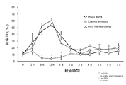
- Rats intraperitoneally administered with anti-neutrophil antibody or control antibody immediately after creation of crush injury (anti-PMN antibody and Ctrl antibody, respectively in the figure), and rats only subjected to crush injury (injury alone ) is a graph showing changes in the blood neutrophil count over time.
- 5 mm distal to the site of sciatic nerve injury 12 hours after crush injury in rats intraperitoneally administered anti-neutrophil antibody or control antibody immediately after crush injury and in rats only subjected to crush injury Representative HE-stained images of longitudinal sections in .
- FIG. 10 is a graph showing the number of neutrophils per wound for each distance from the injured site. 12 hours (upper) and 7 days (lower) after crush injury in rats intraperitoneally administered with anti-neutrophil antibody or control antibody immediately after crush injury, and in rats only subjected to crush injury.
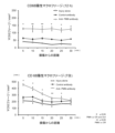
- Fig. 3 is a graph plotting the number of macrophages per unit area of nerve parenchyma detected in the sciatic nerve against the distance from the lesion.
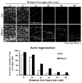
- the rate of sciatic nerve axonal regeneration 7 days after the crush injury was measured in rats intraperitoneally administered with an anti-neutrophil antibody or control antibody immediately after the crush injury, or in rats that received only the crush injury. It is a graph plotted against the distance from the part.
- Representative MBPs of sciatic nerve longitudinal sections 7 days after crush injury in rats intraperitoneally administered with anti-neutrophil antibody or control antibody immediately after crush injury and in rats with only crush injury Myelin Basic Protein
- the bottom row is a high-magnification image of the region corresponding to the epineurium (within the square frame of the corresponding top row image).
- the dashed line in the image indicates the boundary between the neuroparenchyma and the extraneuronal (epineurium), the area surrounded by the dashed line indicates the neuroparenchyma, and the arrows indicate neutrophils.
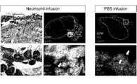
- the number of neutrophils per nerve unit area detected in the sciatic nerve 12 hours after the crush injury in mice that received intravenous administration of neutrophils or PBS and in mice that only received the crush injury was It is a graph represented for each distance from the part.
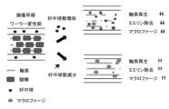
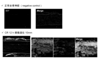
- FIG. 2 is a conceptual diagram of control of axonal regeneration process by neutrophils. Representative immunostaining images of an untreated normal rat sciatic nerve longitudinal section and a rat sciatic nerve longitudinal section 20 mm distal to the injury site 12 hours after creating a crush injury.
- Each image is an image of CitH3 immunostaining, Ly6G immunostaining, CD68 immunostaining, DAPI staining, or a merged image of a plurality of these.
- the upper center and upper right images are weakly magnified images (scale bar 100 ⁇ m), and the strongly magnified images in each rectangular frame are three images each (lower center row and lower right row) (scale bar 10 ⁇ m).
- the dashed line in the image indicates the boundary between the neuroparenchyma and the extraneuronium (epineurium), and the arrow indicates the co-localization of NETs and neutrophils.
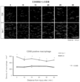
- 12 is a graph of representative CitH3 and Ly-6G immunostaining images and CitH3-positive areas of sciatic nerve longitudinal sections 20 mm distal to the injury site after 12 hours.
- the image on the left side is a weakly magnified image (scale bar 100 ⁇ m)
- the strongly magnified image in each square frame is an image on the right side (scale bar 10 ⁇ m).
- the dashed line in the image indicates the boundary between the neuroparenchyma and the extraneural (epineurium).
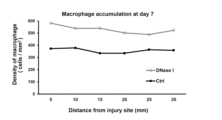
- the number of macrophages per unit area of nerve parenchyma detected in the sciatic nerve 7 days after the creation of the crush injury in rats treated with a sheet containing DNase I at a distance of 5 mm to 25 mm distal to the injury site was calculated as follows: It is a graph plotted against the distance from the part.
- the two upper images and the lower left image are weakly magnified images, and the three images on the lower right side are strongly magnified images of the region corresponding to the epineurium indicated by the rectangular frame in the lower left image.
- the image labeled "Merge" in the lower right is a merged image of MIF immunostaining, Ly-6G immunostaining and DAPI staining.
- the dashed line in the image indicates the demarcation line between the neuroparenchyma and extraneuronal parenchyma (epineurium).
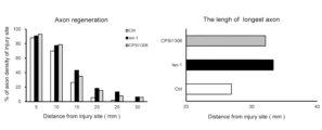
- Fig. 2 is a graph plotting the rate of axonal regeneration against the distance from the injury site (left panel) and a graph of the longest axon length (right panel). Representative immunostaining images of an untreated normal rat sciatic nerve longitudinal section and a rat sciatic nerve longitudinal section 20 mm distal to the injury site 12 hours after creating a crush injury.
- the second image from the left is CXCR4 immunostaining
- the third image from the left is Ly-6G immunostaining
- the other images are merged images of CitH3 immunostaining, Ly-6G immunostaining and DAPI staining.
- the four images on the left are weakly magnified images (scale bar 20 ⁇ m), and the strongly magnified images in the square frame are the images on the right (scale bar 10 ⁇ m).
- the dashed line in the image indicates the boundary between the neuroparenchyma and the extraneuronium (epineurium), and the arrow indicates the co-localization of CXCR4 and neutrophils.
- Representative ⁇ 3 tubulin immunostaining images of longitudinal sections of the sciatic nerve 7 days after the creation of the crush injury in rats treated with a sheet containing AMD3100, 5 mm to 25 mm distal to the injury site, and ⁇ 3 tubulin immunostaining (upper row) 2 is a graph (bottom row) showing the axonal regeneration rate of the sciatic nerve for each distance from the injury site.
- FIG. 2 is a conceptual diagram of control of axonal regeneration process by neutrophils via NETs.
- the present invention provides a pharmaceutical composition for treating peripheral nerve injury, containing a substance that suppresses the extraparenchymal formation of NETs in injured peripheral nerves.
- Neutrophils are a type of granulocyte, and after differentiation and maturation in the bone marrow, they are usually stored in the bone marrow (storage pool), peripheral blood (circulating pool), and blood vessel walls and tissues (marginal pool). There is When inflammation occurs due to tissue damage, neutrophils react to neutrophil migration-stimulating factors such as inflammatory cytokines and chemokines produced at the site of inflammation, accumulate at the site of injury, and participate in tissue repair and regeneration. It is known.
- NETs are mesh-like structures composed of chromatin containing citrullinated histone proteins and intracellular antibacterial proteins released by neutrophils. NETs are known to be involved in host defense and autoimmune diseases (Kely Campos Navegantes et al., Journal of Translational Medicine, 2017, vol. 15, no. 36, DOI 10.1186/s12967-017-1141- 8).
- neutrophils first accumulate outside the nerve parenchyma at the distal site of peripheral nerve injury, and then macrophages accumulate within the nerve parenchyma as neutrophils disappear. Inhibition of accumulation results in early and large accumulation of macrophages in the nerve parenchyma, promoting axon regeneration and myelin clearance. Conversely, increasing the number of accumulated neutrophils suppresses macrophage accumulation. It was also found that axonal regeneration and myelin clearance were suppressed.
- NETs exist outside the nerve parenchyma at the distal site of peripheral nerve injury, and that their localization overlaps with that of neutrophils. We found that it accumulated in the nerve parenchyma and promoted axonal regeneration. Agents that inhibit the formation of NETs are useful in treating peripheral nerve injury.
- the nerve parenchyma of the peripheral nerve means the inner region of the peripheral nerve surrounded by the epineurium, and the parenchyma includes nerve bundles formed by axons wrapped in the perineurium.
- Extraparenchymal of a nerve means the epineurium and its surrounding areas, especially the epineurium.
- Substances that inhibit the formation of NETs outside the parenchyma of injured peripheral nerves include substances that inhibit the accumulation of neutrophils in injured peripheral nerves, substances that inhibit the production of NETs by neutrophils, and substances that degrade NETs. can be mentioned.
- the substance that inhibits the accumulation of neutrophils in injured peripheral nerves used in the present disclosure has the ability to inhibit the accumulation of neutrophils in injured peripheral nerves.
- the group consisting of antibodies to neutrophils and antigen-binding fragments thereof, aptamers to neutrophils, inhibitors to neutrophil migration stimulating factors, and neutrophil migration inhibitory factors At least one substance is selected.
- An example of a substance that inhibits the accumulation of neutrophils in injured peripheral nerves is an antibody against neutrophils or an antigen-binding fragment thereof.
- Antibodies to neutrophils can bind to neutrophils, e.g., to proteins present on the cell surface of neutrophils, and reduce neutrophil numbers or inhibit neutrophil function. Any antibody may be used, and a specific antibody against neutrophils is preferable.
- Specific antibodies against neutrophils include, for example, antibodies against Ly-6G, a GPI-anchored protein of the Ly-6 family (also called myeloid cell differentiation antigen Gr-1), anti-PMN (polymorphonuclear neutrophil) antibodies, and anti-RP3 antibodies. etc.
- a specific antibody can also be referred to as an antibody that can preferentially bind a target protein over a non-target protein, or an antibody that has a high binding affinity for a target protein.
- a specific antibody binds to the target protein with an affinity that is at least 5 times stronger, preferably at least 10 times stronger, more preferably at least 100 times stronger, most preferably at least 1000 times stronger than it binds to the non-target protein. It can be an antibody.
- a specific antibody may also be an antibody that binds to the target protein with a dissociation constant of 10 ⁇ 7 M, preferably 10 ⁇ 8 M, more preferably 10 ⁇ 9 M.
- Antibodies used in the present disclosure can originate from any species including, for example, mouse, rat, shark, rabbit, pig, hamster, camel, llama, goat or human. Antibodies can also be of any class (eg IgG, IgE, IgM, IgD or IgA) and subclass of immunoglobulin molecule, but are preferably IgG.
- Antibodies can also be of any class (eg IgG, IgE, IgM, IgD or IgA) and subclass of immunoglobulin molecule, but are preferably IgG.
- the antibodies used in the present disclosure may be polyclonal antibodies or monoclonal antibodies.
- Antibodies can also be chimeric, humanized or human antibodies.
- an antigen-binding fragment of an antibody is defined as a partial fragment of an antibody that retains the ability to bind to the target protein.
- the antigen-binding fragment of an antibody is preferably a partial fragment of a specific antibody that retains the ability to specifically bind to the target protein, such as Fab (fragment of antigen binding), Fab', F(ab')2, They may be, but are not limited to, single chain antibodies (single chain Fv), disulfide stabilized antibodies (disulfide stabilized Fv), peptides containing CDRs, and the like.
- Antibodies and antigen-binding fragments thereof can be produced using methods known to those skilled in the art.
- a target protein is prepared by genetic recombination based on the amino acid sequence of the target protein or the nucleotide sequence of the DNA encoding it, and by immunizing a suitable animal with this as an antigen, the B cells of the animal are further transformed into myeloma. It can be produced by fusing with cells to obtain a hybridoma. (1997) Immunity 7:283-290; Wright et al. (2000) Immunity 13:233-242; Kaithamana et al. (1999) J. Immunol. 163:5157-5164 Please refer to Etc.
- Antibodies or antigen-binding fragments thereof may be antibody-drug conjugates further bound to other drugs.
- aptamer for neutrophils Another example of a substance that inhibits the accumulation of neutrophils in injured peripheral nerves is an aptamer for neutrophils.
- Aptamers to neutrophils can bind to neutrophils, e.g., to proteins present on the cell surface of neutrophils, and reduce neutrophil numbers or inhibit neutrophil function.
- An example of an aptamer to neutrophils is the aptamer to Ly-6G.
- Aptamers are molecules that have the ability to specifically bind to target substances due to their three-dimensional structure. Aptamers composed of nucleic acids are called nucleic acid aptamers, and aptamers composed of amino acids are called peptide aptamers. In the present disclosure, both nucleic acid aptamers and peptide aptamers can be used.
- the nucleic acid aptamers used in the present disclosure can be deoxyribonucleic acid (DNA), ribonucleic acid (RNA), or chimeric nucleic acids containing both deoxyribonucleotides and ribonucleotides as constituent units.
- Nucleic acid aptamers can be produced by methods known to those skilled in the art, such as the SELEX (systematic evolution of ligands by exponential enrichment) method.
- Peptide aptamers can be produced by methods known to those skilled in the art, such as phage display methods and cell surface display methods.
- IL-8 interleukin-8
- IL-17 IL-17
- Gro ⁇ Rowth-related gene products ⁇
- NAP-2 Neurotrophil Cytokines such as Activating Protein-2
- MIP-2 Macrophage Inflammatory Protein-2
- lipid mediators such as leukotriene B4, fMLP (N-formyl-methionyl-leucyl-phenylanine)
- complement components such as C5a
- a substance that exhibits inhibitory activity against a factor that promotes migration of spheres also called neutrophil migration stimulating factor, neutrophil migration factor, or neutrophil migration activating factor
- inhibitors include antibodies against neutrophil migration stimulating factors, antagonists against neutrophil migration stimulating factor receptors, and the like.
- substances that inhibit neutrophil recruitment to injured peripheral nerves are factors that inhibit neutrophil migration (also called neutrophil migration inhibitory factors) such as colchicine, resolvins, protectins, and 17 ⁇ -estradiol. be.
- NETs a substance that inhibits neutrophil NETs production, is involved in the citrullination of histone proteins by protein arginine deiminase 4 (PAD4) in the neutrophil nucleus, disaggregation of chromatin, and breakdown of the nuclear membrane. It is formed in the cytoplasm by the influx of the accompanying nuclear chromatin into the cytoplasm, the subsequent binding of the chromatin to the antibacterial protein in the granules, and is released outside the cell following the cell membrane disruption of neutrophils. Neutrophils that release NETs undergo cell death called NETosis.
- PAD4 protein arginine deiminase 4
- Substances that inhibit NETs production of neutrophils used in the present disclosure include substances that suppress the expression or function of MIF, substances that suppress the expression or function of CXCR4, and substances that suppress the expression of PAD4. or from the group consisting of substances that inhibit the function, i.e., a substance that suppresses the expression of MIF, a substance that inhibits the function of MIF, a substance that suppresses the expression of CXCR4, a substance that inhibits the function of CXCR4, and a substance that suppresses the expression of PAD4 and substances that inhibit the function of PAD4.
- MIF Glycosylation-Inhibiting Factor
- GIF Glycosylation-Inhibiting Factor
- L-dopachrome isomerase Phenylpyruvate tautomerase
- cytokines expressed in cytoplasm, involved in both innate and adaptive immunity, and is known to induce the production of inflammatory molecules.
- the nucleotide sequence of the human MIF gene has been registered with NCBI Reference Sequence as NC_000022.11, the mRNA nucleotide sequence as NM_002415.2, and the amino acid sequence as NP_002406.1 (all retrieved on July 19, 2021).
- a substance that suppresses the expression or inhibits the function of MIF is used in a pharmaceutical composition for treating peripheral nerve injury.
- a substance that suppresses the expression or functions of MIF used in the present disclosure is any substance that has the ability to suppress the expression of MIF at the gene or protein level or the ability to inhibit the function of MIF.
- it is at least one substance selected from the group consisting of nucleic acids that suppress MIF expression, antibodies against MIF and antigen-binding fragments thereof, aptamers against MIF, and MIF inhibitors.
- Nucleic acids that suppress MIF expression include nucleic acids that can suppress transcription of mRNA from the MIF gene (MIF mRNA), nucleic acids that can degrade MIF mRNA, and nucleic acids that can suppress protein translation from MIF mRNA. Examples of these include antisense nucleic acids or nucleic acids that cause RNA interference, which can be designed and produced by those skilled in the art based on the base sequence of the MIF gene or the base sequence of MIF mRNA. siRNA, shRNA or miRNA may be mentioned.
- An example of a substance that inhibits the function of MIF is an antibody against MIF, preferably a specific antibody against MIF, or an antigen-binding fragment of an antibody against MIF.
- Another example of substances that inhibit the function of MIF are aptamers against MIF. Definitions and other explanations of antibodies, specific antibodies, antigen-binding fragments and aptamers are as described above.
- MIF inhibitor is any substance that has the ability to inhibit the function of MIF.
- MIF inhibitors include ISO-1 (CAS No. 478336-92-4, (S,R)-3-(4-Hydroxyphenyl)-4,5-dihydro-5-isoxazole acetic acid), ( ⁇ )-CPSI 1306 (CAS No.
- the substance that suppresses the expression or functions of MIF is a MIF inhibitor, particularly ISO-1 or ( ⁇ )-CPSI 1306.
- CXCR4 is a G protein-coupled receptor belonging to the CXC motif-type chemokine receptor family. is known to induce Nucleotide sequence of human CXCR4 gene is NC_000002.12 in NCBI Reference Sequence, nucleotide sequence of mRNA is NM_001008540.2, NM_001348056.2, NM_001348059.2, NM_001348060.2, NM_003467.3, amino acid sequence is NP_001008540.1, NP_0010348540.1 .1, NP_001334988.1, NP_001334989.1, and NP_003458.1 (all retrieved December 20, 2021).
- CXCR4 exists outside the nerve parenchyma at the distal site of peripheral nerve injury, and its localization overlaps with that of neutrophils, and that inhibition of CXCR4 inhibits the production of NETs by neutrophils outside the nerve parenchyma. Inhibition of CXCR4 promoted the early and abundant accumulation of macrophages in the nerve parenchyma, promoting axonal regeneration. Substances that suppress the expression or inhibit the function of CXCR4 are useful in treating peripheral nerve injury.
- a substance that suppresses the expression or function of CXCR4 is any substance that has the ability to suppress the expression of CXCR4 at the gene or protein level or has the ability to inhibit the function of CXCR4, preferably CXCR4 , antibodies and antigen-binding fragments thereof against CXCR4, aptamers against CXCR4, and CXCR4 inhibitors.
- Nucleic acids that suppress CXCR4 expression include nucleic acids that can suppress transcription of mRNA from the CXCR4 gene (CXCR4 mRNA), nucleic acids that can degrade CXCR4 mRNA, and nucleic acids that can suppress protein translation from CXCR4 mRNA.
- Examples include nucleic acids that can be designed and produced by those skilled in the art based on the nucleotide sequence of the CXCR4 gene or the nucleotide sequence of CXCR4 mRNA, antisense nucleic acids, or nucleic acids that cause RNA interference, such as siRNA, shRNA or miRNA may be mentioned.
- CXCR4 One example of a substance that inhibits the function of CXCR4 is an antibody against CXCR4, preferably a specific antibody against CXCR4, or an antigen-binding fragment of an antibody against CXCR4.
- Another example of substances that inhibit the function of CXCR4 are aptamers against CXCR4. Definitions and other explanations of antibodies, specific antibodies, antigen-binding fragments and aptamers are as described above.
- a CXCR4 inhibitor is any substance capable of inhibiting the function of CXCR4.
- Examples of CXCR4 inhibitors include Plerixafor (also known as AMD3100; general formula 1,4-Bis[(1,4,8,11-tetraazacyclotetradecan-1-yl)methyl]benzene), WZ811 (CAS No.
- PAD4 is an enzyme that induces NETs formation by returning specific arginine residues of histone proteins (H3, H4, etc.) to citrulline residues.
- the nucleotide sequence of the human PAD4 gene has been registered in the NCBI Reference Sequence as NC_000001.11, the mRNA nucleotide sequence as NM_012387.3, and the amino acid sequence as NP_036519.2 (all retrieved on December 20, 2021).
- the present inventors found that inhibition of PAD4 inhibited the production of neutrophils' NETs outside the parenchyma of injured peripheral nerves, and that inhibition of PAD4 led to an early and abundant accumulation of macrophages in the nerve parenchyma. . Accumulation of macrophages in the nerve parenchyma can promote axonal regeneration, so substances that suppress the expression or function of PAD4 are useful for treating peripheral nerve damage.
- a substance that suppresses the expression or functions of PAD4 used in the present disclosure is any substance that has the ability to suppress the expression of PAD4 at the gene or protein level or has the ability to inhibit the function of PAD4.
- it is at least one substance selected from the group consisting of nucleic acids that suppress PAD4 expression, antibodies against PAD4 and antigen-binding fragments thereof, aptamers against PAD4, and PAD4 inhibitors.
- Nucleic acids that suppress PAD4 expression include nucleic acids that can suppress transcription of mRNA from the PAD4 gene (PAD4 mRNA), nucleic acids that can degrade PAD4 mRNA, and nucleic acids that can suppress protein translation from PAD4 mRNA. Examples of these include antisense nucleic acids or nucleic acids that cause RNA interference, which can be designed and produced by those skilled in the art based on the nucleotide sequence of the PAD4 gene or the nucleotide sequence of PAD4 mRNA, such as siRNA, shRNA or miRNA may be mentioned.
- PAD4 One example of a substance that inhibits the function of PAD4 is an antibody against PAD4, preferably a specific antibody against PAD4, or an antigen-binding fragment of an antibody against PAD4.
- Another example of substances that inhibit the function of PAD4 are aptamers against PAD4. Definitions and other explanations of antibodies, specific antibodies, antigen-binding fragments and aptamers are as described above.
- PAD4 inhibitors are PAD4 inhibitors.
- a PAD4 inhibitor is any substance that has the ability to inhibit the function of PAD4.
- PAD4 inhibitors include Cl-amidine ( N2 - Benzoyl-N5-(1-imino-2-chloroethyl)-L-ornithinamide/trifluoroacetic acid), BMS-P5 (no generic name, Cas #, 1550371 -22-6), GSK121 trifluoroacetate (no generic name, Cas #, 1652591-80-4), and the like.
- DNA capable of inducing transcription of an antisense nucleic acid or a nucleic acid that causes RNA interference against any of the above MIF, CXCR4 and PAD4 by being placed under the control of an appropriate expression promoter and these antisense nucleic acids or Nucleic acids in which part of the nucleotide residues constituting nucleic acids that cause RNA interference are modified are also included in the expression-suppressing nucleic acids of the present disclosure as nucleic acids that are functionally equivalent to antisense nucleic acids or nucleic acids that cause RNA interference. be done. Modifications of nucleotide residues include 2'O-methylation, 2'-Fation, 4'-thiolation, and the like.
- chimeric RNAs in which part of the ribonucleotides of the expression-suppressing RNA are replaced with corresponding deoxyribonucleotides or nucleotide analogues are also included in the expression-suppressing nucleic acids of the present disclosure.
- Nucleotide analogs include, for example, 5-position modified uridine or cytidine, such as 5-(2-amino)propyluridine, 5-bromouridine, etc.; 8-position modified adenosine or guanosine, such as 8-bromoguanosine; deaza nucleotides, Examples include 7-deaza-adenosine and the like; O- or N-alkylated nucleotides such as N6-methyladenosine and the like. There are no particular restrictions on the type or number of modified or substituted bases, as long as they do not lose the ability to suppress the expression of the target molecule.
- Nucleic acids that suppress expression can be artificially synthesized using genetic recombination technology or chemical synthesis technology. Genetic recombination methods, methods for chemically synthesizing nucleic acids, methods for synthesizing unnatural bases, and methods for synthesizing nucleic acids containing these are well known to those skilled in the art.
- substances that inhibit NETs production by neutrophils include lactoferrin described in WO2014/168253; thrombin-like enzymes such as batroxobin, ancrod, crotalase, etc. described in WO2018/131724; and Btk described in WO2019/208805.
- Compounds with inhibitory activity e.g. Immunoglobulins such as sulfonated human immunoglobulin G (Uozumi et al., Modern Rheumatology, 2020, 30(3):544-550.
- PA-dPEG24 peptide: C-terminal of IALILEPICCQERAA PEG-modified peptide with a 24-mer PEG moiety
- Prostaglandin E2 inhibitor of NETosis through cAMP -PKA
- degrading NETs causes macrophages to accumulate early and in large amounts in the damaged peripheral nerve parenchyma, promoting axonal regeneration. Agents that degrade NETs are useful in treating peripheral nerve injury.
- a substance that degrades NETs is any substance that has the ability to degrade chromatin or antibacterial proteins that constitute NETs, especially chromatin. 2013. 342(6160): 863-866. doi: 10.1126/science.1242255).
- compositions comprise an amount of a substance that inhibits extraparenchymal formation of NETs in injured peripheral nerves in an effective amount for treating peripheral nerve injury.
- the pharmaceutical composition contains an effective amount for treating peripheral nerve injury, a substance that inhibits the accumulation of neutrophils in injured peripheral nerves, or a substance that inhibits the expression or function of MIF.
- the effective dose is appropriately determined according to the usage, subject's age, sex, body weight, site and degree of peripheral nerve damage, and other factors.
- the pharmaceutical composition may contain at least one substance selected from the group consisting of substances that inhibit the accumulation of neutrophils in damaged peripheral nerves, substances that suppress the expression of MIF, and substances that inhibit the function of MIF. . That is, the pharmaceutical composition may contain one or more substances that inhibit the accumulation of neutrophils in injured peripheral nerves, may contain one or more substances that suppress the expression of MIF, or It may contain a substance that inhibits the functions of multiple types of MIF.
- the pharmaceutical composition may contain one or more substances that suppress the expression of CXCR4, may contain one or more substances that inhibit the function of CXCR4, and may contain one or more substances that inhibit the expression of PAD4. , may contain one or more substances that inhibit the function of PAD4, and may contain one or more substances that degrade NETs.
- the pharmaceutical composition can contain other agents for the treatment of peripheral nerve damage, or pharmaceutically acceptable additives such as buffers, stabilizers, preservatives, excipients, and the like.
- pharmaceutically acceptable additives are well known to those skilled in the art, and can be appropriately selected and used within the scope of the ordinary ability of those skilled in the art.
- treatment includes all types of medically acceptable therapeutic intervention aimed at curing, temporary remission, etc. of a disease or condition. Therefore, the treatment of peripheral nerve injury includes medical treatment for various purposes, including promotion of regeneration of damaged peripheral nerves, improvement of functional impairment due to peripheral nerve injury, delay or halt of progression of functional impairment, prevention of onset, etc. Includes permissible interventions.
- the pharmaceutical composition can be used for peripheral nerve damage caused by axonal rupture, and is particularly preferably used for severe peripheral nerve damage that cannot be expected to recover spontaneously.
- a preferred example of peripheral nerve injury that can be treated with the pharmaceutical composition is traumatic peripheral nerve injury (wound, compression, traction, amputation, defect, iatrogenic, etc.).
- the pharmaceutical composition can be used in subjects with peripheral nerve damage, e.g., rodents including mice, rats, hamsters, guinea pigs, primates including humans, chimpanzees and rhesus monkeys, livestock including pigs, cattle, goats, horses, sheep, dogs. , to mammals such as companion animals, including cats.
- peripheral nerve damage e.g., rodents including mice, rats, hamsters, guinea pigs, primates including humans, chimpanzees and rhesus monkeys, livestock including pigs, cattle, goats, horses, sheep, dogs.
- mammals such as companion animals, including cats.
- a preferred subject is a human.
- the pharmaceutical composition can suppress the extraparenchymal formation of NETs in the injured peripheral nerve, for example, as long as it can inhibit the accumulation of neutrophils in the injured peripheral nerve, or the expression of MIF in the injured peripheral nerve
- the dosage form and administration method as long as it can suppress or inhibit the function.
- the pharmaceutical composition can be prepared in the form of a solution and administered intravenously or intraperitoneally.
- the pharmaceutical composition may be prepared in the form of a solution, which may be used as it is or impregnated with a biocompatible medical material by placing it around the distal side of the injured peripheral nerve.
- placement may be performed, for example, by injection of a solution agent around the distal side of the injured peripheral nerve, impregnating the solution agent with a biocompatible medical material, which is applied distally to the injured peripheral nerve. It may be done by placing around the sides.
- the pharmaceutical composition contains a substance that suppresses extraparenchymal formation of NETs in injured peripheral nerves, such as a substance that inhibits accumulation of neutrophils in injured peripheral nerves, or suppresses the expression or function of MIF. It can also be used by preparing a form of a biocompatible medical material that retains the inhibitory substance and placing this biocompatible medical material around the distal side of the injured peripheral nerve.
- any method can be used to hold the substance on the biocompatible medical material.
- the substance may be directly fixed to the biocompatible medical material, or fixed via an appropriate linker molecule or adapter molecule. Alternatively, it may be fixed using a binder or coating agent. Said substance may be retained on the biocompatible medical material by non-specific adsorption.
- the biocompatible medical material when the biocompatible medical material is a water-absorbing material, the biocompatible medical material may retain the substance by impregnating the biocompatible medical material with a liquid containing the substance. Furthermore, when the biocompatible medical material is a gel material, the substance may be retained in the biocompatible medical material by including the substance in the raw material solution during the gel preparation process.
- a biocompatible medical material containing a substance that inhibits the formation of NETs outside the parenchyma of injured peripheral nerves such as a substance that inhibits the accumulation of neutrophils in injured peripheral nerves or suppresses the expression of MIF
- biocompatible medical materials include polymeric compounds such as polyethylene fluoride and polystyrene, inorganic compounds such as silica, and biodegradable polymers, with biodegradable polymers being preferred.
- biodegradable polymers include synthetic polymer materials such as polyglycolic acid, polylactic acid, polyethylene glycol, polycaprolactone, polydioxanone, and other copolymers such as lactic acid-glycolic acid copolymers; and inorganic materials such as ⁇ -phosphorus.
- Natural polymer materials include collagen, gelatin, alginic acid, hyaluronic acid, agarose, chitosan, fibrin, fibroin, chitin, cellulose, silk, and the like.
- the shape of the biocompatible medical material may be any shape suitable for placement around the distal side of the injured peripheral nerve, such as sheet-like or gel-like.
- a sheet-shaped medical material can be used by covering the distal portion of a peripheral nerve injury site.
- a gel-like medical material such as fibrin glue, alginic acid, etc. can be used by spraying or coating around the distal side of the injured peripheral nerve, or by indwelling on the distal side of the injured area.
- the pharmaceutical composition was administered at a time from immediately after peripheral nerve injury until neutrophils accumulated outside the nerve parenchyma distal to the injury site and then disappeared.
- Axonal regeneration can be promoted by acting on the distal side of peripheral nerve injury that causes Wallerian degeneration for a relatively short period of time. Therefore, the pharmaceutical composition need not be present throughout the injured peripheral nerve, nor need it be present throughout the period immediately following peripheral nerve injury until axonal regeneration is complete.
- the pharmaceutical composition is administered after peripheral nerve injury anywhere within the period during which neutrophils accumulate outside the nerve parenchyma distal to the injury, e.g.
- distal to the injured peripheral nerve anywhere within the period of time during which neutrophils accumulate outside the nerve parenchyma and then disappear It can be placed and used in the periphery, particularly in the vicinity of the distal side of the injured site.
- the pharmaceutical composition may be administered immediately after, 3 hours, 6 hours or 9 hours after peripheral nerve injury, 12 hours, 15 hours, 18 hours, 21 hours or 24 hours after peripheral nerve injury. It can be used by placing it around the distal side of the injured peripheral nerve for a relatively short period of time during the post-tinal period.
- the pharmaceutical composition can be used for a period from immediately after peripheral nerve injury to 24 hours, from 3 hours to 18 hours, from 6 hours to 15 hours, or from 9 hours to 12 hours after peripheral nerve injury. It can be used by placing it around the distal side of the injured peripheral nerve for a relatively short period of time.
- peripheral nerve reconstruction, suturing, etc. are performed after some time has passed after injury, the surgical invasion may cause neutrophil infiltration in the same way as immediately after peripheral nerve injury.
- the pharmaceutical composition is administered after a peripheral nerve injury, somewhere during the period when neutrophils are accumulating outside the nerve parenchyma distal to the injury, for example immediately after surgical insult to distal to the injury.
- the distal side of the injured peripheral nerve For a relatively short period of time, e.g., 30 minutes to 7 days, around the distal side of the injured peripheral nerve, anywhere during the period of neutrophil accumulation and subsequent disappearance outside the nerve parenchyma, In particular, it can be used by arranging it around the vicinity of the distal side of the injured part.
- the pharmaceutical composition can be administered immediately after, 3 hours, 6 hours or 9 hours after surgical insult, and 12 hours, 15 hours, 18 hours, 21 hours or 24 hours after surgical insult. It can be used by placing it around the distal side of the injured peripheral nerve for a relatively short period of time within the terminal period.
- the pharmaceutical composition can be used for a period from immediately after surgery to 24 hours, from 3 hours to 18 hours, from 6 hours to 15 hours, or from 9 hours to 12 hours after surgery. It can be placed around the distal side of the injured peripheral nerve for a relatively short period of time within the period of .
- the time the pharmaceutical composition is placed around the distal side of the peripheral nerve injury site can be, for example, 30 minutes or more, 60 minutes or more, or 90 minutes or more, and can be 7 days or less, 24 hours or less, 12 hours or less, 6 It can be an hour or less, 4 hours or less, or 3 hours or less.
- the pharmaceutical composition is used by placing it around the distal side of the peripheral nerve injury for 30 minutes to 3 hours within a period of 9 hours to 12 hours after peripheral nerve injury. obtain.
- the pharmaceutical composition can exert its effect even by such a temporally and spatially limited treatment, for example, when performing nerve suturing in the case of peripheral nerve injury. It is also possible to place the pharmaceutical composition around the nerve, perform nerve suture, and remove the pharmaceutical composition after about several tens of minutes to several hours, for example, about 30 minutes to 3 hours have passed since the placement.
- the present invention provides a subject with peripheral nerve damage with an effective amount of a pharmaceutical composition comprising a substance that inhibits the formation of extraparenchymal NETs in the damaged peripheral nerve.
- a method of treating peripheral nerve injury comprising applying a pharmaceutical composition comprising a substance that inhibits accumulation or a substance that inhibits the expression or function of MIF.
- the application is preferably performed by placing the pharmaceutical composition around the periphery of the peripheral nerve distally, particularly near the distally injured site.
- the present invention also provides, in one aspect, a substance that inhibits the formation of NETs extraparenchymal in injured peripheral nerves for use in treating peripheral nerve injury.
- the present invention further provides, in one aspect, use of a substance that suppresses extraparenchymal formation of NETs in injured peripheral nerves in the manufacture of a pharmaceutical composition. Definitions and other explanations for each term are as described above.
- the present invention provides a method for evaluating the peripheral nerve regeneration-promoting activity of a test substance, using the activity of suppressing the formation of extraparenchymal NETs in injured peripheral nerves as an index.
- Activities to suppress the formation of NETs outside the parenchyma of injured peripheral nerves include activity to inhibit accumulation of neutrophils in injured peripheral nerves; activity to inhibit NETs production by neutrophils, such as suppression of MIF expression or function-inhibiting activity, CXCR4 expression-inhibiting or function-inhibiting activity, and PAD4 expression-inhibiting or function-inhibiting activity; and NETs-degrading activity.
- the evaluation method using the activity of inhibiting the accumulation of neutrophils in damaged peripheral nerves as an index is in vitro, in the presence and absence of a test substance, respectively, the survival rate of neutrophils or It includes determining the peripheral nerve regeneration-promoting activity of the test substance by comparing the migration ability.
- a test substance that reduces the survival rate or migration ability of neutrophils can be determined to have peripheral nerve regeneration-promoting activity.
- the evaluation method using the activity of inhibiting the accumulation of neutrophils in damaged peripheral nerves as an indicator is in vivo, the blood neutrophil count in the presence and absence of a test substance Alternatively, by comparing the number of neutrophils accumulated in peripheral nerve injury sites, it includes determining the peripheral nerve regeneration-promoting activity of the test substance. In this embodiment, a test substance that reduces the number of neutrophils in blood or the number of neutrophils accumulated in peripheral nerve injury sites can be determined to have peripheral nerve regeneration-promoting activity.
- the evaluation method using the activity of inhibiting NETs production of neutrophils as an index is in vitro, in the presence and absence of a test substance, and any one of MIF, CXCR4, or PAD4 genes ( hereinafter referred to as an indicator gene) to determine the peripheral nerve regeneration-promoting activity of the test substance.
- the evaluation method includes, for example, a test substance, a cell containing a nucleic acid having a marker gene under the control of a promoter region that controls the expression of an indicator gene, and conditions under which expression of the marker gene by the promoter is induced.
- the marker gene is not particularly limited as long as the expression induction of the gene can be detected, for example, a gene encoding a fluorescent protein such as GFP or a chemiluminescent protein such as luciferase, or a drug resistance gene or auxotrophy of cells Genes that confer a phenotypic change on a cell, such as genes that can complement , can be used.
- Cells containing a nucleic acid having a marker gene under the control of the promoter region of the indicator gene for example, originally, the nucleotide sequence of the open reading frame region of the indicator gene in the cell having the indicator gene is combined with the nucleotide sequence encoding the marker gene. It can be produced by recombination with Also, based on the human genome base sequence information, an expression vector containing the promoter region of a human indicator gene and a marker gene recombined under its control is constructed, and the expression vector is introduced into an appropriate cell. can also
- Incubation can be performed by coexisting the cells and the test substance in a medium or buffer solution in an appropriate container. Conditions such as incubation temperature and time may be conditions that allow the cell to express the marker gene. In addition, marker gene-derived signals can be measured by a method suitable for the marker gene used.
- the evaluation method is to compare the expression of an indicator gene or the expression of any protein of MIF, CXCR4 or PAD4 (hereinafter referred to as an indicator protein) in the presence and absence of a test substance. and determining the peripheral nerve regeneration-promoting activity of the test substance.
- the evaluation method includes, for example, the step of incubating a test substance with cells capable of expressing an indicator gene or indicator protein; measuring the intensity of the indicator gene- or indicator protein-derived signal in the cells after incubation comparing the measured intensity of the indicator gene- or indicator protein-derived signal with the intensity of the indicator gene- or indicator protein-derived signal in the absence of the test substance; and reducing the intensity of the indicator gene- or indicator protein-derived signal
- a step of determining that the test substance has peripheral nerve regeneration-promoting activity may be included.
- a cell capable of expressing an indicator gene or indicator protein preferably expresses a human indicator gene or indicator protein.
- a cultured cell line into which a human indicator gene, such as a human MIF gene, is integrated can be used.
- Incubation can be performed by coexisting the cells and the test substance in a medium or buffer solution in an appropriate container.
- Conditions such as incubation temperature and time may be conditions that allow cells to express the indicator gene or indicator protein.
- Signals derived from indicator genes can be measured by general methods capable of detecting and quantifying specific gene expression, such as hybridization using base sequence information of indicator genes, quantitative PCR, and RNA-Seq. .
- the signal derived from the indicator protein should be measured by a general method that can detect and quantify the expression of a specific protein, such as ELISA using a specific antibody against the indicator protein, or an immunological detection method such as Western blotting. can be done.
- a test substance that reduces the intensity of an indicator gene- or indicator protein-derived signal in cells that have been incubated with the test substance compared to cells that have not been incubated with the test substance under conditions that induce the expression of the indicator gene or protein can be determined to have peripheral nerve regeneration promoting activity.
- the evaluation method using the activity of inhibiting NETs production by neutrophils as an index compares MIF function, CXCR4 function, or PAD4 function in the presence and absence of a test substance. By doing so, it includes determining the peripheral nerve regeneration promoting activity of the test substance. For example, in an evaluation method that uses the activity of inhibiting NETs production by neutrophils as an index, by comparing the macrophage migration inhibitory activity of MIF in the presence and absence of the test substance, peripheral nerve regeneration of the test substance was evaluated. including determining stimulatory activity. In this embodiment, a test substance that has been determined to have activity to inhibit MIF function can be evaluated as having activity to promote peripheral nerve regeneration. A comparison of MIF activity in the presence and absence of a test substance can be performed using methods employed in evaluation or screening of the inhibitory action of known MIF inhibitors.
- the evaluation method using the NETs-degrading activity as an indicator is to compare the degradation of NETs in the presence and absence of the test substance, thereby determining the peripheral nerve regeneration-promoting activity of the test substance. Including judging.
- the evaluation method includes, for example, a step of incubating a test substance with NETs produced by neutrophils; a step of measuring the intensity of the NETs-derived signal after incubation; A step of comparing the intensity of the NETs-derived signal with the intensity of the NETs-derived signal in the absence of the test substance; and a step of determining that the test substance that reduced the intensity of the NETs-derived signal has peripheral nerve regeneration-promoting activity.
- a NETs-derived signal is, for example, a signal derived from a known NETs marker molecule such as citrullinated histone H3, and can be measured by a method suitable for the molecule, such as an immunological detection method.
- a test substance determined to have NETs-degrading activity can be evaluated as having peripheral nerve regeneration-promoting activity.
- the animal's right atrial appendage was incised under anesthesia to exsanguinate blood, and PBS solution was poured into the left heart to exsanguinate blood.
- PBS solution was poured into the left heart to exsanguinate blood.
- GFP-labeled neutrophils used in Test Example 2 were prepared as follows. Bone marrow cells of the femur and crus of C57BL/6 GFP mice (8-10 weeks old) were expelled with PBS and harvested. Red blood cells contained in bone marrow cells were lysed with RBC lysis buffer (Funakoshi) to purify bone marrow cells. Neutrophils were isolated from bone marrow cells using the Neutrophil isolation kit, mouse (Miltenyi Biotec).
- Test Example 1 Spatiotemporal distribution analysis of neutrophils and macrophages in peripheral nerve injury site (1) Neutrophils A rat sciatic nerve was crushed and 12 hours later, the sciatic nerve was excised. Staining and immunohistochemical staining were performed. Representative HE-stained images from 5 mm proximal (-5 mm) to 25 mm distal from the injury site are shown in Figure 1, and immunostained images are shown in Figure 2.
- extraparenchymal is used synonymously with “epineurium”
- 15 mm and In Wallerian degeneration 25 mm distal it was absent in the nerve parenchyma, but only extraparenchymal.
- sciatic nerves were excised from rats subjected to crush injury 1 hour, 6 hours, 12 hours, 1 day, 3 days, or 7 days after injury. Immunohistochemical staining of the sciatic nerve was performed in the same manner as above, and CD68-positive cells present in the nerve parenchyma were counted as macrophages. The number of macrophages distal to the injury site increased 3 days after injury and peaked 7 days after injury (Fig. 6). Like neutrophils, macrophages accumulated in the epineurium 12 hours after injury (Fig. 7).
- Example 1 Evaluation of the effect of anti-neutrophil antibody on nerve regeneration process
- PMN polymorphonuclear
- the sciatic nerve was excised, and longitudinal sections were subjected to HE staining and immunohistochemical staining. The number of neutrophils in the nerve and the number of macrophages in the nerve parenchyma were counted from the stained image, and the axonal regeneration rate was calculated. In addition, the area of the MBP-positive region in the MBP-immunostained image was divided by the total area to calculate the MBP-positive rate, which is an index of the amount of axonal debris.
- Fig. 8 shows the time course of the blood neutrophil count.
- the number of neutrophils in blood peaked at 12 hours after injury, and neutrophil recruitment to the circulating pool was observed.
- the blood neutrophil count did not change significantly after injury.
- Figure 9 shows a representative HE-stained image 5 mm distal to the injury site 12 hours after injury
- Figure 10 shows the distribution of neutrophils distal to the injury site.
- the number of neutrophils observed distal to the injured site in the anti-neutrophil antibody-administered group was lower than in the injury alone group and the control antibody-administered group.
- Fig. 11 shows the distribution of macrophages distal to the sciatic nerve injury 12 hours and 7 days after injury.
- the antineutrophil antibody-administered group accumulation of many macrophages was observed earlier than in the injury alone group and the control antibody-administered group.
- Fig. 12 shows typical ⁇ 3 tubulin immunostaining images from 5 mm to 30 mm distal to the injury site 7 days after injury
- Fig. 13 shows the axonal regeneration rate.
- the antineutrophil antibody-administered group showed a higher axonal regeneration rate than the injury alone group and the control antibody-administered group.
- Fig. 14 shows representative MBP immunostaining images and MBP positive rates 7 days after injury.
- the MBP positive rate was lower and the amount of myelin sheath debris decreased compared to the injury alone group and the control antibody-administered group.
- This example shows that reducing the number of neutrophils that accumulate in Wallerian degeneration sites with anti-neutrophil antibodies increases the accumulation of macrophages, promotes axonal regeneration, and promotes myelin clearance. confirmed.
- the sciatic nerve was removed, and HE staining and immunohistochemical staining of cross sections at 4 mm, 6 mm, 8 mm, and 10 mm distal to the injury site were performed.
- the number of neutrophils in the nerve and the number of macrophages in the nerve parenchyma were counted, and the axonal density and the MBP positive rate were calculated.
- Fig. 15 shows the blood neutrophil count 12 hours after injury
- Fig. 16 shows typical HE-stained images and immunostained images 4 mm distal to the injury site
- Fig. 17 shows the spatial distribution of neutrophils.
- the neutrophil-administered group the number of blood neutrophils increased compared to the injury alone group and the PBS-administered group, and the number of neutrophils distal to the injury site also increased.
- Fig. 18 shows the distribution of macrophages distal to the sciatic nerve injury 7 days after injury.
- the number of macrophages distal to the injury site was lower in the neutrophil-administered group than in the injury alone group and the PBS-administered group.
- FIG. 19 A typical ⁇ 3 tubulin immunostaining image 7 days after injury is shown in Fig. 19, and the axonal density is shown in Fig. 20.
- the axonal density increased closer to the injury site, but the axonal density was generally lower in the neutrophil-administered group.
- a typical MBP immunostaining image 7 days after injury is shown in Figure 21, and the MBP positive rate is shown in Figure 22.
- the MBP positive rate was higher and the amount of myelin sheath debris increased compared to the injury alone group and the PBS-administered group.
- Fig. 23 shows a conceptual diagram of the control of the axonal regeneration process by neutrophils, which is inferred from Test Examples 1 and 2 and Example 1.
- Example 2 Evaluation of NETs formation in the epineurium of Wallerian degeneration and the effect of suppression of NETs formation on nerve regeneration process
- the sciatic nerve was removed and longitudinal sections 20 mm distal to the injury site were prepared for immunohistochemical staining for CitH3, Ly-6G and CD68. The epineurium portion was observed, and the ratio of the area with CitH3 staining to the area of the entire observation field was calculated. In addition, the number of macrophages per unit area within the nerve parenchyma was quantified.
- Fig. 25 shows representative CitH3 and Ly-6G immunostaining images and CitH3-positive areas
- Fig. 26 shows representative CD68 immunostaining images and the number of macrophages.
- a large amount of NETs were found in the epineurium in the control sheet group, but only a few NETs were detected in the Cl-amidine-containing sheet group and the DNase I-containing sheet group.
- accumulation of more macrophages was observed in the nerve parenchyma compared to the control sheet group.
- a representative ⁇ 3 tubulin immunostained image is shown in the upper part of FIG. 27, the axonal regeneration rate is shown in the lower part of FIG. 27, and the number of macrophages is shown in FIG.
- the DNase I-containing sheet group showed a higher axonal regeneration rate over almost the entire evaluated area distal to the injured area, and more macrophages were accumulated in the nerve parenchyma.
- Example 3 Evaluation of the effect of inhibition of MIF on nerve regeneration process
- the sciatic nerve was removed, longitudinally sectioned and immunohistochemically stained for CitH3, Ly-6G and CD68. The epineurium portion was observed, and the ratio of the area with CitH3 staining to the area of the entire observation field was calculated. In addition, the number of macrophages per unit area within the nerve parenchyma was quantified.
- Figure 30 shows the CD68 immunostaining image and macrophage count at 5 mm distal to the sciatic nerve injury site 12 hours after injury
- Figure 31 shows the CD68 immunostaining image and macrophage count at 20 mm distal to the sciatic nerve injury site.
- FIG. 32 shows CitH3 and Ly-6G immunostaining images and CitH3-positive areas in the 20 mm distal region. There was no difference in macrophage accumulation between the iso-1 containing sheet group and the control sheet group at 5 mm distal to the injury site, which was not treated with the sheet. On the other hand, more macrophages were accumulated in the nerve parenchyma in the iso-1 containing sheet group than in the control sheet group at 20 mm distal to the injury site treated with the sheet. In the iso-1 containing sheet group, only a few NETs were detected in the epineurium 20 mm distal to the injury site.
- FIG. 33 A representative CD68 immunostained image and the number of macrophages are shown in FIG. 33, and a CD163 immunostained image and the number of M2 macrophages are shown in FIG. A greater accumulation of macrophages was observed in the nerve parenchyma in the iso-1-containing sheet group compared to the control sheet group throughout the evaluated lesion distal to the lesion.
- FIG. 1 Representative ⁇ 3 tubulin immunostaining images and axonal regeneration rate are shown in FIG.
- the iso-1-containing sheet group showed a higher rate of axonal regeneration than the control sheet group over almost the entire distal injury site evaluated.
- FIG. 36 A typical ⁇ 3 tubulin immunostaining image is shown in FIG. 36, and the axon regeneration rate and longest axon length are shown in FIG.
- the iso-1-containing sheet group and the CPSI-1306-containing sheet group showed a higher rate of axonal regeneration over almost the entire distal area of the injured site, and the longest axonal length was greater than that of the control sheet group.
- Example 4 Evaluation of Effect of CXCR4 Inhibition on Nerve Regeneration Process (1) Distribution of CXCR4 Longitudinal sections of sciatic nerve isolated from untreated rats and sciatic nerves isolated from rats 12 hours after creation of crush injury , immunohistochemical staining for CXCR4 and Ly-6G. No expression of CXCR4 was confirmed in the normal sciatic nerve, but extraparenchymal expression of CXCR4 was observed in the sciatic nerve 12 hours after injury creation, and CXCR4 was double-stained with neutrophils (Fig. 38). This suggests that CXCR4 is produced by accumulated neutrophils outside the nerve parenchyma of the Wallerian degeneration site 12 hours after injury.
- the sciatic nerve was removed and longitudinal sections 20 mm distal to the injury site were prepared for immunohistochemical staining for CitH3, Ly-6G and CD68. The epineurium portion was observed, and the ratio of the area with CitH3 staining to the area of the entire observation field was calculated. In addition, the number of macrophages per unit area within the nerve parenchyma was quantified.
- Fig. 39 shows representative CitH3 and Ly-6G immunostaining images and CitH3-positive areas
- Fig. 40 shows representative CD68 immunostaining images and macrophage counts.
- AMD3100-containing sheet group only a few NETs were detected in the epineurium, and more macrophages were accumulated in the nerve parenchyma than in the control sheet group.
- a representative ⁇ 3 tubulin immunostained image is shown in the upper part of FIG. 41, the axonal regeneration rate is shown in the lower part of FIG. 41, and the number of macrophages is shown in FIG.
- the CXCR4-containing sheet group showed a higher rate of axonal regeneration over almost the entire area distal to the injured site, and more macrophages were accumulated in the nerve parenchyma.
- FIG. 43 shows a conceptual diagram of the control of the axonal regeneration process by neutrophils, which is inferred from Test Examples 1 and 2 and Examples 1-4.
Landscapes
- Health & Medical Sciences (AREA)
- Life Sciences & Earth Sciences (AREA)
- Chemical & Material Sciences (AREA)
- General Health & Medical Sciences (AREA)
- Engineering & Computer Science (AREA)
- Organic Chemistry (AREA)
- Genetics & Genomics (AREA)
- Medicinal Chemistry (AREA)
- Biomedical Technology (AREA)
- Public Health (AREA)
- Veterinary Medicine (AREA)
- Animal Behavior & Ethology (AREA)
- Pharmacology & Pharmacy (AREA)
- Bioinformatics & Cheminformatics (AREA)
- Molecular Biology (AREA)
- Epidemiology (AREA)
- Biochemistry (AREA)
- Biophysics (AREA)
- Biotechnology (AREA)
- Wood Science & Technology (AREA)
- General Engineering & Computer Science (AREA)
- Zoology (AREA)
- Immunology (AREA)
- Proteomics, Peptides & Aminoacids (AREA)
- Physics & Mathematics (AREA)
- Microbiology (AREA)
- Plant Pathology (AREA)
- Neurosurgery (AREA)
- Neurology (AREA)
- Chemical Kinetics & Catalysis (AREA)
- General Chemical & Material Sciences (AREA)
- Nuclear Medicine, Radiotherapy & Molecular Imaging (AREA)
- Gastroenterology & Hepatology (AREA)
- Hospice & Palliative Care (AREA)
- Endocrinology (AREA)
- Psychiatry (AREA)
- Medicines That Contain Protein Lipid Enzymes And Other Medicines (AREA)
- Medicines Containing Antibodies Or Antigens For Use As Internal Diagnostic Agents (AREA)
Abstract
Priority Applications (3)
| Application Number | Priority Date | Filing Date | Title |
|---|---|---|---|
| JP2023538221A JPWO2023007779A1 (fr) | 2021-07-30 | 2022-02-10 | |
| US18/293,199 US20250074976A1 (en) | 2021-07-30 | 2022-02-10 | Pharmaceutical composition for treating peripheral nerve injury |
| EP22848863.1A EP4393507A4 (fr) | 2021-07-30 | 2022-02-10 | Composition pharmaceutique pour le traitement d'une lésion du nerf périphérique |
Applications Claiming Priority (2)
| Application Number | Priority Date | Filing Date | Title |
|---|---|---|---|
| JP2021126207 | 2021-07-30 | ||
| JP2021-126207 | 2021-07-30 |
Publications (1)
| Publication Number | Publication Date |
|---|---|
| WO2023007779A1 true WO2023007779A1 (fr) | 2023-02-02 |
Family
ID=85086454
Family Applications (1)
| Application Number | Title | Priority Date | Filing Date |
|---|---|---|---|
| PCT/JP2022/005504 Ceased WO2023007779A1 (fr) | 2021-07-30 | 2022-02-10 | Composition pharmaceutique pour le traitement d'une lésion du nerf périphérique |
Country Status (4)
| Country | Link |
|---|---|
| US (1) | US20250074976A1 (fr) |
| EP (1) | EP4393507A4 (fr) |
| JP (1) | JPWO2023007779A1 (fr) |
| WO (1) | WO2023007779A1 (fr) |
Citations (6)
| Publication number | Priority date | Publication date | Assignee | Title |
|---|---|---|---|---|
| WO2005074655A2 (fr) | 2004-01-30 | 2005-08-18 | Emory University | Materiaux et procede favorisant la regeneration nerveuse |
| WO2014168253A1 (fr) | 2013-04-09 | 2014-10-16 | 国立大学法人東京大学 | Inhibiteur de la formation de pièges extracellulaires dans les leucocytes |
| WO2018056412A1 (fr) | 2016-09-23 | 2018-03-29 | 国立大学法人大阪大学 | Promoteur de la différenciation des cellules de schwann et promoteur de la régénération des nerfs périphériques |
| WO2018131724A1 (fr) | 2017-01-13 | 2018-07-19 | Tobishi Pharmaceutical Co., Ltd. | Régulateur d'activation des neutrophiles |
| WO2019208805A1 (fr) | 2018-04-27 | 2019-10-31 | 小野薬品工業株式会社 | AGENT PRÉVENTIF ET/OU THÉRAPEUTIQUE POUR MALADIE AUTO-IMMUNE COMPRENANT UN COMPOSÉ AYANT UNE ACTIVITÉ INHIBITRICE DE Btk EN TANT QUE PRINCIPE ACTIF |
| US20210023264A1 (en) * | 2019-07-08 | 2021-01-28 | The Board Of Regents Of The University Of Texas System | Use of immune modulators to improve nerve regeneration |
-
2022
- 2022-02-10 WO PCT/JP2022/005504 patent/WO2023007779A1/fr not_active Ceased
- 2022-02-10 EP EP22848863.1A patent/EP4393507A4/fr active Pending
- 2022-02-10 JP JP2023538221A patent/JPWO2023007779A1/ja active Pending
- 2022-02-10 US US18/293,199 patent/US20250074976A1/en active Pending
Patent Citations (6)
| Publication number | Priority date | Publication date | Assignee | Title |
|---|---|---|---|---|
| WO2005074655A2 (fr) | 2004-01-30 | 2005-08-18 | Emory University | Materiaux et procede favorisant la regeneration nerveuse |
| WO2014168253A1 (fr) | 2013-04-09 | 2014-10-16 | 国立大学法人東京大学 | Inhibiteur de la formation de pièges extracellulaires dans les leucocytes |
| WO2018056412A1 (fr) | 2016-09-23 | 2018-03-29 | 国立大学法人大阪大学 | Promoteur de la différenciation des cellules de schwann et promoteur de la régénération des nerfs périphériques |
| WO2018131724A1 (fr) | 2017-01-13 | 2018-07-19 | Tobishi Pharmaceutical Co., Ltd. | Régulateur d'activation des neutrophiles |
| WO2019208805A1 (fr) | 2018-04-27 | 2019-10-31 | 小野薬品工業株式会社 | AGENT PRÉVENTIF ET/OU THÉRAPEUTIQUE POUR MALADIE AUTO-IMMUNE COMPRENANT UN COMPOSÉ AYANT UNE ACTIVITÉ INHIBITRICE DE Btk EN TANT QUE PRINCIPE ACTIF |
| US20210023264A1 (en) * | 2019-07-08 | 2021-01-28 | The Board Of Regents Of The University Of Texas System | Use of immune modulators to improve nerve regeneration |
Non-Patent Citations (14)
| Title |
|---|
| HAIR ET AL., PLOS ONE., vol. 14, no. 12, 2019, pages 0226875 |
| KAITHAMANA ET AL., J.IMMUNOL., vol. 163, 1999, pages 5157 - 5164 |
| KELY CAMPOS NAVEGANTES ET AL., JOURNAL OF TRANSLATIONAL MEDICINE, vol. 15, no. 36, 2017 |
| MEYAARD ET AL., IMMUNITY, vol. 7, 1997, pages 283 - 290 |
| See also references of EP4393507A4 |
| SHISHIKURA ET AL., BRITISH JOURNAL OF PHARMACOLOGY, vol. 173, no. 2, 2016, pages 319 - 31 |
| SONG HONGHUA; ZHU ZIWEN; ZHOU YUE; DU NAN; SONG TIANCHENG; LIANG HAO; CHEN XIAOJUN; WANG YINGJIE; WANG YONGJUN; HU YUMING: "MIF/CD74 axis participates in inflammatory activation of Schwann cells following sciatic nerve injury", JOURNAL OF MOLECULAR HISTOLOGY, SPRINGER-VERLAG, DORDRECHT, NL, vol. 50, no. 4, 13 June 2019 (2019-06-13), NL , pages 355 - 367, XP036818718, ISSN: 1567-2379, DOI: 10.1007/s10735-019-09832-0 * |
| THAMMAVONGSA ET AL., SCIENCE, vol. 342, no. 6160, 2013, pages 863 - 866 |
| UOZUMI ET AL., MODERN RHEUMATOLOGY, vol. 30, no. 3, 2020, pages 544 - 550 |
| VAIBHAV KUMAR, BRAUN MOLLY, ALVERSON KATELYN, KHODADADI HESAM, KUTIYANAWALLA AMMAR, WARD AYOBAMI, BANERJEE CHRISTOPHER, SPARKS TYL: "Neutrophil extracellular traps exacerbate neurological deficits after traumatic brain injury", SCIENCE ADVANCES, vol. 6, no. 22, 29 May 2020 (2020-05-29), pages eaax8847, XP093029250, DOI: 10.1126/sciadv.aax8847 * |
| WRIGHT ET AL., IMMUNITY, vol. 13, 2000, pages 233 - 242 |
| YAMAMOTO, YASUHIRO ET AL.: "Details of early immune responses after peripheral nerve injuries: Inhibition of healing process by neutrophil-derived MIF", MASSHO SHINKEI = PERIPHERAL NERVE, JAPAN SOCIETY OF PERIPHERAL NERVE, JAPAN, vol. 32, no. 2, 20 August 2021 (2021-08-20) - 11 September 2021 (2021-09-11), JAPAN , pages 268, XP009543353, ISSN: 0917-6772 * |
| ZHAO YONGLIN, WEI XING, LI WEIMIAO, SHAN CHANGYOU, SONG JINNING, ZHANG MING: "Inhibition of Macrophage Migration Inhibitory Factor Protects against Inflammation through a Toll-like Receptor-Related Pathway after Diffuse Axonal Injury in Rats", BIOMED RESEARCH INTERNATIONAL, HINDAWI PUBLISHING CORPORATION, vol. 2020, 7 September 2020 (2020-09-07), pages 1 - 10, XP093029252, ISSN: 2314-6133, DOI: 10.1155/2020/5946205 * |
| ZIVKOVIC SANDRA, AYAZI MARYAM, HAMMEL GRACE, REN YI: "For Better or for Worse: A Look Into Neutrophils in Traumatic Spinal Cord Injury", FRONTIERS IN CELLULAR NEUROSCIENCE, vol. 15, XP093029248, DOI: 10.3389/fncel.2021.648076 * |
Also Published As
| Publication number | Publication date |
|---|---|
| EP4393507A1 (fr) | 2024-07-03 |
| JPWO2023007779A1 (fr) | 2023-02-02 |
| EP4393507A4 (fr) | 2025-04-09 |
| US20250074976A1 (en) | 2025-03-06 |
Similar Documents
| Publication | Publication Date | Title |
|---|---|---|
| Gross et al. | Loss of collagen-receptor DDR1 delays renal fibrosis in hereditary type IV collagen disease | |
| de Kerckhove et al. | Targeting miR‐223 in neutrophils enhances the clearance of Staphylococcus aureus in infected wounds | |
| JP7160683B2 (ja) | 術後癒着の予防及び治療のための方法及び組成物 | |
| Jablonka‐Shariff et al. | Gpr126/Adgrg6 contributes to the terminal Schwann cell response at the neuromuscular junction following peripheral nerve injury | |
| US20130195863A1 (en) | Methods and Pharmaceutical Compositions for the Treatment of Bone Density Related Diseases | |
| US20090041753A1 (en) | Cytokine signaling | |
| Beck et al. | Egr-1 regulates expression of the glial scar component phosphacan in astrocytes after experimental stroke | |
| US20160340674A1 (en) | Compositions and Methods to Inhibit EZH2 for the Treatment of Cardiovascular Diseases | |
| Yamamoto et al. | Neutrophils delay repair process in Wallerian degeneration by releasing NETs outside the parenchyma | |
| Dennis et al. | Collagen XIII induced in vascular endothelium mediates α1β1 integrin-dependent transmigration of monocytes in renal fibrosis | |
| Nevo et al. | Fibrosis inhibition and muscle histopathology improvement in laminin‐α2‐deficient mice | |
| Li et al. | Orchestrating the frontline: HDAC3-miKO recruits macrophage reinforcements for accelerated myelin debris clearance after stroke | |
| Flinn et al. | Myofibroblast Ccn3 is regulated by Yap and Wwtr1 and contributes to adverse cardiac outcomes | |
| JP2024508611A (ja) | 方法 | |
| US20060040253A1 (en) | Use of Smad3 inhibitor in the treatment of fibrosis dependent on epithelial to mesenchymal transition as in the eye and kidney | |
| JP2025513951A (ja) | 進行性骨化性線維異形成症および異所性骨化を処置、寛解、および/または予防する方法ならびにそのためのキット | |
| JP5704722B2 (ja) | 細胞接着阻害剤およびその用途 | |
| JP2010532334A (ja) | 腎疾患を処置するための化合物および方法 | |
| US20250074976A1 (en) | Pharmaceutical composition for treating peripheral nerve injury | |
| US20210198667A1 (en) | Antifibrotic agent and biomarker for fibrosis | |
| US20250297310A1 (en) | Hematopoietic loss of y chromosome leads to cardiac fibrosis and dysfunction and is associated with death due to heart failure | |
| US20130209476A1 (en) | Treatment of inflammatory disorders | |
| KR102179375B1 (ko) | Ctrp3의 건병증 마커 용도 및 ctrp3를 표적으로 하는 건병증 치료용 조성물 및 방법 | |
| Krattli et al. | Complement C5a receptor 1 blockade reverses cognitive deficits following cranial radiation therapy for brain cancer | |
| JP7759032B2 (ja) | 脳血管攣縮抑制剤 |
Legal Events
| Date | Code | Title | Description |
|---|---|---|---|
| 121 | Ep: the epo has been informed by wipo that ep was designated in this application |
Ref document number: 22848863 Country of ref document: EP Kind code of ref document: A1 |
|
| WWE | Wipo information: entry into national phase |
Ref document number: 2023538221 Country of ref document: JP |
|
| WWE | Wipo information: entry into national phase |
Ref document number: 18293199 Country of ref document: US |
|
| WWE | Wipo information: entry into national phase |
Ref document number: 2022848863 Country of ref document: EP |
|
| NENP | Non-entry into the national phase |
Ref country code: DE |
|
| ENP | Entry into the national phase |
Ref document number: 2022848863 Country of ref document: EP Effective date: 20240229 |
|
| WWP | Wipo information: published in national office |
Ref document number: 18293199 Country of ref document: US |