WO2024106941A1 - B7-h3 키메라 항원 수용체 및 이의 용도 - Google Patents
B7-h3 키메라 항원 수용체 및 이의 용도 Download PDFInfo
- Publication number
- WO2024106941A1 WO2024106941A1 PCT/KR2023/018364 KR2023018364W WO2024106941A1 WO 2024106941 A1 WO2024106941 A1 WO 2024106941A1 KR 2023018364 W KR2023018364 W KR 2023018364W WO 2024106941 A1 WO2024106941 A1 WO 2024106941A1
- Authority
- WO
- WIPO (PCT)
- Prior art keywords
- cancer
- cells
- seq
- cell
- variable region
- Prior art date
- Legal status (The legal status is an assumption and is not a legal conclusion. Google has not performed a legal analysis and makes no representation as to the accuracy of the status listed.)
- Ceased
Links
Images


Classifications
-
- A—HUMAN NECESSITIES
- A61—MEDICAL OR VETERINARY SCIENCE; HYGIENE
- A61K—PREPARATIONS FOR MEDICAL, DENTAL OR TOILETRY PURPOSES
- A61K39/00—Medicinal preparations containing antigens or antibodies
-
- A—HUMAN NECESSITIES
- A61—MEDICAL OR VETERINARY SCIENCE; HYGIENE
- A61P—SPECIFIC THERAPEUTIC ACTIVITY OF CHEMICAL COMPOUNDS OR MEDICINAL PREPARATIONS
- A61P35/00—Antineoplastic agents
-
- C—CHEMISTRY; METALLURGY
- C07—ORGANIC CHEMISTRY
- C07K—PEPTIDES
- C07K14/00—Peptides having more than 20 amino acids; Gastrins; Somatostatins; Melanotropins; Derivatives thereof
- C07K14/435—Peptides having more than 20 amino acids; Gastrins; Somatostatins; Melanotropins; Derivatives thereof from animals; from humans
- C07K14/705—Receptors; Cell surface antigens; Cell surface determinants
-
- C—CHEMISTRY; METALLURGY
- C07—ORGANIC CHEMISTRY
- C07K—PEPTIDES
- C07K16/00—Immunoglobulins [IG], e.g. monoclonal or polyclonal antibodies
- C07K16/18—Immunoglobulins [IG], e.g. monoclonal or polyclonal antibodies against material from animals or humans
- C07K16/28—Immunoglobulins [IG], e.g. monoclonal or polyclonal antibodies against material from animals or humans against receptors, cell surface antigens or cell surface determinants
Definitions
- the present invention relates to an expression cassette comprising a polynucleotide encoding a chimeric antigen receptor effective in cancer treatment and to an effector cell transduced with the expression cassette.
- the global solid anticancer drug market is expected to grow at an average annual rate of 12.5% from approximately $101 billion (approximately KRW 126 trillion) in 2021 to approximately $252 billion (approximately KRW 310 trillion) in 2028.
- the market size by detailed disease in 2028 was approximately $52 billion (approximately KRW 65 trillion) for non-small cell lung cancer, approximately $46 billion (approximately KRW 57 trillion) for breast cancer, and approximately $46 billion (approximately KRW 57 trillion) for breast cancer.
- breast cancer stomach cancer, colon cancer, liver cancer, lung cancer, and cervical cancer
- five of the six major cancer types are lung cancer, colon cancer, liver cancer, stomach cancer, and cervical cancer.
- the incidence rate shows a decreasing trend or no significant increase over the past 10 years.
- the incidence rate of the remaining breast cancers has been continuously increasing for the past 20 years.
- Breast cancer is classified based on three indicators: estrogen receptor, progesterone receptor, and epidermal growth factor (HER2). If all three indicators are tested negative (not detected), triple negative breast cancer is diagnosed.
- the five-year survival rate after diagnosis for breast cancer is over 90%, but for triple-negative breast cancer, it is around 70%.
- the 5-year survival rate for stage 3-4 triple negative breast cancer patients is only about 30%. This is a low figure compared to the 5-year survival rate of about 50% for other stage 3 and 4 breast cancer patients, excluding triple-negative breast cancer.
- the progression of triple-negative breast cancer is rapid and aggressive, with approximately 30% of patients diagnosed early experiencing the risk of death within 5 years after surgery.
- triple-negative breast cancer In the case of triple-negative breast cancer, there are many mutations in cancer cells, and because metastasis to other organs is faster than that of other breast cancers, there are several limitations in treatment. In addition, triple-negative breast cancer has a poor prognosis, with approximately 50% of patients experiencing recurrence even if treated early. Current breast cancer treatments target estrogen, progesterone, and epidermal growth factor receptors. Therefore, it is difficult for triple-negative breast cancer patients who are negative for these receptors to expect treatment effects using the drug.
- childhood cancer occurs at a significant rate.
- the incidence of childhood cancer is similar throughout the world, and in Korea, about 1,000 to 1,200 people are diagnosed with childhood cancer every year. This means that approximately 1 per 100,000 children are diagnosed with childhood cancer each year, accounting for approximately 1% of all cancer patients.
- Pediatric cancer is a very dangerous disease that ranks first in disease mortality among children between the ages of 1 and 9, exceeding the number of children who die in traffic accidents.
- lymphoblastic leukemia a leukemia in which lymphoid white blood cells do not mature properly and turn into cancer cells
- has very good treatment results with a cure rate of about 80-90%
- acute myeloid leukemia also shows a cure rate of 70%.
- the prognosis for pediatric brain tumors is poor, so the development of new treatments is necessary.
- 247,952 new cases of cancer occurred in Korea in 2020, of which brain tumors were confirmed to account for 1,795 cases, or 0.7% of all cancer cases.
- brain tumors that occur frequently in children and adolescents include medulloblastoma, astrocytoma, ependymoma, craniopharyngioma, brainstem glioma, and germ cell tumor. cell tumor), and brain tumors can be classified according to their malignancy, location, and constituent cells.
- brainstem glioma is a glioma that occurs in the midbrain, pons, and medulla of the brain. It causes multiple cranial nerve disorders such as dysarthria, dysphagia, strabismus, and facial paralysis, as well as hemiparesis, and has a 2-year survival rate of less than 10%. is the worst pediatric brain tumor.
- the most basic treatment for brain tumors is surgery or a combination of surgery and radiation therapy, and chemotherapy may also be attempted depending on the tumor tissue results.
- Cerebellar mutism syndrome CMS occurs in 25% of surgical patients. Cerebellar mutism syndrome is a condition that can occur in patients who have undergone surgery to remove a tumor from a specific part of the brain, including the cerebellum. Symptoms appear about 1 to 2 days after surgery and include decreased speech, difficulty walking due to difficulty maintaining balance, and loss of muscle tension. Symptoms of cerebellar mutism syndrome gradually disappear over time. However, there are also difficulties in conducting radiation irradiation immediately after surgery. Postoperative adjuvant craniospinal radiotherapy is used to treat the entire brain and spine for vesicles to prevent their spread in the cerebrospinal fluid (CSF). Craniospinal radiotherapy undoubtedly prolongs survival. Unfortunately, radiation therapy comes at the cost of a 2-4 point decline in Intelligence Quotient (IQ) per year.
- IQ Intelligence Quotient
- Leptomeningeal metastatic cancer is known to occur in approximately 5-20% of all solid cancers, of which lung cancer, breast cancer, and melanoma account for a large proportion.
- the proportion of patients with leptomeningeal metastasis is also frequently found in lung cancer, breast cancer, and melanoma (Cancers (Basel). 2021;13).
- Cancers (Basel). 2021;13) According to the 2020 national cancer registration statistics, it is known that 28,949 people were diagnosed with lung cancer and 24,923 people with breast cancer in 2020, so the number of patients with leptomeningeal metastasis from the above two cancers is expected to be at least 2,500 per year in Korea.
- Tagrisso which effectively crosses the blood-brain barrier, are known to be effective in leptomeningeal metastasis cancer, but the number of patients to whom they can be applied is limited and the targeted treatments have the problem of developing resistance (Neurotherapeutics. 2022;19:1782- 1798).
- CAR-T chimeric antigen receptor
- B7-H3 is known to be highly expressed in various carcinomas, including pediatric brain cancer, leptomeningeal metastasis cancer, and triple-negative breast cancer, and such high expression of B7-H3 is associated with negative clinical treatment results.
- the present inventors confirmed the relationship of B7-H3, which is highly expressed in cancer, to various cancers and the possibility of it as a therapeutic agent for chimeric antigen receptor (CAR) in medulloblastoma, leptomeningeal metastasis cancer, and triple-negative breast cancer. I came to invent something.
- CAR chimeric antigen receptor
- the present inventors attempted to develop effector cells containing a chimeric antigen receptor (CAR) targeting B7-H3, which is highly expressed in various carcinomas.
- CAR chimeric antigen receptor
- the object of the present invention is to provide an antigen-binding domain that binds to B7-H3; hinge region; transmembrane domain; co-stimulatory domain; and a polynucleotide encoding a chimeric antigen receptor (CAR) containing an intracellular signaling domain.
- CAR chimeric antigen receptor
- Another object of the present invention is to provide a recombinant expression vector containing the above expression cassette.
- Another object of the present invention is to provide isolated effector cells transduced with the above recombinant expression vector.
- Another object of the present invention is to provide an isolated effector cell comprising a chimeric antigen receptor (CAR) comprising an antigen binding domain that binds to B7-H3.
- CAR chimeric antigen receptor
- Another object of the present invention is to provide a pharmaceutical composition for preventing or treating cancer comprising the effector cells and a pharmaceutically acceptable carrier.
- Another object of the present invention is to provide a method for treating cancer, comprising administering the effector cells to a subject in need thereof.
- the present invention includes an antigen-binding domain that binds to B7-H3; hinge region; transmembrane domain; co-stimulatory domain; and a polynucleotide encoding a chimeric antigen receptor (CAR) comprising an intracellular signaling domain.
- CAR chimeric antigen receptor
- B7-family proteins are expressed on the surface of antigen presenting cells (APC), including cancer cells, and regulate T cell actions through interaction with T cell surface proteins.
- APC antigen presenting cells
- B7-H3 is one of the B7 family proteins expressed in APC and is known as an immune checkpoint that inhibits the action of T cells (Front Immunol. 2021; 12: 701006). Since B7 family proteins regulate T cell actions through different mechanisms, the specificity of antibodies targeting B7-H3 for B7-H3 is very important.
- chimeric antigen receptor refers to an antigen-binding domain, hinge region, transmembrane domain, co-stimulatory domain, and cytoplasm. Refers to a polypeptide containing an intracellular signaling domain.
- First generation CARs contain CD3 ⁇ as the intracellular signaling domain, whereas second generation CARs additionally contain at least one costimulatory domain derived from various proteins.
- Costimulatory domains in second generation CARs include, but are not limited to, for example CD28, CD2, 4-1BB (CD137), and OX-40 (CD134).
- Third generation CARs include, but are not limited to, two costimulatory domains, such as CD28, 4-1BB, OX-40, and CD2.
- an “antibody” is a term in the art and can be used interchangeably herein, and refers to a molecule having an antigen binding site that specifically binds to an antigen. As used herein, the term includes whole antibodies and any antigen-binding fragments (i.e., “antigen-binding portions”) or single chains thereof. In one embodiment, an “antibody” refers to a glycoprotein, or an antigen-binding portion thereof, comprising at least two heavy (H) and two light (L) chains interconnected by disulfide bonds. In other embodiments, “antibody” refers to a single chain antibody comprising a single variable domain, e.g., a VHH domain.
- Each heavy chain consists of a heavy chain variable region (abbreviated as VH) and a heavy chain constant region.
- the heavy chain constant region consists of three domains CH1, CH2, and CH3.
- each light chain consists of a light chain variable region (abbreviated as VL) and a light chain constant region.
- the light chain constant region consists of one domain CL.
- VH and VL regions can be further subdivided into regions of hypervariability, called complementarity-determining regions (CDR), interspersed with more conserved regions, called framework regions (FR).
- CDR complementarity-determining regions
- FR framework regions
- Each VH and VL consists of three CDRs and four FRs, arranged from amino-terminus to carboxy-terminus in the following order: FR1, CDR1, FR2, CDR2, FR3, CDR3, and FR4.
- the variable regions of the heavy and light chains contain binding domains that interact with antigen.
- the constant region of the antibody may mediate the binding of the immunoglobulin to host tissues or factors, including various cells of the immune system (e.g., effector cells) and the first component of the orthodox complement system (C1q).
- Antibodies may be of any type (e.g., IgG, IgE, IgM, IgD, IgA, or IgY), any type (e.g., IgD, IgG2, IgG3, IgG4, IgA1, or IgA2), or any subtype of immunoglobulin molecule. (e.g., IgG1, IgG2, IgG3, and IgG4 in humans; and IgG1, IgG2a, IgG2b, and IgG3 in mice). Immunoglobulins, such as IgG1, exist in several allotypes, which differ from each other by up to several amino acids.
- Antibodies disclosed herein may be derived from any of the commonly known isotypes, types, subtypes, or allotypes.
- the antibodies disclosed herein are of the IgG1, IgG2, IgG3, or IgG4 subtype or any hybrid thereof.
- the antibody is of the human IgG1 subtype or the human IgG2 or human IgG4 subtype.
- Antibody includes, for example, naturally-occurring and non-naturally-occurring antibodies; monoclonal and polyclonal antibodies; chimeric and humanized antibodies; Human and non-human antibodies, fully synthetic antibodies; single chain antibody; monospecific antibodies; multispecific antibodies (including bispecific antibodies); Tetrameric antibodies containing two heavy and two light chain molecules; antibody light chain monomer; antibody heavy chain monomer; antibody light chain dimer; antibody heavy chain dimer; antibody light chain-antibody heavy chain pair; intrabody; heteroconjugate antibodies; monovalent antibody; Camelized antibodies; apibody; A combination consisting of an anti-idiotypic (anti-Id) antibody (including, for example, an anti-anti-Id antibody) and a single monomeric variable antibody domain (e.g., a VH domain or a VL domain) sufficiently capable of antigen binding. Includes single-domain antibodies (sdAb) containing molecules (Harmen M. M. and Haard H. J. Appl Microbiol Biotechnol. 77(1): 13-22 (2007)
- the term “antigen binding domain” refers to any protein or polypeptide domain that is capable of specifically recognizing and binding an antigenic target.
- the domain is an antigen (e.g., human
- the antigen binding domain may be any protein or polypeptide having the ability to specifically bind to (i) the domain of Ig-like-V-type 1 or Ig-like-V-type 2.
- the F(ab')2 fragment which is a bivalent fragment comprising two Fab fragments connected by a disulfide bridge at the hinge region; and a Fd fragment consisting of the CH1 domain;
- an Fv fragment consisting of the VL and VH domains of a single arm of the antibody and
- a dAb fragment consisting of the VH domain (1989) Nature 341: 544-546) and (vi) separate complementarity determining regions (CDRs) or (vii) combinations of two or more separate CDRs which may be optionally joined by a synthetic linker.
- the two domains of the Fv fragment, VL and VH are encoded by separate genes, they can be joined by a synthetic linker using recombinant methods, thereby pairing the VL and VH regions to form a monovalent molecule.
- a single chain Fv e.g., Bird et al., (1988) Science 242:423-426; and Huston et al., (1988) Proc. Natl. Acad. Sci. USA 85:5879-5883.
- the antigen binding domain may be Fab or scFv.
- variable region typically refers to a portion of an antibody, usually a portion of the light or heavy chain, typically about 110 to 120 amino acids in the mature heavy chain and about 90 to 115 amino acids in the mature light chain, which differ extensively in sequence from the antibody. It is used because of the binding and specificity of specific antibodies to specific antigens. Sequence variability is concentrated in regions called complementarity-determining regions (CDRs), while the more highly conserved regions in the variable domains are called framework regions (FRs).
- CDRs complementarity-determining regions
- FRs framework regions
- variable region is a human variable region.
- variable region includes rodent or murine CDRs and human framework regions (FR).
- FR human framework regions
- variable region is a primate (eg, non-human primate) variable region.
- variable region includes rodent or murine CDRs and primate (e.g., non-human primate) framework regions (FR).
- the term "heavy chain" when used in reference to an antibody may be of any of the different types, e.g., alpha ( ⁇ ), delta ( ⁇ ), epsilon ( ⁇ ), etc., based on the amino acid sequence of the constant domain.
- gamma ( ⁇ ) and mu ( ⁇ ) which give rise to the IgA, IgD, IgE, IgG, and IgM types of antibodies, including subtypes of IgG, such as IgG1, IgG2, IgG3, and IgG4, respectively.
- the term “light chain” when used in reference to an antibody can refer to any of the different types, such as kappa ( ⁇ ) and lambda ( ⁇ ), based on the amino acid sequence of the constant domain. Light chain amino acid sequences are well known in the art. In certain embodiments, the light chain is a human light chain.
- VL and “VL domain” are used interchangeably and refer to the light chain variable region of an antibody.
- VH and “VH domain” are used interchangeably and refer to the heavy chain variable region of an antibody.
- the term “antigen” refers to any natural or synthetic immunogenic substance, such as a protein, peptide, or hapten.
- the antigen may be B7-H3 or a fragment thereof.
- epitope is a term in the art and refers to a localized region of an antigen to which an antibody or antigen-binding domain can specifically bind.
- An epitope may be, for example, contiguous amino acids of a polypeptide (a linear or continuous epitope), or an epitope may be, for example, a combination of two or more non-contiguous regions of a polypeptide or polypeptides (a conformational may be a linear, non-linear, discontinuous, or non-contiguous epitope).
- Epitopes formed from sequential amino acids are typically, but not always, retained upon exposure to denaturing solvents, and epitopes formed by tertiary folding are typically lost upon treatment with denaturing solvents.
- Epitopes typically contain at least 1, 2, 3, 4, 5, 6, 7, 8, 9, 10, 11, 12, 13, 14, 15, or 20 amino acids in unique spatial configurations.
- Methods for determining which epitopes are bound by a given antibody i.e. epitope mapping
- epitope mapping include, for example, immunoblotting and immunoprecipitation analyses, where overlapping or consecutive peptides are bound by a given antibody (e.g. tested for reactivity with anti-B7-H3 antibodies).
- Methods for determining the spatial conformation of an epitope include those skilled in the art and disclosed herein, including, for example, X-ray crystallography, two-dimensional nuclear magnetic resonance, and HDX-MS (e.g., Epitope Mapping Protocols in Methods in Molecular Biology, Vol. 66, G. E. Morris, Ed.
- the term “binds to the same epitope” with respect to two or more antibodies or antigen binding domains means that the antibodies or antigen binding domains bind to the same segment of amino acid residues as determined by a given method.
- Techniques for determining whether an antibody or antigen binding domain binds "the same epitope on B7-H3" as an antigen binding domain set forth herein include, for example, epitope mapping methods, such as antigens that provide atomic resolution of the epitope: Includes X-ray analysis and hydrogen/deuterium exchange mass spectrometry (HDX-MS) of crystals of antibody complexes.
- epitope mapping methods such as antigens that provide atomic resolution of the epitope: Includes X-ray analysis and hydrogen/deuterium exchange mass spectrometry (HDX-MS) of crystals of antibody complexes.
- an antigen-binding domain that “cross-competes with other antibodies for binding to the target” refers to an antibody that (partially or completely) inhibits the binding of the remaining antibody to the target. Whether two antibodies or antigen-binding domains compete with each other for binding to the target, i.e. whether and to what extent one antigen-binding domain inhibits the binding of the other antibody to the target can be determined using known competition experiments. You can. In certain embodiments, the antigen binding domain competes with other antibodies for binding to the target and reduces this binding by at least 10%, 20%, 30%, 40%, 50%, 60%, 70%, 80%, 90%, or 100%. Inhibits up to %.
- the level of inhibition or competition may differ depending on whether the antibody is a “blocking antibody” (i.e., a cold antibody that has been first incubated with the target).
- a blocking antibody i.e., a cold antibody that has been first incubated with the target.
- Competitive analysis for example, E.d. Harlow and David Lane, Cold Spring Harbor Protoc; 2006; doi:10.1101/pdb.prot4277 or E.d. It can be performed as set forth in Chapter 11 of “Using Antibodies” by Harlow and David Lane (Cold Spring Harbor Laboratory Press, Cold Spring Harbor, NY, USA 1999).
- Competitive antibodies bind to the same epitope, overlapping epitopes, or adjacent epitopes (e.g., by steric hindrance).
- reference antibody refers to an antibody that serves as a standard for analysis of competing antibodies that bind to the same epitope, overlapping epitope, or adjacent epitope, and that cross-competition (cross) in binding between the competing antibody and the epitope. -compete).
- the antigen binding domain of the present invention is Ig-like-V-type, Ig-like-V-type 1, and/or Ig-like-V-type 2, which are the domains of B7-H3. Binds to an epitope located within the amino acid sequence of the domain.
- the antigen binding domain of the present invention has the Ig-like-V-type 1 sequence of SEQ ID NO: 36, the Ig-like-V-type 2 sequence of SEQ ID NO: 38, and the Ig-like V-type 2 sequence of SEQ ID NO: 40. It binds to an epitope located within the amino acid sequence of the B7-H3 domain selected from the group consisting of like-V-type sequences.
- the antigen binding domain of the present invention comprises amino acid residues 2 to 6 of SEQ ID NO: 36 (i.e., EVQVP), amino acid residues 6 to 10 of SEQ ID NO: 38 (i.e., EVQVP), and amino acid residues of SEQ ID NO: 40. Binds to one or more amino acids corresponding to amino acid residues 2 to 6 (i.e., EVQVS).
- the antigen binding domain is an epitope (e.g. EVQVP) located within the amino acid sequence of the human Ig-like-V-type 1 and/or Ig-like-V-type 2 domain. ) and an epitope (e.g., EVQVS) located within the amino acid sequence of the mouse-derived Ig-like-V-type domain, exhibiting a cancer cell killing effect, and therapeutic effector cells (e.g., CAR-T or CAR-NK) is expected to exhibit efficient anti-cancer effects when developed.
- epitope e.g. EVQVP
- EVQVS epitope.g., EVQVS located within the amino acid sequence of the mouse-derived Ig-like-V-type domain, exhibiting a cancer cell killing effect, and therapeutic effector cells (e.g., CAR-T or CAR-NK) is expected to exhibit efficient anti-cancer effects when developed.
- the antigen binding domain binding to B7-H3 of the invention comprises light chain CDR1, CDR2, and CDR3 and heavy chain CDR1, CDR2, and CDR3, wherein light chain CDR1, CDR2, and CDR3 It consists of SEQ ID NOs: 3, 4, and 5, respectively, and the heavy chain CDR1, CDR2, and CDR3 consist of SEQ ID NOs: 6, 7, and 8, respectively.
- the antigen binding domain of the present invention includes a light chain variable region and a heavy chain variable region.
- the light chain variable region has an amino acid sequence that is at least 80%, at least 85%, at least 90%, at least 95%, at least 96%, at least 97%, at least 98%, at least 99%, or 100% identical to the sequence shown in SEQ ID NO: 9. may include.
- the light chain variable region includes light chain CDR1, CDR2, and CDR3 consisting of SEQ ID NOs: 3, 4, and 5, and is at least 80%, at least 85%, at least 90%, at least 95%, or at least identical to the sequence shown in SEQ ID NO: 9. and may comprise amino acid sequences that are 96%, at least 97%, at least 98%, at least 99%, or 100% identical.
- the heavy chain variable region has an amino acid sequence that is at least 80%, at least 85%, at least 90%, at least 95%, at least 96%, at least 97%, at least 98%, at least 99%, or 100% identical to the sequence shown in SEQ ID NO: 10. may include.
- the heavy chain variable region includes heavy chain CDR1, CDR2, and CDR3 consisting of SEQ ID NOs: 6, 7, and 8, and is at least 80%, at least 85%, at least 90%, at least 95%, or at least identical to the sequence shown in SEQ ID NO: 10. and may comprise amino acid sequences that are 96%, at least 97%, at least 98%, at least 99%, or 100% identical.
- the antigen binding domain of the present invention includes a light chain variable region consisting of SEQ ID NO: 9.
- the antigen binding domain of the present invention includes a heavy chain variable region consisting of SEQ ID NO: 10.
- the light chain variable region is at least 80%, at least 85%, at least 90%, at least 95%, at least 96%, at least 97%, at least 98%, at least 99%, or 100% identical to the sequence shown in SEQ ID NO: 21. It may contain an amino acid sequence.
- the light chain variable region includes light chain CDR1, CDR2, and CDR3 consisting of SEQ ID NOs: 3, 4, and 5, and is at least 80%, at least 85%, at least 90%, at least 95%, or at least identical to the sequence shown in SEQ ID NO: 21. and may comprise amino acid sequences that are 96%, at least 97%, at least 98%, at least 99%, or 100% identical.
- the heavy chain variable region has an amino acid sequence that is at least 80%, at least 85%, at least 90%, at least 95%, at least 96%, at least 97%, at least 98%, at least 99%, or 100% identical to the sequence shown in SEQ ID NO: 22. may include.
- the heavy chain variable region includes heavy chain CDR1, CDR2, and CDR3 consisting of SEQ ID NOs: 6, 7, and 8, and is at least 80%, at least 85%, at least 90%, at least 95%, or at least identical to the sequence shown in SEQ ID NO: 22. and may comprise amino acid sequences that are 96%, at least 97%, at least 98%, at least 99%, or 100% identical.
- the antigen binding domain of the present invention includes a light chain variable region consisting of SEQ ID NO: 21.
- the antigen binding domain of the present invention includes a heavy chain variable region consisting of SEQ ID NO: 22.
- the antigen binding domain of the present invention competes with a reference antibody comprising a light chain variable region consisting of SEQ ID NO: 9 and a heavy chain variable region consisting of SEQ ID NO: 10 in binding to the epitope.
- the antigen binding domain of the present invention competes with a reference antibody comprising a light chain variable region consisting of SEQ ID NO: 21 and a heavy chain variable region consisting of SEQ ID NO: 22 in binding to the epitope.
- the "hinge region” serves to position the antigen-binding domain away from the effector cell surface to allow for proper cell/cell contact, antigen binding and activation, and CARs generally have both the antigen-binding domain and a transmembrane domain. (transmembrane domain, TM) and contains one or more hinge regions between them.
- the hinge region may be natural or synthetic.
- the hinge region may include the amino acid sequence of a native immunoglobulin hinge region or an altered immunoglobulin hinge region.
- hinge regions suitable for use in the CARs described herein include hinge regions derived from the extracellular region of membrane proteins, such as CD8 ⁇ , CD4, CD28, PD1, CD152, and CD7, which are derived from the molecules. It may be the wild-type hinge region of or altered.
- transmembrane domain refers to any oligopeptide or polypeptide known to be able to function across the cell membrane and link extracellular and intracellular signaling domains.
- the “transmembrane domain” is the portion of the CAR that fuses the extracellular binding portion and the intracellular signaling domain and anchors the CAR to the plasma membrane of the immune effector cell.
- the TM domain may be natural or synthetic.
- the transmembrane domain is the alpha, beta or zeta chain of the T-cell receptor, CD28, CD3 epsilon, CD45, CD4, CD5, CD8, CD9, CD16, CD22, CD33, CD37, CD64, CD80, CD86, CD134, CD137 or May be derived from CD154 (i.e. may comprise at least their ‘transmembrane’ domain(s)).
- intracellular signaling domain refers to transmitting a message of effective anti-B7-H3 CAR binding to a B7-H3 polypeptide into the interior of an immune effector cell to perform effector cell functions, e.g.
- the CAR is involved in inducing activation, cytokine production, proliferation and cytotoxic activity, including release of cytotoxic factors to the CAR-bound target cells or other cellular responses induced by antigen binding to the extracellular CAR domain. refers to a part.
- effector function refers to specialized functions of immune effector cells.
- the effector function of a T cell may be or assist in cytolytic activity, including secretion of cytokines.
- intracellular signaling domain refers to the portion of a protein that transmits the effector function signal and directs the cell to perform a specialized function.
- the cytoplasmic signaling domain may be selected from the functional signaling domains of 4-1BB, CD28, OX40, CD3 ⁇ , and combinations thereof.
- costimulatory domain refers to any oligopeptide or polypeptide known to function as a domain that transmits signals that cause activation or inhibition of biological processes within a cell.
- Costimulatory domains include MHC class I molecules, TNF receptor proteins, immunoglobulin-like proteins, cytokine receptors, integrins, signaling lymphocytic activation molecule (SLAM), activating NK cell receptor, and BTLA (B an T lymphocyte attenuator), Toll-like ligand receptor, OX40, CD2, CD7, CD27, CD28, CD30, CD40, CDS, ICAM-1, LFA-1 (CD11a/CD18), 4-1BB ( CD137), B7-H3, CDS, ICAM-1, ICOS (CD278), GITR, BAFFR, LIGHT, HVEM (LIGHTR), KIRDS2, SLAMF7, NKp80 (KLRF1), NKp44, NKp30, NKp46,
- the present invention provides a recombinant expression vector comprising the above-described expression cassette.
- expression is used to encompass the transcription and/or translation of a specific nucleotide sequence driven by a promoter.
- expression cassette refers to a recombinant polynucleotide having an expression control sequence operably linked to the nucleotide sequence to be expressed, or a structure, delivery vehicle, or expression vector containing the polynucleotide.
- expression cassette refers to a chimeric antigen receptor ( It may contain a polynucleotide coding for (CAR).
- vector refers to a carrier capable of inserting a polynucleotide sequence for introduction into cells capable of replicating the polynucleotide (nucleic acid) sequence.
- Polynucleotide sequences may be exogenous or heterologous.
- Vectors include, but are not limited to, plasmids, cosmid vectors, and viral vectors (retrovirus, adenovirus, and adeno-associated virus vectors, etc.).
- expression vector refers to a vector containing a nucleotide sequence encoding at least a portion of the gene product to be transcribed. In some cases, the RNA molecule is then translated into a protein, polypeptide, or peptide. Expression vectors may contain various control sequences. In addition to regulatory sequences that regulate transcription and translation, vectors and expression vectors may also contain nucleic acid sequences that also serve other functions.
- the present invention provides an effector cell transduced with the above-described recombinant expression vector.
- the invention provides an effector cell comprising a chimeric antigen receptor (CAR) comprising an antigen binding domain that binds B7-H3 as described above.
- CAR chimeric antigen receptor
- the antigen binding domain that binds to B7-H3 is amino acid residues 2 to 6 of SEQ ID NO: 36 (i.e., EVQVP), amino acid residues 6 to 10 of SEQ ID NO: 38 (i.e., EVQVP), and Binds to one or more amino acids corresponding to amino acid residues 2 to 6 of SEQ ID NO: 40 (i.e., EVQVS).
- effector cell is any cell of the immune system that has one or more effector functions (e.g., cytotoxic cell killing activity, secretion of cytokines, induction of ADCC and/or CDC).
- the immune effector cells of the invention may be autologous/autologous (“autologous”) or non-autologous (“non-autologous”, eg, allogeneic, syngeneic or xenogeneic).
- autologous tissue refers to cells from the same subject.
- allogeneic refers to cells of the same species that are genetically different from the comparison cells.
- “syngeneic” refers to cells from a different subject that are genetically identical to the comparison cells.
- xenogeneic refers to cells of a different species than the comparison cells.
- the effector cells may be dendritic cells, killer dendritic cells, mast cells, natural killer cells, B lymphocytes, T lymphocytes, macrophages or their progenitor cells, or a combination thereof.
- the T lymphocytes may be cytotoxic T lymphocytes, regulatory T lymphocytes, helper T lymphocytes, or a combination thereof.
- T cell or “T lymphocyte” are art-recognized and are meant to include thymocytes, immature T lymphocytes, mature T lymphocytes, resting T lymphocytes, or activated T lymphocytes.
- the T cells may be T helper (Th) cells, such as T helper 1 (Th1) or T helper 2 (Th2) cells.
- T cells are helper T cells (HTL; CD4 + T cells) CD4 + T cells, cytotoxic T cells (CTL; CD8 + T cells), CD4 + CD8 + T cells, CD4 - CD8 - T cells, or T cells. It could be any other subset.
- Other exemplary populations of T cells suitable for use in certain embodiments include naive T cells and memory T cells.
- the effector cell may be transfected, transduced, or transformed by the vector, which includes a process in which an exogenous polynucleotide (nucleic acid molecule) is delivered or introduced into the cell.
- the term “transduction” is used to include the terms transfected and transformed.
- the cells of the present invention are preferably isolated cells.
- isolated refers to a cell that has been altered, separated, or removed from its natural state.
- a cell that naturally exists in a living animal is not “isolated,” and the same cell that is partially or completely separated from its natural coexisting material is “isolated.”
- Isolated cells may exist in substantially purified form or may exist in a non-natural environment. Thus, isolated cells are in a different environment than that in which the cells naturally occur and are separated from their natural environment, for example by concentrating them to a concentration not found in nature.
- Effector cells can be modified in one or more ways. Effector cells (e.g., T cells) may express at least one non-natural molecule that is a receptor for an antigen present on the surface of one or more types of cells.
- the effector cells include effector cells (e.g., T cells) that are not found in nature because they contain or are engineered to express at least one synthetic molecule not found in nature.
- effector cells e.g., T cells
- CAR chimeric antigen receptor
- the effector cell is a T cell, e.g., a CD4+ T cell, CD8+ T cell, Treg cell, Th1 T cell, Th2 T cell, Th17 T cell, non-specific T cell, or a combination of any of the foregoing. It may be a population of T cells containing. Effector cells (e.g., T cells) engineered with chimeric antigen receptors have great therapeutic potential for cancer treatment. Using CARs, receptors can be programmed to recognize an antigen and, when bound, activate effector cells to kill cells expressing that antigen. Accordingly, effector cells expressing CAR(s) against antigens expressed on tumor cells can target and kill tumor cells.
- T cell e.g., a CD4+ T cell, CD8+ T cell, Treg cell, Th1 T cell, Th2 T cell, Th17 T cell, non-specific T cell, or a combination of any of the foregoing. It may be a population of T cells containing. Effector cells (e.g.,
- CD19-targeted CAR-transduced T cells CD19-CAR-T cells
- CD19-CAR-T cells CD19-targeted CAR-transduced T cells
- the cells of the invention are preferably in-vitro or ex-vivo cells.
- the present invention provides for use as a medicament of the effector cells described above.
- the present invention provides a pharmaceutical composition for preventing or treating cancer comprising the above-described effector cells and a pharmaceutically acceptable carrier.
- the present invention provides a method of treating cancer, comprising administering the above-described effector cell to a subject in need thereof.
- an effector cell an effector cell transduced with a recombinant expression vector or a chimeric cell comprising an antigen binding domain that binds the B7-H3
- an effector cell an effector cell transduced with a recombinant expression vector or a chimeric cell comprising an antigen binding domain that binds the B7-H3
- CAR antigen receptor
- the anti-B7-H3 antibody or antigen-binding fragment thereof specifically binds to B7-H3 and exerts a T cell-mediated cancer cell killing effect.
- the methods disclosed herein inhibit and/or reduce the growth of cancer (or tumor) in a subject.
- tumor growth e.g., tumor volume or weight
- baseline e.g., the corresponding tumor volume or weight in a subject who had not received a composition disclosed herein.
- the methods disclosed herein reduce the frequency of the disease by at least 5%, at least 10%, at least 20%, at least 30% compared to baseline (e.g., the corresponding frequency in subjects who did not receive a composition disclosed herein).
- TGI tumor growth inhibition
- administration of a composition disclosed herein is administered based on a baseline (e.g., a corresponding value in a subject who had not been administered a composition disclosed herein, e.g., an effector cell described above).
- a baseline e.g., a corresponding value in a subject who had not been administered a composition disclosed herein, e.g., an effector cell described above.
- tumors that can be treated with the methods disclosed herein are derived from cancers that are typically responsive to existing anti-cancer agents and cancers that are typically non-responsive to existing anti-cancer agents.
- the cancer is a solid tumor or a cancer with hematological malignancy (liquid tumor).
- Non-limiting examples of cancers requiring treatment include brain tumor, medulloblastoma, leptomeningeal metastasis, squamous cell carcinoma, small-cell lung cancer (SCLC), non-small cell lung cancer, squamous non-small cell lung cancer (NSCLC), non-squamous NSCLC, gastrointestinal cancer, renal cancer (e.g. clear cell carcinoma), ovarian cancer, liver cancer, biliary tract cancer, nasopharyngeal cancer, colorectal cancer, endometrial cancer, kidney cancer (e.g.
- renal cell carcinoma (RCC)
- prostate cancer For example, hormone-refractory prostate adenocarcinoma), thyroid cancer, parathyroid cancer, pancreatic cancer, cervical cancer, stomach cancer, bladder cancer, hepatocellular carcinoma, breast cancer, colon carcinoma, head and neck cancer (or carcinoma), laryngeal cancer, germ cell tumor, childhood cancer, nasal natural killer, Melanoma (e.g.
- metastatic malignant melanoma such as cutaneous or intraocular malignant melanoma
- bone cancer skin cancer, uterine cancer, cancer of the anal area, testicular cancer, carcinoma of the fallopian tubes, carcinoma of the endometrium, carcinoma of the cervix, carcinoma of the vagina, Carcinoma of the vulva, esophageal cancer, small intestine cancer, colon cancer, colon cancer, mast cell tumor, cancer of the endocrine system, cancer of the parathyroid gland, cancer of the adrenal gland, sarcoma of soft tissue, cancer of the urethra, cancer of the genitals, solid tumor in children, cancer of the ureter.
- carcinoma of the renal pelvis carcinoma of the renal pelvis, neoplastic angiogenesis, pituitary adenoma, Kaposi's sarcoma, carcinoma in situ, squamous cell carcinoma, T-cell lymphoma, environmentally induced cancers, including those caused by asbestos, virus-related cancers or cancers of viral origin (e.g. For example, human papillomavirus (HPV-related or -originating tumor), and the two major blood cell lineages: the myeloid cell line (which produces granulocytes, erythrocytes, platelets, macrophages, and mast cells) or the lymphoid cell line.
- myeloid cell line which produces granulocytes, erythrocytes, platelets, macrophages, and mast cells
- lymphoid cell line lymphoid cell line.
- Hematologic malignancies derived from any of the following which produce B, T, NK, and plasma cells
- all types of leukemia, lymphoma, and myeloma including acute, chronic, lymphocytic, and/or myeloid leukemia , such as acute leukemia (ALL), acute myeloid leukemia (AML), chronic lymphocytic leukemia (CLL) and chronic myelogenous leukemia (CML), undifferentiated AML (MO), myeloblastic leukemia (M1), myeloblastic leukemia ( M2; with cell maturation), promyelocytic leukemia (M3 or M3 variant [M3V]), myelomonocytic leukemia (M4 or M4 variant with eosinophilia [M4E]), monocytic leukemia (M5), erythroleukemia (M6).
- ALL acute leukemia
- AML acute myeloid leukemia
- CLL chronic lymph
- lymphomas such as Hodgkin's lymphoma (HL), non-Hodgkin's lymphoma (NHL), B cell hematological malignancies, such as B-cell lymphoma, T-cell lymphoma, lymphoblastoma lymphoma, monocytic B-cell lymphoma , mucosa-associated lymphoid tissue (MALT) lymphoma, anaplastic (e.g.
- Ki 1+ large-cell lymphoma, adult T-cell lymphoma/leukemia, mantle cell lymphoma, angio-immunoblastic T-cell lymphoma, vascular Central lymphoma, intestinal T-cell lymphoma, primary mediastinal B-cell lymphoma, precursor T-lymphoblastic lymphoma, T-lymphoblastic; and lymphoma/leukemia (T-Lbly/T-ALL), peripheral T-cell lymphoma, lymphoblastic lymphoma, post-transplant lymphoproliferative disorder, histiocytic true lymphoma, primary effusion lymphoma, B-cell lymphoma, lymphoblastic lymphoma (LBL).
- T-Lbly/T-ALL lymphoma/leukemia
- peripheral T-cell lymphoma lymphoblastic lymphoma
- post-transplant lymphoproliferative disorder histiocytic true lymphoma
- lymphoid system acute lymphoblastic leukemia, diffuse large B-cell lymphoma, Burkitt lymphoma, follicular lymphoma, diffuse histiocytic lymphoma (DHL), immunoblastic large cell lymphoma, precursor B-lymphoblastic Lymphoma, cutaneous T-cell lymphoma (CTLC) (also called mycosis fungoides or Sizary syndrome), and lymphoblastoma lymphoma with Waldenström macroglobulinemia (LPL); Myeloma, such as IgG myeloma, light chain myeloma, nonsecretory myeloma, asymptomatic multiple myeloma (also called indolent myeloma), solitary plasmocytoma, and multiple myeloma, chronic lymphocytic leukemia (CLL), clast cell lymphoma; Tumors
- T-cells and T-cell tumors including T-cell disorders such as T-prolymphocytic leukemia (T-PLL); Large granular lymphocytic leukemia (LGL) of the T-cell type; a/d T-NHL hepatosplenic lymphoma; Peripheral/post-thymic T cell lymphoma (pleomorphic and immunoblastic subtypes); Angiocentric (nasal) T-cell lymphoma; Cancer of the head or neck, kidney, rectum, or thyroid gland; acute myeloid lymphoma, and any combination thereof.
- the cancer includes brain tumor, pediatric cancer, medulloblastoma, leptomeningeal metastasis cancer, breast cancer, lung cancer, pancreatic cancer, colon cancer, liver cancer, prostate cancer, ovarian cancer, stomach cancer, esophageal cancer, lymphoma, melanoma, Kidney cancer, fibrosarcoma, colon cancer, colorectal cancer, endometrial cancer, thyroid cancer, parathyroid cancer, cervical cancer, bladder cancer, head and neck cancer, bone cancer, skin cancer, uterine cancer, testicular cancer, biliary tract cancer, bronchial cancer, nasopharyngeal cancer, laryngeal cancer, and ureteral cancer. It is one or more cancers selected from the group consisting of.
- the methods disclosed herein also provide treatment for metastatic cancer, unresectable, refractory cancer (e.g., conventional cancer therapy, e.g., immunotherapy, e.g., therapy with a blocking PD-(L)1 antibody. cancer that is resistant to cancer), and/or recurrent cancer.
- the methods disclosed herein can be used to treat recurrent cancer.
- the pharmaceutical composition may be used in combination with additional cancer agents including immunotherapy agents, chemotherapy agents, targeted therapies, or radiotherapy agents.
- the methods disclosed herein include chemotherapy as an existing cancer therapy.
- the methods disclosed herein effectively increase the duration of survival of a subject (e.g., affected by a tumor).
- the duration of survival of a subject can be at least about 1 month, at least about 2 months, at least about 3 months compared to a reference subject (e.g., another subject not treated with a composition disclosed herein, an effector cell described above). months, at least about 4 months, at least about 5 months, at least about 6 months, at least about 7 months, at least about 8 months, at least about 9 months, at least about 10 months, at least about 11 months, or at least about 1 year or more.
- the methods disclosed herein determine the duration of survival of a subject relative to the duration of survival of a reference subject (e.g., another subject not treated with a composition disclosed herein, e.g., an effector cell as described above). At a longer level (about 1 month longer, about 2 months longer, about 3 months longer, about 4 months longer, about 5 months longer, about 6 months longer, about 7 months longer, about 8 months longer) longer, about 9 months longer, about 10 months longer, about 11 months longer, or about 1 year longer).
- the methods of the invention increase the duration of disease progression-free survival of a subject.
- the duration of disease progression-free survival of a subject may be at least about 1 month, at least about 1 month, when compared to a reference subject (e.g., another subject not treated with a composition disclosed herein, e.g., an effector cell as described above). 2 months, at least about 3 months, at least about 4 months, at least about 5 months, at least about 6 months, at least about 7 months, at least about 8 months, at least about 9 months, at least about 10 months, at least about 11 months, or at least Increases to approximately 1 year.
- the methods disclosed herein effectively increase the rate of reaction in a group of subjects.
- the response rate in a group of subjects may be at least about 2%, at least about 3% when compared to a reference subject (e.g., other subjects not treated with a composition disclosed herein, e.g., an effector cell as described above).
- At least about 4% at least about 5%, at least about 10%, at least about 15%, at least about 20%, at least about 25%, at least about 30%, at least about 35%, at least about 40%, at least about 45% %, at least about 50%, at least about 55%, at least about 60%, at least about 70%, at least about 75%, at least about 80%, at least about 85%, at least about 90%, at least about 95%, at least about 99 % or at least increased to about 100%.
- the subject to be treated in the methods is a non-human animal, such as a rat or mouse. In some embodiments, the subject to be treated in the methods is a human.
- the invention also includes methods of treating cancer in a subject in combination with other cancer agents.
- the effector cells useful in the methods of the invention may be administered in combination therapy, i.e., in combination with at least one other anticancer agent and/or immunomodulator agent.
- the above-described effector cells useful in the methods of the invention may be administered in combination with other compounds, drugs, and/or agents used in the treatment of cancer.
- Such compounds, drugs, and/or agents may include, for example, chemotherapy drugs, small molecule drugs, or antibodies that stimulate an immune response against cancer.
- the methods disclosed herein are used in combination with standard treatments (e.g., surgery, radiation, and chemotherapy).
- the methods disclosed herein are used as maintenance therapy, for example, therapy intended to prevent the development or recurrence of a tumor.
- the aforementioned effector cells disclosed herein may be used in combination with one or more additional cancer agents, including immunotherapeutic agents, chemotherapy agents, targeted therapeutic agents, radiotherapy agents, or any combination thereof. there is.
- compositions comprising the effector cells described herein, having the desired degree of purity in a pharmaceutically acceptable carrier, excipient, or stabilizer (Remington's Pharmaceutical Sciences (1990) Mack Publishing Co., Easton, PA), are provided herein. It begins. Acceptable carriers, excipients, or stabilizers are nontoxic to recipients at the dosages and concentrations employed and include buffers such as phosphate, citrate, and other organic acids; Antioxidants including ascorbic acid and methionine; Preservatives (such as octadecyldimethylbenzylammonium chloride; hexamethonium chloride; benzalkonium chloride; phenol, butyl or benzyl alcohol; alkyl parabens such as methyl or propyl paraben; catechol; resorcinol; cyclohexanol; 3-pentanol ; and m-cresol); low molecular weight (less than about 10 residues) polypeptides; Proteins such
- the pharmaceutical composition comprises the aforementioned effector cells disclosed herein in a pharmaceutically acceptable carrier. In certain embodiments, the pharmaceutical composition comprises an effective amount of the aforementioned effector cells disclosed herein, and optionally one or more additional prophylactic or therapeutic agents, in a pharmaceutically acceptable carrier. In some embodiments, the effector cells described above are the only active ingredients included in the pharmaceutical composition.
- Pharmaceutically acceptable carriers used in parenteral preparations include aqueous vehicles, non-aqueous vehicles, antibacterial agents, isotonic agents, buffers, antioxidants, local anesthetics, suspending and dispersing agents, emulsifiers, sequestering or chelating agents of metal ions, and other pharmaceutical agents.
- aqueous vehicles include Sodium Chloride Injection, Ringer's Injection, Isotonic Dextrose Injection, Sterile Water Injection, Dextrose, and Lactated Ringer's Injection.
- Non-aqueous vehicles include fixed oils of vegetable origin, cottonseed oil, corn oil, sesame oil and peanut oil.
- Antibacterial or fungicidal concentrations of antimicrobial agents can be added to parenteral preparations packaged in multi-dose containers, including phenols or cresols, mercury-containing substances, benzyl alcohol, chlorobutanol, methyl and propyl p-hydroxybenzoic acid esters, thimerosols, etc. Includes benzalkonium chloride and benzethonium chloride.
- Isotonic agents include sodium chloride and dextrose. Buffers include phosphate and citrate.
- Antioxidants include sodium bisulfate.
- Local anesthetics include procaine hydrochloride.
- Suspending and dispersing agents include sodium carboxymethylcellulose, hydroxypropyl methylcellulose and polyvinylpyrrolidone.
- Emulsifiers include polysorbate 80 ( TWEEN® 80). Sequestering or chelating agents for metal ions include EDTA. Pharmaceutically acceptable carriers also include ethyl alcohol, polyethylene glycol, and propylene glycol for water-miscible vehicles, and may include sodium hydroxide, hydrochloric acid, citric acid, or lactic acid for pH adjustment.
- compositions can be formulated to suit any route of administration to a subject.
- routes of administration include intranasal, oral, parenteral, intrathecal, intracerebroventricular, pulmonary, subcutaneous, or intraventricular routes.
- Parenteral administration characterized by subcutaneous, intramuscular or intravenous injection, is also contemplated.
- Injectables may be prepared in conventional forms, as liquid solutions or suspensions, solid forms suitable for being brought into solution or suspension in a liquid prior to injection, or emulsions.
- Injectables, solutions and emulsions also contain one or more excipients. Suitable excipients are, for example, water, saline, dextrose, glycerol or ethanol.
- the pharmaceutical composition to be administered may also contain small amounts of non-toxic auxiliary substances, such as wetting or emulsifying agents, pH buffering agents, stabilizers, solubility enhancers, and other agents such as sodium acetate, sorbitan monolaurate, triethanolamine. May contain oleate and cyclodextrin.
- auxiliary substances such as wetting or emulsifying agents, pH buffering agents, stabilizers, solubility enhancers, and other agents such as sodium acetate, sorbitan monolaurate, triethanolamine. May contain oleate and cyclodextrin.
- Preparations for parenteral administration of the effector cells described above include sterile ready-to-injectable solutions, sterile dry soluble products that can be combined with a solvent immediately prior to use, including tablets for subcutaneous injection, such as lyophilized powders, and sterile ready-to-injectable suspensions. , sterile dry insoluble products and sterile emulsions that can be combined with the vehicle immediately prior to use. Solutions may be aqueous or non-aqueous.
- compositions used for in vivo administration may be sterile. For example, it can be sterilized by filtration through a sterile filtration membrane.
- the present invention provides an expression comprising a polynucleotide encoding a chimeric antigen receptor (CAR) comprising an antigen binding domain that binds to B7-H3, a hinge region, a transmembrane domain, a costimulatory domain and a cytoplasmic signaling domain.
- CAR chimeric antigen receptor
- the present invention also provides effector cells transduced with a recombinant expression vector containing the above expression cassette.
- B7-H3 is highly expressed in various carcinomas, including medulloblastoma, leptomeningeal metastasis, and triple-negative breast cancer, and is used to express the chimeric antigen receptor containing the antigen-binding domain of the present invention on effector cells such as T lymphocytes. In this case, it can be useful as an immunotherapy method for cancer diseases related to B7-H3.
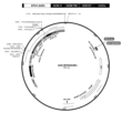
- Figure 1 is a schematic diagram of biopanning using an antibody library.
- Figure 2a is a diagram confirming the binding ability of 2D IgG produced with a complete antibody to recombinant human 4Ig B7-H3, recombinant human 2Ig B7-H3, and recombinant mouse B7-H3 by indirect ELISA.
- Figure 2b is a diagram confirming the binding ability of 2D IgG to Raji and BT-20 cell lines by FACS method.
- Figure 3a is a diagram confirming the binding ability of G2D IgG, a humanized antibody, to recombinant human 4Ig B7-H3, recombinant human 2Ig B7-H3, and recombinant mouse B7-H3 by indirect ELISA
- Figure 3b is a diagram confirming the binding affinity of G2D IgG, a humanized antibody, to recombinant human B7-H3. This is a diagram confirming the binding ability to 4Ig B7-H3, recombinant human 2Ig B7-H3, and recombinant mouse B7-H3 using SPR method.
- Figure 4 is a diagram showing the interaction between B7 family proteins expressed on antigen presenting cells (APC) containing cancer cells and T cells.
- APC antigen presenting cells
- Figure 5 is a diagram confirming the specificity of G2D IgG for B7-H3 among B7 family proteins by indirect ELISA.
- Figure 6 is a diagram confirming the binding capacity of G2D IgG to Raji cell line expressing B7-H3 by FACS method.
- Figure 6a is a diagram confirming the binding affinity to Raji cell line expressing human 4Ig and 2Ig B7-H3
- Figure 6b is a diagram confirming binding affinity to Raji cell line expressing mouse B7-H3.
- Figure 7 is a diagram confirming the epitope of G2D IgG.
- Figure 7a is a diagram confirming the binding of G2D IgG to recombinant 4Ig B7-H3 protein by western blot to confirm the epitope type of G2D IgG
- Figure 7b is a diagram confirming the human 4Ig B7-H3 peptide to which G2D IgG binds. .
- Figure 7c is a diagram confirming the binding of G2D IgG to human 4Ig B7-H3 peptide containing EVQVP, which is expected to be an epitope of G2D IgG
- Figure 7d is a diagram showing the epitope of G2D IgG in all amino acids of human 4Ig B7-H3.
- Figure 7e is a diagram showing the epitope of G2D IgG in the human 4Ig B7-H3 protein structure.
- Figure 8 is a diagram showing the results of G2D antibody binding analysis for colon cancer cell lines.
- Figure 9 is a diagram showing the results of G2D antibody binding analysis for liver cancer cell lines.
- Figure 10 is a diagram showing the results of G2D antibody binding analysis for brain tumor cell lines.
- Figure 11 is a diagram showing the results of G2D antibody binding analysis for pancreatic cancer cell lines.
- Figure 12 is a diagram showing the results of G2D antibody binding analysis for prostate cancer cell lines.
- Figure 13 is a diagram showing the results of G2D antibody binding analysis for lung cancer cell lines.
- Figure 14 is a diagram showing the results of G2D antibody binding analysis for ovarian cancer cell lines.
- Figure 15 is a diagram showing the results of G2D antibody binding analysis for breast cancer cell lines.
- Figure 16 is a diagram showing the results of B7-H3 expression analysis in a triple-negative breast cancer cell line.
- Figure 17 is a diagram showing the number of B7-H3 expression molecules per triple-negative breast cancer cell.
- Figure 18 is a diagram showing the results of G2D antibody binding affinity analysis for mouse cancer cell lines.
- Figure 19 is a diagram showing the results of G2D antibody binding analysis for pediatric brain cancer (medulloblastoma) cell lines.
- Figure 20 is a diagram showing a cleavage map for the construction of a vector having anti-B7-H3 (G2D) CAR, a second-generation chimeric antigen receptor that specifically binds to B7-H3 (CD276).
- G2D anti-B7-H3
- CD276 second-generation chimeric antigen receptor that specifically binds to B7-H3
- Figure 21 is a diagram checking the G2D scFv expression rate of CAR-T cells expressing the Anti-B7-H3 (G2D) gene.
- Figure 22 is a diagram showing the results of measuring the apoptosis ability (LDH) of T cells expressing the anti-B7-H3 (G2D) CAR gene against a triple negative breast cancer cell line (MDA-MB-231) expressing B7-H3.
- LDH apoptosis ability
- Figure 23 is a diagram checking the cytokine (IFN-gamma) production of anti-B7-H3 (G2D) CAR gene-expressing T cells for the triple-negative breast cancer cell lines MDA-MB-231 and BT-20 expressing B7-H3 am.
- IFN-gamma cytokine
- G2D anti-B7-H3
- Figure 24 shows T cell activity expressing anti-B7-H3 (G2D) CAR gene against triple-negative breast cancer cell line (MDA-MB-231), ovarian cancer cell line (A2780), and brain tumor cell line (U87) expressing B7-H3. This is a confirmed drawing.
- Figure 25 shows the targeting of anti-B7-H3 (G2D) CAR gene-expressing T cells to a triple-negative breast cancer cell line (MDA-MB-231), an ovarian cancer cell line (A2780), and a brain tumor cell line (U87) expressing B7-H3.
- MDA-MB-231 triple-negative breast cancer cell line
- A2780 ovarian cancer cell line
- U87 brain tumor cell line
- Figure 26 is a diagram confirming the effectiveness of anti-B7-H3 (G2D) CAR gene-expressing T cells in a brain tumor animal model.
- Figure 27 is a diagram confirming the effectiveness of anti-B7-H3 (G2D) CAR gene-expressing T cells in a non-small cell lung cancer animal model.
- Figure 28 is a diagram confirming the effectiveness of anti-B7-H3 (G2D) CAR gene-expressing T cells in an animal model of leptomeningeal metastasis cancer.
- Figure 29 Measures the apoptotic capacity (LDH) of T cells expressing anti-B7-H3 (G2D) CAR gene against medulloblastoma cell lines (Daoy and D283) expressing B7-H3
- LDH apoptotic capacity
- Figure 30 is a diagram confirming the efficacy of anti-B7-H3 (G2D) CAR gene-expressing T cells in a medulloblastoma animal model.
- Figure 31 shows the results of cytokine (IFN-gamma) secretion by anti-B7-H3 (G2D) CAR gene-expressing T cells against Raji cells expressing human 2Ig and 4Ig B7-H3 structures and mouse 2Ig B7-H3 structure. It is a drawing.
- B7-H3 positive clones were selected using phage display technology.
- 2.5 ⁇ g of recombinant human 4Ig B7-H3 (Sino Biological, China) was conjugated to magnetic beads (ThermoFisher Scientific, USA).
- a non-immune chicken phage library (chicken naive phage library) was reacted with a Raji cell pellet, which is known not to express B7-H3, to remove phages binding to Raji cells in advance, and dispersed in a 3% bovine serum albumin (Millipore, USA) solution.
- 96 clones selected from the plate of the fourth biopanning result were cultured overnight at 37°C with 100 ⁇ g/ml carbenicillin, 70 ⁇ g/ml kanamycin, and VCSM13 helper phage in a 96 deep well plate to produce scFv clones. Proliferation of expressed phage was induced.
- the culture medium obtained above was centrifuged to obtain a medium supernatant containing phages, and recombinant human 4Ig B7-H3, recombinant human 2Ig B7-H3 (R&D Systems, USA), and recombinant mouse B7-H3 (Sino Biological, China) were obtained, respectively.
- ER2738 which has a 2D clone showing a positive signal in the selected B7-H3, was cultured overnight using SB medium and centrifuged to obtain bacterial cells. Plasmid DNA was obtained using a DNA mini prep kit (Geneall, Korea), and the base sequence was analyzed using the primers in Table 1. The 2D clones analyzed had the CDR sequences in Table 2. The light chain and heavy chain variable region amino acid sequences of the 2D clone are shown in Table 3.
- Amino acid sequences of light and heavy chain variable regions of selected clones amino acid sequence number light chain variable region ALTQPSSVSANLGGTVKITCSGGSIGYGWYQQKAPGSAPVTVIYYNDRRPSDIPSRFSGSKSGSANTLTITGVQADDEAIYYCGSADSSSTYTGIFGAGTTLTVL 9 Heavy chain variable region AVTLDESGGGLQTPGGGLSLVCKASGFDFSSYPMVWVRQAPGKGLEYVASINSGGSWTGYGAAVKGRATISRDNGQSTVRLQLNNLRAEDTATYYCARAYGAATIDAWGHGTEVIVSS 10
- the heavy chain was treated with Eco RI and Not I (New England Biolab, UK) enzymes and ligated into pCMV (ThermoFisher Scientific, USA), an expression vector for animal cells, which was also treated with the same restriction enzymes. Additionally, the light chain was treated using Xba I (New England Biolab, UK) enzyme and ligated into the pCMV vector similarly treated with the same restriction enzyme. The ligated plasmid was transformed by applying heat shock to DH5 ⁇ competent cells (New England Biolab, UK), and the obtained colonies were mass-cultured to obtain the plasmid.
- the heavy chain and light chain plasmids converted into complete antibodies were transduced into Expi293F cells (Invitrogen, USA) using polyethylenimine (PEI) (Polysciences, USA) and 150 mM NaCl, and cultured in Freestyle 293 expression medium (Invitrogen, USA). ) was cultured in suspension for 7 days in an Erlenmeyer flask at 37°C, 8% CO 2 and 55% humidity. The expression cell culture was centrifuged at 4,000 rpm for 10 minutes, and the supernatant was taken and filtered through a 0.22 ⁇ m filter.
- PEI polyethylenimine
- Freestyle 293 expression medium Invitrogen, USA
- the filtered supernatant was induced to bind to a HiTrap Mabselect PrismA, 1 mL (GE Healthcare, USA) column at 4°C.
- the bound resin was washed with 10 cv (column volume) of 20 mM sodium phosphate (pH 7.0) and 1 M sodium chloride solution, and then the bound antibody was eluted using 100 mM sodium citrate (pH 3.0) and 150 mM sodium chloride solution. Then, it was neutralized with 1 M Tris-HCL (pH 9.0).
- the binding ability of the 2D IgG produced in Example 2 to the recombinant B7-H3 protein was confirmed by indirect ELISA.
- indirect ELISA recombinant human 4Ig B7-H3, recombinant human 2Ig B7-H3, and recombinant mouse B7-H3 were each diluted to 1 ⁇ g/ml in 50 ⁇ l of PBS, placed in a 96-well immunoplate (Corning, USA), and incubated at 4°C. It was stored and adsorbed overnight.
- the Raji cell line is known to be a B7-H3-negative cell line that does not express B7-H3 (Mol Ther Oncolytics. 2020;17:180-189), and B7-H3 is overexpressed in various breast cancer cells, including the BT-20 cell line. It is known that there is (Mol Cancer Ther. 2011;10(6):960-71).
- the binding capacity of 2D IgG was analyzed by FACS using the Raji cell line, a B7-H3 negative cell line, and the BT-20 cell line, a B7-H3 positive cell line. 5x10 5 BT-20 and Raji cells were suspended in PBS with or without 1 ⁇ g of 2D IgG, reacted at 4°C for 30 minutes, and then centrifuged to remove the supernatant. After removing the supernatant, each cell was washed twice using PBS.
- 2D IgG is a chicken/human chimeric antibody
- humanization was performed as follows to secure a humanized antibody.
- the amino acid residue numbers of the antibody domain are based on the Kabat EU numbering system commonly used in the art (Kabat et al., "Sequences of Proteins of Immunological Interest", 5th Ed., U.S. Department of Health and Human Services, Numbered according to EU index numbers as in NIH Publication No. 91-3242, 1991.
- the J gene of the light chain variable region was fixed as FGGGTKLTVL, referring to US2010-0056386, and the J gene of the heavy chain variable region was fixed as WGQGTTVTVSS, referring to US2014-0206849.
- Humanized 2D was named G2D, and its base sequence is shown in Table 5.
- G2D light and heavy chain variable region amino acid sequences light chain variable region G2D (VL) SYELTQPPSVSVSPGQTARITC SGGSIGYG WYQQKAPGQAPV T VIY YNDRRPS GIPERFSGS K SGTT N TLTISGVQAEDEADYYC GSADSSSTYTGI FGGGTKLTVL SEQ ID NO: 21
- Heavy chain variable region G2D (VH) QVQLVESGGGLVQPGGSLRLSCSASGF D FS SYPMV WVRQAPGKGLEYVS SINSGGSWTGYGAAVKG R
- a TISRDNSKNTLYLQMNSLRAEDTATYYCAR AYGAATIDA WGQGTTVTVSS SEQ ID NO: 22
- the DNA of the G2D variable region described above was synthesized in scFv form (Cosmogenetech, Korea) and converted into a complete antibody through PCR using the primer combinations shown in Table 6 below. Complete antibody conversion and expression/purification were performed in the same manner as in Example 2 described above.
- the binding ability of G2D IgG to B7-H3 was confirmed through SPR analysis, which can measure antigen-antibody binding force and kinetics using optical principles. SPR analysis can confirm the association rate (K a ) and dissociation rate (K d ), and through this, the K D value can be obtained, making quantitative binding force analysis possible.
- B7-family proteins are expressed on the surface of antigen presenting cells (APC), including cancer cells, and regulate T cell activity through interaction with T cell surface proteins (Figure 4).
- APC antigen presenting cells
- B7-H3 is one of the B7 family proteins expressed on APC.
- the T cell receptor has not yet been identified, it is known as an immune checkpoint that inhibits the action of T cells (Front Immunol. 2021; 12: 701006). Since B7 family proteins regulate T cell actions through different mechanisms, the specificity of antibodies targeting B7-H3 for B7-H3 is very important. Therefore, the specificity of G2D IgG for B7-family proteins was analyzed by indirect ELISA.
- Indirect ELISA was performed on: B7-DC (Sino Biological, China), B7-1 (Sino Biological, China), B7-2 (Sino Biological, China), B7-H2 (Sino Biological, China), B7-H4 (Sino Biological, China) China), B7-H1 (Sino Biological, China), 4Ig B7-H3, and 2Ig B7-H3 were used in the same manner as Example 3-1 using a 96-well immune plate coated at 1 ⁇ g/ml.
- G2D IgG was 4Ig among B7 family proteins. It bound only to 2Ig B7-H3 ( Figure 5). These results show the excellent specificity of G2D IgG for B7-H3.
- the recombinant protein and the cell surface protein have the same sequence, there may be structural differences, so it is essential to confirm the binding of the antibody to the cell surface protein.
- a structure containing 4Ig, 2Ig, and mouse B7-H3 extracellular domain was introduced into Raji cell line, a B7-H3 negative cell line. 2Ig, 4Ig, and mouse B7-H3 expressed on the cell surface were confirmed through insertion of green fluorescent protein (GFP). Binding of G2D IgG to B7-H3 expressed on the cell surface was confirmed in the same manner as Example 3-2.
- the secondary antibody used was goat anti-Human IgG (H+L) 594 antibody (Invitrogen, USA). Since binding to cell lines is difficult to confirm quantitatively, enoblituzumab, currently in the clinical stage and registered as a Korean patent (registration number: 10-1828570), was used as a control antibody.
- G2D IgG did not bind to Raji-Vector control, which did not express B7-H3, but bound to Raji-4Ig B7-H3 and Raji-2Ig B7-H3, which expressed 4Ig and 2Ig B7-H3 ( Figure 6a).
- the binding affinity of G2D IgG to 4Ig and 2Ig B7-H3 expressed on the cell surface was higher than that of enoblituzumab, a control antibody. Similar to human B7-H3, G2D IgG did not bind to Raji-Vector control, but bound to Raji-mouse B7-H3 expressing mouse B7-H3 ( Figure 6b).
- the control antibody, enoblituzumab did not bind to mouse B7-H3.
- Therapeutic antibodies are injected into the body and exhibit therapeutic effects by specifically binding to antigens expressed on cells, so the better the binding ability to antigens expressed on cells, the better the therapeutic effect can be expected.
- Figures 6a and b specific and excellent binding of G2D IgG to cell surface B7-H3 can be confirmed.
- Epitope is an antigenic determining site and refers to a specific sequence of an antigen that an antibody can recognize and bind to.
- the therapeutic effect of antibodies may differ depending on the epitope, and in the case of cancer immunotherapy drugs, the degree of immune cell activation may also vary, so identifying the epitope of an antibody is an important part of drug development.
- EVQVP is located in the Ig-like V-type1 and Ig-like V-type2 domains of 4Ig B7-H3 ( Figures 7d and e).
- the epitope of the control antibody enoblituzumab was confirmed in the same way, these epitopes were confirmed to contain the GYPEAE sequence ( Figure 7b), and GYPEAE is the Ig-like C2-type1 and Ig-like C2-type of 4Ig B7-H3. located in type2 ( Figures 7d and e).
- G2D IgG had a different epitope from enoblituzumab, the control antibody.
- the underlined bold portion in the above sequence represents the B7-H3 epitope to which the G2D antibody binds.
- cell lines of colon cancer, liver cancer, brain tumor, pancreas cancer, prostate cancer, lung cancer, ovarian cancer, and breast cancer were used.
- HCT 116 Cat No. CCL-247, ATCC
- HT-29 Cat No. HTB-38, ATCC
- the liver cancer cell line is HepG2 (Cat No. 88065, KCLB).
- HTB-14, ATCC), and MIA-PaCa2 as a pancreatic cancer cell line.
- PanC-1 Cat No. 21469, KCLB
- PC-3 Cat No. 21435, KCLB
- A549 Cat No. 21435, KCLB
- Two cell lines Cat No. CCL-185, ATCC) and NCI-H1975 (Cat No. CRL-5908, ATCC), A2780 (Cat No. 93112519, Sigma) cell line for ovarian cancer, and MDA-MB for breast cancer.
- Flow cytometry was performed using -453 (Cat No. 30131, KCLB), MDA-MB-231 (Cat No. 30026, KCLB) and SK-BR-3 (Cat No. 30030, KCLB) cell lines.
- each cell was reacted with 5 x 10 5 cells and 1 ⁇ g of G2D antibody at 4°C for 30 minutes, and then the supernatant was removed by centrifugation. After removing the supernatant, each cell was washed twice using PBS. After centrifuging the washed cells, add FITC-conjugated Alexa Fluor goat anti-Human IgG (H+L) antibody (Cat No. A11013, Invitrogen), react for 30 minutes at 4°C, and remove the supernatant through centrifugation. did. After removing the supernatant, each cell was washed twice using PBS and then flow cytometric analysis was performed.
- B7-H3 As a comparison group for the flow cytometry results, the expression of B7-H3 (CD276) for each cell line was measured using commercial B7-H3 (APC-conjugated anti-human B7-H3 (CD276) (Cat No. 351006, BioLegend).
- APC-conjugated anti-human B7-H3 (Cat No. 351006, BioLegend).
- a group treated with only FITC-conjugated Alexa Fluor goat anti-Human IgG (H+L) antibody samples was included as a control group for G2D antibody, and anti-human B7-H3 (CD276).
- As an antibody control only Mouse IgG1 kappa Isotype Control APC (Cat No. 17-4714-42, Invitrogen) sample treated (Isotype) was included.
- the binding affinity of G2D and the commercial B7-H3 antibody to the liver cancer cell line HepG2 cell line was confirmed to be more than 95% for both antibodies to the HepG2 cell line (FIG. 9).
- the binding affinity of the G2D and commercial B7-H3 antibodies to the three brain tumor cell lines A172, U251, and U87 was confirmed to be more than 95% for all three cell lines (FIG. 10).
- the binding affinity for the G2D and commercial B7-H3 antibodies to the prostate cancer cell line PC-3 was confirmed to be more than 95% for both antibodies ( Figure 12).
- the binding affinity of G2D and commercial B7-H3 antibodies to the two lung cancer cell lines A549 and NCI-H1975 showed a binding affinity of over 95% for both antibodies to the A549 cell line, and 80% for the G2D antibody to the NCI-H1975 cell line. It showed a binding affinity of more than 95% for the commercial B7-H3 antibody (FIG. 13).
- the binding affinity for the G2D and commercial B7-H3 antibodies to the ovarian cancer cell line A2780 was confirmed to be more than 95% for both antibodies (FIG. 14).
- the binding affinity of the G2D and commercial B7-H3 antibodies to the breast cancer cell lines MDA-MB-453, MDA-MB-231, and SK-BR-3 cell lines showed a binding affinity of over 95% for both antibodies to the MDA-MB-231 cell line.
- the G2D antibody showed a binding affinity of over 35% for the SK-BR-3 cell line
- the commercial B7-H3 antibody showed a binding affinity of over 70%.
- the binding affinity of both antibodies was less than 5% for the MDA-MB-453 cell line (FIG. 15).
- each cell was washed twice using PBS. After removing the supernatant, each cell was washed twice with PBS and the expression rate of B7-H3 (CD276) in the triple-negative breast cancer cell line was analyzed through flow cytometry. As a control, an isotype control (Cat No. 400111, Biolegend) sample was included.
- QuantiBRITE PE Cat No. 340495, BD Biosciences
- flow cytometry method was used to determine the number of B7-H3 expression molecules per cell. Specifically, a new tube of QuantiBRITE beads was reconstituted in the provided tube using 0.5 mL of staining buffer. Samples were acquired on a FACSCanto ⁇ flow cytometer (BD Biosciences). Compensation controls were activated with each analysis to select appropriate instrument settings and at least 10,000 events were collected for each sample. BD FACSDiva TM (BD Biosciences) was used for acquisition control and analysis.
- the QuantiBRITE assay utilizes a bead standard with a population of four beads conjugated with a known number of PE molecules.
- a G1 gate was pulled around the bead singlet on the FSC-H/SSC-H scatter graph. Afterwards, this gated bead population was analyzed using the histogram graph of FL2-H to evaluate the level of PE staining. Individual markers were pulled around the peaks of the four bead populations (M1-4) and the geometric mean for FL2 of each bead population was determined. The FL2 geometric mean of each bead was plotted against the lot specific PE/bead value in a log-log graph. Linear regression was performed to obtain a standard curve using the following equation:
- a G1 gate was pulled around the desired positive fluorescent cell population on the SSC-H/FL4-H dot graph. This gated cell population was then analyzed using the histogram graph of FL2-H to evaluate the level of PE-labeled antibody staining.
- the FL2 geometric mean was determined for each blood cell sample stained with PE-conjugated anti-human B7-H3 monoclonal antibody as well as for unstained control samples. All geometric mean values for FL2 were plotted against the bead standard curve and values for PE per cell were extrapolated. Since both antibody-PE conjugates have a PE:Ab ratio of approximately 1:1, the value for PE per cell corresponds to the number of antibodies bound per cell. The same experiment was performed for each triple-negative breast cancer cell line.
- Mouse lung cancer, pancreatic cancer, and breast cancer cell lines were used to confirm the binding affinity of the G2D antibody, which specifically binds to mouse 2Ig B7-H3 as well as human 2Ig and 4Ig B7-H3, to mouse cell lines.
- 3LL (Cat No. CRL-1642, ATCC) as a lung cancer cell line
- Panc-02 (Cat No. SC078-luc, GeneTarget) as a pancreatic cancer cell line
- 4T1 (Cat No. CRL-2539, ATCC) as a breast cancer cell line.
- Flow cytometry was performed using .
- Each cell was reacted with 5 x 10 5 cells and 1 ⁇ g of G2D antibody at 4°C for 30 minutes, and then the supernatant was removed by centrifugation. After removing the supernatant, each cell was washed twice using PBS. After centrifuging the washed cells, goat anti-Human IgG (H+L) 594 antibody (Cat No. A11014, Invitrogen) was added and reacted for 30 minutes at 4°C, and then centrifuged to remove the supernatant. After removing the supernatant, each cell was washed twice using PBS and then flow cytometric analysis was performed.
- Alexa Fluor 647 Rat Anti-Mouse B7-H3 (Cat No. 562862, BD Biosciences) was used to confirm the expression of B7-H3 (CD276) for each mouse cell line. compared.
- a control group for G2D antibody a group treated only with goat anti-Human IgG (H+L) 594 antibody sample (Secondary) was included, and as a control group for anti-mouse B7-H3 (CD276) antibody, Alexa Fluor 647 Rat sample was included.
- IgG2A, k Isotype Control (Cat No. 557690, BD Biosciences) samples treated only (Isotype) were included.
- the G2D antibody showed a binding affinity of over 90%, and the commercial B7-H3 antibody showed a binding affinity of over 95%.
- the G2D antibody showed a binding affinity of more than 50%, and the commercial mouse B7-H3 antibody showed a binding affinity of more than 90%.
- the pancreatic cancer cell line Panc-02 the G2D antibody showed a binding affinity of more than 85%, and the commercial mouse B7-H3 antibody showed a binding affinity of more than 90% (FIG. 18).
- Example 10 G2D antibody binding analysis on pediatric brain cancer and medulloblastoma cell lines
- Daoy cells were reacted with 5 x 10 5 cells and 1 ⁇ g of G2D antibody at 4°C for 30 minutes, and then the supernatant was removed by centrifugation. After removing the supernatant, each cell was washed twice using PBS. After centrifuging the washed cells, goat anti-Human IgG (H+L) 594 antibody (Cat No. A11014, Invitrogen) was added and reacted for 30 minutes at 4°C, and then centrifuged to remove the supernatant. After removing the supernatant, each cell was washed twice using PBS and then flow cytometric analysis was performed.
- H+L 594 antibody Cat No. A11014, Invitrogen
- Example 11 Construction of a vector with anti-B7-H3 (G2D) CAR, a second-generation chimeric antigen receptor that specifically binds to B7-H3 (CD276)
- scFv Single-chain variable fragment, scFv; SEQ ID NO: 42 and 43
- CD8 ⁇ hinge region single-chain variable fragment, scFv; SEQ ID NO: 42 and 43
- CD8 ⁇ transmembrane domain Transmembrane, TM; SEQ ID NOs: 46 and 47
- CD137 SEQ ID NOs: 48 and 49
- CD3 ⁇ CD3 zeta killing signaling domain
- Peripheral blood mononuclear cells were obtained from whole blood obtained from a healthy donor using Ficoll-Paque premium (GE healthcare, 17-5442-03) to obtain only mononuclear cells of the mononuclear cell layer (buffy coat).
- Isolated PBMCs were mixed with 100 ng/mL of anti-CD3 monoclonal antibody (OKT3, eBioscience, Cat No. 16-0037-81) and anti- OpTmizer TM CTS TM T-Cell Expansion Supplement (Gibco, A10484-02), CTS TM Immune Cell SR (Gibco, A25961-) by adding 100 ng/mL of CD28 monoclonal antibody (eBioscience, Cat No. 16-0289-85).
- T cell fraction was activated by culturing in CTS TM T-Cell Expansion Basal Medium (Gibco, A10221-01) containing GlutaMAX TM -I CTS TM (100X) (Gibco, A12860-01). Activated T cells were transduced using anti-B7-H3 (G2D) CAR virus.
- Anti-B7-H3 (G2D) CAR expressing T cells were proliferated in a 75 T flask using T cell culture medium containing 500 IU/mL of Human IL2. T cells expressing anti-B7-H3 (G2D) CAR propagated in this manner were used in various assays.
- Anti - B7-H3 (G2D) CAR-T cells were 18% against the triple negative breast cancer cell line MDA-MB-231 according to the E:T ratio (0.625:1, 1.25:1, 2.5:1 and 5:1); It showed apoptotic capacity of 30%, 48% and 61%.
- UTD cells not transduced with CAR virus showed LDH values of 3%, 5%, 8% and 19% depending on the E:T ratio (0.625:1, 1.25:1, 2.5:1 and 5:1). shown ( Figure 22).
- anti-B7-H3 (G2D) CAR-T cells have killing ability against triple negative breast cancer cell lines.
- the results of IFN-gamma secretion by anti-B7-H3 (G2D) CAR-T cells against triple negative breast cancer cell lines MDA-MB-231 and BT-20 are shown in Figure 23. Specifically, the amount of IFN-gamma following co-culture with the triple-negative breast cell lines MDA-MB-231 and BT-20 was higher for anti-B7-H3 (G2D) CAR-T than for samples not transduced with CAR virus. IFN-gamma was secreted.
- Triple negative breast cancer cell line ( MDA-MB- 231 , 2 Co-culture was performed on the plate for 48 hours.
- brain tumor cell line U87 1 Live target cell lines were stained using violet solution.
- anti-B7-H3 (G2D) CAR-T had activity and apoptosis ability against triple-negative breast cancer, ovarian cancer, and brain tumor cell lines expressing B7-H3.
- H1299 cells were directly transplanted into the lungs of NSGA mice using 50 ul of 5x10 5 H1299 cells. This induced engraftment. Five days after engraftment, it was confirmed that the tumor size was evenly distributed by measuring the weight of the test animals and using in vivo luciferase imaging.
- the test group consisted of a control group and a test substance administration group ⁇ UTD and B7-H3 (G2D) CAR-T ⁇ . PBS was administered to the control group, and the test substance group was administered a single intravenous dose at a concentration of 5x10 7 anti-B7-H3 (G2D) CAR-T cells.
- BM-H1299 a brain metastasis lung cancer cell line
- Engraftment was induced by transplanting BM-H1299 cells directly into the brain of NSGA mice using 5 ul of 5x10 4 BM-H1299 cells.
- the test group consisted of a control group and a test substance administration group ⁇ UTD and B7-H3 (G2D) CAR-T ⁇ .
- PBS was administered to the control group, and the test substance group was administered a single dose by ICV (intracerebroventricular administration) at a concentration of 1.5x10 6 anti-B7-H3 (G2D) CAR-T cells.
- anti-B7-H3 (G2D) CAR gene-expressing T cells in a brain tumor (glioblastoma) animal model, anti-B7-H3 ( G2D)
- the CAR-T treatment group showed a reduction in the size of brain tumors. From these results, anti-B7-H3 (G2D) CAR-T reduced tumor size in an animal model transplanted with a brain tumor cell line (U87), and thus anti-B7-H3 in a brain tumor model expressing B7-H3. (G2D) It was confirmed that CAR-T worked effectively (Figure 26).
- anti-B7-H3 (G2D) CAR-T can be effectively applied to brain tumors, non-small cell lung cancer, and leptomeningeal metastasis cancer.
- CytoTox96 Non-radio cytotoxicity to measure the cytolytic activity of anti-B7-H3 (G2D) CAR-T cells against B7-H3-expressing medulloblastoma cell lines (Daoy and D283-Cat No. HTB-185, ATCC) Cell death analysis (Cytotoxicity assay) was performed using the assay LDH (Cat No. G1781, Promega) kit. Specifically, anti-B7-H3 (G2D) CAR-T cells (Effector cells) were used after 10 days of culture, and Effector:Target was placed in a 96-well plate at an E:T ratio of 5:1, 2.5:1, and 1.25:1. Cells were added and reacted at 37°C for 18 hours. As a control group in this test, samples that were not transduced with the CAR virus (Untransduced T cells, UTD) were included.
- UTD Untransduced T cells
- Anti - B7-H3 (G2D) CAR-T cells showed 18%, 44% and 66% apoptotic capacity against D283 cell line depending on E:T ratio (1.25:1, 2.5:1 and 5:1). and showed cell death abilities of 50%, 70%, and 84% for the Daoy cell line.
- UTD cells that were not transduced with the CAR virus showed LDH values of 3% and 13% depending on the E:T ratio (2.5: 1 and 5:1).
- anti-B7-H3 (G2D) CAR-T cells have the ability to kill medulloblastoma cells (FIG. 29).
- T cells in an animal model of pediatric brain cancer medulloblastoma, 5 ul of 2x10 5 Daoy cells were transplanted directly into the cerebellum of NSGA mice for engraftment. was derived. Seven days after engraftment, it was confirmed that the tumor size was evenly distributed by measuring the weight of the test animals and using in vivo luciferase imaging.
- the test group consisted of one control group (Vehicle) and two test substance administration groups ⁇ UTD cells, B7-H3 (G2D) CAR-T ⁇ , with 5 animals distributed to each group.
- T cells (UTD) not transduced with CAR virus were administered as a single dose by IT (intratumoral administration) at a concentration of 1.5 x 10 6 cells, and anti-B7-H3 (G2D) CAR-T cells were administered. It was administered as a single dose by IT (intratumoral administration) or ICV (intracerebroventricular administration) at a concentration of 1.5 x 10 6 cells.
- anti-B7-H3 (G2D) CAR-T can be effectively applied to medulloblastoma, and both administration methods (IT and ICV) effectively move anti-B7-H3 (G2D) CAR-T to the tumor. Confirmed.
- Experimental Example 7 Checking cytokine (IFN-gamma) production of T cells expressing anti-B7-H3 (G2D) CAR gene on Raji cells expressing human 2Ig and 4Ig B7-H3 structures and mouse 2Ig B7-H3 structures
- B7-H3 (G2D) CAR-T was analyzed using a cell line (Raji) that does not express B7-H3.
- Raji Raji-2ig B7-H3, Raji-4Ig B7-H3 or Raji-m2Ig B7-H3 cells
- 1 x 10 anti-B7-H3 (G2D) CAR-T cells 5 Add 37°C Cultured in incubator for 18 hours.
- UTD a cell sample not transduced with CAR virus, was included.
- the results of IFN-gamma secretion by anti-B7-H3 (G2D) CAR-T cells against Raji cells expressing human 2Ig and 4Ig B7-H3 structures and mouse 2Ig B7-H3 structure are shown in Figure 31.
- the amount of IFN-gamma following co-culture with Raji cells expressing human 2Ig and 4Ig B7-H3 structures and mouse 2Ig B7-H3 structure was Higher levels of IFN-gamma were secreted compared to non-transduced samples.
- the B7-H3 CAR-T of the present invention binds specifically to not only human but also mouse B7-H3 to treat pediatric brain cancer, adult brain cancer, leptomeningeal metastasis cancer, non-small cell lung cancer, triple-negative breast cancer, and various cancers. It was confirmed that the signaling system was activated.
Landscapes
- Health & Medical Sciences (AREA)
- Chemical & Material Sciences (AREA)
- Life Sciences & Earth Sciences (AREA)
- Organic Chemistry (AREA)
- Immunology (AREA)
- Medicinal Chemistry (AREA)
- General Health & Medical Sciences (AREA)
- Pharmacology & Pharmacy (AREA)
- Animal Behavior & Ethology (AREA)
- Biophysics (AREA)
- Veterinary Medicine (AREA)
- Genetics & Genomics (AREA)
- Biochemistry (AREA)
- Molecular Biology (AREA)
- Proteomics, Peptides & Aminoacids (AREA)
- Public Health (AREA)
- Zoology (AREA)
- Mycology (AREA)
- Cell Biology (AREA)
- Epidemiology (AREA)
- Microbiology (AREA)
- Toxicology (AREA)
- Gastroenterology & Hepatology (AREA)
- Chemical Kinetics & Catalysis (AREA)
- General Chemical & Material Sciences (AREA)
- Nuclear Medicine, Radiotherapy & Molecular Imaging (AREA)
- Peptides Or Proteins (AREA)
Abstract
본 발명은 암 치료에 효과적인 키메릭 항원 수용체를 암호화하는 폴리뉴클레오티드를 포함하는 발현 카세트 및 상기 발현 카세트가 형질도입된 효과기 세포에 관한 것이다. 특히, 본 발명은 다양한 암에서 높게 발현하는 B7-H3 항원에 특이적으로 결합하는 항원 결합 도메인을 포함하는 키메릭 항원 수용체를 T 림프구 등 효과기 세포에 발현시키는 경우 B7-H3와 관련된 암 질환의 면역 치료방법으로서 유용하게 이용될 수 있다.
Description
본 발명은 암 치료에 효과적인 키메릭 항원 수용체를 암호화하는 폴리뉴클레오티드를 포함하는 발현 카세트 및 상기 발현 카세트가 형질도입된 효과기 세포에 관한 것이다.
전 세계 고형항암제 시장은 '21년 약 1,010억 달러(약 126조 원)에서 '28년 약 2,520억 달러(약 310조 원)으로 연 평균 12.5% 성장할 것으로 전망되었다. '28년 세부질환별 시장 규모는 비소세포암(NSCLC, Non-small cell lung cancer) 약 520억 달러(약 65조 원), 유방암(Breast cancer) 약 460억 달러(약 57조 원), 전립선암(Prostate cancer) 약 210억 달러(약 26조 원), 흑색종(Melanoma) 약 160억 달러(약 20조 원), 신세포암(RCC, Renal cell carcinoma) 약 130억 달러(약 16조 원) 등으로 주요 다섯 개 암 종이 전체의 약 60% 비중을 차지할 것으로 예측된다(Evaluate Pharma Market Explorer, 2022).
국가암등록통계사업을 통해 수집된 우리나라 2019년 국가암 등록 통계를 살펴보면, 2015년 이후 암 환자 수는 매년 증가하였으며, 2019년 신규 발생한 암환자 수는 2018년도 대비 8,844명 (3.6%) 증가한 25만 4,718명이다(보건복지부. 2019년 국가암등록 통계자료).
국가암검진사업을 통한 6대암으로 분류되는 유방암, 위암, 대장암, 간암, 폐암 그리고 자궁경부암에 대해서 장기 추세를 보면, 6대암 중 5개의 암종인 폐암, 대장암, 간암, 위암, 그리고 자궁경부암의 발생률은 최근 10여 년간 감소 추세를 보여주거나, 유의미한 증가를 보이지 않는다. 반면 나머지 유방암의 발생률은 20년간 지속적으로 증가하는 추세를 보이고 있다.
유방암은 3가지 지표인 에스트로겐 수용체, 프로게스테론 수용체 그리고 표피성장인자 (HER2)를 기준으로 분류한다. 3가지 지표 검사시 전부 '음성 (발견되지 않음)'으로 확인되면 삼중음성 유방암으로 진단한다.
유방암은 진단 후 5년 생존율이 90% 이상이지만, 삼중음성 유방암은 70% 정도이다. 3~4기의 삼중음성 유방암 환자들의 5년 생존율은 30%정도에 불과하다. 이는 삼중음성 유방암을 제외한 다른 3~4기 유방암 환자들의 5년 생존율이 약 50% 인것과 비교하면 낮은 수치이다. 또한 조기 발견 환자의 30%정도가 외과적 수술 후 5년 내 사망 위험을 경험할 정도로 삼중음성 유방암의 진행이 빠르고 공격적이다.
삼중음성 유방암의 경우 암세포의 변이가 많으며, 다른 장기로의 전이가 다른 유방암보다 빠르기 때문에 치료에 여러 한계가 존재한다. 뿐만 아니라, 삼중음성 유방암은 초기에 치료해도 약 50% 환자가 재발을 경험할 정도로 예후가 나쁘다. 현재 유방암 치료제는 에스트로겐과 프로게스테론 그리고 표피성장인자 수용체를 표적으로 해서 치료한다. 따라서 이들 수용체가 음성인 삼중음성 유방암 환자들은 해당 약물을 이용하여 치료 효과를 기대하기 어렵다.
성인암 뿐만 아니라 소아암의 비율도 적지 않은 범위로 발생되고 있다. 소아암의 발생빈도는 전세계적으로 비슷하며, 대한민국의 경우 매년 1,000~1,200여명이 소아암으로 진단을 받는다. 이는 소아 10만 명당 매년 약 1명이 소아암으로 진단을 받으며 이는 전체 암환자의 약 1%을 차지하고 있다. 소아암은 1세부터 9세 사이 아동의 질병 사망률 1위로 교통사고로 사망한 아동 수보다 많을 정도로 매우 위험한 질병이다.
대표적인 소아암으로는 백혈병, 림프종 그리고 뇌종양으로 나눌 수 있다. 백혈구 중에서도 림프구계 백혈구가 제대로 성숙하지 못하고 암세포로 변하는 백혈병인 림프모구백혈병의 경우 치료 성적이 매우 좋아 완치율이 80~90% 가량이 되며, 급성골수성 백혈병 역시 70%의 완치율을 보이고 있다. 그러나, 소아 뇌종양의 경우 예후가 좋지 않아 새로운 치료법 개발이 필요한 실정이다. 보건복지부 중앙암등록본부 자료에 의하면 2020년에 대한민국에서는 247,952건의 암이 새로이 발생했는데, 그 중 뇌종양은 전체 암 발생의 0.7%인 1,795건으로 확인되었다. 이중에서 소아청소년 뇌종양은 총 152건으로 10세~19세에서 74건 그리고 9세 이하에서 78건이 발생하였다고 보고하였다. 남녀 성비는 1.8:1로 남자에게 더 많이 발생했다. 국가암정보센터에 따르면 소아청소년기에 호발하는 뇌종양으로는 수모세포종 (Medulloblastoma), 성상세포종 (Astrocytoma), 뇌실막세포종 (Ependymoma), 두개인두종 (Craniopharyngioma), 뇌간신경아교종 (Brainstem glioma) 그리고 배세포종 (Germ cell tumor)이며, 뇌종양은 악성도, 발생위치 그리고 구성하는 세포에 따라서 나눌 수 있다.
이중 수모세포종은 소뇌 중앙부에서 자라며, 뇌척수액로를 통해 전이를 일으키기도 한다. 두통 등의 뇌압상승 징후, 운동장애, 뇌신경 마비 징후 그리고 의식장애 등을 나타내며, 소아기 악성 뇌종양 중 가장 흔한 암이다. 뇌종양중 뇌간신경아교종은 뇌의 부위 중 중뇌, 뇌교, 연수에서 발생하는 교종으로 구음장애 연하장애, 사시, 안면 신경마비등 다발성 뇌신경 장애나 반신부전 마비가 오며, 2년 생존율이 10% 미만으로 예후가 가장 나쁜 소아 뇌종양이다. 현, 뇌종양의 치료법으로는 수술 또는 수술과 방사선 치료를 함께하는 것이 가장 기본적이 치료이며 종양 조직 결과에 따라서 항암화학용법을 시도하기도 한다.
수모세포종에 대한 수술적 치료의 경우 소뇌함구증후군 (Cerebellar mutism syndrome CMS) 이 수술 환자의 25%에게 나타나는데, 소뇌함구증후군은 소뇌를 포함한 뇌의 특정 부위를 종양 제거 수술을 받은 환자에서 발생할 수 있는 상태이며, 수술 후 약 1~2일 후 증상이 나타나며 증상으로는 말수가 줄어들고, 균형 잡기가 어려워 걷기가 힘들거나 근육 긴장 소실 등이 포함된다. 소뇌함구증후군에 따른 증상들은 시간이 흐르면서 서서히 사라지게 된다. 하지만, 수술 후 방사선조사를 바로 실시하는데 이 또한, 어려움이 존재한다. 수술 후 보조 두개척추 방사선 치료법은 뇌척수액(Cerebrospinal fluid, CSF)에서 수포종이 전파되는 것을 방지하기 위해 수포종의 전체 뇌와 척추를 치료하는 데 사용되는데, 두개척추 방사선 치료는 의심할 여지없이 생존을 연장하지만, 아쉽게도 방사선 치료법은 매년 2-4포인트의 지능발달 지수 (Intelligence Quotient, IQ) 감소를 희생해야 한다.
연수막 전이암은 전체 고형암의 약 5-20%에서 나타난다고 알려져 있으며 이 중 폐암, 유방암, 흑색종이 많은 비중을 차지하고 있다. 연수막전이암으로 전이되는 환자의 비율 역시 폐암, 유방암, 흑색종에서 많은 빈도수로 나타나고 있다(Cancers (Basel). 2021;13). 2020년 국가암등록 통계에 따르면 2020년 폐암은 28,949명, 유방암은 24,923명이 진단된 것으로 알려져 있기에 위의 두가지 암에서 발생하는 연수막 전이암 환자수만 국내에서 한 해에 최소 2500명 이상이 될 것으로 예상이 되며 다른 적응증 유래 연수막전이암 환자수 및 진단법과 치료제의 발전을 고려하면 그 수는 더욱 늘 것으로 예상된다. 하지만 늘어나는 환자수에 비해 효과적인 치료제 개발은 쉽지 않은 상황이다. 뇌실 내로 메토트렉세이트 (Methotrexate)같은 항암제를 투여하거나 국소 방사선 치료가 증상 개선에 도움이 될 수 있으나 치료를 받더라도 중앙생존기간이 6개월 이내로 예후가 매우 좋지 않다(대한내과학회지. 2011;제81권:제3호). 그나마 최근 혈관-뇌장벽을 효과적으로 통과하는 타그리소 같은 타깃치료제들이 연수막전이암에도 효과적이라고 알려져있으나 적용가능한 환자수가 제한적이며 타깃치료제들은 내성이 생기는 문제를 안고 있다(Neurotherapeutics. 2022;19:1782-1798).
최근, 키메라 항원 수용체 변형 T 세포 (Chimeric antigen receptor, CAR-T)세포의 경우 혈액암을 대상으로 치료제로의 가능성이 입증되었으며, 다양한 연구를 통해서 CAR-T 세포치료제가 혈액암 뿐만 아니라 고형암 치료에도 효과를 나타낼 가능성이 있음을 암시한다.
한편, B7-H3은 소아뇌암, 연수막전이암, 삼중음성 유방암 등을 비롯한 다양한 암종에서 높게 발현된다고 알려져 있으며, 이러한 B7-H3의 고발현은 부정적인 임상적 치료 결과와 관련되어 있다.
따라서 본 발명자는 암에서 높게 발현하고 있는 B7-H3에 대하여 다양한 암에 대한 관계성 및 수모세포종, 연수막전이암 그리고 삼중음성 유방암 등에 있어서 키메라 항원 수용체(CAR)의 치료제로의 가능성을 확인하고 본 발명을 하기에 이르렀다.
상기한 배경기술로서 설명된 사항들은 본 발명의 배경에 대한 이해 증진을 위한 것일 뿐, 이 기술분야에서 통상의 지식을 가진 자에게 이미 알려진 종래기술에 해당함을 인정하는 것으로 받아들여져서는 안 될 것이다.
이에 본 발명자들은 다양한 암종에서 높게 발현하고 있는 B7-H3를 타겟팅하는 키메릭 항원 수용체(CAR)를 포함하는 효과기 세포를 개발하고자 노력하였다. 그 결과 본 발명자들은 B7-H3와 결합하는 항원 결합 도메인이 포함된 효과기 세포가 다양한 종류의 암세포주에 특이적으로 결합하여 암세포 사멸 효과를 나타내는 것을 확인함으로써 본 발명을 완성하게 되었다.
따라서 본 발명의 목적은, B7-H3와 결합하는 항원 결합 도메인(antigen-binding domain); 힌지 영역(hinge region); 막관통 도메인(transmembrane domain); 보조 자극 도메인(co-stimulatory domain); 및 세포질 신호 전달 도메인(intracellular signaling domain)을 포함하는 키메릭 항원 수용체(CAR)를 암호화(coding)하는 폴리뉴클레오티드를 포함하는 발현 카세트를 제공하는데 있다.
본 발명의 다른 목적은, 상기 발현 카세트를 포함하는 재조합 발현 벡터를 제공하는데 있다.
본 발명의 또 다른 목적은, 상기 재조합 발현 벡터로 형질 도입된, 분리된 효과기 세포를 제공하는데 있다.
본 발명의 또 다른 목적은, 상기 B7-H3와 결합하는 항원 결합 도메인을 포함하는 키메릭 항원 수용체(CAR)를 포함하는, 분리된 효과기 세포를 제공하는데 있다.
본 발명의 또 다른 목적은, 상기 효과기 세포 및 약제학적으로 허용되는 담체를 포함하는 암의 예방 또는 치료용 약제학적 조성물을 제공하는데 있다.
본 발명의 또 다른 목적은, 이를 필요로 하는 대상에게, 상기 효과기 세포를 투여하는 단계를 포함하는, 암의 치료방법을 제공하는데 있다.
본 발명의 다른 목적 및 이점은 하기의 발명의 상세한 설명, 청구범위 및 도면에 의해 보다 명확하게 된다.
본 발명의 일 양태에 따르면, 본 발명은 B7-H3와 결합하는 항원 결합 도메인(antigen-binding domain); 힌지 영역(hinge region); 막관통 도메인(transmembrane domain); 보조 자극 도메인(co-stimulatory domain); 및 세포질 신호 전달 도메인(intracellular signaling domain)을 포함하는 키메릭 항원 수용체(CAR)를 암호화(coding)하는 폴리뉴클레오티드를 포함하는 발현 카세트를 제공한다.
암세포를 포함하는 항원발현세포 (Antigen presenting cell, APC)의 표면에는 B7-family protein이 발현되어 있고 T cell 표면 단백질과의 상호작용을 통하여 T cell의 작용을 조절한다. B7-H3는 APC에 발현된 B7 family protein 중 하나로 T cell의 작용을 저해하는 immune checkpoint로 알려져 있다(Front Immunol. 2021; 12: 701006). B7 family protein은 T cell의 작용을 서로 다른 기전으로 조절하므로 B7-H3를 타겟하는 항체의 B7-H3에 대한 특이성이 매우 중요하다.
본 명세서에서 용어 “키메릭 항원 수용체(CAR)”는 항원 결합 도메인(antigen-binding domain), 힌지 영역(hinge region), 막관통 도메인(transmembrane domain), 보조 자극 도메인(co-stimulatory domain) 및 세포질 신호 전달 도메인(intracellular signaling domain)을 포함하는 폴리펩타이드를 지칭한다.
제1 세대 CAR는 CD3ζ를 세포내 신호전달 도메인으로 포함하는 반면, 제2 세대 CAR는 다양한 단백질에서 유래된 적어도 하나의 보조 자극 도메인을 추가로 포함한다. 제2 세대 CAR에서의 보조 자극 도메인은 예를 들어 CD28, CD2, 4-1BB(CD137) 및 OX-40(CD134)을 포함하나, 이에 한정되지 않는다. 제3 세대 CAR은 두 가지 보조 자극 도메인, 예를 들어, CD28, 4-1BB, OX-40, 및 CD2 등을 포함하나, 이에 한정되지 않는다.
본 명세서에서 용어 "항체"는 당업계의 용어이며 본 명세서에서 상호 교환하여 사용될 수 있고, 항원에 특이적으로 결합하는 항원 결합 부위를 가진 분자를 말한다. 본 명세서에서 사용된 용어는 전체 항체 및 임의의 항원 결합 단편(즉 "항원-결합 부분") 또는 이들의 단쇄를 포함한다. 하나의 구체예에서, "항체"는 이황화물 결합에 의해 상호-연결된 적어도 2개의 중쇄(H)와 2개의 경쇄(L)를 포함하는 당단백질, 또는 이의 항원-결합 부분을 말한다. 다른 구체예에서, "항체"는 단일 가변 도메인, 예를 들어 VHH 도메인을 포함하는 단쇄 항체를 말한다. 각 중쇄는 중쇄 가변 영역(VH로 약기)과 중쇄 불변 영역으로 이루어진다. 특정 자연-발생 항체에서, 중쇄 불변 영역은 3개의 도메인 CH1, CH2 및 CH3으로 이루어진다. 특정 자연-발생 항체에서, 각 경쇄는 경쇄 가변 영역(VL로 약기)과 경쇄 불변 영역으로 이루어진다. 경쇄 불변 영역은 하나의 도메인 CL로 이루어진다.
VH 및 VL 영역은 프레임워크 영역(FR)이라 칭하는, 더 보존되는 영역들이 산재된, 상보성 결정 영역(CDR)이라 칭하는, 초가변성의 영역으로 더 세분될 수 있다. 각각의 VH 및 VL은 3개의 CDR과 4개의 FR로 이루어지며, 이들은 FR1, CDR1, FR2, CDR2, FR3, CDR3, 및 FR4의 순서로 아미노-말단에서 카복시-말단을 향해 배열된다. 중쇄 및 경쇄의 가변 영역은 항원과 상호작용하는 결합 도메인을 함유한다. 항체의 불변 영역은 면역 시스템의 다양한 세포(예를 들어 이펙터 세포) 및 정통 보체 시스템의 제1 성분(C1q)를 포함하는 숙주 조직 또는 인자들과 면역글로불린의 결합을 매개할 수 있다.
항체는 면역글로불린 분자의 임의의 타입(예를 들어 IgG, IgE, IgM, IgD, IgA 또는 IgY), 임의의 유형(예를 들어 IgD, IgG2, IgG3, IgG4, IgA1 또는 IgA2), 또는 임의의 아형(예를 들어 인간에서 IgG1, IgG2, IgG3 및 IgG4; 및 마우스에서IgG1, IgG2a, IgG2b 및 IgG3)를 가질 수 있다. 면역글로불린, 예를 들어 IgG1은 몇몇 알로타입으로 존재하며, 이들은 최대 몇 개의 아미노산이 서로 상이하다. 본 명세서에 개시된 항체는 통상 알려진 이소타입, 유형, 아형, 또는 알로타입 중 어느 것으로부터 유래할 수 있다. 특정 구체예에서, 본 명세서에 개시된 항체는 IgG1, IgG2, IgG3, 또는 IgG4 아형 또는 이들의 임의의 하이브리드이다. 특정 구체예에서, 항체는 인간 IgG1 아형 또는 인간 IgG2 또는 인간 IgG4 아형의 것이다.
"항체"는, 예로서, 자연-발생 및 비-자연-발생 항체; 단클론성 및 다클론성 항체; 키메라 및 인간화된 항체; 인간 및 비-인간 항체, 전체 합성 항체; 단쇄 항체; 단일특이적 항체; 다중특이적 항체(이중특이적 항체를 포함); 2개의 중쇄와 2개의 경쇄 분자를 포함하는 테트라머 항체; 항체 경쇄 모노머; 항체 중쇄 모노머; 항체 경쇄 다이머; 항체 중쇄 다이머; 항체 경쇄-항체 중쇄 쌍; 인트라바디; 헤테로콘쥬게이트 항체; 1가 항체; 낙타화 항체; 아피바디; 항-이디오타입(항-Id) 항체(예를 들어 항-항-Id 항체를 포함), 및 충분히 항원 결합할 수 있는 단일 모노머 가변 항체 도메인(예를 들어 VH 도메인 또는 VL 도메인)으로 구성된 결합 분자를 포함하는 단일-도메인 항체(sdAb)를 포함한다(Harmen M. M. and Haard H. J. Appl Microbiol Biotechnol. 77(1): 13-22 (2007)).
본 명세서에서 사용된 용어 “항원 결합 도메인(antigen binding domain)"은 항원 타겟을 특이적으로 인식 및 결합할 수 있는 임의의 단백질 또는 폴리펩타이드 도메인을 지칭한다. 상기 도메인은 항원(예를 들어, 인간 Ig-like-V-type 1 또는 Ig-like-V-type 2의 도메인)에 특이적으로 결합하는 능력을 보유한 임의의 단백질 또는 폴리펩타이드일 수 있다. 상기 항원 결합 도메인은 (i) VL, VH, CL, 및 CH1 도메인으로 구성된 1가 단편인 Fab 단편; (ii) 힌지 영역에서 이황화물 브릿지에 의해 연결된 2개의 Fab 단편을 포함하는 2가 단편인 F(ab')2 단편; (iii) VH 및 CH1 도메인으로 구성된 Fd 단편; (iv) 항체의 단일 팔의 VL 및 VH 도메인으로 구성된 Fv 단편, 및 이황화물-연결된 Fvs(sdFv); (v) VH 도메인으로 구성된 dAb 단편(Ward et al., (1989) Nature 341: 544-546); 및 (vi) 분리된 상보성 결정 영역(CDR) 또는 (vii) 합성 링커에 의해 선택적으로 이어질 수 있는 둘 이상의 분리된 CDR의 조합을 포함하나, 이로 제한되는 것은 아니다. 또한, Fv 단편의 두 도메인인 VL과 VH는 별개의 유전자에 의해 코딩되지만, 이들은 합성 링커에 의해, 재조합 방법을 사용하여 이어질 수 있으며, 이로써 VL과 VH 영역이 쌍을 이루어 1가 분자를 형성한 단일 단백질 사슬이 될 수 있다(단쇄 Fv(scFv)라고 한다)(예를 들어 Bird et al., (1988) Science 242:423-426; 및 Huston et al., (1988) Proc. Natl. Acad. Sci. USA 85:5879-5883 참조). 바람직하게는 상기 항원 결합 도메인은 Fab 또는 scFv일 수 있다.
본 명세서에서 사용된 용어 "가변영역" 또는 "가변도메인"은 당업계에서 통상적으로 상호 교환하여 사용된다. 가변영역은 전형적으로 항체의 일부분, 일반적으로 경쇄 또는 중쇄의 일부분, 전형적으로 성숙한 중쇄에서 약 110 내지 120개 아미노산 및 성숙한 경쇄에서 약 90 내지 115개 아미노산을 말하며, 이들은 항체와 서열이 광범하게 상이하고 이의 특정 항원에 대한 특정 항체의 결합 및 특이성 때문에 사용된다. 서열 변동성은 상보성 결정 영역(CDR)이라고 불리는 영역에 집중되며, 가변 도메인에서 더 고도로 보존된 영역은 프레임워크 영역(FR)이라고 불린다.
경쇄 및 중쇄의 CDR은 항원과 항체의 상호작용 및 특이성을 주로 담당한다고 여겨진다. 특정 구체예에서, 가변영역은 인간 가변영역이다. 특정 구체예에서, 가변영역은 설치류 또는 뮤린 CDR과 인간 프레임워크 영역(FR)을 포함한다. 특정 구체예에서, 가변영역은 영장류(예를 들어 비-인간 영장류) 가변영역이다. 특정 구체예에서, 가변영역은 설치류 또는 뮤린 CDR과 영장류(예를 들어 비-인간 영장류) 프레임워크 영역(FR)을 포함한다.
본 명세서에서 사용된 용어 "중쇄"는 항체와 관련하여 사용되었을 때, 불변도메인의 아미노산 서열에 기초하여, 임의의 상이한 타입, 예를 들어 알파(α), 델타(δ), 엡실론(ε), 감마(γ) 및 뮤(μ)를 말할 수 있으며, 이들은 각각 IgG의 아형, 예를 들어 IgG1, IgG2, IgG3 및 IgG4를 포함하여, 항체의 IgA, IgD, IgE, IgG 및 IgM 유형을 발생시킨다.
본 명세서에서 사용된 용어 "경쇄"는 항체와 관련하여 사용되었을 때, 불변도메인의 아미노산 서열에 기초하여 임의의 상이한 타입, 예를 들어 카파(κ) 및 람다(λ)를 말할 수 있다. 경쇄 아미노산 서열은 당업계에 잘 공지되어 있다. 특정한 구체예에서, 경쇄는 인간 경쇄이다.
본 명세서에서 용어 "VL" 및 "VL 도메인"은 상호 교환하여 사용되며 항체의 경쇄 가변영역을 말한다.
용어 "VH" 및 "VH 도메인"은 상호 교환하여 사용되며 항체의 중쇄 가변영역을 말한다.
본 명세서에서 사용된 용어 "항원"은 임의의 천연 또는 합성 면역원성 물질, 예컨대 단백질, 펩타이드 또는 합텐을 말한다. 항원은 B7-H3 또는 이의 단편일 수 있다.
본 명세서에서 사용된 "에피토프"는 당업계의 용어로서 항체 또는 항원 결합 도메인이 특이적으로 결합할 수 있는 항원의 국소화된 영역을 말한다. 에피토프는, 예를 들어 폴리펩타이드의 연속(contiguous) 아미노산들(선형 또는 연속 에피토프)일 수 있거나, 또는 에피토프는, 예를 들어 폴리펩타이드 또는 폴리펩타이드들의 둘 이상의 비-연속 영역들이 합쳐진 것(입체형태적, 비-선형, 불연속, 또는 비-연속 에피토프)일 수 있다. 연속 아미노산으로부터 형성된 에피토프는 항상 그렇지는 않지만 전형적으로 변성 용매에 노출시 보유되고, 3차 접힘에 의해 형성된 에피토프는 전형적으로 변성 용매로 처리시 상실된다. 에피토프는 전형적으로 독특한 공간적 입체형태에서 적어도 1, 2, 3, 4, 5, 6, 7, 8, 9, 10, 11, 12, 13, 14, 15, 또는 20개 아미노산을 포함한다. 에피토프가 주어진 항체에 의해 결합된 것을 결정하는 방법(즉 에피토프 맵핑)이 당업계에 잘 공지되어 있고, 이것은 예를 들어 면역블롯팅 및 면역침전 분석을 포함하며, 중첩 또는 연속 펩타이드가 주어진 항체(예를 들어 항-B7-H3 항체)와의 반응성에 대해 시험된다. 에피토프의 공간적 입체형태를 결정하는 방법은 당업계의 기술 및 본 명세서에 개시된 것들을 포함하며, 예를 들어 엑스선 결정학, 2-차원 핵자기공명 및 HDX-MS를 포함한다(예를 들어 Epitope Mapping Protocols in Methods in Molecular Biology, Vol. 66, G. E. Morris, Ed. (1996) 참조).
둘 이상의 항체 또는 항원 결합 도메인과 관련하여 용어 "동일한 에피토프에 결합한다"는 주어진 방법에 의해 결정되었을 때 항체 또는 항원 결합 도메인들이 아미노산 잔기의 동일한 세그먼트에 결합한다는 것을 의미한다. 항체 또는 항원 결합 도메인이 본 명세서에 제시된 항원 결합 도메인과 "B7-H3 상의 동일한 에피토프"에 결합하는지의 여부를 결정하는 기술은, 예를 들어 에피토프 맵핑 방법, 예컨대 에피토프의 원자 분해능을 제공하는 항원:항체 복합체의 결정의 엑스선 분석 및 수소/중수소 교환 질량분광법(HDX-MS)을 포함한다. 다른 방법은 항체와 항원 단편 또는 항원의 돌연변이된 변이체의 결합을 모니터링하며, 여기서 항원 서열 내에서 아미노산 잔기의 변형으로 인한 결합의 손실이 주로 에피토프 성분의 표시로서 간주된다. 이에 더하여, 에피토프 맵핑을 위한 컴퓨터 조합 방법이 또한 사용될 수 있다. 이들 방법은 조합 파지 디스플레이 펩타이드 라이브러리로부터 특정한 짧은 펩타이드를 친화성 분리할 수 있는 관심 항체의 능력에 의존한다. 동일한 VH 및 VL 또는 동일한 CDR1, 2 및 3 서열을 가진 항체는 동일한 에피토프에 결합할 것으로 예상된다.
"표적과의 결합에 대해 다른 항체와 상호 경쟁(cross-compete)하는" 항원 결합 도메인은 나머지 항체와 표적의 결합을 (부분적으로 또는 완전히) 저해하는 항체를 말한다. 두 항체 또는 항원 결합 도메인이 표적과의 결합에 대해 서로 경합하는지의 여부, 즉 하나의 항원 결합 도메인이 나머지 항체와 표적의 결합을 저해하는지의 여부와 저해하는 정도는 공지된 경합 실험을 사용하여 결정될 수 있다. 특정 구체예에서, 항원 결합 도메인은 다른 항체와 표적의 결합에서 경쟁하며 이 결합을 적어도 10%, 20%, 30%, 40%, 50%, 60%, 70%, 80%, 90% 또는 100%까지 저해한다. 저해 또는 경쟁의 수준은 항체가 "차단 항체"(즉 표적과 먼저 인큐베이션된 저온 항체)인지에 따라 상이할 수 있다. 경쟁 분석은, 예를 들어 E.d. Harlow and David Lane, Cold Spring Harb Protoc; 2006; doi:10.1101/pdb.prot4277 또는 E.d. Harlow and David Lane에 의한 "Using Antibodies"(Cold Spring Harbor Laboratory Press, Cold Spring Harbor, NY, USA 1999)의 제11장에 제시된 대로 수행될 수 있다. 경합 항체는 (예를 들어 입체 장해에 의해) 동일한 에피토프, 중첩 에피토프 또는 인접 에피토프에 결합한다.
용어 "기준 항체" 또는 "reference antibody"는 상기 동일한 에피토프, 중첩 에피토프 또는 인접 에피토프에 결합하는 경합 항체의 분석의 기준이 되는 항체를 의미하며, 상기 경합 항체와 에피토프와의 결합에서 상호-경쟁(cross-compete)한다.
다른 경합 결합 분석은 고체상 직접 또는 간접 방사성면역분석(RIA), 고체상 직접 또는 간접 효소면역분석(EIA), 샌드위치 경합 분석(Stahli et al., Methods in Enzymology 9:242 (1983) 참조); 고체상 직접 바이오틴-아비딘 EIA(Kirkland et al., J. Immunol. 137:3614 (1986) 참조); 고체상 직접 표지화 분석, 고체상 직접 표지화 샌드위치 분석(Harlow and Lane, Antibodies: A Laboratory Manual, Cold Spring Harbor Press (1988) 참조); 1-125 표지를 사용한 고체상 직접 표지 RIA(Morel et al., Mol. Immunol. 25(1):7 (1988) 참조); 고체상 직접 바이오틴-아비딘 EIA(Cheung et al., Virology 176:546 (1990) 참조); 및 직접 표지화 RIA(Moldenhauer et al., Scand. J. Immunol. 32:77 (1990) 참조)를 포함한다.
본 발명의 바람직한 구현예에 따르면, 본 발명의 항원 결합 도메인은 B7-H3의 도메인인 Ig-like-V-type, Ig-like-V-type 1, 및/또는 Ig-like-V-type 2 도메인의 아미노산 서열 내에 위치된 에피토프에 결합한다.
본 발명의 바람직한 구현예에 따르면, 본 발명의 항원 결합 도메인은 서열번호 36의 Ig-like-V-type 1 서열, 서열번호 38의 Ig-like-V-type 2 서열 및 서열번호 40의 Ig-like-V-type 서열로 구성된 군으로부터 선택되는 B7-H3 도메인의 아미노산 서열 내에 위치된 에피토프에 결합한다.
본 발명의 바람직한 구현예에 따르면, 본 발명의 항원 결합 도메인은 서열번호 36의 아미노산 잔기 2 내지 6(즉, EVQVP), 서열번호 38의 아미노산 잔기 6 내지 10(즉, EVQVP) 및 서열번호 40의 아미노산 잔기 2 내지 6(즉, EVQVS)에 해당하는 하나 이상의 아미노산에 결합한다.
본 발명의 일 실시예에 따르면, 상기 항원 결합 도메인은 인간 유래의 Ig-like-V-type 1, 및/또는 Ig-like-V-type 2 도메인의 아미노산 서열 내에 위치된 에피토프(예를 들어 EVQVP) 및 마우스 유래의 Ig-like-V-type 도메인의 아미노산 서열 내에 위치된 에피토프(예를 들어 EVQVS)에 모두 결합하여 암세포 사멸 효과를 나타내는 바, 치료용 효과기 세포(예를 들어, CAR-T 또는 CAR-NK)로 개발 시 효율적인 항암효과를 나타낼 것으로 예상된다.
본 발명의 바람직한 구현예에 따르면, 본 발명의 B7-H3와 결합하는 항원 결합 도메인은 경쇄 CDR1, CDR2, 및 CDR3 및 중쇄 CDR1, CDR2, 및 CDR3을 포함하며, 여기서 경쇄 CDR1, CDR2, 및 CDR3은 각각 서열번호 3, 4, 및 5로 구성되고, 중쇄 CDR1, CDR2, 및 CDR3은 각각 서열번호 6, 7, 및 8로 구성된다.
본 발명의 바람직한 구현예에 따르면, 본 발명의 항원 결합 도메인은 경쇄 가변영역 및 중쇄 가변영역을 포함한다.
상기 경쇄 가변영역은 서열번호 9에 제시된 서열과 적어도 80%, 적어도 85%, 적어도 90%, 적어도 95%, 적어도 96%, 적어도 97%, 적어도 98%, 적어도 99%, 또는 100% 동일한 아미노산 서열을 포함할 수 있다.
상기 경쇄 가변영역은 서열번호 3, 4, 및 5로 구성된 경쇄 CDR1, CDR2, 및 CDR3을 포함하면서, 서열번호 9에 제시된 서열과 적어도 80%, 적어도 85%, 적어도 90%, 적어도 95%, 적어도 96%, 적어도 97%, 적어도 98%, 적어도 99%, 또는 100% 동일한 아미노산 서열을 포함할 수 있다.
상기 중쇄 가변영역은 서열번호 10에 제시된 서열과 적어도 80%, 적어도 85%, 적어도 90%, 적어도 95%, 적어도 96%, 적어도 97%, 적어도 98%, 적어도 99%, 또는 100% 동일한 아미노산 서열을 포함할 수 있다.
상기 중쇄 가변영역은 서열번호 6, 7, 및 8로 구성된 중쇄 CDR1, CDR2, 및 CDR3을 포함하면서, 서열번호 10에 제시된 서열과 적어도 80%, 적어도 85%, 적어도 90%, 적어도 95%, 적어도 96%, 적어도 97%, 적어도 98%, 적어도 99%, 또는 100% 동일한 아미노산 서열을 포함할 수 있다.
본 발명의 바람직한 구현예에 따르면, 본 발명의 항원 결합 도메인은 서열번호 9로 구성된 경쇄 가변영역을 포함한다.
본 발명의 바람직한 구현예에 따르면, 본 발명의 항원 결합 도메인은 서열번호 10으로 구성된 중쇄 가변영역을 포함한다.
또한, 상기 경쇄 가변영역은 서열번호 21에 제시된 서열과 적어도 80%, 적어도 85%, 적어도 90%, 적어도 95%, 적어도 96%, 적어도 97%, 적어도 98%, 적어도 99%, 또는 100% 동일한 아미노산 서열을 포함할 수 있다.
상기 경쇄 가변영역은 서열번호 3, 4, 및 5로 구성된 경쇄 CDR1, CDR2, 및 CDR3을 포함하면서, 서열번호 21에 제시된 서열과 적어도 80%, 적어도 85%, 적어도 90%, 적어도 95%, 적어도 96%, 적어도 97%, 적어도 98%, 적어도 99%, 또는 100% 동일한 아미노산 서열을 포함할 수 있다.
상기 중쇄 가변영역은 서열번호 22에 제시된 서열과 적어도 80%, 적어도 85%, 적어도 90%, 적어도 95%, 적어도 96%, 적어도 97%, 적어도 98%, 적어도 99%, 또는 100% 동일한 아미노산 서열을 포함할 수 있다.
상기 중쇄 가변영역은 서열번호 6, 7, 및 8로 구성된 중쇄 CDR1, CDR2, 및 CDR3을 포함하면서, 서열번호 22에 제시된 서열과 적어도 80%, 적어도 85%, 적어도 90%, 적어도 95%, 적어도 96%, 적어도 97%, 적어도 98%, 적어도 99%, 또는 100% 동일한 아미노산 서열을 포함할 수 있다.
본 발명의 바람직한 구현예에 따르면, 본 발명의 항원 결합 도메인은 서열번호 21로 구성된 경쇄 가변영역을 포함한다.
본 발명의 바람직한 구현예에 따르면, 본 발명의 항원 결합 도메인은 서열번호 22로 구성된 중쇄 가변영역을 포함한다.
본 발명의 바람직한 구현예에 따르면, 본 발명의 항원 결합 도메인은 서열번호 9로 구성된 경쇄 가변영역 및 서열번호 10으로 구성된 중쇄 가변영역을 포함하는 기준 항체와, 상기 에피토프와의 결합에서 상호 경쟁한다.
본 발명의 바람직한 구현예에 따르면, 본 발명의 항원 결합 도메인은 서열번호 21로 구성된 경쇄 가변영역 및 서열번호 22로 구성된 중쇄 가변영역을 포함하는 기준 항체와, 상기 에피토프와의 결합에서 상호 경쟁한다.
"힌지 영역(hinge region) "은 항원 결합 도메인을 효과기 세포 표면으로부터 떨어져 위치시켜 적합한 세포/세포 접촉, 항원 결합 및 활성화를 가능하게 하는 역할을 하며, CAR는 일반적으로 상기 항원 결합 도메인과 막관통 도메인(transmembrane domain, TM) 사이에 하나 이상의 힌지 영역을 포함한다.
상기 힌지 영역은 천연 또는 합성일 수 있다. 상기 힌지 영역은 천연 면역글로불린 힌지 영역 또는 변경된 면역글로불린 힌지 영역의 아미노산 서열을 포함할 수 있다.
본 명세서에 기재된 CAR에 사용하기에 적합한 다른 예시적인 힌지 영역은 막 단백질의 세포외 영역으로부터 유래된 힌지 영역, 예를 들어 CD8α, CD4, CD28, PD1, CD152 및 CD7을 포함하며, 이들은 상기 분자로부터의 야생형 힌지 영역이거나 변경될 수 있다.
본 명세서에서 용어 “막관통 도메인(transmembrane domain, TM)”은 세포막을 가로지르며 세포외 및 세포내 신호전달 도메인을 연결하는 기능을 할 수 있는 것으로 알려진 임의의 올리고펩타이드 또는 폴리펩타이드를 의미한다.
"막관통 도메인"은 세포외 결합 부분과 세포내 신호전달 도메인을 융합하며 CAR을 면역 효과기 세포의 원형질막에 고정시키는 상기 CAR의 부분이다. 상기 TM 도메인은 천연 또는 합성일 수 있다. 상기 막관통 도메인은 T-세포 수용체의 알파, 베타 또는 제타 쇄, CD28, CD3 엡실론, CD45, CD4, CD5, CD8, CD9, CD16, CD22, CD33, CD37, CD64, CD80, CD86, CD134, CD137 또는 CD154로부터 유래될 수 있다(즉 적어도 이들의 막관통 영역(들)을 포함할 수 있다).
본 명세서에서 용어 "세포내 신호전달 도메인(intracellular signaling domain)"은 B7-H3 폴리펩타이드에 대한 유효한 항-B7-H3 CAR 결합의 메세지를 면역 효과기 세포의 내부로 전달하여 효과기 세포 기능, 예를 들어 상기 CAR-결합된 표적 세포로의 세포독성 인자의 방출 또는 상기 세포외 CAR 도메인에 대한 항원 결합으로 유도된 다른 세포 반응을 포함한 활성화, 사이토킨 생성, 증식 및 세포독성 활성을 유도하는데 관여하는 상기 CAR의 부분을 지칭한다.
본 명세서에서 용어 "효과기 기능"이란 면역 효과기 세포의 전문화된 기능을 지칭한다. 예를 들어 T 세포의 효과기 기능은 사이토킨의 분비를 포함한 세포용해 활성이거나 또는 상기 활성을 도울 수 있다. 따라서, "세포내 신호전달 도메인"이란 용어는 상기 효과기 기능 신호를 전달하고 상기 세포가 전문화된 기능을 수행하도록 지시하는 단백질 부분을 지칭한다. 상기 세포질 신호 전달 도메인은 4-1BB, CD28, OX40, CD3ζ의 기능적 신호 전달 도메인, 및 이들의 조합으로부터 선택될 수 있다.
본 명세서에서 용어 “보조 자극 도메인(costimulatory domain)"은 세포 내에서 생물학적 과정(process)의 활성화 또는 억제를 일으키는 신호를 전송하는 도메인으로 기능하는 것으로 알려진 임의의 올리고펩타이드 또는 폴리펩타이드를 의미한다. 상기 보조 자극 도메인은 MHC 클래스 I 분자, TNF 수용체 단백질, 이뮤노글로불린-유사 단백질, 시토카인 수용체, 인테그린, 신호전달 림프구성 활성화 분자 (signaling lymphocytic activation molecule, SLAM), 활성화 NK 세포 수용체, BTLA(B an T lymphocyte attenuator), 톨-유사 리간드 수용체(Toll-like ligand receptor), OX40, CD2, CD7, CD27, CD28, CD30, CD40, CDS, ICAM-1, LFA-1 (CD11a/CD18), 4-1BB (CD137), B7-H3, CDS, ICAM-1, ICOS (CD278), GITR, BAFFR, LIGHT, HVEM (LIGHTR), KIRDS2, SLAMF7, NKp80 (KLRF1), NKp44, NKp30, NKp46, CD19, CD4, CD8알파, CD8베타, IL2R 베타, IL2R 감마, IL7R 알파, ITGA4, VLA1, CD49a, ITGA4, IA4, CD49D, ITGA6, VLA-6, CD49f, ITGAD, CD11d, ITGAE, CD103, ITGAL, CD11a, LFA-1, ITGAM, CD11b, ITGAX, CD11c, ITGB1, CD29, ITGB2, CD18, LFA-1, ITGB7, NKG2D, NKG2C, TNFR2, TRANCE/RANKL, DNAM1 (CD226), SLAMF4 (CD244, 2B4), CD84, CD96 (Tactile), CEACAM1, CRTAM, Ly9 (CD229), CD160 (BY55), PSGL1, CD100 (SEMA4D), CD69, SLAMF6 (NTB-A, Ly108), SLAM (SLAMF, CD150, IPO-3), BLAME (SLAMF8), SELPLG (CD162), LTBR, LAT, GADS, SLP-76, PAG/Cbp, CD19a, 또는 CD83의 보조 자극 도메인일 수 있다.
본 발명의 다른 양태에 따르면, 본 발명은 전술한 발현 카세트를 포함하는 재조합 발현 벡터를 제공한다.
본 명세서에서 용어 "발현"은 프로모터에 의해 구동되는 특정 뉴클레오타이드 서열의 전사 및/또는 번역을 포괄하는 용어로 사용된다.
본 명세서에서 용어 "발현 카세트"는 발현될 뉴클레오타이드 서열에 작동적으로 연결된 발현 제어 서열을 갖는 재조합 폴리뉴클레오타이드, 또는 상기 폴레뉴클레오타이드를 함유하는 구조체(컨스트럭트), 전달체 또는 발현 벡터를 지칭한다. 본 명세서에서 "발현 카세트"는 항원 결합 도메인, 힌지 영역, 막관통 도메인, 세포질 신호 전달 도메인, 보조 자극 도메인 및 이들의 조합으로 구성된 군으로부터 선택되는 도메인 및/또는 영역을 포함하는 키메릭 항원 수용체(CAR)를 암호화(coding)하는 폴리뉴클레오티드를 포함할 수 있다.
본 명세서에서 용어 "벡터"는 상기 폴리뉴클레오타이드(핵산) 서열을 복제할 수 있는 세포로의 도입을 위해서 폴리뉴클레오타이드 서열을 삽입할 수 있는 전달체를 의미한다. 폴리뉴클레오타이드 서열은 외생(Exogenous) 또는 이종(Heterologous)일 수 있다. 벡터로서는 플라스미드, 코스미드 벡터 및 바이러스 벡터(레트로바이러스, 아데노바이러스 및 아데노-부속 바이러스 벡터 등)를 들 수 있으나, 이에 제한되지 않는다. 당업자는 표준적인 재조합 기술에 의해 벡터를 구축할 수 있다(Maniatis, et al., Molecular Cloning, A Laboratory Manual, Cold Spring Harbor Press, Cold Spring Harbor, N.Y., 1988; 및 Ausubel et al., In: Current Protocols in Molecular Biology, John, Wiley & Sons, Inc, NY, 1994 등).
본 명세서에서 용어 "발현 벡터"는 전사되는 유전자 산물 중 적어도 일부분을 코딩하는 뉴클레오타이드 서열을 포함한 벡터를 의미한다. 일부의 경우에는 그 후 RNA 분자가 단백질, 폴리펩타이드, 또는 펩타이드로 번역된다. 발현 벡터에는 다양한 조절서열을 포함할 수 있다. 전사 및 번역을 조절하는 조절서열과 함께 벡터 및 발현 벡터에는 또 다른 기능도 제공하는 핵산 서열도 포함될 수 있다.
본 발명의 또 다른 양태에 따르면, 본 발명은 전술한 재조합 발현 벡터가 형질 도입된, 효과기 세포를 제공한다.
본 발명의 또 다른 양태에 따르면, 본 발명은 전술한 B7-H3와 결합하는 항원 결합 도메인을 포함하는 키메릭 항원 수용체(CAR)를 포함하는, 효과기 세포를 제공한다.
본 발명의 바람직한 구현예에 따르면, 상기 B7-H3와 결합하는 항원 결합 도메인은 서열번호 36의 아미노산 잔기 2 내지 6(즉, EVQVP), 서열번호 38의 아미노산 잔기 6 내지 10(즉, EVQVP) 및 서열번호 40의 아미노산 잔기 2 내지 6(즉, EVQVS)에 해당하는 하나 이상의 아미노산에 결합한다.
본 명세서에서 용어 "효과기 세포"는 하나 이상의 효과기 기능(예를 들어 세포독성 세포 살해 활성, 사이토킨의 분비, ADCC 및/또는 CDC의 유도)을 갖는 면역계의 임의의 세포이다. 본 발명의 면역 효과기 세포는 자가조직/자생("자기") 또는 비-자가조직("비-자기", 예를 들어 동종이계, 동계 또는 이종)일 수 있다. 본 명세서에서 "자가조직"은 동일한 피실험자로부터의 세포를 지칭한다. 본 명세서에서 "동종이계"는 비교 세포와 유전학적으로 상이한 동일한 종의 세포를 지칭한다. 본 명세서에서 "동계"는 비교 세포와 유전학적으로 일치하는 상이한 피실험자의 세포를 지칭한다. 본 명세서에서 "이종"은 비교 세포와 상이한 종의 세포를 지칭한다. 상기 효과기 세포는 수지상 세포, 킬러 수지상 세포, 비만세포, 자연살해 세포, B 림프구, T 림프구, 대식세포 또는 이들의 전구세포, 또는 이들의 조합일 수 있다. 상기 T 림프구는 세포독성 T 림프구, 조절 T 림프구, 헬퍼 T 림프구 또는 이들의 조합일 수 있다.
"T 세포" 또는 "T 림프구"란 용어는 당해 분야에 인지되어 있으며 흉선세포, 미숙한 T 림프구, 성숙한 T 림프구, 휴지 T 림프구, 또는 활성화된 T 림프구를 포함함을 의미한다. T 세포는 T 헬퍼(Th) 세포, 예를 들어 T 헬퍼 1 (Th1) 또는 T 헬퍼 2 (Th2) 세포일 수 있다. T 세포는 헬퍼 T 세포 (HTL; CD4+ T 세포) CD4+ T 세포, 세포독성 T 세포 (CTL; CD8+ T 세포), CD4+CD8+ T 세포, CD4-CD8- T 세포, 또는 T 세포들의 임의의 다른 부분집합일 수 있다. 특정한 실시 양태에 사용하기에 적합한 T 세포의 다른 예시적인 집단은 나이브 T 세포 및 기억 T 세포를 포함한다.
상기 효과기 세포는 상기 벡터에 의해 형질감염(Transfected), 형질도입(Transduced) 또는 형질전환(Transformed) 될 수 있으며, 이는 외생의 폴리뉴클레오타이드(핵산분자)가 세포 내에 전달되거나 도입되는 과정을 포함한다. 본 명세서에서 용어 "형질도입"은 상기 형질감염(Transfected) 및 형질전환(Transformed)을 포함하는 의미로 사용된다.
본 발명의 세포는 바람직하게는 분리된 세포이다.
본 명세서에서 용어 "분리된" 또는 "분리된 세포"는 자연 상태로부터 변경, 분리되거나 제거된 세포를 지칭한다. 예를 들어, 살아있는 동물에 자연적으로 존재하는 세포는 "분리된" 것이 아니며, 자연 상태의 공존하는 재료로부터 부분적으로 또는 완전히 분리된 동일한 세포는 "분리된" 것이다. 분리된 세포는 실질적으로 정제된 형태로 존재할 수 있거나, 비-자연 환경에 존재할 수 있다. 따라서, 분리된 세포는 세포가 자연적으로 발생하는 것과는 상이한 환경에 있으며, 예를 들어, 자연에서 발견되지 않는 농도로 농축시키는 것과 같이 이의 자연 환경으로부터 분리된다.
효과기 세포(예를 들어, T 세포)는 하나 이상의 방식으로 변형될 수 있다. 효과기 세포(예를 들어, T 세포)는 하나 이상의 유형의 세포 표면 상에 존재하는 항원에 대한 수용체인 적어도 하나의 비-자연 분자를 발현할 수 있다. 일부 실시양태에서, 효과기 세포는 자연에서 발견되지 않는 적어도 하나의 합성 분자를 포함하거나 발현하도록 조작되기 때문에 자연에서 발견되지 않는 효과기 세포 (예를 들어, T 세포)를 포함한다. 특정 실시양태에서, 효과기 세포(예를 들어, T 세포)는 B7-H3와 같은 특정 종양 항원을 표적화하는 CAR을 포함하는 적어도 하나의 키메라 항원 수용체(CAR)를 발현하도록 조작된다. 특정 실시양태에서, 효과기 세포는 T 세포, 예를 들어 CD4+ T 세포, CD8+ T 세포, Treg 세포, Th1 T 세포, Th2 T 세포, Th17 T 세포, 비특이적 T 세포, 또는 전술한 것 중 임의의 것의 조합을 포함하는 T 세포의 집단일 수 있다. 키메라 항원 수용체로 조작된 효과기 세포(예를 들어, T 세포)는 암 치료에 대한 큰 치료 잠재력을 가지고 있다. CAR을 사용하면, 수용체가 항원을 인식하도록 프로그래밍될 수 있으며, 결합되는 경우 효과기 세포를 활성화하여 해당 항원을 발현하는 세포를 사멸시킨다. 따라서, 종양 세포 상에서 발현되는 항원에 대한 CAR(들)을 발현하는 효과기 세포는 종양 세포를 표적화하고 사멸시킬 수 있다. 예를 들어, 혈액학적 암에 대한 CD19-표적 CAR-형질도입 T 세포(CD19-CAR-T 세포)의 최근 임상 시험은 CAR-T 기술의 강력한 효과를 나타내었다. (Kochenderfer, J. N. 등. (2010) Blood 116: 4099-4102; Porter, D. L., 등. (2011) N. Engl. J. Med. 365: 725-733; Grupp, S. A. 등. (2013) N. Engl. J. Med. 368: 1509-1518, Kochenderfer, J. N. 등. (2015) J. Clin. Oncol. 33: 540-549, Brown, C. E. 등. (2016) N. Engl. J. Med. 375: 2561-2569).
본 발명의 세포는 바람직하게는 인-비트로 또는 엑스-비보 세포이다.
본 발명의 또 다른 양태에 따르면, 본 발명은 전술한 효과기 세포의 의약 용도(for use as a medicament)를 제공한다.
본 발명의 또 다른 양태에 따르면, 본 발명은 전술한 효과기 세포 및 약제학적으로 허용되는 담체를 포함하는 암의 예방 또는 치료용 약제학적 조성물을 제공한다.
본 발명의 또 다른 양태에 따르면, 본 발명은 이를 필요로 하는 대상에게, 전술한 효과기 세포를 투여하는 단계를 포함하는, 암의 치료방법을 제공한다.
대상의 암 (또는 종양)을 치료하는 방법이 본 명세서에 개시되며, 이 방법은 상기 효과기 세포(재조합 발현 벡터가 형질 도입된 효과기 세포 또는 상기 B7-H3와 결합하는 항원 결합 도메인을 포함하는 키메릭 항원 수용체(CAR)를 포함하는 효과기 세포)를 대상에 투여하는 것을 포함한다. 특정 구체예에서, 항-B7-H3 항체 또는 이의 항원 결합 단편은 B7-H3에 특이적으로 결합하고 T cell 매개 암세포 사멸 효과를 나타낸다.
일부 구체예에서, 본 명세서에 개시된 방법은 대상의 암(또는 종양)의 성장을 저해 및/또는 감소시킨다. 특정 구체예에서, 종양 성장(예를 들어 종양 부피 또는 중량)은 기준(예를 들어 본 명세서에 개시된 조성물을 투여 받지 않았던 대상에서의 상응하는 종양 부피 또는 중량)과 비교하여 적어도 5%, 적어도 10%, 적어도 20%, 적어도 30%, 적어도 40%, 적어도 50%, 적어도 60%, 적어도 70%, 적어도 80%, 적어도 90%, 또는 100%까지 감소된다. 일부 구체예에서, 본 명세서에 개시된 방법은 기준(예를 들어 본 명세서에 개시된 조성물을 투여 받지 않았던 대상에서의 상응하는 빈도)과 비교하여 적어도 5%, 적어도 10%, 적어도 20%, 적어도 30%, 적어도 40%, 적어도 50%, 적어도 60%, 적어도 70%, 적어도 80%, 적어도 90%, 또는 100%까지 평균 종양 성장 저해(TGI)를 증가시킨다. 일부 구체예에서, 본 명세서에 개시된 조성물(예를 들어 전술한 효과기 세포)의 투여는 기준(예를 들어 본 명세서에 개시된 조성물, 예를 들어 전술한 효과기 세포를 투여 받지 않았던 대상에서의 상응하는 값)과 비교하여 적어도 1일, 2일, 3일, 4일, 5일, 6일 또는 그 이상까지 평균 생존을 증가시킨다.
일부 구체예에서, 본 명세서에 개시된 방법으로 치료될 수 있는 종양은 전형적으로 기존 항암제에 반응성인 암들 및 전형적으로 기존 항암제에 비반응성인 암들로부터 유래된다. 일부 구체예에서, 암은 고상 종양 또는 혈액 악성물(액상 종양)을 가진 암이다.
치료를 요하는 암의 비제한적 예들은 뇌종양, 수모세포종, 연수막전이암, 편평 세포 암종, 소-세포 폐암(SCLC), 비-소 세포 폐암, 편평 비-소 세포 폐암(NSCLC), 비편평 NSCLC, 위장관암, 신암(예를 들어 투명 세포 암종), 난소암, 간암, 담도암, 비인두암, 결직장암, 자궁내막암, 신장암(예를 들어 신장 세포 암종(RCC)), 전립선암(예를 들어 호르몬 난치성 전립선 선암종), 갑상선암, 부갑상선암, 췌장암, 자궁경부암, 위암, 방광암, 간세포암, 유방암, 결장 암종, 두경부암(또는 암종), 후두암, 생식 세포 종양, 소아암, 비강 자연 살해, 흑색종(예를 들어 전이성 악성 흑색종, 예컨대 피부 또는 안내 악성 흑색종), 뼈암, 피부암, 자궁암, 항문 부위의 암, 고환암, 나팔관의 암종, 자궁내막의 암종, 자궁경부의 암종, 질의 암종, 외음부의 암종, 식도암, 소장암, 대장암, 결장암, 비만세포종, 내분비계의 암, 부갑상선의 암, 부신의 암, 연조직의 육종, 요도의 암, 성기의 암, 소아의 고상 종양, 요관의 암, 신우의 암종, 종양 혈관형성, 뇌하수체 선종, 카포시 육종, 상피상 암, 편평 세포 암, T-세포 림프종, 석면에 의해 유발된 것들을 포함하는 환경 유발 암, 바이러스 관련 암 또는 바이러스 기원의 암(예를 들어 인간 유두종 바이러스(HPV-관련 또는 -기원 종양)), 및 두 가지 주요 혈액 세포 계통, 즉 골수성 셀라인(이것은 과립구, 적혈구, 혈소판, 대식세포 및 비만 세포를 생성함) 또는 림프성 셀라인(이것은 B, T, NK 및 혈장 세포를 생성함) 중 어느 하나로부터 유래된 혈액학상의 악성물, 예컨대 모든 종류의 백혈병, 림프종, 및 골수종, 예를 들어 급성, 만성, 림프구성 및/또는 골수성 백혈병, 예컨대 급성 백혈병(ALL), 급성 골수성 백혈병(AML), 만성 림프구성 백혈병(CLL) 및 만성 골수성 백혈병(CML), 비분화 AML(MO), 골수모구성 백혈병(M1), 골수모구성 백혈병(M2; 세포 성숙화 동반), 전골수성 백혈병(M3 또는 M3 변이형[M3V]), 골수단핵구성 백혈병(M4 또는 호산구증가증 동반 M4 변이형[M4E]), 단핵구성 백혈병(M5), 적백혈병(M6), 거핵아구 백혈병(M7), 분리된 과립구성 육종, 및 녹색종; 림프종, 예컨대 호지킨 림프종(HL), 비-호지킨 림프종(NHL), B 세포 혈액학상의 악성물, 예를 들어 B-세포 림프종, T-세포 림프종, 림프모구세포종 림프종, 단구성 B-세포 림프종, 점막-관련 림프성 조직(MALT) 림프종, 역형성(예를 들어 Ki 1+) 거대-세포 림프종, 성인 T-세포 림프종/백혈병, 맨틀 세포 림프종, 혈관-면역아구성 T-세포 림프종, 혈관중심 림프종, 장 T-세포 림프종, 원발성 종격동 B-세포 림프종, 전구체 T-림프모구성 림프종, T-림프모구성; 및 림프종/백혈병(T-Lbly/T-ALL), 말초 T-세포 림프종, 림프모구성 림프종, 이식 후 림프증식 장애, 진성 조직구성 림프종, 원발성 유출 림프종, B세포 림프종, 림프모구성 림프종(LBL), 림프 계통의 조혈성 종양, 급성 림프모구성 백혈병, 미만성 거대 B-세포 림프종, 버킷 림프종, 소포성 림프종, 미만성 조직구성 림프종(DHL), 면역아구성 거대 세포 림프종, 전구체 B-림프모구성 림프종, 피부 T-세포 림프종(CTLC)(균상식육종 또는 시자리 증후군이라고도 한다), 및 왈덴스트룀 마크로글로불린혈증을 동반한 림프모구세포종 림프종(LPL); 골수종, 예컨대 IgG 골수종, 경쇄 골수종, 비분비성 골수종, 무증상 다발성 골수종(smoldering myeloma)(무통성 골수종이라고도 함), 고립 형질세포종(solitary plasmocytoma), 및 다발성 골수종, 만성 림프구성 백혈병(CLL), 모양 세포 림프종; 골수 계통의 조혈성 종양, 섬유육종 및 횡문근육종을 포함하는 간엽 기원의 종양; 섬유육종, 횡문근육종, 및 골육종을 포함하는 간엽 기원의 정상피종, 기형암종, 종양; 및 흑색종, 색소성건피증, 각질가시세포종, 정상피종, 갑상선 소포성 암 및 기형암종을 포함하는 기타 종양들, 림프 계통의 조혈성 종양, 예를 들어 제한은 아니지만 소 세포 및 대뇌양 세포 타입을 포함하여 T-전림프구성 백혈병(T-PLL)과 같은 T-세포 장애를 포함하는 T-세포 및 T-세포 종양; T-세포 타입의 거대 과립 림프구 백혈병(LGL); a/d T-NHL 간비장 림프종; 말초/흉선 뒤(post-thymic) T 세포 림프종(다형성 및 면역아구성 서브타입); 혈관중심(코) T-세포 림프종; 머리 또는 목의 암, 신암, 직장암, 갑상선의 암; 급성 골수성 림프종, 및 이들의 임의의 조합을 포함한다.
본 발명의 일 실시예에 따르면, 상기 암은 뇌종양, 소아암, 수모세포종, 연수막전이암, 유방암, 폐암, 췌장암, 대장암, 간암, 전립선암, 난소암, 위암, 식도암, 림프종, 흑색종, 신장암, 섬유육종, 결장암, 결직장암, 자궁내막암, 갑상선암, 부갑상선암, 자궁경부암, 방광암, 두경부암, 뼈암, 피부암, 자궁암, 고환암, 담도암, 기관지암, 비인두암, 후두암 및 요관암으로 구성된 군으로부터 선택되는 1 이상의 암인 것이다.
일부 구체예에서, 본 명세서에 개시된 방법은 또한 전이성 암, 절제 불가능한, 난치성 암(예를 들어 기존의 암 요법, 예를 들어 면역요법, 예를 들어 차단 PD-(L)1 항체에 의한 요법에 대해 내성인 암), 및/또는 재발성 암의 치료에 사용될 수 있다. 특정 구체예에서, 본 명세서에 개시된 방법은 재발성 암의 치료에 사용될 수 있다.
본 발명의 바람직한 구현예에 따르면, 상기 약제학적 조성물은 면역치료제, 화학치료제, 표적화 치료제, 또는 방사선치료제를 포함하는 추가의 암 제제와 조합하여 사용될 수 있다.
일부 구체예에서, 본 명세서에 개시된 방법은 기존의 암 요법으로서 화학요법을 포함한다.
일부 구체예에서, 본 명세서에 개시된 방법은 하는 대상(예를 들어 종양이 침범된)의 생존 지속기간을 효과적으로 증가시킨다. 예를 들어, 대상의 생존 지속기간은 기준 개체(예를 들어 본 명세서에 개시된 조성물, 전술한 효과기 세포로 치료되지 않은 다른 대상)과 비교했을 때 적어도 약 1개월, 적어도 약 2개월, 적어도 약 3개월, 적어도 약 4개월, 적어도 약 5개월, 적어도 약 6개월, 적어도 약 7개월, 적어도 약 8개월, 적어도 약 9개월, 적어도 약 10개월, 적어도 약 11개월, 또는 적어도 약 1년 또는 그 이상까지 증가된다. 또 다른 구체예에서, 본 명세서에 개시된 방법은 대상의 생존 지속기간을, 기준 대상(예를 들어 본 명세서에 개시된 조성물, 예를 들어 전술한 효과기 세포로 치료되지 않은 다른 대상)의 생존 지속기간보다 더 긴 수준으로(약 1개월 더 길게, 약 2개월 더 길게, 약 3개월 더 길게, 약 4개월 더 길게, 약 5개월 더 길게, 약 6개월 더 길게, 약 7개월 더 길게, 약 8개월 더 길게, 약 9개월 더 길게, 약 10개월 더 길게, 약 11개월 더 길게, 또는 약 1년 더 길게) 증가시킨다.
일부 구체예에서, 본 발명의 방법은 대상의 질환 진행 없는 생존 지속기간을 증가시킨다. 예를 들어, 대상의 질환 진행 없는 생존 지속기간은 기준 대상(예를 들어 본 명세서에 개시된 조성물, 예를 들어 전술한 효과기 세포로 치료되지 않은 다른 대상)과 비교했을 때 적어도 약 1개월, 적어도 약 2개월, 적어도 약 3개월, 적어도 약 4개월, 적어도 약 5개월, 적어도 약 6개월, 적어도 약 7개월, 적어도 약 8개월, 적어도 약 9개월, 적어도 약 10개월, 적어도 약 11개월, 또는 적어도 약 1년까지 증가된다.
일부 구체예에서, 본 명세서에 개시된 방법은 일군의 대상에서의 반응 속도를 효과적으로 증가시킨다. 예를 들어, 일군의 대상에서의 반응 속도는 기준 대상(예를 들어 본 명세서에 개시된 조성물, 예를 들어 전술한 효과기 세포로 치료되지 않은 다른 대상)과 비교했을 때 적어도 약 2%, 적어도 약 3%, 적어도 약 4%, 적어도 약 5%, 적어도 약 10%, 적어도 약 15%, 적어도 약 20%, 적어도 약 25%, 적어도 약 30%, 적어도 약 35%, 적어도 약 40%, 적어도 약 45%, 적어도 약 50%, 적어도 약 55%, 적어도 약 60%, 적어도 약 70%, 적어도 약 75%, 적어도 약 80%, 적어도 약 85%, 적어도 약 90%, 적어도 약 95%, 적어도 약 99% 또는 적어도 약 100%까지 증가된다.
일부 구체예에서, 본 방법에서 치료될 대상은 비-인간 동물, 예컨대 래트 또는 마우스 등이다. 일부 구체예에서, 본 방법에서 치료될 대상은 인간이다.
본 발명은 또한 다른 암 제제와 조합하여 대상의 암을 치료하는 방법을 포함한다. 일부 구체예에서, 본 발명의 방법에서 유용한 상기 효과기 세포는 조합 요법으로, 즉 적어도 하나의 다른 항암제 및/또는 면역조정제 제제와 조합하여 투여될 수 있다. 일부 구체예에서, 본 발명의 방법에서 유용한 전술한 효과기 세포는 암의 치료에 사용되는 다른 화합물, 약물, 및/또는 제제와 조합하여 투여될 수 있다. 이러한 화합물, 약물, 및/또는 제제는, 예를 들어 화학요법 약물, 소 분자 약물, 또는 암에 대한 면역 반응을 자극하는 항체를 포함할 수 있다. 일부 구체예에서, 본 명세서에 개시된 방법은 표준 치료(예를 들어 수술, 방사선 및 화학요법)와 조합하여 사용된다. 다른 구체예에서, 본 명세서에 개시된 방법은 유지 요법, 예를 들어 종양의 발생이나 재발을 방지하고자 하는 요법으로서 사용된다.
일부 구체예에서, 본 명세서에 개시된 전술한 효과기 세포는 면역치료제, 화학치료제, 표적화 치료제, 방사선치료제(방사선 요법), 또는 이들의 임의의 조합을 포함하는 하나 이상의 추가의 암 제제와 조합하여 사용될 수 있다.
약제학적으로 허용되는 담체, 부형제 또는 안정제(Remington's Pharmaceutical Sciences (1990) Mack Publishing Co., Easton, PA) 중에서 원하는 정도의 순도를 가진, 본 명세서에 개시된 전술한 효과기 세포를 포함하는 조성물이 본 명세서에 개시된다. 허용되는 담체, 부형제 또는 안정제는 이용된 투약량 및 농도에서 수혜자에게 비독성이며, 버퍼, 예컨대 포스페이트, 시트레이트 및 다른 유기산; 아스코르브산 및 메티오닌을 포함하는 항산화제; 보존제(예컨대 염화 옥타데실디메틸벤질암모늄; 염화헥사메토늄; 염화벤잘코늄; 페놀, 부틸 또는 벤질 알코올; 알킬 파라벤, 예컨대 메틸 또는 프로필 파라벤; 카테콜; 레졸시놀; 사이클로헥산올; 3-펜타놀; 및 m-크레졸); 저 분자량(약 10개 잔기 미만) 폴리펩타이드; 단백질, 예컨대 혈청 알부민, 젤라틴, 또는 면역글로불린; 친수성 중합체, 예컨대 폴리비닐피롤리돈; 아미노산, 예컨대 글리신, 글루타민, 아스파라긴, 히스티딘, 아르기닌, 또는 리신; 단당류, 이당류, 및 글루코오스, 만노오스, 또는 덱스트린을 포함하는 다른 탄수화물들; 킬레이트화제, 예컨대 EDTA; 당, 예컨대 수크로오스, 만니톨, 트레할로오스 또는 소르비톨; 염-형성 반대-이온, 예컨대 나트륨; 금속 복합체(예를 들어 Zn-단백질 복합체); 및/또는 비-이온 계면활성제, 예컨대 TWEEN®, PLURONICS® 또는 폴리에틸렌 글리콜(PEG)을 포함한다.
일부 구체예에서, 약제학적 조성물은 본 명세서에 개시된 전술한 효과기 세포를, 약제학적으로 허용되는 담체 중에 포함한다. 특정 구체예에서, 약제학적 조성물은 본 명세서에 개시된 전술한 효과기 세포의 유효량, 및 선택적으로 하나 이상의 추가의 예방 또는 치료제를, 약제학적으로 허용되는 담체 중에 포함한다. 일부 구체예에서, 전술한 효과기 세포는 약제학적 조성물에 포함된 유일한 활성 성분이다.
비경구 제조물에서 사용된 약제학적으로 허용되는 담체는 수성 비히클, 비수성 비히클, 항균제, 등장제, 버퍼, 항산화제, 국소 마취제, 현탁 및 분산제, 유화제, 금소 이온의 봉쇄 또는 킬레이트화제 및 다른 제약학적으로 허용되는 물질을 포함한다. 수성 비히클의 예들은 염화나트륨 주사액, 링거 주사액, 등장 덱스트로오스 주사액, 멸균수 주사액, 덱스트로오스 및 락테이트 링거 주사액을 포함한다. 비수성 비히클은 식물 기원의 고정유, 면실유, 옥수수유, 참깨유 및 땅콩유를 포함한다. 정균 또는 정진균 농도의 항균제가 다수-용량 용기에 패키지된 비경구 제조물에 첨가될 수 있으며, 이들은 페놀 또는 크레졸, 수은함유물질, 벤질 알코올, 클로로부탄올, 메틸 및 프로필 p-하이드록시벤조산 에스테르, 티메로살, 염화벤잘코늄 및 염화벤제토늄을 포함한다. 등장제는 염화나트륨 및 덱스트로오스를 포함한다. 버퍼는 포스페이트 및 시트레이트를 포함한다. 항산화제는 중황산나트륨을 포함한다. 국소 마취제는 프로카인 염산염을 포함한다. 현탁 및 분산제는 나트륨 카복시메틸셀룰로오스, 하이드록시프로필 메틸셀룰로오스 및 폴리비닐피롤리돈을 포함한다. 유화제는 폴리소르베이트 80(TWEEN® 80)을 포함한다. 금속 이온의 봉쇄 또는 킬레이트화제는 EDTA를 포함한다. 약제학적으로 허용되는 담체는 또한 수 혼화성 비히클용 에틸 알코올, 폴리에틸렌 글리콜 및 프로필렌 글리콜을 포함하고, pH 조정용 수산화나트륨, 염산, 시트르산 또는 락트산을 포함할 수 있다.
약제학적 조성물은 대상에 대한 임의의 투여 경로에 맞게 제제화될 수 있다. 투여 경로의 구체적인 예들은 비내, 경구, 비경구, 척추강내, 뇌실내, 폐, 피하, 또는 심실내 경로를 포함한다. 피하, 근육내 또는 정맥내 주사를 특징으로 하는 비경구 투여가 또한 고려된다. 주사가능물질이 종래의 형태로, 액체 용액 또는 현탁액, 주사 전에 액체 중에서 용액 또는 현탁액으로 되기에 적합한 고체 형태, 또는 에멀젼으로서 제조될 수 있다. 주사가능물질, 용액 및 에멀젼은 또한 하나 이상의 부형제를 함유한다. 적합한 부형제는, 예를 들어 물, 식염수, 덱스트로오스, 글리세롤 또는 에탄올이다. 이에 더하여, 원한다면, 투여될 약제학적 조성물은 또한 소량의 비-독성 보조 물질, 예컨대 습윤 또는 유화제, pH 완충제, 안정제, 용해성 증진제, 및 다른 제제들, 예컨대 나트륨 아세테이트, 소르비탄 모노라우레이트, 트리에탄올아민 올레에이트 및 사이클로덱스트린을 함유할 수 있다.
전술한 효과기 세포의 비경구 투여용 제조물은 바로 주사 가능한 멸균 용액, 피하주사용 정제를 포함하는 사용 직전에 용매와 바로 조합될 수 있는 멸균 건조 가용성 제품, 예컨대 동결건조된 분말, 바로 주사 가능한 멸균 현탁액, 사용 직전에 비히클과 바로 조합될 수 있는 멸균 건조 불용성 제품 및 멸균 에멀젼을 포함한다. 용액은 수성 또는 비수성일 수 있다.
생체내 투여에 사용되는 조성물은 멸균될 수 있다. 예를 들어, 멸균 여과 막을 통한 여과에 의해 멸균될 수 있다.
본 발명의 특징 및 이점을 요약하면 다음과 같다:
(i) 본 발명은 B7-H3와 결합하는 항원 결합 도메인, 힌지 영역, 막관통 도메인, 보조 자극 도메인 및 세포질 신호 전달 도메인을 포함하는 키메릭 항원 수용체(CAR)를 암호화하는 폴리뉴클레오티드를 포함하는 발현 카세트를 제공한다.
(ii) 또한, 본 발명은 상기 발현 카세트를 포함하는 재조합 발현 벡터로 형질 도입된 효과기 세포를 제공한다.
(iii) B7-H3는 수모세포종, 연수막전이암 그리고 삼중음성 유방암을 비롯한 다양한 암종에서 높게 발현되는 바, 본 발명의 항원 결합 도메인을 포함하는 키메릭 항원 수용체를 T 림프구 등 효과기 세포에 발현시키는 경우 B7-H3와 관련된 암 질환의 면역 치료방법으로서 유용하게 이용될 수 있다.
도 1은 항체 라이브러리를 이용한 바이오패닝 (Biopanning)의 개요도다.
도 2a는 완전항체로 제작한 2D IgG의 recombinant human 4Ig B7-H3, recombinant human 2Ig B7-H3 및 recombinant mouse B7-H3에 대한 결합력을 indirect ELISA로 확인한 도면이다.
도 2b는 2D IgG의 Raji 및 BT-20 세포주에 대한 결합력을 FACS 방법으로 확인한 도면이다.
도 3a는 인간화 항체인 G2D IgG의 recombinant human 4Ig B7-H3, recombinant human 2Ig B7-H3 및 recombinant mouse B7-H3에 대한 결합력을 indirect ELISA로 확인한 도면이고, 도 3b는 인간화 항체인 G2D IgG의 recombinant human 4Ig B7-H3, recombinant human 2Ig B7-H3 및 recombinant mouse B7-H3에 대한 결합력을 SPR 방법으로 확인한 도면이다.
도 4는 암세포를 포함하는 antigen presenting cell (APC)에 발현된 B7 family protein과 T cell의 상호작용을 보여주는 도면이다.
도 5는 G2D IgG의 B7 family protein 중 B7-H3에 대한 특이성을 indirect ELISA로 확인한 도면이다.
도 6은 B7-H3 발현 Raji 세포주에 대한 G2D IgG의 결합력을 FACS 방법으로 확인한 도면이다. 도 6a는 human 4Ig, 2Ig B7-H3 발현 Raji 세포주에 대한 결합력을 확인한 도면이고, 도 6b는 mouse B7-H3 발현 Raji 세포주에 대한 결합력을 확인한 도면이다.
도 7은 G2D IgG의 epitope을 확인한 도면이다. 도 7a는 G2D IgG의 epitope 유형을 확인하기 위하여 recombinant 4Ig B7-H3 단백질에 대한 G2D IgG의 결합을 western blot으로 확인한 도면이고, 도 7b는 G2D IgG가 결합하는 human 4Ig B7-H3 peptide를 확인한 도면이다. 도 7c는 G2D IgG의 epitope으로 예상되는 EVQVP가 포함된 human 4Ig B7-H3 peptide에 대한 G2D IgG의 결합을 확인한 도면이며, 도 7d는 human 4Ig B7-H3 전체 아미노산에서 G2D IgG의 epitope을 표시한 도면이다. 도 7e는 human 4Ig B7-H3 단백질 구조에서 G2D IgG의 epitope을 표시한 도면이다.
도 8은 대장암 세포주에 대한 G2D 항체 결합 분석 결과를 나타낸 도면이다.
도 9는 간암 세포주에 대한 G2D 항체 결합 분석 결과를 나타낸 도면이다.
도 10은 뇌종양 세포주에 대한 G2D 항체 결합 분석 결과를 나타낸 도면이다.
도 11은 췌장암 세포주에 대한 G2D 항체 결합 분석 결과를 나타낸 도면이다.
도 12는 전립선암 세포주에 대한 G2D 항체 결합 분석 결과를 나타낸 도면이다.
도 13은 폐암 세포주에 대한 G2D 항체 결합 분석 결과를 나타낸 도면이다.
도 14는 난소암 세포주에 대한 G2D 항체 결합 분석 결과를 나타낸 도면이다.
도 15는 유방암 세포주에 대한 G2D 항체 결합 분석 결과를 나타낸 도면이다.
도 16은 삼중음성 유방암 세포주에서 B7-H3 발현 분석 결과를 나타낸 도면이다.
도 17은 삼중음성 유방암 세포 하나당 B7-H3 발현 분자수를 나타낸 도면이다.
도 18은 Mouse 암세포주에 대한 G2D 항체 결합력 분석 결과를 나타낸 도면이다.
도 19는 소아뇌암(수모세포종) 세포주에 대한 G2D항체 결합 분석 결과를 나타낸 도면이다.
도 20은 B7-H3 (CD276)에 특이적으로 결합하는 2세대 키메라 항원 수용체, anti-B7-H3 (G2D) CAR를 갖는 벡터의 구축에 대한 개열지도를 나타낸 도면이다.
도 21은 Anti-B7-H3 (G2D) 유전자 발현 CAR-T 세포의 G2D scFv 발현률을 체크한 도면이다.
도 22는 B7-H3을 발현하는 삼중음성 유방암 세포주 (MDA-MB-231)에 대한 anti-B7-H3 (G2D) CAR 유전자 발현 T 세포의 세포사멸 능력 (LDH) 측정한 결과를 나타낸 도면이다.
도 23은 B7-H3을 발현하는 삼중음성 유방암 세포주 MDA-MB-231 그리고 BT-20에 대한 anti-B7-H3 (G2D) CAR 유전자 발현 T 세포의 사이토카인 (IFN-gamma) 생성을 체크한 도면이다.
도 24는 B7-H3을 발현하는 삼중음성 유방암 세포주 (MDA-MB-231), 난소암 세포주 (A2780) 그리고 뇌종양 세포주 (U87)에 대한 anti-B7-H3 (G2D) CAR 유전자 발현 T 세포 활성을 확인한 도면이다.
도 25는 B7-H3을 발현하는 삼중음성 유방암 세포주 (MDA-MB-231), 난소암 세포주 (A2780) 그리고 뇌종양 세포주 (U87)에 대한 anti-B7-H3 (G2D) CAR 유전자 발현 T 세포의 표적세포 사멸 현상 (Crystal violet)을 확인한 도면이다.
도 26은 뇌종양 동물모델에서의 anti-B7-H3 (G2D) CAR 유전자 발현 T 세포의 효력을 확인한 도면이다.
도 27은 비소세포폐암 동물모델에서의 anti-B7-H3 (G2D) CAR 유전자 발현 T 세포의 효력을 확인한 도면이다.
도 28은 연수막전이암 동물모델에서의 anti-B7-H3 (G2D) CAR 유전자 발현 T 세포의 효력을 확인한 도면이다.
도 29는 B7-H3을 발현하는 수모세포종 세포주 (Daoy 그리고 D283)에 대한 anti-B7-H3 (G2D) CAR 유전자 발현 T 세포의 세포사멸 능력 (LDH) 측정
도 30은 수모세포종 동물모델에서의 anti-B7-H3 (G2D) CAR 유전자 발현 T 세포의 효력을 확인한 도면이다.
도 31은 Human 2Ig 그리고 4Ig B7-H3 구조 및 mouse 2Ig B7-H3 구조를 발현하는 Raji 세포에 대한 anti-B7-H3 (G2D) CAR 유전자 발현 T 세포의 사이토카인 (IFN-gamma) 분비 결과를 나타낸 도면이다.
이하, 실시 예를 통하여 본 발명을 더욱 상세히 설명하고자 한다. 이들 실시 예는 오로지 본 발명을 보다 구체적으로 설명하기 위한 것으로, 본 발명의 요지에 따라 본 발명의 범위가 이들 실시 예에 의해 제한되지 않는다는 것은 당업계에서 통상의 지식을 가진 자에 있어서 자명할 것이다.
실시예
실시예 1: B7-H3 양성 클론 스크리닝 (B7-H3 positive clone screening)
1-1.
B7-H3에 결합하는 클론 선별
도 1과 같이 phage display 기술을 이용하여 B7-H3 양성 클론을 선별하였다. 먼저, 마그네틱 비드 (ThermoFisher Scientific, 미국)에 recombinant human 4Ig B7-H3 (Sino Biological, 중국) 2.5 μg을 접합시켰다. B7-H3가 발현하지 않는다고 알려진 Raji cell pellet과 반응시켜 Raji cell에 결합하는 파아지를 미리 제거하고 3% 우혈청 알부민 (Millipore, 미국) 용액에 분산시킨 비면역 닭 파아지 라이브러리 (chicken naive phage library)와 마그네틱 비드에 접합된 4Ig B7-H3를 상온에서 2시간 동안 반응하게 하여 B7-H3에 친화력을 가진 파아지를 결합시킨 후, 이를 0.5% Tween 20 (Amresco, 미국)이 포함된 완충용액으로 세척하고, 산성 용액을 이용하여 용출하였다. 용출된 파아지는 다음 라운드 바이오패닝을 위해 대장균 ER2738에 감염시켜 밤새 배양하여 증식시켰다. 이러한 과정을 4차례 반복하며 바이오패닝을 진행하였다. 1차에서 1회, 2~3차에서 3회, 4차에서 5회 세척하여 바이오패닝 횟수가 증가할수록 세척 횟수가 증가하여 결합력이 높은 파아지를 축적하였다.
4차 바이오패닝 결과물의 플레이트로부터 선별된 96개의 클론을 96 딥웰 (deep well) 플레이트에서 100 μg/ml 카베니실린, 70 μg/ml 카나마이신 및 VCSM13 헬퍼 파아지를 넣고 37℃에서 밤새 배양하여 scFv 클론이 발현된 파아지의 증식을 유도하였다. 상기에서 수득한 배양액을 원심분리를 통해 파아지를 포함한 배지 상등액을 얻어 recombinant human 4Ig B7-H3, recombinant human 2Ig B7-H3 (R&D Systems, 미국) 및 recombinant mouse B7-H3 (Sino Biological, 중국)가 각각 코팅되어 있는 ELISA 플레이트에 넣고 37℃에서 2시간 동안 반응하였으며, HRP가 접합되어 있는 항-M13 항체 (Sino Biological, 중국)를 이차항체로 이용하여 각각의 B7-H3에 결합하는 클론을 phage ELISA로 확인하고 강한 신호를 나타내는 2D 클론을 선별하였다.
1-2.
선별한 항체의 염기서열 분석
선별한 B7-H3에 양성 signal을 나타내는 2D 클론을 가지고 있는 ER2738을 SB 배지를 이용하여 밤새 배양한 후 원심분리하여 균체를 얻었다. DNA mini prep kit (Geneall, 한국)를 이용하여 plasmid DNA를 확보하고 표 1의 프라이머를 이용하여 염기서열을 분석하였다. 분석한 2D 클론은 표 2의 CDR 서열을 가진다. 2D 클론의 경쇄 및 중쇄 가변영역 아미노산 서열을 표 3에 나타냈다.
| scFv 시퀀싱 프라이머 | ||
| 1 | ACA CTT TAT GCT TCC GGC | 정방향 (서열번호 1) |
| 2 | CAA AAT CAC CGG AAC CAG AG | 역방향 (서열번호 2) |
| 경쇄 가변영역 CDR | ||
| LCDR1 | LCDR2 | LCDR3 |
| SGGSIGYG(서열번호 3) | YNDRRPS (서열번호 4) |
GSADSSSTYTGI (서열번호 5) |
| 중쇄 가변영역 CDR | ||
| HCDR1 | HCDR2 | HCDR3 |
| SYPMV(서열번호 6) | SINSGGSWTGYGAAVKG (서열번호 7) |
AYGAATIDA (서열번호 8) |
| 아미노산 | 서열번호 | |
| 경쇄 가변영역 | ALTQPSSVSANLGGTVKITCSGGSIGYGWYQQKAPGSAPVTVIYYNDRRPSDIPSRFSGSKSGSANTLTITGVQADDEAIYYCGSADSSSTYTGIFGAGTTLTVL | 9 |
| 중쇄 가변영역 | AVTLDESGGGLQTPGGGLSLVCKASGFDFSSYPMVWVRQAPGKGLEYVASINSGGSWTGYGAAVKGRATISRDNGQSTVRLQLNNLRAEDTATYYCARAYGAATIDAWGHGTEVIVSS | 10 |
실시예 2: 선별한 클론의 완전항체 전환 및 발현/정제
2-1. 선별한 클론의 완전항체 전환
상기 실시예 1에 따라 선별한 클론인 2D는 scFv 형태이므로 PCR을 통해 완전 항체 (full-length IgG)로 전환하였다. 먼저 scFv를 포함하는 pComb3x로부터 중쇄 및 경쇄의 가변영역과 불변영역의 절편을 하기 표 4의 VL, CK 및 VH, CH 프라이머 조합을 이용해서 PCR을 통하여 수득하였다. 수득한 항체의 가변영역과 불변영역을 이용하여 하기 표 4의 LC 및 HC 프라이머 조합을 사용하여 PCR을 수행하여 각각의 항체의 중쇄와 경쇄를 확보하였다. 중쇄는 EcoRI과 NotI (New England Biolab, 영국) 효소를 처리하였고 마찬가지로 동일한 제한효소로 처리된 동물세포 발현용 벡터인 pCMV (ThermoFisher Scientific, 미국)에 라이게이션 하였다. 또한 경쇄는 XbaI (New England Biolab, 영국) 효소를 사용하여 처리하였으며 마찬가지로 동일한 제한효소로 처리된 pCMV 벡터에 라이게이션 하였다. 라이게이션이 완료된 플라스미드는 DH5α competent cell (New England Biolab, 영국)에 열충격을 가하여 형질전환하였고, 확보한 콜로니를 대량 배양하여 플라스미드를 확보하였다.
| 프라이머 | 서열 | 서열번호 | |
| VL | 정방향 | GGT CTT TGT ATA CAT GTT GCT GTG GTT GTC TGG TGT TGA AGG AGC CCT GAC TCA GCC GT | 11 |
| 역방향 | GGC CAC GGT CCG TAG GAC GGT CAG GGT | 12 | |
| CK | 정방향 | CGG ACC GTG GCC GCC CCC TC | 13 |
| 역방향 | TAG TTC TAG AAC TAG CAC TCG CCC CG | 14 | |
| LC | 정방향 | GGG AAT TCT AGA GGA TCG AAC CCT TTG CAA GCT TCG GCA CGA GCA GAC CAG CAT GGG CAT CAA GAT GGA GAC ACA TTC TCA GGT CTT TGT ATA CAT GTT G | 15 |
| 역방향 | TAG TTC TAG AAC TAG CAC TCG CCC CG | 14 | |
| VH | 정방향 | CTT CCT GTC AGT AAC TAC AGG TGT CCA CTC CGC CGT GAC GTT GGA C | 16 |
| 역방향 | CCC TTG GTG GAG GCG GAG GAG ACG ATG AC | 17 | |
| CH | 정방향 | GCC TCC ACC AAG GGC CC | 18 |
| 역방향 | GTG AGC GGC CGC TCA CTT GCC GGG GGA | 19 | |
| HC | 정방향 | CAG AAT TCA CTC TAA CCA TGG AAT GGA GCT GGG TCT TTC TCT TCT TCC TGT CAG TAA CTA CAG | 20 |
| 역방향 | GTG AGC GGC CGC TCA CTT GCC GGG GGA | 19 |
2-2. 발현/정제
완전항체로 전환시킨 중쇄와 경쇄 각각의 플라스미드를 폴리에틸렌이민(polyethylenimine, PEI) (Polysciences, 미국)과 150 mM NaCl을 이용하여 Expi293F 세포 (Invitrogen, 미국)에 형질도입하고 Freestyle 293 발현배지 (Invitrogen, 미국)에서 37℃, 8% CO2 및 55% 습도 조건으로 Erlenmeyer 플라스크에서 7일간 부유배양 하였다. 발현 세포 배양액을 4,000 rpm로 10분 간 원심분리한 후, 상등액을 취해 0.22 μm 필터로 여과하였다. 여과된 상등액은 4℃에서 HiTrap Mabselect PrismA, 1 mL (GE Healthcare, 미국) 컬럼에 결합을 유도하였다. 결합된 레진은 10 cv (column volume)의 20 mM Sodium phosphate (pH 7.0)과 1 M 염화나트륨 용액으로 세척 후, 100 mM Sodium citrate (pH 3.0)과 150 mM 염화나트륨 용액을 사용하여 결합된 항체를 용출한 후, 1 M Tris-HCL (pH 9.0)으로 중화하였다. Slide-A-Lyzer Dialysis 카세트 (ThermoFisher Scientific, 미국)를 이용하여 pH 7.2 ~ 7.4 PBS로 완충액 교환을 진행 후 SDS-PAGE를 통해 정제된 항체의 경쇄와 중쇄의 크기 및 순도를 최종적으로 확인하였으며, 그 결과 이론적 계산치와 일치하는 분자량과 고순도의 항체가 생산되었음을 확인할 수 있었다.
실시예 3: 항원 결합력 분석
3-1. 재조합 항원 단백질에 대한 결합력 분석
상기 실시예 2에서 제작한 2D IgG의 재조합 B7-H3 단백질에 대한 결합력을 indirect ELISA로 확인하였다. Indirect ELISA는 50 μl의 PBS에 1 μg/ml로 recombinant human 4Ig B7-H3, recombinant human 2Ig B7-H3 및 recombinant mouse B7-H3를 각각 희석하여 96웰 면역 플레이트 (Corning, 미국)에 넣고 4℃에서 보관하여 밤새 흡착시켰다. 3% 우혈청 알부민이 포함된 완충용액으로 상온에서 1시간 반응시킨 후, 0.5% Tween 20이 포함된 완충용액으로 3회 세척하고, 순차적 농도(0.0001, 0.0003, 0.001, 0.003, 0.01, 0.03, 0.1, 0.3, 1, 3, 10 nM)로 희석한 각각의 항체를 웰 당 50 μl씩 처리하였다. 항원에 항체가 결합할 수 있도록 상온에서 2시간 반응시킨 후, 0.5% Tween 20이 포함된 완충용액으로 3회 세척하였다. 항-인간 면역글로불린 Fc-HRP 항체 (Jackson Immunoresearch, 미국)를 1:3,000으로 희석하여 웰당 50 μl씩 처리한 후, 상온에서 1시간 반응시켰다. 반응이 끝난 후, 0.5% Tween 20이 포함된 완충용액으로 3회 세척한 후, 3,3',5,5'-테트라메틸벤지딘 (TMB) (ThermoFisher Scientific, 미국) 50 μl씩 넣고 10분간 발색 시켰다. 분광 광도계 (Biotek, 미국)를 이용하여 450 nm에서 흡광도를 측정한 결과, 2D IgG는 B7-H3에 결합하는 결과를 나타냈고 결합력은 4Ig B7-H3, 2Ig B7-H3, mouse B7-H3 순이었다 (도 2a).
3-2. B7-H3 발현 세포주에 대한 결합력 분석
Raji 세포주는 B7-H3가 발현하지 않는 B7-H3-음성 세포주로 알려져 있으며 (Mol Ther Oncolytics. 2020;17:180-189), BT-20 세포주를 포함하는 다양한 유방암 세포에 B7-H3가 과발현 되어 있다고 알려져 있다 (Mol Cancer Ther. 2011;10(6):960-71).
B7-H3 음성 세포주인 Raji 세포주와 B7-H3 양성 세포주인 BT-20세포주를 사용하여 2D IgG의 결합력을 FACS로 분석하였다. 5x10⁵개의 BT-20 및 Raji 세포를 2D IgG 1 μg이 포함되거나 포함되지 않은 PBS로 현탁하고 30분간 4℃에서 반응후에 원심분리를 통해 상층액을 제거하였다. 상층액 제거 후 각각의 세포를 PBS를 이용하여 2회 세척하였다. 세척이 완료된 세포를 원심분리후 FITC-conjugated Alexa Fluor goat anti-Human IgG (H+L) 항체 (Invitrogen, 미국)을 넣고 4℃조건에서 3분간 반응 후 원심분리를 통해 상층액을 제거하였다. 상층액 제거 후 각각의 세포를 PBS를 이용하여 2회 세척 후 유세포분석을 진행하고, 그 결과를 도 2b에 나타냈다. 2D IgG는 B7-H3 음성 세포주인 Raji에 결합하지 않았고, B7-H3 양성 세포주인 BT-20에 강하게 결합하였다 (도 2b).
실시예 4: 2D의 인간화 항체 (humanized antibody)인 G2D 제작
4-1. 2D의 인간화
2D IgG는 chicken/human chimeric antibody이므로 humanized antibody를 확보하기 위하여 다음과 같은 방법으로 인간화를 수행하였다.
2D의 인간화를 위해, 2D의 경쇄 가변영역 내 3개의 FR(LFR1, LFR2, LFR3)을 IGLV3-25*02의 FR로 치환하였고 중쇄 가변영역 내 3개의 FR(HFR1, HFR2, HFR3)을 IGHV3-64*04의 FR로 치환하였다. 그 후, 아미노산의 물리화학적 성질의 유사성 또는 비유사성을 고려하여 특정한 아미노산을 모항체 (2D)의 아미노산으로 다시 치환하였다. 모항체 (2D)로 다시 치환된 아미노산은 경쇄 가변영역에서 L46T, S66K, V71N이고, 중쇄 가변영역에서 T28D, F67A이다. 항체 도메인의 아미노산 잔기 번호는 당업계에서 통상적으로 사용되는 카밧 EU 넘버링 시스템(Kabat EU numbering system, Kabat et al., "Sequences of Proteins of Immunological Interest", 5th Ed., U.S. Department of Health and Human Services, NIH Publication No. 91-3242, 1991에서와 같은 EU 지수번호에 따름)에 따라 넘버링하였다. 경쇄 가변영역의 J gene은 US2010-0056386을 참고하여 FGGGTKLTVL로 고정하였고, 중쇄 가변영역의 J gene은 US2014-0206849를 참고하여 WGQGTTVTVSS로 고정하였다. 인간화 2D를 G2D로 명명하였고, 그 염기서열을 표 5에 표시하였다.
| 경쇄 가변영역 | ||
| G2D (VL) | SYELTQPPSVSVSPGQTARITCSGGSIGYGWYQQKAPGQAPVTVIYYNDRRPSGIPERFSGSKSGTTNTLTISGVQAEDEADYYCGSADSSSTYTGIFGGGTKLTVL | 서열번호 21 |
| 중쇄 가변영역 | ||
| G2D (VH) | QVQLVESGGGLVQPGGSLRLSCSASGFDFSSYPMVWVRQAPGKGLEYVSSINSGGSWTGYGAAVKGRATISRDNSKNTLYLQMNSLRAEDTATYYCARAYGAATIDAWGQGTTVTVSS | 서열번호 22 |
상기 표에서 밑줄친 부분은 CDR 영역이고, 볼드체는 모항체 (2D)로 다시 치환된 아미노산을 나타낸다.
4-2. G2D의 완전 항체 전환 및 발현/정제
상기에 기술한 G2D 가변영역의 DNA를 scFv 형태로 합성하고 (Cosmogenetech, 한국), 하기 표 6의 프라이머 조합을 사용하여 PCR을 통해 완전 항체로 전환하였다. 완전항체 전환 및 발현/정제는 전술한 실시예 2와 동일한 방법으로 수행하였다.
| 프라이머 | 서열 | 서열번호 | |
| VL | 정방향 | TCT CAG GTC TTT GTA TAC ATG TTG CTG TGG TTG TCT GGT GTT GAA GGA TCC TAC GAG TTG ACG CAG | 23 |
| 역방향 | GGG CGG CCA CGG TCC GGA GAA CTG TCA GTT TCG TTC C | 24 | |
| CK | 정방향 | CGG ACC GTG GCC GCC CCC TC | 25 |
| 역방향 | TAG TTC TAG AAC TAG CAC TCG CCC CG | 26 | |
| LC | 정방향 | GGG AAT TCT AGA GGA TCG AAC CCT TTG CAA GCT TCG GCA CGA GCA GAC CAG CAT GGG CAT CAA GAT GGA GAC ACA TTC TCA GGT CTT TGT ATA CAT GTT G | 27 |
| 역방향 | TAG TTC TAG AAC TAG CAC TCG CCC CG | 26 | |
| VH | 정방향 | CTT CCT GTC AGT AAC TAC AGG TGT CCA CTC CCA GGT CCA GCT GGT TGA AAG | 28 |
| 역방향 | GGC CCT TGG TGG AGG CGC TAG ACA CTG TCA CGG TTG | 29 | |
| CH | 정방향 | GCC TCC ACC AAG GGC CC | 30 |
| 역방향 | GTG AGC GGC CGC TCA CTT GCC GGG GGA | 31 | |
| HC | 정방향 | CAG AAT TCA CTC TAA CCA TGG AAT GGA GCT GGG TCT TTC TCT TCT TCC TGT CAG TAA CTA CAG | 32 |
| 역방향 | GTG AGC GGC CGC TCA CTT GCC GGG GGA | 31 |
실시예 5: G2D IgG의 항원 결합력 및 특이성 분석
5-1. Indirect ELISA를 이용한 재조합 항원 단백질에 대한 결합력 분석
상기 실시예 4에서 제작한 G2D IgG의 재조합 B7-H3 단백질에 대한 결합력을 indirect ELISA로 확인하였다. Indirect ELISA는 전술한 실시예 3-1과 동일한 방법으로 수행하였다.
Indirect ELISA 결과 G2D IgG는 2D IgG와 마찬가지로 B7-H3에 결합하였고 결합력은 4Ig B7-H3, 2Ig B7-H3, mouse B7-H3 순이었다 (도 3).
5-2. Surface Plasmon Resonance Measurement System (SPR)을 이용한 재조합 항원 단백질에 대한 결합력 분석
광학적 원리를 이용하여 항원-항체 결합력 및 kinetics 측정이 가능한 SPR 분석을 통하여 G2D IgG의 B7-H3에 대한 결합력을 확인하였다. SPR 분석은 결합 속도 (Ka)와 해리 속도 (Kd)의 확인이 가능하고 이를 통하여 KD 값을 구할 수 있으므로 정량적인 결합력 분석이 가능하다.
SPR 분석은 Biacore T200 (Cytiva, 미국) 장비를 사용하여 수행하였다. CM5 chip (Cytiva, 미국)에 Anti-human Fc antibody (Jackson Immunoresearch, 미국)를 7658~7908 RU로 고정하고 그 위에 G2D IgG를 400~460 RU로 캡쳐하였다. 4Ig B7-H3는 0.3125~10 nM, 2Ig B7-H3는 25~800 nM, mouse B7-H3는 15.625~500 nM로 주입하였다. 각각의 B7-H3에 대한 association/dissociation time을 4분/7분으로 설정하고 분석을 수행하였다. 분석 결과 4Ig B7-H3에 대하여 10-9 M 수준의 결합력을 보였고 2Ig B7-H3에 대하여 10-7 M 수준, mouse B7-H3에 대하여 10-6 M 수준의 결합력을 보였다 (도 3b, 표 7). Indirect ELISA와 유사한 결과를 확인하였고, G2D IgG가 recombinant 4Ig, 2Ig 및 mouse B7-H3에 모두 결합할 수 있음을 확인하였다.
| Analyte | Ligand | Ka(1/Ms) | Kd(1/s) | KD(M) |
| 4Ig B7-H3 | G2D IgG | 2.357x10⁶ | 1.465x10⁻² | 6.216x10⁻⁹ |
| 2Ig B7-H3 | 2.494x10⁴ | 2.875x10⁻³ | 1.153x10⁻⁷ | |
| Mouse B7-H3 | 5.270x10⁵ | 5.961x10⁻¹ | 1.131x10⁻⁶ |
5-3. 재조합 B7-family protein에 대한 특이성 분석
암세포를 포함하는 항원발현세포 (Antigen presenting cell, APC)의 표면에는 B7-family protein이 발현되어 있고 T cell 표면 단백질과의 상호작용을 통하여 T cell의 작용을 조절한다 (도 4). B7-H3는 APC에 발현된 B7 family protein 중 하나로 T cell receptor는 아직 규명되지 않았지만 T cell의 작용을 저해하는 immune checkpoint로 알려져 있다 (Front Immunol. 2021; 12: 701006). B7 family protein은 T cell의 작용을 서로 다른 기전으로 조절하므로 B7-H3를 타겟하는 항체의 B7-H3에 대한 특이성이 매우 중요하다. 그러므로 G2D IgG의 B7-family protein에 대한 특이성을 indirect ELISA로 분석하였다.
Indirect ELISA는 B7-DC (Sino Biological, 중국), B7-1 (Sino Biological, 중국), B7-2 (Sino Biological, 중국), B7-H2 (Sino Biological, 중국), B7-H4 (Sino Biological, 중국), B7-H1 (Sino Biological, 중국), 4Ig B7-H3, 2Ig B7-H3를 1 μg/ml로 코팅한 96웰 면역 플레이트를 사용하여 실시예 3-1과 동일한 방법으로 수행하였다.
Indirect ELISA 결과 G2D IgG는 B7 family protein 중 4Ig. 2Ig B7-H3에만 결합하였다 (도 5). 이러한 결과는 G2D IgG의 B7-H3에 대한 우수한 특이성을 보여준다.
5-4. 세포 표면에 발현된 B7-H3에 대한 결합력 분석
재조합 단백질과 세포 표면 단백질은 서열이 동일하지만 구조적인 차이가 있을 수 있으므로 세포 표면 단백질에 대한 항체의 결합 확인은 필수적이다. 세포표면에 발현하는 B7-H3에 대한 결합여부를 확인하기 위하여 B7-H3 음성 세포주인 Raji 세포주에 4Ig, 2Ig 및 mouse B7-H3 세포 외 도메인을 포함하는 구조를 도입하였다. 녹색 형광 단백질 (Green fluorescent protein, GFP) 삽입을 통하여 세포 표면에 발현되는 2Ig, 4Ig 및 mouse B7-H3를 확인하였다. 세포 표면에 발현된 B7-H3에 대한 G2D IgG의 결합을 실시예 3-2와 동일한 방법으로 확인하였다. 이차항체는 goat anti-Human IgG (H+L) 594 항체 (Invitrogen, 미국)을 사용하였다. 세포주에 대한 결합은 정량적인 확인이 어려우므로 대조 항체로 현재 임상단계에 진입하였으며 대한민국 특허로 등록된 (등록번호: 10-1828570) enoblituzumab을 사용하였다.
G2D IgG는 B7-H3가 발현하지 않은 Raji-Vector control에 결합하지 않고 4Ig, 2Ig B7-H3를 발현시킨 Raji-4Ig B7-H3, Raji-2Ig B7-H3에 결합하였다 (도 6a). 세포 표면에 발현된 4Ig 및 2Ig B7-H3에 대한 G2D IgG의 결합력은 대조항체인 enoblituzumab 보다 높았다. Human B7-H3와 유사하게 G2D IgG는 Raji-Vector control에는 결합하지 않고 mouse B7-H3를 발현시킨 Raji-mouse B7-H3에는 결합하였다 (도 6b). 대조항체인 enoblituzumab은 mouse B7-H3에는 결합하지 않았다. 치료용 항체는 체내에 주입되어 세포에 발현된 항원에 특이적으로 결합하여 치료효과를 나타내므로 세포에 발현된 항원에 대한 결합력이 우수할수록 좋은 치료효과를 기대할 수 있다. 도 6a 및 b를 통하여 G2D IgG의 세포 표면 B7-H3에 대한 특이적이고 우수한 결합을 확인할 수 있다.
5-5. G2D IgG의 epitope 확인
Epitope은 항원 결정 부위로써 항체가 인식하고 결합할 수 있는 항원의 특정한 서열을 의미한다. Epitope에 따라 항체의 치료효과가 상이할 수 있고 면역항암제의 경우 면역세포 활성 정도 역시 상이할 수 있으므로 항체의 epitope 확인은 의약품 개발에서 중요한 부분이다.
Western blot을 통하여 G2D가 4Ig B7-H3를 인식하는 것을 확인하여 (도 7a), G2D IgG가 linear epitope을 가짐을 알 수 있었다. 따라서 linear epitope mapping을 진행하였다. Linear epitope mapping을 앱클론㈜에 의뢰하였고 독일의 Pepperprint GmbH에서 수행하였다. 4Ig B7-H3 아미노산을 15개씩 14개가 겹치도록 합성하여 peptide microarray를 제작하고 항체를 반응시키고 이차항체로 peptide에 결합하는 항체를 확인하였다 (도 7b). 그 결과 G2D IgG는 EVQVP 서열을 가지는 peptide에 결합하였다 (도 7c). EVQVP는 4Ig B7-H3의 Ig-like V-type1 및 Ig-like V-type2 domain에 위치한다 (도 7d 및 e). 같은 방법으로 대조항체인 enoblituzumab의 epitope을 확인하였을 때 이들의 epitope은 GYPEAE 서열이 포함된 부분으로 확인되었고 (도 7b), GYPEAE는 4Ig B7-H3의 Ig-like C2-type1 및 Ig-like C2-type2에 위치한다 (도 7d 및 e). 결과적으로 G2D IgG가 대조항체인 enoblituzumab과 상이한 epitope을 가짐을 확인하였다.
B7-H3의 서열 및 이의 도메인 서열과 G2D 항체가 결합하는 에피토프 서열은 다음과 같다(표 8).
B7-H3 서열
Human 4Ig B7-H3 (서열번호 33)
MLRRRGSPGMGVHVGAALGALWFCLTGAL EVQVP
EDPVVALVGTDATLCCSFSPEPGFSLAQLNLIWQLTDTKQLVHSFAEGQDQGSAYANRTALFPDLLAQGNASLRLQRVRVADEGSFTCFVSIRDFGSAAVSLQVAAPYSKPSMTLEPNKDLRPGDTVTITCSSYQGYPEAEVFWQDGQGVPLTGNVTTSQMANEQGLFDVHSILRVVLGANGTYSCLVRNPVLQQDAHSSVTITPQRSPTGAV EVQVP
EDPVVALVGTDATLRCSFSPEPGFSLAQLNLIWQLTDTKQLVHSFTEGRDQGSAYANRTALFPDLLAQGNASLRLQRVRVADEGSFTCFVSIRDFGSAAVSLQVAAPYSKPSMTLEPNKDLRPGDTVTITCSSYRGYPEAEVFWQDGQGVPLTGNVTTSQMANEQGLFDVHSVLRVVLGANGTYSCLVRNPVLQQDAHGSVTITGQPMTFPPEALWVTVGLSVCLIALLVALAFCWRKIKQSCEEENAGAEDQDGEGEGSKTALQPLKHSDSKEDDGQEIA
Human 2Ig B7-H3 (서열번호 34)
MLRRRGSPGMGVHVGAALGALWFCLTGAL EVQVP
EDPVVALVGTDATLCCSFSPEPGFSLAQLNLIWQLTDTKQLVHSFAEGQDQGSAYANRTALFPDLLAQGNASLRLQRVRVADEGSFTCFVSIRDFGSAAVSLQVAAPYSKPSMTLEPNKDLRPGDTVTITCSSYRGYPEAEVFWQDGQGVPLTGNVTTSQMANEQGLFDVHSVLRVVLGANGTYSCLVRNPVLQQDAHGSVTITGQPMTFPPEALWVTVGLSVCLIALLVALAFVCWRKIKQSCEEENAGAEDQDGEGEGSKTALQPLKHSDSKEDDGQEIA
Mouse B7-H3 (서열번호 35)
MLRGWGGPSVGVCVRTALGVLCLCLTGAV EVQVS
EDPVVALVDTDATLRCSFSPEPGFSLAQLNLIWQLTDTKQLVHSFTEGRDQGSAYSNRTALFPDLLVQGNASLRLQRVRVTDEGSYTCFVSIQDFDSAAVSQVAAPYSKPSMTLEPNKDLRPGNMVTITCSSYQGYPEAEVFWKDGQGVPLTGNVTTSQMANERGLFDVHSVLRVVLGANGTYSCLVRNPVLQQDAHGSVTITGQPLTFPPEALWVTVGLSVCLVVLLVALAFVCWRKIKQSCEEENAGAEDQDGDGEGSKTALRPLKPSENKEDDGQEIA
| 도메인 | 서열 | 서열번호 | |
| Human 4Ig B7-H3 | Ig-like-V-type 1 | L EVQVP EDPV VALVGTDATL CCSFSPEPGF SLAQLNLIWQ LTDTKQLVHS FAEGQDQGSA YANRTALFPD LLAQGNASLR LQRVRVADEG SFTCFVSIRD FGSAAVSLQV A | 36 |
| Ig-like-C2-type 1 | PSMTLEPNKD LRPGDTVTIT CSSYQGYPEA EVFWQDGQGV PLTGNVTTSQ MANEQGLFDV HSILRVVLGA NGTYSCLVRN PVLQQDAHSS VTIT | 37 | |
| Ig-like-V-type 2 | PTGAV EVQVP EDPVVALVGT DATLRCSFSP EPGFSLAQLN LIWQLTDTKQ LVHSFTEGRD QGSAYANRTA LFPDLLAQGN ASLRLQRVRV ADEGSFTCFV SIRDFGSAAV SLQVA | 38 | |
| Ig-like-C2-type 2 | PSMTLEPNKD LRPGDTVTIT CSSYRGYPEA EVFWQDGQGV PLTGNVTTSQ MANEQGLFDV HSVLRVVLGA NGTYSCLVRN PVLQQDAHGS VTIT | 39 | |
| Human 2Ig B7-H3 | Ig-like-V-type 1 | L EVQVP EDPV VALVGTDATL CCSFSPEPGF SLAQLNLIWQ LTDTKQLVHS FAEGQDQGSA YANRTALFPD LLAQGNASLR LQRVRVADEG SFTCFVSIRD FGSAAVSLQV A | 36 |
| Ig-like-C2-type 1 | PSMTLEPNKD LRPGDTVTIT CSSYRGYPEA EVFWQDGQGV PLTGNVTTSQ MANEQGLFDV HSVLRVVLGA NGTYSCLVRN PVLQQDAHGS VTIT | 39 | |
| Mouse B7-H3 | Ig-like V-type | V EVQVS EDPV VALVDTDATL RCSFSPEPGF SLAQLNLIWQ LTDTKQLVHS FTEGRDQGSA YSNRTALFPD LLVQGNASLR LQRVRVTDEG SYTCFVSIQD FDSAAVSLQV A | 40 |
| Ig-like C2-type | PSMTLEPNKD LRPGNMVTIT CSSYQGYPEA EVFWKDGQGV PLTGNVTTSQ MANERGLFDV HSVLRVVLGA NGTYSCLVRN PVLQQDAHGS VTIT | 41 |
상기 서열에서 밑줄 볼드체 부분은 G2D 항체가 결합하는 B7-H3의 에피토프를 나타낸다.
실시예 6: 암 세포주에 대한 G2D항체 결합 분석
6-1. 실험 방법
B7-H3에 특이적으로 결합하는 G2D 항체에 대한 암 세포주와의 결합력을 확인하기 위해서 대장암, 간암, 뇌종양, 췌장암, 전립선암, 폐암, 난소암, 그리고 유방암의 세포주를 사용하였다. 대장암 세포주로는 HCT 116 (Cat No. CCL-247, ATCC)과 HT-29 (Cat No. HTB-38, ATCC) 두 종의 세포주, 간암 세포주로는 HepG2 (Cat No. 88065, KCLB) 세포주, 뇌종양 세포주로는 A172 (Cat No. 21620, KCLB), U251 (Cat No. HTB-17, ATCC) 그리고 U87 (Cat No. HTB-14, ATCC) 세 종의 세포주, 췌장암 세포주로는 MIA-PaCa2 (Cat No. CRL-1420, ATCC)와 PanC-1 (Cat No. 21469, KCLB) 두 종의 세포주, 전립선암으로는 PC-3 (Cat No. 21435, KCLB) 세포주, 폐암 세포주로는 A549 (Cat No. CCL-185, ATCC)와 NCI-H1975 (Cat No. CRL-5908, ATCC) 두 종의 세포주, 난소암으로는 A2780 (Cat No. 93112519, Sigma) 세포주 그리고 유방암 세포주로는 MDA-MB-453 (Cat No. 30131, KCLB), MDA-MB-231 (Cat No. 30026, KCLB) 그리고 SK-BR-3 (Cat No. 30030, KCLB) 세포주를 이용하여 유세포 분석을 진행하였다.
각각의 세포를 5 x 10⁵ cells의 세포와 1 μg의 G2D 항체를 30분간 4℃에서 반응후에 원심분리를 통해 상층액을 제거하였다. 상층액 제거 후 각각의 세포를 PBS를 이용하여 2회 세척하였다. 세척이 완료된 세포를 원심분리후 FITC-conjugated Alexa Fluor goat anti-Human IgG (H+L) 항체 (Cat No. A11013, Invitrogen)을 넣고 4℃조건에서 30분간 반응 후 원심분리를 통해 상층액을 제거하였다. 상층액 제거 후 각각의 세포를 PBS를 이용하여 2회 세척 후 유세포분석을 진행하였다.
유세포 분석 결과에 대한 비교군으로 commercial B7-H3 (APC-conjugated anti-human B7-H3 (CD276) (Cat No. 351006, BioLegend)을 이용하여 각각의 세포주에 대한 B7-H3 (CD276)의 발현을 확인 및 비교하였다. 또한, G2D 항체의 대조군으로는 FITC-conjugated Alexa Fluor goat anti-Human IgG (H+L) 항체 시료만 처리한 군 (Secondary)을 포함시켰으며, anti-human B7-H3 (CD276) 항체의 대조군으로 Mouse IgG1 kappa Isotype Control APC (Cat No. 17-4714-42, Invitrogen) 시료만 처리한 (Isotype)을 포함시켰다.
6-2. 실험 결과
대장암 세포주 HCT 116과 HT-29 두 종의 세포주에 대한 G2D와 commercial B7-H3 항체에 대한 결합력은 두 항체에 대한 두 종의 세포주 모두 95% 이상의 결합력을 확인하였다 (도 8).
간암 세포주 HepG2세포주에 대한 G2D와 commercial B7-H3 항체에 대한 결합력은 HepG2 세포주에 대해서 두 항체 모두 95% 이상의 결합력을 확인하였다 (도 9).
뇌종양 세포주 A172, U251 그리고 U87 세 종의 세포주에 대한 G2D와 commercial B7-H3 항체에 대한 결합력은 두 항체에 대한 세 종의 세포주 모두 95% 이상의 결합력을 확인하였다 (도 10).
췌장암 세포주 MIA-PaCa2와 PanC-1 두 종의 세포주에 대한 G2D와 commercial B7-H3 항체에 대한 결합력은 두 항체에 대한 두 종의 세포주 모두 95% 이상의 결합력을 확인하였다 (도 11).
전립선암 세포주 PC-3에 대한 G2D와 commercial B7-H3 항체에 대한 결합력은 두 항체에 대하여 95% 이상의 결합력을 확인하였다 (도 12).
폐암 세포주 A549와 NCI-H1975 두 종의 세포주에 대한 G2D와 commercial B7-H3 항체에 대한 결합력은 A549 세포주에 대해서 두 항체 모두 95% 이상의 결합력을 보여주었으며, NCI-H1975 세포주에 대해서 G2D 항체는 80% 이상의 결합력을 보여주었으며, commercial B7-H3 항체의 경우 95% 이상의 결합력을 확인하였다 (도 13).
난소암 세포주 A2780에 대한 G2D와 commercial B7-H3 항체에 대한 결합력은 두 항체에 대하여 95% 이상의 결합력을 확인하였다 (도 14).
유방암 세포주 MDA-MB-453, MDA-MB-231 그리고 SK-BR-3 세포주에 대한 G2D와 commercial B7-H3 항체에 대한 결합력은 MDA-MB-231 세포주에 대해서 두 항체 모두 95% 이상의 결합력을 보여주었으며, SK-BR-3 세포주에 대해서 G2D 항체는 35% 이상의 결합력을 보여주었으며, commercial B7-H3 항체의 경우 70% 이상의 결합력을 확인하였다. 반면, MDA-MB-453 세포주에 대해서 두 항체 모두 5% 이하의 결합력을 확인하였다 (도 15).
상기 결과로부터, 본 발명의 G2D 항체와 commercial B7-H3 항체는 다양한 암 세포주에 대한 B7-H3 단백질과의 결합양상이 유사함을 확인할 수 있었다. 결과적으로, commercial B7-H3 항체와 결합력이 유사한 G2D 항체를 이용하여 B7-H3가 고발현되는 암을 표적 할 수 있음을 확인할 수 있었다.
실시예 7: 삼중음성 유방암 세포주에서 B7-H3 발현 분석
7-1. 실험 방법
삼중음성 유방암세포주에서의 B7-H3의 발현을 분석하기 위해서 총 8종의 삼중음성 유방암세포주 MDA-MB-453, MDA-MB-231, HCC-70 (Cat No. 9S0070, KCLB), Hs 578T (Cat No. 30126, KCLB), HCC-1143 (Cat No. 9S1143, KCLB), BT-20 (Cat No. 60061, KCLB), HCC-1937 (Cat No. 9S1937, KCLB) 그리고 HCC-38 (Cat No. 9S0038, KLCB)를 사용하였다. 구체적으로는 각각의 삼중음성 유방암세포를 5 x 10⁵ cells의 세포와 1 μg의 PE-conjugated anti-human B7-H3 단일클론 항체 (Cat No. 331606, Biolegend)를 30분간 4℃에서 반응후에 원심분리를 통해 상층액을 제거하였다. 상층액 제거 후 각각의 세포를 PBS를 이용하여 2회 세척하였다. 상층액 제거 후 각각의 세포를 PBS를 이용하여 2회 세척 후 유세포분석을 통해서 삼중음성 유방암세포주에서 B7-H3 (CD276) 발현률을 분석하였다. 대조군으로 isotype control (Cat No. 400111, Biolegend) 시료를 포함시켰다.
7-2. 실험 결과
8종의 삼중음성 유방암세포주의 B7-H3을 분석해 본 결과, MDA-MB-453을 제외하고는 나머지 7종의 삼중음성 유방암세포주에서는 90% 이상의 B7-H3을 발현하고 있음을 확인하였다(도 16).
실시예 8: 삼중음성 유방암 세포 하나당 B7-H3 발현 분자수
8-1. 실험 방법
B7-H3을 발현하는 삼중음성 유방암세포주에 대하여 세포 하나에 B7-H3 발현 분자수를 확인하기 위해서 QuantiBRITE PE (Cat No. 340495, BD Biosciences) 유세포분석 방법을 사용하였다. 구체적으로는 QuantiBRITE 비드의 새로운 튜브를 0.5 mL의 염색 완충액을 이용하여 제공된 튜브 내에서 재구성하였다. 샘플을 FACSCanto ±유세포분석기 (BD Biosciences) 상에서 습득하였다. 각 분석과 함께 보상 제어를 작동시켜 적절한 기기 설정을 선택하고 각 샘플에 대하여 적어도 10,000개의 이벤트를 수집하였다. 습득 제어 및 분석을 위해 BD FACSDivaTM (BD Biosciences)를 사용하였다.
QuantiBRITE 분석은 공지된 수의 PE 분자와 접합된 4개의 비드 집단을 갖는 비드 표준을 활용한다. 데이터 분석을 위해, G1 게이트를 FSC-H/SSC-H 분산 그래프 상의 비드 단일항 (singlet) 주위로 끌어당겼다. 이후, 이 게이트된 비드 집단을 FL2-H의 히스토그램 그래프를 이용하여 분석하여 PE 염색 수준을 평가하였다. 개별적인 마커를 4개의 비드 집단 (M1-4)의 피크 주위로 끌어당기고 각 비드 집단의 FL2에 대한 기하 평균을 결정하였다. 각비드의 FL2 기하 평균을 로그-로그 (log-log) 그래프 내의 롯트 (lot) 특이적 PE/비드 값에 대해 도시하였다. 선형회귀를 수행하여 하기 방정식을 이용한 표준곡선을 얻었다:
y = mx + c, "m"은 기울기이고, "c"는 y-절편임.
삼중음성 유방암세포주 샘플 분석을 위해, G1 게이트를 SSC-H/FL4-H 점 그래프 상의 목적하는 양성 형광 세포 집단 주위로 끌어당겼다. 이후, 이 게이트된 세포 집단을 FL2-H의 히스토그램 그래프를 이용하여 분석하여 PE-표지된 항체 염색 수준을 평가하였다. PE-conjugated anti-human B7-H3 단일클론 항체로 염색된 각 혈액 세포 샘플뿐만 아니라 염색되지 않은 대조군 샘플에 대하여 FL2 기하 평균을 결정하였다. FL2에 대한 모든 기하 평균값을 비드 표준 곡선에 대해 도시하고 세포당 PE에 대한 값을 외삽하였다. 두 항체-PE 접합체가 대략 1:1의 PE:Ab 비율이므로, 세포당 PE에 대한 값은 세포당 결합된 항체의 수 값에 해당한다. 각 삼중음성 유방암세포주당 동일한 실험을 수행하였다.
8-2. 실험 결과
B7-H3를 발현하는 7종의 삼중음성 유방암세포주 중에서 HCC-38, HCC-1937, BT-20, HCC-1143, Hs 578T, MDA-MB-231 그리고 HCC-70 순으로 세포 하나에 B7-H3 발현 분자수가 높게 분석되었다 (도 17).
실시예 9: Mouse 암세포주에 대한 G2D 항체 결합력 분석
9-1. 실험 방법
Human 2Ig 그리고 4Ig B7-H3 뿐만 아니라 mouse 2Ig B7-H3에 특이적으로 결합하는 G2D 항체에 대한 마우스 세포주와의 결합력을 확인하기 위해서 마우스 폐암, 췌장암, 그리고 유방암의 세포주를 사용하였다.
폐암 세포주로는 3LL (Cat No. CRL-1642, ATCC), 췌장암 세포주로는 Panc-02 (Cat No. SC078-luc, GeneTarget) 그리고 유방암 세포주로는 4T1 (Cat No. CRL-2539, ATCC)를 이용하여 유세포 분석을 진행하였다.
각각의 세포를 5 x 10⁵ cells의 세포와 1 μg의 G2D 항체를 30분간 4℃에서 반응후에 원심분리를 통해 상층액을 제거하였다. 상층액 제거 후 각각의 세포를 PBS를 이용하여 2회 세척하였다. 세척이 완료된 세포를 원심분리후 goat anti-Human IgG (H+L) 594 항체 (Cat No. A11014, Invitrogen)을 넣고 4℃조건에서 30분간 반응 후 원심분리를 통해 상층액을 제거하였다. 상층액 제거 후 각각의 세포를 PBS를 이용하여 2회 세척 후 유세포분석을 진행하였다.
유세포 분석 결과에 대한 비교군으로 Alexa Fluor 647 Rat Anti-Mouse B7-H3 (CD276) (Cat No. 562862, BD Biosciences)을 이용하여 각각의 마우스 세포주에 대한 B7-H3 (CD276)의 발현을 확인 및 비교하였다. 또한, G2D 항체의 대조군으로는 goat anti-Human IgG (H+L) 594 항체 시료만 처리한 군 (Secondary)을 포함시켰으며, Anti-Mouse B7-H3 (CD276) 항체의 대조군으로 Alexa Fluor 647 Rat IgG2A, k Isotype Control (Cat No. 557690, BD Biosciences) 시료만 처리한 (Isotype)을 포함시켰다.
9-2. 실험 결과
폐암 세포주 3LL에 대해서 G2D 항체는 90% 이상의 결합력을 보여주었으며, commercial B7-H3 항체의 경우 95% 이상의 결합력을 확인하였다. 유방암 세포주 4T1에 대해서 G2D 항체는 50 % 이상의 결합력을 보여주었으며, commercial mouse B7-H3 항체의 경우 90 % 이상의 결합력을 보여주었다. 다른 마우스 암세포주인 췌장암 세포주 Panc-02에 대해서 G2D 항체는 85 % 이상의 결합력을 보여주었으며, commercial mouse B7-H3 항체의 경우 90 % 이상의 결합력을 보여주었 다 (도 18).
실시예 10: 소아뇌암, 수모세포종 세포주에 대한 G2D항체 결합 분석
10-1. 실험 방법
B7-H3에 특이적으로 결합하는 G2D 항체에 대한 소아뇌암중 수모세포종 세포주와의 결합력을 확인하기 위해서 수모세포종 세포주 Daoy (Cat No. HTB-186, ATCC)를 이용하여 유세포분석을 진행하였다.
Daoy 세포를 5 x 10⁵ cells의 세포와 1 μg의 G2D 항체를 30분간 4℃에서 반응후에 원심분리를 통해 상층액을 제거하였다. 상층액 제거 후 각각의 세포를 PBS를 이용하여 2회 세척하였다. 세척이 완료된 세포를 원심분리후 goat anti-Human IgG (H+L) 594 항체 (Cat No. A11014, Invitrogen)을 넣고 4℃조건에서 30분간 반응 후 원심분리를 통해 상층액을 제거하였다. 상층액 제거 후 각각의 세포를 PBS를 이용하여 2회 세척 후 유세포분석을 진행하였다.
10-2. 실험 결과
그 결과는 도 19에 나타내었다. 수모세포종 Daoy 세포주에 대한 B7-H3 발현 G2D 항체에 대한 결합력은 90% 이상의 결합력을 확인하였다. 본 발명으로부터 B7-H3 (G2D) CAR-T 가 소아뇌암인 수모세포종을 표적할수 있음을 확인하였다.
실시예 11: B7-H3 (CD276)에 특이적으로 결합하는 2세대 키메라 항원 수용체, anti-B7-H3 (G2D) CAR를 갖는 벡터의 구축
B7-H3 (CD276)에 특이적으로 결합하는 G2D 항체 단편 (Single-chain variable fragment, scFv; 서열번호 42 및 43)을 이용하여 CAR-콘스트럭트를 제작하기 위하여 CD8α 힌지 (hinge) 영역(서열번호 44 및 45), CD8 막횡단 도메인 (Transmembrane, TM; 서열번호 46 및 47)을 사용하였으며 T 세포 활성화를 위한 세포질 신호전달 도메인으로 CD137(서열번호 48 및 49) 및 CD3ζ(서열번호 50 및 51)를 사용하였다. CD137은 tumor necrosis factor superfamily member 9 활성 신호 전달 도메인 (coactivation signaling domain)을, CD3ζ는 CD3 제타 살해 (killing) 신호 도메인을 나타낸다(도 20).
실시예 12: Anti-B7-H3 (G2D) 유전자 발현 CAR-T 세포의 G2D scFv 발현률 체크
12-1. 실험 방법
말초 혈액 단핵 세포는 건강한 공여자로부터 얻은 전혈 (whole blood)을 Ficoll-Paque premium (GE healthcare, 17-5442-03)를 이용하여 단핵구층 (buffy coat)의 단핵 세포만을 획득하였다. 분리된 PBMC를 1 x 10⁶ 세포에 Human IL2 (NOVARTIS) 500IU/mL 조건으로 있는 상태에서 항-CD3 단일클론 항체 (OKT3, eBioscience, Cat No. 16-0037-81) 100 ng/mL와 항-CD28 단일클론 항체 (eBioscience, Cat No. 16-0289-85) 100 ng/mL을 첨가하여 OpTmizerTM CTSTM T-Cell Expansion Supplement (Gibco, A10484-02), CTSTM Immune Cell SR (Gibco, A25961-01) 그리고 GlutaMAXTM-I CTSTM (100X) (Gibco, A12860-01) 가 함유된 CTSTM T-Cell Expansion Basal Medium (Gibco, A10221-01)에 배양함으로써 T 세포 분획을 활성화시켰다. 활성화된 T 세포에 anti-B7-H3 (G2D) CAR 바이러스를 이용하여 형질도입을 진행하였다.
Anti-B7-H3 (G2D) CAR 발현 T 세포는 75 T 플라스크에서 Human IL2를500IU/mL이 포함된 T 세포 배양배지를 이용하여 증식하였다. 이러한 방식으로 증식된 anti-B7-H3 (G2D) CAR을 발현하는 T 세포는 다양한 분석 실험에 사용되었다.
T 세포 배양 배지(OpTmizer CTS 배지 (Cat No. A10221-01, Gibco))에 배양 중 (배양일, 10일) anti-B7-H3 (G2D) CAR 발현 T 세포 (1 x 10⁶) 를 원심분리 하였다. 원심분리 후 상층액을 제거하고, 2% bovine serum albumin을 함유한 PBS을 이용하여 anti-B7-H3 (G2D) CAR 발현 T 세포를 2회 세척하였다. 세척이 완료된 세포를 원심분리후 APC-conjugated anti-Human F(ab)2 항체 (Cat No. 109-136-097, Jackson ImmunoResearch)을 넣고 4℃조건에서 30분간 반응 후 원심분리를 통해 상층액을 제거하였다. 상층액 제거 후 각각의 세포를 PBS를 이용하여 2회 세척 후 유세포분석을 진행하였다. 유세포 분석 결과에 대한 대조군으로 CAR 바이러스에 형질도입 되지 않은 (Untransduced T 세포, UTD) 시료를 포함시켰다.
12-2. 실험 결과
Anti-B7-H3 (G2D) CAR 발현 T 세포에서 anti-B7-H3 (G2D) CAR 발현을 확인하기 위해서 실험방법에 따라 유세포 분석을 진행하였다. CAR 바이러스에 형질도입 되지 않은 T 세포의 B7-H3 (G2D) 발현은 0.0 % 이며, anti-B7-H3 (G2D) CAR 유전자가 전달된 T 세포의 경우 42.9 % 발현률을 나타내었다 (도 21). 결과적으로, anti-B7-H3 (G2D) 구조가 정상적으로 T 세포의 표면에 발현됨을 확인할 수 있었다.
실험예 1. B7-H3을 발현하는 삼중음성 유방암 세포주 (MDA-MB-231)에 대한 anti-B7-H3 (G2D) CAR 유전자 발현 T 세포의 세포사멸 능력 (LDH) 측정
1-1. 실험방법
B7-H3을 발현하는 삼중음성 유방암 세포주 (MDA-MB-231)에 대한 anti-B7-H3 (G2D) CAR-T 세포의 세포 용해 활성을 측정하기 위해서 CytoTox96 Non-radio cytotoxicity assay LDH (Cat No. G1781, Promega) 키트를 사용하여 세포사멸 분석 (Cytotoxicity assay)을 진행하였다. 구체적으로는 anti-B7-H3 (G2D) CAR-T 세포 (Effector 세포)는 10일 배양 후에 사용하였으며 96 웰 플레이트 에 (Effector:Target, E:T ratio)의 비율 (5:1, 2.5:1, 1.25:1 그리고 0.625:1)로 세포를 넣고 37℃에서 18시간 동안 반응시켰다. 본 시험의 대조군으로 CAR 바이러스에 형질도입 되지 않은 (Untransduced T 세포, UTD) 시료를 포함시켰다.
1-2. 실험 결과
Anti-B7-H3 (G2D) CAR-T 세포는 E:T ratio (0.625:1, 1.25:1, 2.5:1 및 5:1)에 따라 삼중음성 유방암 세포주 MDA-MB-231에 대해 18%, 30%, 48% 그리고 61%의 세포사멸 능력을 보였다. 대조군으로 CAR 바이러스에 형질도입 되지 않은 UTD 세포는 E:T ratio (0.625:1, 1.25:1, 2.5:1 및 5:1)에 따라 3%, 5%, 8% 그리고 19%의 LDH 값을 나타내었다 (도 22). 결과적으로 anti-B7-H3 (G2D) CAR-T 세포의 경우 삼중음성유방암 세포주에 대하여 사멸능력이 있음을 확인하였다.
실험예 2. B7-H3을 발현하는 삼중음성 유방암 세포주 MDA-MB-231 그리고 BT-20에 대한 anti-B7-H3 (G2D) CAR 유전자 발현 T 세포의 사이토카인 (IFN-gamma) 생성 체크
2-1. 실험 방법
B7-H3을 발현하는 삼중음성 유방암세포주 (MDA-MB-231 그리고 BT-20)에 대한 anti-B7-H3 (G2D) CAR-T 세포의 활성을 확인하기 위해서, 96웰 플레이트에 삼중음성 유방암세포주 MDA-MB-231 또는 BT-20 세포를 4 x 10⁵ 넣고, anti-B7-H3 (G2D) CAR-T 세포를 1 x 10⁵ 넣고 37℃2 incubator에서 18 시간 배양하였다. 대조군으로 CAR 바이러스에 형질도입 되지 않은 세포 시료를 포함시켰다. 삼중음성 유방암세포주 MDA-MB-231 그리고 BT-20 세포주에 대한 anti-B7-H3 (G2D) CAR-T 세포의 IFN-gamma의 양을 분석하기 위해서 human IFN-gamma immunoassay kit (Cat No. DIF50C, R&D)을 이용하여 진행하였다. 본 시험의 대조군으로 CAR 바이러스에 형질도입 되지 않은 (Untransduced T 세포, UTD) 시료를 포함시켰다.
2-2. 실험 결과
삼중음성 유방암세포주 MDA-MB-231 그리고 BT-20에 대한 anti-B7-H3 (G2D) CAR-T 세포의 IFN-gamma 분비 결과는 도 23에 나타내었다. 구체적으로, 삼중음성 유방 세포주 MDA-MB-231와 BT-20와의 공배양에 따른 IFN-gamma의 양은 anti-B7-H3 (G2D) CAR-T의 경우 CAR 바이러스에 형질 도입되지 않은 시료에 비해 높은 IFN-gamma을 분비하였다.
결과적으로, 삼중음성 유방암세포주인 MDA-MB-231 그리고 BT-20 세포에 대한 anti-B7-H3 (G2D) CAR-T의 하위 신호체계가 활성화되는 것을 확인할 수 있었다.
실험예 3. B7-H3을 발현하는 삼중음성 유방암세포주 (MDA-MB-231), 난소암 세포주 (A2780) 그리고 뇌종양 세포주 (U87)에 대한 anti-B7-H3 (G2D) CAR 유전자 발현 T 세포의 표적세포 사멸현상 (Crystal violet) 확인
3-1. 실험 방법
B7-H3을 발현하는 다양한 고형암에 대한 anti-B7-H3 (G2D) CAR-T 세포의 항암 활성능을 다른 방법을 통해 확인하기 위해서 삼중음성 유방암세포주 뿐만 아니라 B7-H3를 발현하는 난소암 세포주 (A2780) 그리고 뇌종양 세포주 (U87)와 공배양 후 살아 있는 표적세포에만 염색되는 crystal violet 용액 (Cat No. V5265, Sigma)을 처리하여 anti-B7-H3 (G2D) CAR-T 세포의 표적세포 사멸현상을 확인하였다.
삼중음성 유방암세포주 (MDA-MB-231, 2 x 10⁵ 세포), 난소암 세포주 (A2780, 2 x 10⁵ 세포)와 anti-B7-H3 (G2D) CAR-T 세포 5 x 10⁵를 12웰 플레이트에 48시간 공배양을 진행하였다. 뇌종양 세포주 U87의 경우 1 x 10⁵와 anti-B7-H3 (G2D) CAR-T 세포 2.5 x 10⁵를 12웰 플레이트에 48시간 공배양 후 PBS을 이용하여 죽은 세포를 완벽하게 제거하고 0.1 % crystal violet 용액을 이용하여 살아 있는 표적 세포주를 염색하였다.
살아 있는 표적세포주 (MDA-MB-231, A2780 그리고 U87)의 crystal violet 염색이 완료된 후, PBS을 이용하여 1회 세척한 후 anti-B7-H3 (G2D) CAR-T 세포의 표적세포주에 대한 사멸현상을 확인하였다. 대조군으로 CAR 바이러스에 형질도입 되지 않은 (Untransduced T 세포, UTD) 시료를 포함시켰다.
3-2. 실험 결과
표적세포와 anti-B7-H3 (G2D) CAR-T 세포와의 공배양 24시간 경과 후 (도 24) 항원과의 결합을 통하여 anti-B7-H3 (G2D) CAR-T 세포의 활성이 이루어짐을 aggregation을 통해서 확인하였다.
B7-H3 발현하는 고형암 세포주인 MDA-MB-231, A2780 그리고 U87 세포와 anti-B7-H3 (G2D) CAR-T와 공배양을 진행할 경우 대조군인 UTD 세포에 비해 crystal violet 용액에 대한 염색이 적게 되었다. 대조군으로 CAR 바이러스에 형질도입 되지 않은 UTD 세포 시료의 경우 MDA-MB-231, A2780 그리고 U87 세포의 crystal violet 용액에 대한 염색이 유지되는 것을 확인할 수 있었다 (도 25).
결과적으로, B7-H3가 발현하는 삼중음성 유방암, 난소암 그리고 뇌종양 세포주에 대해서 anti-B7-H3 (G2D) CAR-T에 대한 활성 및 세포사멸능력을 지님을 확인할 수 있었다.
실험예 4. B7-H3을 발현하는 뇌종양 세포주 (U87), 비소세포폐암 세포주 (H1299) 그리고 연수막전이암 세포주 (Brain-meta, BM-H1299) 세포주에 대한 anti-B7-H3 (G2D) CAR 유전자 발현 T 세포의 동물 효력 평가 확인
4-1. 실험 방법
뇌종양 (교모세포종) 동물 모델에서의 anti-B7-H3 (G2D) CAR 유전자 발현 T 세포의 항암 효과를 확인하기 위해서, NSGA 마우스에 1x10⁵ U87 세포 5 ul를 이용하여 NSGA 마우스의 뇌에 직접 U87세포를 이식하여 생착을 유도하였다. 생착 5일 후, 시험동물의 체중 측정 및 in vivo luciferase 이미징을 이용하여 종양의 크기가 고르게 분배되었음을 확인하였다. 시험군은 대조군 (Control)과 시험물질의 투여군 {B7-H3 (G2D) CAR-T}으로 구성하였다. 대조군에는 PBS을 투여하였으며, 시험물질 투여군에는 5x10⁷ anti-B7-H3 (G2D) CAR-T 세포 농도로 정맥으로 단회 투여하였다.
비소세포폐암 동물 모델에서의 anti-B7-H3 (G2D) CAR 유전자 발현 T 세포의 항암 효과를 확인하기 위해서, NSGA 마우스에 5x10⁵ H1299 세포 50 ul를 이용하여 NSGA 마우스의 폐에 직접 H1299세포를 이식하여 생착을 유도하였다. 생착 5일 후, 시험동물의 체중 측정 및 in vivo luciferase 이미징을 이용하여 종양의 크기가 고르게 분배되었음을 확인하였다. 시험군은 대조군 (Control)과 시험물질의 투여군 {UTD 그리고 B7-H3 (G2D) CAR-T}으로 구성하였다. 대조군에는 PBS을 투여하였으며, 시험물질 투여군에는 5x10⁷ anti-B7-H3 (G2D) CAR-T세포 농도로 정맥으로 단회 투여하였다.
연수막전이암 동물 모델에서의 anti-B7-H3 (G2D) CAR 유전자 발현 T 세포의 항암 효과를 확인하기 위해서, 뇌전이 폐암 세포주 (BM-H1299)를 제작하였다. NSGA 마우스에 5x10⁴ BM-H1299 세포 5 ul를 이용하여 NSGA 마우스의 뇌에 직접 BM-H1299세포를 이식하여 생착을 유도하였다. 생착 3일 후, 시험동물의 체중 측정 및 in vivo luciferase 이미징을 이용하여 종양의 크기가 고르게 분배되었음을 확인하였다. 시험군은 대조군 (Control)과 시험물질의 투여군 {UTD 그리고 B7-H3 (G2D) CAR-T}으로 구성하였다. 대조군에는 PBS을 투여하였으며, 시험물질 투여군에는 1.5x10⁶ anti-B7-H3 (G2D) CAR-T세포 농도로 ICV (뇌실내투여)로 단회 투여하였다.
4-2. 실험 결과
뇌종양 (교모세포종) 동물 모델에서의 anti-B7-H3 (G2D) CAR 유전자 발현 T 세포의 항암 효과를 확인해 본 결과, 약물을 처리하지 않은 대조군 (Control)의 뇌종양 크기에 비해서 anti-B7-H3 (G2D) CAR-T 투여군에서 뇌종양의 크기가 감소된 결과를 보여주었다. 이러한 결과로부터, 뇌종양 세포주 (U87)가 이식된 동물 모델에 대해서 anti-B7-H3 (G2D) CAR-T가 종양 크기를 감소시킴에 따라서, B7-H3를 발현하는 뇌종양 모델에서 anti-B7-H3 (G2D) CAR-T가 효과적으로 작용함을 확인할 수 있었다(도 26).
비소세포폐암 동물 모델에서의 항암 효과를 확인해 본 결과, anti-B7-H3 (G2D) CAR-T 투여 초기에는 비교그룹 (Control 그리고 UTD)과의 차이점이 없지만 17일 경과 후 anti-B7-H3 (G2D) CAR-T 그룹의 종양이 비교그룹 (Control 그리고 UTD) 보다 감소됨을 확인하였다(도 27).
연수막전이암 동물 모델에서의 항암 효과를 확인해 본 결과, Control과 UTD 그룹의 경우 대부분 개체의 종양이 척수로 전이됨을 확인하였다. 반면 anti-B7-H3 (G2D) CAR-T 투여 그룹의 경우 대부분의 개체의 종양이 척수로 전이가 되지 않음을 확인하였다(도 28).
결과적으로 anti-B7-H3 (G2D) CAR-T의 경우 뇌종양, 비소세포폐암 그리고 연수막전이암에 효과적으로 적용 가능하다.
실험예 5. B7-H3을 발현하는 수모세포종 세포주 (Daoy 그리고 D283)에 대한 anti-B7-H3 (G2D) CAR 유전자 발현 T 세포의 세포사멸 능력 (LDH) 측정
5-1. 실험 방법
B7-H3을 발현하는 수모세포종 세포주 (Daoy 그리고 D283-Cat No. HTB-185, ATCC)에 대한 anti-B7-H3 (G2D) CAR-T 세포의 세포 용해 활성을 측정하기 위해서 CytoTox96 Non-radio cytotoxicity assay LDH (Cat No. G1781, Promega) 키트를 사용하여 세포사멸 분석 (Cytotoxicity assay)을 진행하였다. 구체적으로는 anti-B7-H3 (G2D) CAR-T 세포 (Effector 세포)는 10일 배양 후에 사용하였으며 96 웰 플레이트에 Effector:Target를 E:T ratio의 비율 5:1 2.5:1 그리고 1.25:1로 세포를 넣고 37℃에서 18시간 동안 반응시켰다. 본 시험의 대조군으로 CAR 바이러스에 형질도입 되지 않은 (Untransduced T 세포, UTD) 시료를 포함시켰다.
5-2. 실험 결과
Anti-B7-H3 (G2D) CAR-T 세포는 E:T ratio (1.25:1, 2.5:1 및 5:1)에 따라 D283 세포주에 대해 18 %, 44 % 그리고 66 %의 세포사멸 능력을 보였으며, Daoy 세포주에 대해 50%, 70% 그리고 84%의 세포사멸 능력을 보였다. Daoy 세포주의 대조군으로 CAR 바이러스에 형질도입 되지 않은 UTD 세포는 E:T ratio (2.5: 1 및 5:1)에 따라 3% 그리고 13%의 LDH 값을 나타내었다. 결과적으로 anti-B7-H3 (G2D) CAR-T 세포의 경우 수모세포종 세포에 대하여 사멸능력이 있음을 확인하였다(도 29).
실험예 6. 소아뇌암, 수모세포종 세포주 (Daoy)에 대한 anti-B7-H3 (G2D) CAR 유전자 발현 T 세포의 항암 효과 확인
6-1. 실험 방법
소아뇌암 수모세포종 동물 모델에서의anti-B7-H3 (G2D) CAR 유전자 발현 T 세포의 항암 효과를 확인하기 위해서, NSGA 마우스에 2x10⁵ Daoy 세포 5 ul를 이용하여 NSGA 마우스의 소뇌에 직접 이식하여 생착을 유도하였다. 생착 7일 후, 시험동물의 체중 측정 및 in vivo luciferase 이미징을 이용하여 종양의 크기가 고르게 분배되었음을 확인하였다. 시험군은 1개의 대조군 (Vehicle)과 2개의 시험물질의 투여군 {UTD 세포, B7-H3 (G2D) CAR-T}으로 구성하였으며, 각 군당 5마리씩 분배하였다. 종양이식 후 7일에 CAR 바이러스에 형질도입 되지 않은 T 세포 (UTD)를 1.5 x 10⁶ 세포 농도로 IT (종양내 투여)로 단회 투여하였으며, anti-B7-H3 (G2D) CAR-T 세포를 1.5 x 10⁶ 세포 농도로 IT (종양내 투여) 또는 ICV (뇌실내투여)으로 단회 투여하였다.
6-2. 실험 결과
그 결과는 도 30에 나타내었다. 대조군 (Control과 UTD)의 종양의 경우 생착시점으로 부터 종양의 성장이 확인되었다. 반면, anti-B7-H3 (G2D) CAR-T 세포를 투여한 그룹의 경우 투여 방법 (IT 그리고 ICV)에 차이점이 없이 대부분 개체에서 종양이 제거됨을 확인하였다. 이러한 결과로부터 수모세포종 세포 (Daoy)가 이식된 동물 모델에 대해서 anti-B7-H3 (G2D) CAR-T가 종양 크기를 감소함에 따라서, B7-H3를 발현하는 수모세포종 모델에서 anti-B7-H3 (G2D) CAR-T가 효과적으로 작용함을 확인할 수 있었다. 결과적으로 anti-B7-H3 (G2D) CAR-T의 경우 수모세포종에 효과적으로 적용 가능하며, 두 가지 투여 방법 (IT 그리고 ICV) 모두 효과적으로 anti-B7-H3 (G2D) CAR-T가 종양으로 이동됨을 확인하였다.
실험예 7. Human 2Ig 그리고 4Ig B7-H3 구조 및 mouse 2Ig B7-H3 구조를 발현하는 Raji 세포에 대한 anti-B7-H3 (G2D) CAR 유전자 발현 T 세포의 사이토카인 (IFN-gamma) 생성 체크
7-1. 실험 방법
Anti-B7-H3 (G2D) CAR-T의 표적 특이적인 활성을 확인하기 위해서 B7-H3가 발현하지 않은 세포주 (Raji)을 이용하여 B7-H3 (G2D) CAR-T의 활성을 분석하였다. 96웰 플레이트에 Raji, Raji-2ig B7-H3, Raji-4Ig B7-H3 또는 Raji-m2Ig B7-H3 세포를 4 x 10⁵ 넣고, anti-B7-H3 (G2D) CAR-T 세포를 1 x 10⁵ 넣고 37℃ incubator에서 18시간 배양하였다. 대조군으로 CAR 바이러스에 형질도입 되지 않은 세포 시료인 UTD를 포함시켰다. Human 2Ig 그리고 4Ig B7-H3 구조 및 mouse 2Ig B7-H3 구조를 발현하는 Raji 세포에 대한 anti-B7-H3 (G2D) CAR-T 세포의 IFN-gamma의 양을 분석하기 위해서 human IFN-gamma immunoassay kit (R&D)을 이용하여 진행하였다.
7-2. 실험 결과
Human 2Ig 그리고 4Ig B7-H3 구조 및 mouse 2Ig B7-H3 구조를 발현하는 Raji 세포에 대한 anti-B7-H3 (G2D) CAR-T 세포의 IFN-gamma 분비 결과는 도 31에 나타내었다. 구체적으로, Human 2Ig 그리고 4Ig B7-H3 구조 및 mouse 2Ig B7-H3 구조를 발현하는 Raji 세포와의 공배양에 따른 IFN-gamma의 양은 anti-B7-H3 (G2D) CAR-T의 경우 CAR 바이러스에 형질 도입되지 않은 시료에 비해 높은 IFN-gamma을 분비하였다.
결과적으로, 본 발명의 B7-H3 CAR-T의 경우 human 뿐만 아니라 mouse B7-H3에 특이적으로 결합하여 소아뇌암, 성인뇌암, 연수막전이암, 비소세포폐암, 삼중음성 유방암 그리고 다양한 암에 대한 신호체계가 활성화되는 것을 확인할 수 있었다.
이상, 본 발명의 실시예들에 대하여 설명하였으나, 해당 기술 분야에서 통상의 지식을 가진 자라면 특허청구범위에 기재된 본 발명의 사상으로부터 벗어나지 않는 범위 내에서, 구성 요소의 부가, 변경, 삭제 또는 추가 등에 의해 본 발명을 다양하게 수정 및 변경시킬 수 있을 것이며, 이 또한 본 발명의 권리범위 내에 포함된다고 할 것이다.
Claims (22)
- B7-H3와 결합하는 항원 결합 도메인(antigen-binding domain); 힌지 영역(hinge region); 막관통 도메인(transmembrane domain); 보조 자극 도메인(co-stimulatory domain); 및 세포질 신호 전달 도메인(intracellular signaling domain)을 포함하는 키메릭 항원 수용체(CAR)를 암호화(coding)하는 폴리뉴클레오티드를 포함하는 발현 카세트.
- 제 1 항에 있어서,상기 B7-H3와 결합하는 항원 결합 도메인은 서열번호 36의 Ig-like-V-type 1 서열, 서열번호 38의 Ig-like-V-type 2 서열 및 서열번호 40의 Ig-like-V-type 서열로 구성된 군으로부터 선택되는 B7-H3 도메인의 아미노산 서열 내에 위치된 에피토프에 결합하는 것을 특징으로 하는, 발현 카세트.
- 제 2 항에 있어서,상기 B7-H3와 결합하는 항원 결합 도메인은 서열번호 36의 아미노산 잔기 2 내지 6(즉, EVQVP), 서열번호 38의 아미노산 잔기 6 내지 10(즉, EVQVP) 및 서열번호 40의 아미노산 잔기 2 내지 6(즉, EVQVS)에 해당하는 하나 이상의 아미노산에 결합하는 것을 특징으로 하는, 발현 카세트.
- 제 1 항에 있어서,상기 B7-H3와 결합하는 항원 결합 도메인은 경쇄 CDR1, CDR2, 및 CDR3 및 중쇄 CDR1, CDR2, 및 CDR3을 포함하며, 여기서 경쇄 CDR1, CDR2, 및 CDR3은 각각 서열번호 3, 4, 및 5로 구성되고, 중쇄 CDR1, CDR2, 및 CDR3은 각각 서열번호 6, 7, 및 8로 구성되는 것을 특징으로 하는, 발현 카세트.
- 제 4 항에 있어서, 상기 B7-H3와 결합하는 항원 결합 도메인은 경쇄 가변영역 및 중쇄 가변영역을 포함하며,(i) 상기 경쇄 가변영역은 서열번호 9에 제시된 서열과 적어도 80%, 적어도 85%, 적어도 90%, 적어도 95%, 적어도 96%, 적어도 97%, 적어도 98%, 적어도 99%, 또는 100% 동일한 아미노산 서열을 포함하고; 및(ii) 상기 중쇄 가변영역은 서열번호 10에 제시된 서열과 적어도 80%, 적어도 85%, 적어도 90%, 적어도 95%, 적어도 96%, 적어도 97%, 적어도 98%, 적어도 99%, 또는 100% 동일한 아미노산 서열을 포함하는 것을 특징으로 하는, 발현 카세트.
- 제 5 항에 있어서, 상기 B7-H3와 결합하는 항원 결합 도메인은 경쇄 가변영역 및 중쇄 가변영역을 포함하며,상기 경쇄 가변영역은 서열번호 9로 구성되고, 상기 중쇄 가변영역은 서열번호 10으로 구성되는 것을 특징으로 하는, 발현 카세트.
- 제 4 항에 있어서, 상기 B7-H3와 결합하는 항원 결합 도메인은 경쇄 가변영역 및 중쇄 가변영역을 포함하며,(i) 상기 경쇄 가변영역은 서열번호 21에 제시된 서열과 적어도 80%, 적어도 85%, 적어도 90%, 적어도 95%, 적어도 96%, 적어도 97%, 적어도 98%, 적어도 99%, 또는 100% 동일한 아미노산 서열을 포함; 및(ii) 상기 중쇄 가변영역은 서열번호 22에 제시된 서열과 적어도 80%, 적어도 85%, 적어도 90%, 적어도 95%, 적어도 96%, 적어도 97%, 적어도 98%, 적어도 99%, 또는 100% 동일한 아미노산 서열을 포함하는 것을 특징으로 하는, 발현 카세트.
- 제 7 항에 있어서, 상기 B7-H3와 결합하는 항원 결합 도메인은 경쇄 가변영역 및 중쇄 가변영역을 포함하며,상기 경쇄 가변영역은 서열번호 21로 구성되고, 상기 중쇄 가변영역은 서열번호 22로 구성되는 것을 특징으로 하는, 발현 카세트.
- 제 1 항에 있어서,상기 CAR의 힌지 영역은 CD8α, CD4, CD28, PD1, CD152 또는 CD7의 힌지 영역인 것을 특징으로 하는, 발현 카세트.
- 제 1 항에 있어서,상기 CAR의 막관통 도메인은 T-세포 수용체의 알파, 베타 또는 제타 쇄, CD28, CD3 엡실론, CD45, CD4, CD5, CD8, CD9, CD16, CD22, CD33, CD37, CD64, CD80, CD86, CD134, CD137 또는 CD154의 막관통 도메인인 것을 특징으로 하는, 발현 카세트.
- 제 1 항에 있어서,상기 CAR의 보조 자극 도메인은 MHC 클래스 I 분자, TNF 수용체 단백질, 이뮤노글로불린-유사 단백질, 시토카인 수용체, 인테그린, 신호전달 림프구성 활성화 분자 (signaling lymphocytic activation molecule, SLAM), 활성화 NK 세포 수용체, BTLA(B an T lymphocyte attenuator), 톨-유사 리간드 수용체(Toll-like ligand receptor), OX40, CD2, CD7, CD27, CD28, CD30, CD40, CDS, ICAM-1, LFA-1 (CD11a/CD18), 4-1BB (CD137), B7-H3, CDS, ICAM-1, ICOS (CD278), GITR, BAFFR, LIGHT, HVEM (LIGHTR), KIRDS2, SLAMF7, NKp80 (KLRF1), NKp44, NKp30, NKp46, CD19, CD4, CD8알파, CD8베타, IL2R 베타, IL2R 감마, IL7R 알파, ITGA4, VLA1, CD49a, ITGA4, IA4, CD49D, ITGA6, VLA-6, CD49f, ITGAD, CD11d, ITGAE, CD103, ITGAL, CD11a, LFA-1, ITGAM, CD11b, ITGAX, CD11c, ITGB1, CD29, ITGB2, CD18, LFA-1, ITGB7, NKG2D, NKG2C, TNFR2, TRANCE/RANKL, DNAM1 (CD226), SLAMF4 (CD244, 2B4), CD84, CD96 (Tactile), CEACAM1, CRTAM, Ly9 (CD229), CD160 (BY55), PSGL1, CD100 (SEMA4D), CD69, SLAMF6 (NTB-A, Ly108), SLAM (SLAMF, CD150, IPO-3), BLAME (SLAMF8), SELPLG (CD162), LTBR, LAT, GADS, SLP-76, PAG/Cbp, CD19a, 또는 CD83의 보조 자극 도메인을 포함하는 것을 특징으로 하는, 발현 카세트.
- 제 1 항에 있어서,상기 CAR의 세포질 신호 전달 도메인은 4-1BB, CD28, OX40, CD3ζ의 기능적 신호 전달 도메인, 및 이들의 조합으로부터 선택되는 세포질 신호 전달 도메인을 포함하는 것을 특징으로 하는, 발현 카세트.
- 제 1 항의 발현 카세트를 포함하는 재조합 발현 벡터.
- 제 13 항의 재조합 발현 벡터로 형질 도입된, 분리된 효과기 세포.
- B7-H3와 결합하는 항원 결합 도메인을 포함하는 키메릭 항원 수용체(CAR)를 포함하는, 분리된 효과기 세포로서,상기 B7-H3와 결합하는 항원 결합 도메인은 서열번호 36의 아미노산 잔기 2 내지 6(즉, EVQVP), 서열번호 38의 아미노산 잔기 6 내지 10(즉, EVQVP) 및 서열번호 40의 아미노산 잔기 2 내지 6(즉, EVQVS)에 해당하는 하나 이상의 아미노산에 결합하는 것을 특징으로 하는, 효과기 세포.
- 제 14 항 또는 제 15 항에 있어서, 상기 효과기 세포가 수지상 세포, 킬러 수지상 세포, 비만세포, 자연살해 세포, B 림프구, T 림프구, 대식세포 또는 이들의 전구세포, 또는 이들의 조합인 것을 특징으로 하는, 효과기 세포.
- 제 16 항에 있어서, 상기 T 림프구가 세포독성 T 림프구, 조절 T 림프구, 헬퍼 T 림프구 또는 이들의 조합인 것을 특징으로 하는, 효과기 세포.
- 제 14 항 또는 제 15 항의 효과기 세포 및 약제학적으로 허용되는 담체를 포함하는 암의 예방 또는 치료용 약제학적 조성물.
- 제 18 항에 있어서, 상기 암은 B7-H3를 발현하는 암종에 의한 것을 특징으로 하는, 암의 예방 또는 치료용 약제학적 조성물.
- 제 18 항에 있어서, 상기 암은 뇌종양, 소아암, 수모세포종, 연수막전이암, 유방암, 폐암, 췌장암, 대장암, 간암, 전립선암, 난소암, 위암, 식도암, 림프종, 흑색종, 신장암, 섬유육종, 결장암, 결직장암, 자궁내막암, 갑상선암, 부갑상선암, 자궁경부암, 방광암, 두경부암, 뼈암, 피부암, 자궁암, 고환암, 담도암, 기관지암, 비인두암, 후두암 및 요관암으로 구성된 군으로부터 선택되는 것을 특징으로 하는, 암의 예방 또는 치료용 약제학적 조성물.
- 이를 필요로 하는 대상에게, 제 14 항 또는 제 15 항의 효과기 세포를 투여하는 단계를 포함하는 암의 치료방법.
- 제 14 항 또는 제 15 항의 효과기 세포의 의약 용도(for use as a medicament).
Applications Claiming Priority (4)
| Application Number | Priority Date | Filing Date | Title |
|---|---|---|---|
| KR20220152914 | 2022-11-15 | ||
| KR10-2022-0152914 | 2022-11-15 | ||
| KR1020230154995A KR20240082201A (ko) | 2022-11-15 | 2023-11-10 | B7-h3 키메라 항원 수용체 및 이의 용도 |
| KR10-2023-0154995 | 2023-11-10 |
Publications (1)
| Publication Number | Publication Date |
|---|---|
| WO2024106941A1 true WO2024106941A1 (ko) | 2024-05-23 |
Family
ID=91084760
Family Applications (1)
| Application Number | Title | Priority Date | Filing Date |
|---|---|---|---|
| PCT/KR2023/018364 Ceased WO2024106941A1 (ko) | 2022-11-15 | 2023-11-15 | B7-h3 키메라 항원 수용체 및 이의 용도 |
Country Status (1)
| Country | Link |
|---|---|
| WO (1) | WO2024106941A1 (ko) |
Citations (4)
| Publication number | Priority date | Publication date | Assignee | Title |
|---|---|---|---|---|
| US9381244B2 (en) * | 2012-09-07 | 2016-07-05 | King's College London | VISTA modulators for diagnosis and treatment of cancer |
| KR102011789B1 (ko) * | 2015-08-05 | 2019-08-19 | 주식회사 셀랩메드 | 키메라 항원 수용체 및 키메라 항원 수용체가 발현된 t 세포 |
| WO2021244721A1 (en) * | 2020-06-04 | 2021-12-09 | Y-Mabs Therapeutics, Inc. | Anti-b7h3 antibodies for the treatment of cancer |
| JP2021534784A (ja) * | 2018-08-31 | 2021-12-16 | シアトル チルドレンズ ホスピタル (ディービーエイ シアトル チルドレンズ リサーチ インスティテュート) | B7h3キメラ抗原受容体を含む方法および組成物 |
-
2023
- 2023-11-15 WO PCT/KR2023/018364 patent/WO2024106941A1/ko not_active Ceased
Patent Citations (4)
| Publication number | Priority date | Publication date | Assignee | Title |
|---|---|---|---|---|
| US9381244B2 (en) * | 2012-09-07 | 2016-07-05 | King's College London | VISTA modulators for diagnosis and treatment of cancer |
| KR102011789B1 (ko) * | 2015-08-05 | 2019-08-19 | 주식회사 셀랩메드 | 키메라 항원 수용체 및 키메라 항원 수용체가 발현된 t 세포 |
| JP2021534784A (ja) * | 2018-08-31 | 2021-12-16 | シアトル チルドレンズ ホスピタル (ディービーエイ シアトル チルドレンズ リサーチ インスティテュート) | B7h3キメラ抗原受容体を含む方法および組成物 |
| WO2021244721A1 (en) * | 2020-06-04 | 2021-12-09 | Y-Mabs Therapeutics, Inc. | Anti-b7h3 antibodies for the treatment of cancer |
Non-Patent Citations (1)
| Title |
|---|
| MAJZNER ROBBIE G., THERUVATH JOHANNA L., NELLAN ANANDANI, HEITZENEDER SABINE, CUI YONGZHI, MOUNT CHRISTOPHER W., RIETBERG SKYLER P: "CAR T Cells Targeting B7-H3, a Pan-Cancer Antigen, Demonstrate Potent Preclinical Activity Against Pediatric Solid Tumors and Brain Tumors", CLINICAL CANCER RESEARCH, ASSOCIATION FOR CANCER RESEARCH, US, vol. 25, no. 8, 15 April 2019 (2019-04-15), US, pages 2560 - 2574, XP055913475, ISSN: 1078-0432, DOI: 10.1158/1078-0432.CCR-18-0432 * |
Similar Documents
| Publication | Publication Date | Title |
|---|---|---|
| WO2019225787A1 (ko) | 항-b7-h3 항체 및 그 용도 | |
| WO2019225777A1 (ko) | 항-ror1 항체 및 그 용도 | |
| WO2015133817A1 (ko) | B 세포 림프종 세포를 특이적으로 인지하는 단일클론항체 및 이의 용도 | |
| WO2015058573A1 (zh) | 拮抗抑制程序性死亡受体pd-1与其配体结合的单克隆抗体及其编码序列与用途 | |
| WO2022124866A1 (ko) | 항-pd-1 항체 및 이의 용도 | |
| WO2022039490A1 (en) | Anti-b7-h4/anti-4-1bb bispecific antibodies and use thereof | |
| WO2023277361A1 (ko) | 메소텔린 특이적 항체 및 이의 용도 | |
| WO2022124765A1 (ko) | Gpc3에 특이적인 항체 및 이의 용도 | |
| WO2024025343A1 (ko) | 항-ror1 항체 및 이의 용도 | |
| WO2022169269A1 (ko) | 항 ctla-4 항체 및 이의 용도 | |
| WO2021210939A1 (ko) | 항-her2 어피바디 및 이를 스위치 분자로 이용하는 스위처블 키메라 항원 수용체 | |
| WO2023090704A1 (ko) | 인간화된 cd22에 특이적인 항체 및 이를 이용한 키메라 항원 수용체 | |
| WO2022216079A1 (ko) | Gucy2c 결합 폴리펩타이드 및 그의 용도 | |
| WO2022181992A1 (ko) | Cd22에 특이적인 인간화 항체 및 이의 용도 | |
| WO2024106941A1 (ko) | B7-h3 키메라 항원 수용체 및 이의 용도 | |
| WO2022145739A1 (en) | Humanized antibody specific for cd22 and chimeric antigen receptor using the same | |
| WO2024172363A1 (ko) | 항-cntn4 항체 및 그의 용도 | |
| WO2023191526A1 (ko) | Cd30 유래의 세포내 신호전달 도메인을 포함하는 키메라 항원 수용체, 이를 발현하는 면역세포 및 이들의 용도 | |
| WO2024085632A1 (ko) | 인간 항체 fc 도메인 변이체 및 그 용도 | |
| WO2023224412A1 (ko) | Pd-l1 및 cd47에 대한 이중특이적 인간화 단일 도메인 항체 및 이의 용도 | |
| WO2022250433A1 (ko) | Cd300c 항원 또는 그의 수용체에 특이적으로 결합하는 키메라 항원 수용체 | |
| WO2022124864A1 (ko) | 항-tigit 항체 및 이의 용도 | |
| WO2023027561A1 (ko) | B7-h3 및 tgfβ에 특이적으로 결합하는 이중 특이적 분자 및 이의 용도 | |
| WO2025188047A1 (en) | Anti-lilrb4 antibodies and anti-lilrb4/anti-4-1bb bispecific antibodies and uses thereof | |
| WO2024106939A1 (ko) | B7-h3에 특이적으로 결합하는 항체 |
Legal Events
| Date | Code | Title | Description |
|---|---|---|---|
| 121 | Ep: the epo has been informed by wipo that ep was designated in this application |
Ref document number: 23892007 Country of ref document: EP Kind code of ref document: A1 |
|
| NENP | Non-entry into the national phase |
Ref country code: DE |
|
| 122 | Ep: pct application non-entry in european phase |
Ref document number: 23892007 Country of ref document: EP Kind code of ref document: A1 |