WO2024143784A1 - Échafaudage nanofibreux pour la régénération de la peau, comprenant du polydésoxyribonucléotide dérivé de patiria pectinifera - Google Patents
Échafaudage nanofibreux pour la régénération de la peau, comprenant du polydésoxyribonucléotide dérivé de patiria pectinifera Download PDFInfo
- Publication number
- WO2024143784A1 WO2024143784A1 PCT/KR2023/014358 KR2023014358W WO2024143784A1 WO 2024143784 A1 WO2024143784 A1 WO 2024143784A1 KR 2023014358 W KR2023014358 W KR 2023014358W WO 2024143784 A1 WO2024143784 A1 WO 2024143784A1
- Authority
- WO
- WIPO (PCT)
- Prior art keywords
- nanofibers
- pdrn
- wound
- gelatin
- pectinifera
- Prior art date
- Legal status (The legal status is an assumption and is not a legal conclusion. Google has not performed a legal analysis and makes no representation as to the accuracy of the status listed.)
- Ceased
Links
Images







Classifications
-
- A—HUMAN NECESSITIES
- A61—MEDICAL OR VETERINARY SCIENCE; HYGIENE
- A61L—METHODS OR APPARATUS FOR STERILISING MATERIALS OR OBJECTS IN GENERAL; DISINFECTION, STERILISATION OR DEODORISATION OF AIR; CHEMICAL ASPECTS OF BANDAGES, DRESSINGS, ABSORBENT PADS OR SURGICAL ARTICLES; MATERIALS FOR BANDAGES, DRESSINGS, ABSORBENT PADS OR SURGICAL ARTICLES
- A61L15/00—Chemical aspects of, or use of materials for, bandages, dressings or absorbent pads
- A61L15/16—Bandages, dressings or absorbent pads for physiological fluids such as urine or blood, e.g. sanitary towels, tampons
- A61L15/22—Bandages, dressings or absorbent pads for physiological fluids such as urine or blood, e.g. sanitary towels, tampons containing macromolecular materials
- A61L15/225—Mixtures of macromolecular compounds
-
- A—HUMAN NECESSITIES
- A61—MEDICAL OR VETERINARY SCIENCE; HYGIENE
- A61L—METHODS OR APPARATUS FOR STERILISING MATERIALS OR OBJECTS IN GENERAL; DISINFECTION, STERILISATION OR DEODORISATION OF AIR; CHEMICAL ASPECTS OF BANDAGES, DRESSINGS, ABSORBENT PADS OR SURGICAL ARTICLES; MATERIALS FOR BANDAGES, DRESSINGS, ABSORBENT PADS OR SURGICAL ARTICLES
- A61L15/00—Chemical aspects of, or use of materials for, bandages, dressings or absorbent pads
- A61L15/16—Bandages, dressings or absorbent pads for physiological fluids such as urine or blood, e.g. sanitary towels, tampons
- A61L15/22—Bandages, dressings or absorbent pads for physiological fluids such as urine or blood, e.g. sanitary towels, tampons containing macromolecular materials
-
- A—HUMAN NECESSITIES
- A61—MEDICAL OR VETERINARY SCIENCE; HYGIENE
- A61L—METHODS OR APPARATUS FOR STERILISING MATERIALS OR OBJECTS IN GENERAL; DISINFECTION, STERILISATION OR DEODORISATION OF AIR; CHEMICAL ASPECTS OF BANDAGES, DRESSINGS, ABSORBENT PADS OR SURGICAL ARTICLES; MATERIALS FOR BANDAGES, DRESSINGS, ABSORBENT PADS OR SURGICAL ARTICLES
- A61L15/00—Chemical aspects of, or use of materials for, bandages, dressings or absorbent pads
- A61L15/16—Bandages, dressings or absorbent pads for physiological fluids such as urine or blood, e.g. sanitary towels, tampons
- A61L15/42—Use of materials characterised by their function or physical properties
-
- A—HUMAN NECESSITIES
- A61—MEDICAL OR VETERINARY SCIENCE; HYGIENE
- A61L—METHODS OR APPARATUS FOR STERILISING MATERIALS OR OBJECTS IN GENERAL; DISINFECTION, STERILISATION OR DEODORISATION OF AIR; CHEMICAL ASPECTS OF BANDAGES, DRESSINGS, ABSORBENT PADS OR SURGICAL ARTICLES; MATERIALS FOR BANDAGES, DRESSINGS, ABSORBENT PADS OR SURGICAL ARTICLES
- A61L15/00—Chemical aspects of, or use of materials for, bandages, dressings or absorbent pads
- A61L15/16—Bandages, dressings or absorbent pads for physiological fluids such as urine or blood, e.g. sanitary towels, tampons
- A61L15/42—Use of materials characterised by their function or physical properties
- A61L15/425—Porous materials, e.g. foams or sponges
-
- D—TEXTILES; PAPER
- D04—BRAIDING; LACE-MAKING; KNITTING; TRIMMINGS; NON-WOVEN FABRICS
- D04H—MAKING TEXTILE FABRICS, e.g. FROM FIBRES OR FILAMENTARY MATERIAL; FABRICS MADE BY SUCH PROCESSES OR APPARATUS, e.g. FELTS, NON-WOVEN FABRICS; COTTON-WOOL; WADDING ; NON-WOVEN FABRICS FROM STAPLE FIBRES, FILAMENTS OR YARNS, BONDED WITH AT LEAST ONE WEB-LIKE MATERIAL DURING THEIR CONSOLIDATION
- D04H1/00—Non-woven fabrics formed wholly or mainly of staple fibres or like relatively short fibres
- D04H1/70—Non-woven fabrics formed wholly or mainly of staple fibres or like relatively short fibres characterised by the method of forming fleeces or layers, e.g. reorientation of fibres
- D04H1/72—Non-woven fabrics formed wholly or mainly of staple fibres or like relatively short fibres characterised by the method of forming fleeces or layers, e.g. reorientation of fibres the fibres being randomly arranged
- D04H1/728—Non-woven fabrics formed wholly or mainly of staple fibres or like relatively short fibres characterised by the method of forming fleeces or layers, e.g. reorientation of fibres the fibres being randomly arranged by electro-spinning
-
- A—HUMAN NECESSITIES
- A61—MEDICAL OR VETERINARY SCIENCE; HYGIENE
- A61L—METHODS OR APPARATUS FOR STERILISING MATERIALS OR OBJECTS IN GENERAL; DISINFECTION, STERILISATION OR DEODORISATION OF AIR; CHEMICAL ASPECTS OF BANDAGES, DRESSINGS, ABSORBENT PADS OR SURGICAL ARTICLES; MATERIALS FOR BANDAGES, DRESSINGS, ABSORBENT PADS OR SURGICAL ARTICLES
- A61L2400/00—Materials characterised by their function or physical properties
- A61L2400/12—Nanosized materials, e.g. nanofibres, nanoparticles, nanowires, nanotubes; Nanostructured surfaces
Definitions
- Wet dressings are designed to seal the wound surface and maintain a moist state.
- various hydrophilic and hydrophobic polymers they are available in the form of film, sheet, non-woven fabric, sponge, and foam ( It is rapidly developing into various forms such as foam, rope, pellet, and powder.
- the material used for such wound healing is a product that can absorb and contain a large amount of exudate discharged from the wound surface. It must have the form of a three-dimensional network structure made by cross-linking hydrophilic polymers with covalent or non-covalent bonds. Due to its hydrophilic nature, it absorbs a large amount of moisture and swells in aqueous solutions and in aqueous environments, but must have the property of not dissolving due to the cross-linked structure.
- Nanofibers produced using electrospinning technology are receiving considerable attention in skin tissue engineering applications because they have structural characteristics similar to extracellular matrix (ECM) in terms of their interconnected porous structure.
- Electrospinning can be used with a variety of synthetic polymers, including polycaprolactone (PCL), due to their unique properties such as mechanical properties, biodegradability, and biocompatibility.
- PCL is a widely used semi-crystalline aliphatic polyester that has been approved for biomedical use by the U.S. Food and Drug Administration (FDA).
- FDA U.S. Food and Drug Administration
- it due to its hydrophobicity and low response to cells, it is mixed with various natural polymers such as collagen, gelatin, alginate, and chitosan to control biological and mechanical properties and provide structural functions suitable for tissue regeneration.
- PDRN polydeoxyribonucleotides
- Oncorhynchus mykiss an active mixture with a molecular weight ranging from 50 to 1,500 kDa
- PDRN is a type of DNA-derived medicine currently approved by the Food and Drug Administration and is widely used for tissue repair and wound treatment.
- PDRN exhibits multiple efficacies including pro-angiogenic, anti-apoptotic, anti-inflammatory and tissue repair activities.
- O. mykiss and O. keta are used as extraction raw materials for PDRN.
- the purpose of the present invention is to provide a nanofiber scaffold containing polydeoxyribonucleotide (PDRN) derived from starfish ( Patiria pectinifera ) and a method for manufacturing the same.
- PDRN polydeoxyribonucleotide
- the present invention provides a nanofiber scaffold containing polycaprolactone (poly( ⁇ -caprolactone; PCL); gelatin; and polydeoxyribonucleotide (PDRN) derived from starfish ( Patiria pectinifera ).
- polycaprolactone poly( ⁇ -caprolactone; PCL); gelatin; and polydeoxyribonucleotide (PDRN) derived from starfish ( Patiria pectinifera ).
- the present invention includes the steps of isolating polydeoxyribonucleotide (PDRN) from starfish ( Patiria pectinifera ) (step 1); Mixing the polydeoxyribonucleotide with polycaprolactone and gelatin (step 2); and electrospinning the mixture to produce a nanofiber mat (step 3).
- PDRN polydeoxyribonucleotide
- Figure 5 shows the results of FDA/PI staining confirming the cytotoxicity of P. pectinifera- derived PDRN against HaCaT.
- Figure 8 shows the effect on the expression and (B) mechanism of action of several proteins (type I collagen, type III collagen, ⁇ -SMA) related to skin regeneration in the HDF of PDRN extracted from P. pectinifera (A).
- proteins type I collagen, type III collagen, ⁇ -SMA
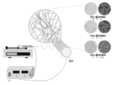
- Figure 9 is a schematic diagram showing the manufacturing process of nanofibers using electrospinning.
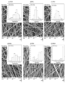
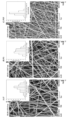
- Figure 10 shows SEM micrographs and the corresponding nanofiber diameter distributions (percentage and frequency of distribution): (a) P10G0, (b) P8G2, (c) P6G4, (d) P4G6, (e) P2G8, and (f) P0G10 nanofiber.
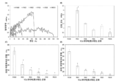
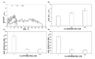
- Figure 11 shows the mechanical properties of PCL/Gel nanofibers: (A) typical stress-strain curve, (B) tensile strength, (C) strain at maximum load, and (D) strain at maximum extension. # p ⁇ 0.05 was considered to be a statistically significant difference compared to P10G0 nanofiber.
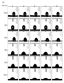
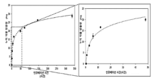
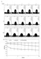
- Figure 21 is a graph showing (A) a representative image and (B) a dynamic water contact angle of the water contact angle of the manufactured nanofibers (n > 3). # p ⁇ 0.05 was considered to be a statistically significant difference compared to the P nanofiber group at the same time.
- Figures 32A and 32B show wound closure of nanofibers in a rectangular full-thickness wound model at 28 days.
- A H&E, Masson's trichrome, and Picro-Sirius Red staining results of mouse skin tissue 28 days after wounding. Scale bar 1000 ⁇ m at 1X and 200 ⁇ m at 5X.
- B Relative wound length and
- C wound thickness of the rectangular full-thickness wound model at day 28. Error bars represent standard deviation (n ⁇ 3). * p ⁇ 0.05 was considered a statistically significant difference compared to the untreated (blank) group.
- the present invention relates to a nanofiber scaffold containing polycaprolactone (poly( ⁇ -caprolactone; PCL); gelatin; and polydeoxyribonucleotide (PDRN) derived from starfish ( Patiria pectinifera ); and a method for manufacturing the same. will be.
- polycaprolactone poly( ⁇ -caprolactone; PCL); gelatin; and polydeoxyribonucleotide (PDRN) derived from starfish ( Patiria pectinifera ); and a method for manufacturing the same.
- the polycaprolactone, gelatin, and polydeoxyribonucleotide may be mixed at a weight ratio of 4 to 8:2 to 6:0.005 to 0.1, for example, 6:4:0.02. It can be done, but is not limited to this.
- PDRN Polydeoxyribonucleotide
- P. pectinifera starfish
- AccuPrep Genomic DNA extraction kit Booneer, Daejeon, Korea
- P. pectinifera was washed three times with tap water to remove epiphytes, salt, and sand attached to the surface, and then carefully washed again with fresh water.
- P. pectinifera frozen at -20°C was freeze-dried and then homogenized to powder.
- Crushed P. pectinifera (1 g) was extracted in 4 ml TL buffer containing Proteinase K (2 mg/ml) and RNase A (2 mg/ml) at 60°C for 2 hours.
- HDF Human dermal fibroblasts
- HaCaT human keratinocytes
- HDFs and HaCaT were incubated with FDA (10 ⁇ g/ml) in serum-free medium for 15 min at 37 °C. and PI (20 ⁇ g/ml). The stained cells were then washed with PBS to remove untreated FDA and PI and residues. FDA appears as green fluorescence in live cells and PI appears as red in dead cells, and was quantitatively analyzed using a fluorescence microscope (Leica DMI3000B).
- HDF was inoculated into 2 ml of culture medium in a 6-well plate at a density of 4 Cells were incubated for 24 hours to attach at a density of approximately 90%. After cells were attached to the plate, a wound line was created with a 2 mm wide plastic pipette tip, and unattached cells were washed with PBS. Then, cells were treated with PDRN at different concentrations and allowed to migrate. Pictures were taken at a magnification of 50 was carried out.
- Collagen production was assessed by Picro-Sirius red staining.
- HDFs were incubated with a staining solution made of Sirius red (0.1%, Sigma) dissolved in a saturated aqueous solution of picric acid (1.3% in water, Sigma) for 2 hours, washed three times with PBS, and then dehydrated.
- the stained crystals were dissolved in 0.1 M NaOH, and the absorbance was measured using a PowerWave XS2 microplate reader (BioTek Instruments, Inc., Winooski, VT, USA). Data were expressed as a percentage of the average collagen production rate ⁇ standard deviation of visualized cells in three replicate experiments.
- HDFs were grown in 100 cm 2 dishes at a density of approximately 2 ⁇ 10 6 cells.
- Cells were treated with PDRN at various concentrations and incubated for 24 hours to check the collagen expression level, and incubated for 30 minutes to analyze the degree of activation of Smad2/3 and mitogen-activated protein kinase (MAPK) pathways.
- MAPK mitogen-activated protein kinase
- mice were sacrificed, skin tissue was removed, and tissue samples were stored in a nitrogen gas tank until further analysis. Frozen tissue samples were homogenized with a Tissue Lyser (SpeedMill PLUS, Jena, Germany) in lysis buffer. Specimens were destroyed using steel beads at 30 cycles/second for 5 minutes.
- Tissue Lyser SpeedMill PLUS, Jena, Germany
- PDRN derived from P. pectinifera were evaluated using MTT assay and FDA/PI staining.
- MTT assay HDF and HaCaT cells were treated with increasing concentrations of PDRN for 1, 3, and 5 days, and for FDA/PI staining, they were treated with increasing concentrations of PDRN for 1 and 3 days.
- PDRN 5-200 ⁇ g/ml significantly increased cell proliferation after 3 days of treatment, with treatment at 50 ⁇ g/ml PDRN showing the highest proliferation ( Figures 2 and 3).
- PDRN did not show toxicity to HaCaT cells, but also showed no significant proliferative effect ( Figures 4 and 5). Based on these results, it was concluded that PDRN at the above concentration (5-50 ⁇ g/ml) showed a proliferative effect on HDF cells without showing toxicity to HaCaT cells.
- PDRN extracted from P. pectinifera The effect on activation of MAPK and Smad2/3 pathways was assessed by Western blot analysis. PDRN significantly increased phosphorylation of ERK and Smad2/3 compared to untreated cells ( Figure 8B). These results suggest that PDRN increases the expression of proteins related to wound healing by activating the phosphorylation of ERK and Smad2/3.
- nanofibers The wetting behavior of nanofibers was evaluated using a contact angle analyzer (SEO Phoenix MT, Suwon, Gyeonggi-do, Korea). The volume of the droplet was 2 ⁇ l, and the contact angle was measured at five separate random locations and averaged. DMEM medium without serum was used as the test solution. Nanofibers of the same size were placed on the sample stage at ambient temperature ( ⁇ 296 K) and the contact angle was measured. Pictures were taken with a digital camera over time and analyzed using image processing software (Image Pro 300).
- the tensile strength of nanofibers was measured using a universal tensile machine (LR5K Plus, Lloyd Instruments). Each sample was cut into dumbbell-shaped strips (15 mm I ordered it.
- the functional groups of pure PCL, pure gelatin, pure PDRN, P nanofiber, PG nanofiber, and PGP nanofiber were analyzed using Fourier transform infrared (FT-IR) spectroscopy (FT-4100, JASCO).
- FT-IR Fourier transform infrared
- FT-4100 Fourier transform infrared spectroscopy
- the IR spectrum showed an average of 30 scans at a frequency of 650-4000 cm -1 at a resolution of 4 cm -1 .
- Thermogravimetric analysis was performed using a Pyris 1 TGA analyzer (PerkinElmer TGA-7, Waltham, MA, USA) with a scan range of 30 °C to 700 °C and a constant heating rate of 20 °C under continuous nitrogen. Calorimetry was performed using differential scanning calorimetry (DSC) under nitrogen flowing at a rate of 10 ml/min. The specimen was pressed into a sealed aluminum pan. Heating cycles were performed until the glass transition temperature (T g ) and melting temperature (T m ) were reached. During the cycle, the sample was heated from 30°C to 180°C at a rate of 10°C. The sample was then cooled using nitrogen at an exponentially decreasing rate.
- DSC differential scanning calorimetry
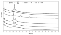
- X-ray diffraction (XRD) analysis of the nanofibers was performed using X-ray diffraction (X'Pert3-Powder, PANalytical, Netherlands) with Cu-K ⁇ radiation. Diffraction intensity was recorded at a scanning speed of 2.4° min -1 in the range of 5 to 90°.
- the weak band at 1440 cm -1 represents the aliphatic CH bending vibration.
- the bands at 1524 and 1237 cm -1 represent stretching vibrations of NH bending and CN stretching, respectively, and are attributed to the characteristic bands of amide II and amide III in gelatin, respectively.
- the FT-IR spectrum of PDRN showed several characteristic peaks of DNA.
- the third peak shows vibration as the -PO 2 vibration of the phosphodieter skeleton in nucleic acid and was recorded at 1287 cm -1 .
- the peak corresponding to the deoxyribose CO stretching vibration appeared at 1033 cm -1 .
- the strong peak at 1020 cm -1 represents furanose vibration, and the next strongest peak at 981 cm -1 is due to CC stretching of the DNA deoxyribose-phosphate backbone.
- the weak vibration appearing near 788 cm -1 is related to deoxy c3'-endo-OPO and represents the A-type DNA conformation. All characteristic peaks of pure PCL, pure gelatin and PDRN are shown in Table 5.
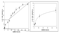
- elution solutions of nanofibers prepared for 1 and 3 days were prepared and analyzed by MTT analysis and FDA and PI fluorescence in HDF and HaCaT. Indirect cytotoxicity was studied by performing live/dead cell staining. The MTT results showed that the elution solution of the prepared nanofibers was not cytotoxic ( Figures 24, 25, and 26). The live cell/dead cell staining results also demonstrated no cytotoxicity, similar to the MTT analysis results.
- H&E hematoxylin and eosin
- the blank group showed a lower density of collagen fibers in the wound area compared to the PGP group. Moreover, the deposition of collagen fibers was smaller and thicker in the nanofiber-implanted group.
Landscapes
- Health & Medical Sciences (AREA)
- Engineering & Computer Science (AREA)
- Chemical & Material Sciences (AREA)
- Animal Behavior & Ethology (AREA)
- Hematology (AREA)
- Materials Engineering (AREA)
- Epidemiology (AREA)
- Life Sciences & Earth Sciences (AREA)
- General Health & Medical Sciences (AREA)
- Public Health (AREA)
- Veterinary Medicine (AREA)
- Textile Engineering (AREA)
- Dispersion Chemistry (AREA)
- Materials For Medical Uses (AREA)
Abstract
La présente invention concerne un échafaudage nanofibreux et son procédé de préparation, l'échafaudage nanofibreux comprenant : poly(ε-caprolactone) (PCL); gélatine; et polydésoxyribonucléotide (PDRN) dérivé de Patiria pectinifera. L'échafaudage nanofibreux de la présente invention a une structure de nanofibres uniformes, une excellente absorption et rétention de fluide, une vitesse de libération adéquate, une stabilité mécanique élevée, une stabilité thermique, et est ainsi approprié pour un pansement pour la cicatrisation de plaies.
Applications Claiming Priority (2)
| Application Number | Priority Date | Filing Date | Title |
|---|---|---|---|
| KR1020220185655A KR102873915B1 (ko) | 2022-12-27 | 2022-12-27 | 별불가사리 유래 폴리데옥시리보뉴클레오티드를 포함하는 피부 재생용 나노파이버 스캐폴드 |
| KR10-2022-0185655 | 2022-12-27 |
Publications (1)
| Publication Number | Publication Date |
|---|---|
| WO2024143784A1 true WO2024143784A1 (fr) | 2024-07-04 |
Family
ID=91718052
Family Applications (1)
| Application Number | Title | Priority Date | Filing Date |
|---|---|---|---|
| PCT/KR2023/014358 Ceased WO2024143784A1 (fr) | 2022-12-27 | 2023-09-21 | Échafaudage nanofibreux pour la régénération de la peau, comprenant du polydésoxyribonucléotide dérivé de patiria pectinifera |
Country Status (2)
| Country | Link |
|---|---|
| KR (1) | KR102873915B1 (fr) |
| WO (1) | WO2024143784A1 (fr) |
Families Citing this family (4)
| Publication number | Priority date | Publication date | Assignee | Title |
|---|---|---|---|---|
| KR102855769B1 (ko) | 2024-10-23 | 2025-09-09 | 애경산업(주) | 쑥 유래 폴리데옥시리보뉴클레오티드를 유효성분으로 함유하는 피부외용제 조성물 및 약학 조성물 |
| KR102798269B1 (ko) | 2024-10-23 | 2025-04-24 | 애경산업(주) | 브루셀스프라우트 유래 폴리데옥시리보뉴클레오티드를 유효성분으로 함유하는 피부외용제 조성물 및 약학 조성물 |
| KR102866930B1 (ko) | 2025-06-10 | 2025-10-02 | 애경산업(주) | 자작나무속 식물 유래 폴리데옥시리보뉴클레오티드를 유효성분으로 함유하는 화장료 조성물 및 약학 조성물 |
| KR102866929B1 (ko) | 2025-06-10 | 2025-10-02 | 애경산업(주) | 루콜라 유래 폴리데옥시리보뉴클레오티드를 유효성분으로 함유하는 화장료 조성물 및 약학 조성물 |
Citations (5)
| Publication number | Priority date | Publication date | Assignee | Title |
|---|---|---|---|---|
| JP2011525495A (ja) * | 2008-06-23 | 2011-09-22 | イノバクティブ インコーポレイティド | ヒトデ体液を含んでなる化粧組成物及びその使用方法。 |
| KR20170096842A (ko) * | 2016-02-17 | 2017-08-25 | 남기종 | 불가사리로부터 피부 재생 인자 및 세포 성장 인자의 추출 방법 및 이의 응용 |
| KR20180048520A (ko) * | 2018-04-27 | 2018-05-10 | 주식회사 한국비엔씨 | 폴리데옥시리보뉴클레오타이드를 이용한 항염 및 피부 재생용 조성물의 제조방법 |
| KR20200112256A (ko) * | 2019-03-21 | 2020-10-05 | 순천향대학교 산학협력단 | 전기방사 폴리카프로락톤/젤라틴/β-TCP 매트와 알지네이트/젤라틴 하이드로젤로 구성된 골 유도형 이중막 제조방법 |
| KR102308773B1 (ko) * | 2018-09-06 | 2021-10-06 | 미국 마린 에센스 바이오사이언시즈 코퍼레이션 | 조직 유도 재생용 바이오물질 장치 및 국소 조성물 |
Family Cites Families (3)
| Publication number | Priority date | Publication date | Assignee | Title |
|---|---|---|---|---|
| KR101005079B1 (ko) * | 2008-10-23 | 2010-12-30 | 금오공과대학교 산학협력단 | 유착방지막 용도의 생분해성 나노섬유시트 및 그 제조방법 |
| KR20100133117A (ko) | 2009-06-11 | 2010-12-21 | 주식회사 아모텍 | 드레싱용 나노섬유와, 이를 이용한 드레싱 복합체 및 그의 제조방법 |
| KR102694006B1 (ko) * | 2021-03-08 | 2024-08-09 | 차의과학대학교 산학협력단 | 생리활성물질을 포함하는 생분해성 고분자 지지체 및 이의 제조방법 |
-
2022
- 2022-12-27 KR KR1020220185655A patent/KR102873915B1/ko active Active
-
2023
- 2023-09-21 WO PCT/KR2023/014358 patent/WO2024143784A1/fr not_active Ceased
Patent Citations (5)
| Publication number | Priority date | Publication date | Assignee | Title |
|---|---|---|---|---|
| JP2011525495A (ja) * | 2008-06-23 | 2011-09-22 | イノバクティブ インコーポレイティド | ヒトデ体液を含んでなる化粧組成物及びその使用方法。 |
| KR20170096842A (ko) * | 2016-02-17 | 2017-08-25 | 남기종 | 불가사리로부터 피부 재생 인자 및 세포 성장 인자의 추출 방법 및 이의 응용 |
| KR20180048520A (ko) * | 2018-04-27 | 2018-05-10 | 주식회사 한국비엔씨 | 폴리데옥시리보뉴클레오타이드를 이용한 항염 및 피부 재생용 조성물의 제조방법 |
| KR102308773B1 (ko) * | 2018-09-06 | 2021-10-06 | 미국 마린 에센스 바이오사이언시즈 코퍼레이션 | 조직 유도 재생용 바이오물질 장치 및 국소 조성물 |
| KR20200112256A (ko) * | 2019-03-21 | 2020-10-05 | 순천향대학교 산학협력단 | 전기방사 폴리카프로락톤/젤라틴/β-TCP 매트와 알지네이트/젤라틴 하이드로젤로 구성된 골 유도형 이중막 제조방법 |
Also Published As
| Publication number | Publication date |
|---|---|
| KR20240103455A (ko) | 2024-07-04 |
| KR102873915B1 (ko) | 2025-10-20 |
Similar Documents
| Publication | Publication Date | Title |
|---|---|---|
| WO2024143784A1 (fr) | Échafaudage nanofibreux pour la régénération de la peau, comprenant du polydésoxyribonucléotide dérivé de patiria pectinifera | |
| Siritienthong et al. | Development of ethyl alcohol-precipitated silk sericin/polyvinyl alcohol scaffolds for accelerated healing of full-thickness wounds | |
| Lashkari et al. | Cell-based wound dressing: Bilayered PCL/gelatin nanofibers-alginate/collagen hydrogel scaffold loaded with mesenchymal stem cells | |
| Zhou et al. | An injectable carboxymethyl chitosan hydrogel scaffold formed via coordination bond for antibacterial and osteogenesis in osteomyelitis | |
| Shen et al. | Bilayer silk fibroin/sodium alginate scaffold promotes vascularization and advances inflammation stage in full-thickness wound | |
| Huang et al. | Functional improvement and neurogenesis after collagen-GAG matrix implantation into surgical brain trauma | |
| Arasteh et al. | Fabrication and characterization of nano-fibrous bilayer composite for skin regeneration application | |
| Kim et al. | PCL/gelatin nanofibers incorporated with starfish polydeoxyribonucleotides for potential wound healing applications | |
| Sapru et al. | Non-immunogenic, porous and antibacterial chitosan and Antheraea mylitta silk sericin hydrogels as potential dermal substitute | |
| Tabaei et al. | Chitosan-based nano-scaffolds as antileishmanial wound dressing in BALB/c mice treatment: Characterization and design of tissue regeneration | |
| Augustine et al. | Stromal cell-derived factor loaded co-electrospun hydrophilic/hydrophobic bicomponent membranes for wound protection and healing | |
| WO2016010330A1 (fr) | Compostion de mousse polymère, procédé de préparation de la composition de mousse polymère à l'aide de celle-ci, et mousse polymère pour emballage | |
| Singaravelu et al. | Dual-layered 3D nanofibrous matrix incorporated with dual drugs and their synergetic effect on accelerating wound healing through growth factor regulation | |
| WO2014092239A1 (fr) | Produit d'étanchéité de tissu, dans lequel sont mélangés du collagène et de la fibrine, et son procédé de préparation | |
| Indrakumar et al. | Photopolymerized silk fibroin gel for advanced burn wound care | |
| Foroughi et al. | Manufacturing ulvan biopolymer for wound dressings | |
| WO2019221360A1 (fr) | Pansement partiellement durci de membrane amniotique de type lentille de contact et procédé pour sa fabrication | |
| WO2017069365A1 (fr) | Biomembrane artificielle utilisant une matrice de soie, et son procédé de fabrication | |
| WO2021167330A1 (fr) | Développement de couche dermique à retrait régulé, et fabrication de peau artificielle ayant une performance uniforme à l'aide de ce dernier | |
| WO1994017840A1 (fr) | Compositions pharmaceutiques comprenant une substance spongieuse composee de derives ester d'acide hyaluronique en combinaison avec d'autres substances pharmacologiquement actives | |
| Liu et al. | An injectable photocuring silk fibroin-based hydrogel for constructing an antioxidant microenvironment for skin repair | |
| WO2016195152A1 (fr) | Procédé de fabrication de film de collagène à l'aide de lumière ultraviolette, film de collagène fabriqué a l'aide de ce dernier, et matériau biologique préparé à l'aide du film de collagène | |
| CN106139238A (zh) | 一种壳聚糖胶原海绵敷料及其制备方法 | |
| WO2012081944A2 (fr) | Membrane dentaire et procédé de fabrication associé | |
| KR101714695B1 (ko) | 가교화된 pva-ecm 복합체를 제조하는 방법 및 그에 의하여 제조된 pva-ecm 복합체 |
Legal Events
| Date | Code | Title | Description |
|---|---|---|---|
| 121 | Ep: the epo has been informed by wipo that ep was designated in this application |
Ref document number: 23912467 Country of ref document: EP Kind code of ref document: A1 |
|
| NENP | Non-entry into the national phase |
Ref country code: DE |
|
| 122 | Ep: pct application non-entry in european phase |
Ref document number: 23912467 Country of ref document: EP Kind code of ref document: A1 |