WO2024257872A1 - Procédé pour prédire le taux de transfert génique - Google Patents
Procédé pour prédire le taux de transfert génique Download PDFInfo
- Publication number
- WO2024257872A1 WO2024257872A1 PCT/JP2024/021754 JP2024021754W WO2024257872A1 WO 2024257872 A1 WO2024257872 A1 WO 2024257872A1 JP 2024021754 W JP2024021754 W JP 2024021754W WO 2024257872 A1 WO2024257872 A1 WO 2024257872A1
- Authority
- WO
- WIPO (PCT)
- Prior art keywords
- acid
- cells
- gene
- car
- cell
- Prior art date
- Legal status (The legal status is an assumption and is not a legal conclusion. Google has not performed a legal analysis and makes no representation as to the accuracy of the status listed.)
- Ceased
Links
Images








Classifications
-
- C—CHEMISTRY; METALLURGY
- C07—ORGANIC CHEMISTRY
- C07K—PEPTIDES
- C07K14/00—Peptides having more than 20 amino acids; Gastrins; Somatostatins; Melanotropins; Derivatives thereof
- C07K14/435—Peptides having more than 20 amino acids; Gastrins; Somatostatins; Melanotropins; Derivatives thereof from animals; from humans
-
- C—CHEMISTRY; METALLURGY
- C07—ORGANIC CHEMISTRY
- C07K—PEPTIDES
- C07K16/00—Immunoglobulins [IG], e.g. monoclonal or polyclonal antibodies
-
- C—CHEMISTRY; METALLURGY
- C12—BIOCHEMISTRY; BEER; SPIRITS; WINE; VINEGAR; MICROBIOLOGY; ENZYMOLOGY; MUTATION OR GENETIC ENGINEERING
- C12N—MICROORGANISMS OR ENZYMES; COMPOSITIONS THEREOF; PROPAGATING, PRESERVING, OR MAINTAINING MICROORGANISMS; MUTATION OR GENETIC ENGINEERING; CULTURE MEDIA
- C12N15/00—Mutation or genetic engineering; DNA or RNA concerning genetic engineering, vectors, e.g. plasmids, or their isolation, preparation or purification; Use of hosts therefor
- C12N15/09—Recombinant DNA-technology
- C12N15/11—DNA or RNA fragments; Modified forms thereof; Non-coding nucleic acids having a biological activity
- C12N15/62—DNA sequences coding for fusion proteins
-
- C—CHEMISTRY; METALLURGY
- C12—BIOCHEMISTRY; BEER; SPIRITS; WINE; VINEGAR; MICROBIOLOGY; ENZYMOLOGY; MUTATION OR GENETIC ENGINEERING
- C12N—MICROORGANISMS OR ENZYMES; COMPOSITIONS THEREOF; PROPAGATING, PRESERVING, OR MAINTAINING MICROORGANISMS; MUTATION OR GENETIC ENGINEERING; CULTURE MEDIA
- C12N5/00—Undifferentiated human, animal or plant cells, e.g. cell lines; Tissues; Cultivation or maintenance thereof; Culture media therefor
- C12N5/06—Animal cells or tissues; Human cells or tissues
-
- C—CHEMISTRY; METALLURGY
- C12—BIOCHEMISTRY; BEER; SPIRITS; WINE; VINEGAR; MICROBIOLOGY; ENZYMOLOGY; MUTATION OR GENETIC ENGINEERING
- C12N—MICROORGANISMS OR ENZYMES; COMPOSITIONS THEREOF; PROPAGATING, PRESERVING, OR MAINTAINING MICROORGANISMS; MUTATION OR GENETIC ENGINEERING; CULTURE MEDIA
- C12N5/00—Undifferentiated human, animal or plant cells, e.g. cell lines; Tissues; Cultivation or maintenance thereof; Culture media therefor
- C12N5/10—Cells modified by introduction of foreign genetic material
-
- C—CHEMISTRY; METALLURGY
- C12—BIOCHEMISTRY; BEER; SPIRITS; WINE; VINEGAR; MICROBIOLOGY; ENZYMOLOGY; MUTATION OR GENETIC ENGINEERING
- C12Q—MEASURING OR TESTING PROCESSES INVOLVING ENZYMES, NUCLEIC ACIDS OR MICROORGANISMS; COMPOSITIONS OR TEST PAPERS THEREFOR; PROCESSES OF PREPARING SUCH COMPOSITIONS; CONDITION-RESPONSIVE CONTROL IN MICROBIOLOGICAL OR ENZYMOLOGICAL PROCESSES
- C12Q1/00—Measuring or testing processes involving enzymes, nucleic acids or microorganisms; Compositions therefor; Processes of preparing such compositions
- C12Q1/02—Measuring or testing processes involving enzymes, nucleic acids or microorganisms; Compositions therefor; Processes of preparing such compositions involving viable microorganisms
- C12Q1/04—Determining presence or kind of microorganism; Use of selective media for testing antibiotics or bacteriocides; Compositions containing a chemical indicator therefor
- C12Q1/06—Quantitative determination
-
- C—CHEMISTRY; METALLURGY
- C12—BIOCHEMISTRY; BEER; SPIRITS; WINE; VINEGAR; MICROBIOLOGY; ENZYMOLOGY; MUTATION OR GENETIC ENGINEERING
- C12Q—MEASURING OR TESTING PROCESSES INVOLVING ENZYMES, NUCLEIC ACIDS OR MICROORGANISMS; COMPOSITIONS OR TEST PAPERS THEREFOR; PROCESSES OF PREPARING SUCH COMPOSITIONS; CONDITION-RESPONSIVE CONTROL IN MICROBIOLOGICAL OR ENZYMOLOGICAL PROCESSES
- C12Q1/00—Measuring or testing processes involving enzymes, nucleic acids or microorganisms; Compositions therefor; Processes of preparing such compositions
- C12Q1/68—Measuring or testing processes involving enzymes, nucleic acids or microorganisms; Compositions therefor; Processes of preparing such compositions involving nucleic acids
-
- G—PHYSICS
- G01—MEASURING; TESTING
- G01N—INVESTIGATING OR ANALYSING MATERIALS BY DETERMINING THEIR CHEMICAL OR PHYSICAL PROPERTIES
- G01N33/00—Investigating or analysing materials by specific methods not covered by groups G01N1/00 - G01N31/00
- G01N33/48—Biological material, e.g. blood, urine; Haemocytometers
- G01N33/50—Chemical analysis of biological material, e.g. blood, urine; Testing involving biospecific ligand binding methods; Immunological testing
Definitions
- the present invention relates to a method for predicting a gene transfer rate and/or the number of gene-transferred cells.
- Non-Patent Document 1 reports that CAR-T cells were measured using flow cytometry.
- Cytometry Part B Clinical Cytometry, Volume 100, Issue 2, p. 218-224
- the present invention aims to provide a method for predicting the gene transfer rate and/or the number of gene-transferred cells, which enables the measurement of the gene transfer rate and/or the number of gene-transferred cells in a simpler and more efficient manner than conventional methods.
- the inventors discovered that there are differences in the components of the culture supernatant between transfected and non-transfected cells, and discovered that by analyzing the medium in which the cells are cultured and using the medium components (e.g., metabolites in the medium, pH, extracellular vesicles) as indicators, it is possible to simply and efficiently predict the gene transfer rate and/or the number of transfected cells in animal cells.
- the medium components e.g., metabolites in the medium, pH, extracellular vesicles
- the present invention was completed based on these findings and through further investigation, and provides the following method for predicting the gene transfer rate and/or the number of gene-transferred cells in animal cells.
- a method for predicting a gene transfer rate and/or a number of gene-transferred cells in an animal cell comprising the steps of: A method comprising: (1) measuring components of a culture supernatant in which genetically-introduced animal cells have been cultured; and (2) predicting the gene introduction rate and/or the number of genetically-introduced cells based on the values measured in step (1).
- a method comprising: (1) measuring components of a culture supernatant in which genetically-introduced animal cells have been cultured; and (2) predicting the gene introduction rate and/or the number of genetically-introduced cells based on the values measured in step (1).
- the metabolite is at least one selected from the group consisting of organic acids, sugars, lipids, vitamins, amines, nucleic acids, amino acids and other metabolite ions.
- the organic acid is at least one selected from the group consisting of decenoic acid, dodecenoic acid, myristoleic acid, palmitic acid, palmitoleic acid, hexadecadienoic acid, stearic acid, oleic acid, linoleic acid, ⁇ -linolenic acid, ⁇ -linolenic acid, arachidic acid, eicosenoic acid, eicosadienoic acid, mead acid, arachidonic acid, docosapentaenoic acid, docosahexaenoic acid, nervonic acid, 2-hydroxyvaleric acid, 2-oxoglutaric acid, indoxyl s
- [5] The method according to [3] or [4], wherein the sugar is at least one selected from the group consisting of fructose, inositol, galactose, glucose, and sucrose.
- the lipid is at least one selected from the group consisting of ceramide, Hex2-ceramide, Hex-ceramide, cholesterol, cholesterol sulfate, cardiolipin, monoglycerol, lysophosphatidylcholine, lysophosphatidylethanolamine, phosphatidic acid, phosphatidylcholine, phosphatidylethanolamine, phosphatidylglycerol, phosphatidylinositol, phosphatidylserine, sphingomyelin, dihydrosphingomyelin, triglyceride, glycerol-3-phosphate, and ganglioside GM3.
- amino acid is at least one selected from the group consisting of acylcarnitine, alanine, arginine, asparagine, aspartic acid, ⁇ -alanine, cysteine, cysteine-HPE-AM, cysteine-SS-HPE-AM, dimethylglycine, glutamic acid, glutamine, glycine, isoleucine, leucine, leucine acid, lysine, methionine, homocysteine-HPE-AM, N-acetyl- ⁇ -alanine, N-acetylcysteine, N-acetylserine, N-acetyltryptophan, N-carbamoyl-aspartate, O-acetylserine, ornithine, pyroglutamic acid, phenylalanine, proline, serine, S-sulfocysteine, taurine
- a method for producing a gene-introduced cell preparation comprising the steps of measuring components of a culture supernatant in which animal cells are cultured, and determining the gene introducion rate and/or the number of gene-introduced cells based on the measured values.
- the method for predicting the gene introduction rate and/or the number of gene-introduced cells of the present invention does not require complicated steps compared to conventional methods, and allows the gene introduction rate and/or the number of gene-introduced cells to be measured with simple operations (e.g., the only operation required is automatic measurement using a measuring device). Furthermore, the method for predicting the gene introduction rate and/or the number of gene-introduced cells of the present invention allows the gene introduction rate of cells to be measured efficiently (e.g., in a short time of less than 5 minutes). In addition, since the culture supernatant is used, cell sampling is not required, so there is no stirring process and damage to the cells is minimal.
- the CAR introduction rate and the number of introduced cells can be grasped easily and quickly without damaging the cells, making it possible to confirm whether a product that meets the standards has been manufactured.
- Example 1 shows the results of Example 1.
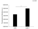
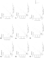
- Fig. 1 shows the results of Example 1. Graph showing each component in the supernatant after culture of control T cells (UTD) not transfected with a gene and CAR-T cells transfected with a CAR gene.
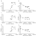
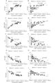
- Figure 4 shows the results of Example 4. Graphs showing the correlation between each component in the culture supernatant on the final day of culture and cell density or CAR-introduced T cell (CAR-T) density (C-D: lactic acid, E-F: ammonium ion). Figure 4 shows the results of Example 4. Graphs showing the correlation between each component in the culture supernatant on the final day of culture and cell density or CAR-introduced T cell (CAR-T) density (GH: pH, I-J: glutamine, K-L: glucose). Fig. 4 shows the results of Example 4.
- CAR-T CAR-introduced T cell
- Fig. 6 shows the results of Example 5.
- Fig. 6 is a graph showing (A) cell density and (B) CAR introduction efficiency at each date during the amplification step.
- Figure 5 shows the results of Example 5.
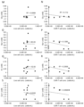
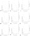
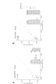
- Example 6 Graph showing the correlation between each component in the culture supernatant and cell density or CAR-T cell density throughout the entire period of the amplification process (A-B: lactate, C-D: ammonium ion, E-F: pH, G-H: glutamine, I-J: glucose). This shows the results of Example 6.
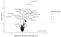
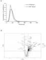
- This graph uses a volcano plot to show the differences in components in the supernatant between UTD and CAR-T cells.
- the horizontal axis shows the estimated amount of change under conditions with the highest CAR introduction efficiency, obtained by performing a Williams test on the difference in the amount of each component between UTD and CAR-T cells.
- the left side shows the components that decreased in CAR-T cells, and the right side shows the components that increased in CAR-T cells.
- the vertical axis also shows the -Log(p value) of the Williams test on the difference in the amount of each component at each date during the amplification process.
- Fig. 7 shows the results of Example 7.
- Fig. 7 shows the results of Example 7.
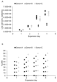
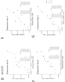
- A-C Graphs showing prediction of the number of CAR-T cells (cells/mL) by LASSO regression analysis from components in the culture supernatant at each date during the amplification process.
- Fig. 7 shows the results of Example 7.
- Figure showing the results of Example 7. Graph showing prediction of CAR-T cell count (cells/mL) by simple regression analysis from components in the culture supernatant during the entire amplification process.
- FIG. 7 shows the results of Example 7.
- Fig. 1 shows the results of Example 8.
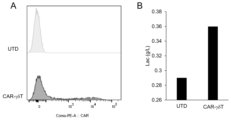
- Graphs showing (A) CAR transfection efficiency and (B) lactic acid in the supernatant after culture of non-gene-transfected control SK-HEP-1 cells (UTD) and CAR gene-transfected SK-HEP-1 cells (N 4, *p ⁇ 0.05, p value analyzed by t-test).
- Fig. 13 shows the results of Example 9.
- Fig. 1 shows the results of Example 10.
- Fig. 11 shows the results of Example 11.
- A Graph showing the particle size of exosomes in the supernatants of UTD and CAR-T cells
- C A table showing proteins whose expression levels are predominantly changed in UTD and exosomes derived from CAR-T cells.
- Fig. 1 shows the results of Example 11.
- D Graph showing the correlation between the number of CAR-T cells and CD63-positive exosomes.
- Fig. 12 shows the results of Example 12.
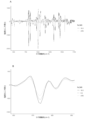
- A shows an example of the entire second derivative of the Raman spectroscopy spectrum obtained for the UTD and the culture supernatant of CAR-T cells
- B shows an example of a peak that has a difference.
- Comprise(s) means the inclusion, but not the limitation, of the elements that follow the phrase. Thus, it indicates the inclusion of the elements that follow the phrase, but not the exclusion of any other elements.
- Consist(s) of means the inclusion, and the limitation, of any elements that follow the phrase. Thus, the phrase “consisting of” indicates that the recited elements are required or essential, and that other elements are substantially absent.
- Consist(s) essentially of means the inclusion, and the limitation, of any elements that follow the phrase, and that other elements do not affect the activity or function of the element identified in this disclosure. Thus, the phrase “consisting essentially of” indicates that the recited elements are required or essential, but that other elements are optional and may or may not be present depending on whether they affect the activity or function of the recited elements.
- pluripotent stem cells refers to embryonic stem cells (ES cells) and cells that have the same differentiation pluripotency, i.e., the potential ability to differentiate into various tissues of the body (all of the endoderm, mesoderm, and ectoderm).
- Cells that have the same differentiation pluripotency as ES cells include "induced pluripotent stem cells” (sometimes referred to as “iPS cells” in this specification).
- iPS cells induced pluripotent stem cells
- the pluripotent stem cells are ES cells or any cells derived from a human embryo, the cells may be cells produced by destroying an embryo or cells produced without destroying an embryo, and are preferably cells produced without destroying an embryo.
- a “cell population” refers to two or more cells of the same or different types.
- a “cell population” also refers to a mass of cells of the same or different types.
- culture refers to maintaining and/or growing cells in an in vitro environment.
- “Culturing” refers to sustaining and/or growing cells outside a tissue or outside the body, for example in a cell culture dish or flask.
- the present invention provides a method for predicting the gene introduction rate and/or the number of gene-introduced cells in animal cells, comprising: (1) a step of measuring components of a culture supernatant in which gene-introduced animal cells have been cultured; and (2) a step of predicting the gene introduction rate and/or the number of gene-introduced cells based on the values measured in step (1).
- the "gene transfer rate” refers to the rate of transfer of a nucleic acid into a cell, and is, for example, the proportion of cells into which a nucleic acid has been transferred among all cells, the amount of nucleic acid taken up in a cell population, or the expression rate of a transferred gene in a whole cell population.
- the transferred gene is a CAR, it may also be referred to as the CAR positive rate.
- the "number of gene-transduced cells” refers to the number of cells into which a gene has been introduced in a cell population.
- the transduced gene is CAR, it may also be referred to as the number of CAR-positive cells.
- the term "and/or" in the "method for predicting the gene transfer rate and/or the number of gene-transferred cells in animal cells" of the present invention means that the prediction method of the present invention is a method for predicting both the gene transfer rate and the number of gene-transferred cells, or a method for measuring either one of them. More preferably, it is a method for predicting the number of gene-transferred cells.
- Step (1) components of the culture supernatant in which gene-transfected animal cells have been cultured are measured.
- animal cell is not particularly limited, and various animal cells can be widely used.
- animal cell types include spleen cells, nerve cells, glial cells, pancreatic ⁇ cells, bone marrow cells, mesangial cells, Langerhans cells, epidermal cells, epithelial cells, endothelial cells, fibroblasts, fibrocytes, muscle cells (e.g., skeletal muscle cells, cardiac muscle cells, myoblasts, muscle satellite cells), adipocytes, immune cells (e.g., macrophages, T cells, B cells, natural killer cells (NK cells), mast cells, neutrophils, basophils, eosinophils, monocytes, megakaryocytes), synovial cells, chondrocytes, bone cells, osteoblasts, osteoclasts, mammary gland cells, hepatocytes, interstitial cells, egg cells, and sperm cells, as well as stem cells that can be induced to differentiate into these cells (including pluripotent stem cells such as neural
- T cells include ⁇ T cells, ⁇ T cells, helper T cells, cytotoxic T cells, regulatory T cells, suppressor T cells, tumor-infiltrating T cells, memory T cells, naive T cells, NKT cells, TCR-T cells, STAR receptor T cells, CAR-T cells (e.g., CAR- ⁇ T cells, CAR-gdT cells), etc.
- animal cells also include the above cells produced by inducing differentiation in vitro of primary cells, the above stem cells (e.g., iPS cells), etc.
- animal cells also include various types of cancer cells. Animal cells may include only one type, or two or more types.
- the organism from which the animal cells are derived is not particularly limited, and examples of such organisms include mammals, such as humans, mice, rats, cows, horses, pigs, rabbits, dogs, cats, goats, monkeys, and chimpanzees. Among these, humans are preferred.
- Animal cells are animal cells into which a foreign gene has been introduced.
- An “exogenous gene” is a gene or monomeric nucleotide that is introduced from the outside in order to cause an animal cell to express a desired protein, and can be selected appropriately depending on the use of the animal cell.
- animal cells into which a gene has been introduced includes both animal cells into which a gene has actually been introduced in an attempt to introduce a gene, and animal cells into which no gene has been introduced.
- (animal) cells into which a gene has been introduced” means (animal) cells into which a gene has actually been introduced.
- Cells into which a gene has been introduced and “cells into which a gene has been introduced” can be used interchangeably depending on the context.
- the foreign gene may be, for example, a gene for expressing a CAR (chimeric antigen receptor).
- the foreign gene may be, for example, a gene for expressing a CAR and a gene for expressing a cytokine and/or a chemokine.
- a CAR expressed by an animal cell is basically, like a general or known CAR, composed of peptides at each of the following sites linked via a spacer as necessary: (i) an antigen recognition site (e.g., a single-chain antibody, a ligand, a peptide, etc.) that recognizes a cell surface antigen of a cancer cell; (ii) a cell membrane-spanning domain; and (iii) a signal transduction domain that induces T cell activation.
- an antigen recognition site e.g., a single-chain antibody, a ligand, a peptide, etc.
- TCR T cell receptor
- STAR synthetic T cell receptor and antigen receptor
- chimeric TAC T cell antigen coupler
- suicide genes iCas9, HSV-TK, etc.
- cytokines interleukins, chemokines, etc.
- T cell activation inhibitors molecules that suppress the expression of immune checkpoint factors (CTLA-4, PD-1, TIM-3, LAG-3, TGIT, BTLA, VISTA (PD-1H), etc.), CD160, Cbl-b, endogenous TCR, etc.
- T cell activation promoters CD28, ICOS, CD137, 0X40, CD27, GITR, BAFFR, TACI, BMCA, CD40L, etc.
- the expression vector may be linear or circular, and may be a non-viral vector such as a plasmid, a viral vector, or a transposon-based vector.
- the technique for introducing the expression vector into the animal cell can be an appropriate one depending on the embodiment.
- the expression vector can be introduced into the animal cell by known methods such as the viral infection method, the calcium phosphate method, the lipofection method, the microinjection method, and the electroporation method.
- the expression vector can be prepared in a form suitable for use in each technique by known means or, as appropriate, using a commercially available kit (following the instructions for the kit).
- the expression vector can be introduced into animal cells by viral infection.
- viral vectors include retroviral vectors, lentiviral vectors, adenoviral vectors, and adeno-associated viral vectors.
- retroviral vectors When using these viral vectors, a vector containing the desired foreign gene and the packaging vector (plasmid) of each virus are transfected into host cells using a corresponding commercially available kit to produce a recombinant virus, and then the resulting recombinant virus is used to infect animal cells.
- all of the foreign genes may be contained in one expression vector, all of the foreign genes may be contained in separate expression vectors, or some of the multiple foreign genes may be contained in one expression vector and the rest may be contained in separate expression vectors.
- multiple foreign genes are contained in one expression vector, there is no particular limitation on the order in which the foreign genes are arranged from upstream to downstream.
- the foreign gene can be composed of a nucleic acid (polynucleotide) having a base sequence that codes for the amino acid sequence of the desired protein or polypeptide.
- a person skilled in the art can design and prepare an expression vector that can express the desired protein (polypeptide) in an animal cell.
- the nucleic acid contained in the expression vector can be prepared by a chemical DNA synthesis reaction, or can be prepared as cDNA (cloned).
- the expression vector may contain sequences such as a promoter, terminator, enhancer, initiation codon, termination codon, polyadenylation signal, nuclear localization signal (NLS), multiple cloning site (MCS), etc., as necessary.
- the expression vector may further include nucleic acids (base sequences) encoding "functional genes" such as reporter genes (e.g., genes encoding fluorescent proteins of various colors), drug selection genes (e.g., kanamycin resistance genes, ampicillin resistance genes, puromycin resistance genes), and suicide genes (e.g., genes encoding diphtheria A toxin, herpes simplex thymidine kinase (HSV-TK), carboxypeptidase G2 (CPG2), carboxylesterase (CA), cytosine deaminase (CD), cytochrome P450 (cyt-450), deoxycytidine kinase (dCK), nitroreductase (NR), purine nucleoside phosphorylase (PNP), thymidine phosphorylase (TP), varicella zoster virus thymidine kinase (VZV-TK), xanthine-guanine phosphoribosyltransfera
- Nucleic acid refers to any molecule formed by polymerizing monomeric nucleotides or nucleotides and molecules having equivalent functions to the nucleotides, such as RNA, which is a polymer of ribonucleotides, DNA, which is a polymer of deoxyribonucleotides, a mixed polymer of ribonucleotides and deoxyribonucleotides, and a nucleotide polymer containing a nucleotide analogue, and may also be a nucleotide polymer containing a nucleic acid derivative.
- the nucleic acid may be a single-stranded nucleic acid or a double-stranded nucleic acid. Double-stranded nucleic acids also include double-stranded nucleic acids in which one strand hybridizes to the other strand under stringent conditions.
- Nucleotide analogues may be any molecule that is a ribonucleotide, deoxyribonucleotide, RNA or DNA modified to improve or stabilize nuclease resistance, increase affinity with complementary nucleic acid strands, increase cell permeability or to make it visible, as compared to RNA or DNA.
- Nucleotide analogues may be naturally occurring or non-naturally occurring molecules, such as sugar-modified nucleotide analogues (e.g., nucleotide analogues substituted with 2'-O-methylribose, nucleotide analogues substituted with 2'-O-propylribose, nucleotide analogues substituted with 2'-methoxyethoxyribose, nucleotide analogues substituted with 2'-O-methoxyethylribose, nucleotide analogues substituted with 2'-O-[2-(guanidinium)ethyl]ribose, nucleotide analogues substituted with 2'-fluororibose, bridged structure-type artificial nucleotide analogues, etc.
- sugar-modified nucleotide analogues e.g., nucleotide analogues substituted with
- nucleic acids BNA: Bridged Nucleic Acid
- locked artificial nucleic acid LNA: Locked Nucleic Acid
- ethylene bridged artificial nucleic acid ENA: Ethylene bridged nucleic acid acid
- PNA peptide nucleic acid
- OPNA oxypeptide nucleic acid
- PRNA peptide ribonucleic acid
- nucleotide analogues modified with phosphodiester bonds e.g. nucleotide analogues substituted with phosphorothioate bonds, nucleotide analogues substituted with N3'-P5' phosphoamidate bonds
- a nucleic acid derivative may be any molecule in which another chemical substance has been added to the nucleic acid in order to improve nuclease resistance, stabilize the nucleic acid, increase affinity with a complementary nucleic acid strand, increase cell permeability, or make it visible, and specific examples include 5'-polyamine-added derivatives, cholesterol-added derivatives, steroid-added derivatives, bile acid-added derivatives, vitamin-added derivatives, Cy5-added derivatives, Cy3-added derivatives, 6-FAM-added derivatives, and biotin-added derivatives.
- a protein, siRNA, shRNA, dsRNA, miRNA, antisense nucleic acid, etc. may be introduced into the animal cell, and even if other substances besides a foreign gene are introduced into the animal cell, the gene introduction rate of the cells can be measured by measuring the components of the culture supernatant.
- the medium used in the culture in step (1) can be a basal medium that is used for culturing animal cells.
- the basal medium is not particularly limited as long as it can be used for culturing animal cells, and examples thereof include AIMV, X-VIVO-15, NeuroBasal, EGM2, TeSR, BME, BGJb, CMRL 1066, Glasgow MEM, improved MEM zinc option, IMDM, 199 medium, Eagle MEM, ⁇ MEM, DMEM, Ham, RPMI-1640, Fisher medium, and various commercially available products for culturing T cells (e.g., CTS TM OpTmizer TM T-Cell Expansion Basal Medium (Thermo Fisher Scientific), CTS OpTmizer Pro Serum Free Medium (Thermo Fisher Scientific), CTS OpTmizer Pro TM (Thermo Fisher Scientific), CTS TM OpTmizer TM T-Cell Expansion Supplement (Thermo Fisher Scientific), 4Cell Nutri-T GMP lymphocyte medium (
- the medium may contain serum or may be serum-free.
- the medium may also contain serum substitutes (e.g., albumin, transferrin, Knockout Serum Replacement (KSR), CTS Immune Cell SR (Thermo Fisher Scientific), fatty acids, insulin, collagen precursors, trace elements, 2-mercaptoethanol, 3'-thiolglycerol, ITS-supplement, B27 (trademark) supplement, etc.).
- serum substitutes e.g., albumin, transferrin, Knockout Serum Replacement (KSR), CTS Immune Cell SR (Thermo Fisher Scientific)
- fatty acids e.g., insulin, collagen precursors, trace elements, 2-mercaptoethanol, 3'-thiolglycerol, ITS-supplement, B27 (trademark) supplement, etc.
- the medium may also contain one or more substances such as lipids, amino acids (non-essential amino acids, etc.), L-glutamine, vitamins, growth factors, cytokines, antibiotics, antioxidant
- the pH of the medium at the start of the culture is usually 7.0 to 7.8, preferably 7.2 to 7.6. After the start of culture, the pH varies depending on the type of cells to be cultured and the culture period, but is, for example, 6.8 to 7.8.
- the medium is preferably sterilized by a method such as filtration, ultraviolet irradiation, heat sterilization, or radiation exposure before use to prevent contamination.
- the culture conditions are not particularly limited, and the culture temperature is, for example, about 37 to 42°C, preferably about 37 to 39°C, the CO2 concentration is, for example, 2% to 10%, preferably 2 to 5%, and the oxygen concentration is, for example, 1 to 20%, preferably 5 to 20%.
- culture supernatant components are not particularly limited as long as they can be used to predict the gene transfer rate and/or the number of cells into which genes have been transferred.
- examples of the culture supernatant components include the pH of the culture supernatant, metabolites, and extracellular vesicles.
- the components of the culture supernatant also include the pH of the culture supernatant.
- examples of metabolites include organic acids, sugars, lipids, vitamins, amines, nucleic acids, amino acids, and other metabolite ions.
- Organic acids in metabolites include, for example, decenoic acid, lauric acid, dodecenoic acid, myristic acid, myristoleic acid, palmitic acid, palmitoleic acid, hexadecadienoic acid, stearic acid, oleic acid, vaccenic acid, linoleic acid, gamma-linolenic acid, alpha-linolenic acid, eleostearic acid, arachidic acid, eicosenoic acid, eicosadienoic acid, mead acid, arachidonic acid, behenic acid, docosapentaenoic acid, and docosahexaenoic acid.
- organic acids examples include lactic acid, lignoceric acid, nervonic acid, 2-hydroxyvaleric acid, 2-oxoglutaric acid, indoxyl sulfate, 4-pyridoxy acid, 4-ethylphenyl sulfate, adipic acid, citric acid, cis-aconitic acid, isocitric acid, oxalosuccinic acid, oxaloacetic acid, fumaric acid, succinic acid, lactic acid, malic acid, phenyllactic acid, pantothenic acid, pipecolic acid, pyruvic acid, tartronic acid, paracresol sulfate, and xanthurenic acid.
- preferred organic acids are lactic acid, 2-hydroxyvaleric acid, indoxyl sulfate, 4-pyridoxy acid, phenyllactic acid, and xanthurenic acid.
- sugars in metabolites include fructose, inositol, galactose, glucose, sucrose, maltose, and lactose. Of these, glucose and fructose are preferred.
- Lipids in the metabolism include, for example, ceramide, Hex2-ceramide, Hex-ceramide, cholesterol, cholesterol sulfate, cardiolipin, monoglycerol, diglycerol, lysophosphatidylcholine, lysophosphatidylethanolamine, lysophosphatidylglycerol, lysophosphatidylinositol, lysophosphatidylserine, phosphatidic acid, phosphatidylcholine, phosphatidylethanolamine, phosphatidylglycerol, phosphatidylinositol, phosphatidylserine, sphingomyelin, dihydrosphingomyelin, triglycerides, glycerol 3-phosphate, and ganglioside GM3.
- the lipids include ceramide (16:0), Hex2-ceramide ((16:0) and (24:1)), Hex-ceramide ((16:0) and (24:1)), cholesterol, cholesterol sulfate, cardiolipin ((aaaa-70:5), (aaaa-70:6), (aaaa-72:6), and (aaa a-72:7), monoglycerol ((e-16:0), (a-16:1), (a-18:1), and (a-20:0)), lysophosphatidylcholine ((a-14:0), (a-16:0), (a-16:1), (a-18:0), (a-18:1), (a-18:2), and (a-20:4)), lysophosphatidylethanolamine ((a-14:0), (a-16:0), (a-16:1), (a-18:0), (a-18:1), (a-18:2), and (a-20:4)), amines (a-18:0,
- cholesterol sulfate Among the lipids, cholesterol sulfate, FFA (C10:1), FFA (C16:2), FFA (C22:5), MG (a-16:1), PC (aa-28:0), PC (aa-30:0), PC (aa-30:1), PC (aa-32:1), PC (aa-34:2), PE (aa-34:2), PE (aa-36:2), PE (aa-36:3), and SM (16:0) are preferred.
- vitamins in metabolites include alpha-tocopherol, nicotinamide, choline, retinol, ergocalciferol, cholecalciferol, tocotrienol, phylloquinone, menaquinone, thiamine, riboflavin, nicotinic acid, pantothenic acid, pyridoxal, pyridoxamine, pyridoxine, biotin, folic acid, cyanocobalamin, methylcobalamin, hydroxocobalamin, and ascorbic acid.
- Examples of amines in metabolites include ethanolamine and ethanolamine phosphate.
- nucleic acids in metabolites include adenosine, methylthioadenosine, guanosine, 5-methyluridine, uridine, and cytidine. Of these, methylthioadenosine is preferred as a nucleic acid.
- Amino acids in metabolites include, for example, acylcarnitines (e.g., acylcarnitine (C5:0)), alanine, arginine, asparagine, aspartic acid, ⁇ -alanine, cysteine, cysteine-HPE-AM, cysteine-SS-HPE-AM, dimethylglycine, glutamic acid, glutamine, glycine, isoleucine, leucine, leucine acid, lysine, methionine, homocysteine-HPE-AM, N-acetyl- ⁇ -alanine, N-acetylcysteine, N-acetylserine, N-acetyltryptophan, N-carbamoyl-aspartate, O-acetylserine, ornithine, pyroglutamic acid, phenylalanine, proline, serine, S-sulfocysteine, taurine
- amino acids alanine, arginine, cysteine-SS-HPE-AM, leucic acid, N-acetylcysteine, N-acetylserine, pyroglutamic acid, and S-sulfocysteine are preferred.
- metabolite ions in metabolites include, for example, ammonium ion, potassium ion, calcium ion, sodium ion, pyrophosphate, sulfate, sulfite, phenol sulfate, SS-bis-HPE-AM, etc.
- ammonium ion, potassium ion, calcium ion, and SS-bis-HPE-AM are preferred.
- Extracellular vesicles are minute vesicles with a membrane structure that are secreted or released from various types of cells. Extracellular vesicles are secreted from various cells containing functional molecules such as proteins, microRNA, and mRNA, and are known to function as a mediator of intercellular communication in the body. Examples of extracellular vesicles include exosomes, microvesicles, and apoptotic bodies.
- the size of exosomes is, for example, about 30 nm to 100 nm in diameter
- the size of microvesicles is, for example, about 100 nm to 1.0 ⁇ m in diameter
- the size of apoptotic bodies is, for example, about 1.0 ⁇ m to 5.0 ⁇ m in diameter.
- Extracellular vesicles can be purified, separated, and concentrated from the culture supernatant according to or in accordance with known methods, such as ultracentrifugation (e.g., pellet down method, sucrose cushion method, density gradient centrifugation, etc.), methods using immunoaffinity carriers, gel filtration, field-flow fractionation, FACS, etc.
- ultracentrifugation e.g., pellet down method, sucrose cushion method, density gradient centrifugation, etc.
- methods using immunoaffinity carriers e.g., gel filtration, field-flow fractionation, FACS, etc.
- purification, separation, and concentration of extracellular vesicles can be performed using commercially available kits. Exosomes are preferred as extracellular vesicles.
- the method for measuring the components of the culture supernatant is not particularly limited as long as it can measure (particularly, quantify) the components of the culture supernatant, and examples thereof include a method using various commercially available measuring devices, a method using various commercially available measuring reagents and measuring kits, and the like.
- the components of the culture supernatant can also be measured by Raman spectroscopy.
- the basic parameters for physically evaluating the particles are particle size and zeta potential, and examples of particle size analyzers include transmission electron microscopes (TEM), submerged atomic force microscopes (AFM), nanoparticle tracking analysis (NTA), particle size analyzers using the Coulter counter method, and flow cytometers.
- Examples of devices for measuring metabolites in the culture supernatant include FLEX2 (Novo biomedical), STAT PROFILE Prime series (Novo biomedical), Cedex analyzer series (Roche Diagnostics), ABL800 FLEX blood gas analyzer series (RADIOMETER), REBEL cell culture component analyzer (ST Japan Co., Ltd.), Bioflow BF series (Oji Instruments Co., Ltd.), RapidPoint 500e blood gas analyzer (SIEMENS Healthineers), and Extracellular Flux Analyzer XF series (Prime Tech Co., Ltd.).
- Examples of devices for measuring the pH of the culture supernatant include various commercially available pH meters.
- Examples of devices for measuring extracellular vesicles in culture supernatants include the nanoparticle analysis system NANOSIGHT series (Japan Quantum Design Co., Ltd.), EXOVIEW IMAGER (Quantum Design), ExoCounter (Sysmex Corporation), VIDEODROP nanoparticle imaging analyzer (Meiwafosis Co., Ltd.), NanoFCM (Meiwafosis Co., Ltd.), Exoid nanoparticle multianalyzer (Meiwafosis Co., Ltd.), ZE5 Cell Analyzer series (Bio-Rad Laboratories), CytoFLEX series (Thermo Fisher Scientific), BD FACSymphony TM series (BD Bioscience), nanoparticle size/concentration counter nCS1 (Spectradyne), disc centrifuge particle size distribution measurement device CPS Disc Centrifuge series (CPS Instruments), and laser diffraction/scattering particle size distribution measurement device Bettersizer2600
- Examples of measuring devices using Raman spectroscopy include the RamanRxn Systems series (Endless+Hauser), LabRAM HR Evolution, LC-Raman System, AFM Raman, LabRAM Soleil, XploRA PLUS, Standard Microscope Spectroscopy Systems (HORIBA, Ltd.), inVia series, Virsa fiber Raman device, RA816, RA802 (Renishaw), TRS100, RapID, Vaya (Agilent Technologies), ProCellics (Merck Millipore), Ramina Process Analyzer (Thermo Fisher Scientific), BioPAT Spectro (Sartorius), BRAVO, SENTERRAII, MultiRAM, RAM II (Bruker), etc. Measurement of the components of the culture supernatant using a measuring device can be performed according to a known method, for example, according to the manual of the measuring device.
- Step (2) the gene transfer rate and/or the number of gene-transfected cells is predicted based on the values measured in step (1).
- a method for using the measured values of the components of the culture supernatant to predict the gene transfer rate and/or the number of gene-transferred cells of a cell population includes, for example, creating a calibration curve based on the gene transfer rate and/or the number of gene-transferred cells in cells with a known gene transfer rate and/or number of gene-transferred cells and the measured values of the components of the culture supernatant, and using the calibration curve to determine the gene transfer rate and/or the number of gene-transferred cells from parameters obtained from the values measured in step (1).
- the calibration curve can be created using software or the like by a standard method such as the least squares method.
- the gene transfer rate and/or the number of cells into which the gene has been transferred may be predicted based on one type of component of the culture supernatant, or the gene transfer rate and/or the number of cells into which the gene has been transferred may be predicted based on a combination of two or more types of components of the culture supernatant.
- the combination may be 3 or more types, 4 or more types, 5 or more types, 6 or more types, 7 or more types, 8 or more types, 9 or more types, 10 or more types, etc., with no particular upper limit, and examples of the combination include 30 or less types, 20 or less types, 10 or less types, etc.
- the metabolic products of the culture supernatant, glutamine, glucose, potassium ions, and calcium ions, are negatively correlated with the gene transfer rate and the number of gene-transfected cells, while lactate, ammonium ions, PC(aa-28:0), PC(aa-30:0), PC(aa-30:1), PC(aa-32:1), PC(aa-34:2), PC(aa-36:2), PC(aa-36:3), FFA(C10:1), and phenyllactic acid are positively correlated with the number of gene-transfected cells.
- the pH of the culture supernatant is negatively correlated with the gene transfer rate and the number of gene-transfected cells.
- glutamine, glutamic acid, glucose, potassium ion, calcium ion, lactic acid, ammonium ion, pH, PC(aa-28:0), PC(aa-30:0), PC(aa-30:1), PC(aa-32:1), PC(aa-36:2), FFA(C10:1), phenyllactic acid, etc. can be used individually to predict the gene transfer rate and/or the number of gene-transfected cells of a cell population for each number of culture days.
- predictions can also be made by combining multiple components.
- components used in such predictions include alanine, arginine, N-acetylcysteine, S-methyl-5-thioadenosine, indoxyl sulfate, SS-bis-HPE-AM, N-acetylserine, 2-hydroxyvaleric acid, PC(aa-28:0), PC(aa-30:0), PC(aa-30:1), PC(aa-32:1), PE(aa-34:2), PE(aa-36:2), PE(aa-36:3), FFA(C10:1), FFA(C16:2), FFA(C22:5), lactic acid, leucine acid, phenyllactic acid, SM(16:0), fructose, 4-pyridoxalic acid, MG(a-16:1), xanthurenic acid, pyroglutamic acid, S-sulfocysteine, and cholesterol sulfate.
- PC(aa-28:0), PC(aa-30:0), PC(aa-30:1), PC(aa-32:1), PC(aa-34:2), PC(aa-36:2), PC(aa-36:3), FFA(C10:1), phenyllactic acid, etc. can be used alone to predict the gene transfer rate and/or the number of gene-transfected cells of a cell population.
- Components used for such prediction include, for example, PC(aa-28:0), PC(aa-30:0), PC(aa-30:1), PC(aa-32:1), PC(aa-34:2), PE(aa-34:2), PE(aa-36:2), PE(aa-36:3), FFA(C10:1), FFA(C16:2), lactic acid, leucic acid, phenyllactic acid, cysteine-SS-HPE-AM, etc.
- the prediction can be performed, for example, by multivariate analysis, and in particular, a method using a prediction model obtained by regression analysis can be used.
- a regression analysis method can be any method that can perform regression analysis, and examples include lasso regression, PLS (Partial Least Squares), OPLS (Orthogonal Partial Least Squares), ridge regression, elastic net regression, principal component regression, partial least squares regression, random forest, neural network, deep learning, and support vector machine.
- PLS Partial Least Squares
- OPLS Orthogonal Partial Least Squares
- ridge regression elastic net regression
- principal component regression principal component regression
- partial least squares regression random forest
- neural network deep learning
- support vector machine support vector machine
- the gene transfer rate and/or the number of gene-transferred cells measured by Raman spectroscopy can be predicted by using the Raman spectroscopy measured values of the components of the culture supernatant.
- the height of the peak in the Raman spectroscopy spectrum can be used to predict the gene transfer rate and/or the number of gene-transferred cells of a cell population.
- step (2) the change in the composition of the culture supernatant before and after gene introduction can also be used to predict the gene introduction rate and/or the number of cells into which the gene has been introduced.
- the composition of the culture supernatant changes before and after gene introduction.
- the component to be measured in step (1) is preferably at least one selected from the group consisting of lactic acid, ammonium ion, pH, glutamine, glucose, phenyllactic acid, FFA (C10:1), PC (aa-28:0), PC (aa-30:0), PC (aa-30:1), PC (aa-32:1), PE (aa-34:2), PE (aa-36:2) and PE (aa-36:3), and more preferably at least one selected from the group consisting of lactic acid, ammonium ion, pH, glutamine and glucose.
- the component to be measured in step (1) is preferably at least one selected from the group consisting of lactate, ammonium ion, glucose, glutamate, sodium ion, and pH.
- the component to be measured in step (1) is preferably lactic acid.
- the method for producing a gene-introduced cell preparation of the present invention comprises the steps of: (I) The method is characterized by comprising a step of measuring components of a culture supernatant in which animal cells have been cultured, and determining the gene transfer rate and/or the number of gene-transferred cells based on the measured values.
- the manufacturing method of the present invention comprises the steps of: (II) the step of introducing a gene into an animal cell may be further included.
- the steps (I) and (II) of the production method of the present invention can be carried out by the method described above.
- the cell preparation produced by the production method of the present invention (hereinafter, sometimes referred to as the cell preparation of the present invention) is preferably produced as a parenteral preparation by mixing an effective amount of gene-transduced animal cells with a pharma- ceutically acceptable carrier according to known means (e.g., the method described in the Japanese Pharmacopoeia, etc.).
- the cell preparation of the present invention is preferably produced as a parenteral preparation such as an injection, suspension, or drip infusion.
- Parenteral administration methods include intravenous, intraarterial, intramuscular, intraperitoneal, and subcutaneous administration.
- Pharmaceutically acceptable carriers include, for example, solvents, bases, diluents, excipients, soothing agents, buffers, preservatives, stabilizers, suspending agents, isotonic agents, surfactants, and solubilizing agents.
- the dosage of the cell preparation of the present invention can be appropriately determined depending on various conditions such as the patient's weight, age, sex, and symptoms.
- the cell preparation of the present invention can be administered once or multiple times.
- the cell preparation of the present invention can be in a known form suitable for parenteral administration, such as an injection or infusion.
- the cell preparation of the present invention may also contain physiological saline, phosphate buffered saline (PBS), a culture medium, etc., in order to stably maintain the cells. Examples of culture media include, but are not limited to, RPMI, AIM-V, and X-VIVO10.
- the cell preparation may also contain a medicamentically acceptable carrier (e.g., human serum albumin), a preservative, etc. for the purpose of stabilization.
- the cell preparation of the present invention is applicable to mammals, including humans.
- the method for predicting the gene transfer rate and/or the number of gene-transferred cells of the present invention does not require complicated steps, unlike the conventional method using flow cytometry, which requires manual work, and for example, the gene transfer rate and/or the number of gene-transferred cells can be measured by a simple operation of automatic measurement using a measuring device. Furthermore, the method for predicting the gene transfer rate and/or the number of gene-transferred cells of the present invention makes it possible to efficiently measure the gene transfer rate of cells in a short time of, for example, less than 5 minutes, whereas the conventional method requires 6-7 hours. In addition, since sampling is not required, there is no stirring step and the damage to the cells is small. Furthermore, the method of the present invention is not particularly limited to the type of cells, and can be applied to various cells. It is anticipated that the method of the present invention will be applied to gene therapy such as CAR-T cell therapy.
- CAR-T cells Culture medium for CAR- ⁇ T cells (hereinafter, also simply referred to as CAR-T cells) and CAR-gdT cells: OpTmizer CTS T-Cell Expansion basal medium (Thermo Fisher Scientific) was supplemented with 2.6% OpTmizer Expansion Basal Supplement (Thermo Fisher Scientific), 1% L-Glutamine (Thermo Fisher Scientific), 1% Streptomycin, and 2% CTS Immune Cell SR (Thermo Fisher Scientific) to prepare a basal medium.
- MACS GMP IL-2 (Miltenyi Biotec) was added to the medium at a concentration of 20 IU/mL or 40 IU/mL.
- SK-HEP-1 cell culture medium MEM, L-Gln(+) (Thermo Fisher Scientific) was prepared by adding 10% FBS (Biosera), 1% non-essential amino acids (Fujifilm Wako Pure Chemical Corporation), 1% penicillin-streptomycin solution (Fujifilm Wako Pure Chemical Corporation), and 1 mM sodium pyruvate (Fujifilm Wako Pure Chemical Corporation).
- the cells were diluted with culture medium using a LOVO Cell processing system (Fresenius Kabi) or a centrifuge, and seeded at 6.07x10 5 cells/cm 2 or less into a culture bag that had been previously coated with RetroNectinTM (Takara Bio Inc.) and a retrovirus carrying the CAR gene, and cultured until the next day.
- the cells were seeded in culture bottles (G-REX, Wilson Wolf) at a density of 2.2x10⁶ cells/cm2 or less and cultured for 3-7 days to produce CAR-T cells and CAR-gdT cells (amplification step).
- the CAR gene used has the base sequence shown in SEQ ID NO: 1 (the same applies to the SK-HEP-1 cells described below).
- PBMCs Charles River Laboratories
- BINKITTM for NK cells expansion from PBMCs
- RetroNectinTM Retrovirus carrying a CAR gene
- SK-HEP-1 cells (ATCC) were diluted with SK-HEP-1 cell culture medium to a concentration of 2.0x10⁶ cells/mL or less, seeded at a density of 6.07x10⁵ cells/cm2 or less onto a culture plate that had been pre-coated with RetroNectin (Takara Bio Inc.) and a retrovirus carrying the CAR gene, and cultured for 4 days to introduce the CAR gene into the SK-HEP-1 cells.
- Glucose uptake was measured using a Glucose Uptake Detection Kit Green (Dojindo Laboratories, Ltd.) according to the manual.
- ⁇ Metabolome and lipidome analysis> The culture supernatant was homogenized by adding 9 volumes of methanol and vortexing, and then analyzed by hydrophilic interaction chromatography/tandem mass spectrometry (HILIC/MS/MS), lipidomics, and gas chromatography/tandem mass spectrometry (GC/MS/MS).
- HILIC/MS/MS hydrophilic interaction chromatography/tandem mass spectrometry
- lipidomics lipidomics
- GC/MS/MS gas chromatography/tandem mass spectrometry
- LC/MS/MS liquid chromatography/tandem mass spectrometry
- SRM Selected reaction monitoring
- GC/MS/MS analysis was performed by centrifuging the homogenate, drying 10 ⁇ L of the supernatant with a nitrogen stream, and then derivatizing the compounds through a two-step reaction of oximation and trimethylsilylation.
- 1 ⁇ L of the reaction mixture was injected into an Agilent 7890B series gas chromatography system using a GC Injector 80 autosampler (Agilent Technologies).
- Exosomes in the culture supernatant were isolated using MagCapture TM Exosome Isolation Kit PS ver.2 (Fujifilm Wako Pure Chemical Corporation). Eight volumes of cold acetone (Fujifilm Wako Pure Chemical Corporation) were added to the exosome suspension, and the suspension was left to stand overnight at -20°C. The supernatant was removed by centrifugation (15,000 ⁇ g, 4°C, 20 minutes), and the precipitate was suspended in cold acetone. The suspension was mixed by inversion and centrifuged again (15,000 ⁇ g, 4°C, 5 minutes), and the supernatant was removed. Finally, the precipitate was air-dried (left at room temperature for 30 minutes).
- the dried precipitate was dissolved in 10 ⁇ L of solubilization buffer [8 M urea (Fujifilm Wako Pure Chemical Corporation), 50 mM Tris-HCl, pH 8.0 (Nippon Gene Co., Ltd., 312-90061)] (protein solution).
- solubilization buffer [8 M urea (Fujifilm Wako Pure Chemical Corporation), 50 mM Tris-HCl, pH 8.0 (Nippon Gene Co., Ltd., 312-90061)] (protein solution).
- the entire protein solution was subjected to hydrolysis with trypsin (Promega). That is, 1.1 ⁇ L of 100 mM dithiothreitol (DTT) (FUJIFILM Wako Pure Chemical Co., Ltd.) solution was added to the protein solution and incubated at 37°C for 30 minutes (final DTT concentration: 10 mM).
- DTT dithiothreitol
- the peptide solution after the reaction was desalted using a C18 + SDB STAGE Tip (Rappsilber, J. et al., Anal. Chem. 2003, 75: 663-70).
- the desalted sample was dried under reduced pressure.
- the dried peptide sample was dissolved in a solvent (volume ratio 98:2:0.1) consisting of water (Fujifilm Wako Pure Chemical Corporation, 210-01303), acetonitrile (same company, 018-19853), and trifluoroacetic acid (same company, 204-02743).
- the volume of the added solvent was adjusted to 0.1 ⁇ g/ ⁇ L based on the total protein amount (equivalent to 2 ⁇ g) and the solution was subjected to LC-MS/MS.
- the measurement data obtained from each sample by LC-MS/MS was processed as follows. Specifically, peak alignment and sequence database search of each LC-MS/MS data were performed using Thermo Fisher Scientific's software Proteome Discoverer (ver. 3.0) (hereafter referred to as "PD 3.0") (https://www.thermofisher.com/jp/ja/home/industrial/mass-spectrometry/liquid-chromatography-mass-spectrometry-lc-ms/lc-ms-software/multi-omics-data-analysis/proteome-discoverer-software.html). Sequest HT software was used for the sequence database search.
- the identification information of peptides deemed significant was linked to each detection peak, and the intensity value of the linked detection peak was taken as the detection intensity of the peptide in question (two types: detection intensity before normalization "Abundance” and intensity after normalization “Abundances (Normalized)”). Peak detection intensity normalization was performed according to the PD 3.0 algorithm.
- ⁇ Raman spectroscopy measurement> The Raman spectroscopy of the culture supernatant was measured using an Endress+Hauser Raman analyzer, RamanRxn2 system. The obtained spectra were then subjected to second-order differentiation using statistical analysis software R, and the spectra of samples with different CAR gene transfection rates were compared.
- Example 1 Changes in the composition of culture supernatant in T cells by CAR gene introduction CAR-T cells were produced, and the cell density, viability, and gene introduction efficiency after production were measured. The results are shown in Figure 1. As a result, a slight decrease in viability was observed depending on the presence or absence of CAR gene introduction (Figure 1B), but there was no change in cell density ( Figure 1A). However, in the culture supernatant containing phenol red, it was observed that the pH of the CAR-T cells was lower and more acidic than that of T cells not introduced with the gene (UTD) ( Figures 1D-1E). Furthermore, metabolites were measured to investigate changes in the supernatant components. The results are shown in Figure 2.
- Example 2 Changes in metabolism in T cells due to CAR gene introduction Since changes were observed in the culture supernatant in Example 1, changes in metabolism in T cells due to CAR gene introduction were confirmed. The results are shown in Figure 3. After producing non-gene-introduced T cells (UTD) and CAR-T cells, the medium was replaced with fresh medium and cultured. After 1, 3, 6, or 24 hours, cell counts and analysis of the culture supernatant were performed ( Figure 3A). During the 24-hour culture, no increase in cell number was observed in UTD and CAR-T cells ( Figure 3B). However, a decrease in glucose and an increase in lactate were observed in the culture supernatant of CAR-T cells, confirming the enhancement of glycolysis ( Figures 3C-3D).
- Example 3 Change in glucose uptake in T cells by CAR gene introduction Since it was confirmed from the results of Example 2 that metabolism was changed by CAR gene introduction, the actual glucose uptake was quantified. The results are shown in Figure 4. As a result, it was confirmed that the glucose uptake increased by CAR gene introduction.
- Example 4 Prediction of the number of CAR-T cells on the final day of production from the components in the culture supernatant From the results of Examples 1-3, it is inferred that the number of CAR-T cells correlates with the components in the culture supernatant.
- T cells with and without the CAR gene introduced from T cells of one donor were prepared. After that, the cells were mixed so that the gene-introduced T cells were 0, 25, 50, 75, and 100%, and cultured for 4 days. Although there was no significant difference in the number of cells, a cell population containing CAR-T cells at a ratio according to the mixture ratio was prepared with respect to the CAR introduction rate (FIGS. 5-1A and 5-1B).
- Example 5 Prediction of CAR-T cell numbers over time from components in culture supernatants using multiple donors Using the same production method as in Example 4, CAR-T cells were prepared from T cells of three donors, and the cell density and CAR introduction rate were measured on all culture days from days 1 to 4 of expansion. The results are shown in Figures 6A-B. After that, when the correlation between cell density and CAR-T cell density was confirmed from the culture components found in Example 4, a correlation was also shown with CAR-T cell density, but a higher correlation was shown with cell density ( Figures 7A-J).
- Example 6 Comprehensive analysis of culture supernatant using metabolomic and lipidomic analysis Metabolomic and lipidomic analysis was performed using the culture supernatant used in Example 5. Regarding the difference in the supernatant components between non-gene-transduced T cells (UTD) and CAR-T cells, assuming that the difference between each component increases according to the CAR transduction rate when UTD is used as the control group, a Williams test was performed to extract components whose difference increases according to the CAR transduction rate. The results are shown in FIG. 8.
- the components that accumulate in the supernatant depending on the CAR transduction rate are plotted in the upper right, and the components that are consumed depending on the CAR transduction rate are plotted in the upper left.
- the abbreviations of the lipid components in the figure are as follows.
- PC phosphatidylcholine
- PE phosphatidylethanolamine
- SM sphingomyelin
- FFA free fatty acid.
- the bond between glycerol and fatty acid is represented as a if it is an ester bond, and e if it is an ether bond, and the numbers of a and e indicate the number of fatty acid residues bound to the glycerol part.
- the total number of carbon atoms (x) and the number of double bonds (y) of the bound fatty acid residues are represented as x:y.
- PC (aa-34:1) indicates that two fatty acids are bound to the glycerol part of phosphatidylcholine by ester bonds, the total number of carbon atoms of the fatty acid residue is 34, and there is one double bond among them.
- HPE-AM is an abbreviation for ⁇ -(4-hydroxyphenyl)ethyl iodoacetamide, and is used to stabilize and detect sulfur compounds such as persulfides and polysulfides. Therefore, Cysteine-SS-HPE-AM in the figure indicates cysteine persulfide, etc., and SS-bis-HPE-AM indicates sulfur metabolites such as H 2 S 2 .
- PE aa-34:2), PC (aa-30:1), PE (aa-36:2), PC (aa-30:0), FFA (C10:1), etc. were observed to accumulate in the supernatant in a CAR introduction rate-dependent manner, while FFA (C16:2), arginine, glutamine, etc. were observed to be consumed in a CAR introduction rate-dependent manner.
- Example 7 Prediction of CAR-T cell number from components in culture supernatant using metabolomic/lipidomic analysis Metabolomic/lipidomic analysis was performed using the culture supernatant used in Example 5 to confirm whether there was a component capable of predicting the number of CAR-T cells.
- LASSO regression and OPLS regression were performed as multivariate analysis for the same corrected values, and the selection of components contributing to the regression to the number of CAR-T cells or the importance of variables was analyzed. The results are shown in FIG. 10.
- Fig. 10A-C the actual measurements and predictions of the number of CAR-T cells on days 2, 3, and 4 of amplification during the amplification process
- Fig. 10D-G the actual measurements and predictions of the number of CAR-T cells on days 1, 2, 3, and 4 of amplification during the amplification process
- metabolites with high variable importance at that time are shown in the same graph.
- Example 8 Change in metabolism in SK-Hep-1 cells by CAR gene introduction Using SK-Hep-1 cells, a hepatic cancer cell line, it was confirmed whether the introduction of the CAR gene used in Example 1 changes cell metabolism, regardless of the introduced cells (i.e., other than T cells). The results are shown in Figure 13. As in Example 1, an increase in lactate in the culture supernatant was observed by CAR gene introduction.
- Example 9 Change in metabolism in gdT cells by CAR gene introduction Using gdT cells, it was confirmed whether the introduction of the CAR gene used in Example 1 changes the metabolism of the cells. The results are shown in Figure 14. As a result, an increase in lactic acid in the culture supernatant was observed due to the introduction of the CAR gene.
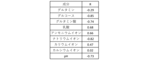
- Example 10 Changes in metabolism in NK cells due to CAR gene introduction CAR-NK cells were produced, and correlations between the components in the culture supernatant and the number of CAR-NK cells were confirmed. The results are shown in Figure 15. The number of CAR-NK cells was positively correlated with lactate and ammonium ions, and negatively correlated with glucose, glutamate, sodium ions, and pH. The number of CAR-NK cells can be predicted from the components of the culture supernatant.
- Example 11 Changes in exosomes by CAR gene introduction
- exosomes which are cell vesicles in the culture supernatant, change due to the introduction of the CAR gene used in Example 1.
- the results are shown in Figure 16. It was confirmed that the particle size of exosomes becomes smaller than that of T cells by the introduction of the CAR gene ( Figure 16A).
- proteome analysis of exosomes derived from CAR-T cells or T cells was performed to confirm the exosome profile.
- the profile of exosome proteins changed between CAR-T cells and T cells ( Figure 16B).
- Example 12 Change in Raman spectroscopy due to introduction of CAR gene It was confirmed whether the Raman spectroscopy of the culture supernatant changes due to introduction of the CAR gene used in Example 1. The results are shown in FIG. 17.
- FIG. 17A shows the entire second derivative of the acquired Raman spectroscopy spectrum. Differences were observed in various peaks between CAR-introduced cells and non-introduced cells.
- FIG. 17B shows an example of the peaks.
Landscapes
- Health & Medical Sciences (AREA)
- Life Sciences & Earth Sciences (AREA)
- Chemical & Material Sciences (AREA)
- Engineering & Computer Science (AREA)
- Organic Chemistry (AREA)
- Genetics & Genomics (AREA)
- Zoology (AREA)
- Biomedical Technology (AREA)
- Wood Science & Technology (AREA)
- Biotechnology (AREA)
- Biochemistry (AREA)
- Molecular Biology (AREA)
- Bioinformatics & Cheminformatics (AREA)
- General Health & Medical Sciences (AREA)
- Proteomics, Peptides & Aminoacids (AREA)
- General Engineering & Computer Science (AREA)
- Microbiology (AREA)
- Biophysics (AREA)
- Immunology (AREA)
- Physics & Mathematics (AREA)
- Cell Biology (AREA)
- Medicinal Chemistry (AREA)
- Analytical Chemistry (AREA)
- Urology & Nephrology (AREA)
- Hematology (AREA)
- Toxicology (AREA)
- Pathology (AREA)
- General Physics & Mathematics (AREA)
- Food Science & Technology (AREA)
- Plant Pathology (AREA)
- Gastroenterology & Hepatology (AREA)
- Measuring Or Testing Involving Enzymes Or Micro-Organisms (AREA)
Abstract
L'invention concerne un procédé de prédiction du taux de transfert de gène dans une cellule animale et/ou le nombre de cellules transférées par gène dans les cellules animales, le procédé comprenant (1) une étape de mesure d'un composant dans un surnageant d'une culture obtenue par la culture de cellules animales transférées par gène et (2) une étape de prédiction du taux de transfert de gène et/ou du nombre de cellules transférées par gène sur la base d'une valeur mesurée à l'étape (1).
Applications Claiming Priority (2)
| Application Number | Priority Date | Filing Date | Title |
|---|---|---|---|
| JP2023099695 | 2023-06-16 | ||
| JP2023-099695 | 2023-06-16 |
Publications (1)
| Publication Number | Publication Date |
|---|---|
| WO2024257872A1 true WO2024257872A1 (fr) | 2024-12-19 |
Family
ID=93852263
Family Applications (1)
| Application Number | Title | Priority Date | Filing Date |
|---|---|---|---|
| PCT/JP2024/021754 Ceased WO2024257872A1 (fr) | 2023-06-16 | 2024-06-14 | Procédé pour prédire le taux de transfert génique |
Country Status (1)
| Country | Link |
|---|---|
| WO (1) | WO2024257872A1 (fr) |
Citations (4)
| Publication number | Priority date | Publication date | Assignee | Title |
|---|---|---|---|---|
| JPH10509318A (ja) * | 1994-11-08 | 1998-09-14 | チャン,ラン−ジ | 組換えcmv−ie/hiv−tar/モロニーマウス白血病ウイルスltrを含有するレトロウイルスベクター |
| JP2007524375A (ja) * | 2003-03-07 | 2007-08-30 | ダイヴァーサ コーポレイション | ヒドロラーゼ、それをコードする核酸並びにその製造および使用方法 |
| US20190226994A1 (en) * | 2016-07-04 | 2019-07-25 | Celltool Gmbh | Device and Method for the Determination of Transfection |
| WO2021005729A1 (fr) * | 2019-07-09 | 2021-01-14 | 株式会社島津製作所 | Procédé de construction d'un modèle de prédiction d'efficacité de différenciation de cellules ips et procédé de prédiction de l'efficacité de différenciation de cellules ips |
-
2024
- 2024-06-14 WO PCT/JP2024/021754 patent/WO2024257872A1/fr not_active Ceased
Patent Citations (4)
| Publication number | Priority date | Publication date | Assignee | Title |
|---|---|---|---|---|
| JPH10509318A (ja) * | 1994-11-08 | 1998-09-14 | チャン,ラン−ジ | 組換えcmv−ie/hiv−tar/モロニーマウス白血病ウイルスltrを含有するレトロウイルスベクター |
| JP2007524375A (ja) * | 2003-03-07 | 2007-08-30 | ダイヴァーサ コーポレイション | ヒドロラーゼ、それをコードする核酸並びにその製造および使用方法 |
| US20190226994A1 (en) * | 2016-07-04 | 2019-07-25 | Celltool Gmbh | Device and Method for the Determination of Transfection |
| WO2021005729A1 (fr) * | 2019-07-09 | 2021-01-14 | 株式会社島津製作所 | Procédé de construction d'un modèle de prédiction d'efficacité de différenciation de cellules ips et procédé de prédiction de l'efficacité de différenciation de cellules ips |
Similar Documents
| Publication | Publication Date | Title |
|---|---|---|
| Zhou et al. | Transfer of cGAMP into bystander cells via LRRC8 volume-regulated anion channels augments STING-mediated interferon responses and anti-viral immunity | |
| Haneke et al. | CDK1 couples proliferation with protein synthesis | |
| Tajan et al. | A role for p53 in the adaptation to glutamine starvation through the expression of SLC1A3 | |
| JP5892939B2 (ja) | 細胞状態をモニタリングするための方法及び間葉系幹細胞を不死化するための方法 | |
| Yoon et al. | Wnt signaling regulates mitochondrial physiology and insulin sensitivity | |
| US20200048616A1 (en) | Methods and products for transfection | |
| Namgaladze et al. | Polarization of human macrophages by interleukin-4 does not require ATP-citrate lyase | |
| SG183579A1 (en) | Methods of detecting therapeutic exosomes | |
| EP2616540A1 (fr) | Procédé d'établissement efficace de cellules souches pluripotentes induites | |
| Romero-Moya et al. | Genetic rescue of mitochondrial and skeletal muscle impairment in an induced pluripotent stem cells model of coenzyme Q10 deficiency | |
| Franzolin et al. | The pyrimidine nucleotide carrier PNC1 and mitochondrial trafficking of thymidine phosphates in cultured human cells | |
| TW202345878A (zh) | 控制性t細胞之製造方法 | |
| Xia et al. | CBP-dependent Wnt/β-catenin signaling is crucial in regulation of MDR1 transcription | |
| Miyazaki et al. | Glutamine protects mouse spermatogonial stem cells against NOX1-derived ROS for sustaining self-renewal division in vitro | |
| CA2964229A1 (fr) | Cellules dendritiques tolerogenes, leurs methodes de production, et utilisations associees | |
| Alborzinia et al. | Selenocysteine metabolism is a targetable vulnerability in MYCN-amplified cancers | |
| Martin et al. | Therapeutic extracellular vesicle production is substantially increased by inhibition of cellular cholesterol biosynthesis | |
| Xian et al. | DUSP facilitates RPMI8226 myeloma cell aging and inhibited TLR4 expression | |
| EP4545632A1 (fr) | Procédé de culture cellulaire | |
| EP4561602A1 (fr) | Polynucléotides codant pour des antigènes liés et leurs utilisations | |
| Alkan et al. | N-acetylaspartate improves cell survival when glucose is limiting | |
| EP4502172A1 (fr) | Procédé pour prédire le taux de transfert génique | |
| Kim et al. | Cytoplasmic fusion between an enlarged embryonic stem cell and a somatic cell using a microtunnel device | |
| WO2020196926A1 (fr) | Inhibiteur de compétition cellulaire | |
| Salmond et al. | DMEM, or Opti-MEM, that is the Question: An Important Consideration for Extracellular Vesicle Isolation and their Downstream Applications |
Legal Events
| Date | Code | Title | Description |
|---|---|---|---|
| 121 | Ep: the epo has been informed by wipo that ep was designated in this application |
Ref document number: 24823485 Country of ref document: EP Kind code of ref document: A1 |
|
| NENP | Non-entry into the national phase |
Ref country code: DE |