EP0151030A2 - Anticorps monoclonaux spécifiques de tumeur - Google Patents
Anticorps monoclonaux spécifiques de tumeur Download PDFInfo
- Publication number
- EP0151030A2 EP0151030A2 EP85300610A EP85300610A EP0151030A2 EP 0151030 A2 EP0151030 A2 EP 0151030A2 EP 85300610 A EP85300610 A EP 85300610A EP 85300610 A EP85300610 A EP 85300610A EP 0151030 A2 EP0151030 A2 EP 0151030A2
- Authority
- EP
- European Patent Office
- Prior art keywords
- tumor
- human
- cells
- cell
- antigens
- Prior art date
- Legal status (The legal status is an assumption and is not a legal conclusion. Google has not performed a legal analysis and makes no representation as to the accuracy of the status listed.)
- Granted
Links
Images


Classifications
-
- C—CHEMISTRY; METALLURGY
- C07—ORGANIC CHEMISTRY
- C07K—PEPTIDES
- C07K16/00—Immunoglobulins [IG], e.g. monoclonal or polyclonal antibodies
- C07K16/18—Immunoglobulins [IG], e.g. monoclonal or polyclonal antibodies against material from animals or humans
- C07K16/28—Immunoglobulins [IG], e.g. monoclonal or polyclonal antibodies against material from animals or humans against receptors, cell surface antigens or cell surface determinants
- C07K16/30—Immunoglobulins [IG], e.g. monoclonal or polyclonal antibodies against material from animals or humans against receptors, cell surface antigens or cell surface determinants from tumour cells
- C07K16/3046—Stomach, Intestines
-
- A—HUMAN NECESSITIES
- A61—MEDICAL OR VETERINARY SCIENCE; HYGIENE
- A61P—SPECIFIC THERAPEUTIC ACTIVITY OF CHEMICAL COMPOUNDS OR MEDICINAL PREPARATIONS
- A61P35/00—Antineoplastic agents
-
- C—CHEMISTRY; METALLURGY
- C07—ORGANIC CHEMISTRY
- C07K—PEPTIDES
- C07K16/00—Immunoglobulins [IG], e.g. monoclonal or polyclonal antibodies
- C07K16/18—Immunoglobulins [IG], e.g. monoclonal or polyclonal antibodies against material from animals or humans
- C07K16/28—Immunoglobulins [IG], e.g. monoclonal or polyclonal antibodies against material from animals or humans against receptors, cell surface antigens or cell surface determinants
- C07K16/30—Immunoglobulins [IG], e.g. monoclonal or polyclonal antibodies against material from animals or humans against receptors, cell surface antigens or cell surface determinants from tumour cells
-
- A—HUMAN NECESSITIES
- A61—MEDICAL OR VETERINARY SCIENCE; HYGIENE
- A61K—PREPARATIONS FOR MEDICAL, DENTAL OR TOILETRY PURPOSES
- A61K38/00—Medicinal preparations containing peptides
Definitions
- This invention relates to monoclonal antibodies produced by hybridoma or transformed B-cell lines derived from B-cells of cancer patients actively immunized with autologous tumor antigen. These monoclonal antibodies can be used in both diagnostic procedures and therapy for .human cancers. This invention also relates to diagnostic procedures and therapeutic approaches using these monoclonal antibodies.
- This invention relates to new human monoclonal antibodies which react specifically with antigens associated with particular cancers and to hybridoma and transformed B-cell lines for their production derived from peripheral blood B-cells of actively immunized patients.
- This invention also relates to methods having general applicability to all solid cancers for preparing hybridomas and monoclonal antibodies and to diagnostic procedures and cancer therapy using these monoclonal antibodies.
- Antibodies are protein molecules normally synthesized by the B-cell lymphocytes produced by bone marrow and carried in the blood stream. For any antigen entering the body, i.e., any foreign molecule from a simple organic chemical to a complex protein, antibodies are produced which recognize and attach to that particular chemical structure.
- the unique chemical structure on the antigen to which a particular antibody can bind is referred to as an antigenic determinant or epitope.
- B-cell lymphocytes in the body referred to as B-cells, lymphocytes, or leukocytes, exist as hundreds of millions of different genetically programmed cells, each producing an antibody specific for a different determinant.
- An antigen, which stimulates antibody production can have several determinants on its surface. On encountering an antigen, a B-cell carrying on its surface an antibody specific for a determinant on that antigen will replicate. This clonal expansion results in many daughter cells which secrete that antibody into the blood stream.
- hybrid cells do not grow in a continuous culture unless they have been altered by hybridization with an "immortal” cell or by being transformed with either viral or tumor DNA.
- Kohler and Milstein demonstrated that hybrid cells could be prepared by somatic cell fusion between lymphocytes and myeloma cells which grow in culture and produce an antibody specific for a single determinant. These hybrids are referred to as "hybridoma cells.”
- Hybridoma cells are prepared by fusing lymphocytes, which have been activated to produce a particular antibody, with myeloma cells. When cultured, hybridomas produce antibodies specific for a single determinant on a particular antigen. Such antibodies are referred to as “monoclonal antibodies.”
- Monoclonal antibodies may also be produced by B-lymphocyte cell lines that have been spontaneously transformed, either prior to or subsequent to being placed in culture. These cells, in distinction to hybridoma cells, possess a normal human diploid number (46) of chromosomes. This invention permits the isolation of both hybridomas and transformed B-cell lines that produce monoclonal antibodies. For sake of simplicity, both cell types will be referred to as monoclonal antibody producing cells below.
- Monoclonal antibodies are synthesized in pure form by a monoclonal antibody producing cell cultures uncontaminated by other immunoglobulins. With such a cell culture, it is possible to produce virtually unlimited quantities of an antibody that is specific for one determinant on a particular antigen.
- antibodies specific for particular cancer cells could be used in various methods of treatment and diagnosis. Such antibodies could inactivate or kill particular tumor cells merely by attaching to the cell at the determinant for which they are specific. Alternatively, these antibodies may bind to the surface of effector lymphocytes or macrophages, converting them into tumor antigen-specific killer cells.
- Monoclonal antibodies can also increase the specificity of chemotherapeutic drugs, toxins and radioactive isotopes, thus increasing their efficacy while decreasing their toxicity.
- a monoclonal antibody can be conjugated with a toxin, radionuclide or chemotherapeutic drug; this conjugated antibody may be simplistically viewed as a guided missile with the antibody as the guidance system and the drug as the warhead.
- antibodies conjugated with radionuclides or metallic tracers can be used for proton emission (PET) and nuclear magnetic resonance (NMR) imaging for in vivo diagnosis and localization of metastases.
- PET proton emission
- NMR nuclear magnetic resonance
- the antibodies can also be used for detecting the presence of tumor antigens in blood, as a diagnostic and/or prognostic test for cancer.
- monoclonal antibodies can be used to isolate the tumor antigens for potential use in a standardized vaccine.
- mice immunized against human tumors have too broad a reactivity. That is, most of the mouse monoclonal antibodies generated react with human antigens present on normal as well as on tumor tissue. An antibody that reacts only with tumor cells is very difficult to select from among the large variety of antibodies produced. For example, 20,000 hybridomas derived from mice immunized with human small-cell lung carcinoma were screened for reactivity with tumor cells (Science, 1982, 216:283). In contrast to a very low frequency ( ⁇ 0.4%) observed by this research group, the present invention results in up to 16% of the hybridomas derived from immunized colon patients producing monoclonal antibodies that react specifically with tumor cells.
- B-cells extracted from either peripheral blood or lymph nodes from patients bearing tumors. It was believed that the presence of the antigenic tumor would cause a tumor-bearing individual to mount an immune response against his tumor and produce specifically immune B-cells. Thus, B-cells were taken from lymph nodes draining tumors in cancer patients or from circulating lymphocytes found in peripheral blood.
- lymph nodes draining tumors in cancer patients or from circulating lymphocytes found in peripheral blood.
- One object of the present invention was to develop monoclonal antibodies reactive with tumor-specific antigens that induce an immune response in patients having particular cancers.
- a valid in vivo assay for the immunogenicity of tumor-specific antigens in tumor immunized patients is by delayed cutaneous hypersensitivity. Such antibodies provide a means for detecting and diagnosing tumors.
- a second objective of this invention was to obtain monoclonal antibodies which would be effective in treating patients with particular types of cancer.
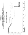
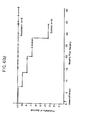
- the treatment also proved to be highly beneficial. Forty-two months after the immunization of the first patients there has been an objective and significant improvement in the patients with respect to duration of the disease-free period following surgery, and the survival data are encouraging. Only 3 of 20 treated patients had recurrences and none have died. Comparatively, 9 of 20 patients in a control group had recurrences and four have died.
- B-cells which produce antibodies having reactivity specific for tumor cell antigens, particularly cell surface antigens as in the majority of cases, is an advantageous result that was speculative, at best, when the immunization studies were begun. Only the immunization treatment was observed and measured during the animal studies on which the human immunization procedures were based, not the production of tumor specific antibodies.
- the general immune response accompanied by an improvement in the subject's condition was indicative of a cellular response in which macrophages and T-cells become activated in the presence of tumor cell antigens and destroy the tumor cells.
- an antibody response would predictably be triggered by immunization under most circumstances, the time course of the antibody response and the cellular response would in most instances be different.
- the fact that the patients were being immunized with autologous tumor cells, and the experience of previous investigators that little or no antibody production is triggered by a patient's own tumor made our discovery that B-cells which produce tumor specific antibodies are generated after immunization an unexpected beneficial result.
- a third objective of this invention was to prepare a standardized vaccine for use in detecting and treating specific cancers in the general population which did not require the custom preparation of a new immunogen suitable for each individual patient. Without a standardized vaccine, only a vaccine prepared for each individual patient from his own tumor tissue could be used for therapy, and only known cancers could have been treated on a limited scale in large institutions. It would not have been possible to make individual preparations for treating the approximately 139,000 cases of colorectal cancer that are discovered in the United States every year.
- This invention comprises the preparation of successful vaccines for active specific immunization, procedures for extracting immunized B-cells, the production of monoclonal antibody producing cells and the production of monoclonal antibodies.
- Malignant tumors are digested using enzyme preparations. The cells obtained are treated to yield a non-tumorigenic tumor cell preparation having the requisite cell viability, which is injected as a vaccine into the subject from which the tumor was obtained.
- Peripheral blood B-cells are obtained from the inoculated subject after a predetermined interval and are used to prepare monoclonal antibody producing cells by fusing with myeloma cells, after which the fused cells are screened for the synthesis of immunoglobulin. Cells that synthesize immunoglobulin are tested for production of antibodies which react with antigens characteristic of the malignant tissue. Those selected are cultured to produce monoclonal antibodies that react with the particular type of tumor with which the subject was afflicted.
- Tumor tissue was obtained from patients suffering from the particular solid cancer for which monoclonal antibodies were to be prepared.
- the tumor tissue was surgically removed from the patient, separated from any non-tumor tissue, and cut into small pieces. We found it satisfactory to cut the tumor tissue into fragments 2-3 mm in diameter. The tumor fragments were then digested to free individual tumor cells by incubation in an enzyme solution.
- the freed cells were pooled and counted, and cell viability was assessed. The trypan blue exclusion test was found to be an acceptable measure of cell viability. The tumor cells were then cryopreserved and stored in liquid nitrogen.
- the vaccine was prepared for injection by rapidly thawing cryopreserved cells, diluting the cells, washing with HBSS, resuspending, counting, and assessing viability.
- Viable tumor cells were irradiated to render them non-tumorigenic. We found that irradiation with 4020 rads/min for a total of 20,000 rads resulted in non-tumorigenic but viable cells. The volume of the cell suspension in HBSS was adjusted such that 10 viable cells remained in the tube. The cells were centrifuged, the supernatant was removed, and 10 viable BCG were added in a volume of 0.1 ml. Hank's Balanced Salt Solution (HBSS) was added in sufficient quantity for a final volume of 0.2 ml. A third vaccine was similarly prepared, omitting the BCG.
- HBSS Hank's Balanced Salt Solution
- Venous blood was collected from the immunized patients one week after each vaccination.
- Peripheral blood lymphocytes PBL were separated from the collected blood for use in hybridoma production.
- the lymphocyte separation medium Separation of lymphocytes from the blood was accomplished using two different methods. The first comprised dilution with calcium and magnesium-free HBSS, layering on lymphocyte separation medium, centrifuging, and removing cells at the interface. These cells were diluted with HBSS and pelleted. The lymphocytes were then resuspended in serum-free Hepes-buffered Dulbecco's MEM (DMEM), counted, and assayed for viability (GIBCO Biologics, Grand Island, New York).
- DMEM serum-free Hepes-buffered Dulbecco's MEM
- PBLs peripheral blood lymphocytes
- AET 2-aminoethylisothio uronium bromide hydrobromide
- Peripheral blood lymphocytes (PBLs) and cultured myeloma cells were mixed together, pelleted, and resuspended in a serum-free medium.
- Polyethylene glycol (PEG) was added, the cells pelleted and resuspended in HT medium (DMEM containing 20% fetal bovine serum, hypcxan- thine and thymidine) and distributed into microtiter wells.
- HT medium containing aminopterin
- HAT medium HT medium containing aminopterin
- the hybridomas were pre-screened for the synthesis of human immunoglobulin using the standard enzyme immunoassay. Hybridomas synthesizing human immunoglobulin in sufficient amounts were tested on tissues. Particular tissue samples were incubated with hybridoma supernatant fluids. Supernatants which demonstrated reactivity with particular tumor tissues indicated that hybridoma cells in the wells from which the particular supernatants were drawn produced tumor-specific antibodies. If the same supernatants failed to show a reaction with samples of normal tissue after extensive screenings, the hybridomas in that particular well were considered tumor-specific. These tumor-specific supernatants were further tested against carcinoembryonic antigen (CEA) to be sure of their narrow specificity.
- CEA carcinoembryonic antigen
- transformed human B-cells diploid cells
- the transformed B-cells were detected in the same way as tumor-specific antibody-producing hybridomas.
- well supernatants which tested positively for reactions with tumor tissue and negatively for reactions with normal tissue and with CEA contained either hybridomas or transformed B-cells.
- the two types of cells were differentiated by observing that the transformed B-cells contained 46 human chromosomes, whereas the hybridomas contained many more chromosomes, not all of which were of the human type.
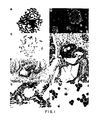
- LiCo 21B27 was incubated with colcemid (0.05 pg/ml) for two hours and treated with hypertonic (0.075M) KCl for three minutes.
- the cells were fixed with methanol-acetic acid (3:1), dropped onto microscope slides, air-dried and stained with Giemsa. Both human and mouse chromosomes are present.
- G-banded chromosome spread of the cell line shown in Figure 1D (X1360). Note the absence of mouse chromosomes.
- the cells were incubated with colcemid (0.01 ⁇ g/ml) overnight.
- the chromosome spreads were prepared as described above. The unstained slide was aged for 10 days.
- the chromosomes were treated with trypsin (0.19% for 30 seconds at room temperature), dehydrated with ethanol and stained with Giemsa.
- Colon tumor as in Figure 1D reacted with normal human IgM (4 pg/ml) (x380). No staining is observed.
- Cryostat section of a colon tumor stained by LiCo 16-88 (x640). Note the intense label of the periphery of the tumor cells (arrows). The section was air dried and stored at -30°C. This section was post-fixed (20 min. at 4°C) with PLP in PBS and processed as described in Figure 1D, except that peroxidase-labeled goat antibody specific to human pchains was used.
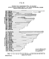
- Two monoclonal antibodies react with most colorectal tumors.
- the reactivity of two monoclonal antibodies to paraffin sections of 15 colorectal tumors and air-dried cytospin preparations of dissociated tumors from 9 patients are compared. Shaded area indicates positive indirect immunoperoxidase staining.
- Randomization was done with stratification according to pathologic stage and tumor was obtained from all patients who met the clinical criteria.
- Candidates for the study were colorectal cancer patients with no previous history of cancer, who had received no prior chemotherapy or radiation therapy, and who were in suitable medical condition to comply with the outpatient treatment protocol.
- Patients eligible for the trial were those with tumor extending through the bowel wall (Astler-Coller B2), positive lymph nodes (stages Cl, C2) or patients with metastatic disease (stage D). Within these classifications, patients were randomly selected for participation in treatment and nontreatment groups. Randomization cards were computer generated and sequentially drawn from each category postoperatively.
- HBSS Hank's Balanced Salt Solution
- Tissue fragments were placed in a 75 ml flask with 20-40 ml of 0.14% (200 units/ml) Collagenase Type 1 (Sigma C - 0130) and 0.1% (500 Kunitz units/ml) deoxyribonuclease type 1 (Sigma D - 0876) (DNAase 1, Sigma D-0876) prewarmed to 37°C. Flasks were placed in a 37°C waterbath with submersible magnetic stirrers at a speed which caused tumbling, but not foaming. After a 30-minute incubation, free cells were decanted through three layers of sterile medium-wet nylon mesh (166t: Martin Supply Co., Baltimore, Maryland) into a 50 ml centrifuge tube.
- sterile medium-wet nylon mesh 166t: Martin Supply Co., Baltimore, Maryland
- the cells were centrifuged at 1200 rpm (250 x g) in a refrigerated centrifuge for 10 minutes. The supernatant was poured off and the cells were resuspended in 5-10 ml of DNAase (0.1% in HBSS) and held at 37°C for 5-10 minutes. The tube was filled with HBSS, washed by centrifugation, resuspended to 15 ml in HBSS and held on ice. The procedure was repeated until sufficient cells were obtained, usually three times for tumor cells. Cells from the different digests were then pooled, counted, and cell viability assessed by the trypan blue exclusion test. The cells were centrifuged for a final wash prior to cryopreservation.
- Optimal cryopreservation was a primary concern.
- the dissociated tumor cells were adjusted to 5-8 x 10 7 /ml in HB S S and added in equal volume to chilled 2 X freezing medium containing 15% dimethylsulfoxide (DMSO) and 4% human serum albumin (HSA).
- DMSO dimethylsulfoxide
- HSA human serum albumin
- the final suspension of 2 to 4 x 10 7 cells/ml were placed in 1.2 ml Nunc freezer vials.
- DCH cell testing the procedure was the same except that no HSA was used.
- the Nunc vials were transferred on ice to a Cryo-Med model 990 Biological Freezer with a model 700 Controller and a model 500 Temperature Recorder for controlled-rate freezing.
- stage D patients patients with tumors of the appropriate pathologic stages were randomized to receive either the autologous tumor cell-BCG vaccine or to have no further therapy.
- stage D patients all received 5-fluoroura- cil chemotherapy and all patients with lesions below the peritoneal reflection (rectal cancer) received 5040 rads of pelvic X-irradiation two weeks after immunotherapy was completed.
- the vaccines were started at 4-5 weeks after tumor resection to allow sufficient time for recovery of immunologic suppression induced by anesthesia and surgery. At 3-4 weeks after resection, both control and treatment patients were skin tested with standard recall antigens as well as graded doses of their autologous tumor cells.
- Mumps skin test antigen USP, Eli Lilly, Indianapolis, Indiana
- Aplisol, PPD (Tuberculin Purified Protein Derivative), Parke-Davis, Detroit, Michigan
- Trichophyton diluted 1:30, Center Laboratories, Port Washington, New York
- Candida albicans diluted 1:100, Center Laboratories, Port Washington, New York, 0.1 ml of each was placed intradermally on the forearm and examined for erythema and induration at 24 and 48 hours.
- Patents selected for treatment protocol received 3 weekly intradermal vaccine injections consisting of 10 7 irradiated, autologous tumor cells and 10 7 BCG in the first 2 vaccines with 10 7 tumor cells alone in the final.
- Fresh-frozen Tice BCG supplied by Dr. Ray Crispen, University of Illinois Medical Center, Chicago, Illinois, was stored at -70°C.
- the first vaccine was placed on the left anterior thigh approximately 10 cm below the groin crease, the second in a comparable location on the right thigh and the third in the right deltoid area.
- tumor cells were diluted slowly to 15 ml in HBSS, washed once by centrifugation at 1200 rpm and resuspended to 15 ml in BBSS.
- Cell counts and viability determinations were made using the trypan blue exclusion test. Viability ranged between 70 and 90%, with a mean of 80%.
- the cells were washed once by centrifugation at 1200 rpm and resuspended to 15 ml in HBSS.
- the suspension of tumor cells was placed on ice and irradiated at 4020 rads/min for a total of 20,000 rads.
- the volume of the cell suspension was adjusted such that 10 7 viable tumor cells remained in the tube ( 1 . 3 x 10 7 viable cells are included to allow for cell loss in tubes and syringes, and for the possibility of approximately 20% misidentifi- cation of lymphoid cells).
- the cells were centrifuged, the supernatant removed and 10 7 BCG were added in a volume of 0.1 m1.
- HBSS was added in sufficient quantity for a final volume of 0.2 ml.
- the third vaccine was similarly prepared, omitting the BCG.
- the vaccine suspension was drawn up through a 20 gauge needle into a 1.0 ml tuberculin syringe.
- the 20 gauge needle was replaced with a 27 gauge needle for the intradermal injection, and the syringe was placed on ice for transport to the clinic.
- the patients were observed closely after each vaccine for erytherma and induration at the site of injections, fever, lymphadenopathy or any adverse reactions.
- the first two vaccine sites ulcerated after 2-3 weeks but always healed within 10 to 12 weeks.
- DCH Delayed Cutaneous Hypersensitivity
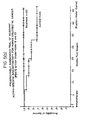
- the delayed cutaneous hypersensitivity reaction to 10 6 autologous tumor cells in 24 immunized and 11 nonimmunized control patients is shown in Table 2.
- a 48-hour induration measurement of greater than 5 mm was considered positive.
- Of significance (p ⁇ 0.01) all of the initially four positive responders and 12 of the negative responders in the immunization group boosted to greater DCH reactivity following a course of immunotherapy (67% became positive). Seven of these patients have been tested at one year, with three maintaining a positive response. Only three of the 16 objectively immunized patients demonstrated a positive DCH response to 10 5 tumor cells at 6 weeks, with none showing a response to 104 cells.
- Venous blood was collected ascep- tically in the presence of preserative-free heparin (O'Neill, Jones and Feldman, St. Louis, Missouri) at a final concentration of 17 units/ml. The blood was maintained at room temperature and transported to the laboratory expeditiously, within a few hours of collection.
- preserative-free heparin O'Neill, Jones and Feldman, St. Louis, Missouri
- the blood diluted 1:2 with calcium and magnesium-free HBSS, was layered (4 ml) over 3 ml of lymphocyte separation medium (LSM, Litton Bionetics) and centrifuged in a 15 ml centrifuge tube for 30 minutes at 400 x g.
- LSM lymphocyte separation medium
- the cells at the interface were removed, diluted with three times their volume of HBSS and pelleted (1000 rpm for 10 minutes).
- the peripheral blood lymphocytes (PBL) were resuspended in 10 ml of serum free Hepes buffered Dulbecco's MEM (DMEM), counted and viability determined.
- DMEM Dulbecco's MEM
- T-lymphocytes were removed by rosetting with AET-treated sheep erythrocytes. Sheep erythrocytes (in Alsever's solution) were washed three time with balanced salt solution (BSS) and incubated at 37°C for 20 minutes with four times the packed cell volume with 0.14 M AET (Sigma). The treated cells were then washed three times with HBSS and resuspended to a 10% suspension. The treated erythrocytes were layered over LSM, centrifuged at 2500 rpm and the pellet collected.
- BSS balanced salt solution
- the sheep erythrocytes were resuspended to a 10% suspension in undiluted fetal bovine serum and used within two weeks.
- the PBL (up to 80 million cells) were mixed with 1 ml of AET-treated sheep erythrocytes and pelleted at 1000 rpm for 10 minutes at 4°C. The pellet was incubated on ice for 45 minutes, gently resuspended with a wide bore pipette and layered over 3 ml LSM. The rosetted cells were centrifuged at 400 x g for 40 minutes at room temperature. The T-cell depleted PBLs were collected at the interface, washed with three times the volume HBSS, and pelleted. Following counting and viability determination, the PBLs enriched for B-cells were then used for hybridoma generation.
- Mouse myeloma cells (NS-1) were grown in the presence of 8-azaguanine (20 ug/ml). Three days before fusion, the cells were pelleted and passaged in medium free of 8-azaguanine. The cells were passaged again the day before fusion to maintain them in log phase. The myeloma cells were washed once with serum-free medium, counted, and viability determined. The PBL and myeloma cells were mixed at a ratio of 3:1 and pelleted together at 1000 rpm for 10 minutes. All supernatant fluid was removed and the cell pellet resuspended in less than 100 ⁇ l of serum-free medium.
- HAT medium HT medium containing .18 ug/ml aminopterin
- HAT medium HT medium containing .18 ug/ml aminopterin
- co-cultivation of PBL with myeloma cells may be used to generate transformed diploid B-cells.
- PBL and myeloma cells were mixed (at a ratio of 3:1), pelleted at 800 rpm and selected in HAT medium, as described above.
- the hybridomas were first quantified and isotyped by a capture enzyme-linked immunoassay (ELISA) for the synthesis of human immunoglobulin (IgA, IgG and IgM).
- ELISA capture enzyme-linked immunoassay
- the standard Bio-EnzaBead method was utilized, which is sensitive in the range of 10-300 ng/ml.
- the hybridoma supernatant fluids were diluted 1:30 with an effective range of .3-9 ug/ml. Only hybridomas that synthesized human immunoglobulin at a concentration of greater than or equal to 1 ⁇ g/ml were tested by indirect immunoperoxidase on tissues after the isotype of the antibody (IgA, IgG or IgM) was determined.
- Polycarbonate-coated metallic beads (Bio-EnzaBead TM , Litton Bionetics) were incubated with goat antibodies to human immunoglobulins (IgG + IgA + IgM) overnight at 4°C and then blocked (30 min at room temperature) with 2.5% BSA to prevent non-specific binding. The beads were then air dried and stored at 4°C.
- the ELISA for detection of immunoglobulin was performed as follows.
- Supernatant fluid from a 96-well culture plate was diluted, incubated with the antibody-capture bead for 1 hr at 37°C, washed, and then incubated for 1 hr at 37°C with peroxidase-labeled affinity-purified goat antibody to human immunoglobulins (IgG + IgA + IgM). The washed beads were then incubated (10 min at room temperature) with 2,2'-Azino-di[3-ethyl-benzthiazoline-6-sulfonic acid], and the optical density was determined at 405 nm.
- the immunoglobulin concentrations were interpolated mathematically from the linear portion of a standard curve (30-1000 ng/ml) of human gamma globulin. Supernatant fluids containing >1 pg/ml were then isotyped using this ELISA with peroxidase-labeled goat antibodies to human Y, a, and p chains. Subsequent quantitative assays used an immunoglobulin standard appropriate for the monoclonal antibody isotype. Mouse immunoglobulins were assayed with Bio-EnzaBeads coated with goat antimouse IgG + IgM (H + L) and peroxidase-conjugated goat antimouse IgG + IgM (H + L). In other experiments, supernatant fluids were incubated with the antihuman Ig beads and the peroxidase-conjugated goat antimouse IgG + IgM (H + L).
- human-mouse heterohybridoma 7a2 was passaged for more than 20 generations from a recently cloned seed stock of 5 x 10 6 cells without a decrease in antibody production.
- the cells theoretically could be expanded to 7 x 1 0 13 cells.
- This hybrid produced approximately 30 ug/ml/ 10 6 cells and thus 7 x 10 13 cells could conceivably produce over 2 kg of antibody.
- Human monoclonal antibody producing cells were grown in RPMI 1640 medium (Sigma Chemical Co., St. Louis, Missouri) supplemented with 10% fetal bovine serum, 3 mM L-glutamine and 5 ug/ml gentamicin. The medium was in some cases further supplemented with 25% D-glucose (final concentration 0.25%). The cells were at 37°C (35-38°C) under a humidified atmosphere of 7.5% C0 2 in air. The antibody was harvested from the highly metabolized spent medium by pelletizing the medium free of cells (e.g., by centrifuging at 500 rpm for 15 minutes.
- Example III Reactivity of Monoclonal Antibodies
- the antibodies reactive with paraffin sections were also tested for reactivity with normal breast, lung, gall bladder and liver and were found to be negative.
- the pattern of reactivity of 10 of the human monoclonal antibodies (MCA) to histological sections of colorectal adenocarcinomas from 15 patients is shown in Figure 2.
- the matrix of reactivity of the antibodies tested indicates that individual antibodies reacted to between 47 and 80% of the tumor specimens tested. No monoclonal antibodies reacted to all 15 tumors.
- tissue sections from individual patients the range of reactivity varied from tissues reactive to all 10 antibodies to tissues reactive to as few as 1 or 2 antibodies. All of the tissue specimens used for determination of monoclonal antibody reactivity were taken from patients other than the 10 donors of B-cells for the original fusions.
- Monoclonal antibody LiCo 16-88 reacted with an antigen preserved in paraffin-embedded sections of colorectal carcinoma that was either absent or greatly reduced in normal colonic mucosa.
- tumor cells exhibited surface-like staining ( Figure ID). This binding was specific, as demonstrated by the absence of staining by normal human immunoglobulin matched in concentration and isotype to the monoclonal antibody. Also noteworthy is the observation that this antibody reacted with both primary tumors and metastases.
- Antibody LiCo 16-88 reacted with cryostat sections. As seen in Figure lE, intense staining of the periphery of tumor cells was observed with LiCo 16-88 but not with normal human immunoglobulin (Figure IF).
- the lack of staining of some of the cells may be due to either clonal or cell cycle variations in the expression of the antigen(s).
- the greatest advantage of this invention which uses immunized patients as the source of sensitized B-cells, is the extremely high frequency of antibodies reactive with cell surface antigens produced.
- the antibodies produced according to the invention have the greatest potential for the diagnosis and treatment of cancer.
- Protein (PBS and 3.0 M KC1) and lipid (chloroform-methanol) extracts were prepared from HT-29 and SW-1463 cells. Thirteen of the antibodies were found to react with these extracts. The most striking finding was that all the antibodies react with the protein extracts, treatment of the extracts with protease significantly reduced the binding. These results contrast markedly with those obtained with murine monoclonal antibodies which are often directed against glycolipid antigens of colon tumors (Morgan et al., Hybridoma, 3:3, page 233, 1984), and Lindholm et al., Int. Arch. Allergy Appl. Immuno., 71:178-181, 1983).
- antigens extracted by the chloroform-methanol treatment may either represent proteins not denatured by this treatment or alternatively glycolipids which share a common epitope (i.e., the carbohydrate moiety) with a glycoprotein.
- the specificity of antibodies for tumor cells versus normal cells is difficult to evaluate by indirect staining methods on Cytospin preparations and cryostat sections.
- the peroxidase-labeled antihuman Ig antibodies used to detect the human antibodies also recognize endogenous human immunoglobulin present on all human tissues. Normal tissues contain greater amounts of endogenous immunoglobulin than do corresponding tumor tissues, consequently the background is higher for normal than for tumor tissue.
- Direct labeling of the antibodies overcomes this problem and permits inclusion of an excess of irrelevant human immunoglobulin with the monoclonal antibodies to block nonspecific immunoglobulin binding, another problem associated with indirect techniques.
- Frozen tissue sections of normal breast, stomach, kidney, liver, muscle and skin showed no staining by biotin-labeled human antibodies except antibody 19b2 which exhibited a low level of binding to normal stomach tissue.
- An overall background stain of connective tissue components was observed. This background staining was nonspecific and has been observed by others using biotin-labeled monoclonal antibodies.
- the invention provides monoclonal antibodies which will be useful as probes to isolate and characterize the antigens relevant to human cancer immunity. These antigens may ultimately prove useful as a tumor vaccine.
- the generation of antibody producing diploid cells adds a dimension of genetic stability to the production of human monoclonal antibodies reactive with tumor cell surface antigens.
- Table 3 shows the tissue reactivity of monoclonal antibodies produced by the monoclonal antibody cell lines prepared according to these procedures.
- carcinoma tumors particularly well-differentiated colorectal adenocarcinomas.
- the invention pertains to all carcinomas, such as lung, breast, and other malignancies in areas which arise from the same type of embryonic tissue.
- the procedures described can be adjusted, if necessary, by one skilled in the art to be used to apply this invention to other types of cancer.
Landscapes
- Health & Medical Sciences (AREA)
- Chemical & Material Sciences (AREA)
- Life Sciences & Earth Sciences (AREA)
- Organic Chemistry (AREA)
- Immunology (AREA)
- Medicinal Chemistry (AREA)
- General Health & Medical Sciences (AREA)
- Molecular Biology (AREA)
- Cell Biology (AREA)
- Proteomics, Peptides & Aminoacids (AREA)
- Genetics & Genomics (AREA)
- Biophysics (AREA)
- Biochemistry (AREA)
- Veterinary Medicine (AREA)
- General Chemical & Material Sciences (AREA)
- Pharmacology & Pharmacy (AREA)
- Public Health (AREA)
- Nuclear Medicine, Radiotherapy & Molecular Imaging (AREA)
- Chemical Kinetics & Catalysis (AREA)
- Animal Behavior & Ethology (AREA)
- Preparation Of Compounds By Using Micro-Organisms (AREA)
- Medicines Containing Antibodies Or Antigens For Use As Internal Diagnostic Agents (AREA)
- Peptides Or Proteins (AREA)
- Micro-Organisms Or Cultivation Processes Thereof (AREA)
Priority Applications (1)
| Application Number | Priority Date | Filing Date | Title |
|---|---|---|---|
| AT85300610T ATE71410T1 (de) | 1984-01-31 | 1985-01-30 | Tumorspezifische monoklonale antikoerper. |
Applications Claiming Priority (2)
| Application Number | Priority Date | Filing Date | Title |
|---|---|---|---|
| US57553384A | 1984-01-31 | 1984-01-31 | |
| US575533 | 1984-01-31 |
Publications (3)
| Publication Number | Publication Date |
|---|---|
| EP0151030A2 true EP0151030A2 (fr) | 1985-08-07 |
| EP0151030A3 EP0151030A3 (en) | 1987-11-25 |
| EP0151030B1 EP0151030B1 (fr) | 1992-01-08 |
Family
ID=24300700
Family Applications (1)
| Application Number | Title | Priority Date | Filing Date |
|---|---|---|---|
| EP85300610A Expired - Lifetime EP0151030B1 (fr) | 1984-01-31 | 1985-01-30 | Anticorps monoclonaux spécifiques de tumeur |
Country Status (15)
| Country | Link |
|---|---|
| EP (1) | EP0151030B1 (fr) |
| JP (2) | JPH0759518B2 (fr) |
| AT (1) | ATE71410T1 (fr) |
| AU (2) | AU589351B2 (fr) |
| CA (1) | CA1340781C (fr) |
| DE (1) | DE3585093D1 (fr) |
| DK (1) | DK171896B1 (fr) |
| ES (1) | ES8802621A1 (fr) |
| GR (1) | GR850179B (fr) |
| HU (1) | HU209519B (fr) |
| IE (1) | IE58859B1 (fr) |
| IL (1) | IL74156A (fr) |
| NZ (1) | NZ210867A (fr) |
| PT (1) | PT79894B (fr) |
| ZA (1) | ZA85689B (fr) |
Cited By (21)
| Publication number | Priority date | Publication date | Assignee | Title |
|---|---|---|---|---|
| EP0127173A3 (fr) * | 1983-05-31 | 1987-07-22 | Birchmeier, Walter, Prof. Dr. | Procédé d'obtention d'anticorps monoclonaux agissant contre des antigènes tumoraux et médicament les contenant |
| EP0258817A3 (en) * | 1986-08-30 | 1988-06-15 | Behringwerke Aktiengesellschaft | Use of monoclonal antibody in tumor therapy |
| WO1988007377A1 (fr) * | 1987-04-03 | 1988-10-06 | Jens Christian Jensenius | Antigene associe a une tumeur humaine |
| EP0222360A3 (fr) * | 1985-11-12 | 1989-03-15 | Biotherapeutics Inc. | Procédé de production d'un réactif cytotoxique spécifique du patient et composition |
| EP0218257A3 (en) * | 1985-10-09 | 1989-08-02 | Kyowa Hakko Kogyo Co., Ltd. | Anti-human pulmonary adenocarcinoma specific monoclonal antibody |
| EP0235817A3 (fr) * | 1986-03-06 | 1990-08-22 | Kyowa Hakko Kogyo Co., Ltd. | Anticorps monoclonal contre le cancer de l'estomac humain |
| EP0265156A3 (fr) * | 1986-10-14 | 1990-08-29 | Bunge (Australia) Proprietary Limited | Anticorps monoclonaux |
| WO1990015625A1 (fr) * | 1989-06-19 | 1990-12-27 | Akzo N.V. | RADIOIMMUNOTHERAPIE UTILISANT UNE EMISSION DE PARTICULES $g(a) |
| WO1992000763A1 (fr) * | 1990-07-03 | 1992-01-23 | Akzo N.V. | Compose immunoreactif |
| US5281710A (en) * | 1990-08-01 | 1994-01-25 | The Scripps Research Institute | Dynemicin analogs: synthesis, methods of preparation and use |
| EP0546634A3 (en) * | 1991-12-13 | 1994-09-21 | Akzo Nv | Tumor associated monoclonal antibody 88bv59 |
| EP0328578B1 (fr) * | 1987-07-02 | 1996-05-08 | Akzo Nobel N.V. | Antigene identifie par mca 16-88 |
| US5552291A (en) * | 1985-10-09 | 1996-09-03 | Kyowa, Hakko Kogyo Co., Ltd. | Anti-human pulmonary adenocarcinoma specific monoclonal antibody |
| EP0650735A3 (fr) * | 1993-07-09 | 1999-04-14 | Akzo Nobel N.V. | Kit et méthode de préciblage |
| US6228361B1 (en) | 1987-11-30 | 2001-05-08 | Roger Williams General Hospital | IgG-1 human monoclonal antibody reactive with an HIV-1 antigen and methods of use |
| US6294172B1 (en) | 1983-08-12 | 2001-09-25 | Dade Behring Marburg Gmbh | Monoclonal antibodies with specificity for membrane-associated antigens |
| US6926896B2 (en) | 1989-03-24 | 2005-08-09 | Aventis Pharma Deutschland Gmbh | Monoclonal antibodies (MAbs) against tumor-associated antigens, the preparation and use thereof |
| US7628996B2 (en) | 2002-02-22 | 2009-12-08 | Intracel Resources Llc | Sterile immunogenic non-tumorigenic tumor cell compositions and methods |
| US9433204B2 (en) | 2002-12-20 | 2016-09-06 | Aduro Gvax Inc. | Directly injectable formulations which provide enhanced cryoprotection of cell products |
| EP3409297A1 (fr) | 2017-05-30 | 2018-12-05 | AlfaRim Medial Holding B.V. | Générateur optimal de 225-actinium - 213-bismuth pour la radio-immunothérapie à particules alpha |
| WO2019057598A1 (fr) | 2017-09-20 | 2019-03-28 | Alfarim Medical Holding B.V. | Générateur optimal de 225actinium--213bismuth pour la radio-immunothérapie à particules alpha |
Families Citing this family (6)
| Publication number | Priority date | Publication date | Assignee | Title |
|---|---|---|---|---|
| US5011684A (en) * | 1985-09-05 | 1991-04-30 | Beth Israel Hospital Association | Lysing or blocking unwanted cells with IL-2 receptor-specific binding substance |
| JPH06100843B2 (ja) * | 1986-06-23 | 1994-12-12 | キヤノン株式会社 | 絶縁性磁性乾式現像剤 |
| WO1991016629A1 (fr) * | 1990-04-12 | 1991-10-31 | Akzo N.V. | Ctaa 28a32, l'antigene reconnu par mca 28a32 |
| FI935038A0 (fi) * | 1991-05-16 | 1993-11-15 | Akzo Nv | Tumoer associerad monoklonal antikropp 81AV78 |
| JPH06113831A (ja) * | 1992-04-08 | 1994-04-26 | Dev Center For Biotechnol | ハイブリドーマおよびその抗体 |
| US20160237163A1 (en) * | 2013-10-02 | 2016-08-18 | Suri Technologies Ltd. | Patient-specific immunotherapy for treating heterogeneous tumors |
Family Cites Families (5)
| Publication number | Priority date | Publication date | Assignee | Title |
|---|---|---|---|---|
| AU4322879A (en) * | 1978-01-11 | 1979-07-19 | Massachusetts General Hospital, The | Producing antibodies |
| DE3211356A1 (de) * | 1982-03-27 | 1983-09-29 | Behringwerke Ag, 3550 Marburg | Zell-hybrid, verfahren zu seiner herstellung sowie seine verwendung |
| JPS58201994A (ja) * | 1982-05-21 | 1983-11-25 | Hideaki Hagiwara | 抗原特異的ヒト免疫グロブリンの生産方法 |
| JPS59137497A (ja) * | 1983-01-20 | 1984-08-07 | ザ・リ−ジエンツ・オブ・ザ・ユニバ−シテイ・オブ・カリフオルニア | 抗原特異的免疫グロブリン生産性ヒト/ヒトハイブリド−マ及びその生産する抗体 |
| US4613576A (en) * | 1983-03-09 | 1986-09-23 | Sloan-Kettering Institute For Cancer Research | Human monoclonal antibodies to cancer cells |
-
1985
- 1985-01-17 NZ NZ210867A patent/NZ210867A/xx unknown
- 1985-01-22 GR GR850179A patent/GR850179B/el unknown
- 1985-01-23 AU AU37992/85A patent/AU589351B2/en not_active Ceased
- 1985-01-25 IL IL74156A patent/IL74156A/xx not_active IP Right Cessation
- 1985-01-25 IE IE19285A patent/IE58859B1/en not_active IP Right Cessation
- 1985-01-28 PT PT79894A patent/PT79894B/pt not_active IP Right Cessation
- 1985-01-29 ZA ZA85689A patent/ZA85689B/xx unknown
- 1985-01-30 AT AT85300610T patent/ATE71410T1/de not_active IP Right Cessation
- 1985-01-30 DK DK040885A patent/DK171896B1/da not_active IP Right Cessation
- 1985-01-30 ES ES539987A patent/ES8802621A1/es not_active Expired
- 1985-01-30 HU HU85344A patent/HU209519B/hu not_active IP Right Cessation
- 1985-01-30 DE DE8585300610T patent/DE3585093D1/de not_active Expired - Lifetime
- 1985-01-30 EP EP85300610A patent/EP0151030B1/fr not_active Expired - Lifetime
- 1985-01-30 CA CA000473130A patent/CA1340781C/fr not_active Expired - Lifetime
- 1985-01-31 JP JP60017692A patent/JPH0759518B2/ja not_active Expired - Lifetime
-
1989
- 1989-12-29 AU AU47367/89A patent/AU635511B2/en not_active Ceased
-
1993
- 1993-10-27 JP JP5269230A patent/JP2516731B2/ja not_active Expired - Lifetime
Cited By (26)
| Publication number | Priority date | Publication date | Assignee | Title |
|---|---|---|---|---|
| EP0127173A3 (fr) * | 1983-05-31 | 1987-07-22 | Birchmeier, Walter, Prof. Dr. | Procédé d'obtention d'anticorps monoclonaux agissant contre des antigènes tumoraux et médicament les contenant |
| US6294172B1 (en) | 1983-08-12 | 2001-09-25 | Dade Behring Marburg Gmbh | Monoclonal antibodies with specificity for membrane-associated antigens |
| US5552291A (en) * | 1985-10-09 | 1996-09-03 | Kyowa, Hakko Kogyo Co., Ltd. | Anti-human pulmonary adenocarcinoma specific monoclonal antibody |
| EP0218257A3 (en) * | 1985-10-09 | 1989-08-02 | Kyowa Hakko Kogyo Co., Ltd. | Anti-human pulmonary adenocarcinoma specific monoclonal antibody |
| EP0222360A3 (fr) * | 1985-11-12 | 1989-03-15 | Biotherapeutics Inc. | Procédé de production d'un réactif cytotoxique spécifique du patient et composition |
| EP0235817A3 (fr) * | 1986-03-06 | 1990-08-22 | Kyowa Hakko Kogyo Co., Ltd. | Anticorps monoclonal contre le cancer de l'estomac humain |
| EP0258817A3 (en) * | 1986-08-30 | 1988-06-15 | Behringwerke Aktiengesellschaft | Use of monoclonal antibody in tumor therapy |
| EP0265156A3 (fr) * | 1986-10-14 | 1990-08-29 | Bunge (Australia) Proprietary Limited | Anticorps monoclonaux |
| EP0285173A3 (en) * | 1987-04-03 | 1990-01-31 | Jens Christian Jensenius | Human tumour-associated antigen |
| US5338661A (en) * | 1987-04-03 | 1994-08-16 | Jensenius Jens C | Monoclonal antibody specific for a human tumour-associated antigen |
| WO1988007377A1 (fr) * | 1987-04-03 | 1988-10-06 | Jens Christian Jensenius | Antigene associe a une tumeur humaine |
| EP0328578B1 (fr) * | 1987-07-02 | 1996-05-08 | Akzo Nobel N.V. | Antigene identifie par mca 16-88 |
| US6228361B1 (en) | 1987-11-30 | 2001-05-08 | Roger Williams General Hospital | IgG-1 human monoclonal antibody reactive with an HIV-1 antigen and methods of use |
| US7662383B2 (en) | 1989-03-24 | 2010-02-16 | Aventis Pharma Deutschland Gmbh | Chimeric antibody against tumor associated antigens and its use in diagnosing tumors |
| US6926896B2 (en) | 1989-03-24 | 2005-08-09 | Aventis Pharma Deutschland Gmbh | Monoclonal antibodies (MAbs) against tumor-associated antigens, the preparation and use thereof |
| WO1990015625A1 (fr) * | 1989-06-19 | 1990-12-27 | Akzo N.V. | RADIOIMMUNOTHERAPIE UTILISANT UNE EMISSION DE PARTICULES $g(a) |
| WO1992000763A1 (fr) * | 1990-07-03 | 1992-01-23 | Akzo N.V. | Compose immunoreactif |
| US5527805A (en) * | 1990-08-01 | 1996-06-18 | The Scripps Research Institute | Dynemicin analogs: syntheses, methods of preparation and use |
| US5281710A (en) * | 1990-08-01 | 1994-01-25 | The Scripps Research Institute | Dynemicin analogs: synthesis, methods of preparation and use |
| EP0546634A3 (en) * | 1991-12-13 | 1994-09-21 | Akzo Nv | Tumor associated monoclonal antibody 88bv59 |
| EP0650735A3 (fr) * | 1993-07-09 | 1999-04-14 | Akzo Nobel N.V. | Kit et méthode de préciblage |
| US7628996B2 (en) | 2002-02-22 | 2009-12-08 | Intracel Resources Llc | Sterile immunogenic non-tumorigenic tumor cell compositions and methods |
| US9433204B2 (en) | 2002-12-20 | 2016-09-06 | Aduro Gvax Inc. | Directly injectable formulations which provide enhanced cryoprotection of cell products |
| US9848598B2 (en) | 2002-12-20 | 2017-12-26 | Aduro Gvax Inc. | Cryoprotected, frozen recombinant cancer cells |
| EP3409297A1 (fr) | 2017-05-30 | 2018-12-05 | AlfaRim Medial Holding B.V. | Générateur optimal de 225-actinium - 213-bismuth pour la radio-immunothérapie à particules alpha |
| WO2019057598A1 (fr) | 2017-09-20 | 2019-03-28 | Alfarim Medical Holding B.V. | Générateur optimal de 225actinium--213bismuth pour la radio-immunothérapie à particules alpha |
Also Published As
| Publication number | Publication date |
|---|---|
| JPH0759518B2 (ja) | 1995-06-28 |
| ZA85689B (en) | 1986-04-30 |
| EP0151030A3 (en) | 1987-11-25 |
| HUT36859A (en) | 1985-10-28 |
| ES8802621A1 (es) | 1988-09-16 |
| DK40885D0 (da) | 1985-01-30 |
| JPS60260597A (ja) | 1985-12-23 |
| IL74156A (en) | 1990-08-31 |
| HU209519B (en) | 1994-06-28 |
| ES539987A0 (es) | 1988-09-16 |
| AU3799285A (en) | 1985-08-08 |
| EP0151030B1 (fr) | 1992-01-08 |
| ATE71410T1 (de) | 1992-01-15 |
| DK171896B1 (da) | 1997-08-04 |
| AU635511B2 (en) | 1993-03-25 |
| NZ210867A (en) | 1989-01-06 |
| CA1340781C (fr) | 1999-10-12 |
| PT79894B (en) | 1986-10-23 |
| DE3585093D1 (de) | 1992-02-20 |
| IL74156A0 (en) | 1985-04-30 |
| DK40885A (da) | 1985-10-25 |
| JP2516731B2 (ja) | 1996-07-24 |
| PT79894A (en) | 1985-02-01 |
| AU589351B2 (en) | 1989-10-12 |
| IE58859B1 (en) | 1993-11-17 |
| AU4736789A (en) | 1990-05-03 |
| GR850179B (fr) | 1985-05-23 |
| IE850192L (en) | 1985-07-31 |
| JPH06315395A (ja) | 1994-11-15 |
Similar Documents
| Publication | Publication Date | Title |
|---|---|---|
| US4828991A (en) | Tumor specific monoclonal antibodies | |
| EP0151030B1 (fr) | Anticorps monoclonaux spécifiques de tumeur | |
| US4997762A (en) | Tumor associated monocoloal antibodies derived from human B-cell line | |
| US5529903A (en) | Extraction and cultivation of transformed cells and production of antibodies directed against them | |
| US5106738A (en) | Tumor specific monoclonal antibodies | |
| US5474755A (en) | Tumor associated monoclonal antibodies | |
| US5180814A (en) | Tumor specific monoclonal antibodies | |
| CA1341552C (fr) | Procede de production d'hybridomes humain-humain, production d'anticorps monoclonaux ou polyclonaux a partir de ceux-ci, et leur utilisation therapeutique | |
| US5348880A (en) | Tumor associated monoclonal antibody 81AV/78 | |
| US6491926B1 (en) | 35 kD tumor associated protein antigen: uses and methods of detection | |
| EP0584267B1 (fr) | Anticorps monoclonal 81av78 specifique des tumeurs | |
| AU698184B2 (en) | Monoclonal antibody 88BV59, subclones and method of making | |
| US5495002A (en) | Tumor associated monoclonal antibody 123AV16 | |
| AU651261B2 (en) | Tumor associated monoclonal antibody 88BV59 | |
| Hellström et al. | Antigens in human melanomas detected by using monoclonal antibodies as probes |
Legal Events
| Date | Code | Title | Description |
|---|---|---|---|
| PUAI | Public reference made under article 153(3) epc to a published international application that has entered the european phase |
Free format text: ORIGINAL CODE: 0009012 |
|
| AK | Designated contracting states |
Designated state(s): AT BE CH DE FR GB IT LI LU NL SE |
|
| PUAL | Search report despatched |
Free format text: ORIGINAL CODE: 0009013 |
|
| AK | Designated contracting states |
Kind code of ref document: A3 Designated state(s): AT BE CH DE FR GB IT LI LU NL SE |
|
| 17P | Request for examination filed |
Effective date: 19880127 |
|
| 17Q | First examination report despatched |
Effective date: 19890925 |
|
| RAP1 | Party data changed (applicant data changed or rights of an application transferred) |
Owner name: AKZO N.V. |
|
| GRAA | (expected) grant |
Free format text: ORIGINAL CODE: 0009210 |
|
| AK | Designated contracting states |
Kind code of ref document: B1 Designated state(s): AT BE CH DE FR GB IT LI LU NL SE |
|
| REF | Corresponds to: |
Ref document number: 71410 Country of ref document: AT Date of ref document: 19920115 Kind code of ref document: T |
|
| REF | Corresponds to: |
Ref document number: 3585093 Country of ref document: DE Date of ref document: 19920220 |
|
| ITF | It: translation for a ep patent filed | ||
| ET | Fr: translation filed | ||
| PLBE | No opposition filed within time limit |
Free format text: ORIGINAL CODE: 0009261 |
|
| STAA | Information on the status of an ep patent application or granted ep patent |
Free format text: STATUS: NO OPPOSITION FILED WITHIN TIME LIMIT |
|
| 26N | No opposition filed | ||
| ITTA | It: last paid annual fee | ||
| EPTA | Lu: last paid annual fee | ||
| EAL | Se: european patent in force in sweden |
Ref document number: 85300610.4 |
|
| NLS | Nl: assignments of ep-patents |
Owner name: PERIMMUNE HOLDINGS, INC. |
|
| NLT1 | Nl: modifications of names registered in virtue of documents presented to the patent office pursuant to art. 16 a, paragraph 1 |
Owner name: AKZO NOBEL N.V. |
|
| PGFP | Annual fee paid to national office [announced via postgrant information from national office to epo] |
Ref country code: FR Payment date: 20010123 Year of fee payment: 17 |
|
| PGFP | Annual fee paid to national office [announced via postgrant information from national office to epo] |
Ref country code: AT Payment date: 20010125 Year of fee payment: 17 Ref country code: SE Payment date: 20010125 Year of fee payment: 17 |
|
| PGFP | Annual fee paid to national office [announced via postgrant information from national office to epo] |
Ref country code: NL Payment date: 20010131 Year of fee payment: 17 |
|
| REG | Reference to a national code |
Ref country code: CH Ref legal event code: PUE Owner name: AKZO NOBEL N.V. TRANSFER- PERIMMUNE HOLDINGS, INC. Ref country code: CH Ref legal event code: PFA Free format text: AKZO N.V. TRANSFER- AKZO NOBEL N.V. |
|
| PGFP | Annual fee paid to national office [announced via postgrant information from national office to epo] |
Ref country code: CH Payment date: 20010219 Year of fee payment: 17 |
|
| PGFP | Annual fee paid to national office [announced via postgrant information from national office to epo] |
Ref country code: BE Payment date: 20010313 Year of fee payment: 17 Ref country code: LU Payment date: 20010313 Year of fee payment: 17 |
|
| REG | Reference to a national code |
Ref country code: GB Ref legal event code: IF02 |
|
| REG | Reference to a national code |
Ref country code: CH Ref legal event code: PVP |
|
| PG25 | Lapsed in a contracting state [announced via postgrant information from national office to epo] |
Ref country code: LU Free format text: LAPSE BECAUSE OF NON-PAYMENT OF DUE FEES Effective date: 20020130 Ref country code: AT Free format text: LAPSE BECAUSE OF NON-PAYMENT OF DUE FEES Effective date: 20020130 |
|
| PG25 | Lapsed in a contracting state [announced via postgrant information from national office to epo] |
Ref country code: LI Free format text: LAPSE BECAUSE OF NON-PAYMENT OF DUE FEES Effective date: 20020131 Ref country code: SE Free format text: LAPSE BECAUSE OF NON-PAYMENT OF DUE FEES Effective date: 20020131 Ref country code: BE Free format text: LAPSE BECAUSE OF NON-PAYMENT OF DUE FEES Effective date: 20020131 Ref country code: CH Free format text: LAPSE BECAUSE OF NON-PAYMENT OF DUE FEES Effective date: 20020131 |
|
| BERE | Be: lapsed |
Owner name: PERIMMUNE HOLDINGS INC. Effective date: 20020131 |
|
| PG25 | Lapsed in a contracting state [announced via postgrant information from national office to epo] |
Ref country code: NL Free format text: LAPSE BECAUSE OF NON-PAYMENT OF DUE FEES Effective date: 20020801 |
|
| EUG | Se: european patent has lapsed |
Ref document number: 85300610.4 |
|
| REG | Reference to a national code |
Ref country code: CH Ref legal event code: PL |
|
| PG25 | Lapsed in a contracting state [announced via postgrant information from national office to epo] |
Ref country code: FR Free format text: LAPSE BECAUSE OF NON-PAYMENT OF DUE FEES Effective date: 20020930 |
|
| NLV4 | Nl: lapsed or anulled due to non-payment of the annual fee |
Effective date: 20020801 |
|
| REG | Reference to a national code |
Ref country code: FR Ref legal event code: ST |
|
| NLXE | Nl: other communications concerning ep-patents (part 3 heading xe) |
Free format text: A REQUEST FOR RESTORATION TO THE PRIOR STATE (ARTICLE 23 OF THE PATENTS ACT 1995) HAS BEEN FILED ON 20030731. |
|
| PGFP | Annual fee paid to national office [announced via postgrant information from national office to epo] |
Ref country code: DE Payment date: 20040116 Year of fee payment: 20 |
|
| PGFP | Annual fee paid to national office [announced via postgrant information from national office to epo] |
Ref country code: GB Payment date: 20040129 Year of fee payment: 20 |
|
| NLXE | Nl: other communications concerning ep-patents (part 3 heading xe) |
Free format text: THE REQUEST FOR RESTORATION OF THE PRIOR STATE AS PROVIDED FOR IN ARTICLE 23 OF THE PATENTS ACT 1995 (SEE PUBLICATION IN HEADING XE OF THE PATENT BULLETIN OF 20031001/10) HAS BEEN REJECTED. |
|
| PG25 | Lapsed in a contracting state [announced via postgrant information from national office to epo] |
Ref country code: GB Free format text: LAPSE BECAUSE OF EXPIRATION OF PROTECTION Effective date: 20050129 |
|
| REG | Reference to a national code |
Ref country code: GB Ref legal event code: PE20 |