EP0194212B2 - Procédé de mise en évidence d'agglutinats érythrocytaires - Google Patents
Procédé de mise en évidence d'agglutinats érythrocytaires Download PDFInfo
- Publication number
- EP0194212B2 EP0194212B2 EP86420040A EP86420040A EP0194212B2 EP 0194212 B2 EP0194212 B2 EP 0194212B2 EP 86420040 A EP86420040 A EP 86420040A EP 86420040 A EP86420040 A EP 86420040A EP 0194212 B2 EP0194212 B2 EP 0194212B2
- Authority
- EP
- European Patent Office
- Prior art keywords
- gel
- red blood
- blood cells
- serum
- antibodies
- Prior art date
- Legal status (The legal status is an assumption and is not a legal conclusion. Google has not performed a legal analysis and makes no representation as to the accuracy of the status listed.)
- Expired - Lifetime
Links
Images
Classifications
-
- G—PHYSICS
- G01—MEASURING; TESTING
- G01N—INVESTIGATING OR ANALYSING MATERIALS BY DETERMINING THEIR CHEMICAL OR PHYSICAL PROPERTIES
- G01N33/00—Investigating or analysing materials by specific methods not covered by groups G01N1/00 - G01N31/00
- G01N33/48—Biological material, e.g. blood, urine; Haemocytometers
- G01N33/50—Chemical analysis of biological material, e.g. blood, urine; Testing involving biospecific ligand binding methods; Immunological testing
- G01N33/53—Immunoassay; Biospecific binding assay; Materials therefor
- G01N33/558—Immunoassay; Biospecific binding assay; Materials therefor using diffusion or migration of antigen or antibody
- G01N33/559—Immunoassay; Biospecific binding assay; Materials therefor using diffusion or migration of antigen or antibody through a gel, e.g. Ouchterlony technique
-
- G—PHYSICS
- G01—MEASURING; TESTING
- G01N—INVESTIGATING OR ANALYSING MATERIALS BY DETERMINING THEIR CHEMICAL OR PHYSICAL PROPERTIES
- G01N33/00—Investigating or analysing materials by specific methods not covered by groups G01N1/00 - G01N31/00
- G01N33/48—Biological material, e.g. blood, urine; Haemocytometers
- G01N33/50—Chemical analysis of biological material, e.g. blood, urine; Testing involving biospecific ligand binding methods; Immunological testing
- G01N33/53—Immunoassay; Biospecific binding assay; Materials therefor
- G01N33/543—Immunoassay; Biospecific binding assay; Materials therefor with an insoluble carrier for immobilising immunochemicals
- G01N33/554—Immunoassay; Biospecific binding assay; Materials therefor with an insoluble carrier for immobilising immunochemicals the carrier being a biological cell or cell fragment, e.g. bacteria, yeast cells
- G01N33/555—Red blood cell
Definitions
- the present invention relates to a method for detecting erythrocyte agglutinates, frequently used in biology, in particular in immuno-hematology and transfusion.
- the present invention aims to eliminate some of the drawbacks of known methods, by using an original method for demonstrating agglutination, in order to obtain tests which meet the following criteria:
- Sensitivity improving the sensitivity of the tests, due to the particular conditions, and the reading mode, is essential.
- Reproducibility The absence, in the last phase, of human intervention that is difficult to quantify (according to the tests: manual washes, agitation, addition of a reagent, reading time, etc.), by achieving relative standardization, must allow better reproducibility, especially titrations.
- the size of the pores of the filter constituted by the gel is such that it lets the free globules pass more easily than the agglutinates, this during a gentle centrifugation, meeting specific criteria.
- a particular conical shape of the bottom of the reaction tube by slowing the progression of the globules during centrifugation, increases the phenomenon. Reading is then particularly easy: in negative reactions, the globules have sedimented completely, in positive reactions, they remain trapped, at least in part, in the gel filter. This reading can easily be postponed for several hours, taken by several people successively, photographed.
- the filter consists of a gel obtained as follows:
- the gel is prepared simply, by adding the low ionic strength solution to Sephadex or vice versa, gentle agitation, addition of antiglobulin, new agitation. It is not imperative to use a low ionic strength solution, and conclusive tests have been carried out with other solutions (saline, phosphate buffers, carbonate buffers, citrate-dextrose resuspension solution from Lalezari (Lalezari P., Jiang AF, The manual polybrene test: A simple and rapid procedure for detection of red cell antibodies, Transfusion 1980; 20: 206-211)).
- the antiglobulin used in the application was an antiglobulin anti-IgG goat or rabbit, manufactured by the Blood Transfusion Center of Lyon (animal serum diluted 20 to 50 times in saline to which is added dextran sulfate to obtain a concentration 1 g / l).
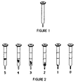
- the tubes used are small (internal diameter 4 mm, length 45 mm) and terminated by a conical part, extended by a point (see Figure 1 ).
- Each tube receives, with an automatic pipette, 200 microliters of gel, and is then centrifuged briefly to approximately 1000 g (5 seconds, but this centrifugation can without inconvenience be much longer), to pack this gel.
- the angular speed is expressed in radian / second
- the centrifugation radius is measured between the axis of the centrifuge and the bottom of the tube.
- the centrifugal force must be in the axis of the tube.
- the reading is carried out directly at the end of the centrifugation, and remains possible for several hours: the positive reactions are characterized by the retention of red blood cells in the gel.
- Sera donors or patients
- Sera corresponding to the main anticorpal specificities of immune irregular antibody systems, were titrated by this technique and by a classic low ionic strength Coombs test (Low B., Messeter L., Antiglobulin test in low-ionic strength salt solution for rapid antibody screening and cross-matching, Vox Sang. 1974; 26: 53-61).
- the dilutions of the sera were prepared in advance, separated into 2 fractions, numbered at random, and frozen.
- the tests were carried out the same day, after thawing, by different technicians, working blind.
- the results were expressed in terms of positivity or negativity, with a rating test of 1 to 5 for the positives.
- FIG. 3 shows the titles obtained by each of the methods and the superiority of the gel test. It should also be noted that the last positive dilutions in the classic test corresponded to extremely weak reactions, visible only by a confirmed technician immediately reading, while the last positivities, with the gel test, could have been interpreted the next day, by a person who has never used this technique.
- the minimum amount of anti-D detectable by this technique is 0.4 to 0.8 ng / ml, which corresponds to a sensitivity significantly higher than that of the Coombs test, and practically equivalent to that of the polybrene® test. in an automatic device (Mollison PL, Blood transfusion in clinical medicine, Blackwell Oxford, 1983, pp. 487-557).
- the immunized animal serum can be replaced in the gel with a neutral serum.
- the incubation conditions time, temperature are then modified according to the nature of the antibody involved.
- Polybrene® test mixed polybrene®-antiglobulin test.
- the resuspension solution then replaces the low ionic strength solution in the gel.
- the polybrene is, as in the Lalezari technique, added to the serum-globule mixture, and the centrifugation first induces agglutination, then deagglutination (except positive reaction) when the globules meet the citrated solution.
- the antiglobulin contained in the gel acts as described above.
- the advantage is the relative standardization of the agglutination and deagglutination operations, which are always difficult in the manual polybrene test.
Landscapes
- Health & Medical Sciences (AREA)
- Life Sciences & Earth Sciences (AREA)
- Immunology (AREA)
- Engineering & Computer Science (AREA)
- Hematology (AREA)
- Chemical & Material Sciences (AREA)
- Biomedical Technology (AREA)
- Molecular Biology (AREA)
- Urology & Nephrology (AREA)
- Cell Biology (AREA)
- Microbiology (AREA)
- Biochemistry (AREA)
- General Health & Medical Sciences (AREA)
- Food Science & Technology (AREA)
- Medicinal Chemistry (AREA)
- Physics & Mathematics (AREA)
- Analytical Chemistry (AREA)
- Biotechnology (AREA)
- Pathology (AREA)
- General Physics & Mathematics (AREA)
- Dispersion Chemistry (AREA)
- Mycology (AREA)
- Investigating Or Analysing Biological Materials (AREA)
- Measurement Of The Respiration, Hearing Ability, Form, And Blood Characteristics Of Living Organisms (AREA)
- Low-Molecular Organic Synthesis Reactions Using Catalysts (AREA)
Description
- La présente invention concerne un procédé pour la mise en évidence d'agglutinats érythrocytaires, fréquemment utilisé en biologie, notamment en immuno-hématologie et transfusion.
- On connaît les techniques classiques de mise en évidence de la réaction antigène-anticorps en immuno-hématologie érythrocytaire. Les plus courantes font appel à l'agglutination:
Agglutination spontanée en milieu salin
Agglutination en milieu macromoléculaire
Agglutination d'hématies traitées par des enzymes
Test de Coombs Mourant Race (agglutination après lavage d'hématies revêtues par des anticorps ou du complément, au moyen d'antiglobuline humaine)
Maintien d'une agglutination non spécifique (telle celle induite par l'hexadimethrine bromide ou polybrène®, par exemple) - Méthodes dérivées des procédés cités ci-dessus, ou associant plusieurs d'entre eux (test de Coombs sur hématies trypsinées, test manuel polybrène®-antiglobuline, en deux phases ...).
- Ces techniques peuvent généralement être réalisées sur plaque, en tube, ou dans des dispositifs automatiques.
- On connaît des développements récents à ces techniques:
- Graham décrit en 1982 une méthode originale de séparation sérum-globules, évitant les lavages du test de Coombs classique. Pour cela il utilise, dans un tube particulier, un milieu composé d'albumine et de dextran, de densité intermédiaire entre sérum et globules (Mollison P. L., Blood transfusion in clinical medicine, Blackwell, Oxford, 1983, p. 512). Ortho Diagnostic Systems a commercialisé, sous forme de kit, ce procédé (Simwash®). Il est original, mais, d'après l'auteur, pas plus sensible que le test classique dont il ne diffère pas fondamentalement, dans sa phase finale.
- Des techniques dites "en phase solide" voient le jour, avec des méthodologies variables.
- La synthèse des deux procédés exposés ci-dessus (séparation sérum-globules sans lavage et phase solide) aurait abouti à la technique suivante: le mélange sérum-globules est centrifugé à travers un liquide de séparation, dans un puits dont le fond est revêtu d'antiglobuline. La lecture est faite automatiquement. Cette méthode, présentée au symposium transfusionnel de Munich (1984) par une équipe parisienne (Muller A. Beige D., Richard D., Matte C., CNTS Paris/Orsay), donnerait d'excellents résultats, en tous cas nettement supérieurs à ceux obtenus par les tests manuels.
- La présente invention vise à supprimer certains des inconvénients des méthodes connues, en utilisant un procédé original de mise en évidence de l'agglutination, pour obtenir des tests répondant aux critères suivants:
- Praticabilité: Les tests manuels les plus couramment utilisés en immuno-hématologie, s'ils nécessitent des précautions de manipulation et d'interprétation, restent, pour des personnes entraînées, simples et rapides. Le test proposé doit donc répondre aux mêmes exigences de praticabilité. Ceci n'exclut d'ailleurs pas son utilisation dans des systèmes plus ou moins automatisés, pour des applications en séries.
- Fiabilité: l'interprétation de la réaction, d'une très grande simplicité doit améliorer la fiabilité de la réponse dans un certain nombre de situations difficiles (réaction faible, urgence, utilisateurs occasionnels...). D'autre part, les précautions à apporter à la manipulation concernant surtout la phase de préparation du milieu, peuvent, dans une certaine mesure, être dissociées, dans le temps, de ces situations difficiles.
- Sensibilité: l'amélioration de la sensibilité des tests, du fait des conditions particulières, et du mode de lecture, est primordiale.
- Reproducibilité: L'absence, dans la dernière phase, d'intervention humain difficilement quantifiable (selon les tests: lavages manuels, agitation, ajout d'un réactif, temps de lecture...), en réalisant une relative standardisation, doit permettre une meilleure reproductibilité, notamment des titrages.
- Dans la phase finale des tests en tubes couramment utilisés en immuno-hématologie, on peut trouver les manipulations suivantes:
- Lavages (Coombs et phase "antiglobuline" du test polybrène®-Coombs).
- Ajout de réactifs (citrate dans le test manuel au polybrène® antiglobuline dans le test de Coombs ou la phase "antiglobuline" du test polybrène®-Coombs).
- Agitation pour remise en suspension des hématies.
- Lecture immédiate, par une personne entraînée (ces deux dernières manipulations sont communes à tous les tests d'agglutination en tube, quand la lecture est macroscopique).
- Le procédé que nous allons décrire agit sur cette phase. En effet l'incubation ne diffère pas fondamentalement de celle des tests classiques. Cette technique permet toutefois l'utilisation de quantités de réactifs qui sont en moyenne plus faibles que celles utilisées habituellement, notamment pour les hématies-tests, ce qui améliore le rapport sérum/globules pendant l'incubation.
- Les lavages, quand ceux-ci sont nécessaires, sont remplacés par une centrifugation douce du mélange sérum-globules à travers un filtre (dans la technique détaillée décrite plus loin, ce filtre est un gel). Les globules, qui sédimentent rapidement, sont alors séparés du sérum. Cet aspect de la technique, s'il est intéressant, n'est pas à proprement parler nouveau, comme signalé précédemment (sauf par le fait que le milieu utilisé est un gel).
- L'ajout de réactifs quand il est nécessaire, est remplacé par leur incorporation au milieu de gel: c'est le cas de l'antiglobuline, ou de la solution citratée de resuspension utilisée dans le test manuel au polybrène®. Lors de la centrifugation, on a donc une séparation globules-sérum, mais aussi une réaction avec les molécules incluses dans le filtre.
- La taille des pores du filtre constitué par le gel est telle que celui-ci laisse passer les globules libres plus facilement que les agglutinats, ceci lors d'une centrifugation douce, répondant à des critères précis. Une forme particulière, conique, du fond du tube de réaction, en ralentissant la progression des globules lors de la centrifugation, majore le phénomène. La lecture est alors particulièrement aisée: dans les réactions négatives, les globules ont sédimenté complètement, dans les réactions positives, ils restent pris, au moins en partie, dans le filtre de gel. Cette lecture peut sans inconvénient être différée de plusieurs heures, effectuée par plusieurs personnes successivement, photographiée.
- Cet aspect du procédé, nouveau, constitue la base de la demande de brevet.
- A titre d'exemple non limitatif, on trouvera ci-dessous une description détaillée d'une application: le test à l'antiglobuline.
- Préparation du gel. Dans cette application, le filtre est constitué par un gel obtenu de la façon suivante:
- Mouiller du Séphadex® G100 Superfine (Pharmacia Fine Chemicals, Upssala, Sweden) avec une solution basse force ionique, à raison de 18 ml de solution par gramme de Séphadex. Dans l'application, la solution basse force ionique utilisée était celle du Centre de Transfusion Sanguine de Lyon, préparée en accord avec la formule de Löw et Messeter (Low B., Messeter L., Antiglobulin test in low-ionic strength salt solution for rapid antibody screening and cross-matching, Vox Sang. 1974, 26:53-61).
- Ajouter une antiglobuline contenant 1 g/l de sulfate de dextran (Pharmacia Fine Chemicals, Uppsala, Sweden), à raison d'1 volume pour 10 volumes du mélange Séphadex-solution basse force ionique.
- Le gel est préparé simplement, par ajout de la solution basse force ionique au Séphadex ou l'inverse, agitation douce, ajout de l'antiglobuline, nouvelle agitation. Il n'est pas impératif d'utiliser une solution basse force ionique, et des essais concluants ont été réalisés avec d'autres solutions (saline, tampons phosphate, tampons carbonate, solution de resuspension citrate-dextrose de Lalezari (Lalezari P., Jiang A. F., The manual polybrene test: A simple and rapid procedure for detection of red cell antibodies, Transfusion 1980; 20:206-211)).
- Il convient par contre d'observer un certain équilibre entre les différentes constituants de l'antiglobuline, pour améliorer la sédimentation des globules libres, et la conservation des réactifs (sérum animal ou albumine, sulfate de dextran). En l'absence de cet équilibre, les globules sont incapables de sédimenter à travers le gel, même si le sérum avec lequel ils ont été incubés ne contient pas d'anticorps. L'antiglobuline utilisée dans l'application était une antiglobuline anti-lgG de chèvre ou de lapin, fabriquée par le Centre de Transfusion Sanguine de Lyon (sérum animal dilué 20 à 50 fois en saline auquel est ajouté du sulfate de dextran pour obtenir une concentration de 1 g/l).
- Préparation des tubes à réaction. Les tubes utilisés (tubes pour extraction des micro sédiments réf. 72702 Starstedt, Romnelsdorf, West Germany) sont de petite taille (diamètre intérieur 4 mm, longueur 45 mm) et terminés par une partie conique, prolongée par une pointe (cf. figure 1).
- Leur faible capacité permet de travailler avec de petites quantités de réactifs, assure une sédimentation correcte et rend la lecture possible à travers le plastique et le gel. La partie terminale conique, en ralentissant la sédimentation des hématies, d'autant plus qu'elles sont agglutinées, améliore la sensibilité, du test.
- D'autres tubes ont été utilisés avec succès, mais celui-ci est particulièrement bien adapté, et donne les meilleures résultats.
- Chaque tube reçoit, à la pipette automatique, 200 microlitres de gel, et est ensuite centrifugé brièvement à environ 1000 g (5 secondes, mais cette centrifugation peut sans inconvénient être beaucoup plus longue), pour tasser ce gel.
- Réaction. Dans un autre récipient (tube, puits de microplaque...), on mélange 30 microlitres de sérum à 120 microlitres d'une suspension érythrocytaire à 0,25% en solution basse force ionique (Low et Messeter). Après une incubation de 10 minutes à 37°C, on transfère ce mélange à la pipette (Pasteur, par exemple) dans la partie supérieure du tube, au dessus du gel. Peu importe que le contact entre gel et mélange soit immédiat ou non, pourvu que le mode de dépôt n'ait pas fait pénétrer profondément les globules dans le gel.
- Le tube est alors centrifugé 10 minutes à 200 unités RCF (radial centrifugation force).
1 unité RCF=(rayon de centrifugation) (vitesse angulaire)²/9,8
9,8 étant la constante de Newton - La vitesse angulaire est exprimée en radian/seconde
- Le rayon de centrifugation, exprimé en mètres, est mesuré entre l'axe de la centrifugeuse et le fond du tube.
- En cours de centrifugation, la force centrifuge doit être dans l'axe du tube.
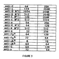
- La lecture est effectuée directement à la fin de la centrifugation, et reste possible plusieurs heures: les réactions positives sont caractérisées par la rétention d'hématies dans le gel.
- Cette rétention est haute et totale pour les réactions fortement positives (figure 2, tubes 3 à 5), plus basse, et parfois incomplète pour les réactions faiblement positives (figure 2, tubes 1 et 2).
- Dans les réactions négatives (figure 2, tube 0), tous les globules ont sédimenté dans la pointe.
- Titrage d'anticorps immuns irréguliers. Des sérums (donneurs ou malades) correspondant aux principales spécificités anticorpales des systèmes à anticorps irréguliers immuns, ont été titrés par cette technique et par un test de Coombs basse force ionique classique (Low B., Messeter L., Antiglobulin test in low-ionic strength salt solution for rapid antibody screening and cross-matching, Vox Sang. 1974; 26:53-61). Les dilutions des sérums ont été préparées à l'avance, séparées en 2 fractions, numérotées au hasard, et congelées. Les tests ont été réalisés le même jour, après décongélation, par des techniciens différents, travaillant à l'aveugle. Les résultats étaient exprimés en terme de positivité ou négativité, avec essai de cotation de 1 à 5 pour les positifs.
- La figure 3 montre les titres obtenus par chacune des méthodes et la supériorité du test sur gel. Il faut noter également qu'aux dernières dilutions positives en test classique correspondaient des réactions extrêmement faibles, visibles seulement par un technicien confirmé effectuant immédiatement la lecture, alors que les dernières positivités, avec le test sur gel, auraient pu être interprétées le lendemain, par une personne n'ayant jamais utilisé cette technique.
- Quelques anticorps de nature habituellement IgM (anti-Lewis, anti-M anti-N...) ont été testés, avec des résultats variables.
- Recherche de la quantité minimale détectable d'anti-D par titrage d'un sérum étalon. La quantité minimale d'anti-D détectable par cette technique est de 0,4 à 0,8 ng/ml, ce qui correspond à une sensibilité nettement supérieure à celle du test de Coombs, et pratiquement équivalente à celle du test au polybrène® en appareil automatique (Mollison P. L., Blood transfusion in clinical medicine, Blackwell Oxford, 1983, pp. 487-557).
- Groupage érythrocytaire, pour les antigènes C, c, D, E, e, K, k, Fya, Fyb, Jka, Jkb, S, s. Les sérums utilisés sont des sérums test homologués par le CNRGS, purs ou dilués au 1/2 ou au 1/4. Ces sérums étant suffisamment forts, les réactions sont particulièrement faciles à lire: dans les réactions positives les globules sont tous retenus dans la partie haute du gel. Dans ces conditions, la présence simultanée de globules retenus dans le gel et de globules ayant bien sédimenté, correspond évidemment à une double population. Les hématies minoritaires sont détectables à condition de représenter:
- 10 à 15% des hématies totales, quand elles ne portent pas l'antigène cherché.
- 1,5 à 3% des hématies totales, quand elles portent l'antigène cherché. Pour les deux dernières applications présentées (groupage et estimation de la sensibilité), les résultats cités ne correspondent pas aux meilleurs résultats, mais à des résultats habituels.
- Dans les conditions d'utilisation décrites ci-dessus, il n'a pas été noté de fausse réaction positive sur les sérums testés. L'utilisation de témoins reste tout de même indispensable.
- On peut, à titre d'exemple, citer d'autres applications du procédé décrit:
- Test de Coombs direct. Le principe est exactement le même: préparation d'une suspension globulaire à 1% en saline, par exemple, dépôt à la surface du gel, centrifugation, lecture.
- Agglutination en milieu salin, ou albumineux. On peut remplacer, dans le gel, le sérum d'animal immunisé par un sérum neutre. Les conditions (temps, température) d'incubation sont alors modifiées en fonction de la nature de l'anticorps mis en jeu.
- Test au polybrène®, test mixte polybrène®-antiglobuline. La solution de resuspension remplace alors la solution basse force ionique dans le gel. Le polybrène est, comme dans la technique de Lalezari, ajouté dans le mélange sérum-globules, et la centrifugation induit d'abord une agglutination, puis une désagglutination (sauf réaction positive) quand les globules rencontrent la solution citratée. Dans les cas (anti-Kell) où la désagglutination a lieu malgré la présence d'anticorps, l'antiglobuline contenue dans le gel agit comme il a été décrit précédemment. L'avantage est la relative standardisation des opérations d'agglutination et désagglutination, toujours délicates dans le test manuel au polybrène.
- Séparation immunologique des doubles populations. Il est simple, dans ce système, de récupérer, d'une part les globules libres, d'autre par les globules agglutinés.
- On peut également citer des modifications possibles de la procédure:
- Utilisation d'autres gels (Séphacryl® par exemple), en ne perdant pas de vue que, compte tenu de l'utilisation prévue, le coût doit rester modéré.
- Simplification de la procédure. En l'état actuel, le test n'est ni complexe, ni long, mais certaines étapes peuvent être supprimées, notamment le transfert du mélange sérum-globules si ce mélange est réalisé directement dans la partie haute d'un tube réaction dont la partie basse aurait été remplie au préalable par le gel.
- Modalités différentes de préparation et conservation du gel, ou de ses constituants.
- Intégration dans des systèmes semi-automatiques.
- Ce procédé est susceptible d'intéresser tous les laboratoires ayant une activité d'immuno-hématologie, qu'ils soient intra ou extra-hospitaliers.
Claims (5)
- Procédé pour la mise en évidence d'une agglutination érythrocytaire, permettant une lecture facile, directe et prolongée, caracterisé en ce qu'un mélange sérum-hématies est incorporé, après incubation, dans un milieu de gel contenant des anticorps ou non, et que ce mélange est soumis à des conditions particulières de centrifugation, permettant d'un seul trait et en un temps:a) la séparation du sérum et des hématies à l'intérieur du système de test,b) le contact des hématies sensibilisées ou non-sensibilisées avec des anticorps contenus dans le milieu de gel,c) la rétention relative des hématies agglutinées par le gel par rapport aux hématies non agglutinées qui sédimentent.
- Procédé selon la revendication 1, caractérisé en ce que le milieu de gel est préparé par reconstitution dans un tampon physiologique.
- Procédé selon la revendication 1, caractérisé en ce que le gel est à base de dextran.
- Procédé selon la revendication 2, caractérisé en ce que la conservation des anticorps dans le gel et la séparation des hématies agglutinées et non-agglutinées sont améliorées, par l'ajout d'albumine, de dextrans, de polyvinylpyrrolidone ou de sérums animaux.
- Procédé selon la revendication 2, caractérisé en ce que le milieu de gel comprend une antiglobuline humaine comme anticorps.
Priority Applications (1)
| Application Number | Priority Date | Filing Date | Title |
|---|---|---|---|
| AT86420040T ATE53127T1 (de) | 1985-02-08 | 1986-02-07 | Verfahren zum nachweis von agglutinierten erythrozyten. |
Applications Claiming Priority (2)
| Application Number | Priority Date | Filing Date | Title |
|---|---|---|---|
| FR8502010A FR2577321B1 (fr) | 1985-02-08 | 1985-02-08 | Dispositif et procede de mise en evidence d'agglutinats erythrocytaires |
| FR8502010 | 1985-02-08 |
Publications (3)
| Publication Number | Publication Date |
|---|---|
| EP0194212A1 EP0194212A1 (fr) | 1986-09-10 |
| EP0194212B1 EP0194212B1 (fr) | 1990-05-23 |
| EP0194212B2 true EP0194212B2 (fr) | 1995-10-11 |
Family
ID=9316209
Family Applications (1)
| Application Number | Title | Priority Date | Filing Date |
|---|---|---|---|
| EP86420040A Expired - Lifetime EP0194212B2 (fr) | 1985-02-08 | 1986-02-07 | Procédé de mise en évidence d'agglutinats érythrocytaires |
Country Status (4)
| Country | Link |
|---|---|
| EP (1) | EP0194212B2 (fr) |
| AT (1) | ATE53127T1 (fr) |
| DE (1) | DE3671534D1 (fr) |
| FR (1) | FR2577321B1 (fr) |
Families Citing this family (28)
| Publication number | Priority date | Publication date | Assignee | Title |
|---|---|---|---|---|
| US5338689A (en) * | 1987-08-24 | 1994-08-16 | Stiftung Fur Diagnostische Forschung | Method and card for detecting antigens and/or antibodies |
| JPH087215B2 (ja) * | 1987-08-24 | 1996-01-29 | シュティフツング・フュア・ディアグノスティッシュ・フォルシュンク | 抗原および/又は抗体の検出方法および検出用の試験キット |
| FR2660437B1 (fr) * | 1990-03-27 | 1993-11-19 | Lille Ctre Rgl Transfusion Sang | Procede de mise en evidence d'agglutination erythrocytaire destine aux analyses de comptabilites sanguines. |
| GR1002306B (el) * | 1990-11-09 | 1996-05-08 | Ortho Diagnostic Systems Inc. | Αναλυση και διαταξη συγκολλησεως στηλης. |
| FR2673472B1 (fr) * | 1991-02-28 | 1993-06-25 | Boy Sa Inst Jacques | Procede pour la mise en evidence d'agglutinats eythrocytaires. |
| FR2688311B1 (fr) * | 1991-11-12 | 1995-03-10 | Boy Inst Jacques | Procede pour la mise en evidence d'agglutinats erythrocytaires. |
| DK0557546T3 (da) * | 1992-02-25 | 1997-10-20 | Diagnostische Forsch Stiftung | Kobling og antigener og antistoffer til ikke-fikserende erytrocytter. |
| US5232828A (en) * | 1992-03-09 | 1993-08-03 | Becton, Dickinson And Company | Coating agents for cell recovery |
| FR2697633B1 (fr) * | 1992-10-21 | 1995-01-20 | Pasteur Sanofi Diagnostics | Procédé d'immunodiagnostic en matrice poreuse avec particules sensibilisées et dispositif pour sa mise en Óoeuvre. |
| US5552064A (en) * | 1993-02-26 | 1996-09-03 | Ortho Diagnostic Systems, Inc. | Column agglutination assay and device using biphasic centrifugation |
| DE4410633C1 (de) * | 1994-03-26 | 1995-07-20 | Biotest Ag | Filtersystem |
| US5905028A (en) * | 1994-05-17 | 1999-05-18 | Gamma Biologicals, Inc. | Method and apparatus useful for detecting bloodgroup antigens and antibodies |
| US5665558A (en) * | 1994-05-17 | 1997-09-09 | Gamma Biologicals, Inc. | Method and apparatus useful for detecting bloodgroup antigens and antibodies |
| FR2732037B1 (fr) * | 1995-03-20 | 1997-05-30 | Dbv | Procede de detection de microorganismes par separation et culture sur un systeme gelifie, systeme gelifie et necessaire de dosage pour mettre en oeuvre ce procede, utilisation en microbiologie |
| DE59608443D1 (de) * | 1996-03-18 | 2002-01-24 | Diagnostische Forsch Stiftung | Partikel-Immunoassay mit kompakter Matrix |
| NL1003570C2 (nl) | 1996-07-11 | 1998-01-15 | Stichting Centraal Lab | Methode voor antigeen- en antistofbepaling in de bloedgroepserologie. |
| DE19631855C2 (de) * | 1996-08-07 | 1998-10-15 | Komanns Aribert | Verfahren und Vorrichtung zur Erfassung von Oberflächenantigenen oder Strukturmerkmalen von Zellen, Partikeln oder Makromolekülen |
| PT849595E (pt) | 1996-12-18 | 2001-10-31 | Stiftung Fur Diagnostische For | Particulas sinteticas como reagentes de aglutinacao |
| ES2126521B1 (es) * | 1997-06-05 | 1999-11-16 | Transfusion De La Comunidad Va | Metodo para la deteccion de antigenos presentes en la membrana de hematies, propios o acoplados y de anticuerpos irregulares en muestras de suero. |
| DE19856703C2 (de) * | 1998-12-09 | 2001-02-01 | Deutsches Rotes Kreuz Blutspen | Verfahren zum Nachweis von Antikörpern oder Antigenen |
| US7425309B2 (en) * | 2000-01-31 | 2008-09-16 | Emory University | Immunological assay system and method |
| DE102004005193B4 (de) * | 2004-02-02 | 2006-08-24 | Medion Diagnostics Gmbh | Vorrichtung zur Separation einzelner Partikel von Partikel-Agglutinationen |
| FR2869996B1 (fr) | 2004-05-05 | 2006-12-15 | Diagast Soc Par Actions Simpli | Utilisation de ferrofluides pour le phenotypage sanguin et applications derivees |
| ES2264403B1 (es) | 2006-06-22 | 2007-11-01 | Grifols S.A. | Medio de suspension de hematies. |
| FR2963108B1 (fr) | 2010-07-21 | 2017-06-23 | Diagast | Procede magnetique d'immunodiagnostic et kit pour la mise en evidence de complexe anticorps/antigene de groupe/phenotype sanguin |
| CN108627664B (zh) * | 2017-03-20 | 2019-06-07 | 苏州长光华医生物医学工程有限公司 | 血型分析仪中提高微柱凝胶卡使用效率和准确性的方法 |
| CN113341163B (zh) * | 2021-06-03 | 2024-01-19 | 上海市血液中心 | 一种-d-稀有血型筛选试剂及其应用 |
| CN114324888A (zh) * | 2021-11-12 | 2022-04-12 | 南昌市第九医院 | 一种微柱凝集法间接抗人球蛋白试验方法 |
Family Cites Families (8)
| Publication number | Priority date | Publication date | Assignee | Title |
|---|---|---|---|---|
| DE1598151A1 (de) * | 1966-11-19 | 1970-05-27 | Kleine Dr Rer Nat Norbert | Agglutinationsverstaerker |
| DE2035784A1 (en) * | 1970-07-18 | 1972-01-27 | Theuer K | Tubes part-filled with antigens - in gel for antigen-antibody reaction in body fluids visible as a narrow precipitation zone |
| US3876376A (en) * | 1974-05-09 | 1975-04-08 | American Cyanamid Co | Linear determination of hemolytic complement activity in undiluted serum |
| US4105415A (en) * | 1976-04-21 | 1978-08-08 | Lovett Wayne D | Multi-purpose test tube |
| FR2395780A1 (fr) * | 1977-07-01 | 1979-01-26 | Boutroy Raymond | Tube et support de tubes pour analyses medicales |
| US4254082A (en) * | 1978-06-12 | 1981-03-03 | Miles Laboratories, Inc. | Specific binding-adsorbent assay test means |
| EP0039195B1 (fr) * | 1980-04-28 | 1986-06-18 | Montefiore Hospital and Medical Center | Procédé pour la détection d'anticorps |
| DE3175955D1 (en) * | 1981-02-19 | 1987-04-09 | Hoffmann La Roche | Process for the deternination of antigens or antibodies |
-
1985
- 1985-02-08 FR FR8502010A patent/FR2577321B1/fr not_active Expired
-
1986
- 1986-02-07 EP EP86420040A patent/EP0194212B2/fr not_active Expired - Lifetime
- 1986-02-07 DE DE8686420040T patent/DE3671534D1/de not_active Expired - Lifetime
- 1986-02-07 AT AT86420040T patent/ATE53127T1/de not_active IP Right Cessation
Also Published As
| Publication number | Publication date |
|---|---|
| EP0194212B1 (fr) | 1990-05-23 |
| EP0194212A1 (fr) | 1986-09-10 |
| FR2577321B1 (fr) | 1989-04-28 |
| DE3671534D1 (de) | 1990-06-28 |
| ATE53127T1 (de) | 1990-06-15 |
| FR2577321A1 (fr) | 1986-08-14 |
Similar Documents
| Publication | Publication Date | Title |
|---|---|---|
| EP0194212B2 (fr) | Procédé de mise en évidence d'agglutinats érythrocytaires | |
| US5432054A (en) | Method for separating rare cells from a population of cells | |
| FR2917174A1 (fr) | Analyse multiple d'echantillons sanguins | |
| ES2681972T3 (es) | Dispositivo de diagnóstico in vitro y utilizaciones | |
| FR2679660A1 (fr) | Procede et dispositif magnetique d'analyse immunologique sur phase solide. | |
| EP0311492B1 (fr) | Trousse et méthode de dosage immunométrique applicables à des cellules entières | |
| Pantin et al. | Blood groups of Brazilian Indians | |
| EP2942626A1 (fr) | Dispositif et procede d'identification et de determination de groupes sanguins | |
| EP1745290B1 (fr) | Utilisation de ferrofluides pour le phenotypage sanguin et applications derivees | |
| FR2892820A1 (fr) | Procede magnetique d'immunodiagnostic pour la mise en evidence de complexe anticorps/antigene, en particulier de groupe sanguin | |
| EP0512896B1 (fr) | Complexe agglutinant comme réactif de groupage sanguin | |
| US4640897A (en) | Immunoanalysis of basophil-containing blood fraction for diagnosing parasitoses and allergies | |
| EP0266278A1 (fr) | Procédé et dispositif de dosage de substances d'intérêt clinique réactives immunologiquement | |
| FR2660437A1 (fr) | Procede de mise en evidence d'agglutination erythrocytaire destine aux analyses de comptabilites sanguines. | |
| FR2688311A1 (fr) | Procede pour la mise en evidence d'agglutinats erythrocytaires. | |
| WO1990007118A2 (fr) | Anticorps polymerises, diriges contre des immunoglobulines - leur utilisation dans des tests de diagnostic | |
| FR2549230A1 (fr) | Procede de detection d'un oligomere dans un milieu liquide contenant des monomeres correspondants, dispositif pour la mise en oeuvre de ce procede et application notamment a la recherche d'allergenes | |
| Darmon et al. | A method for measuring anion transfer across membranes of hemoglobin-free cells and vesicles by continuous monitoring of fluorescence | |
| EP0594506B1 (fr) | Procédé d'immunodiagnostic en matrice d'affinité poreuse avec particules sensibilisées et dispositif pour sa mise en oeuvre | |
| EP0544578B1 (fr) | Trousse pour le dénombrement rapide des granulocytes et procédé utilisant ladite trousse | |
| FR2673472A1 (fr) | Procede pour la mise en evidence d'agglutinats eythrocytaires. | |
| EP0558361B1 (fr) | Hématies indicatrices, une méthode de préparation et leur utilisation | |
| WO2002016942A1 (fr) | Procede d'analyse immunologique par reaction d'adherence | |
| Lamy et al. | Red cell antibody screening, red cell antibody identification and compatibility testing with the Column Agglutination Technology (CAT). The BioVue system | |
| BE877090A (fr) | Procede et necessaire utilisant des microcapsules semi-permeables active et libre. |
Legal Events
| Date | Code | Title | Description |
|---|---|---|---|
| PUAI | Public reference made under article 153(3) epc to a published international application that has entered the european phase |
Free format text: ORIGINAL CODE: 0009012 |
|
| AK | Designated contracting states |
Kind code of ref document: A1 Designated state(s): AT BE CH DE FR GB IT LI LU NL SE |
|
| RAP1 | Party data changed (applicant data changed or rights of an application transferred) |
Owner name: CENTRE REGIONAL DE TRANSFUSION SANGUINE DE LYON-BE |
|
| RIN1 | Information on inventor provided before grant (corrected) |
Inventor name: LAPIERRE, YVES |
|
| 17P | Request for examination filed |
Effective date: 19870228 |
|
| RAP1 | Party data changed (applicant data changed or rights of an application transferred) |
Owner name: FONDATION POUR LA RECHERCHE DE DIAGNOSTIQUES DE LA |
|
| 17Q | First examination report despatched |
Effective date: 19881026 |
|
| GRAA | (expected) grant |
Free format text: ORIGINAL CODE: 0009210 |
|
| AK | Designated contracting states |
Kind code of ref document: B1 Designated state(s): AT BE CH DE FR GB IT LI LU NL SE |
|
| REF | Corresponds to: |
Ref document number: 53127 Country of ref document: AT Date of ref document: 19900615 Kind code of ref document: T |
|
| REF | Corresponds to: |
Ref document number: 3671534 Country of ref document: DE Date of ref document: 19900628 |
|
| ITF | It: translation for a ep patent filed | ||
| GBT | Gb: translation of ep patent filed (gb section 77(6)(a)/1977) | ||
| K2 | Correction of patent specification published | ||
| PLBI | Opposition filed |
Free format text: ORIGINAL CODE: 0009260 |
|
| 26 | Opposition filed |
Opponent name: ORTHO DIAGNOSTIC SYSTEMS, INC. Effective date: 19910222 |
|
| NLR1 | Nl: opposition has been filed with the epo |
Opponent name: ORTHO DIAGNOSTIC SYSTEMS, INC. |
|
| ITTA | It: last paid annual fee | ||
| EPTA | Lu: last paid annual fee | ||
| EAL | Se: european patent in force in sweden |
Ref document number: 86420040.7 |
|
| PUAH | Patent maintained in amended form |
Free format text: ORIGINAL CODE: 0009272 |
|
| STAA | Information on the status of an ep patent application or granted ep patent |
Free format text: STATUS: PATENT MAINTAINED AS AMENDED |
|
| 27A | Patent maintained in amended form |
Effective date: 19951011 |
|
| AK | Designated contracting states |
Kind code of ref document: B2 Designated state(s): AT BE CH DE FR GB IT LI LU NL SE |
|
| GBTA | Gb: translation of amended ep patent filed (gb section 77(6)(b)/1977) |
Effective date: 19951011 |
|
| REG | Reference to a national code |
Ref country code: CH Ref legal event code: AEN Free format text: MAINTIEN DU BREVET DONT L'ETENDUE A ETE MODIFIEE |
|
| NLR2 | Nl: decision of opposition | ||
| ITF | It: translation for a ep patent filed | ||
| NLR3 | Nl: receipt of modified translations in the netherlands language after an opposition procedure | ||
| APAC | Appeal dossier modified |
Free format text: ORIGINAL CODE: EPIDOS NOAPO |
|
| APAC | Appeal dossier modified |
Free format text: ORIGINAL CODE: EPIDOS NOAPO |
|
| REG | Reference to a national code |
Ref country code: GB Ref legal event code: IF02 |
|
| PGFP | Annual fee paid to national office [announced via postgrant information from national office to epo] |
Ref country code: AT Payment date: 20050111 Year of fee payment: 20 |
|
| PGFP | Annual fee paid to national office [announced via postgrant information from national office to epo] |
Ref country code: FR Payment date: 20050113 Year of fee payment: 20 |
|
| PGFP | Annual fee paid to national office [announced via postgrant information from national office to epo] |
Ref country code: GB Payment date: 20050118 Year of fee payment: 20 |
|
| PGFP | Annual fee paid to national office [announced via postgrant information from national office to epo] |
Ref country code: NL Payment date: 20050119 Year of fee payment: 20 |
|
| PGFP | Annual fee paid to national office [announced via postgrant information from national office to epo] |
Ref country code: DE Payment date: 20050121 Year of fee payment: 20 |
|
| PGFP | Annual fee paid to national office [announced via postgrant information from national office to epo] |
Ref country code: SE Payment date: 20050124 Year of fee payment: 20 |
|
| PGFP | Annual fee paid to national office [announced via postgrant information from national office to epo] |
Ref country code: LU Payment date: 20050208 Year of fee payment: 20 |
|
| PGFP | Annual fee paid to national office [announced via postgrant information from national office to epo] |
Ref country code: CH Payment date: 20050209 Year of fee payment: 20 |
|
| PGFP | Annual fee paid to national office [announced via postgrant information from national office to epo] |
Ref country code: BE Payment date: 20050221 Year of fee payment: 20 |
|
| PGFP | Annual fee paid to national office [announced via postgrant information from national office to epo] |
Ref country code: IT Payment date: 20050226 Year of fee payment: 20 |
|
| APAH | Appeal reference modified |
Free format text: ORIGINAL CODE: EPIDOSCREFNO |
|
| PG25 | Lapsed in a contracting state [announced via postgrant information from national office to epo] |
Ref country code: GB Free format text: LAPSE BECAUSE OF EXPIRATION OF PROTECTION Effective date: 20060206 |
|
| PG25 | Lapsed in a contracting state [announced via postgrant information from national office to epo] |
Ref country code: NL Free format text: LAPSE BECAUSE OF EXPIRATION OF PROTECTION Effective date: 20060207 |
|
| REG | Reference to a national code |
Ref country code: CH Ref legal event code: PL |
|
| REG | Reference to a national code |
Ref country code: GB Ref legal event code: PE20 |
|
| NLV7 | Nl: ceased due to reaching the maximum lifetime of a patent |
Effective date: 20060207 |
|
| EUG | Se: european patent has lapsed | ||
| BE20 | Be: patent expired |
Owner name: *FONDATION POUR LA RECHERCHE DE DIAGNOSTIQUES DE L Effective date: 20060207 |