EP0197435A2 - Monoklonale Choriokarzinom-Antikörper und Antikörpergarnitur - Google Patents
Monoklonale Choriokarzinom-Antikörper und Antikörpergarnitur Download PDFInfo
- Publication number
- EP0197435A2 EP0197435A2 EP86104170A EP86104170A EP0197435A2 EP 0197435 A2 EP0197435 A2 EP 0197435A2 EP 86104170 A EP86104170 A EP 86104170A EP 86104170 A EP86104170 A EP 86104170A EP 0197435 A2 EP0197435 A2 EP 0197435A2
- Authority
- EP
- European Patent Office
- Prior art keywords
- cells
- choriocarcinoma
- monoclonal antibodies
- cell
- panel
- Prior art date
- Legal status (The legal status is an assumption and is not a legal conclusion. Google has not performed a legal analysis and makes no representation as to the accuracy of the status listed.)
- Granted
Links
Images

Classifications
-
- C—CHEMISTRY; METALLURGY
- C07—ORGANIC CHEMISTRY
- C07K—PEPTIDES
- C07K16/00—Immunoglobulins [IG], e.g. monoclonal or polyclonal antibodies
- C07K16/18—Immunoglobulins [IG], e.g. monoclonal or polyclonal antibodies against material from animals or humans
- C07K16/28—Immunoglobulins [IG], e.g. monoclonal or polyclonal antibodies against material from animals or humans against receptors, cell surface antigens or cell surface determinants
- C07K16/30—Immunoglobulins [IG], e.g. monoclonal or polyclonal antibodies against material from animals or humans against receptors, cell surface antigens or cell surface determinants from tumour cells
Definitions
- This invention relates to new monoclonal antibodies which are specific to surface glycoproteins of cultured choriocarcinoma and teratocarcinoma cells, and choriocarcinoma and teratocarcinoma tumor tissues. Additionally, the invention relates to panels of monoclonal antibodies which are used to phenotype these cultured cells and tumor tissues.
- hybridomas produce a single antibody with uniform characteristics.
- the Köhler-Millstein procedure entails the fusion of spleen cells from an immunized animal with an immortal myeloma cell line. From the fused cells (hybridomas), clones are selected that produce antibody of the desired specificity.
- hybridoma cells can be cultured indefinitely (or stored frozen in liquid nitrogen), a constant supply of antibody is assured.
- Antibodies are proteins that have the ability to combine with and recognize other molecules, known as antigens. Monoclonal antibodies are no different from other antibodies except that they are very uniform in their properties and recognize only one antigen or a portion of an antigen known as a determinant.
- the determinant recognized is an antigen on or in the cell which reacts with the antibody. It is through these cell antigens that a particular antibody recognizes, i.e., reacts with, a particular kind of cell. Thus the cell antigens are markers by which the cell is identified.
- Antigenic markers may be used to observe normal processes of cell differentiation and to locate abnormalities, such as cancer, within a given cell system.
- the process of differentiation is accompanied by changes in the surface antigenic phenotype, antigens that distinguish cells at different phases, or transformed cells, may be observed if a correct antibody is available.
- Transformation of normal cells into carcinogenic tumor cells is accompanied by changes in surface protein phenotypes.
- Monoclonal antibodies specific for surface proteins of carcinogenic cells may be used to identify those cells.
- different cancers exhibit different surface protein phenotypes. Hence, it is possible to determine the presence of a particular type of cancer, given the proper monoclonal antibodies.
- Choriocarcinomas are cancers which result from transformation of trophoblasts (in the case of gestational choriocarcinomas) or from transformation of germ cells (in the case of germ cell tumors). The transformation alters the surface antigens of the resulting choriocarcinoma, enabling one to distinguish between cancerous and non-cancerous materials.
- Panels of monoclonal antibodies are provided which phenotypically identify choriocarcinoma and teratocarcinoma cells and tissues.
- Figure 1 Fluorogram of immunoprecipitates obtained with monoclonal antibodies to cell surface antigens using extracts of [ 35 S]-methionine-labeled choriocarcinoma cells GCC-SV(c). Con A bound fractions of extract were used for experiments A-H and unfractionated extracts for experiments I and J. Immunoprecipitates were separated on 9% SDS-NaDodSO 4 /polyacrylamide gels.
- Monoclonal antibodies used for immunoprecipitation tests were -Lane A: normal nu/nu mouse serum; Lane B: AbLK26; Lane C: AbSV19; Lane D: AbSV63; Lane E: AbK8; Lane F: AbSV63 (cell extract precleared with AbK8); Lane G: AbSV13; Lane H: AbS4; Lane I: AbS4; Lane J: normal nu/nu mouse serum. Position of molecular weight markers is indicated on the right.
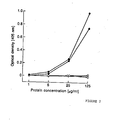
- Figure 2 Analysis of binding of alkaline phosphatase activity by monoclonal antibodies to cell surface glycoproteins of human choriocarcinoma using a solid-phase immunosorbent assay.
- Hybridoma cell lines producing the monoclonal antibodies of the present invention have been deposited prior to the filing of the present application at the American Type Culture Collection,. Bethesda, Maryland, and will be made available to the public during the pendancy of any patent issuing therefrom, in accordance with the Budapest Treaty.
- the monoclonal antibodies bear the following designations:
- the cell line OCC-MM(c) established from a heterotransplant of human ovarian choriocarcinoma propagated in nu/nu mice, as reported by Kim, supra SCH, a choriocarcinoma derived from gastric tumor and described by Kameya et al, Cancer Res., 35 2025-2032 (1975), were used as well, in immunization.
- Spleen cells were then fused with cells of the mouse myeloma cell line MOPC-21 NS/1, and antibody producing clones were then isolated by repeated subcloning.
- Hybridoma cells were injected, subcutaneously or intraperitoneally, into nu/nu mice (Swiss background), and ascites fluid and serum of mice with progressively growing tumors was used as a source of antibody for serological and biochemical studies, with cultured cells.
- Antibody subclass was determined by double diffusion in agar, with anti-Ig heavy chain specific antisera.
- MHA Mixed hemadsorption
- Frozen sections ( 5 m) of tissues were air- dried, fixed for 10 minutes in 3.7% formaldehyde in phosphate-buffered saline (PBS; 0.01 M sodium phosphate, 0.15 M NaCI, pH 7.3), washed, and incubated for 1 hour at room temperature with undiluted hybridoma culture supernatant or a 1/50 dilution of nu/nu mouse serum. The slides were washed and incubated for 45 minutes at room temperature with a 1/40 dilution of fluorescein-conjugated goat-anti-mouse Ig, washed again, and wet- mounted in 90% glycerol in PBS.
- PBS phosphate-buffered saline
- Cells were metabolically labeled with [ 3 H]-glucosamine or [ 35 S]methionine and extracted in Nonidet P-40 buffer following the procedures of Ueda, et aI., Proc. Natl. Acad. Sci., U.S.A. 78, 5122-5126 (1981).
- FIG. 1 shows the results of immunoprecipitation tests with the five antibodies which precipitate glycoproteins from choriocarcinoma cells: LK26 (gp about 30,000 to 35,000), SV19 (gp 40,000 under reduced conditions, 25,000 under unreduced conditions), SV63 and K8 (gp 68,000) and S4 (gp 160,000).
- LK26 shows a highly restricted distribution. It is expressed on all choriocarcinoma, teratocarcinoma and renal cancer cell lines. Among the other tumor cell lines tested, only a small subset of epithelial cancer lines bears the LK26 determinant, whereas leukemias, lymphomas and neuroectodermally derived tumors do not. In striking contrast to the positive LK26 phenotype of renal cancer lines, normal kidney epithelial cells lack LK26 expression. In normal tissues, LK26 is restricted to the trophoblast. All other adult and fetal tissues tested, including normal kidney, were negative for LK26.
- SV19 is expressed on 7/7 choriocarcinoma lines (titration endpoints 4 X 10- s to 1 10- 6 ), 3/4 teratocarcinoma lines, and 4/13 breast cancer lines (titration endpoints 1 X 10- 3 to 1 X 10 -"). Over- 120 other cell lines were SV19 negative in MHA direct tests. In absorption tests, low levels of SV19 antigen could be detected on 3 additional breast cancer lines and 5/6 colon cancer lines but not on 35 other tumor lines tested by this method (5 renal cancers, 4 bladder cancers, 9 lung cancers, 1 pancreatic cancer, 6 melanomas, 4 astrocytomas, 5 EBV-transformed B cell lines, 1 normal kidney epithelial cell culture).
- SV19 expression is restricted to the normal trophoblast, mammary gland epithelium and colonic epithelium.
- teratocarcinomas, breast, bladder, colon and lung cancers were SV19 positive.
- additional tissues e.g., fetal testis and fetal ovary, a distinct nuclear staining pattern was found. The relationship between this nuclear reactivity and the cell surface expression of SV19 remains to be determined.
- AbSV19 detected glycoproteins of molecular weight from about 40 to about 45,000 daltons (under reducing conditions) or molecular weight of about 25,000 daltons (non-reducing conditions).
- SV63 is found in the placenta (prominent staining of the syn- cytiotrophoblast and weaker staining of the cytotrophoblast), intestinal epithelium and germ cells, whereas K8 reactivity is restricted to the trophoblast.
- the SV63 and K8 antigens were shown to have alkaline phosphatase activity'using a solid-phase immunosorbent-assay - ( Figure 2).
- LK24 expressors 6/7 choriocarcinoma cell lines are strong LK24 expressors.
- LK24 is also present on most breast cancer lines and a small subset of epithelial cancer lines, as well as on EBV-transformed B cells, certain leukemias and lymphomas.
- titration endpoints in MHA tests with breast cancers and lymphoid cells were more than 10-fold lower than with choriocarcinoma cells, and reactivity was limited to a small fraction (5-20%) of cells within a culture. This low percentage of LK24 positive cells, which was not seen with choriocarcinomas, was unaffected by changing the density of the target cells used in MHA tests or the time between target cell plating and testing ( 1 to 5 days).
- LK24 is expressed in the trophoblast, colonic epithelium, distal and collecting tubules of the kidney, urothelium and a subpopulation of cells in the fetal thymus. In the tumor panel, LK24 was found in a variety of epithelial cancers and also in lymphomas. Reactivity of cultured cells with AbLK24 is resistant to boiling (5 min at 100°C) and trypsin treatment. Immunoprecipitation tests with extracts of ('H]glucosamine-and [ 35 S]methionine- labeled cells failed to show any specific components. These biochemical properties suggest that AbLK24 may recognize a carbohydrate or lipid moiety.
- Type O erythrocytes remove LK24 reactivity in absorption tests, but no agglutination was seen with either type 0 or type A, B, and AB erythrocytes.
- AbLK24 failed to show reactivity with any of the purified blood group glycoproteins and glycolipids carrying A, B, H, I, Le a , Le b , X and Y determinants, suggesting that LK24 represents an unrelated antigenic system.
- K66 shows an intermediate distribution on the cultured cell panel, being expressed on choriocarcinomas and many epithelial cancer lines, 5/10 astrocytomas, 1/10 melanomas and on normal kidney epithelial cells.
- two fibroblast cultures derived from fetal tissues were K66 positive.
- K66 reactivity with cultured cells was very strong (e.g., titration endpoints in MHA tests on renal cancer cells 1 X 10- 6 to 1 X'10 - 7 ), no reactivity was seen in tests with the normal tissue panel or the tumors.
- the S4 antigen which has been described by Ueda, et al., Proc. Natl. Acad. Sci., U.S.A. 708, 5122-5126 (1981) and Cordon-Cardo, et al., J. Histochem. Cytochem. (in press) (1984), is a marker for normal and malignant kidney epithelial cells. S4 is also expressed on 6/6 choriocarcinoma cell lines.
- AbS4 precipitates glycoproteins of molecular weight of about 160,000 daltons from both [ 3 H]glucosamine- labeled extracts and ConA-bound fractions of [ 35 S]-methionine-labeled extracts of GCC-SV(c), OCC-MM or JEG-3 choriocarcinoma cells, which is consistent with the molecular weight reported for S4 on renal cancer cells.
- LK26 is a highly restricted trophoblast antigen. It is found in the placenta (but not in any other normal adult or fetal tissue tested) and is expressed on trophoblastic tumors, including hydatidiform mole gestational choriocarcinoma. In sharp contrast to the similarity in LK26 expression between normal and malignant trophoblastic cells, normal and malignant kidney epithelial cells differ with respect to this antigen. 15/15 renal cancer cell lines were LK26 positive whereas 5/5 cultures of normal kidney epithelial cells were LK26 negative. Also, tissues of normal adult and fetal kidney lacked LK26, whereas a subset of renal cancer showed the antigen. Thus, the antigen seems to be restricted to normal trophoblastic cells and certain tumors (choriocarcinoma, subsets of other teratocarcinomas, and epithelial cancers).
- S4 is a marker of normal and malignant kidney epithelial cells, both in vitro and in tissues.
- S4 antigen is present on choriocarcinoma cell lines.
- the S4 distribution resembles that of LK26, but the two antigens can be distinguished by their molecular weights and important differences in their serological distribution: both S4 and LK26 are present on malignant kidney epithelial cells and on choriocarcinoma cells but S4 is found on normal kidney epithelial cells and not on normal trophoblastic cells, whereas LK26 is strongly expressed on normal trophoblast, but not on normal kidney epithelial cells.
- S4 and LK26 represent antigens shared between trophoblastic cells and cells of renal origin, a different relationship is found for the other antigens described herein: SV19, SV63 and LK24 are common to trophoblastic cells and colon and/or breast epithelial cells, with SV19 being the most restricted of this group of antigens.
- Antibodies AbSV63 and AbK8 recognize different epitopes of the PALP molecule,. and their patterns of reactivity with the cell panel are quite distinct from the LK26 pattern.
- ALP alkaline phosphatase
- K66 belongs to this group of antigens. AbK66 has very high titers when tested in MHA assays on cultured cells (e.g., titration endpoints of1 X 10- 6 to 1 X 1 0- 7 on kidney epithelial cells). Therefore, low affinity of the antibody or differences in sensitivity between the serological assays used (MHA assay vs. indirect immunofluorescence) are probably not responsible for the observed discrepancy in K66 distribution in vitro and in vivo.
- this discrepancy is related to the presence of antigen only on rapidly proliferating cells or on cells adapted to the growth conditions in vitro. If this is true, an additional explanation is required to account for the clear distinction between K66 positive and K66 negative cell types in vitro, both of which have comparable rates of proliferation. Of particular interest is the finding that among short term cultures of fibroblasts, those derived from adults were K66 negative whereas those derived from fetal tissues were K66 positive.
- the antigenic systems described herein differ from the cell surface antigens and secreted proteins of choriocarcinoma and normal trophoblastic cells that have been studies by other investigators as reported by Lipinski, et al., Proc. Natl. Acad. Sci., U.S.A. 78 5147-5150 - (1980); Sunderland, et al., Immunol. 43 541-546 (1981); McLaughlin, et al., Int. J. Cancer 30 21-26 - (1982); Travers, et al., lnt. J. Cancer 33 633-641 - (1984).
- NDOG-1, NDOG-2, Trop-1 and Trop-2 are different from LK26, SV19, LK24, K66 and S4.
- NDOG-2 and SV63 co-type and may both react with ALP.
- NDOG-1 did not show any cell surface reactivity with the cell lines used in this study.
- Trop-1 and Trop-2 showed a broader representation on the cell panel and cannot be distinguished from each other as the anti-Trop-2 antibody showed higher reactivity and a serological pattern that overlapped the Trop-1 pattern.
- LK26, S4, SV19 and PALP restriction of antigens LK26, S4, SV19 and PALP to only a small number of normal human tissues make them useful diagnostic markers for gestational choriocarcinomas and germ cell tumors.
- the reciprocal expression of SV63 and SV19/P12 in subsets of breast cancers and the presence of LK26 on renal cancer cells (but not normal kidney epithelium) indicate that these antigens may be of interest in the study of other epithelial cancers as well.
- the highly restricted distribution of LK26 which is undetectable in normal tissues other than the trophoblast, makes this antigen a possible target for immunological imaging and immunotherapy of choriocarcinoma.
- phenotyping panel described, e.g., in Tables 2 and 3, may be used.
- Table 2 shows that LK26 shows affinity, to one degree or another, with lung, renal, ovarian/uterine, breast, teratocarcinoma, and choriocarcinoma cancer cells, and contacting a cell sample to this mAb would limit the possibilities of the identity of the cell sample accordingly. Further contact with SV19 would eliminate the possibility of the sample being lung, renal, or ovarian/uterine cancer, as SV19 is distributed only on breast, teratocarcinoma, and choriocarcinoma cancer cells.
- LK24 antigen is distributed over more than one cancer cell type, the passage through LK26, SV19, and K8 has limited the cell possibilities to either of choriocarcinoma or teratocarcinoma. Binding to the cells of LK24 indicates choriocarcinoma, no binding indicates teratocarcinoma. Hence, the panel LK26, SV19, K8 and LK24 will determine the presence, or lack therof, of both choriocarcinoma and teratocarcinoma.
Landscapes
- Health & Medical Sciences (AREA)
- Chemical & Material Sciences (AREA)
- Organic Chemistry (AREA)
- Immunology (AREA)
- Life Sciences & Earth Sciences (AREA)
- Biophysics (AREA)
- Biochemistry (AREA)
- General Health & Medical Sciences (AREA)
- Genetics & Genomics (AREA)
- Medicinal Chemistry (AREA)
- Molecular Biology (AREA)
- Proteomics, Peptides & Aminoacids (AREA)
- Cell Biology (AREA)
- Preparation Of Compounds By Using Micro-Organisms (AREA)
- Peptides Or Proteins (AREA)
- Micro-Organisms Or Cultivation Processes Thereof (AREA)
- Medicines That Contain Protein Lipid Enzymes And Other Medicines (AREA)
- Medicines Containing Antibodies Or Antigens For Use As Internal Diagnostic Agents (AREA)
Applications Claiming Priority (2)
| Application Number | Priority Date | Filing Date | Title |
|---|---|---|---|
| US718162 | 1985-04-01 | ||
| US06/718,162 US4851332A (en) | 1985-04-01 | 1985-04-01 | Choriocarcinoma monoclonal antibodies and antibody panels |
Publications (3)
| Publication Number | Publication Date |
|---|---|
| EP0197435A2 true EP0197435A2 (de) | 1986-10-15 |
| EP0197435A3 EP0197435A3 (en) | 1989-03-22 |
| EP0197435B1 EP0197435B1 (de) | 1992-07-29 |
Family
ID=24885068
Family Applications (1)
| Application Number | Title | Priority Date | Filing Date |
|---|---|---|---|
| EP86104170A Expired EP0197435B1 (de) | 1985-04-01 | 1986-03-26 | Monoklonale Choriokarzinom-Antikörper und Antikörpergarnitur |
Country Status (5)
| Country | Link |
|---|---|
| US (1) | US4851332A (de) |
| EP (1) | EP0197435B1 (de) |
| JP (1) | JPS61293382A (de) |
| CA (1) | CA1303531C (de) |
| DE (1) | DE3686187T2 (de) |
Cited By (6)
| Publication number | Priority date | Publication date | Assignee | Title |
|---|---|---|---|---|
| EP0287030A3 (en) * | 1987-04-14 | 1988-11-09 | Abbott Laboratories | Immunological methods and materials for detection of tumor associated antigens |
| WO1995024482A1 (en) * | 1994-03-08 | 1995-09-14 | Scotgen Biopharmaceuticals, Inc. | Recombinant humanized anti-lk26 antibodies |
| EP2172487A1 (de) | 2005-04-22 | 2010-04-07 | Morphotek Inc. | Antikörper mit Immuneffektoraktivität, die in Folatrezeptor alpha-positive Zellen internalisiert werden |
| EP2316857A1 (de) | 2004-02-12 | 2011-05-04 | Morphotek, Inc. | Monoklonaler Antikörper spezifisch für Folatrezeptor alpha |
| WO2014087863A1 (ja) | 2012-12-07 | 2014-06-12 | 協和発酵キリン株式会社 | 抗folr1抗体 |
| WO2014205342A2 (en) | 2013-06-20 | 2014-12-24 | Morphotek, Inc. | Methods for treatment of ovarian cancer |
Families Citing this family (42)
| Publication number | Priority date | Publication date | Assignee | Title |
|---|---|---|---|---|
| AU2004249673A1 (en) * | 2003-05-23 | 2004-12-29 | Morphotek, Inc. | Anti alpha - folate - receptor - tetramer antibodies |
| EP1861425B1 (de) | 2005-03-10 | 2012-05-16 | Morphotek, Inc. | Anti-mesothelin-antikörper |
| AU2006221823B2 (en) | 2005-03-11 | 2011-06-09 | Syngenta Limited | Rodent pest control |
| US20170335281A1 (en) | 2014-03-15 | 2017-11-23 | Novartis Ag | Treatment of cancer using chimeric antigen receptor |
| JP2017528433A (ja) | 2014-07-21 | 2017-09-28 | ノバルティス アーゲー | 低い免疫増強用量のmTOR阻害剤とCARの組み合わせ |
| US20180334490A1 (en) | 2014-12-03 | 2018-11-22 | Qilong H. Wu | Methods for b cell preconditioning in car therapy |
| WO2016126608A1 (en) | 2015-02-02 | 2016-08-11 | Novartis Ag | Car-expressing cells against multiple tumor antigens and uses thereof |
| EP3286211A1 (de) | 2015-04-23 | 2018-02-28 | Novartis AG | Behandlung von krebs mit chimärem antigenrezeptor- und proteinkinase-a-blocker |
| US11667691B2 (en) | 2015-08-07 | 2023-06-06 | Novartis Ag | Treatment of cancer using chimeric CD3 receptor proteins |
| FI3380620T3 (fi) | 2015-11-23 | 2024-08-01 | Novartis Ag | Optimoituja lentiviruksen siirtovektoreita ja niiden käyttötapoja |
| RU2018127657A (ru) | 2015-12-30 | 2020-01-31 | Новартис Аг | Виды терапии на основе иммуноэффекторных клеток с улучшенной эффективностью |
| KR20180118175A (ko) | 2016-03-04 | 2018-10-30 | 노파르티스 아게 | 다중 키메라 항원 수용체 (car) 분자를 발현하는 세포 및 그에 따른 용도 |
| EP3432924A1 (de) | 2016-03-23 | 2019-01-30 | Novartis AG | Zellsekretierte minikörper und verwendungen davon |
| CN109715808A (zh) | 2016-04-15 | 2019-05-03 | 诺华股份有限公司 | 用于选择性蛋白质表达的组合物和方法 |
| BR112019002035A2 (pt) | 2016-08-01 | 2019-05-14 | Novartis Ag | tratamento de câncer usando um receptor de antígeno quimérico em combinação com um inibidor de uma molécula pró-macrófago m2 |
| WO2018111340A1 (en) | 2016-12-16 | 2018-06-21 | Novartis Ag | Methods for determining potency and proliferative function of chimeric antigen receptor (car)-t cells |
| ES2912408T3 (es) | 2017-01-26 | 2022-05-25 | Novartis Ag | Composiciones de CD28 y métodos para terapia con receptores quiméricos para antígenos |
| CN110582509A (zh) | 2017-01-31 | 2019-12-17 | 诺华股份有限公司 | 使用具有多特异性的嵌合t细胞受体蛋白治疗癌症 |
| WO2018160731A1 (en) | 2017-02-28 | 2018-09-07 | Novartis Ag | Shp inhibitor compositions and uses for chimeric antigen receptor therapy |
| WO2018229715A1 (en) | 2017-06-16 | 2018-12-20 | Novartis Ag | Compositions comprising anti-cd32b antibodies and methods of use thereof |
| AU2018351050B2 (en) | 2017-10-18 | 2025-09-18 | Novartis Ag | Compositions and methods for selective protein degradation |
| WO2019081983A1 (en) | 2017-10-25 | 2019-05-02 | Novartis Ag | CD32B TARGETING ANTIBODIES AND METHODS OF USE |
| RU2020116579A (ru) | 2017-10-25 | 2021-11-25 | Новартис Аг | Способы получения клеток, экспрессирующих химерный антигенный рецептор |
| US20210179709A1 (en) | 2017-10-31 | 2021-06-17 | Novartis Ag | Anti-car compositions and methods |
| WO2019210153A1 (en) | 2018-04-27 | 2019-10-31 | Novartis Ag | Car t cell therapies with enhanced efficacy |
| EP3788369A1 (de) | 2018-05-01 | 2021-03-10 | Novartis Ag | Biomarker zur auswertung von car-t-zellen zur vorhersage des klinischen ergebnisses |
| EP3801769A1 (de) | 2018-05-25 | 2021-04-14 | Novartis AG | Kombinationstherapie mit therapien mit chimärem antigen-rezeptor (car) |
| WO2019237035A1 (en) | 2018-06-08 | 2019-12-12 | Intellia Therapeutics, Inc. | Compositions and methods for immunooncology |
| AR116109A1 (es) | 2018-07-10 | 2021-03-31 | Novartis Ag | Derivados de 3-(5-amino-1-oxoisoindolin-2-il)piperidina-2,6-diona y usos de los mismos |
| KR20210106437A (ko) | 2018-12-20 | 2021-08-30 | 노파르티스 아게 | 3-(1-옥소이소인돌린-2-일)피페리딘-2,6-디온 유도체를 포함하는 투약 요법 및 약학적 조합물 |
| CN113490528B (zh) | 2019-02-15 | 2024-12-03 | 诺华股份有限公司 | 3-(1-氧代-5-(哌啶-4-基)异吲哚啉-2-基)哌啶-2,6-二酮衍生物及其用途 |
| CA3123519A1 (en) | 2019-02-15 | 2020-08-20 | Novartis Ag | Substituted 3-(1-oxoisoindolin-2-yl)piperidine-2,6-dione derivatives and uses thereof |
| EP3959320A1 (de) | 2019-04-24 | 2022-03-02 | Novartis AG | Zusammensetzungen und verfahren für selektiven proteinabbau |
| BR112022011902A2 (pt) | 2019-12-20 | 2022-09-06 | Novartis Ag | Terapias de combinação |
| JP2023529211A (ja) | 2020-06-11 | 2023-07-07 | ノバルティス アーゲー | Zbtb32阻害剤及びその使用 |
| KR20230027056A (ko) | 2020-06-23 | 2023-02-27 | 노파르티스 아게 | 3-(1-옥소이소인돌린-2-일)피페리딘-2,6-디온 유도체를 포함하는 투약 요법 |
| JP7819176B2 (ja) | 2020-08-03 | 2026-02-24 | ノバルティス アーゲー | ヘテロアリール置換3-(1-オキソイソインドリン-2-イル)ピペリジン-2,6-ジオン誘導体及びその使用 |
| TW202304979A (zh) | 2021-04-07 | 2023-02-01 | 瑞士商諾華公司 | 抗TGFβ抗體及其他治療劑用於治療增殖性疾病之用途 |
| WO2022229853A1 (en) | 2021-04-27 | 2022-11-03 | Novartis Ag | Viral vector production system |
| WO2023214325A1 (en) | 2022-05-05 | 2023-11-09 | Novartis Ag | Pyrazolopyrimidine derivatives and uses thereof as tet2 inhibitors |
| AU2023369684A1 (en) | 2022-10-26 | 2025-04-17 | Novartis Ag | Lentiviral formulations |
| PE20252789A1 (es) | 2023-05-24 | 2025-12-22 | Kumquat Biosciences Inc | Compuestos heterociclicos y usos de estos |
Family Cites Families (4)
| Publication number | Priority date | Publication date | Assignee | Title |
|---|---|---|---|---|
| GB8305967D0 (en) * | 1983-03-04 | 1983-04-07 | Univ Liverpool | Material for treatment of spontaneous abortions |
| US4643971A (en) * | 1983-03-11 | 1987-02-17 | Sloan-Kettering Institute | Monoclonal antibodies to human bladder and ureter cancers and method |
| DE3329184A1 (de) * | 1983-08-12 | 1985-02-21 | Behringwerke Ag, 3550 Marburg | Monoklonale antikoerper mit spezifitaet fuer membran-assoziierte antigene |
| EP0143367A3 (de) * | 1983-11-30 | 1987-11-25 | Sloan-Kettering Institute For Cancer Research | Verfahren zur Herstellung monoklonaler Antikörper für Krebsdiagnose und Therapie |
-
1985
- 1985-04-01 US US06/718,162 patent/US4851332A/en not_active Expired - Lifetime
-
1986
- 1986-03-26 DE DE8686104170T patent/DE3686187T2/de not_active Expired - Fee Related
- 1986-03-26 EP EP86104170A patent/EP0197435B1/de not_active Expired
- 1986-04-01 JP JP61075407A patent/JPS61293382A/ja active Pending
- 1986-04-01 CA CA000505505A patent/CA1303531C/en not_active Expired - Lifetime
Non-Patent Citations (27)
| Title |
|---|
| ALBINO ET AL., J. IMMUNOL., vol. 131, 1983, pages 1595 - 1599 |
| ANGER ET AL., HYBRIDOMA, vol. 1, 1982, pages 139 - 146 |
| CAIRNCROSS ET AL., PROC. NATL. ACAD. SCI., U.S.A., vol. 79, 1982, pages 5641 - 5645 |
| CAREY ET AL., PROC. NATL. ACAD. SCI., U.S.A., vol. 73, 1976, pages 3278 - 3282 |
| CORDON-CARDO ET AL., J. HISTOCHEM. CYTOCHEM., 1984 |
| DIPPOLD ET AL., PROC. NATL. ACAD. SCI., U.S.A., vol. 77, 1980, pages 6114 - 6118 |
| FRADET ET AL., PROC. NATL, ACAD. SCI., U.S.A., vol. 81, 1984, pages 224 - 228 |
| FRADET ET AL., PROC. NATL. ACAD. SCI., U.S.A., vol. 81, 1984, pages 224 - 228 |
| HARRIS, HARVEY LECT., vol. 76, 1982, pages 95 - 124 |
| KAMEYA ET AL., CANCER RES., vol. 35, 1975, pages 2025 - 2032 |
| KIM ET AL., GYNECOL. ONCOL., vol. 6, 1978, pages 165 - 182 |
| LIPINSKI ET AL., PROC. NATL. ACAD. SCI., U.S.A., vol. 78, 1980, pages 5147 - 5150 |
| LIPINSKI ET AL., PROC. NATL. ACAD. SCI., U.S.A., vol. 78, 1981, pages 5147 - 5150 |
| LLOYD ET AL.: "Basic and Clinical Tumor Immunologv", 1983, MARTINUS NIIHOFF, pages: 159 - 214 |
| LLOYD, K.O.: "Basic and Clinical Tumor Immunology", 1983, MARTINUS NYTOFF, article "Human Tumor Antigens: Detection and Characterization with Monoclonal Antibodies", pages: 159 - 214 |
| MATTES ET AL., J. IMMUNOL. METH., vol. 61, 1983, pages 145 - 150 |
| MATTES ET AL., PROC. NATL. ACAD. SCI., U.S.A., vol. 81, 1984, pages 568 - 572 |
| MCDICKEN ET AL., INT. J. CANCER, vol. 32, 1983, pages 205 - 209 |
| MCLAUGHLIN ET AL., INT. J. CANCER, vol. 30, 1982, pages 21 - 26 |
| MILLAN ET AL., CANCER RES., vol. 42, 1982, pages 2444 - 2449 |
| PROC. NATL. ACAD. SCI., U.S.A., vol. 81, 1984, pages 6437 - 6441 |
| SLAUGHTER ET AL., PROC. NATL. ACAD. SCI., U.S.A., vol. 78, 1981, pages 1124 - 1128 |
| SUNDERLAND ET AL., IMMUNOL., vol. 43, 1981, pages 541 - 546 |
| TRAVERS ET AL., INT. J. CANCER, vol. 33, 1984, pages 633 - 641 |
| TRAVERS; BODMER, INT. J. CANCER, vol. 33, 1984, pages 633 - 641 |
| UEDA ET AL., PROC. NATL. ACAD. SCI., U.S.A., vol. 708, 1981, pages 5122 - 5126 |
| UEDA ET AL., PROC. NATL. ACAD. SCI., U.S.A., vol. 78, 1981, pages 5122 - 5126 |
Cited By (12)
| Publication number | Priority date | Publication date | Assignee | Title |
|---|---|---|---|---|
| EP0287030A3 (en) * | 1987-04-14 | 1988-11-09 | Abbott Laboratories | Immunological methods and materials for detection of tumor associated antigens |
| WO1995024482A1 (en) * | 1994-03-08 | 1995-09-14 | Scotgen Biopharmaceuticals, Inc. | Recombinant humanized anti-lk26 antibodies |
| US5646253A (en) * | 1994-03-08 | 1997-07-08 | Memorial Sloan-Kettering Cancer Center | Recombinant human anti-LK26 antibodies |
| US5952484A (en) * | 1994-03-08 | 1999-09-14 | Sloan-Kettering Cancer Center | Recombinant human anti-LK26 antibodies |
| US6124106A (en) * | 1994-03-08 | 2000-09-26 | Ludwig Institute For Cancer Research | Method for detecting cancers |
| EP2316857A1 (de) | 2004-02-12 | 2011-05-04 | Morphotek, Inc. | Monoklonaler Antikörper spezifisch für Folatrezeptor alpha |
| EP3101034A1 (de) | 2004-02-12 | 2016-12-07 | Morphotek, Inc. | Monoklonale, spezifisch an folatrezeptor-alpha bindende antikörper |
| EP2172487A1 (de) | 2005-04-22 | 2010-04-07 | Morphotek Inc. | Antikörper mit Immuneffektoraktivität, die in Folatrezeptor alpha-positive Zellen internalisiert werden |
| US9522195B2 (en) | 2005-04-22 | 2016-12-20 | Morphotek, Inc. | Antibodies with immune effector activity and that internalize in folate receptor alpha-positive cells |
| WO2014087863A1 (ja) | 2012-12-07 | 2014-06-12 | 協和発酵キリン株式会社 | 抗folr1抗体 |
| US9695237B2 (en) | 2012-12-07 | 2017-07-04 | Kyowa Hakko Kirin Co., Ltd | Anti-FOLR1 antibody |
| WO2014205342A2 (en) | 2013-06-20 | 2014-12-24 | Morphotek, Inc. | Methods for treatment of ovarian cancer |
Also Published As
| Publication number | Publication date |
|---|---|
| DE3686187T2 (de) | 1993-03-04 |
| EP0197435A3 (en) | 1989-03-22 |
| DE3686187D1 (de) | 1992-09-03 |
| JPS61293382A (ja) | 1986-12-24 |
| EP0197435B1 (de) | 1992-07-29 |
| CA1303531C (en) | 1992-06-16 |
| US4851332A (en) | 1989-07-25 |
Similar Documents
| Publication | Publication Date | Title |
|---|---|---|
| EP0197435B1 (de) | Monoklonale Choriokarzinom-Antikörper und Antikörpergarnitur | |
| KR910000956B1 (ko) | 인체의 비-소세포 폐암치료용 단클론의 항체 및 항원 | |
| Takahashi et al. | Immunoglobulin G3 monoclonal antibody directed to Tn antigen (tumor-associated α-N-acetylgalactosaminyl epitope) that does not cross-react with blood group A antigen | |
| Southall et al. | Immunohistological distribution of 5T4 antigen in normal and malignant tissues | |
| CA1320459C (en) | Anti-human pulmonary carcinoma monoclonal antibody | |
| US4650756A (en) | Monoclonal antibodies to cell surface antigens of human renal cancer | |
| WO1986002945A1 (en) | Monoclonal antibody to a human carcinoma tumor associated antigen | |
| Rettig et al. | Cell surface antigens of human trophoblast and choriocarcinoma defined by monoclonal antibodies | |
| IE60286B1 (en) | Monoclonal antibodies and antigen for human non-small cell lung carcinoma and certain other human carcinomas | |
| US5084380A (en) | Monoclonal antibodies reactive with activated and oncogenic ras p21 proteins | |
| EP0287030B1 (de) | Immunologische Methoden und Materialien zur Bestimmung von Tumor assoziierten Antigenen | |
| Xu et al. | Development of two new monoclonal antibodies reactive to a surface antigen present on human ovarian epithelial cancer cells | |
| US4898932A (en) | Monoclonal antibodies reactive with activated and oncogenic ras p21 proteins | |
| CA1296660C (en) | Monoclonal antibody against a ras oncogene p21 related dodecapeptide | |
| EP0203552B1 (de) | Monoklonale Antikörper für humane Lungenkarzinome des "Nicht-kleine-Zellen"-Typs | |
| JPH0734741B2 (ja) | ヒト腎臓がん抗原に対するモノクローナル抗体生産ハイブリドーマ細胞系 | |
| EP0218257A2 (de) | Spezifischer monoklonaler Antikörper gegen menschliches Lungenadenokarzinom | |
| NZ233147A (en) | Retinoblastoma gene product antibodies and their use in assays for tumor cells | |
| EP0143367A2 (de) | Verfahren zur Herstellung monoklonaler Antikörper für Krebsdiagnose und Therapie | |
| Bhattacharya et al. | Production and Characterization of Monoclonal Antibody to a 60-kD Glycoprotein in Ovarian Carcinoma1 | |
| Lindgren et al. | Monoclonal antibodies to carcinoembryonic antigen (CEA): characterization and use in a radioimmunoassay for CEA | |
| US5081032A (en) | Anti-human pulmonary adenocarcinoma monoclonal antibody | |
| US5143843A (en) | Method for production of monoclonal antibodies for cancer diagnosis and therapy | |
| AU601680B2 (en) | Monoclonal antibodies for human non-small cell lung carcinomas | |
| CA1221926A (en) | Monoclonal, antibodies to cell surface antigens of human malignant melanoma |
Legal Events
| Date | Code | Title | Description |
|---|---|---|---|
| PUAI | Public reference made under article 153(3) epc to a published international application that has entered the european phase |
Free format text: ORIGINAL CODE: 0009012 |
|
| AK | Designated contracting states |
Kind code of ref document: A2 Designated state(s): DE GB |
|
| PUAL | Search report despatched |
Free format text: ORIGINAL CODE: 0009013 |
|
| AK | Designated contracting states |
Kind code of ref document: A3 Designated state(s): DE GB |
|
| 17P | Request for examination filed |
Effective date: 19890920 |
|
| 17Q | First examination report despatched |
Effective date: 19901109 |
|
| GRAA | (expected) grant |
Free format text: ORIGINAL CODE: 0009210 |
|
| AK | Designated contracting states |
Kind code of ref document: B1 Designated state(s): DE GB |
|
| REF | Corresponds to: |
Ref document number: 3686187 Country of ref document: DE Date of ref document: 19920903 |
|
| PLBE | No opposition filed within time limit |
Free format text: ORIGINAL CODE: 0009261 |
|
| STAA | Information on the status of an ep patent application or granted ep patent |
Free format text: STATUS: NO OPPOSITION FILED WITHIN TIME LIMIT |
|
| 26N | No opposition filed | ||
| PGFP | Annual fee paid to national office [announced via postgrant information from national office to epo] |
Ref country code: GB Payment date: 19980220 Year of fee payment: 13 |
|
| PGFP | Annual fee paid to national office [announced via postgrant information from national office to epo] |
Ref country code: DE Payment date: 19980225 Year of fee payment: 13 |
|
| PG25 | Lapsed in a contracting state [announced via postgrant information from national office to epo] |
Ref country code: GB Free format text: LAPSE BECAUSE OF NON-PAYMENT OF DUE FEES Effective date: 19990326 |
|
| GBPC | Gb: european patent ceased through non-payment of renewal fee |
Effective date: 19990326 |
|
| PG25 | Lapsed in a contracting state [announced via postgrant information from national office to epo] |
Ref country code: DE Free format text: LAPSE BECAUSE OF NON-PAYMENT OF DUE FEES Effective date: 20000101 |