EP0220534A2 - Dispositif d'étalonnage de cathéter optique - Google Patents
Dispositif d'étalonnage de cathéter optique Download PDFInfo
- Publication number
- EP0220534A2 EP0220534A2 EP86113656A EP86113656A EP0220534A2 EP 0220534 A2 EP0220534 A2 EP 0220534A2 EP 86113656 A EP86113656 A EP 86113656A EP 86113656 A EP86113656 A EP 86113656A EP 0220534 A2 EP0220534 A2 EP 0220534A2
- Authority
- EP
- European Patent Office
- Prior art keywords
- catheter
- enclosure
- distal end
- reference element
- passage
- Prior art date
- Legal status (The legal status is an assumption and is not a legal conclusion. Google has not performed a legal analysis and makes no representation as to the accuracy of the status listed.)
- Granted
Links
Images

Classifications
-
- G—PHYSICS
- G01—MEASURING; TESTING
- G01N—INVESTIGATING OR ANALYSING MATERIALS BY DETERMINING THEIR CHEMICAL OR PHYSICAL PROPERTIES
- G01N21/00—Investigating or analysing materials by the use of optical means, i.e. using sub-millimetre waves, infrared, visible or ultraviolet light
- G01N21/17—Systems in which incident light is modified in accordance with the properties of the material investigated
- G01N21/47—Scattering, i.e. diffuse reflection
- G01N21/4785—Standardising light scatter apparatus; Standards therefor
-
- A—HUMAN NECESSITIES
- A61—MEDICAL OR VETERINARY SCIENCE; HYGIENE
- A61B—DIAGNOSIS; SURGERY; IDENTIFICATION
- A61B5/00—Measuring for diagnostic purposes; Identification of persons
- A61B5/145—Measuring characteristics of blood in vivo, e.g. gas concentration or pH-value ; Measuring characteristics of body fluids or tissues, e.g. interstitial fluid or cerebral tissue
- A61B5/1495—Calibrating or testing of in-vivo probes
-
- A—HUMAN NECESSITIES
- A61—MEDICAL OR VETERINARY SCIENCE; HYGIENE
- A61B—DIAGNOSIS; SURGERY; IDENTIFICATION
- A61B2560/00—Constructional details of operational features of apparatus; Accessories for medical measuring apparatus
- A61B2560/02—Operational features
- A61B2560/0223—Operational features of calibration, e.g. protocols for calibrating sensors
- A61B2560/0228—Operational features of calibration, e.g. protocols for calibrating sensors using calibration standards
- A61B2560/0233—Optical standards
Definitions
- This invention relates to a calibration device and calibrating system for optical catheters used in a catheter oximetry system, and more particularly, it relates to a calibrating device which may remain with the sealed and sterilized distal end of the catheter within a package while the proximal end of the catheter is plugged into a computer or processor in order to perform the calibrating operation.
- a catheter oximetry system provides accurate, continuous, real-time measurement of mixed venous oxygen saturation using multiple wavelength reflection spectrophotometry.
- the color of red blood cells progressively changes from scarlet to purple as the amount of oxygen the red blood cells are carrying decreases.
- the amount of light backscattered, or reflected, at each wavelength depends upon the color, and therefore, oxygen level of the blood.
- Careful choice of wavelengths in the transmittal light allows accurate measurement of oxygenated hemoglobin with minimal interference by other blood characteristics such as temperature, pH, and hematocrit.
- the relative amounts of oxygenated hemoglobin and deoxygenated hemoglobin in the blood can be determined by measuring the relative absorption of light at different selected wavelengths.
- the percentage of hemoglobin which is in the oxygenated form is defined as the oxygen saturation of the blood in the equation: where HbO2 is the oxygenated hemoglobin concentration and Hb is the deoxygenated hemoglobin concentration.
- a widely used catheter oximetry system consists of three basic components: (1) a disposable fiberoptic pulmonary artery catheter that has a distal end adapted to be inserted into a vein of a patient and that interfaces at its other end with (2) an optical module containing light emitting diodes, a photodetector and associated electronics, which in turn, interfaces with the electrical leads of (3) a computer-based instrument that performs all of the data processing and control functions with displays, alarms and associated read-out devices.
- the instrument and optical module may be reused many times with different patients, but the catheter is used only with a single patient during a single operation or monitoring process.
- the catheters are disposable and are arranged to be separately packaged in sealed aseptic packages each with a specially designed optical connector plug adapted to be plugged into the optical module when the catheter is ready for use.
- each catheter be separately calibrated immediately before it is used so as to relate the actual light intensities received from the sample under test to the unknown concentrations of the substances being quantified in the sample under test. This may be accomplished by initially measuring a given sample of blood with the catheter and then wholly independently measuring the same blood in the laboratory by a different technique in order to match the laboratory calculated actual oxygen saturation content with the instrument calculated content and adjusting the latter accordingly.
- the catheter can be pulled loose from the tube and reference block and placed in a patient for obtaining blood oxygen saturation measurements in the manner intended.
- the initial calibration readings are obtained with the reference block and catheter remaining in the package in a sealed and sterilized condition while the connector plug end of the catheter is connected to the optical module and oximetry processor.
- a catheter having transmitting and receiving light guides therein is arranged to be packaged in a tray with the distal end of the catheter being received in a calibrating device.
- the calibrating device includes a tubular enclosure within which a reference element is urged into compliant engagement with the distal end of the catheter and with means being provided for tightly gripping and holding the catheter to the enclosure so that the catheter and calibrating device are ready for an immediate calibration operation in the package without any additional movement of either catheter or reference block being required.
- the package is sealed with a cover material that encloses the catheter and calibrating device in the tray in a sealed and sterile condition.
- a portion of the sealing material can be removed while the remainder is left in its sealed and sterile condition so as to only expose the optical connector at the proximal end of the catheter permitting it to be connected to an oximetry system to provide a calibration reading for the catheter -- all while the distal end and insertable major length of the catheter remains in a sealed, sterile condition.
- the remainder of the sealing material can be removed, the calibrating device readily removed from the distal end of the catheter, and the catheter placed directly in the patient for continuous blood oxygen saturation readings.
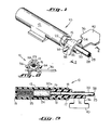
- the calibrating device 10 of the present invention is shown in Figures 1-3 wherein it will be seen to be comprised of a cylinder 12 which is closed at one end 13 and open at the other end thereof (Fig. 3). Received within the open end of the cylinder 12 is a plug 15 which is provided near the center thereof with a circumferencially extending rib 17 adapted to be received within a detent 18 just within the open end of the cylinder 12. The plug is inserted in the cylinder during the assembly of the calibrating device, and, as indicated in Figure 3, it is snapped into snug engagement therewith.
- an optical catheter 20 is adapted to be inserted through the plug 15 within an axial passage 22 extending therethrough.
- the axial passage 22 is just slightly larger than the outer diameter of the catheter and its balloon 21 so as to snugly confine the catheter therewithin.
- a reference block 24 and a coil spring 25 Positioned within the inner end of the tubular member 12 is a reference block 24 and a coil spring 25 with the spring urging the reference block into firm engagement with the flat distal end 20a of the catheter 20 at a position spaced inwardly from the end of the plug 15.
- the reference block 24 is the same as that shown and described in the aforementioned prior United States Patent No. 4,322,164 to Robert F. Shaw et al. Briefly, the reference block comprises a solid cylindrical element formed of a silicone resin and having a plurality of tiny particles scattered throughout its mass to provide scattering and reflecting surfaces for the light beams transmitted by the catheter 20. The particles will typically have dimensions within the range of from about 0.02 to about 20 microns and should be uniformly dis persed within the solid mass of the reference block 24.
- the mass is translucent is nature and has compliant characteristics at the surface thereof so that it will yield when pressed against the rigid surface 20a of the catheter thereby insuring a snug fit which will not become easily dislodged during handling of the catheter and attached calibrating device.
- the catheter 20 comprises a conventional optical catheter useful in oximetry measurements having a pair of separated lumens with a transmitting light guide 28 formed of a single fiber and a receiving light guide 29 likewise formed of a single fiber extending side-by-side along the length of the catheter to an exposed position at the flattened surface 20a at the very end of the catheter.
- Light carried along the transmitting fiber 28 is directed into the reference block 24 where it is backscattered and a portion thereof is reflected back into the receiving fiber 29 for transmission back to the oximetry processing apparatus to provide readings useful for calibrating the catheter and associated optical components.
- the strap includes an enlarged tab 40 at the outwardly projecting end thereof which tab is of a size whereby it can be readily gripped between the fingers in order to pull the strap loose from the prongs at the conclusion of the calibration operation in order to release the catheter from the calibrating device.
- FIG. 4 The use of the calibrating device 10 of the present invention in a catheter oximetry system is shown sequentially in Figures 4, 5 and 6.
- the catheter 20 is arranged to be packaged within conforming recesses set in a rectangularly shaped plastic tray 42.
- a piece of plastic sealing material 44 is laid atop the tray and sealed thereto, and the tray and enclosed catheter are then sterilized using conventional sterilization techniques.
- the distal end of the catheter is connected directly to the calibrating device 10 in the aforedescribed manner and clamped thereto by the strap 36 with the tab 40 of the strap extending to the side in a position adapting it to ready removability.
- the proximal end of the catheter includes the optical connector plug 46 and a plurality of other conven tional output connections including lumen connections for pressure readings, samplings, or infusion, a thermistor connection for cardiac outputs and a mechanism connected to pressurize the balloon 21 at the tip of the catheter -- all of such elements being conventional with the details thereof having no relevance with respect to the present invention.
- the first step in the calibration operation is to remove the plastic sealing material 44 from atop the tray to allow the fiberoptic connector plug 46 to be removed and coupled to the computer or processor 48.
- the sealing material 44 is provided with two sections separated by a seam or scoreline 43 whereby only one portion thereof is removed during the initial peeling of the material, as shown in Figure 5, exposing only the proximal end of the catheter and the connections thereto (including the connector plug 46) but leaving the main body of the catheter, which will later be placed in the patient, within the package in its original sealed and sterilized condition.
- the connector plug can then be placed in a receptacle in an optical module 50 which provides the electro-optical coupling between the connector plug 44 and the processing circuitry of the computer 48.
- the computer is turned on to provide signals to the optical module 50 creating the light sources which are directed via the coupling 46 down the length of the catheter to the reference block 24 wherein the light is backscattered and reflected back to the optical module.
- the module then converts these light signals into electrical signals for processing by the computer. In this way the appropriate calibration readings are obtained and stored in the computer.
- the catheter is calibrated and immediately ready for use in monitoring the blood oxygenation of a patient.
- the remainder of the sealing material 44 is then removed, and a simple pulling away of the strap 36 from its secured position on the calibrating device 10 leaves the catheter 20 free from its locked engagement therewith. The nurse or doctor can then directly take the catheter and place it in the patient.
- the calibrating device of the present invention permits the catheter to be directly locked to a calibrating device and packaged in such a manner so that no additional steps are required other than to connect the proximal end of the catheter to suitable processing circuitry in order to obtain appropriate calibration readings. Once the readings have been obtained, the catheter is ready for immediate use, and the protective and sealing material can be removed to permit the catheter to be immediately used. It has been found that the packaging method as aforedescribed will stand up under repeated jostling or dropping without dislodging the reference block from the catheter.
Landscapes
- Health & Medical Sciences (AREA)
- Life Sciences & Earth Sciences (AREA)
- Physics & Mathematics (AREA)
- General Health & Medical Sciences (AREA)
- Pathology (AREA)
- Optics & Photonics (AREA)
- Heart & Thoracic Surgery (AREA)
- General Physics & Mathematics (AREA)
- Immunology (AREA)
- Analytical Chemistry (AREA)
- Chemical & Material Sciences (AREA)
- Biophysics (AREA)
- Engineering & Computer Science (AREA)
- Biomedical Technology (AREA)
- Biochemistry (AREA)
- Medical Informatics (AREA)
- Molecular Biology (AREA)
- Surgery (AREA)
- Animal Behavior & Ethology (AREA)
- Public Health (AREA)
- Veterinary Medicine (AREA)
- Measurement Of The Respiration, Hearing Ability, Form, And Blood Characteristics Of Living Organisms (AREA)
- Investigating Or Analysing Materials By Optical Means (AREA)
Applications Claiming Priority (2)
| Application Number | Priority Date | Filing Date | Title |
|---|---|---|---|
| US06/791,706 US4650327A (en) | 1985-10-28 | 1985-10-28 | Optical catheter calibrating assembly |
| US791706 | 1985-10-28 |
Publications (3)
| Publication Number | Publication Date |
|---|---|
| EP0220534A2 true EP0220534A2 (fr) | 1987-05-06 |
| EP0220534A3 EP0220534A3 (en) | 1987-12-09 |
| EP0220534B1 EP0220534B1 (fr) | 1991-05-22 |
Family
ID=25154545
Family Applications (1)
| Application Number | Title | Priority Date | Filing Date |
|---|---|---|---|
| EP86113656A Expired EP0220534B1 (fr) | 1985-10-28 | 1986-10-03 | Dispositif d'étalonnage de cathéter optique |
Country Status (5)
| Country | Link |
|---|---|
| US (1) | US4650327A (fr) |
| EP (1) | EP0220534B1 (fr) |
| JP (1) | JPH0667382B2 (fr) |
| CA (1) | CA1277848C (fr) |
| DE (1) | DE3679369D1 (fr) |
Cited By (5)
| Publication number | Priority date | Publication date | Assignee | Title |
|---|---|---|---|---|
| EP0271304A1 (fr) * | 1986-12-08 | 1988-06-15 | Spectramed, Inc. | Manchon de calibrage à usage unique pour cathéter cardiovasculaire du type optique |
| EP0366840A1 (fr) * | 1986-12-16 | 1990-05-09 | BAXTER INTERNATIONAL INC. (a Delaware corporation) | Appareil de calibrage d'un cathéter optique et méthode |
| EP0397496A1 (fr) * | 1989-05-12 | 1990-11-14 | Baxter International Inc. | Enveloppe de calibrage pour calibrer in vitro un moniteur de saturation d'oxygène et sa méthode d'utilisation |
| EP0572133A1 (fr) * | 1992-05-22 | 1993-12-01 | Biomedical Sensors Ltd | Emballage pour un instrument médical actif |
| EP1401328A4 (fr) * | 2001-05-31 | 2006-04-05 | Infraredx Inc | Catheters optiques de referencage |
Families Citing this family (101)
| Publication number | Priority date | Publication date | Assignee | Title |
|---|---|---|---|---|
| US4834532A (en) * | 1986-12-05 | 1989-05-30 | The State Of Oregon Acting By And Through The State Board Of Higher Education On Behalf Of Oregon Health Sciences University | Devices and procedures for in vitro calibration of pulse oximetry monitors |
| US4968137A (en) * | 1986-12-05 | 1990-11-06 | The State Of Oregon Acting By And Through The State Board Of Higher Education On Behalf Of Oregon Health Sciences University | Devices and procedures for in vitro testing of pulse oximetry monitors |
| US5596986A (en) * | 1989-03-17 | 1997-01-28 | Scico, Inc. | Blood oximeter |
| US5762609A (en) * | 1992-09-14 | 1998-06-09 | Sextant Medical Corporation | Device and method for analysis of surgical tissue interventions |
| US5460182A (en) * | 1992-09-14 | 1995-10-24 | Sextant Medical Corporation | Tissue penetrating apparatus and methods |
| US5772597A (en) * | 1992-09-14 | 1998-06-30 | Sextant Medical Corporation | Surgical tool end effector |
| US5365925A (en) * | 1993-08-13 | 1994-11-22 | Ohmeda Inc. | Disposable calibration boot for multi-point calibration in fiber optic sensors |
| US5777731A (en) * | 1997-02-21 | 1998-07-07 | The Goodyear Tire & Rubber Company | Calibration of optical properties to measure depth of a liquid |
| US6271920B1 (en) | 1997-12-19 | 2001-08-07 | Chromatics Color Sciences International, Inc. | Methods and apparatus for color calibration and verification |
| ATE343346T1 (de) * | 1999-08-31 | 2006-11-15 | Nir Diagnostics Inc | Vorrichtung zur überprüfung der genauigkeit eines spektralanalysators |
| DE60141090D1 (de) | 2000-10-30 | 2010-03-04 | Gen Hospital Corp | Optische systeme zur gewebeanalyse |
| US9295391B1 (en) | 2000-11-10 | 2016-03-29 | The General Hospital Corporation | Spectrally encoded miniature endoscopic imaging probe |
| US7865231B2 (en) * | 2001-05-01 | 2011-01-04 | The General Hospital Corporation | Method and apparatus for determination of atherosclerotic plaque type by measurement of tissue optical properties |
| US6923762B1 (en) * | 2001-11-01 | 2005-08-02 | Frank C. Creaghan, Jr. | Venoscope apparatus |
| US7355716B2 (en) * | 2002-01-24 | 2008-04-08 | The General Hospital Corporation | Apparatus and method for ranging and noise reduction of low coherence interferometry LCI and optical coherence tomography OCT signals by parallel detection of spectral bands |
| US20110201924A1 (en) * | 2002-04-30 | 2011-08-18 | The General Hospital Corporation | Method and Apparatus for Improving Image Clarity and Sensitivity in Optical Tomography Using Dynamic Feedback to Control Focal Properties and Coherence Gating |
| US7643153B2 (en) * | 2003-01-24 | 2010-01-05 | The General Hospital Corporation | Apparatus and method for ranging and noise reduction of low coherence interferometry LCI and optical coherence tomography OCT signals by parallel detection of spectral bands |
| US7761139B2 (en) * | 2003-01-24 | 2010-07-20 | The General Hospital Corporation | System and method for identifying tissue using low-coherence interferometry |
| CA2519937C (fr) | 2003-03-31 | 2012-11-20 | Guillermo J. Tearney | Reduction de granularite dans la tomographie par coherence optique au moyen d'une composition angulaire par codage de longueur de trajet |
| KR101204370B1 (ko) | 2003-06-06 | 2012-11-23 | 더 제너럴 하스피탈 코포레이션 | 파장 동조 소스용 방법 및 장치 |
| WO2005047813A1 (fr) | 2003-10-27 | 2005-05-26 | The General Hospital Corporation | Procede et appareil d'imagerie optique par interferometrie dans le domaine frequentiel |
| DE102004024164A1 (de) * | 2004-05-14 | 2005-12-08 | Kaltenbach & Voigt Gmbh & Co. Kg | Zahnärztliches System zum Untersuchen der optischen Eigenschaften von Zahngewebe mit optischer Untersuchungsvorrichtung und Abgleicheinrichtung |
| WO2005117534A2 (fr) | 2004-05-29 | 2005-12-15 | The General Hospital Corporation | Procede, systeme et logiciel destines a la compensation de dispersion chromatique au moyen de couches reflechissantes dans l'imagerie par tomographie par coherence optique (oct) |
| JP4995720B2 (ja) * | 2004-07-02 | 2012-08-08 | ザ ジェネラル ホスピタル コーポレイション | ダブルクラッドファイバを有する内視鏡撮像プローブ |
| JP5053845B2 (ja) | 2004-08-06 | 2012-10-24 | ザ ジェネラル ホスピタル コーポレイション | 光学コヒーレンス断層撮影法を使用して試料中の少なくとも1つの位置を決定するための方法、システムおよびソフトウェア装置 |
| ATE538714T1 (de) * | 2004-08-24 | 2012-01-15 | Gen Hospital Corp | Verfahren, system und software-anordnung zur bestimmung des elastizitätsmoduls |
| JP5324095B2 (ja) | 2004-08-24 | 2013-10-23 | ザ ジェネラル ホスピタル コーポレイション | 血管セグメントを画像化する方法および装置 |
| KR101269455B1 (ko) | 2004-09-10 | 2013-05-30 | 더 제너럴 하스피탈 코포레이션 | 광 간섭 영상화를 위한 시스템 및 방법 |
| EP1804638B1 (fr) | 2004-09-29 | 2012-12-19 | The General Hospital Corporation | Systeme et procede d'imagerie a coherence optique |
| EP1825214A1 (fr) * | 2004-11-24 | 2007-08-29 | The General Hospital Corporation | Interferometre a trajet commun pour oct endoscopique |
| EP1816949A1 (fr) | 2004-11-29 | 2007-08-15 | The General Hospital Corporation | Ensembles, dispositifs, endoscopes, catheters et methodes d'imagerie optique permettant d'eclairer et de detecter simultanement plusieurs points sur un echantillon |
| US8351665B2 (en) | 2005-04-28 | 2013-01-08 | The General Hospital Corporation | Systems, processes and software arrangements for evaluating information associated with an anatomical structure by an optical coherence ranging technique |
| EP1886121A1 (fr) * | 2005-05-13 | 2008-02-13 | The General Hospital Corporation | Arrangements, systemes et procedes permettant d'obtenir une reflectrometrie de coherence optique du domaine spectral pour une detection sensible d'un echantillon chimique et biologique |
| EP1889037A2 (fr) * | 2005-06-01 | 2008-02-20 | The General Hospital Corporation | Appareil, methode et systeme pour effectuer une imagerie de domaine de frequence optique a resolution de phase |
| EP1913332B1 (fr) * | 2005-08-09 | 2010-10-13 | The General Hospital Corporation | Dispositif et procede servant a effectuer une demodulation en quadrature a base de polarisation en tomographie a coherence optique |
| US7426407B2 (en) * | 2005-09-13 | 2008-09-16 | Edwards Lifesciences Corp | Continuous spectroscopic measurement of total hemoglobin |
| US20070121196A1 (en) * | 2005-09-29 | 2007-05-31 | The General Hospital Corporation | Method and apparatus for method for viewing and analyzing of one or more biological samples with progressively increasing resolutions |
| US7889348B2 (en) | 2005-10-14 | 2011-02-15 | The General Hospital Corporation | Arrangements and methods for facilitating photoluminescence imaging |
| WO2007082228A1 (fr) * | 2006-01-10 | 2007-07-19 | The General Hospital Corporation | Systèmes et procédés de génération de données basés sur une ou plusieurs technique(s) d'endoscopie spectralement codées |
| US20070238955A1 (en) * | 2006-01-18 | 2007-10-11 | The General Hospital Corporation | Systems and methods for generating data using one or more endoscopic microscopy techniques |
| WO2007084903A2 (fr) * | 2006-01-19 | 2007-07-26 | The General Hospital Corporation | Dispositif de collecte d'information pour une structure utilisant des techniques d'endoscopie à codage spectral, et procédé d'élaboration correspondant |
| EP2289397A3 (fr) * | 2006-01-19 | 2011-04-06 | The General Hospital Corporation | Procédés et systèmes pour visualiser de manière optique des organes intracavitaires épithéliaux lors d'un balayage au faisceau |
| WO2007084933A2 (fr) * | 2006-01-20 | 2007-07-26 | The General Hospital Corporation | Systemes et procedes pour produire une imagerie moleculaire endogene avec un eclairage mid-infrarouge |
| WO2007149602A2 (fr) * | 2006-02-01 | 2007-12-27 | The General Hospital Corporation | Procédés et systèmes d'application d'un rayonnement électromagnétique sur au moins une partie d'un échantillon au moyen de procédures thérapeutiques laser conformationnelles |
| EP1986545A2 (fr) | 2006-02-01 | 2008-11-05 | The General Hospital Corporation | Appareil destiné à appliquer une pluralité de rayonnements électromagnétiques à un échantillon |
| JP2009526224A (ja) * | 2006-02-07 | 2009-07-16 | アストラゼネカ・アクチエボラーグ | 分光計システムの性能を解析するための装置 |
| JP5519152B2 (ja) * | 2006-02-08 | 2014-06-11 | ザ ジェネラル ホスピタル コーポレイション | 光学顕微鏡法を用いて解剖学的サンプルに関わる情報を取得するための装置 |
| WO2007101026A2 (fr) * | 2006-02-24 | 2007-09-07 | The General Hospital Corporation | Procédés et systèmes destinés à réaliser une tomographie par cohérence optique dans le domaine de fourier avec résolution angulaire |
| EP2564769B1 (fr) | 2006-04-05 | 2015-06-03 | The General Hospital Corporation | Dispositif pour une imagerie de domaine de fréquence optique sensible à la polarisation d'un échantillon |
| EP2015669A2 (fr) | 2006-05-10 | 2009-01-21 | The General Hospital Corporation | Processus, agencements et systèmes pour obtenir une imagerie de domaine de fréquence d'un échantillon |
| WO2007133964A2 (fr) | 2006-05-12 | 2007-11-22 | The General Hospital Corporation | Processus, agencements et systèmes pour produire une carte d'épaisseur de couche de fibres sur la base d'images de tomographie à cohérence optique |
| US7920271B2 (en) * | 2006-08-25 | 2011-04-05 | The General Hospital Corporation | Apparatus and methods for enhancing optical coherence tomography imaging using volumetric filtering techniques |
| US20080287808A1 (en) * | 2006-09-12 | 2008-11-20 | The General Hospital Corporation | Apparatus, probe and method for providing depth assessment in an anatomical structure |
| US8838213B2 (en) | 2006-10-19 | 2014-09-16 | The General Hospital Corporation | Apparatus and method for obtaining and providing imaging information associated with at least one portion of a sample, and effecting such portion(s) |
| US7949019B2 (en) * | 2007-01-19 | 2011-05-24 | The General Hospital | Wavelength tuning source based on a rotatable reflector |
| WO2008115965A1 (fr) * | 2007-03-19 | 2008-09-25 | The General Hospital Corporation | Appareil et methode de diagnostic non invasif d'une hemorragie interne |
| US9176319B2 (en) | 2007-03-23 | 2015-11-03 | The General Hospital Corporation | Methods, arrangements and apparatus for utilizing a wavelength-swept laser using angular scanning and dispersion procedures |
| US10534129B2 (en) * | 2007-03-30 | 2020-01-14 | The General Hospital Corporation | System and method providing intracoronary laser speckle imaging for the detection of vulnerable plaque |
| US8045177B2 (en) | 2007-04-17 | 2011-10-25 | The General Hospital Corporation | Apparatus and methods for measuring vibrations using spectrally-encoded endoscopy |
| US8115919B2 (en) * | 2007-05-04 | 2012-02-14 | The General Hospital Corporation | Methods, arrangements and systems for obtaining information associated with a sample using optical microscopy |
| WO2009018456A2 (fr) | 2007-07-31 | 2009-02-05 | The General Hospital Corporation | Systèmes et procédés pour fournir des motifs de balayage de faisceau pour une imagerie dans le domaine de la fréquence optique doppler de vitesse élevée |
| WO2009029843A1 (fr) * | 2007-08-31 | 2009-03-05 | The General Hospital Corporation | Système et procédé pour une microscopie par fluorescence à auto-interférence, et support lisible par ordinateur associé à ceux-ci |
| WO2009036453A1 (fr) * | 2007-09-15 | 2009-03-19 | The General Hospital Corporation | Appareil, support accessible par ordinateur et procédé de mesure de compositions chimiques et/ou moléculaires de plaques athéroscléreuses coronariennes dans des structures anatomiques |
| WO2009059034A1 (fr) | 2007-10-30 | 2009-05-07 | The General Hospital Corporation | Système et procédé permettant une détection de mode de gaine |
| US8100893B2 (en) * | 2007-11-28 | 2012-01-24 | The Spectranetics Corporation | Laser catheter calibrator |
| US7898656B2 (en) * | 2008-04-30 | 2011-03-01 | The General Hospital Corporation | Apparatus and method for cross axis parallel spectroscopy |
| JP5607610B2 (ja) * | 2008-05-07 | 2014-10-15 | ザ ジェネラル ホスピタル コーポレイション | 構造の特徴を決定する装置、装置の作動方法およびコンピュータアクセス可能な媒体 |
| JP5795531B2 (ja) | 2008-06-20 | 2015-10-14 | ザ ジェネラル ホスピタル コーポレイション | フューズドファイバオプティックカプラ構造、及びその使用方法 |
| EP2309923B1 (fr) | 2008-07-14 | 2020-11-25 | The General Hospital Corporation | Appareil et procédés d'endoscopie couleur |
| JP5731394B2 (ja) * | 2008-12-10 | 2015-06-10 | ザ ジェネラル ホスピタル コーポレイション | 光サブサンプリングを通じて、光コヒーレンストモグラヒィーのイメージング深度範囲を伸ばすためのシステム、装置及び方法 |
| US9615748B2 (en) * | 2009-01-20 | 2017-04-11 | The General Hospital Corporation | Endoscopic biopsy apparatus, system and method |
| EP2382456A4 (fr) * | 2009-01-26 | 2012-07-25 | Gen Hospital Corp | Système, procédé et support accessible par ordinateur permettant de fournir une microscopie de super-résolution à large champ |
| WO2010105197A2 (fr) | 2009-03-12 | 2010-09-16 | The General Hospital Corporation | Système optique sans contact, support accessible par ordinateur et procédé de mesure d'au moins une propriété mécanique d'un tissu à l'aide d'une ou plusieurs techniques cohérentes de dispersion |
| CN102469943A (zh) * | 2009-07-14 | 2012-05-23 | 通用医疗公司 | 用于测量脉管内流动和压力的设备、系统和方法 |
| JP5856061B2 (ja) * | 2009-10-06 | 2016-02-09 | ザ ジェネラル ホスピタル コーポレイション | スペクトル符号化共焦点顕微鏡法を用いた特定の細胞を撮像するための装置及び方法 |
| JP5711260B2 (ja) * | 2009-12-08 | 2015-04-30 | ザ ジェネラル ホスピタル コーポレイション | 光コヒーレンストモグラフィにより声帯襞を分析、診断及び治療モニタリングする方法及び装置 |
| SMT202000624T1 (it) | 2010-03-05 | 2021-01-05 | Massachusetts Gen Hospital | Apparecchio per fornire radiazione elettromagnetica a un campione |
| US9069130B2 (en) | 2010-05-03 | 2015-06-30 | The General Hospital Corporation | Apparatus, method and system for generating optical radiation from biological gain media |
| EP2575598A2 (fr) | 2010-05-25 | 2013-04-10 | The General Hospital Corporation | Appareil, systèmes, procédés et support accessible par ordinateur pour l'analyse spectrale d'images de tomographie par cohérence optique |
| US9557154B2 (en) | 2010-05-25 | 2017-01-31 | The General Hospital Corporation | Systems, devices, methods, apparatus and computer-accessible media for providing optical imaging of structures and compositions |
| US10285568B2 (en) | 2010-06-03 | 2019-05-14 | The General Hospital Corporation | Apparatus and method for devices for imaging structures in or at one or more luminal organs |
| WO2012058381A2 (fr) | 2010-10-27 | 2012-05-03 | The General Hospital Corporation | Appareil, systèmes et méthodes de mesure de la pression sanguine dans au moins un vaisseau |
| US20130301031A1 (en) * | 2011-01-27 | 2013-11-14 | Koninklijke Philips N.V. | Templates for optical shape sensing calibration during clinical use |
| WO2013013049A1 (fr) | 2011-07-19 | 2013-01-24 | The General Hospital Corporation | Systèmes, procédés, appareils et supports accessibles par ordinateur permettant de produire une compensation de dispersion en mode polarisation dans la tomographie à cohérence optique |
| WO2013029047A1 (fr) | 2011-08-25 | 2013-02-28 | The General Hospital Corporation | Procédés, systèmes, configurations et support accessible par ordinateur permettant de mettre en œuvre des procédures de tomographie par cohérence micro-optique |
| EP2769491A4 (fr) | 2011-10-18 | 2015-07-22 | Gen Hospital Corp | Appareil et procédés de production et/ou d'utilisation de retard(s) optique(s) de recirculation |
| WO2013148306A1 (fr) | 2012-03-30 | 2013-10-03 | The General Hospital Corporation | Système d'imagerie, procédé et fixation distale permettant une endoscopie à champ de vision multidirectionnel |
| US11490797B2 (en) | 2012-05-21 | 2022-11-08 | The General Hospital Corporation | Apparatus, device and method for capsule microscopy |
| WO2014117130A1 (fr) | 2013-01-28 | 2014-07-31 | The General Hospital Corporation | Appareil et procédé devant fournir une spectroscopie diffuse co-enregistrée avec imagerie de domaine de fréquence optique |
| WO2014120791A1 (fr) | 2013-01-29 | 2014-08-07 | The General Hospital Corporation | Appareil, systèmes et procédés pour donner des informations sur la valvule aortique |
| US11179028B2 (en) | 2013-02-01 | 2021-11-23 | The General Hospital Corporation | Objective lens arrangement for confocal endomicroscopy |
| WO2014144709A2 (fr) | 2013-03-15 | 2014-09-18 | The General Hospital Corporation | Procédés et systèmes de caractérisation d'objet |
| US9784681B2 (en) | 2013-05-13 | 2017-10-10 | The General Hospital Corporation | System and method for efficient detection of the phase and amplitude of a periodic modulation associated with self-interfering fluorescence |
| US10117576B2 (en) | 2013-07-19 | 2018-11-06 | The General Hospital Corporation | System, method and computer accessible medium for determining eye motion by imaging retina and providing feedback for acquisition of signals from the retina |
| WO2015009932A1 (fr) | 2013-07-19 | 2015-01-22 | The General Hospital Corporation | Appareil d'imagerie et procédé utilisant une endoscopie à champ de vision multidirectionnel |
| WO2015013651A2 (fr) | 2013-07-26 | 2015-01-29 | The General Hospital Corporation | Système, appareil et procédé utilisant une dispersion optique pour réaliser une tomographie en cohérence optique dans le domaine de fourier |
| WO2015105870A1 (fr) | 2014-01-08 | 2015-07-16 | The General Hospital Corporation | Procédé et appareil pour imagerie microscopique |
| US10736494B2 (en) | 2014-01-31 | 2020-08-11 | The General Hospital Corporation | System and method for facilitating manual and/or automatic volumetric imaging with real-time tension or force feedback using a tethered imaging device |
| WO2015153982A1 (fr) | 2014-04-04 | 2015-10-08 | The General Hospital Corporation | Appareil et procédé de commande de la propagation et/ou de la transmission d'un rayonnement électromagnétique dans un ou des guides d'ondes flexibles |
| WO2016015052A1 (fr) | 2014-07-25 | 2016-01-28 | The General Hospital Corporation | Appareil, dispositifs et procédés d'imagerie in vivo et de diagnostic |
| JP6833371B2 (ja) * | 2016-07-12 | 2021-02-24 | 浜松ホトニクス株式会社 | 光出力モニタ装置、光出力モニタ方法、保護キャップおよびアダプタ |
Citations (1)
| Publication number | Priority date | Publication date | Assignee | Title |
|---|---|---|---|---|
| US4322164A (en) | 1976-10-18 | 1982-03-30 | Oximetrix, Inc. | Sterilizable, disposable optical scattering reference medium and container assembly |
Family Cites Families (2)
| Publication number | Priority date | Publication date | Assignee | Title |
|---|---|---|---|---|
| CA1094341A (fr) * | 1976-10-18 | 1981-01-27 | Robert F. Shaw | Milieu de reference jetable et sterilisable pour diffusion optique |
| US4416285A (en) * | 1978-11-29 | 1983-11-22 | Oximetrix, Inc. | Improved optical catheter and method for making same |
-
1985
- 1985-10-28 US US06/791,706 patent/US4650327A/en not_active Expired - Lifetime
-
1986
- 1986-10-03 DE DE8686113656T patent/DE3679369D1/de not_active Expired - Lifetime
- 1986-10-03 EP EP86113656A patent/EP0220534B1/fr not_active Expired
- 1986-10-22 CA CA000521131A patent/CA1277848C/fr not_active Expired - Lifetime
- 1986-10-28 JP JP61254828A patent/JPH0667382B2/ja not_active Expired - Fee Related
Patent Citations (1)
| Publication number | Priority date | Publication date | Assignee | Title |
|---|---|---|---|---|
| US4322164A (en) | 1976-10-18 | 1982-03-30 | Oximetrix, Inc. | Sterilizable, disposable optical scattering reference medium and container assembly |
Cited By (6)
| Publication number | Priority date | Publication date | Assignee | Title |
|---|---|---|---|---|
| EP0271304A1 (fr) * | 1986-12-08 | 1988-06-15 | Spectramed, Inc. | Manchon de calibrage à usage unique pour cathéter cardiovasculaire du type optique |
| EP0366840A1 (fr) * | 1986-12-16 | 1990-05-09 | BAXTER INTERNATIONAL INC. (a Delaware corporation) | Appareil de calibrage d'un cathéter optique et méthode |
| EP0613654A1 (fr) * | 1986-12-16 | 1994-09-07 | Baxter International Inc. | Dispositif d'étalonnage de cathéter optique |
| EP0397496A1 (fr) * | 1989-05-12 | 1990-11-14 | Baxter International Inc. | Enveloppe de calibrage pour calibrer in vitro un moniteur de saturation d'oxygène et sa méthode d'utilisation |
| EP0572133A1 (fr) * | 1992-05-22 | 1993-12-01 | Biomedical Sensors Ltd | Emballage pour un instrument médical actif |
| EP1401328A4 (fr) * | 2001-05-31 | 2006-04-05 | Infraredx Inc | Catheters optiques de referencage |
Also Published As
| Publication number | Publication date |
|---|---|
| DE3679369D1 (de) | 1991-06-27 |
| JPH0667382B2 (ja) | 1994-08-31 |
| EP0220534B1 (fr) | 1991-05-22 |
| CA1277848C (fr) | 1990-12-18 |
| EP0220534A3 (en) | 1987-12-09 |
| JPS62102737A (ja) | 1987-05-13 |
| US4650327A (en) | 1987-03-17 |
Similar Documents
| Publication | Publication Date | Title |
|---|---|---|
| US4650327A (en) | Optical catheter calibrating assembly | |
| US4295470A (en) | Optical catheters and method for making same | |
| US4322164A (en) | Sterilizable, disposable optical scattering reference medium and container assembly | |
| US4416285A (en) | Improved optical catheter and method for making same | |
| US5803908A (en) | System for noninvasive hematocrit monitoring | |
| EP1491135A2 (fr) | Méthode et appareil de mesure de constituants du sang | |
| US8073517B1 (en) | System and method for measuring blood constituents using a catheter | |
| US4598715A (en) | Instrument for spectral measurement in the bloodstream | |
| US6754515B1 (en) | Stabilization of noisy optical sources in photoplethysmography | |
| US5944660A (en) | Disposable cartridge assembly with optional integrated temperature control system, and systems containing same | |
| US7729735B1 (en) | System and method for venous oximetry using a catheter | |
| JP3643842B2 (ja) | グルコース濃度検査装置 | |
| US6882873B2 (en) | Method and system for determining bilirubin concentration | |
| EP0073558A2 (fr) | Capteur à fibres optiques pour la mesure de pH de tissus | |
| EP0210417A1 (fr) | Méthode et dispositif pour la détermination des composants du sang | |
| EP0104772A2 (fr) | Capteur optique étalonné pour oxymétrie | |
| ATE78389T1 (de) | Vorrichtung zur messung eines parameters ins blutgefaess. | |
| EP1207782A1 (fr) | Sonde photoplethysmographique segmentee a extremite universelle | |
| JPH05261088A (ja) | 哺乳動物における血液酸素濃度を測定する方法及び酸素濃度計 | |
| WO1993005702A1 (fr) | Systeme detecteur a fibre optique preetalonne | |
| JP2637698B2 (ja) | 光ファイバセンサーにおける多点較正用使い捨て可能な較正ブーツ | |
| JPH05184556A (ja) | センサーを血管壁から離れた位置に配置するための装置 | |
| JPS63214227A (ja) | 心臓脈管用光学カテーテルのための較正用スリーブ | |
| US20070177130A1 (en) | Modified method and apparatus for measuring analytes | |
| US4856527A (en) | Amnioscope |
Legal Events
| Date | Code | Title | Description |
|---|---|---|---|
| PUAI | Public reference made under article 153(3) epc to a published international application that has entered the european phase |
Free format text: ORIGINAL CODE: 0009012 |
|
| AK | Designated contracting states |
Kind code of ref document: A2 Designated state(s): BE DE FR GB IT NL |
|
| PUAL | Search report despatched |
Free format text: ORIGINAL CODE: 0009013 |
|
| AK | Designated contracting states |
Kind code of ref document: A3 Designated state(s): BE DE FR GB IT NL |
|
| 17P | Request for examination filed |
Effective date: 19880526 |
|
| 17Q | First examination report despatched |
Effective date: 19891012 |
|
| GRAA | (expected) grant |
Free format text: ORIGINAL CODE: 0009210 |
|
| AK | Designated contracting states |
Kind code of ref document: B1 Designated state(s): BE DE FR GB IT NL |
|
| REF | Corresponds to: |
Ref document number: 3679369 Country of ref document: DE Date of ref document: 19910627 |
|
| ET | Fr: translation filed | ||
| ITF | It: translation for a ep patent filed | ||
| PLBE | No opposition filed within time limit |
Free format text: ORIGINAL CODE: 0009261 |
|
| STAA | Information on the status of an ep patent application or granted ep patent |
Free format text: STATUS: NO OPPOSITION FILED WITHIN TIME LIMIT |
|
| 26N | No opposition filed | ||
| REG | Reference to a national code |
Ref country code: GB Ref legal event code: IF02 |
|
| PGFP | Annual fee paid to national office [announced via postgrant information from national office to epo] |
Ref country code: GB Payment date: 20040915 Year of fee payment: 19 |
|
| PGFP | Annual fee paid to national office [announced via postgrant information from national office to epo] |
Ref country code: NL Payment date: 20040921 Year of fee payment: 19 |
|
| PGFP | Annual fee paid to national office [announced via postgrant information from national office to epo] |
Ref country code: FR Payment date: 20041004 Year of fee payment: 19 |
|
| PGFP | Annual fee paid to national office [announced via postgrant information from national office to epo] |
Ref country code: DE Payment date: 20041029 Year of fee payment: 19 |
|
| PGFP | Annual fee paid to national office [announced via postgrant information from national office to epo] |
Ref country code: BE Payment date: 20041105 Year of fee payment: 19 |
|
| PG25 | Lapsed in a contracting state [announced via postgrant information from national office to epo] |
Ref country code: IT Free format text: LAPSE BECAUSE OF NON-PAYMENT OF DUE FEES;WARNING: LAPSES OF ITALIAN PATENTS WITH EFFECTIVE DATE BEFORE 2007 MAY HAVE OCCURRED AT ANY TIME BEFORE 2007. THE CORRECT EFFECTIVE DATE MAY BE DIFFERENT FROM THE ONE RECORDED. Effective date: 20051003 Ref country code: GB Free format text: LAPSE BECAUSE OF NON-PAYMENT OF DUE FEES Effective date: 20051003 |
|
| PG25 | Lapsed in a contracting state [announced via postgrant information from national office to epo] |
Ref country code: BE Free format text: LAPSE BECAUSE OF NON-PAYMENT OF DUE FEES Effective date: 20051031 |
|
| PG25 | Lapsed in a contracting state [announced via postgrant information from national office to epo] |
Ref country code: NL Free format text: LAPSE BECAUSE OF NON-PAYMENT OF DUE FEES Effective date: 20060501 |
|
| PG25 | Lapsed in a contracting state [announced via postgrant information from national office to epo] |
Ref country code: DE Free format text: LAPSE BECAUSE OF NON-PAYMENT OF DUE FEES Effective date: 20060503 |
|
| GBPC | Gb: european patent ceased through non-payment of renewal fee |
Effective date: 20051003 |
|
| PG25 | Lapsed in a contracting state [announced via postgrant information from national office to epo] |
Ref country code: FR Free format text: LAPSE BECAUSE OF NON-PAYMENT OF DUE FEES Effective date: 20060630 |
|
| NLV4 | Nl: lapsed or anulled due to non-payment of the annual fee |
Effective date: 20060501 |
|
| REG | Reference to a national code |
Ref country code: FR Ref legal event code: ST Effective date: 20060630 |
|
| BERE | Be: lapsed |
Owner name: *OXIMETRIX INC. Effective date: 20051031 |