EP0240942B1 - Verfahren und Gerät zur Bestimmung der Teilchenzahl pro Volumeneinheit - Google Patents
Verfahren und Gerät zur Bestimmung der Teilchenzahl pro Volumeneinheit Download PDFInfo
- Publication number
- EP0240942B1 EP0240942B1 EP87104908A EP87104908A EP0240942B1 EP 0240942 B1 EP0240942 B1 EP 0240942B1 EP 87104908 A EP87104908 A EP 87104908A EP 87104908 A EP87104908 A EP 87104908A EP 0240942 B1 EP0240942 B1 EP 0240942B1
- Authority
- EP
- European Patent Office
- Prior art keywords
- particles
- volume
- refraction
- fluid
- index
- Prior art date
- Legal status (The legal status is an assumption and is not a legal conclusion. Google has not performed a legal analysis and makes no representation as to the accuracy of the status listed.)
- Expired
Links
- 239000002245 particle Substances 0.000 title claims description 91
- 238000000034 method Methods 0.000 title claims description 14
- 239000012530 fluid Substances 0.000 claims description 44
- 210000003743 erythrocyte Anatomy 0.000 claims description 14
- 238000000149 argon plasma sintering Methods 0.000 claims description 11
- 210000004027 cell Anatomy 0.000 description 33
- 210000000601 blood cell Anatomy 0.000 description 14
- 239000000725 suspension Substances 0.000 description 11
- 238000001514 detection method Methods 0.000 description 5
- 238000004820 blood count Methods 0.000 description 4
- 238000010586 diagram Methods 0.000 description 4
- 238000004062 sedimentation Methods 0.000 description 3
- 230000009881 electrostatic interaction Effects 0.000 description 2
- 238000005286 illumination Methods 0.000 description 2
- 238000011534 incubation Methods 0.000 description 2
- 238000005259 measurement Methods 0.000 description 2
- 238000000691 measurement method Methods 0.000 description 2
- 238000000569 multi-angle light scattering Methods 0.000 description 2
- 210000004369 blood Anatomy 0.000 description 1
- 239000008280 blood Substances 0.000 description 1
- 230000006727 cell loss Effects 0.000 description 1
- 239000003153 chemical reaction reagent Substances 0.000 description 1
- 239000003795 chemical substances by application Substances 0.000 description 1
- 230000003111 delayed effect Effects 0.000 description 1
- 238000013461 design Methods 0.000 description 1
- 230000000694 effects Effects 0.000 description 1
- 238000000684 flow cytometry Methods 0.000 description 1
- 238000012986 modification Methods 0.000 description 1
- 230000004048 modification Effects 0.000 description 1
- 230000003287 optical effect Effects 0.000 description 1
- 238000005070 sampling Methods 0.000 description 1
- 238000000926 separation method Methods 0.000 description 1
- 238000010561 standard procedure Methods 0.000 description 1
Images


Classifications
-
- G—PHYSICS
- G01—MEASURING; TESTING
- G01N—INVESTIGATING OR ANALYSING MATERIALS BY DETERMINING THEIR CHEMICAL OR PHYSICAL PROPERTIES
- G01N15/00—Investigating characteristics of particles; Investigating permeability, pore-volume or surface-area of porous materials
- G01N15/10—Investigating individual particles
- G01N15/14—Optical investigation techniques, e.g. flow cytometry
- G01N15/1456—Optical investigation techniques, e.g. flow cytometry without spatial resolution of the texture or inner structure of the particle, e.g. processing of pulse signals
- G01N15/1459—Optical investigation techniques, e.g. flow cytometry without spatial resolution of the texture or inner structure of the particle, e.g. processing of pulse signals the analysis being performed on a sample stream
-
- G—PHYSICS
- G01—MEASURING; TESTING
- G01N—INVESTIGATING OR ANALYSING MATERIALS BY DETERMINING THEIR CHEMICAL OR PHYSICAL PROPERTIES
- G01N15/00—Investigating characteristics of particles; Investigating permeability, pore-volume or surface-area of porous materials
- G01N15/10—Investigating individual particles
- G01N15/14—Optical investigation techniques, e.g. flow cytometry
- G01N2015/1486—Counting the particles
-
- G—PHYSICS
- G01—MEASURING; TESTING
- G01N—INVESTIGATING OR ANALYSING MATERIALS BY DETERMINING THEIR CHEMICAL OR PHYSICAL PROPERTIES
- G01N35/00—Automatic analysis not limited to methods or materials provided for in any single one of groups G01N1/00 - G01N33/00; Handling materials therefor
- G01N2035/00346—Heating or cooling arrangements
- G01N2035/00356—Holding samples at elevated temperature (incubation)
-
- G—PHYSICS
- G01—MEASURING; TESTING
- G01N—INVESTIGATING OR ANALYSING MATERIALS BY DETERMINING THEIR CHEMICAL OR PHYSICAL PROPERTIES
- G01N35/00—Automatic analysis not limited to methods or materials provided for in any single one of groups G01N1/00 - G01N33/00; Handling materials therefor
- G01N2035/00465—Separating and mixing arrangements
Definitions
- This invention is directed to a method and apparatus for accurately determining the count per unit volume of particles suspended in a fluid containing one or more undesirable particles. More particularly, the invention pertains to determining the count per unit volume of blood cells in a flow cytometric system wherein the count is corrected for air bubbles present in the blood suspension medium.
- the suspension of blood cells is mixed prior to the cells being passed through the counting mechanism.
- air bubbles will be introduced into the fluid which may interfere with the accuracy of the blood cell count. This interference is generally caused by the trapped air increasing the fluid volume measured by the system which erroneously lowers the cell count and also by small air bubbles which may be counted as cells erroneously increasing the cell count.
- EP-A-0 140 616 There is described in EP-A-0 140 616 a method and apparatus for determining the volume and index of refraction of particles. Object of EP-A-0 140 616 is to obtain the index of refraction as well as the volume of individual particles of the same type simultaneously.
- the present invention is directed to a method for accurately determining the count per unit volume of fluid of first particles suspended in a fluid containing said first particles having a first index of refraction and at least one second particle having a second index of refraction different from the first index of refraction.
- the apparatus of the present invention accurately determines the count per unit volume of fluid of first particles suspended in a fluid containing said first particles having a first index of refraction and at least one second particle having a second index of refraction different from the first index of refraction.
- the present invention is directed to a method and apparatus for accurately determining the count per unit volume of particles that overcomes the sources of potential error in a system utilizing flow cytometric techniques.
- the invention discriminates between undesirable particles and desired particles on the basis of a difference in relative index of refraction.
- the count of desired particles is then corrected to take into account the undesirable particles to provide an accurate count per unit volume.
- the particles desired to be counted such as red blood cells, are suspended in a fluid.
- the suspension fluid may contain one or more air bubbles that become introduced as a result of mixing of the fluid.
- the red blood cells have an index of refraction different from the index of refraction of the air bubbles.
- a predetermined volume of fluid containing both cells and air bubbles is aspirated from a mixing chamber and each particle, in turn, is passed through a light beam.
- Each particle produces at least one forward light scattering pattern.
- the above-mentioned application utilizes a multiple angle light scattering measurement technique from which there can be determined the volume and index of refraction of particles passing through a flow cytometer.
- the intensity of light from at least a portion of the scattering pattern is detected and measured and a signal representative of such intensity if generated.
- All the particles in the aspirated volume of fluid are passed through the light beam and the combined total number of particles is counted.
- the volume and index of refraction of each of the particles is thereafter determined using the multiple angle technique.
- a discriminator separates the cells and air bubbles by their respective indices of refraction. Thereafter, the count and volume of particles is corrected by the count and volume of air bubbles to determine the count per unit volume of red blood cells.
- the intensity of light from the scattering pattern is detected within a plurality of angular intervals and a signal is generated from each angular interval.
- the angular intervals are uniquely selected such that the light contains sufficient information to determine the volume and index of refraction of the particles. It is preferred that two angular intervals be selected.
- a two-dimensional graphical representation may be provided which shows a first region of particles having the index of refraction of blood cells and second region of particles having the index of refraction of air.
- a threshold technique is utilized to separate the blood cells from air.
- a threshold value is set in accordance with the indices of refraction of the blood cells and air so that only those particles above or below the threshold are counted. Since the number of cells will be much more than the number of air bubbles, only the number and volume of air bubbles are determined and then the total count initially measured and detected can be corrected for the amount of air to provide an accurate count per unit volume of red blood cells.
- Figure 1 is a block diagram of a preferred embodiment of the present invention.
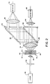
- Figure 2 is a schematic diagram of the flow cell and detection optics used in the present invention.
- Figure 3 is a graphical representation of the separation of particles by their indices of refraction and
- Figure 4 is a block diagram showing separate counters between the discriminator and the microprocessor.
- Fig. 1 shows a block diagram of the system of the present invention wherein blood cells are introduced through conduit 10 into a mixing and incubation chamber 12.
- the conduit 10 comprises a conventional sampling apparatus, not shown, for introducing blood cells into the chamber 12.
- a suspension fluid is introduced through conduit 14 to suspend the cells therein.
- An isovolumetric sphering reagent may also be introduced along conduit 14 to sphere the red blood cells contained in the sample. The isovolumetric sphering of the red blood cells ensures that the measurement is fully independent of the orientation of the cell during optical measurement in a flow cytometer.
- the individual cells are first mixed in the chamber 12 to mix with the suspension fluid and to react with the sphering agent.
- the cells are then permitted to incubate during which a number of cells may stick to the chamber walls and/or settle on the chamber bottom.
- the sedimentation will occur as mentioned, due to the blood cells being denser than the suspension fluid.
- the cells may have an electrostatic interaction with the walls of the container 12 thereby causing them to be attracted to the walls.
- the cells will become spatially inhomogenous which will interfere with the accuracy of the blood count.
- the cells are again mixed in the chamber 12 prior to being passed to a flow cell 16 in order to avoid the problem of spatial inhomogeneity.
- mixing generally will cause a plurality of air bubbles to be formed in the suspension fluid.
- a predetermined volume of suspension fluid having cells and air bubbles is aspirated from the mixing chamber 12. Therefore, each cell is passed through the flow cell 16 for detection. In addition, any air bubbles in the aspirated volume will also be detected.
- a conventional aspirating device may be used to pass the predetermined volume of fluid from the mixing chamber 12 to the flow cell 16.
- the flow cell 16 can be of conventional design, for example, as described in U.S. Patent No. 3,661,460, assigned to a common assignee which discloses a sheath-stream flow cell. As shown in Figure 2, the fluid stream 40 is passed through the flow cell 16 which is designed so that individual cells flow successively through an optically defined view volume.
- the stream 40 is encased in a sheath 42 having an index of refraction the same as the stream 40.
- the red blood cells 44 flow through the passageway 46 which has a narrowing internal diameter thereby constricting the stream 40 to cause individual cells to flow through the view volume.
- Illumination optics 18 directs a light beam through the view volume to illuminate, in turn, each particle as it passes through the cell 16.
- the illumination comprises a laser 48, precision slit aperture 50 and a lens system 52 for condensing the light beam into the center of the view volume, as shown in Figure 2 while maintaining the substantially collimated light from the laser 48.
- the detection optics utilizes a multiple angle light scattering measurement technique. As shown in Figure 2, the detection optics includes a lens 54 which collects the forward scattered light within two critical angular intervals which are seperately detected and measured from which the volume and index of refraction of each individual particle is determined.
- the light from the lens is directed to a beam splitter 56 which transmits approximately half the light and reflects the remaining light which is directed to and reflected from a mirror 58.
- the light passed through the beam splitter 56 is directed through a dark field stop 60 adapted to pass light scattered within a low angular interval.
- light reflected from mirror 58 is directed through a dark field stop 62 adapted to pass light scattered within a high angular interval.
- the light from the two different angles is collected by lenses 64 and 66 then detected by two separate detectors 68 and 70, respectively.
- a counter 22 counts the combined total of particles passing through the flow cell 16. The signal is then transmitted to index of refraction processor 24 where the volume and index of refraction of each particle is determined.
- a discriminator 26 distinguishes between the blood cells and air bubbles by their respective indices of refraction.
- the index of refraction of air is approximately 1.00, while the index of refraction of red blood cells is approximately in the range of 1.38 to 1.44.
- the discriminator 26 separates the signals in accordance with the measured index of refraction.
- a graphical representation may be obtained by plotting the results for each particle. The graph will depict one region of the particles having one index of refraction and a second region of particles having a second index of refraction. An example of such a graph is shown in Figure 3.
- Region A includes the concentration of red blood cells while region B includes the air bubbles.
- Region C shows the effect of occassional coincidence of red cells in the view volume. Errors due to coincidence may be corrected by standard techniques commonly used in cell counters.
- processor 28 is utilized to correct the count and volume for the detected air bubbles to provide an accurate determination of the count per unit volume of red blood cells.
- the total count of particles passed through the flow cell is inputted to the processor 28 together with the predetermined volume of fluid aspirated.
- Processor 28 is programmed with an algorithm that will take the total number of particles counted and subtract the number of air bubbles to provide an accurate count of red blood cells.
- processor 28 will subtract the volume of air bubbles from the total volume of fluid aspirated to determine the accurate volume of fluid. The number of cells is then divided by the accurate volume of fluid to provide an accurate measure of the total count of red blood cells per unit volume of fluid.
- the processor 28 can be a microprocessor programmed with an algorithm that sets a threshold value at either of the measured indices of refraction.
- Processor 28 can include a counter to count only those particles above or below the threshold value. An adder will then determine the total volume of particles counted.
- the mean volume per air bubbles can be calculated and multiplied by the number of air bubbles to determine the total volume of air in the sample stream.
- Figure 4 illustrates the use of separate counting means 30 and 32 coupled between the discriminator 26 and processor 28. After discriminator 26 separates signals in accordance with the indices of refraction, counters 30 and 32 then individually count each of the signals in the two regions. This information is fed to the processor 28 together with the combined count and volume originally measured and detected to determine the accurate count per unit volume of blood cells.
Landscapes
- Chemical & Material Sciences (AREA)
- Dispersion Chemistry (AREA)
- Physics & Mathematics (AREA)
- Health & Medical Sciences (AREA)
- Life Sciences & Earth Sciences (AREA)
- Analytical Chemistry (AREA)
- Biochemistry (AREA)
- General Health & Medical Sciences (AREA)
- General Physics & Mathematics (AREA)
- Immunology (AREA)
- Pathology (AREA)
- Investigating Or Analysing Biological Materials (AREA)
- Ship Loading And Unloading (AREA)
Claims (8)
- Verfahren zur genauen Bestimmung der Anzahl erster, in einem Fluid suspendierter Partikel in einer Volumeneinheit des Fluids, wobei das Fluid diese ersten Partikel mit einem ersten Refraktionsgrad und zumindest ein zweites Partikel mit einem zweiten Refraktionsgrad. der vom ersten Refraktionsgrad verschieden ist, enthält, mittels Hindsurchführen des Partikel beider Refraktionsgrade enthaltenden Fluids durch einen Lichtstrahl, wobei die ersten Partikel mit dem ersten Refraktionsgrad und die zweiten Partikel mit dem zweiten Refraktionsgrad im Wechsel zumindest ein vorderes Lichtstreuungsmuster erzeugen, wobei die Gesamtzahl erster und zweiter Partikel durch Messen der Lichtintensität von zumindest einem Teil jedes dieser Lichtstreuungsmuster ermittelt wird, gekennzeichnet durch die folgenden Verfahrensschritte:
Ermitteln (20) und anschließendem Zählen (22) der Gesamtzahl der ersten und zweiten Partikel, die den Lichtstrahl passieren aus dem Lichtstreuungsmuster;
Bestimmen (24) des Volumens und Refraktionsgrads sowohl der ersten als auch der zweiten Partikel, die den Lichtstrahl passieren aus zumindest einem vorderen Lichtstreuungsmuster;
Unterscheiden (26) zwischen ersten und zweiten Partikeln entsprechend ihrem ersten bzw. zweiten Refraktionsgrad;
Bestimmung der Gesamtanzahl und des Gesamtvolumens der zweiten Partikel;
Bestimmung (28) der Anzahl und des Volumens der den Lichtstrahl passierenden ersten Partikel und eines korrigierten Fluidvolumens mit der Anzahl bzw. dem Volumen der zweiten Partikel und
unterscheidende Bestimmung (28) der Anzahl erster Partikel je Fluidvolmeneinheit aus der korrigierten Anzahl und dem korrigierten Volumen. - Verfahren des Anspruchs 1, bei dem die ersten Partikel rote Blutkörper und die zweiten Partikel Luftblasen sind und das weiter die folgenden Verfahrensschritte einschließt:
Ansaugen eines Volumens des Fluids aus einer Mischkammer (12) vor der Hindurchführung des Fluids durch den Lichtstrahl und
Mischen des rote Blutkörper enthaltenden Fluids, um die Bildung von Luftblasen in dem Fluid vor dem Ansaugen zu veranlassen. - Verfahren des Anspruchs 1 oder 2, bei dem der Verfahrensschritt der Volumenbestimmung und der Refraktionsgradbestimmung von ersten und zweiten Partikeln als weitere Verfahrensschritte einschließt;
Ermitteln und Messen der Lichtintensität in jedem einer Mehrzahl von Winkelintervallen des Lichtstreuungsmusters zur Erzeugung eines Signals aus jedem Winkelintervall, wobei die Winkelintervalle so ausgewählt sind, daß das in ihnen vorliegende Licht ausreichende Informationen für die genaue Bestimmung von Volumen und Refraktionsgrad von ersten und zweiten Partikeln enthält und
unterscheidende Bestimmung des Volumens und des Refraktionsgrads der Partikel erster und zweiter Art aus den Signalgrößen. - Verfahren eines beliebigen der Ansprüche 1 bis 3, bei dem der Verfahrensschritt der unterscheidenden Bestimmung als weiteren Verfahrensschritt die Lieferung einer zweidimensionalen grafischen Wiedergabe einschließt, die einen ersten Bereich mit Partikeln mit dem ersten Refraktionsgrad und einem zweiten Bereich mit Partikeln mit dem zweiten Refraktionsgrad wiedergibt.
- Verfahren des Anspruchs 4, bei dem der Verfahrensschritt der unterscheidenden Bestimmung als weiteren Verfahrensschritt die Festlegung einer Schwelle für jeden der beiden Refraktionsgrade und Zählen lediglich der Partikel oberhalb oder unterhalb dieser Schwelle einschließt, als weiteren Verfahrensschritt die Bestimmung der Zahl von ersten Partikeln je Fluidvolumen durch Dividieren der Zahl erster Partikel durch das Volumen des vorbestimmten Fluids weniger dem Volumen von zweiten Partikeln und als weiteren Verfahrensschritt das Errechnen des mittleren Volumens je zweitem Partikel und Multiplikation des mittleren Volumens mit der Anzahl der zweiten Partikel zur Bestimmung des Gesamtvolumens der zweiten Partikel.
- Vorrichtung zur genauen Bestimmung der Anzahl erster, in einem Fluid suspendierter Partikel in einer Volumeneinheit des Fluids, wobei das Fluid diese ersten Partikel mit einem ersten Refraktionsgrad und zumindest ein zweites Partikel mit einem zweiten Refraktionsgrad, der vom ersten Refraktionsgrad verschieden ist, enthält, mittels Hindurchführen des Partikel beider Refraktionsgrade enthaltenden Fluids durch einen Lichtstrahl, wobei die ersten Partikel mit dem ersten Refraktionsgrad und die zweiten Partikel mit dem zweiten Refraktionsgrad im Wechsel zumindest ein vorderes Lichtstreuungsmuster ezeugen, wobei die Gesamtzahl erster und zweiter Partikel durch Messen der Lichtintensität von zumindest einem Teil jedes dieser Lichtstreuungsmuster ermittelt wird, mit
einem Mittel (20,22) zum Ermitteln und anschließendem Zählen der Gesamtanzahl der ersten und zweiten Partikel, die den Lichtstrahl passieren aus dem Lichtstreuungmuster;
einem Mittel (24) zum Bestimmen des Volumens und Refraktionsgrads sowohl der ersten als auch der zweiten Partikel, die den Lichtstrahl passieren aus zumindest einem vorderen Lichtstreuungsmuster;
einem Mittel (26) zum Unterscheiden zwischen ersten und zweiten Partikeln entsprechend ihrem ersten bzw. zweiten Refraktionsgrad;
einem Mittel zur Bestimmung der Gesamtanzahl und des Gesamtvolumens der zweiten Partikel;
einem Mittel (28) zur Bestimmung der Anzahl und des Volumens der ersten, den Lichtstrahl passierenden ersten Partikel und eines korrigierten Fluidvolumens durch Korrektur der gesamten Partikelanzahl und des vorbestimmten Fluidvolumens mit der Anzahl bzw. dem Volumen der zweiten Partikel und
einem Mittel zum Bestimmen der Anzahl erster Partikel je Fluidvolumeneinheit aus der korrigierten Anzahl und dem korrigierten Volumen. - Vorrichtung des Anspruchs 6, bei der das Mittel (20,22) zum Ermitteln der Gesamtanzahl von ersten und zweiten Partikeln Mittel zum Messen der Lichtintensität in zumindest einem Teil der Lichtstreuungsmuster einschließt und die ferner einschließt:
ein Mittel (24) zum Bestimmen des Volumens und des Refraktionsgrads von jedem der ersten und zweiten Patikel, die den Lichtstrahl an zumindest einem der vorderen Lichtstreuungsmuster passieren und
einem Mittel (26) zum Unterscheiden zwischen den ersten und zweiten Partikeln mittel ihres jeweiligen ersten und zweiten Refraktionsgrades. - Vorrichtung des Anspruchs 6 oder 7, bei der das Korrekturmittel (28) einschließt:
ein Mittel zur Bestimmung der Gesamtanzahl und des Gesamtvolumens der zweiten Partikel;
ein Mittel zur Bestimmung der Anzahl und des Volumens der ersten Partikel, die den Lichtstrahl und das korrigierte Volumen des Fluids passieren durch Modifikation der Partikelgesamtanzahl und des vorbestimmten Fluidvolumens mit der Anzahl bzw. dem Volumen der zweiten Partikel und
ein Mittel zur Bestimmung der Anzahl der ersten Partikel je Volumeneinheit des Fluid aus modifizierter Anzahl und modifiziertem Volumen.
Applications Claiming Priority (2)
| Application Number | Priority Date | Filing Date | Title |
|---|---|---|---|
| US848172 | 1986-04-04 | ||
| US06/848,172 US4989978A (en) | 1986-04-04 | 1986-04-04 | Method and apparatus for determining the count per unit volume of particles |
Publications (3)
| Publication Number | Publication Date |
|---|---|
| EP0240942A2 EP0240942A2 (de) | 1987-10-14 |
| EP0240942A3 EP0240942A3 (en) | 1988-04-06 |
| EP0240942B1 true EP0240942B1 (de) | 1992-01-15 |
Family
ID=25302552
Family Applications (1)
| Application Number | Title | Priority Date | Filing Date |
|---|---|---|---|
| EP87104908A Expired EP0240942B1 (de) | 1986-04-04 | 1987-04-02 | Verfahren und Gerät zur Bestimmung der Teilchenzahl pro Volumeneinheit |
Country Status (7)
| Country | Link |
|---|---|
| US (1) | US4989978A (de) |
| EP (1) | EP0240942B1 (de) |
| JP (1) | JPS62239289A (de) |
| AU (1) | AU590683B2 (de) |
| CA (1) | CA1272805C (de) |
| DE (1) | DE3775961D1 (de) |
| ES (1) | ES2027985T3 (de) |
Families Citing this family (35)
| Publication number | Priority date | Publication date | Assignee | Title |
|---|---|---|---|---|
| US5488469A (en) * | 1991-08-30 | 1996-01-30 | Omron Corporation | Cell analyzing apparatus |
| DE69230902D1 (de) * | 1991-08-30 | 2000-05-18 | Omron Tateisi Electronics Co | Vorrichtung zur Zellenanalyse |
| US5451525A (en) * | 1992-02-14 | 1995-09-19 | Coulter Corporation | Method and materials for determining particle count in a flow cytometer |
| DE69327182T2 (de) * | 1992-02-18 | 2000-06-15 | Hitachi, Ltd. | Gerät und Verfahren zur Untersuchung von Teilchen in einem Fluid |
| WO1994027146A1 (en) * | 1993-05-14 | 1994-11-24 | Coulter Corporation | Reticulocyte analyzing method and apparatus utilizing light scatter techniques |
| US6790366B2 (en) * | 1996-06-07 | 2004-09-14 | Immunivest Corporation | Magnetic separation apparatus and methods |
| US6660159B1 (en) | 1996-06-07 | 2003-12-09 | Immunivest Corporation | Magnetic separation apparatus and methods |
| US6890426B2 (en) * | 1996-06-07 | 2005-05-10 | Immunivest Corporation | Magnetic separation apparatus and methods |
| US6136182A (en) * | 1996-06-07 | 2000-10-24 | Immunivest Corporation | Magnetic devices and sample chambers for examination and manipulation of cells |
| US7666308B2 (en) * | 1996-06-07 | 2010-02-23 | Veridex, Llc. | Magnetic separation apparatus and methods |
| AU3374597A (en) * | 1996-06-07 | 1998-01-05 | Immunivest Corporation | Magnetic separation employing external and internal gradients |
| US6172376B1 (en) | 1997-12-17 | 2001-01-09 | American Air Liquide Inc. | Method and system for measuring particles in a liquid sample |
| US6813017B1 (en) | 1999-10-20 | 2004-11-02 | Becton, Dickinson And Company | Apparatus and method employing incoherent light emitting semiconductor devices as particle detection light sources in a flow cytometer |
| EP1222451A1 (de) * | 1999-10-20 | 2002-07-17 | Becton, Dickinson and Company | Vorrichtung und verfahren mit verwendung von nicht-kohärentes licht emittierenden halbleitervorrichtungen als lichtquellen für die teilchendetektion in einem durchflusszytometer |
| US7630063B2 (en) * | 2000-08-02 | 2009-12-08 | Honeywell International Inc. | Miniaturized cytometer for detecting multiple species in a sample |
| US7641856B2 (en) * | 2004-05-14 | 2010-01-05 | Honeywell International Inc. | Portable sample analyzer with removable cartridge |
| WO2002025247A2 (en) | 2000-09-20 | 2002-03-28 | Menguc M Pinar | A non-intrusive method and apparatus for characterizing particles based on scattering of elliptically polarized radiation |
| JP3720799B2 (ja) * | 2002-10-02 | 2005-11-30 | 神栄株式会社 | 花粉センサ |
| US7092078B2 (en) | 2003-03-31 | 2006-08-15 | Nihon Kohden Corporation | Flow cytometer for classifying leukocytes and method for determining detection angle range of the same |
| US7356365B2 (en) * | 2003-07-09 | 2008-04-08 | Glucolight Corporation | Method and apparatus for tissue oximetry |
| SE0302676D0 (sv) * | 2003-10-09 | 2003-10-09 | Neural Ab | Method and apparatus for holographic refractometry |
| US7510849B2 (en) * | 2004-01-29 | 2009-03-31 | Glucolight Corporation | OCT based method for diagnosis and therapy |
| CN1985168B (zh) * | 2004-05-14 | 2013-01-02 | 霍尼韦尔国际公司 | 具有可拆卸盒的便携式样本分析仪 |
| US8036727B2 (en) | 2004-08-11 | 2011-10-11 | Glt Acquisition Corp. | Methods for noninvasively measuring analyte levels in a subject |
| US7254429B2 (en) | 2004-08-11 | 2007-08-07 | Glucolight Corporation | Method and apparatus for monitoring glucose levels in a biological tissue |
| JP2008537903A (ja) | 2005-04-13 | 2008-10-02 | グルコライト・コーポレーシヨン | Octが基になった血糖モニターのデータ処理および較正方法 |
| US8768423B2 (en) | 2008-03-04 | 2014-07-01 | Glt Acquisition Corp. | Multispot monitoring for use in optical coherence tomography |
| EP4180794A3 (de) | 2011-12-01 | 2023-08-09 | Particle Measuring Systems Inc. | Schema zur erkennung von partikelgrössen und -konzentrationen |
| JP2015031665A (ja) * | 2013-08-06 | 2015-02-16 | トライボテックス株式会社 | 粒子計数装置及び粒子計数方法 |
| CN107870139B (zh) * | 2016-09-27 | 2023-10-27 | 深圳长城开发科技股份有限公司 | 一种无损检测产品表面颗粒污染物的装置及方法 |
| WO2019082186A1 (en) | 2017-10-26 | 2019-05-02 | Particle Measuring Systems, Inc. | SYSTEM AND METHOD FOR MEASURING PARTICLES |
| WO2020146967A1 (zh) * | 2019-01-14 | 2020-07-23 | 深圳迈瑞生物医疗电子股份有限公司 | 一种样本光学检测装置 |
| EP3959505B1 (de) | 2019-04-25 | 2024-05-08 | Particle Measuring Systems, Inc. | Teilchendetektionssysteme und verfahren zur teilchendetektion in der achse und/oder zur differenziellen detektion |
| CN114729868A (zh) | 2019-11-22 | 2022-07-08 | 粒子监测系统有限公司 | 先进的用于干涉测量颗粒检测和具有小大小尺寸的颗粒的检测的系统和方法 |
| CN119880716B (zh) * | 2025-03-27 | 2025-06-24 | 烟台格藻生物科技有限公司 | 应用于生物信息的细胞数量测量方法 |
Family Cites Families (8)
| Publication number | Priority date | Publication date | Assignee | Title |
|---|---|---|---|---|
| US3520316A (en) * | 1963-12-12 | 1970-07-14 | Bowles Eng Corp | Pressure-to-pressure transducer |
| US3740143A (en) * | 1970-10-30 | 1973-06-19 | Technicon Instr | Automatic apparatus for determining the percentage population of particulates in a medium |
| US4202625A (en) * | 1978-08-18 | 1980-05-13 | Ortho Diagnostics, Inc. | Method and apparatus for discriminating red blood cells from platelets |
| US4577964A (en) * | 1978-09-06 | 1986-03-25 | Ortho Diagnostics, Inc. | Apparatus and method for detecting platelets in whole blood |
| US4429995A (en) * | 1980-07-21 | 1984-02-07 | National Research Development Corporation | Two dimensinal flow analyzer |
| US4548500A (en) * | 1982-06-22 | 1985-10-22 | Wyatt Philip J | Process and apparatus for identifying or characterizing small particles |
| JPS5994037A (ja) * | 1982-11-19 | 1984-05-30 | Shimadzu Corp | 血球計数装置 |
| US4735504A (en) * | 1983-10-31 | 1988-04-05 | Technicon Instruments Corporation | Method and apparatus for determining the volume & index of refraction of particles |
-
1986
- 1986-04-04 US US06/848,172 patent/US4989978A/en not_active Expired - Lifetime
-
1987
- 1987-01-29 CA CA528467A patent/CA1272805C/en not_active Expired
- 1987-02-26 AU AU69585/87A patent/AU590683B2/en not_active Ceased
- 1987-04-02 ES ES198787104908T patent/ES2027985T3/es not_active Expired - Lifetime
- 1987-04-02 EP EP87104908A patent/EP0240942B1/de not_active Expired
- 1987-04-02 DE DE8787104908T patent/DE3775961D1/de not_active Expired - Lifetime
- 1987-04-03 JP JP62082774A patent/JPS62239289A/ja active Granted
Also Published As
| Publication number | Publication date |
|---|---|
| ES2027985T3 (es) | 1992-07-01 |
| EP0240942A2 (de) | 1987-10-14 |
| EP0240942A3 (en) | 1988-04-06 |
| CA1272805A (en) | 1990-08-14 |
| AU590683B2 (en) | 1989-11-09 |
| JPH0543154B2 (de) | 1993-06-30 |
| JPS62239289A (ja) | 1987-10-20 |
| US4989978A (en) | 1991-02-05 |
| AU6958587A (en) | 1987-10-08 |
| CA1272805C (en) | 1990-08-14 |
| DE3775961D1 (de) | 1992-02-27 |
Similar Documents
| Publication | Publication Date | Title |
|---|---|---|
| US4989978A (en) | Method and apparatus for determining the count per unit volume of particles | |
| US6630990B2 (en) | Optical method and apparatus for red blood cell differentiation on a cell-by-cell basis, and simultaneous analysis of white blood cell differentiation | |
| US7390662B2 (en) | Method and apparatus for performing platelet measurement | |
| JP4965561B2 (ja) | サイトメータ細胞計数及びサイズ測定システム | |
| US5162863A (en) | Method and apparatus for inspecting a specimen by optical detection of antibody/antigen sensitized carriers | |
| US7092078B2 (en) | Flow cytometer for classifying leukocytes and method for determining detection angle range of the same | |
| CA1230752A (en) | Method and apparatus for determining the volume and index of refraction of particles | |
| JP2815435B2 (ja) | 粒子解析装置及び血球カウンタ | |
| US3740143A (en) | Automatic apparatus for determining the percentage population of particulates in a medium | |
| US5050987A (en) | Particle analyzing apparatus and method for determining nuclear shift index | |
| US4188121A (en) | Multiple ratio single particle counter | |
| EP2302377A2 (de) | Blutzellenzähler, Diagnosehilfsverfahren und Computerprogrammprodukt | |
| US6670191B2 (en) | Method for quantitatively analyzing fragmented red blood cells | |
| EP0633462A2 (de) | Dreifarben Durchflusszytometrie mit automatischer Auswertung | |
| WO1994029800A9 (en) | Three-color flow cytometry with automatic gating function | |
| Mayer et al. | Automated platelet counters. A comparative evaluation of latest instrumentation | |
| Gray et al. | A new method for cell volume measurement based on volume exclusion of a fluorescent dye | |
| Dumoulin-Lagrange et al. | Evaluation of automated platelet counters for the enumeration and sizing of platelets in the diagnosis and management of hemostatic problems | |
| JPH1130580A (ja) | 粒子測定装置 | |
| US20250334503A1 (en) | Sample analyzer | |
| US20240344961A1 (en) | Sample analysis apparatus and sample analysis method | |
| JP2714629B2 (ja) | 粒子計数方法および装置 | |
| Samama et al. | STANDARDIZATION OF PLATELET COUNTS‐PROBLEMS AND PITFALLS | |
| JPS63100351A (ja) | 粒子計数装置 | |
| JPS61126447A (ja) | 粒子分析装置 |
Legal Events
| Date | Code | Title | Description |
|---|---|---|---|
| PUAI | Public reference made under article 153(3) epc to a published international application that has entered the european phase |
Free format text: ORIGINAL CODE: 0009012 |
|
| AK | Designated contracting states |
Kind code of ref document: A2 Designated state(s): BE CH DE ES FR GB IT LI NL SE |
|
| PUAL | Search report despatched |
Free format text: ORIGINAL CODE: 0009013 |
|
| AK | Designated contracting states |
Kind code of ref document: A3 Designated state(s): BE CH DE ES FR GB IT LI NL SE |
|
| 17P | Request for examination filed |
Effective date: 19881005 |
|
| RAP1 | Party data changed (applicant data changed or rights of an application transferred) |
Owner name: TECHNICON INSTRUMENTS CORPORATION(A DELAWARE CORPO |
|
| 17Q | First examination report despatched |
Effective date: 19900531 |
|
| GRAA | (expected) grant |
Free format text: ORIGINAL CODE: 0009210 |
|
| AK | Designated contracting states |
Kind code of ref document: B1 Designated state(s): BE CH DE ES FR GB IT LI NL SE |
|
| ET | Fr: translation filed | ||
| REF | Corresponds to: |
Ref document number: 3775961 Country of ref document: DE Date of ref document: 19920227 |
|
| ITF | It: translation for a ep patent filed | ||
| RAP2 | Party data changed (patent owner data changed or rights of a patent transferred) |
Owner name: MILES INC.(AN INDIANA CORP.) |
|
| REG | Reference to a national code |
Ref country code: CH Ref legal event code: PUE Owner name: MILES INC. (AN INDIANA CORP.) |
|
| REG | Reference to a national code |
Ref country code: ES Ref legal event code: FG2A Ref document number: 2027985 Country of ref document: ES Kind code of ref document: T3 |
|
| NLT2 | Nl: modifications (of names), taken from the european patent patent bulletin |
Owner name: MILES INC. (AN INDIANA CORP.) TE TARRYTOWN, NEW YO |
|
| PLBE | No opposition filed within time limit |
Free format text: ORIGINAL CODE: 0009261 |
|
| STAA | Information on the status of an ep patent application or granted ep patent |
Free format text: STATUS: NO OPPOSITION FILED WITHIN TIME LIMIT |
|
| BECN | Be: change of holder's name |
Effective date: 19920115 |
|
| 26N | No opposition filed | ||
| EAL | Se: european patent in force in sweden |
Ref document number: 87104908.6 |
|
| REG | Reference to a national code |
Ref country code: GB Ref legal event code: IF02 |
|
| PGFP | Annual fee paid to national office [announced via postgrant information from national office to epo] |
Ref country code: SE Payment date: 20020320 Year of fee payment: 16 |
|
| PGFP | Annual fee paid to national office [announced via postgrant information from national office to epo] |
Ref country code: NL Payment date: 20020321 Year of fee payment: 16 |
|
| PGFP | Annual fee paid to national office [announced via postgrant information from national office to epo] |
Ref country code: CH Payment date: 20020322 Year of fee payment: 16 |
|
| PGFP | Annual fee paid to national office [announced via postgrant information from national office to epo] |
Ref country code: BE Payment date: 20020416 Year of fee payment: 16 |
|
| PGFP | Annual fee paid to national office [announced via postgrant information from national office to epo] |
Ref country code: ES Payment date: 20020509 Year of fee payment: 16 |
|
| PGFP | Annual fee paid to national office [announced via postgrant information from national office to epo] |
Ref country code: GB Payment date: 20030326 Year of fee payment: 17 |
|
| PG25 | Lapsed in a contracting state [announced via postgrant information from national office to epo] |
Ref country code: SE Free format text: LAPSE BECAUSE OF NON-PAYMENT OF DUE FEES Effective date: 20030403 Ref country code: ES Free format text: LAPSE BECAUSE OF NON-PAYMENT OF DUE FEES Effective date: 20030403 |
|
| PGFP | Annual fee paid to national office [announced via postgrant information from national office to epo] |
Ref country code: FR Payment date: 20030418 Year of fee payment: 17 |
|
| PG25 | Lapsed in a contracting state [announced via postgrant information from national office to epo] |
Ref country code: LI Free format text: LAPSE BECAUSE OF NON-PAYMENT OF DUE FEES Effective date: 20030430 Ref country code: CH Free format text: LAPSE BECAUSE OF NON-PAYMENT OF DUE FEES Effective date: 20030430 Ref country code: BE Free format text: LAPSE BECAUSE OF NON-PAYMENT OF DUE FEES Effective date: 20030430 |
|
| PGFP | Annual fee paid to national office [announced via postgrant information from national office to epo] |
Ref country code: DE Payment date: 20030430 Year of fee payment: 17 |
|
| BERE | Be: lapsed |
Owner name: *MILES INC.(AN INDIANA CORP.) Effective date: 20030430 |
|
| PG25 | Lapsed in a contracting state [announced via postgrant information from national office to epo] |
Ref country code: NL Free format text: LAPSE BECAUSE OF NON-PAYMENT OF DUE FEES Effective date: 20031101 |
|
| NLV4 | Nl: lapsed or anulled due to non-payment of the annual fee |
Effective date: 20031101 |
|
| EUG | Se: european patent has lapsed | ||
| REG | Reference to a national code |
Ref country code: CH Ref legal event code: PL |
|
| PG25 | Lapsed in a contracting state [announced via postgrant information from national office to epo] |
Ref country code: GB Free format text: LAPSE BECAUSE OF NON-PAYMENT OF DUE FEES Effective date: 20040402 |
|
| REG | Reference to a national code |
Ref country code: ES Ref legal event code: FD2A Effective date: 20030403 |
|
| PG25 | Lapsed in a contracting state [announced via postgrant information from national office to epo] |
Ref country code: DE Free format text: LAPSE BECAUSE OF NON-PAYMENT OF DUE FEES Effective date: 20041103 |
|
| GBPC | Gb: european patent ceased through non-payment of renewal fee | ||
| PG25 | Lapsed in a contracting state [announced via postgrant information from national office to epo] |
Ref country code: FR Free format text: LAPSE BECAUSE OF NON-PAYMENT OF DUE FEES Effective date: 20041231 |
|
| REG | Reference to a national code |
Ref country code: FR Ref legal event code: ST |
|
| PG25 | Lapsed in a contracting state [announced via postgrant information from national office to epo] |
Ref country code: IT Free format text: LAPSE BECAUSE OF NON-PAYMENT OF DUE FEES;WARNING: LAPSES OF ITALIAN PATENTS WITH EFFECTIVE DATE BEFORE 2007 MAY HAVE OCCURRED AT ANY TIME BEFORE 2007. THE CORRECT EFFECTIVE DATE MAY BE DIFFERENT FROM THE ONE RECORDED. Effective date: 20050402 |