EP0256677A2 - Pseudorabiesvirus-Rekombinanten und ihre Verwendung zur Herstellung von Proteinen - Google Patents
Pseudorabiesvirus-Rekombinanten und ihre Verwendung zur Herstellung von Proteinen Download PDFInfo
- Publication number
- EP0256677A2 EP0256677A2 EP87306364A EP87306364A EP0256677A2 EP 0256677 A2 EP0256677 A2 EP 0256677A2 EP 87306364 A EP87306364 A EP 87306364A EP 87306364 A EP87306364 A EP 87306364A EP 0256677 A2 EP0256677 A2 EP 0256677A2
- Authority
- EP
- European Patent Office
- Prior art keywords
- region
- pseudorabies virus
- giii
- prv
- gene
- Prior art date
- Legal status (The legal status is an assumption and is not a legal conclusion. Google has not performed a legal analysis and makes no representation as to the accuracy of the status listed.)
- Withdrawn
Links
- 108090000623 proteins and genes Proteins 0.000 title claims abstract description 183
- 241000701093 Suid alphaherpesvirus 1 Species 0.000 title claims abstract description 164
- 102000004169 proteins and genes Human genes 0.000 title claims abstract description 73
- 238000004519 manufacturing process Methods 0.000 title claims abstract description 15
- 239000012634 fragment Substances 0.000 claims abstract description 101
- 241000700605 Viruses Species 0.000 claims abstract description 63
- 238000003780 insertion Methods 0.000 claims abstract description 29
- 230000037431 insertion Effects 0.000 claims abstract description 29
- 229960005486 vaccine Drugs 0.000 claims abstract description 12
- 108020004414 DNA Proteins 0.000 claims description 148
- 210000004027 cell Anatomy 0.000 claims description 55
- 238000000034 method Methods 0.000 claims description 33
- 238000012217 deletion Methods 0.000 claims description 28
- 230000037430 deletion Effects 0.000 claims description 28
- 108091008146 restriction endonucleases Proteins 0.000 claims description 25
- 238000003776 cleavage reaction Methods 0.000 claims description 20
- 230000007017 scission Effects 0.000 claims description 20
- 239000013598 vector Substances 0.000 claims description 15
- 230000003612 virological effect Effects 0.000 claims description 12
- 108020004705 Codon Proteins 0.000 claims description 11
- 230000000890 antigenic effect Effects 0.000 claims description 10
- 210000003292 kidney cell Anatomy 0.000 claims description 9
- 101150003725 TK gene Proteins 0.000 claims description 8
- 230000008569 process Effects 0.000 claims description 8
- 101150030521 gI gene Proteins 0.000 claims description 7
- 102100024209 CD177 antigen Human genes 0.000 claims description 6
- 101000980845 Homo sapiens CD177 antigen Proteins 0.000 claims description 6
- 241000282898 Sus scrofa Species 0.000 claims description 6
- 108020005202 Viral DNA Proteins 0.000 claims description 5
- 208000009305 pseudorabies Diseases 0.000 claims description 5
- 210000001124 body fluid Anatomy 0.000 claims description 4
- 239000010839 body fluid Substances 0.000 claims description 4
- 238000012258 culturing Methods 0.000 claims description 3
- 239000003085 diluting agent Substances 0.000 claims description 3
- 239000003937 drug carrier Substances 0.000 claims description 3
- 230000006798 recombination Effects 0.000 claims description 3
- 238000005215 recombination Methods 0.000 claims description 3
- 230000015572 biosynthetic process Effects 0.000 claims description 2
- 230000004048 modification Effects 0.000 claims description 2
- 238000012986 modification Methods 0.000 claims description 2
- JEIPFZHSYJVQDO-UHFFFAOYSA-N ferric oxide Chemical compound O=[Fe]O[Fe]=O JEIPFZHSYJVQDO-UHFFFAOYSA-N 0.000 claims 1
- 239000013612 plasmid Substances 0.000 description 70
- 241000588724 Escherichia coli Species 0.000 description 26
- 238000010276 construction Methods 0.000 description 23
- 239000000499 gel Substances 0.000 description 22
- LFQSCWFLJHTTHZ-UHFFFAOYSA-N Ethanol Chemical compound CCO LFQSCWFLJHTTHZ-UHFFFAOYSA-N 0.000 description 21
- 238000011144 upstream manufacturing Methods 0.000 description 17
- FAPWRFPIFSIZLT-UHFFFAOYSA-M Sodium chloride Chemical compound [Na+].[Cl-] FAPWRFPIFSIZLT-UHFFFAOYSA-M 0.000 description 14
- 108091026890 Coding region Proteins 0.000 description 13
- 239000000523 sample Substances 0.000 description 12
- 241000894006 Bacteria Species 0.000 description 11
- 108091028043 Nucleic acid sequence Proteins 0.000 description 11
- 239000000047 product Substances 0.000 description 11
- 108020001507 fusion proteins Proteins 0.000 description 10
- 102000037865 fusion proteins Human genes 0.000 description 10
- 230000014509 gene expression Effects 0.000 description 10
- 101150066555 lacZ gene Proteins 0.000 description 10
- 108090000288 Glycoproteins Proteins 0.000 description 9
- 239000000427 antigen Substances 0.000 description 9
- 108091007433 antigens Proteins 0.000 description 9
- 102000036639 antigens Human genes 0.000 description 9
- 102000003960 Ligases Human genes 0.000 description 8
- 108090000364 Ligases Proteins 0.000 description 8
- 238000004458 analytical method Methods 0.000 description 8
- 239000000203 mixture Substances 0.000 description 8
- 210000002966 serum Anatomy 0.000 description 8
- 241001465754 Metazoa Species 0.000 description 7
- DBMJMQXJHONAFJ-UHFFFAOYSA-M Sodium laurylsulphate Chemical compound [Na+].CCCCCCCCCCCCOS([O-])(=O)=O DBMJMQXJHONAFJ-UHFFFAOYSA-M 0.000 description 7
- 239000007984 Tris EDTA buffer Substances 0.000 description 7
- 239000007983 Tris buffer Substances 0.000 description 7
- 150000001413 amino acids Chemical class 0.000 description 7
- 239000000872 buffer Substances 0.000 description 7
- 238000001962 electrophoresis Methods 0.000 description 7
- 208000015181 infectious disease Diseases 0.000 description 7
- 239000011780 sodium chloride Substances 0.000 description 7
- 241001515965 unidentified phage Species 0.000 description 7
- 229920000936 Agarose Polymers 0.000 description 6
- 102000004594 DNA Polymerase I Human genes 0.000 description 6
- 108010017826 DNA Polymerase I Proteins 0.000 description 6
- KCXVZYZYPLLWCC-UHFFFAOYSA-N EDTA Chemical compound OC(=O)CN(CC(O)=O)CCN(CC(O)=O)CC(O)=O KCXVZYZYPLLWCC-UHFFFAOYSA-N 0.000 description 6
- PEDCQBHIVMGVHV-UHFFFAOYSA-N Glycerine Chemical compound OCC(O)CO PEDCQBHIVMGVHV-UHFFFAOYSA-N 0.000 description 6
- 239000000020 Nitrocellulose Substances 0.000 description 6
- 101710090322 Truncated surface protein Proteins 0.000 description 6
- 102000005936 beta-Galactosidase Human genes 0.000 description 6
- 108010005774 beta-Galactosidase Proteins 0.000 description 6
- 238000002474 experimental method Methods 0.000 description 6
- 238000009396 hybridization Methods 0.000 description 6
- 229920001220 nitrocellulos Polymers 0.000 description 6
- 239000002773 nucleotide Substances 0.000 description 6
- 125000003729 nucleotide group Chemical group 0.000 description 6
- 229920002401 polyacrylamide Polymers 0.000 description 6
- 108010062697 pseudorabies virus glycoproteins Proteins 0.000 description 6
- LENZDBCJOHFCAS-UHFFFAOYSA-N tris Chemical compound OCC(N)(CO)CO LENZDBCJOHFCAS-UHFFFAOYSA-N 0.000 description 6
- 241000238631 Hexapoda Species 0.000 description 5
- 241000700588 Human alphaherpesvirus 1 Species 0.000 description 5
- 241000700618 Vaccinia virus Species 0.000 description 5
- 229960000723 ampicillin Drugs 0.000 description 5
- 230000001580 bacterial effect Effects 0.000 description 5
- 238000005119 centrifugation Methods 0.000 description 5
- 239000000284 extract Substances 0.000 description 5
- 230000001717 pathogenic effect Effects 0.000 description 5
- 230000022532 regulation of transcription, DNA-dependent Effects 0.000 description 5
- 230000010076 replication Effects 0.000 description 5
- XLYOFNOQVPJJNP-UHFFFAOYSA-N water Substances O XLYOFNOQVPJJNP-UHFFFAOYSA-N 0.000 description 5
- QKNYBSVHEMOAJP-UHFFFAOYSA-N 2-amino-2-(hydroxymethyl)propane-1,3-diol;hydron;chloride Chemical compound Cl.OCC(N)(CO)CO QKNYBSVHEMOAJP-UHFFFAOYSA-N 0.000 description 4
- 108091003079 Bovine Serum Albumin Proteins 0.000 description 4
- 102000004190 Enzymes Human genes 0.000 description 4
- 108090000790 Enzymes Proteins 0.000 description 4
- 102000003886 Glycoproteins Human genes 0.000 description 4
- 241000829100 Macaca mulatta polyomavirus 1 Species 0.000 description 4
- TWRXJAOTZQYOKJ-UHFFFAOYSA-L Magnesium chloride Chemical compound [Mg+2].[Cl-].[Cl-] TWRXJAOTZQYOKJ-UHFFFAOYSA-L 0.000 description 4
- 108020004511 Recombinant DNA Proteins 0.000 description 4
- 240000004808 Saccharomyces cerevisiae Species 0.000 description 4
- 238000002105 Southern blotting Methods 0.000 description 4
- 108091081024 Start codon Proteins 0.000 description 4
- AVKUERGKIZMTKX-NJBDSQKTSA-N ampicillin Chemical compound C1([C@@H](N)C(=O)N[C@H]2[C@H]3SC([C@@H](N3C2=O)C(O)=O)(C)C)=CC=CC=C1 AVKUERGKIZMTKX-NJBDSQKTSA-N 0.000 description 4
- 230000001413 cellular effect Effects 0.000 description 4
- 230000000295 complement effect Effects 0.000 description 4
- 239000013604 expression vector Substances 0.000 description 4
- 230000004927 fusion Effects 0.000 description 4
- 239000002609 medium Substances 0.000 description 4
- 108020004999 messenger RNA Proteins 0.000 description 4
- 238000010369 molecular cloning Methods 0.000 description 4
- 244000052769 pathogen Species 0.000 description 4
- 239000013600 plasmid vector Substances 0.000 description 4
- 238000012360 testing method Methods 0.000 description 4
- 230000014621 translational initiation Effects 0.000 description 4
- QTBSBXVTEAMEQO-UHFFFAOYSA-N Acetic acid Chemical compound CC(O)=O QTBSBXVTEAMEQO-UHFFFAOYSA-N 0.000 description 3
- 229920001817 Agar Polymers 0.000 description 3
- 102000012410 DNA Ligases Human genes 0.000 description 3
- 108010061982 DNA Ligases Proteins 0.000 description 3
- 101710117538 Endogenous retrovirus group FC1 Env polyprotein Proteins 0.000 description 3
- 101710167714 Endogenous retrovirus group K member 18 Env polyprotein Proteins 0.000 description 3
- 101710152279 Endogenous retrovirus group K member 21 Env polyprotein Proteins 0.000 description 3
- 101710197529 Endogenous retrovirus group K member 25 Env polyprotein Proteins 0.000 description 3
- 101710141424 Endogenous retrovirus group K member 6 Env polyprotein Proteins 0.000 description 3
- 101710159911 Endogenous retrovirus group K member 8 Env polyprotein Proteins 0.000 description 3
- 101710205628 Endogenous retrovirus group K member 9 Env polyprotein Proteins 0.000 description 3
- 241000282412 Homo Species 0.000 description 3
- 101710203526 Integrase Proteins 0.000 description 3
- OKKJLVBELUTLKV-UHFFFAOYSA-N Methanol Chemical compound OC OKKJLVBELUTLKV-UHFFFAOYSA-N 0.000 description 3
- 101710091286 Syncytin-1 Proteins 0.000 description 3
- 101710091284 Syncytin-2 Proteins 0.000 description 3
- 101710184535 Transmembrane protein Proteins 0.000 description 3
- 101710141239 Transmembrane protein domain Proteins 0.000 description 3
- 101800001690 Transmembrane protein gp41 Proteins 0.000 description 3
- 101710110267 Truncated transmembrane protein Proteins 0.000 description 3
- 239000008272 agar Substances 0.000 description 3
- 239000013602 bacteriophage vector Substances 0.000 description 3
- MSWZFWKMSRAUBD-QZABAPFNSA-N beta-D-glucosamine Chemical compound N[C@H]1[C@H](O)O[C@H](CO)[C@@H](O)[C@@H]1O MSWZFWKMSRAUBD-QZABAPFNSA-N 0.000 description 3
- 229940098773 bovine serum albumin Drugs 0.000 description 3
- 238000004113 cell culture Methods 0.000 description 3
- 238000006243 chemical reaction Methods 0.000 description 3
- 230000002950 deficient Effects 0.000 description 3
- 201000010099 disease Diseases 0.000 description 3
- 208000037265 diseases, disorders, signs and symptoms Diseases 0.000 description 3
- 230000000694 effects Effects 0.000 description 3
- 108700004025 env Genes Proteins 0.000 description 3
- 101150030339 env gene Proteins 0.000 description 3
- 239000013613 expression plasmid Substances 0.000 description 3
- 210000002950 fibroblast Anatomy 0.000 description 3
- 239000012530 fluid Substances 0.000 description 3
- 230000013595 glycosylation Effects 0.000 description 3
- 238000006206 glycosylation reaction Methods 0.000 description 3
- 239000001963 growth medium Substances 0.000 description 3
- 239000005556 hormone Substances 0.000 description 3
- 229940088597 hormone Drugs 0.000 description 3
- 244000052637 human pathogen Species 0.000 description 3
- 238000002955 isolation Methods 0.000 description 3
- BPHPUYQFMNQIOC-NXRLNHOXSA-N isopropyl beta-D-thiogalactopyranoside Chemical compound CC(C)S[C@@H]1O[C@H](CO)[C@H](O)[C@H](O)[C@H]1O BPHPUYQFMNQIOC-NXRLNHOXSA-N 0.000 description 3
- 239000006166 lysate Substances 0.000 description 3
- 239000008188 pellet Substances 0.000 description 3
- 239000000243 solution Substances 0.000 description 3
- 241000894007 species Species 0.000 description 3
- 239000000126 substance Substances 0.000 description 3
- 210000001519 tissue Anatomy 0.000 description 3
- 230000029812 viral genome replication Effects 0.000 description 3
- IHPYMWDTONKSCO-UHFFFAOYSA-N 2,2'-piperazine-1,4-diylbisethanesulfonic acid Chemical compound OS(=O)(=O)CCN1CCN(CCS(O)(=O)=O)CC1 IHPYMWDTONKSCO-UHFFFAOYSA-N 0.000 description 2
- OSBLTNPMIGYQGY-UHFFFAOYSA-N 2-amino-2-(hydroxymethyl)propane-1,3-diol;2-[2-[bis(carboxymethyl)amino]ethyl-(carboxymethyl)amino]acetic acid;boric acid Chemical compound OB(O)O.OCC(N)(CO)CO.OC(=O)CN(CC(O)=O)CCN(CC(O)=O)CC(O)=O OSBLTNPMIGYQGY-UHFFFAOYSA-N 0.000 description 2
- 241000700199 Cavia porcellus Species 0.000 description 2
- 241000282693 Cercopithecidae Species 0.000 description 2
- 108091033380 Coding strand Proteins 0.000 description 2
- 241000699800 Cricetinae Species 0.000 description 2
- 239000003109 Disodium ethylene diamine tetraacetate Substances 0.000 description 2
- 238000002965 ELISA Methods 0.000 description 2
- 102100038132 Endogenous retrovirus group K member 6 Pro protein Human genes 0.000 description 2
- 101710121417 Envelope glycoprotein Proteins 0.000 description 2
- 108060002716 Exonuclease Proteins 0.000 description 2
- ZHNUHDYFZUAESO-UHFFFAOYSA-N Formamide Chemical compound NC=O ZHNUHDYFZUAESO-UHFFFAOYSA-N 0.000 description 2
- 241000700721 Hepatitis B virus Species 0.000 description 2
- KFZMGEQAYNKOFK-UHFFFAOYSA-N Isopropanol Chemical compound CC(C)O KFZMGEQAYNKOFK-UHFFFAOYSA-N 0.000 description 2
- 108010054278 Lac Repressors Proteins 0.000 description 2
- 241000124008 Mammalia Species 0.000 description 2
- 206010028980 Neoplasm Diseases 0.000 description 2
- 241000283973 Oryctolagus cuniculus Species 0.000 description 2
- 108091000080 Phosphotransferase Proteins 0.000 description 2
- 238000012300 Sequence Analysis Methods 0.000 description 2
- 241000282887 Suidae Species 0.000 description 2
- 239000008051 TBE buffer Substances 0.000 description 2
- 108020005038 Terminator Codon Proteins 0.000 description 2
- 108020004440 Thymidine kinase Proteins 0.000 description 2
- 229920004890 Triton X-100 Polymers 0.000 description 2
- 230000004075 alteration Effects 0.000 description 2
- 238000003556 assay Methods 0.000 description 2
- 210000004899 c-terminal region Anatomy 0.000 description 2
- 244000309466 calf Species 0.000 description 2
- 238000012832 cell culture technique Methods 0.000 description 2
- 238000007796 conventional method Methods 0.000 description 2
- 238000011161 development Methods 0.000 description 2
- 230000018109 developmental process Effects 0.000 description 2
- 230000029087 digestion Effects 0.000 description 2
- 235000019301 disodium ethylene diamine tetraacetate Nutrition 0.000 description 2
- ZMMJGEGLRURXTF-UHFFFAOYSA-N ethidium bromide Chemical compound [Br-].C12=CC(N)=CC=C2C2=CC=C(N)C=C2[N+](CC)=C1C1=CC=CC=C1 ZMMJGEGLRURXTF-UHFFFAOYSA-N 0.000 description 2
- 229960005542 ethidium bromide Drugs 0.000 description 2
- 102000013165 exonuclease Human genes 0.000 description 2
- 238000011049 filling Methods 0.000 description 2
- 238000010353 genetic engineering Methods 0.000 description 2
- 230000012010 growth Effects 0.000 description 2
- 238000002744 homologous recombination Methods 0.000 description 2
- 230000006801 homologous recombination Effects 0.000 description 2
- 239000012133 immunoprecipitate Substances 0.000 description 2
- 230000006698 induction Effects 0.000 description 2
- 230000002458 infectious effect Effects 0.000 description 2
- KWGKDLIKAYFUFQ-UHFFFAOYSA-M lithium chloride Chemical compound [Li+].[Cl-] KWGKDLIKAYFUFQ-UHFFFAOYSA-M 0.000 description 2
- 229910001629 magnesium chloride Inorganic materials 0.000 description 2
- 239000012528 membrane Substances 0.000 description 2
- 244000005700 microbiome Species 0.000 description 2
- 108010083127 phage repressor proteins Proteins 0.000 description 2
- YBYRMVIVWMBXKQ-UHFFFAOYSA-N phenylmethanesulfonyl fluoride Chemical compound FS(=O)(=O)CC1=CC=CC=C1 YBYRMVIVWMBXKQ-UHFFFAOYSA-N 0.000 description 2
- 102000020233 phosphotransferase Human genes 0.000 description 2
- 108091033319 polynucleotide Proteins 0.000 description 2
- 239000002157 polynucleotide Substances 0.000 description 2
- 102000040430 polynucleotide Human genes 0.000 description 2
- 238000002360 preparation method Methods 0.000 description 2
- 125000002924 primary amino group Chemical group [H]N([H])* 0.000 description 2
- 238000012545 processing Methods 0.000 description 2
- 230000000644 propagated effect Effects 0.000 description 2
- 238000011160 research Methods 0.000 description 2
- 239000001509 sodium citrate Substances 0.000 description 2
- NLJMYIDDQXHKNR-UHFFFAOYSA-K sodium citrate Chemical compound O.O.[Na+].[Na+].[Na+].[O-]C(=O)CC(O)(CC([O-])=O)C([O-])=O NLJMYIDDQXHKNR-UHFFFAOYSA-K 0.000 description 2
- 238000010561 standard procedure Methods 0.000 description 2
- 239000007858 starting material Substances 0.000 description 2
- 239000006228 supernatant Substances 0.000 description 2
- 238000013518 transcription Methods 0.000 description 2
- 230000035897 transcription Effects 0.000 description 2
- 238000001890 transfection Methods 0.000 description 2
- 230000009466 transformation Effects 0.000 description 2
- 230000014616 translation Effects 0.000 description 2
- 238000013519 translation Methods 0.000 description 2
- 241000712461 unidentified influenza virus Species 0.000 description 2
- 210000002845 virion Anatomy 0.000 description 2
- DGVVWUTYPXICAM-UHFFFAOYSA-N β‐Mercaptoethanol Chemical compound OCCS DGVVWUTYPXICAM-UHFFFAOYSA-N 0.000 description 2
- 108091032973 (ribonucleotides)n+m Proteins 0.000 description 1
- BHNQPLPANNDEGL-UHFFFAOYSA-N 2-(4-octylphenoxy)ethanol Chemical compound CCCCCCCCC1=CC=C(OCCO)C=C1 BHNQPLPANNDEGL-UHFFFAOYSA-N 0.000 description 1
- MSWZFWKMSRAUBD-IVMDWMLBSA-N 2-amino-2-deoxy-D-glucopyranose Chemical compound N[C@H]1C(O)O[C@H](CO)[C@@H](O)[C@@H]1O MSWZFWKMSRAUBD-IVMDWMLBSA-N 0.000 description 1
- QFVHZQCOUORWEI-UHFFFAOYSA-N 4-[(4-anilino-5-sulfonaphthalen-1-yl)diazenyl]-5-hydroxynaphthalene-2,7-disulfonic acid Chemical compound C=12C(O)=CC(S(O)(=O)=O)=CC2=CC(S(O)(=O)=O)=CC=1N=NC(C1=CC=CC(=C11)S(O)(=O)=O)=CC=C1NC1=CC=CC=C1 QFVHZQCOUORWEI-UHFFFAOYSA-N 0.000 description 1
- 102000002260 Alkaline Phosphatase Human genes 0.000 description 1
- 108020004774 Alkaline Phosphatase Proteins 0.000 description 1
- USFZMSVCRYTOJT-UHFFFAOYSA-N Ammonium acetate Chemical compound N.CC(O)=O USFZMSVCRYTOJT-UHFFFAOYSA-N 0.000 description 1
- 239000005695 Ammonium acetate Substances 0.000 description 1
- 241000271566 Aves Species 0.000 description 1
- BTBUEUYNUDRHOZ-UHFFFAOYSA-N Borate Chemical compound [O-]B([O-])[O-] BTBUEUYNUDRHOZ-UHFFFAOYSA-N 0.000 description 1
- 208000035473 Communicable disease Diseases 0.000 description 1
- 102000053602 DNA Human genes 0.000 description 1
- 108050009160 DNA polymerase 1 Proteins 0.000 description 1
- 239000003298 DNA probe Substances 0.000 description 1
- 230000007023 DNA restriction-modification system Effects 0.000 description 1
- 241000450599 DNA viruses Species 0.000 description 1
- 238000009007 Diagnostic Kit Methods 0.000 description 1
- 108010042407 Endonucleases Proteins 0.000 description 1
- 102000004533 Endonucleases Human genes 0.000 description 1
- 241000702224 Enterobacteria phage M13 Species 0.000 description 1
- 241000701959 Escherichia virus Lambda Species 0.000 description 1
- 229920001917 Ficoll Polymers 0.000 description 1
- 101710154606 Hemagglutinin Proteins 0.000 description 1
- 208000004898 Herpes Labialis Diseases 0.000 description 1
- 101000735217 Homo sapiens Paralemmin-2 Proteins 0.000 description 1
- QIVBCDIJIAJPQS-VIFPVBQESA-N L-tryptophane Chemical compound C1=CC=C2C(C[C@H](N)C(O)=O)=CNC2=C1 QIVBCDIJIAJPQS-VIFPVBQESA-N 0.000 description 1
- 235000019687 Lamb Nutrition 0.000 description 1
- 108091036060 Linker DNA Proteins 0.000 description 1
- 206010067152 Oral herpes Diseases 0.000 description 1
- 101710093908 Outer capsid protein VP4 Proteins 0.000 description 1
- 101710135467 Outer capsid protein sigma-1 Proteins 0.000 description 1
- 239000007990 PIPES buffer Substances 0.000 description 1
- 102100035032 Paralemmin-2 Human genes 0.000 description 1
- 239000002202 Polyethylene glycol Substances 0.000 description 1
- 108010021757 Polynucleotide 5'-Hydroxyl-Kinase Proteins 0.000 description 1
- 102000008422 Polynucleotide 5'-hydroxyl-kinase Human genes 0.000 description 1
- 101710176177 Protein A56 Proteins 0.000 description 1
- 239000012083 RIPA buffer Substances 0.000 description 1
- 108091081062 Repeated sequence (DNA) Proteins 0.000 description 1
- VMHLLURERBWHNL-UHFFFAOYSA-M Sodium acetate Chemical compound [Na+].CC([O-])=O VMHLLURERBWHNL-UHFFFAOYSA-M 0.000 description 1
- ABBQHOQBGMUPJH-UHFFFAOYSA-M Sodium salicylate Chemical compound [Na+].OC1=CC=CC=C1C([O-])=O ABBQHOQBGMUPJH-UHFFFAOYSA-M 0.000 description 1
- QIVBCDIJIAJPQS-UHFFFAOYSA-N Tryptophan Natural products C1=CC=C2C(CC(N)C(O)=O)=CNC2=C1 QIVBCDIJIAJPQS-UHFFFAOYSA-N 0.000 description 1
- 206010046865 Vaccinia virus infection Diseases 0.000 description 1
- 241000251539 Vertebrata <Metazoa> Species 0.000 description 1
- 239000011543 agarose gel Substances 0.000 description 1
- 229940043376 ammonium acetate Drugs 0.000 description 1
- 235000019257 ammonium acetate Nutrition 0.000 description 1
- 210000004102 animal cell Anatomy 0.000 description 1
- 230000003466 anti-cipated effect Effects 0.000 description 1
- 230000002238 attenuated effect Effects 0.000 description 1
- 238000000211 autoradiogram Methods 0.000 description 1
- 238000000376 autoradiography Methods 0.000 description 1
- MSWZFWKMSRAUBD-UHFFFAOYSA-N beta-D-galactosamine Natural products NC1C(O)OC(CO)C(O)C1O MSWZFWKMSRAUBD-UHFFFAOYSA-N 0.000 description 1
- 239000012888 bovine serum Substances 0.000 description 1
- UDSAIICHUKSCKT-UHFFFAOYSA-N bromophenol blue Chemical compound C1=C(Br)C(O)=C(Br)C=C1C1(C=2C=C(Br)C(O)=C(Br)C=2)C2=CC=CC=C2S(=O)(=O)O1 UDSAIICHUKSCKT-UHFFFAOYSA-N 0.000 description 1
- 239000003153 chemical reaction reagent Substances 0.000 description 1
- 239000003795 chemical substances by application Substances 0.000 description 1
- 210000003837 chick embryo Anatomy 0.000 description 1
- 238000010367 cloning Methods 0.000 description 1
- 238000012761 co-transfection Methods 0.000 description 1
- 230000009918 complex formation Effects 0.000 description 1
- 101150054175 cro gene Proteins 0.000 description 1
- 238000005520 cutting process Methods 0.000 description 1
- 230000000120 cytopathologic effect Effects 0.000 description 1
- 230000002939 deleterious effect Effects 0.000 description 1
- 229940009976 deoxycholate Drugs 0.000 description 1
- KXGVEGMKQFWNSR-LLQZFEROSA-N deoxycholic acid Chemical compound C([C@H]1CC2)[C@H](O)CC[C@]1(C)[C@@H]1[C@@H]2[C@@H]2CC[C@H]([C@@H](CCC(O)=O)C)[C@@]2(C)[C@@H](O)C1 KXGVEGMKQFWNSR-LLQZFEROSA-N 0.000 description 1
- 239000005547 deoxyribonucleotide Substances 0.000 description 1
- 125000002637 deoxyribonucleotide group Chemical group 0.000 description 1
- 238000001514 detection method Methods 0.000 description 1
- 230000001627 detrimental effect Effects 0.000 description 1
- LOKCTEFSRHRXRJ-UHFFFAOYSA-I dipotassium trisodium dihydrogen phosphate hydrogen phosphate dichloride Chemical compound P(=O)(O)(O)[O-].[K+].P(=O)(O)([O-])[O-].[Na+].[Na+].[Cl-].[K+].[Cl-].[Na+] LOKCTEFSRHRXRJ-UHFFFAOYSA-I 0.000 description 1
- VHJLVAABSRFDPM-QWWZWVQMSA-N dithiothreitol Chemical compound SC[C@@H](O)[C@H](O)CS VHJLVAABSRFDPM-QWWZWVQMSA-N 0.000 description 1
- 108010078428 env Gene Products Proteins 0.000 description 1
- 239000012091 fetal bovine serum Substances 0.000 description 1
- 230000002068 genetic effect Effects 0.000 description 1
- 229960002442 glucosamine Drugs 0.000 description 1
- 239000000185 hemagglutinin Substances 0.000 description 1
- 208000006454 hepatitis Diseases 0.000 description 1
- 231100000283 hepatitis Toxicity 0.000 description 1
- 238000007654 immersion Methods 0.000 description 1
- 230000001900 immune effect Effects 0.000 description 1
- 230000036039 immunity Effects 0.000 description 1
- 238000010166 immunofluorescence Methods 0.000 description 1
- 238000001114 immunoprecipitation Methods 0.000 description 1
- 238000000338 in vitro Methods 0.000 description 1
- 238000011534 incubation Methods 0.000 description 1
- 239000000411 inducer Substances 0.000 description 1
- 231100000518 lethal Toxicity 0.000 description 1
- 230000001665 lethal effect Effects 0.000 description 1
- 150000002632 lipids Chemical class 0.000 description 1
- 239000003550 marker Substances 0.000 description 1
- 238000005259 measurement Methods 0.000 description 1
- 230000011987 methylation Effects 0.000 description 1
- 238000007069 methylation reaction Methods 0.000 description 1
- 238000002156 mixing Methods 0.000 description 1
- 230000035772 mutation Effects 0.000 description 1
- 238000012261 overproduction Methods 0.000 description 1
- 239000002953 phosphate buffered saline Substances 0.000 description 1
- 150000008300 phosphoramidites Chemical class 0.000 description 1
- 230000026731 phosphorylation Effects 0.000 description 1
- 238000006366 phosphorylation reaction Methods 0.000 description 1
- 229920001223 polyethylene glycol Polymers 0.000 description 1
- 239000002244 precipitate Substances 0.000 description 1
- 230000020978 protein processing Effects 0.000 description 1
- 238000003127 radioimmunoassay Methods 0.000 description 1
- 239000011541 reaction mixture Substances 0.000 description 1
- 230000009257 reactivity Effects 0.000 description 1
- 238000011084 recovery Methods 0.000 description 1
- 238000004007 reversed phase HPLC Methods 0.000 description 1
- 230000035939 shock Effects 0.000 description 1
- 238000002741 site-directed mutagenesis Methods 0.000 description 1
- 239000001632 sodium acetate Substances 0.000 description 1
- 235000017281 sodium acetate Nutrition 0.000 description 1
- 229960004025 sodium salicylate Drugs 0.000 description 1
- 208000011580 syndromic disease Diseases 0.000 description 1
- 210000001541 thymus gland Anatomy 0.000 description 1
- 238000012546 transfer Methods 0.000 description 1
- 230000001131 transforming effect Effects 0.000 description 1
- 241001529453 unidentified herpesvirus Species 0.000 description 1
- 238000002255 vaccination Methods 0.000 description 1
- 208000007089 vaccinia Diseases 0.000 description 1
- 239000003981 vehicle Substances 0.000 description 1
- 230000009385 viral infection Effects 0.000 description 1
- 239000013603 viral vector Substances 0.000 description 1
Images




Classifications
-
- C—CHEMISTRY; METALLURGY
- C12—BIOCHEMISTRY; BEER; SPIRITS; WINE; VINEGAR; MICROBIOLOGY; ENZYMOLOGY; MUTATION OR GENETIC ENGINEERING
- C12N—MICROORGANISMS OR ENZYMES; COMPOSITIONS THEREOF; PROPAGATING, PRESERVING, OR MAINTAINING MICROORGANISMS; MUTATION OR GENETIC ENGINEERING; CULTURE MEDIA
- C12N15/00—Mutation or genetic engineering; DNA or RNA concerning genetic engineering, vectors, e.g. plasmids, or their isolation, preparation or purification; Use of hosts therefor
- C12N15/09—Recombinant DNA-technology
- C12N15/63—Introduction of foreign genetic material using vectors; Vectors; Use of hosts therefor; Regulation of expression
- C12N15/79—Vectors or expression systems specially adapted for eukaryotic hosts
- C12N15/85—Vectors or expression systems specially adapted for eukaryotic hosts for animal cells
- C12N15/86—Viral vectors
-
- C—CHEMISTRY; METALLURGY
- C07—ORGANIC CHEMISTRY
- C07K—PEPTIDES
- C07K14/00—Peptides having more than 20 amino acids; Gastrins; Somatostatins; Melanotropins; Derivatives thereof
- C07K14/005—Peptides having more than 20 amino acids; Gastrins; Somatostatins; Melanotropins; Derivatives thereof from viruses
-
- A—HUMAN NECESSITIES
- A61—MEDICAL OR VETERINARY SCIENCE; HYGIENE
- A61K—PREPARATIONS FOR MEDICAL, DENTAL OR TOILETRY PURPOSES
- A61K39/00—Medicinal preparations containing antigens or antibodies
-
- C—CHEMISTRY; METALLURGY
- C07—ORGANIC CHEMISTRY
- C07K—PEPTIDES
- C07K2319/00—Fusion polypeptide
-
- C—CHEMISTRY; METALLURGY
- C07—ORGANIC CHEMISTRY
- C07K—PEPTIDES
- C07K2319/00—Fusion polypeptide
- C07K2319/40—Fusion polypeptide containing a tag for immunodetection, or an epitope for immunisation
-
- C—CHEMISTRY; METALLURGY
- C07—ORGANIC CHEMISTRY
- C07K—PEPTIDES
- C07K2319/00—Fusion polypeptide
- C07K2319/70—Fusion polypeptide containing domain for protein-protein interaction
- C07K2319/735—Fusion polypeptide containing domain for protein-protein interaction containing a domain for self-assembly, e.g. a viral coat protein (includes phage display)
-
- C—CHEMISTRY; METALLURGY
- C12—BIOCHEMISTRY; BEER; SPIRITS; WINE; VINEGAR; MICROBIOLOGY; ENZYMOLOGY; MUTATION OR GENETIC ENGINEERING
- C12N—MICROORGANISMS OR ENZYMES; COMPOSITIONS THEREOF; PROPAGATING, PRESERVING, OR MAINTAINING MICROORGANISMS; MUTATION OR GENETIC ENGINEERING; CULTURE MEDIA
- C12N2710/00—MICROORGANISMS OR ENZYMES; COMPOSITIONS THEREOF; PROPAGATING, PRESERVING, OR MAINTAINING MICROORGANISMS; MUTATION OR GENETIC ENGINEERING; CULTURE MEDIA dsDNA viruses
- C12N2710/00011—Details
- C12N2710/16011—Herpesviridae
- C12N2710/16311—Mardivirus, e.g. Gallid herpesvirus 2, Marek-like viruses, turkey HV
- C12N2710/16322—New viral proteins or individual genes, new structural or functional aspects of known viral proteins or genes
-
- C—CHEMISTRY; METALLURGY
- C12—BIOCHEMISTRY; BEER; SPIRITS; WINE; VINEGAR; MICROBIOLOGY; ENZYMOLOGY; MUTATION OR GENETIC ENGINEERING
- C12N—MICROORGANISMS OR ENZYMES; COMPOSITIONS THEREOF; PROPAGATING, PRESERVING, OR MAINTAINING MICROORGANISMS; MUTATION OR GENETIC ENGINEERING; CULTURE MEDIA
- C12N2710/00—MICROORGANISMS OR ENZYMES; COMPOSITIONS THEREOF; PROPAGATING, PRESERVING, OR MAINTAINING MICROORGANISMS; MUTATION OR GENETIC ENGINEERING; CULTURE MEDIA dsDNA viruses
- C12N2710/00011—Details
- C12N2710/16011—Herpesviridae
- C12N2710/16311—Mardivirus, e.g. Gallid herpesvirus 2, Marek-like viruses, turkey HV
- C12N2710/16341—Use of virus, viral particle or viral elements as a vector
- C12N2710/16343—Use of virus, viral particle or viral elements as a vector viral genome or elements thereof as genetic vector
-
- C—CHEMISTRY; METALLURGY
- C12—BIOCHEMISTRY; BEER; SPIRITS; WINE; VINEGAR; MICROBIOLOGY; ENZYMOLOGY; MUTATION OR GENETIC ENGINEERING
- C12N—MICROORGANISMS OR ENZYMES; COMPOSITIONS THEREOF; PROPAGATING, PRESERVING, OR MAINTAINING MICROORGANISMS; MUTATION OR GENETIC ENGINEERING; CULTURE MEDIA
- C12N2740/00—Reverse transcribing RNA viruses
- C12N2740/00011—Details
- C12N2740/10011—Retroviridae
- C12N2740/16011—Human Immunodeficiency Virus, HIV
- C12N2740/16211—Human Immunodeficiency Virus, HIV concerning HIV gagpol
- C12N2740/16222—New viral proteins or individual genes, new structural or functional aspects of known viral proteins or genes
Definitions
- the present invention concerns novel Pseudorabies virus recombinants and the use of such recombinants in the production of desired proteins.
- DNA fragments containing specific genes or portions thereof from the genome of one organism or microorganism and insert those fragments into another DNA molecule called a vector.
- the recombinant construct thus created is then introduced into a host such as a bacterial, yeast, insect or animal cell, where it is propagated and, if desired, the protein encoded by the fragment insert produced.
- the genetic information required for the recombinant construct's propagation in the host is generally provided by the vector molecule.
- Viruses with their ability to infect, replicate and express proteins in animal and insect cells as well as other hosts, provide a starting point for the creation of recombinants directed to these ends. Viruses engineered to contain foreign genes coding for antigens could also be employed as live vaccines.
- European patent application 0,073,656 dated August 26, 1982 discusses, inter alia , the production of hepatitis surface antigen in vertebrate cell cultures tranformed with recombinant DNA constructed using a simian virus 40 (SV40) with an excised VP-1 gene, E . coli plasmid pBR322, and the gene encoding the surface antigen of hepatitis B virus, HBsAg.
- SV40 is a small DNA virus infecting some strains of monkeys. It is not known to be pathogenic for humans but has been found to cause tumors in animals.
- hybrid vaccinia viruses by genetic engineering to produce live vaccines is described by Smith et al., Proc. Natl. Acad. Sci. USA, 80 :7155-7159, (1983). The authors discuss the insertion and expression of the influenza virus hemagglutinin gene in vaccinia virus, a virus with a large DNA genome. The foreign gene was inserted into the the thymidine kinase gene because that gene is nonessential for viral replication and simultaneously provided a selective process for recombinant viruses. Cells infected with the engineered vaccinia virus produced glycosylated hemagglutinins and the virus successfully immunized hamsters against infection with influenza virus. The vaccinia virus can be grown in the cells of numerous animal and avian species. However, a general concern with vaccinia virus is that it is infectious for humans and has been known to cause disease.
- HSV-1 herpes simplex virus type 1
- Shih et al. Proc. Natl. Acad. Sci. USA, 81 : 5867-5870, (1984), describe a HSV-1 engineered to cause infected cells to produce and excrete the Hepatitis B virus surface antigen.
- HSV-1 is a common human pathogen that establishes itself in the latent state often resulting in a recurring infection - cold sores. The latent state and recurrence of infections persists for the rest of the life of the infected person.
- U. S. 4,514,497 discloses the use of the pseudorabies virus as a vaccine for pigs.
- Pseudorabies is a large DNA virus and is a known pathogen for swine, causing the syndrome known as Aujusky's disease.
- the virus was attenuated by excising or altering its thymidine kinase gene. No additional genes were added to the virus and it vaccinated the pigs only against pseudorabies.
- the present invention shows that the swine herpesvirus, pseudorabies virus, which has no record as a human pathogen but which has a wide host range, can be used as a vector for the introduction and expression of foreign genes in cells.
- the invention comprises all essential portions of the pseudorabies virus genome and optionally includes foreign gene inserts and generated EcoRI sites. The engineering of pseudorabies virus DNA to produce recombinant viruses is described.
- XhoI/gIII region a region heretofore not known to be nonessential to viral infectivity and replication. Portions of this region can be deleted, and alternatively or in addition, foreign genes can be inserted and expressed.
- this region codes for a protein which is subsequently glycosylated in the cell
- this region and in particular the gIII gene located therein, presents possiblities for the insertion of foreign genes and the cellular processing, particularily glycosylation, of proteins produced thereby.
- desired proteins such as enzymes, hormones, antigens and antibodies having medical, agricultural and industrial utility
- Antigenic proteins produced by the described process should be useful in diagnostic kits and as vaccine agents.
- the pseudorabies virus engineered to contain the genes for appropriate antigens also should have utility as a live vaccine.
- a further aspect of the invention involves the creation of unique EcoRI restriction enzyme cleavage sites in the pseudorabies virus genome, resulting in a large viral DNA vector capable of easy manipulation.
- the present invention involves a recombinant pseudorabies virus comprising: a pseudorabies virus genome optionally having at least a portion of at least one nonessential region deleted therefrom; and a foreign DNA fragment coding for a desired protein and capable of being expressed, wherein the foreign DNA fragment is inserted in the pseudorabies virus genome at a point selected from within a nonessential XhoI/gIII region of the genome.
- the foreign DNA fragment is inserted into the gIII gene segment of the XhoI/gIII region. Most preferably, it is inserted into this segment downstream from a remaining ATG codon located in the gene. Deletions may be made in any nonessential region.
- Prefered are deletions in the XhoI/gIII region, the tk gene region and the gI gene region. Most preferably, the deleted portion of the XhoI/gIII region is selected from within the gIII segment of the region.
- the invention also concerns a recombinant pseudorabies virus comprising a pseudorabies virus genome having at least a portion of a nonessential XhoI/gIII region of the genome deleted therefrom and optionally having at least a portion of at least one other nonessential region deleted therefrom.
- the deleted portion of the XhoI/gIII region is selected from within the gIII gene segment of the region.
- Other nonessential regions from which deletions can be made are preferably the tk gene region and the gI gene region.
- the present invention involves a recombinant pseudorabies virus comprising: a pseudorabies virus genome optionally having at least a portion of at least one nonessential region deleted therefrom; and an EcoRI site generated in a remaining nonessential region of the pseudorabies virus genome.
- the EcoRI site is generated at a point selected from within the XhoI/gIII region. More preferably, it is generated at a point selected from within the gIII gene segment of the XhoI/gIII region. Most preferably, it is generated at a point selected from within the gIII gene segment, downstream from a remaining ATG codon. Deletions may be made in any region that is nonessential. Prefered are deletions in the XhoI/gIII region, the tk gene region and the gI region. Most preferably the deleted portion of the XhoI/gIII region is selected from within the gIII gene segment of the region.
- the invention contemplates a process for producing a protein comprising: (1) infecting cells with recombinant pseudorabies virus containing a foreign DNA fragment coding for a desired protein and capable of being expressed; (2) culturing the infected cells until the desired protein is produced; and (3) recovering the protein.
- the invention also encompasses those proteins produced by this process.
- PRV pseudorabies virus
- PRV without alterations in the genome is refered to throughout as either parental or unaltered PRV.
- PRV with alterations in the genome is termed recombinant PRV.
- PRV with deletions and/or insertions in a nonessential region of the genome which begins with a first XhoI restriction enzyme cleavage site immediately upstream from the pseudorabies gIII glycoprotein gene (hereinafter referred to as the "first XhoI restriction enzyme cleavage site") and ends after the final base pair of that gene, referred to hereinafter as the "XhoI/gIII region".
- upstream is defined as being on the 5 ⁇ side of the mRNA of any given gene.
- Downstream is on the 3 ⁇ side of the mRNA of any given gene.
- the specific PRV strain employed as a starting material in the present invention is not critical, as long as it contains a gIII gene region.
- suitable strains include the Becker strain, the Ka strain and the Bartha strain, described in Robbins et al. J. Virol., 58 : 339-347 (1986) (Becker); Ben Porat et al., J. Virol., 57 : 191-196 (1986) (Ka and Bartha); and Lomniczi et al., J. Virol., 52 : 198-205 (1984) (Ka and Bartha).
- the Becker strain is the preferred strain employed in the present invention.
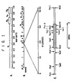
- the XhoI/gIII region is located at approximately 0.4 on the PRV genome within a 4.3 kb PstI fragment ( Figure 1).
- the gIII gene contained within this region is a structural gene which codes for a PRV envelope glycoprotein.
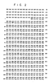
- the complete base pair sequence of the gIII gene (noncoding strand) is given in Figure 2.
- the DNA sequence for 196 bp upstream and 90 bp downstream from the gene is also given in Figure 2.
- the XhoI/gIII region, and any other nonessential region of the PRV genome can be manipulated to produce the deletions and insertions required in the invention by using standard recombinant DNA techniques known in the art such as those described in Maniatis et al., Molecular Cloning, A Laboratory Manual, Cold Spring Harbor Laboratory, Cold Spring Harbor, N.Y. (1982); Myers et al., Science, 229 : 242-247 (1985); Botstein et al., Science, 229 : 1193-1201 (1985); Roizman et al., Science, 229 : 1208-1214 (1985); and Graham et al., Virology, 52 : 456-467 (1973).
- the procedure would involve cleavage of the PRV genome using one or more restriction enzymes to produce genomic fragments, followed by isolation of the fragment containing the region which is to be the subject of the manipulation. This fragment could then be cloned into a vector.
- Any bacterial plasmid or bacteriophage vector could be utilized provided it contains a selectable marker. Suitable plasmid and bacteriophage vectors include, but are not limited to, E . coli plasmid pBR322, E . coli pSC1O1, bacteriophage ⁇ , and bacteriophage M13. Other vector systems, including yeast, can also be used.
- Preferred for ease of manipulation is E. coli plasmid pBR322.
- deletions within the PRV fragment could be effected by conventional techniques employing endonucleases, exonucleases and the like. Insertions, if desired, could also be made using techniques known to those skilled in the art.
- a plasmid available for such use is the plasmid pALM20 carried by E. coli strain NF1829, on deposit with the American Type Culture Collection in Rockville, Maryland bearing the accession number ATCC No. 67151.
- This plasmid is an Amp S , Tet R , pBR322 plasmid having cloned at the PstI site a 4.3 kb PstI PRV fragment containing the gIII gene (shown in figure 1) taken from the Becker strain of PRV.
- the plasmid or bacteriophage DNA having the desired manipulations can then be selected for using conventional methods, and introduced for propagation purposes into a host cell or organism using standard transformation procedures. Any host cell or organism in which the plasmid or bacteriophage can be propagated could be utilized. For example, in the case of a manipulated pALM20, the host organism would be E. coli .
- the plasmid or bacteriophage could be isolated from the host organism, mixed with unaltered PRV DNA and cotransfected into host cells.
- the plasmid or the bacteriophage and the PRV DNA would enter the cells where homologous recombination would take place, resulting in the replacement of the unaltered region in the viral DNA with the corresponding altered region from the plasmid or bacteriophage DNA.
- the plasmid or bacteriophage DNA may be linearized prior to mixing with the unaltered PRV DNA. Any host cell suitable for plasmid or bacteriophage and PRV DNA transfection and subsequent recombinant virus propogation can be utilized in this procedure. The recombinant PRV DNA is then replicated within the cell and the viruses which have undergone the desired recombination could be selected for using standard techniques.
- the present invention involves PRV engineered to carry foreign DNA fragments coding for a desired protein and capable of being expressed.
- the foreign fragment is inserted in the PRV genome at a point selected from within the nonessential XhoI/gIII region of the genome.
- insertions are made into the gIII gene segment of the XhoI/gIII region.
- insertions are made into this segment downstream from a remaining ATG codon sequence.
- the positions of the ATG codons in the gIII gene are illustrated in Figure 2.
- gIII gene segment it is meant the gIII glycoprotein coding sequence.
- the present invention in all of its embodiments requires that all essential regions of the PRV genome be present. However, nonessential regions within the genome may, but need not, be deleted in whole or in part in all embodiments.
- an "essential region” is any region of the PRV genome, the deletion of which would result in a complete cessation of viral propagation.
- propagation encompasses viral infectivity replication and release. Cessation of viral propagation has occurred where the virus is unable to propagate even with the assistance of a helper virus or a complementing cell line.
- Helper viruses are replication competent infectious viruses that provide gene products required for the propagation of coinfecting defective viruses that, by definition, cannot propagate themselves.
- helper viruses suitable for use in the present invention include unaltered PRV as well as other viruses that express the genes contained within the deleted region whose products are necessary for propagation of the recombinant PRV.
- "Complementing cell lines” are cell lines that provide gene products required for the propagation of defective viruses that, by definition, cannot propagate themselves.
- a "nonessential region”, as that phrase is defined herein, is any region, the deletion of which would not result in a complete cessation of viral propagation. Cessation of viral propagation has not occurred where the virus is able to propogate, with or without the assistance of a helper virus. It is believed that approximately 40% of the viral genome is nonessential.
- deletions are made in nonessential regions of the PRV genome such as the XhoI/gIII region, the tk gene region, as described in U.S. Patent 4,514,497, and the gI gene region, as described in Ben Porat et al., J. Virol., 57 : 191-196 (1986); and Mettenleiter et al., J. Virol., 53 : 52-57 (1985).
- Deletions in the aforementioned regions are preferred because such deletions do not necessitate the use of a helper virus or a complementing cell line in order for the PRV to infect and replicate.
- the deleted portion of the XhoI/gIII region is selected from within the gIII gene segment of the region.
- the definition of the insertion region, the XhoI/gIII region is based upon the unaltered genome having this region present. As nonessential, this region can be deleted in whole or in part, and the insertion made in the remaining portion of the region, or if entirely deleted, within the portion of the remaining genome where XhoI/gIII would have, if present, been located.
- the foreign fragment insert can be any fragment which codes for a desired protein and is capable of being expressed.
- the maximum size of such an insert is determined by adding the cloning capacity of unaltered PRV, which at least 3,000 and possibly 15,000 base pairs, to the number of base pairs removed in any genomic deletion.
- Desired proteins may be, but are not limited to, enzymes, hormones, antigens and antibodies.
- the phrase "capable of being expressed” requires that: (1) the resulting recombinant PRV DNA contain a transcription control region and a translation initiation codon sequence (ATG) upstream from the coding region of the foreign fragment; (2) the coding region of the foreign fragment be in reading frame with the translation initiation codon sequence; and (3) the desired protein produced be compatable with PRV propagation (i.e., is not lethal to the cells or the virus).
- the upstream translation initiation codon sequences can be supplied by either the PRV genomic portion of the recombinant virus or alternatively, by a translation initiation codon sequence insert, as desired, or as deletions and/or insertions require.
- Figure 2 shows the location of the ATG codon sequences within the unaltered PRV gIII gene.
- the upstream transcription control region can be supplied by either the remaining PRV genome or a transcription control region insert, again as desired or as deletions and/or insertions require.
- the transcription control region is located upstream from the gIII gene within a region of the PRV genome which begins with the first XhoI restriction enzyme cleavage site immediately upstream from the gIII glycoprotein gene and which ends at the first base pair of the gIII gene.
- Suitable transcription control region inserts include the long terminal repeat portion of the RNA tumor virus and the early or late promoter of the SV-40 virus. Such control regions are described in Experimental Manipulation of Gene Expression, M. Inouye, ed., (Academic Press 1983).
- PRV-24 One example of a recombinant of the above embodiment is PRV-24, described below.
- the present invention involves PRV engineered to remove all or part of the nonessential XhoI/gIII region.
- other nonessential regions may also, but need not, be deleted in whole or in part.
- the preferences as to deleted portions are as described above. Examples of this embodiment are PRV-2 and PRV-10 described below.
- a further aspect provides recombinant PRV comprising a pseudorabies virus genome having generated therein an EcoRI restriction enzyme cleavage site (coding sequence 5 ⁇ -GAATTC-3 ⁇ ), wherein the EcoRI site is located in a nonessential region of the PRV genome.
- Preferred are recombinants with insertions in the XhoI/gIII region, the tk gene region and the gI gene region. More preferred are recombinants with insertions in the XhoI/gIII region. Most preferably, the insertion is within the gIII gene segment of the XhoI/gIII region.
- the most preferred embodiment involves recombinants with insertions in the gIII gene segment downstream from an ATG codon sequence.
- EcoRI sites can be generated alternatively by site directed mutagenesis, or by the insertion of synthetic or naturally occurring foreign DNA fragments containing EcoRI sites.
- recombinants containing this EcoRI site are cut with EcoRI restriction enzyme and, employing standard recombinant DNA techniques, the desired DNA fragment inserted.
- PRV-1 and PRV-32 are examples of a recombinant of this type.
- the proteins of the instant invention are produced by: (1) infecting cells with recombinant PRV containing a foreign DNA fragment coding for a desired protein and capable of being expressed; (2) culturing the infected cells until the desired protein is produced; and (3) recovering the protein.
- the PRV is simply applied to the cells under standard cell culture conditions.
- To recover the protein standard techniques are employed. Cell culture techniques and recovery methods are described in Maniatis et al., Molecular Cloning, A Laboratory Manual, Cold Spring Harbor Laboratory, Cold Spring Harbor, N.Y. (1982); Experimental Manipulation of Gene Expression, M. Inouye, ed., (Academic Press 1983); and Robbins et al., J. Mol. Appl. Gen.
- the specific host cells employed in the present invention are not critical as long as they allow replication and expression of the recombinant PRV DNA.
- Suitable cells include, but are not limited to, animal and insect cells.
- Preferred are mouse fibroblast cells, HeLa cells, rabbit kidney cells, swine kidney cells, lamb kidney cells, dog kidney cells, monkey kidney cells, chick embryo fibroblast cells, baby hamster kidney cells, human fibroblast cells, and any and all combinations thereof.
- Most preferred are swine kidney cells, in particular PK15, available from National Veterinary Services Laboratories, Ames, Iowa.
- a pharmaceutically effective amount of the above-described live recombinant viruses containing foreign genes coding for an antigenic protein or an antigenic protein produced by such recombinants in accordance with the above-disclosed process can be employed along with a pharmaceutically acceptable carrier or diluent as a vaccine against disease in mammals.
- the antigenic protein need not be in pure form, but may if desired consist of cellular extracts containing any and all cellular parts and culture medium.
- the useful dosage to be administered and the mode of administration will vary depending upon the age, weight and mammal vaccinated and the type of pathogen against which vaccination is sought.
- the antigenic proteins produced by such viruses can also be employed as diagnostic reagents in a pathogen detection kit.
- a pathogen detection kit Specifically, the presence of a pathogen in samples of body fluids could be detected by contacting the protein with body fluids suspected of containing antibodies to that protein and measuring the formation of antigen-antibody complexes. Measurement of the complex formation can be accomplished using methods such as ELISA (enzyme-linked immunosorbent assay), strip radioimmunoassay or indirect immunofluorescence assay known to those skilled in the art and discussed in U.S. Patent 4,520,113.
- ELISA enzyme-linked immunosorbent assay
- strip radioimmunoassay or indirect immunofluorescence assay known to those skilled in the art and discussed in U.S. Patent 4,520,113.
- Swine kidney cells PK15, available from National Veterinary Services Laboratories, Ames, Iowa, were grown in Dulbecco's Eagle's Medium supplemented with 10% heat inactivated fetal bovine serum The bovine serum and the Medium were obtained from GIBCO, Grand Island, N.Y. The Becker strain of PRV was used, although any strain of PRV is satisfactory. Cells, the virus and culture conditions are described by Robbins et al., J. Mol. Appl. Gen. 2 : 485-496 (1984).
- M1 and M16 - specific for the gIII envelope glycoprotein were obtained from Dr. T. Ben Porat, Vanderbilt University, Nashville, Tennessee. Although the monoclonal antibodies were made using the Ka strain of PRV, they show essentially the same reactivity with the Becker strain of PRV and they neutralize the virus in the presence or absence of complement. They also immunoprecipitate gIII proteins from infected cells. Although M1 and M16 were utilized herein, any monoclonal antibodies raised against the gIII gene product could be employed.
- the procedure used to purify the viruses was that described by Ben Porat et al.,Virology, 60 : 29-37 (1974). The procedure was modified in order to prepare 3H-glucosamine labelled virus in that 100 ⁇ Ci/mL of 3H-glucosamine, obtained from New England Nuclear, Boston, Massachusetts, was added. The techniques for making cell extracts for immunological analysis were described previously by Robbins et al., J. Mol. Appl. Gen., 2 : 485-496 (1984).
- PRV DNA was purified from PRV nucleocapsids as described by Ben Porat et al., Virology, 41 : 265-273 (1970). DNA so obtained was analyzed by the Southern blot method as described by Southern, E. M., J. Mol. Biol., 98 : 503-517 (1975).
- DNA/DNA hybridizations were done at 37 °C for 16 hours in 60% formamide, 10 mM PIPES (piperazine-N,N ⁇ -bis[2-ethanesulphonic acid]) pH 6.5, 5 ⁇ SSC (0.75 M sodium chloride and 0.075 M sodium citrate, pH 6.5), sonicated calf thymus DNA (100 ug/mL) and 5 ⁇ Denhardt's solution (0.1% each of ficoll 400 [Pharmacia Inc., Piscataway, N.J.], bovine serum albumin and polyvinylpyrollidone [Sigma Chemical Company, St. Louis, MO]).
- DNA probes were prepared by nick-translation using 32P-labelled nucleotides.
- the hybridized nitrocellulose membranes were washed four times for 30 minutes each at 65° in 2 ⁇ SSC (0.3 M sodium chloride and 0.03 M sodium citrate, pH 7.0). The nitrocellulose membrane was then prepared for autoradiography on Kodak XAR-5 film.
- E . coli bacterial plasmid vectors were utilized in making the recombinant PRV DNA. They are designated by having a small p immediately preceding letters or numbers or a mixture of letters and numbers. For certain experiments, fragments of PRV DNA were inserted into an E . coli expression vector so that significant amounts of the DNA fragment and also the PRV gene product could be produced for purposes of DNA and protein analysis as well as for use in cotransfection gene replacement experiments.
- E . coli strain NF1829 carrying an F ⁇ plasmid with the laci q mutation which results in the overproduction of the Lac repressor was used to regulate the expression of PRV genes in the Lac expression vectors. This is important because excessive expression of the PRV gIII gene in E . coli results in growth arrest of the cells as described by Robbins et al., J. Virol. 58 : 339-347 (1986).
- the gel was cut above and below the required fragments and the DE81 strips were then placed within the cut gel and the DNA electrophoresed onto the strip.
- the strips into which the DNA was electophoresed were then rinsed with water and the DNA eluted off with 1.5M NaCl, 10mm Tris pH 7.5 and 1mm EDTA for 2 hours at 37°.
- the DNA was precipitated in equal volume of isopropanol at room temperature for one hour. The precipitate was collected by centrifugation. To the resulting pellets were added TEN (10mm Tris pH 7.5, 1mm EDTA, 100mm NaCl). The solution was ethanol precipitated overnight at -20° and the pellets recoved by centrifugation.
- 100ng of the pellet containing the linearized pBR322 isolate and 200ng of the 4.3 PRV PstI fragment were added in the presence of 1 ⁇ l 10 ⁇ ligase buffer, 1 ⁇ l 1mg/ml of bovine serum albumin (obtained from Sigma Chemical Company), 1 ⁇ l of ligase. and H2O up to 10 ⁇ l. After 3 hours at room temperature the resulting ligation mix was transformed into E . coli strain NF 1829, described above. The bacteria were then plated on Tet plates overnight at 37°. Replica plate colonies from the Tet plates were next plated on both Tet and Amp plates. Plasmid that were Tet R and Amp S were selected and analysed by Southern blot analysis.
- Plasmid DNA's were then digested with PstI and EcoRI, subjected to electrophoresis on a 1% agarose/TEA gel, transferred to nitrocellulose and probed using nick-translated pALM15.
- a clone designated pALM20 that contained a 4.3 kb PstI fragment of PRV genomic DNA containing the gIII gene was isolated.
- Deletions affecting gIII were constructed by altering the E . coli pBR322 derived expression plasmid pALM3 (figure 3A).
- pALM3 is described in European Patent Application 0,162,738 dated November 27, 1985 (referred to therein as pNT 7/123).
- E. coli strain K12, NF1829 carrying the plasmid pALM3 (pNT7/123) is on deposit with In Vitro International, Inc., Ann Arbor, Michigan, bearing the accession number IVI-10049.
- transcription of the Cro-PRV gIII hybrid gene is directed from the tac promoter under control of the Lac repressor.
- E . coli strain NF1829 containing pALM3 were grown in L-broth containing 100 ⁇ g/mL of ampicillin. Fusion proteins were induced by adding isopropyl beta-D thiogalactopyranoside to a final concentration of 1mM. Three mL of culture was harvested 4 hours after the inducer was added. The Cro-PRVgIII fusion proteins were collected as insoluble aggregates. The proteins were fractionatedby electrophoresis on a 10% SDS-polyacrylamide gel and then stained with Coomassie blue.
- pALM3 produces significant quantities of a CRO-PRV gIII fusion protein that accumulates to approximately 3-5% of total cell protein ( Figure 4, lane 2).
- the amino terminal 23 amino acids derived from the bacteriophage Lambda Cro protein, are fused to the first amino acid (Met) of gIII.
- the coding sequence terminates at the natural translation stop signal (TGA) after amino acid 479 followed by approximately 1000 bp of PRV sequence.
- pALM2 ( Figure 3A) was constructed by digesting pALM3 with the restriction enzyme SacI, and re-ligating.
- pALM2 contains an in-frame deletion of 402 bp - the internal SacI fragment - within the gIII gene and produces (as described above for pALM3) a truncated protein ( Figure 4, lane 3).
- pALM10 is another expression plasmid containing part of the gIII gene of PRV ( Figure 3B). In this case, about 87% of the gIII gen of PRV has been removed. It was constructed as follows. First pALM11 was constructed to serve as an intermediate. pALM11 was constructed by digesting pALM12 with SphI and removing the resulting single-stranded ends by using the 3 ⁇ -5 ⁇ exonuclease activity of the Klenow fragment of DNA polymerase I. The construction of pALM12, an E. coli pBR322 derivative, is described in European Patent Application 0,162,738 dated November 27, I985 (referred to therein as p7/123).
- pHK412 Another pBR322 derivative, is described in European Patent Application 0,162,738 dated November 27, 1985 (referred to therein as pHK412).
- DNA from both pALM3 and pALM11 were digested with PstI and XhoI.
- the resulting 1840 bp fragment of pALM11 and 3670 bp fragment of pALM3 were purified and ligated with T4 DNA ligase.
- the plasmid formed, pALM10 lacks all of the gIII gene (including 235 bp of upstream DNA) except for 186 bp encoding the carboxy-terminus portion. It also retains the tac promoter/operator and translation elements of the pALM3 expression vector. It produces no proteins upon induction ( Figure 4, lane 4).
- Recombinant pseudorabies virus PRV-2 were produced as follows.
- the DNA of pALM2 was digested by incubating it with the restriction enzyme NcoI.
- One ⁇ g of the resulting NcoI digested DNA from pALM2 was cotransfected with 2 ⁇ g of DNA from parental (unaltered) PRV DNA into PK15 cells employing the procedure described by Graham et al., Virology, 52 : 456-467 (1973).
- the transfected cells were incubated for 4 hours at 37°, washed with growth medium, and subjected to a (15% glycerol) shock for three minutes. The cells were again washed with medium, two times, and then placed into a fresh batch of growth medium.
- gIII monoclonal antibodies M1 and M16 (to a concentration of 0.6 ⁇ g/mL of each) and guinea pig complement (to a concentration of 5%) were added to enrich for gIII-defective recombinant virus.
- these monoclonal antibodies neutralize the infectivity of parental PRV, but they do not neutralize PRV that no longer express gIII protein or that express truncated forms of the gIII glycoprotein.
- Neither M1 nor M16 monoclonal antibodies reacted with the gIII protein expressed by PRV-2. The total virus yield from each transfection was harvested when complete cytopathic effect was observed, which occurred after approximately 36 hours.
- PRV-10 recombinant viruses were produced and selected essentially as PRV-2 described above except that pALM10 was linearized with PstI and cotransfected with parental virus DNA.
- the DNA of PRV-2 and PRV-10 were analyzed to show that the deletions defined by pALM2 and pALM10 had been crossed into the selected viruses.
- Parental PRV was also analyzed for comparison.
- Viral DNA was isolated from nucleocapsids as described in the General Procedures. The DNA was digested with PstI and XhoI, fractionated on a 1% agarose gel, transferred to nitrocellulose and hybridized with a 32P-labelled probe from pALM15 ( Figure 5). As shown in the restriction enzyme map of the parental genome included in Figure 5, the gIII gene is located approximately in the center of a 4.3 kb PstI fragment.
- DNA from PRV-2 (lane 2 in Figure 5) which contains a 402 bp deletion within the gIII gene, the 1.3 kb and 0.95 kb fragments are unchanged, but the 1.48 kb internal XhoI fragment now migrates as a 1.1 kb fragment.
- DNA from PRV-10 (lane 3 in Figure 5) which contains a precise deletion of the 1.48 kb internal XhoI fragment shows only the 1.3 and 0.95 kb fragments.
- the SacI/BglII linker is shown below. 5 ⁇ GAGCTCAATGAGTCCGA 3 ⁇ 3 ⁇ CTCGAGTTACTCAGGCTCTAG 5 ⁇
- the following were added to an appropriate reaction vessel: 4.0 ⁇ L annealed linker polynucleotides 5.5 ⁇ L of water 1.0 ⁇ L of 10 ⁇ linker kinase buffer (700 mM tris-HCl, pH 7.6, 100 mM MgCl2, 50 mM dithiothreitol) ⁇ L 10 mM ATP, pH 7.0 2 uL T4 kinase (from Bethesda Research Laboratory (BRL)), Gaithersburg, MD, 20 units) The mixture was incubated for 1 hour at 37°, and then heated at 65° for 5 minutes.
- 10 ⁇ linker kinase buffer 700 mM tris-HCl, pH 7.6, 100 mM MgCl2, 50 mM dithiothreitol
- pLORO contains the fragment of the HTLVIII envelope polyprotein gene coding region from bp7196 - bp8621. Included within this fragment are 135 bp of glycoprotein 120 (gp120) and all of glycoprotein 41 (gp41), the latter consisting of 1425 bp.
- pEXC An HTLVIII fragment obtained from ⁇ BH10 by BglII digestion and cloned into the unique BglII site of plasmid pEXC, forming pLORO-1.
- pEXC was constructed by combining a PstI-BglII fragment. from p ⁇ ROP and a PstI fragment from pKGP36.
- trp . p ⁇ ROP is a derivative of pBR322 in which a BglII linker has been inserted into the PvuII site. To create pKGP36.
- trp tryptophan promotor-operator
- Shine-Delgarno sequences derived from pLD102 as described in Christie et al. J. Mol. Biol., 143 : 335-341 (1980), were inserted into the ClaI site of plasmid pBR322.
- TBE buffer consists of 50 mM tris-HCl, pH 8.2, 50 mM borate and 2.5 mM disodium EDTA.
- the gel was stained by immersion in a solution of ethidium bromide (1 ⁇ g/mL).
- the kinased SacI/BglII linker was ligated onto the HTLVIII fragment as follows. Added to an appropriate reaction vessel were: 15 ⁇ L of DNA fragment in TE buffer (from part 2, above) 11.5 ⁇ L 5 ⁇ ligase buffer (500 mM tris-HCl, pH 7.8, 10 mM MgCl2, 100 ⁇ g/mL bovine serum albumin) 6 ⁇ L 10 mM ATP, pH 7.0 22.5 ⁇ L 40% polyethylene glycol, molecular weight size 8000 5 ⁇ L kinased linker 5 ⁇ L water 1 ⁇ L T4 DNA ligase (BRL, 1 unit) The mixture was incubated about 16 hours and the resulting DNA ethanol precipitated and resuspended in 10 ⁇ L of TE buffer.
- 5 ⁇ ligase buffer 500 mM tris-HCl, pH 7.8, 10 mM MgCl2, 100 ⁇ g/mL bovine serum albumin
- the DNA was then digested with 100 units of SacI for 3 hours at 37° in a total volume of 100 ⁇ L.
- the resulting HTLVIII-linker DNA fragment was ethanol precipitated and resuspended in 10 ⁇ L of TE buffer. This fragment is illustrated in Figure 6(D).
- DNA (20 ⁇ L) from pALM24 was digested with 30 units of SstI in a total of 100 ⁇ L for 2 hours at 37°.
- the resulting DNA was precipitated with ethanol, resuspended in 10 ⁇ L of TE buffer and electrophoresed through an 0.8% agarose/TBE gel for about 16 hours at 30 volts.
- the gel was stained with ethidium bromide as previously described and the presence of the 1.43 kb band of DNA (HTLVIII DNA-linker fragment) verified.
- the orientation of the cloned fragment was checked by digesting 20 ⁇ L of the plasmid DNA with 40 units of HindIII in a total volume 100 ⁇ L for 2 hours at 37°.
- the DNA was precipitated with ethanol, resuspended in 20 ⁇ L of TE buffer and electrophoresed as described. The presence of a band of DNA with 1000 bp shows that the cloned fragment was in the correct orientation and that the construction was complete.
- Recombinant PRV-24 was produced essentially as described above for the production of PRV-2 and PRV-10 except that the source of DNA used to transfect PK15 cells were obtained from pALM24 cut with NcoI and parental PRV.
- Recombinant viruses were enriched by incubating the infected cultures in the presence of guinea pig complement and M1 and M16 monoclonal antibodies as previously described.
- Recombinant viruses were selected by means of the black plaque selection technique previously described. About 4 white plaques were selected from the approximately 18,000 total plaques (white and black) which appeared. One of the resulting white plaques was then purified by two successive single plaque isolations and grown into a stock of virus, PRV-24.
- PRV-24 contained and expressed HTLVIII glycosylated antigen
- PRV-24 as well as PRV-2 and PRV were separately grown in PK15 cells in the presence of 0.5 mCi of 3H-glucosamine.
- the cells were scraped into phosphate buffered saline and collected by centrifugation.
- the infected cells were lysed by suspending them in RIPA buffer (150 mM NaCl, 10 mM tris, pH 7.9, 1% Triton X100, 1% deoxycholate, 0.1% sodium dodecyl sulfate) containing phenylmethylsulfonyl fluoride at a concentration of 100 ⁇ g/mL.
- Triton X100 is obtained from Sigma Chemical Company, St. Louis, MO. The lysed cells and suspending fluid were then centrifuged at 100,000 ⁇ g for 30 minutes. To 400 ⁇ L of supernatant fluid from each of the samples were added in separate tubes, 20 ⁇ L of 490 serum (a polyclonal antiserum to the gIII protein of PRV, raised in rabbits against the Cro-PRV gIII fusion protein expressed by pALM3), 10 ⁇ L of antisera raised against the protein produced by a BglII to BamHI fragment of the HTLVIII gene from bp 7198 to 8052, as described in Ratner et al., Nature 313 :277-284 (1985) containing a portion of the gp120 and the gp41 gene (anti-HTLVIII serum), and 20 ⁇ L of M2 antibody (against the gII family of PRV glycoproteins).
- 490 serum a polyclonal antiserum to the
- the recovered pelleted Staph A's were resuspended in 25 ⁇ L of SB (3% sodium dodecyl sulfate, 700 mM 2-mercaptoethanol, 10 mM tris buffer pH 6.8, 1% glycerol plus bromophenol blue) and boiled 2 minutes.
- SB 3% sodium dodecyl sulfate, 700 mM 2-mercaptoethanol, 10 mM tris buffer pH 6.8, 1% glycerol plus bromophenol blue
- the Staph A's were pelleted by centrifugation and the supernatant then electrophoresed on a 10% polyacrylamide/SDS gel.
- the gels were then fixed in methanol/acetic acid, washed twice in H2O and then treated for 30 minutes in 1M sodium salicylate.
- the gels were dried and exposed on Kodak XAR5 film for one week using an intensifying screen.
- PRV-24 contains the DNA sequence encoding a portion of the HTLVIII env gene.
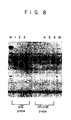
- DNA was isolated from virions of parental PRV, PRV-2 and PRV-24 as described above. Each DNA (6 ⁇ g) was digested with PstI. The digested DNA was electrophoresed in two sets of three on a 1% agarose TEA (tris, EDTA, sodium acetate) gel in TEA. Southern Blot analysis was carried out as described earlier. After the DNA was transferred to nitrocellulose, the nitrocellulose was cut in half yielding two identical blots. One blot was hybridized with a gIII specific probe, pALM15.
- the other blot was hybridized with an HTLVIII specific probe, a KpnI fragment from bp 5928 to 8596 of the HTLVIII genome, as described in Ratner et al., Nature, 313 : 277-284 (1985).
- the blot was exposed to Kodak XAR film, using an intensifying screen, at -70 °C for 2 days. The results are shown in Figure 8.
- the autoradiogram of the blot using gIII as a probe showed hybridization to a PRV-24 DNA species of 5.3 kb. This DNA species is 1 kb larger than parental PRV DNA and 1.4 kb larger than PRV-2 DNA. This corresponds to the anticipated fragment size for the inserted HTLVIII DNA sequence.
- the second blot using the HTLVIII probe only showed specific hybridization to a 5.3 kb species in the PRV-24 lane. No specific hybridization was seen to either parental PRV or PRV-2. This indicates that PRV-24 contains HTLVIII envelope sequences inserted with the gIII coding sequence.
- This example describes the insertion of a novel EcoRI restriction enzyme site into the SacI restriction enzyme site of pALM2 generating plasmid pALM1.
- pALM2 was digested with EcoRI at 37° for 60 minutes.
- the reaction mixture was then incubated with the Klenow fragment of DNA polymerase I in the presence of all four deoxyribonucleotides to fill in the ends of the DNA.
- the blunt ends of the resulting DNA were then rejoined.
- This derivative of PALM2, free of any EcoRI sites is named pALM7.
- pALM7 DNA was digested with SacI at 37° for 60 minutes.
- the polynucleotide linker fragment consisting of 5 ⁇ CGAATTCGAGCT 3 ⁇ was synthesized on an Applied Biosystem DNA synthesizer using standard phosphoramidite chemistry and purified by reverse-phase high performance liquid chromatography.
- the linker DNA was melted at 68°, reannealed slowly and then phosphorylated using T4 polynucleotide kinase.
- the phosphorylated linker was then ligated to the SacI digested pALM7 DNA with T4 DNA ligase to create pALM1, shown in Figure 10.
- the structure of pALM1 was verified by restriction enzyme and DNA sequence analysis.
- This linker maintains the reading frame of the truncated gIII protein as indicated by the spacing (see Figure 10) and also introduces an EcoRI site bracketed by SacI sites.
- the linker adds 4 extra amino acids to the truncated gIII protein. It produced the expected fusion protein upon induction of E. coli bearing the pALM1 plasmid.
- PRV can tolerate an EcoRI site
- PRV With the EcoRI site was produced by cotransfecting PK15 cells with parental PRV DNA and pALM1 DNA as described above for PRV-2.
- Virus defective for the gIII gene product was selected, isolated and characterized again in the same manner.
- One such isolat was designated PRV-1.
- PRV-1 was then grown in tissue culture. No deleterious effects of the EcoRI site on virus reproduction were observed.
- This example demonstrates the construction of a hybrid gIII gene and the E . coli gene encoding beta-galactosidase (the lac z gene) and the expression and glycosylation of a fusion protein containing a gene product not normally glycosylated. Further, the example shows the use of EcoRI sites engineered into the gIII gene.
- pALM1 DNA contains a truncated gIII gene with a synthetic EcoRI site as described in Example 2.
- the source of the lac z DNA sequence is the E. coli derived plasmid pMLB1034, a plasmid that contains a unique EcoRI site at the beginning of the lac z coding sequence.
- pMLB1034 is described in T.J. Silhavy, M.L. Berman, and L.W. Enquist, Experiments With Gene Fusions, pp. 250-251 (Cold Spring Harbor Laboratory 1984).
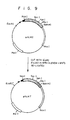
- pALM1 DNA is digested with the enzymes EcoRI and PstI and ligated to pMLB1034 plasmid DNA which has also been digested with PstI and EcoRI ( Figure 9).
- the ligation of a 5.5 kb PstI-EcoRI fragment from pMLB1034 to a 1.7 kb PstI-EcoRI fragment from pALM1 gave rise to a 7.2 kb plasmid pCL1403.
- the lac z gene is fused to the gIII gene at the EcoRI site in pCL1403, the two genes are not in the same reading frame. A protein coded from such a construction would thus result in a nonsense fusion protein.
- NTP's T-T-A-A
- T-T-A-A Four nucleotide pairs (NTP's: T-T-A-A) were therefore added to place the two genes in the same frame. This was accomplished by digesting pCL1403 DNA with EcoRI and filling in the resulting single stranded ends left by the EcoRI cleavage by incubating the digested DNA with the Klenow fragment of DNA polymerase I and the four deoxynucleotides. The blunt ends were ligated by means of T4 ligase creating pCL1404. This plasmid was selected by transforming E . coli strain NF1829 with the altered DNA containing pCL1404 and selecting ampicillin resistant colonies containing plasmid DNA resistant to digestion with EcoRI.
- bacteria containing pCL1404 produced a gIII- lac z fusion protein by growing colonies of the transformed bacteria on agar medium in the presence of 5-bromo-4-chloro-3-indoyl-beta-D-galactoside (x-Gal) and noting that the colonies were blue as a result of the beta- galactosidase activity.
- bacteria containing PCL1404 grown in the presence of isopropyl-beta-D-thiogalactopyranoside (IPTG) produced a 130 kd protein when analyzed on a polyacrylamide/SDS gel.
- the DNA sequence containing the lac Z gene must be flanked by regions of DNA homologous to gIII sequences in the PRV genome.
- the NcoI-BamHI fragment of pCL1404 contains a gIII DNA sequence adjacent to and upstream of the lac Z gene. However there is no gIII DNA sequence adjacent to the downstream end of the lac Z gene. This was remedied as follows. First, pZ186, a pMLB524 derivative, was created. pMLB524, described in T.J. Silhavy, M.L. Berman, and L.W.
- DNA from pCL1407 was digested with NcoI and the resulting DNA along with the DNA from parental PRV were co-transfected into PK 15 cells.
- a recombinant virus containing an altered gIII gene was selected by previously described methods. Viruses from a representative viral plaque was isolated, purified and analyzed. The virus was designated PRV-Z1.
- Southern analysis of the DNA of PRV-Z1 it was shown to contain a 3.0 kb insertion of lac Z specific DNA within the 4.3 kb PstI fragment containing the gIII gene.
- This example describes the insertion of a synthetic EcoRI site into the KpnI site of the unaltered gIII gene and the construction of a PRV recombinant (PRV-32) containing this novel gene.
- PRV-32 PRV recombinant
- pALM3 was constructed as described above. This plasmid contains an EcoRI site just upstream of the tac promoter which was removed by cutting pALM3 DNA with EcoRI, filling in the single-stranded ends left by EcoRI cleavage using DNA polymerase 1 (see Example 2) and religating the linear plasmid with T4 ligase. The re-ligated DNA was transformed into E . coli strain NF1829 and plasmids were isolated that were resistant to cleavage by EcoRI. One such plasmid was isolated, characterized and named pALM-28 (See Figure 14).
- PRV-2 The novel gIII gene in pALM32 was inserted by recombination into the DNA of PRV-2 using standard co-transfection as above.
- PRV-2 is described in Example 2.
- PRV-2 expresses a truncated gIII protein that does not react with M1 or M16 monoclonal antibodies. Therefore, PRV-2 makes white plaques in the "Black Plaque Test".
- black plaques were recovered at frequencies of about 0.5% of total plaques. Five black plaques were picked, purified and analyzed. Of the 5 virus isolates, two were analyzed and found to contain a novel EcoRI site at the KpnI site of gIII. One stock was chosen and called PRV-32.
- This example shows the construction of a PRV vector containing a synthetic EcoRI site in a full length gIII gene.
- the new gene expresses a gIII protein that reacts with M1 and M16 monoclonal antibodies in the "Black Plaque Test”.
- this example teaches the utility of PRV vectors with gIII deletions (e.g. PRV-2 or PRV-10) as vehicles for insertion of gIII genes using the "Black Plaque Test" as a screen.
- viruses and plasmid have been deposited under the listed accession numbers with the American Tissue Culture Collection in Rockville, MD, on 3 July 1986:
- the present invention is not to be limited in scope by the viruses deposited.
Landscapes
- Life Sciences & Earth Sciences (AREA)
- Health & Medical Sciences (AREA)
- Chemical & Material Sciences (AREA)
- Genetics & Genomics (AREA)
- Organic Chemistry (AREA)
- Engineering & Computer Science (AREA)
- Biophysics (AREA)
- Zoology (AREA)
- Biochemistry (AREA)
- General Health & Medical Sciences (AREA)
- Molecular Biology (AREA)
- Wood Science & Technology (AREA)
- Bioinformatics & Cheminformatics (AREA)
- General Engineering & Computer Science (AREA)
- Virology (AREA)
- Biomedical Technology (AREA)
- Biotechnology (AREA)
- Physics & Mathematics (AREA)
- Plant Pathology (AREA)
- Gastroenterology & Hepatology (AREA)
- Microbiology (AREA)
- Proteomics, Peptides & Aminoacids (AREA)
- Medicinal Chemistry (AREA)
- Micro-Organisms Or Cultivation Processes Thereof (AREA)
- Preparation Of Compounds By Using Micro-Organisms (AREA)
- Peptides Or Proteins (AREA)
- Medicines Containing Antibodies Or Antigens For Use As Internal Diagnostic Agents (AREA)
Applications Claiming Priority (2)
| Application Number | Priority Date | Filing Date | Title |
|---|---|---|---|
| US886691 | 1986-07-18 | ||
| US06/886,691 US5037742A (en) | 1986-07-18 | 1986-07-18 | Pseudorabies virus recombinants and their use in the production of proteins |
Publications (2)
| Publication Number | Publication Date |
|---|---|
| EP0256677A2 true EP0256677A2 (de) | 1988-02-24 |
| EP0256677A3 EP0256677A3 (de) | 1988-12-28 |
Family
ID=25389547
Family Applications (1)
| Application Number | Title | Priority Date | Filing Date |
|---|---|---|---|
| EP87306364A Withdrawn EP0256677A3 (de) | 1986-07-18 | 1987-07-17 | Pseudorabiesvirus-Rekombinanten und ihre Verwendung zur Herstellung von Proteinen |
Country Status (5)
| Country | Link |
|---|---|
| US (1) | US5037742A (de) |
| EP (1) | EP0256677A3 (de) |
| JP (1) | JPS6387976A (de) |
| CA (1) | CA1301674C (de) |
| DK (1) | DK375487A (de) |
Cited By (8)
| Publication number | Priority date | Publication date | Assignee | Title |
|---|---|---|---|---|
| FR2601689A1 (fr) * | 1986-07-17 | 1988-01-22 | Syntro Corp | Herpes virus attenues, herpes virus qui renferment de l'adn etranger codant pour une sequence d'amino-acides, et vaccins les contenant |
| EP0332677A4 (en) * | 1987-07-27 | 1990-10-10 | Syntro Corporation | Attenuated herpes viruses, herpes viruses which include foreign dna encoding an amino acid sequence and vaccines containing same |
| WO1993001833A1 (en) * | 1991-07-17 | 1993-02-04 | Commonwealth Scientific And Industrial Research Organisation | Improved vaccine |
| FR2693472A1 (fr) * | 1992-06-26 | 1994-01-14 | Rhone Merieux | Mutants du virus de la rhinotrachéite infectieuse bovine, délétés dans l'un des gènes des glycoprotéines mineures, vaccins préparés à partir de ces souches, méthodes de production et méthodes d'utilisation. |
| WO1994008000A1 (en) * | 1992-10-06 | 1994-04-14 | Akzo Nobel N.V. | Pseudorabies virus vaccine |
| EP0256092B1 (de) * | 1986-01-27 | 1998-04-08 | Syntro Corporation | Abgeschwächte herpesviren, herpesviren, die eine für aminosäuresequenzen kodierende fremd-dns enthalten, sowie impfstoffe, die diese enthalten |
| US5738854A (en) * | 1992-10-06 | 1998-04-14 | Akzo Nobel N.V. | Pseudorabies virus vaccine |
| US5763269A (en) * | 1985-09-06 | 1998-06-09 | Syntro Corporation | Recombinant infectious bovine rhinotrocheitis virus |
Families Citing this family (3)
| Publication number | Priority date | Publication date | Assignee | Title |
|---|---|---|---|---|
| US5240703A (en) * | 1991-03-01 | 1993-08-31 | Syntro Corporation | Attenuated, genetically-engineered pseudorabies virus s-prv-155 and uses thereof |
| US20060216302A1 (en) * | 2003-02-28 | 2006-09-28 | Robert Root-Bernstein | Immunological markers |
| US20050180822A1 (en) * | 2004-02-18 | 2005-08-18 | Andrew Barmakian | Composite structure |
Family Cites Families (8)
| Publication number | Priority date | Publication date | Assignee | Title |
|---|---|---|---|---|
| EP0074808A3 (de) * | 1981-09-16 | 1984-07-04 | University Patents, Inc. | Rekombinantes Verfahren und Materialien |
| NL8303501A (nl) * | 1983-10-12 | 1985-05-01 | Centraal Diergeneeskundig Inst | Deletiemutant van een herpesvirus en vaccin, dat dit virus bevat. |
| US4514497B1 (en) * | 1983-12-30 | 1998-02-24 | Novagene Inc | Modified live pseudorabies viruses |
| EP0162738A1 (de) * | 1984-04-09 | 1985-11-27 | MOLECULAR GENETICS RESEARCH & DEVELOPMENT LIMITED PARTNERSHIP | Herstellung von Untereinheiten von Pseudorabiesvirus-Vakzinen |
| JP2559384B2 (ja) * | 1985-07-29 | 1996-12-04 | ジ・アップジョン・カンパニ− | ウイルスワクチン |
| US4877737A (en) * | 1985-09-06 | 1989-10-31 | Prutech Research And Development Partnership | Attenuated pseudorabies virus which has a deletion in at least a portion of a repeat sequence and vaccine containing same |
| AU623333B2 (en) * | 1986-01-27 | 1992-05-14 | Syntro Corporation | Attenuated herpesviruses, herpesviruses which include foreign dna encoding an amino acid sequence and vaccine containing same |
| US4711850A (en) * | 1986-01-28 | 1987-12-08 | Novagene, Inc. | Pseudorabies virus mutants, vaccines containing same, methods for the production of same and methods for the use of same |
-
1986
- 1986-07-18 US US06/886,691 patent/US5037742A/en not_active Expired - Fee Related
-
1987
- 1987-07-09 CA CA000541717A patent/CA1301674C/en not_active Expired - Lifetime
- 1987-07-17 DK DK375487A patent/DK375487A/da not_active Application Discontinuation
- 1987-07-17 JP JP62177368A patent/JPS6387976A/ja active Pending
- 1987-07-17 EP EP87306364A patent/EP0256677A3/de not_active Withdrawn
Cited By (9)
| Publication number | Priority date | Publication date | Assignee | Title |
|---|---|---|---|---|
| US5763269A (en) * | 1985-09-06 | 1998-06-09 | Syntro Corporation | Recombinant infectious bovine rhinotrocheitis virus |
| EP0256092B1 (de) * | 1986-01-27 | 1998-04-08 | Syntro Corporation | Abgeschwächte herpesviren, herpesviren, die eine für aminosäuresequenzen kodierende fremd-dns enthalten, sowie impfstoffe, die diese enthalten |
| FR2601689A1 (fr) * | 1986-07-17 | 1988-01-22 | Syntro Corp | Herpes virus attenues, herpes virus qui renferment de l'adn etranger codant pour une sequence d'amino-acides, et vaccins les contenant |
| EP0332677A4 (en) * | 1987-07-27 | 1990-10-10 | Syntro Corporation | Attenuated herpes viruses, herpes viruses which include foreign dna encoding an amino acid sequence and vaccines containing same |
| WO1993001833A1 (en) * | 1991-07-17 | 1993-02-04 | Commonwealth Scientific And Industrial Research Organisation | Improved vaccine |
| FR2693472A1 (fr) * | 1992-06-26 | 1994-01-14 | Rhone Merieux | Mutants du virus de la rhinotrachéite infectieuse bovine, délétés dans l'un des gènes des glycoprotéines mineures, vaccins préparés à partir de ces souches, méthodes de production et méthodes d'utilisation. |
| WO1994000586A3 (fr) * | 1992-06-26 | 1994-02-17 | Rhone Merieux | Mutants et vaccins du virus de la rhinotracheite infectieuse bovine |
| WO1994008000A1 (en) * | 1992-10-06 | 1994-04-14 | Akzo Nobel N.V. | Pseudorabies virus vaccine |
| US5738854A (en) * | 1992-10-06 | 1998-04-14 | Akzo Nobel N.V. | Pseudorabies virus vaccine |
Also Published As
| Publication number | Publication date |
|---|---|
| JPS6387976A (ja) | 1988-04-19 |
| EP0256677A3 (de) | 1988-12-28 |
| DK375487A (da) | 1988-01-19 |
| DK375487D0 (da) | 1987-07-17 |
| US5037742A (en) | 1991-08-06 |
| CA1301674C (en) | 1992-05-26 |
Similar Documents
| Publication | Publication Date | Title |
|---|---|---|
| US6998252B1 (en) | Recombinant poxviruses having foreign DNA expressed under the control of poxvirus regulatory sequences | |
| Robbins et al. | Pseudorabies virus gene encoding glycoprotein gIII is not essential for growth in tissue culture | |
| Wei et al. | Construction and isolation of a transmissible retrovirus containing the src gene of Harvey murine sarcoma virus and the thymidine kinase gene of herpes simplex virus type 1 | |
| EP0110385B1 (de) | Verfahren zur Herstellung von Poxvirus-Rekombinanten zur Expression fremder Gene | |
| Straus et al. | Molecular cloning and physical mapping of varicella-zoster virus DNA. | |
| AU645333B2 (en) | Recombinant marek's disease virus | |
| JPH01112986A (ja) | 組換えウイルスベクター | |
| JPH02430A (ja) | 組み換え家禽痘疹ウイルス | |
| Grunwald et al. | Loss of viral gene expression and retention of tumorigenicity by Abelson lymphoma cells | |
| Cho et al. | A second Epstein-Barr virus early antigen gene in BamHI fragment M encodes a 48-to 50-kilodalton nuclear protein | |
| EP0261940A2 (de) | Pseudorabies-Impfstoffe und DNS-Vektoren zur Rekombination mit Poxviren | |
| US5037742A (en) | Pseudorabies virus recombinants and their use in the production of proteins | |
| US4642333A (en) | Immunologically reactive non-glycosylated amino acid chains of glycoprotein B of herpes virus types 1 and 2 | |
| AU607399B2 (en) | Infectious recombinant virus vaccine for feline leukemia | |
| Cho et al. | Localization of the coding region for an Epstein-Barr virus early antigen and inducible expression of this 60-kilodalton nuclear protein in transfected fibroblast cell lines | |
| US7767449B1 (en) | Methods using modified vaccinia virus | |
| US5369025A (en) | Recombinant fowlpox vaccine for protection against Marek's disease | |
| Jenson et al. | Palindromic structure and polypeptide expression of 36 kilobase pairs of heterogeneous Epstein-Barr virus (P3HR-1) DNA | |
| US5231023A (en) | Recombinant Marek's disease virus | |
| EP0576092B1 (de) | Rekombinanter Katzen-Herpesvirus-Impfstoff | |
| EP0062574A2 (de) | Synthese von Virusproteinen | |
| Machida et al. | Reduced leukemogenicity caused by mutations in the membrane glycoprotein gene of Rauscher spleen focus-forming virus | |
| EP0422072B1 (de) | Verfahren zur selektion von rekombinanten pockviren | |
| Querat et al. | Characteristics of a novel lentivirus derived from South African sheep with pulmonary adenocarcinoma (jaagsiekte) | |
| US5004693A (en) | Pseudorabies virus recombinants and their use in the production of proteins |
Legal Events
| Date | Code | Title | Description |
|---|---|---|---|
| PUAI | Public reference made under article 153(3) epc to a published international application that has entered the european phase |
Free format text: ORIGINAL CODE: 0009012 |
|
| AK | Designated contracting states |
Kind code of ref document: A2 Designated state(s): AT BE CH DE ES FR GB GR IT LI LU NL SE |
|
| PUAL | Search report despatched |
Free format text: ORIGINAL CODE: 0009013 |
|
| AK | Designated contracting states |
Kind code of ref document: A3 Designated state(s): AT BE CH DE ES FR GB GR IT LI LU NL SE |
|
| 17P | Request for examination filed |
Effective date: 19890526 |
|
| 17Q | First examination report despatched |
Effective date: 19911210 |
|
| STAA | Information on the status of an ep patent application or granted ep patent |
Free format text: STATUS: THE APPLICATION IS DEEMED TO BE WITHDRAWN |
|
| 18D | Application deemed to be withdrawn |
Effective date: 19940226 |
|
| RIN1 | Information on inventor provided before grant (corrected) |
Inventor name: ENQUIST, LYNN WILLIAM Inventor name: WHEALY, MARY ELAINE Inventor name: ROBBINS, ALAN KEITH |