EP0263025A2 - Fractions contenant des antigènes spécifiques du virus de l'herpès type 1 (HSV-1) et type 2 (HSV-2), procédé d'isolement de ces fractions et méthode de diagnostic spécifique du virus de l'herpès HSV-1 et/ou HSV-2 à l'aide desdites fractions - Google Patents
Fractions contenant des antigènes spécifiques du virus de l'herpès type 1 (HSV-1) et type 2 (HSV-2), procédé d'isolement de ces fractions et méthode de diagnostic spécifique du virus de l'herpès HSV-1 et/ou HSV-2 à l'aide desdites fractions Download PDFInfo
- Publication number
- EP0263025A2 EP0263025A2 EP87402162A EP87402162A EP0263025A2 EP 0263025 A2 EP0263025 A2 EP 0263025A2 EP 87402162 A EP87402162 A EP 87402162A EP 87402162 A EP87402162 A EP 87402162A EP 0263025 A2 EP0263025 A2 EP 0263025A2
- Authority
- EP
- European Patent Office
- Prior art keywords
- hsv
- fractions
- sucrose
- specific
- cells
- Prior art date
- Legal status (The legal status is an assumption and is not a legal conclusion. Google has not performed a legal analysis and makes no representation as to the accuracy of the status listed.)
- Granted
Links
Images




Classifications
-
- G—PHYSICS
- G01—MEASURING; TESTING
- G01N—INVESTIGATING OR ANALYSING MATERIALS BY DETERMINING THEIR CHEMICAL OR PHYSICAL PROPERTIES
- G01N33/00—Investigating or analysing materials by specific methods not covered by groups G01N1/00 - G01N31/00
- G01N33/48—Biological material, e.g. blood, urine; Haemocytometers
- G01N33/50—Chemical analysis of biological material, e.g. blood, urine; Testing involving biospecific ligand binding methods; Immunological testing
- G01N33/53—Immunoassay; Biospecific binding assay; Materials therefor
- G01N33/569—Immunoassay; Biospecific binding assay; Materials therefor for microorganisms, e.g. protozoa, bacteria, viruses
- G01N33/56983—Viruses
- G01N33/56994—Herpetoviridae, e.g. cytomegalovirus, Epstein-Barr virus
-
- A—HUMAN NECESSITIES
- A61—MEDICAL OR VETERINARY SCIENCE; HYGIENE
- A61K—PREPARATIONS FOR MEDICAL, DENTAL OR TOILETRY PURPOSES
- A61K39/00—Medicinal preparations containing antigens or antibodies
- A61K39/12—Viral antigens
-
- A—HUMAN NECESSITIES
- A61—MEDICAL OR VETERINARY SCIENCE; HYGIENE
- A61K—PREPARATIONS FOR MEDICAL, DENTAL OR TOILETRY PURPOSES
- A61K39/00—Medicinal preparations containing antigens or antibodies
- A61K39/12—Viral antigens
- A61K39/245—Herpetoviridae, e.g. herpes simplex virus
-
- C—CHEMISTRY; METALLURGY
- C07—ORGANIC CHEMISTRY
- C07K—PEPTIDES
- C07K14/00—Peptides having more than 20 amino acids; Gastrins; Somatostatins; Melanotropins; Derivatives thereof
- C07K14/005—Peptides having more than 20 amino acids; Gastrins; Somatostatins; Melanotropins; Derivatives thereof from viruses
-
- A—HUMAN NECESSITIES
- A61—MEDICAL OR VETERINARY SCIENCE; HYGIENE
- A61K—PREPARATIONS FOR MEDICAL, DENTAL OR TOILETRY PURPOSES
- A61K39/00—Medicinal preparations containing antigens or antibodies
- A61K2039/555—Medicinal preparations containing antigens or antibodies characterised by a specific combination antigen/adjuvant
- A61K2039/55511—Organic adjuvants
- A61K2039/55566—Emulsions, e.g. Freund's adjuvant, MF59
-
- A—HUMAN NECESSITIES
- A61—MEDICAL OR VETERINARY SCIENCE; HYGIENE
- A61K—PREPARATIONS FOR MEDICAL, DENTAL OR TOILETRY PURPOSES
- A61K39/00—Medicinal preparations containing antigens or antibodies
-
- C—CHEMISTRY; METALLURGY
- C12—BIOCHEMISTRY; BEER; SPIRITS; WINE; VINEGAR; MICROBIOLOGY; ENZYMOLOGY; MUTATION OR GENETIC ENGINEERING
- C12N—MICROORGANISMS OR ENZYMES; COMPOSITIONS THEREOF; PROPAGATING, PRESERVING, OR MAINTAINING MICROORGANISMS; MUTATION OR GENETIC ENGINEERING; CULTURE MEDIA
- C12N2710/00—MICROORGANISMS OR ENZYMES; COMPOSITIONS THEREOF; PROPAGATING, PRESERVING, OR MAINTAINING MICROORGANISMS; MUTATION OR GENETIC ENGINEERING; CULTURE MEDIA dsDNA viruses
- C12N2710/00011—Details
- C12N2710/16011—Herpesviridae
- C12N2710/16611—Simplexvirus, e.g. human herpesvirus 1, 2
- C12N2710/16622—New viral proteins or individual genes, new structural or functional aspects of known viral proteins or genes
-
- C—CHEMISTRY; METALLURGY
- C12—BIOCHEMISTRY; BEER; SPIRITS; WINE; VINEGAR; MICROBIOLOGY; ENZYMOLOGY; MUTATION OR GENETIC ENGINEERING
- C12N—MICROORGANISMS OR ENZYMES; COMPOSITIONS THEREOF; PROPAGATING, PRESERVING, OR MAINTAINING MICROORGANISMS; MUTATION OR GENETIC ENGINEERING; CULTURE MEDIA
- C12N2710/00—MICROORGANISMS OR ENZYMES; COMPOSITIONS THEREOF; PROPAGATING, PRESERVING, OR MAINTAINING MICROORGANISMS; MUTATION OR GENETIC ENGINEERING; CULTURE MEDIA dsDNA viruses
- C12N2710/00011—Details
- C12N2710/16011—Herpesviridae
- C12N2710/16611—Simplexvirus, e.g. human herpesvirus 1, 2
- C12N2710/16634—Use of virus or viral component as vaccine, e.g. live-attenuated or inactivated virus, VLP, viral protein
Definitions
- the present invention relates to obtaining fractions containing specific antigens of the herpes virus type 1 (HSV-1) and type 2 (HSV-2), and more particularly of the gC glycoprotein of HSV-1 and of HSV-2 gD glycoprotein, as well as the use of these fractions for the differentiated diagnosis of HSV-1 and HSV-2 infections.
- HSV-1 herpes virus type 1
- HSV-2 type 2
- Types 1 (HSV-1) and 2 (HSV-2) of the herpes virus can be differentiated by their antigenic profile (Dowdle et al, 1967, Journal of Immunology, 99, p.974-980), and by their genetic composition (Ludwig et al, 1972, Virology, 49, p.95-101).
- the structural polypeptides of the virus are incorporated into cell membranes (Norrild et al, 1979, Journal of Virology, 32, p.741-748); among these polypeptides, the specific glycoproteins of the virus will form targets for humoral and cytotoxic immune responses (Norrild, 1980, Current Topics in Microbiology and Immunology, 90, p.67-106).
- the serological determination of the types 1 and 2 viruses responsible for the immunological response can be based on the detection of antibodies directed against one or more of these glycoproteins.
- HSV-1 or HSV-2 viruses exhibit a very large intertypical cross-reactivity (Hampar and Martos, 1973, Immunological relationships, p.221-259 in AS Kaplan, The Herpes viruses).
- This strong cross-reactivity between the HSV-1 and HSV-2 viruses constitutes the main problem in the serodiagnosis of infections with the herpes virus, this problem being due to the difficulty of obtaining viral antigens having a sufficient degree of specificity to allow a differentiated diagnosis.
- the gC and gD glycoproteins are highly immunogenic and constitute the target antigens present in the viral envelopes and on the surface of infected cells (Norrild, 1980, Current Topics in Microbiology and Immunology 90, p.67-106). Although it was thought that the HSV-1 gC glycoprotein had only specific epitopes specific to the serotype, it has been shown recently that this glycoprotein can also have antigenic determinants responsible for cross-reactions (Zweig et al, 1983, Journal of Virology 47, p. 185-192; Zezulak et al, 1983, Journal of Virology 47 , p.553-562).
- HSV-1 and HSV-2 polypeptides express cross-reactive antigenic determinants, as well as type-specific antigenic determinants (Pereira et al, 1980, Infection and Immunity 29, p.724-732; Showalter et al, 1981, Infection and Immunity 34, p.684-692; Pereira et al, 1982, Infection and Immunity 35, p. 363-367; Zweig et al, 1983, Journal of Virology 47, p.185-192).
- EP 0 183 215 proposes a method for the determination of anti-herpes virus antibodies by a passive agglutination test, but the latter is unable to distinguish between infections by HSV-1 or by HSV- 2 and the sensitivity of the test is also very low.
- the ELISA technique is an immunoenzymatic method of antibody titration: an antigen fixed on a solid support binds with the specific antibody contained in the biological medium to be tested and the immun-complex obtained is detected by a labeled anti-species antibody by an enzyme; an enzyme-specific substrate is then added, the degradation of which by the enzyme reveals a colored product detected in an appropriate manner.
- McEwen (Analytical Biochemistry, vol. 20, No. 1, July 1967, p. 114-149) has developed a method of calculating erythrocyte sedimentation rate of particles in sucrose density gradients, wherein it calculates coefficient of sedimentation of a particle in a linear sucrose gradient, using the formula in which Z0 is the concentration of the solute corresponding to the extrapolation of the concentration of the linear gradient of of the solute at radius zero, Z1 and Z2 are respectively the maximum and minimum percentages of the sucrose gradient of the solute expressed by weight relative to the total weight of the solute, r1 and r2 are respectively the minimum radial distances (top of the centrifuge tube) and maximum (bottom of the centrifuge tube) relative to the centrifuge axis (r1 and r2 are specific for each type of device used).
- the calculation of the concentration of sucrose at the centrifugation axis, when r 0, determines the sign of the value of Z0, and generally this value is negative.
- the sedimentation of a protein from the initial position Z y (minimum percentage of the sucrose gradient) to a new position Z x (percentage of the sucrose gradient where the protein is detected at the end of the centrifugation) depends of the difference between the two values Z x and Z y , expressed by two corresponding values of "integral time" I2 and I1, calculated using the McEwen Tables according to the density of the particle (in general we admit densities from 1.3 to 1.4 for most proteins), temperature and Z0 value.
- the object of the present invention is therefore to provide a process for obtaining fractions containing specific antigens of the herpes virus type 1 (HSV-1) and type 2 (HSV-2), and more particularly HSV-1 gC glycoprotein and HSV-2 gD glycoprotein, which method responds better to the needs of the practice than the methods of obtaining previously known antigenic fractions, in particular in that it is specific, reliable, inexpensive , and allows obtaining antigenic fractions with a high degree of specificity, for the implementation of a differentiated diagnostic test for an infection with the herpes virus, which is capable of determining with precision an infection by HSV-1 or by HSV- 2.
- the subject of the present invention is fractions containing antigens specific respectively for the herpes virus type 1 (HSV-1) and the herpes virus type 2 (HSV-2), characterized in that they contain polypeptides having a molecular weight, determined by electrophoresis, of the order of 125,000 daltons respectively for the HSV-1 polypeptide and of the order of 46,000 daltons for the HSV-2 polypeptide, in that said polypeptides react positively, respectively with rabbit antiserum, or of another mammal, anti-HSV-1 and with rabbit antiserum, or of another mammal, anti-HSV-2 monospecific and in that said polypeptides are identified by immunoblotting as the HSV-1 gC glycoprotein for the 125,000 dalton polypeptide and as the HSV-2 gD glycoprotein for the 46,000 dalton polypeptide.
- HSV-1 herpes virus type 1
- HSV-2 herpes virus type 2
- said antigenic fractions are obtained by centrifugation in a linear gradient of sucrose or of another suitable medium, of a suspension of virus HSV-1 and / or HSV-2 intra- or extracellular, previously lysed and disaggregated, the conditions of said centrifugation being determined as a function of the sedimentation coefficient calculated for each of the specific antigens of the type concerned (1 or 2).
- the suspension of HSV-1 and / or HSV 2 virus is itself obtained from a culture of mammalian cells infected with HSV-1 and HSV-2, centrifuged to provide a soluble fraction which, after being lysed and disaggregated, is subjected to the above process of centrifugation in a sucrose gradient.
- said antigenic fractions have a protein concentration of the order of 200 to 300 ⁇ g / ml.
- the present invention also relates to a method for the isolation of the fractions containing the specific antigens HSV-1 and / or HSV-2, defined in the above, characterized in that cells of an appropriate mammal are infected , with cultures of HSV-1 and / or HSV-2 viruses, in which the virus multiplies until the lysis of at least a significant proportion of the cells, in that the remaining cells are lysed by treatment with agents appropriate chemicals, then their wall is disintegrated by physical treatment, in particular by ultrasound, and they are subjected to centrifugation, in that the supernatant solution is centrifuged in a linear gradient of sucrose or of another suitable medium, and in that the fractions containing the specific antigens HSV-1 and / or HSV-2 are recovered by any appropriate means.
- the virus cultures include both the viral suspensions obtained after complete lysis of the cells, as well as the cultures of cells containing the virus, and that a mixture of infected cells and virus released from lysed cells.
- the polypeptides endowed with antigenic properties HSV-1 and / or HSV-2 contained in the above-mentioned antigenic fractions are isolated on a linear gradient of sucrose or of another suitable medium. , determined according to their calculated sedimentation coefficient.
- the sedimentation coefficient is calculated according to the formula in which ⁇ I represents the difference between two extreme integral sedimentation values estimated as a function of the density of the protein, the temperature and the concentration of the sucrose solute, in which w represents the centrifugation speed and in which t represents the time.
- the calculated sedimentation coefficient is between 9 and 9.6 S for the glycoprotein gC.
- the calculated sedimentation coefficient is between 4.3 and 4.5 S for the gD glycoprotein.
- the linear sucrose gradient for the isolation of the specific fractions HSV-1 and / or HSV-2 is carried out at sucrose concentrations of between 6 and 27% by weight total sucrose solution.
- the concentration of sucrose necessary to isolate the gC glycoprotein from HSV-1 is preferably between 10 and 27% of the total weight of the sucrose solution.
- the concentration of sucrose necessary to isolate the gD glycoprotein from HSV-2 is preferably between 6 and 19% of the total weight of the sucrose solution.
- the fractions containing the specific antigens HSV-1 and / or HSV-2 are identified separately by electrophoresis and immunoblotting, each fraction being respectively collected in the fraction of solute containing from 6 to 10% of sucrose relative to the total weight of the solution of sucrose for the polypeptide of 46,000 daltons, and in the fraction of solute containing from 20 to 27% of sucrose by relative to the total weight of the sucrose solution for the 125,000 dalton polypeptide.
- the present invention further relates to a method for diagnosing infections with the herpes virus of types 1 or 2, by determination of anti-HSV-1 and / or anti-HSV-2 antibodies in a biological medium, characterized in that fractions containing the specific antigens HSV-1 and / or HSV-2 in accordance with the present invention are brought into contact, suitably fixed on an appropriate solid support, with the biological medium presumed to contain anti-HSV-1 and / or anti-HSV-2 antibody examined, in that the immune complex obtained in the case where anti-HSV-1 and / or anti-HSV-2 antibodies are actually present, is detected by an anti-species antibody labeled with an enzyme, in the presence of a specific substrate for the enzyme.
- the fractions containing the specific antigens HSV-1 and / or HSV-2 are fixed on said support at dilutions advantageously between 0.375 and 6 ⁇ g of proteins per ml.
- the solid support is chosen from the group of supports comprising in particular microplates, tubes, red blood cells, polystyrene beads and nitrocellulose sheets.
- the present invention also relates to a set, or kit, for diagnosis, for determining, in bodily fluids, anti-HSV-1 and / or anti-HSV-2 antibodies, which comprises as main components: - an appropriate quantity, optionally subdivided into unit doses, of various dilutions of fractions in accordance with the present invention containing the specific antigens HSV-1 and of fractions containing the specific antigens HSV-2; - an appropriate quantity, possibly subdivided into unit doses, sufficient for the previous determination, of anti-species antibodies labeled with an enzyme, for the revelation of the antigen-antibody reaction between the antibody to be determined and the corresponding specific antigen; - Optionally an appropriate quantity of buffers, diluents, reagents, necessary for the implementation of the diagnostic method in accordance with the present invention.
- RL Rabbit kidney primary cells
- HSV-1 and HSV-2 herpes virus type 1 (HSV-1) and type 2 (HSV-2) (ATCC strains VR / 733 and VR / 734 respectively) at a multiplicity of infection (MOI) equal to 0.01 infectious virus particle per ml.
- MOI multiplicity of infection
- the viral stock is obtained in the extracellular culture liquid when the virus has lyzed all of the cells.
- the culture liquid is then centrifuged at 1000 g for 10 minutes and the supernatant is distributed into 2 ml aliquots and frozen at -70 ° C.
- the RL monolayer cells lysed between 3+ and 4+ 24 hours after infection are washed twice in cold PBS (137 mM NaCl, 3 mM KCl, 8 mM Na2HPO4, 2 mM KH2PO4, pH 7.4 ) containing protease inhibitors such as tosyl-lysyl-chloromethyl ketone, p-hydroxy mercuribenzoate and phenylmethylsulfonyl fluoride (Sigma Chemicals) at a final concentration of 1 mM.
- protease inhibitors such as tosyl-lysyl-chloromethyl ketone, p-hydroxy mercuribenzoate and phenylmethylsulfonyl fluoride (Sigma Chemicals) at a final concentration of 1 mM.
- the cells are scraped in 0.020 M cold glycine and 0.0076 M Tris buffer (pH 8.6) containing 3% (vol./vol.) Of Triton X-100 and protease inhibitors as indicated above.
- Tris buffer pH 8.6
- the lysed cells are disintegrated by ultrasound (18 kc / sec, twice for 15 seconds on ice), then centrifuged at 1000 g for 10 minutes.
- the supernatants are centrifuged at 100,000 g for one hour.
- the supernatant of the antigen preparations for HSV-1 and HSV-2 and control cells are sampled and stored at -70 ° C.
- the protein concentration in the soluble fractions is determined by the modified Lowry method (Journal of Biological Chemistry 192: 265-275)) to carry out the measurement in so lutions containing Triton X-100 (Dulley and Grieve, 1975, Analytical Biochemistry 64: 136-141); the protein concentration of the solubilized antigen preparations is adjusted to 10 mg / ml.
- the gradients are then centrifuged for 24 hours (40,000 revolutions / minute, + 5 ° C, SW 41 Ti rotor, Bechman) and then 0.4 ml of fractions containing the specific antigens are collected which will be subjected to immunoelectrophoresis using a cross rocket with an intermediate gel (Portocala et al., 1983, Analysis of centrifugation gradients by fused rocket immunoelectrophoresis, 82, p.587-597).
- Polyspecific anti-HSV-1 and anti-HSV-2 antibodies are obtained after infection of rabbits by successive subcutaneous injections of antigen preparations (from the infected RL cells of Example 1) mixed in equal volume with l 'incomplete Freund's adjuvant (2 x 0.1 ml); injections are given once a month for 12 months.
- the rabbit immunoglobulins are then purified (Harboe and Ingild, 1973, Scandinavian Journal of Immunology 2 (suppl.1): 161-164) and their concentration adjusted to 40 mg / l.
- the anti-HSV-1 and anti-HSV-2 antibodies are placed in 1% of HSB agarose gel (Litex, Denmark) with Tris-tricine buffer (9.7 g Tris, 4.3 g Tricine, 0, 92 g of calcium lactate, 0.2 g of NaN3, pH 8.6) and 1% (vol./vol.) Of Triton X-100.
- Tris-tricine buffer 9.7 g Tris, 4.3 g Tricine, 0, 92 g of calcium lactate, 0.2 g of NaN3, pH 8.6
- Triton X-100 Triton X-100.
- the quantity of solubilized antigen used is 7 ⁇ l for rocket immunoelectrophoresis and 20 ⁇ l for cross-rocket immunoelectrophoresis.
- the operation takes place under the following conditions: 1.5 V / cm for 18 hours at 16 ° C (Axelsen, 1973, Scandinavian Journal of Immunology 2 (suppl.1): p.71-77). The results are shown in
- Figure 1 shows a rocket immunoelectrophoresis with an intermediate gel of a fractionated HSV-1 antigen preparation. 29 fractions were analyzed, from fraction 1 on the left to fraction 29 located on the far right.
- the antibodies present in the intermediate gel G i come from hyperimmune rabbit serum vis-à-vis HSV-2 (25 ⁇ l / cm2) and in the reference gel Gr, there is the hyperimmune rabbit serum vis-à-vis of HSV-1 (10 ⁇ l / cm2).
- the gel was colored with brilliant Coomassie blue.
- Figure 2 shows a rocket immunoelectrophoresis with an intermediate gel of a fractionated HSV-2 antigen preparation. 29 fractions were analyzed, from fraction 30 on the left to fraction 59 located on the far right.
- the antibodies present in the intermediate gel G i come from rabbit serum hyperimmune with respect to HSV-1 (25 ⁇ l / cm2) and in the reference gel Gr, there is the rabbit serum hyperimmune with respect to of HSV-2 (10 ⁇ l / cm2).
- the gel was colored with brilliant Coomassie blue.
- Rocket immunoelectrophoresis with an intermediate gel of HSV-1 or HSV-2 antigens reveals the distribution of an identified antigen in the reference gel (which is superimposed on the intermediate gel) and the degree of contamination with other antigens (intermediate gel) of these fractions. Control cell preparations do not give peaks in the reference gel.
- the fractions which show peaks in the reference gel are the fractions containing the specific antigens.
- the protein concentration of each fraction varies from 200 to 300 ⁇ g / ml.
- Z1 and Z2 are respectively 10 and 27%.
- Table I shows the values of I2 and I1 calculated according to the McEwen Tables for a value of Z x of 19% and a value of Z y of 10%, and the sedimentation coefficient obtained for densities of proteins equal to 1.3 and 1.4.
- Z1 and Z2 are 6 and 19% respectively.
- Table II shows the I2 and I1 values calculated from the McEwen Tables for a value of Z x of 10% and a value of Z y of 6%, and the sedimentation coefficient obtained for protein densities equal to 1.3 and 1.4.
- EXAMPLE 4 Determination of the molecular weight of the glycoproteins gC of HSV-1 and gD of HSV-2.
- Confluent monolayers of RL cells are infected according to the procedure of Example 1 at an MOI of 5. Then these cells are labeled 4 to 20 hours after infection, either with 10 ⁇ Ci of 35S-methionine per ml (1.370 Ci / mmol) coming from a medium containing 0.05% in concentration of cold methionine, or with 0.8 ⁇ Ci of D- (1-14C) -glucosamine per ml (54 mCi / mmol) coming from a medium containing 10% of glucose (the radioisotopes come from the Company Amersham, England).
- Type-specific antigens labeled as described above are subjected to cross-rocket immunoelectrophoresis through an intermediate gel. 15 peaks are cut and collected in the reference gel and the polypeptides contained in these peaks are eluted in 400 ⁇ l of a 0.06 M Tris-H3PO4 buffer (pH 6.9) containing 2% (by weight / vol.) sodium dodecyl sulfate (SDS) and 2.5% (vol./vol.) 2-mercaptoethanol (Norrild et al., 1977, Analytical Biochemistry 81: 432-441).
- Radiolabelled proteins are visualized by autora diography carried out on a Kodak-X Omat S film and exposed for 4 to 6 days at a temperature of -70 ° C. The results are shown in Figures 3, 4 and 5 attached.
- Figure 3 shows a cross-rocket immunoelectrophoresis through an intermediate gel of preparations of fractionated HSV-1 and HSV-2 antigens.
- Band (a) corresponds to a fraction containing the specific antigen of the HSV-2 type.
- the antibodies present in the intermediate gel G i come from anti-HSV-1 hyperimmune rabbit serum, as prepared in Example 2 (25 ⁇ l / cm2) and in the reference gel G r , the rabbit serum is found anti-HSV-2 hyperimmune (15 ⁇ l / cm2).
- Band (b) corresponds to the fraction containing the specific antigen of the HSV-1 type.
- the antibodies present in the intermediate gel G i come from anti-HSV-2 hyperimmune rabbit serum (25 ⁇ l / cm2) and in the reference gel G r , there is anti-HSV-1 hyperimmune rabbit serum (15 ⁇ l / cm2).
- the gels are colored with brilliant Coomassie blue.
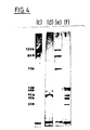
- Figure 4 shows an autoradiogram of SDS-PAGE.
- the peptides present in the viral antigen solutions are analyzed by cross-rocket immunoelectrophoresis and solubilization.
- lane (c) 20 ⁇ l of specific HSV-1 antigen labeled with 35S-methionine precipitated in the reference gel containing 15 ⁇ l / cm2 of rabbit anti-HSV-1 antibodies
- lane (d) 20 ⁇ l of specific HSV-2 antigen labeled with 35S-methionine precipitated in the reference gel containing 15 ⁇ l / cm2 of rabbit anti-HSV-2 antibodies.
- Figure 5 shows an autoradiogram of glycoproteins, labeled as indicated at the beginning of this example, separated by electrophoresis, originating from RL cells infected with HSV-1 in the case of band (h) and by HSV-2 in the case of band (i); the band (g) corresponds to the molecular weight markers, which are proteins labeled with carbon 14. The molecular weights of the proteins are indicated in kilodaltons to the left of the three bands of this Figure.
- the viral polypeptides which have precipitated [FIG. 4, bands (c) and (d)], are identified by their molecular weights estimated by electrophoretic migration relative to the standard proteins which mark molecular weight.
- the 125,000 dalton polypeptide, band (c) is identified as corresponding to the HSV-1 gC glycoprotein (Spear, 1976, Journal of Virology 17: 991-1008).
- the 95,000 dalton polypeptide also seen on this band probably represents a precursor of the gC glycoprotein, which is also precipitated in cross-rocket immunoelectrophoresis, by rabbit anti-HSV-1 hyperimmune sera, because the existence of common antigenic determinants.
- the 46,000 dalton polypeptide, band (d), is identified as corresponding to the HSV-2 gD glycoprotein, and for the same reasons as above, there is a 42,000 dalton polypeptide which is probably a precursor of the gD glycoprotein.
- Lanes (e) and (f) in Figure 4 show that the fractions containing type-specific antigens also include three or four antigens common to both types of virus.
- the radiolabelling of the viral glycoproteins by D- (1-14C) -glucosamine and the autoradiography of the glycoproteins separated by electrophoresis (FIG. 5) effectively show that the gD glycoprotein of HSV-2 (band i) has a molecular weight of 46,000. daltons and that the HSV-1 gC glycoprotein (h-band) has a molecular weight of 125,000 daltons.
- EXAMPLE 5 Correspondence between the polypeptides of 46,000 and 125,000 daltons and the glycoproteins gC of HSV-1 and gD of HSV-2.
- polypeptides collected from cells infected with HSV-1, by HSV-2, as well as control cells, are separated by electrophoresis on 7.5% polyacrylamide gel, as described in Example 4, then the proteins are transferred onto a nitrocellulose sheet (pore size: 0.22 m) at 220 mA and 60 V for 2 hours, using a Bio-Rad transfer cell (Towbin et al, 1979, Proceedings of the National Academy of Science, USA 79: 4350-4354). Before reacting the proteins with the corresponding monospecific antisera, the nitrocellulose sheets are blocked with PBS containing 3% bovine serum albumin.
- FIG. 6 shows the reaction, after transfer to the nitrocellulose sheet, between the polypeptides of HSV-1, of HSV-2 and of control cells and, on the one hand, the monospecific antiserum anti-polypeptide of 125,000 daltons, bands (j), (k), and (l), and on the other hand the 46,000 dalton anti-polypeptide monospecific antiserum, bands (m), (n) and (o).
- Lanes (j) and (m) correspond to the HSV-1 polypeptides
- bands (k) and (n) correspond to the control cell polypeptides
- bands (l) and (o) correspond to the HSV-2 polypeptides.
- the molecular weights are given to the left of the bands, in kilodaltons.
- Lanes (j) and (l) correspond to the reaction between the HSV-1 and HSV-2 polypeptides respectively, and the 125,000 dalton anti-polypeptide monospecific antiserum.
- This serum reacts strongly with the 125,000 dalton polypeptide of HSV-1, as shown in strip (j), and weakly with a 67,000 dalton polypeptide common to HSV-1 and HSV-2, as shown in strip ( l).
- the denaturation of certain antigenic determinants in the presence of SDS could contribute to the detection of the 67,000 dalton polypeptide common to HSV-1 and HSV-2 by the monospecific anti-HSV-1 antiserum.
- Lanes (m) and (o) correspond to the reaction between the polypeptides of HSV-1 and HSV-2 respectively and the monospecific antiserum anti-polypeptide of 46,000 daltons. The strongest reaction is observed in this case with the 46,000 dalton polypeptide of HSV-2, as shown in band (o), and a weak reaction is observed with a 60,000 dalton polypeptide such as shows it the strip (m).
- polypeptides of 125,000 and 46,000 daltons are identified as being respectively the glycoprotein gC of HSV-1 and the glycoprotein gD of HSV-2.
- anti-HSV-1 and anti-HSV-2 antibodies-immunoglobulins G (IgG) in human serum is carried out on polystyrene microtiter plates (Immunological Plates, Nunc, 1-96F, Denmark).
- the specific antigens of each of the two types diluted in 0.05 M carbonate buffer (pH 9.6), are respectively fixed on the plates by incubation at 4 ° C overnight. These plates are then washed twice in PBS-Tween 20 (0.05%), and 100 ⁇ l of PBS-Tween 20 (0.05%) containing 1% bovine serum albumin are added to each well in order to avoid non-specific links.
- the plates are incubated for one hour at 37 ° C., washed twice with PBS-Tween 20 (0.05%), then 120 ⁇ l of serum diluted in PBS-Tween 20 (0.05) is added to each well. %) containing 1% bovine serum albumin (the successive serum dolutions are multiplied by 2, and range from 1: 150 to 1: 19200).
- Each plate includes series of wells containing positive and negative test sera.
- the plates are incubated at 37 ° C. for two hours in a room in a humid atmosphere and washed four times in PBS-Tween, as above.
- a human anti-globulin IgG immunoglobulin is conjugated to horseradish peroxidase specific for the ⁇ chains (Dako, Denmark), and diluted 1: 1000 in PBS-Tween containing 1% bovine serum albumin. 120 ⁇ l of this conjugated antiserum are added to each well and incubated at 37 ° C for 1 hour in a humid chamber. After having washed the plates four times in PBS-Tween 20, 200 ⁇ l of substrate solution (10 mg of o-phenylenediamine, 0.03% H2O2 added immediately before use in 100 ml) are added to each well.
- substrate buffer at pH 5 containing 25.7 ml of 0.2 M disodium phosphate, 24.3 ml of 0.1 M citric acid, and 50 ml of deionized water).
- the reaction is completed by adding 50 ⁇ l per well of 3N HCl after 20 minutes.
- the absorbance is then measured at a wavelength of 492 nm on a Titertek Multiskan device (Flow Laboratories, England).
- Negative control sera are used to determine a limit value, defined as the mean specific absorbance of negative control sera increased by 3 standard deviations from this mean. Any serum with a specific absorbance above this limit value is considered positive for anti-virus antibodies.
- the IgM antibodies against Epstein-Barr virus were determined by an indirect immunofluorescence technique, using P3HR1 cells fixed on microscope slides and antibodies against human goat IgM, conjugated with fluorescein isothiocyanate at a dilution of 1: 100 (Pasteur Diagnostic France).
- the ELISA method was used with 3 anti-CMV sera, 3 anti-VZV sera or 3 anti-EBV sera, in the presence of fractions containing the specific antigens HSV-1 and HSV-2 ( Figure 8). No positive reaction was found in these sera, which clearly shows the specificity of the 2 antigenic fractions for the herpes virus.
- a serum is classified as negative for anti-virus antibodies if the absorbance values (at 492 nm) are less than 0.2, and for CSFs if these absorbance values are less than 0.15.
- Patients who are infected with HSV-1 or HSV-2 show responses to both homologous and heterologous antigens.
- Patients infected with HSV-1 have a strong specific type response, so the percentage of sera positive for the heterologous HSV-2 antigen is 4.5% compared to the total number of sera positive for with respect to the HSV-1 homologous antigen, and the percentage of LCR positive with respect to the HSV-2 heterologous antigen is 0% relative to the total number of LCR positive with respect to l HSV-1 homologous antigen (results are shown in Tables III and IV).
- patients infected with HSV-2 have a very strong common type response with regard to the sera, thus the percentage of sera positive with respect to the heterologous antigen HSV-1 is 71.4%. relative to the total number of sera positive with respect to the homologous antigen HSV-2, this response of common type being zero with regard to the CRL (the results are shown in Tables III and IV).
- LCR examined are negative with regard to the two antigens. LCRs are examined at dilutions ranging from 1:20 to 1: 2,560.
Landscapes
- Health & Medical Sciences (AREA)
- Life Sciences & Earth Sciences (AREA)
- Virology (AREA)
- Chemical & Material Sciences (AREA)
- Immunology (AREA)
- General Health & Medical Sciences (AREA)
- Medicinal Chemistry (AREA)
- Molecular Biology (AREA)
- Microbiology (AREA)
- Engineering & Computer Science (AREA)
- Mycology (AREA)
- Urology & Nephrology (AREA)
- Organic Chemistry (AREA)
- Veterinary Medicine (AREA)
- Public Health (AREA)
- Animal Behavior & Ethology (AREA)
- Biomedical Technology (AREA)
- Epidemiology (AREA)
- Pharmacology & Pharmacy (AREA)
- Hematology (AREA)
- Biochemistry (AREA)
- Biotechnology (AREA)
- Cell Biology (AREA)
- Physics & Mathematics (AREA)
- Analytical Chemistry (AREA)
- General Physics & Mathematics (AREA)
- Pathology (AREA)
- Gastroenterology & Hepatology (AREA)
- Food Science & Technology (AREA)
- Proteomics, Peptides & Aminoacids (AREA)
- Biophysics (AREA)
- Genetics & Genomics (AREA)
- Tropical Medicine & Parasitology (AREA)
- Peptides Or Proteins (AREA)
- Micro-Organisms Or Cultivation Processes Thereof (AREA)
- Measuring Or Testing Involving Enzymes Or Micro-Organisms (AREA)
- Preparation Of Compounds By Using Micro-Organisms (AREA)
Abstract
Application : Diagnostic spécifique du virus de l'herpès HSV-1 et/ou HSV-2 à l'aide desdites fractions.
Description
- La présente invention est relative à l'obtention de fractions contenant des antigènes spécifiques du virus de l'herpès type 1 (HSV-1) et type 2 (HSV-2), et plus particulièrement de la glycoprotéine gC de HSV-1 et de la glycoprotéine gD de HSV-2, ainsi qu'à l'utilisation de ces fractions pour le diagnostic différencié d'infections par HSV-1 et par HSV-2.
- Les types 1 (HSV-1) et 2 (HSV-2) du virus de l'herpès peuvent être différenciés par leur profil antigénique (Dowdle et al, 1967, Journal of Immunology, 99, p.974-980), et par leur composition génétique (Ludwig et al, 1972, Virology, 49, p.95-101). Lors d'une infection par le virus de l'herpès, HSV, les polypeptides structuraux du virus sont incorporés dans les membranes cellulaires (Norrild et al, 1979, Journal of Virology, 32, p.741-748) ; parmi ces polypeptides, les glycoprotéines spécifiques du virus vont former des cibles pour les réponses immunes humorales et cytotoxiques (Norrild, 1980, Current Topics in Microbiology and Immunology, 90, p.67-106). La détermination sérologique des virus de types 1 et 2, responsables de la réponse immunologique, peut être basée sur la détection d'anticorps dirigés contre une ou plusieurs de ces glycoprotéines. Néanmoins, les anticorps qui se développent contre les virus HSV-1 ou HSV-2 présentent une réactivité croisée intertypique très grande (Hampar et Martos, 1973, Immunological relationships, p.221-259 in AS Kaplan, The Herpes viruses). Cette forte réactivité croisée entre les virus HSV-1 et HSV-2 constitue le principal problème dans le sérodiagnostic des infections par le virus de l'herpès, ce problème étant dû à la difficulté d'obtenir des antigènes viraux présentant un degré de spécificité suffisant pour permettre un diagnostic différencié.
- Or, il est indispensable de pouvoir établir un tel diagnostic, notamment par exemple en ce qui concerne la pathologie la plus grave due au virus herpétique, à savoir l'encéphalomyélite, pour laquelle le virus de l'herpès type a été identifié comme antigène responsable de cette maladie.
- De nombreux tests sérologiques ont été décrits dans l'Art antérieur, en vue de distinguer les anticorps anti-HSV-1 des anticorps anti-HSV-2 dans le sérum humain. On peut ainsi citer les tests cinétiques de microneutralisation (Pauls and Dowdle, 1967, Journal of Immunology 98, p.941-947) et de neutralisation (Rawls et al, 1968, Herpes-virus type 2:association with carcinoma of the cervix, Science 161, p.1255-1256), l'hemagglutination indirecte (Fucilo et al, 1970, Proceedings of Society of Experimental Biology and Medicine 133, p. 735-739), les radioimmuno-essais en phase solide (Forganni et al, 1975, Journal of Clinical Microbiology 2, p.410-418), les radioimmuno-essais avec des protéines virales purifiées (Heilman et al, 1979, Journal of Virology 29, p.34-42), l'immunoélectrophorèse (Vestergaard, 1979, Infection and Immunity 23, p.553-558) et les méthodes immunoenzymatiques comme la technique ELISA (Vestergaard et Grauballe, 1979, Acta Pathologica Microbiologica Scandinavia, Section B 87 : p.261-263 ; Vestergaard, 1982, Developments in Biological Standardization 52, p.95-98).Mais ces tests ne permettent pas de distinguer nettement entre les infections par les virus HSV-1 et celles par les virus HSV-2, essentiellement par manque d'antigènes spécifiques du type, suffisamment purifiés. Il est donc important de pouvoir identifier de façon différenciée les virus des types HSV-1 et HSV-2 respectivement, responsables des deux types d'infections par HSV.
- Les glycoprotéines gC et gD sont fortement immunogènes et constituent les antigènes cibles présents dans les enveloppes virales et à la surface des cellules infectées (Norrild, 1980, Current Topics in Microbiology and Immunology 90,p.67-106). Bien que l'on ait pensé que la glycoprotéine gC de HSV-1 possédait seulement des épitopes spé cifiques du sérotype, il a été montré récemment que cette glycoprotéine peut aussi avoir des déterminants antigéniques responsables de réactions croisées (Zweig et al, 1983, Journal of Virology 47, p. 185-192 ; Zezulak et al, 1983, Journal of Virology 47, p.553-562). L'utilisation d'anticorps monoclonaux a mis en évidence que plusieurs polypeptides de HSV-1 et HSV-2, y compris les glycoprotéines gC et gD, expriment des déterminants antigéniques à réactivité croisée, aussi bien que des déterminants antigéniques spécifiques du type (Pereira et al, 1980, Infection and Immunity 29, p.724-732 ; Showalter et al, 1981, Infection and Immunity 34, p.684-692 ; Pereira et al, 1982, Infection and Immunity 35, p. 363-367 ; Zweig et al, 1983, Journal of Virology 47, p.185-192).
- Arvin et al (1983, Infection and Immunity 40, p.184-189), en utilisant la glycoprotéine gC de HSV-1 purifiée par immunoaffinité, Hampar et al (1983, Journal of Clinical Microbiology 21, p.496-500), en utilisant les glycoprotéines gC et gD de HSV-1 et gD et gF de HSV-2 purifiées par immunoaffinité également, de même que Svennerholm et al (1984, Herpes simplex virus type-selective enzyme-linked immunosorbent assay with Helix pomatia lectin-purified antigens), Suchankova et al (1984, Journal of Infectious Diseases 149, p.964-971), en utilisant des antigènes de HSV-1 (glycoprotéine gC) et de HSV-2 (polypeptide de 130 000 daltons), purifiés par la lectine de Helix pomatia, ont développé des tests immunoenzymatiques (ELISA) ou des radioimmunoessais avec ces différentes protéines pour la détection d'anticorps anti-HSV-1 et/ou anti-HSV-2. Bien que des protéines individuelles puissent, en principe, être utilisées comme antigène cible, des précautions particulières devraient être prises en cas d'utilisation de tels antigènes, car la variation antigénique observée d'une souche de virus herpétique à une autre peut conduire à des taux d'anticorps modifiés en réponse à des antigènes individuels. Ceci pourrait réduire la sensibilité des tests jusqu'à des niveaux trop faibles pour être interprétables et conduire à des diagnostics imprécis.
- En outre, toutes ces fractions contenant des antigènes spécifiques de type ont été préparées par des méthodes immunologiques ou par des procédés de chromatographie par affinité ; ces procédés sont longs à mettre en oeuvre, coûtent cher (exemple : la lectine d'Helix pomatia) ou dépendent de la disponibilité d'anticorps monoclonaux (exemple : les méthodes par immunoaffinité).
- La Demande de Brevet Européen (EP 0 183 215) propose une méthode pour la détermination d'anticorps anti-virus herpétique par un test d'agglutination passive, mais cette dernière est incapable de distinguer entre les infections par HSV-1 ou par HSV-2 et la sensibilité du test est en outre très faible.
- La technique ELISA est une méthode immunoenzymatique de titrage d'anticorps : un antigène fixé sur un support solide se lie avec l'anticorps spécifique contenu dans le milieu biologique à tester et l'immun-complexe obtenu est détecté par un anticorps anti-espèce marqué par une enzyme ; on ajoute ensuite un substrat spécifique de l'enzyme, dont la dégradation par l'enzyme fait apparaître un produit coloré détecté d'une manière appropriée.
- McEwen (Analytical Biochemistry, vol. 20, no1, Juillet 1967, p. 114-149) a mis au point une méthode de calcul de la vitesse de sédimentation de particules dans des gradients de densité de saccharose, dans laquelle il calcule le coefficient de sédimentation d'une particule dans un gradient linéaire de saccharose, à l'aide de la formule


- La présente invention s'est en conséquence donné pour but de pourvoir à un procédé d'obtention de fractions contenant des antigènes spécifiques du virus de l'herpès type 1 (HSV-1) et type 2 (HSV-2), et plus particulièrement la glycoprotéine gC de HSV-1 et la glycoprotéine gD de HSV-2, lequel procédé répond mieux aux nécessités de la pratique que les procédés d'obtention de fractions antigéniques antérieurement connus, notamment en ce qu'il est spécifique, fiable, peu onéreux, et permet l'obtention de fractions antigéniques à degré de spécificité élevé, pour la mise en oeuvre d'un test de diagnostic différencié d'une infection par le virus de l'herpès, qui est apte à déterminer avec précision une infection par HSV-1 ou par HSV-2.
- La présente invention a pour objet des fractions contenant des antigènes spécifiques respectivement du virus de l'herpès type 1 (HSV-1) et du virus de l'herpès type 2 (HSV-2), caractérisées en ce qu'elles contiennent des polypeptides présentant un poids moléculaire, déterminé par électrophorèse, respectivement de l'ordre de 125 000 daltons pour le polypeptide de HSV-1 et de l'ordre de 46 000 daltons pour le polypeptide de HSV-2, en ce que lesdits polypeptides réagissent positivement, respectivement avec de l'antisérum de lapin, ou d'un autre mammifère, anti-HSV-1 et avec de l'antisérum de lapin, ou d'un autre mammifère, anti-HSV-2 monospécifiques et en ce que lesdits polypeptides sont identifiés par immunotransfert comme étant la glycoprotéine gC de HSV-1 pour le polypeptide de 125 000 daltons et comme étant la glycoprotéine gD de HSV-2 pour le polypeptide de 46 000 daltons.
- Selon un mode de réalisation avantageux de la présente invention, lesdites fractions antigéniques sont obtenues par centrifugation dans un gradient linéaire de saccharose ou d'un autre milieu approprié,d'une suspension de virus HSV-1 et/ou HSV-2 intra- ou extracellulaire, préalablement lysée et désagrégée, les conditions de ladite centrifugation étant déterminées en fonction du coefficient de sédimentation calculé pour chacun des antigènes spécifiques du type concerné (1 ou 2).
- Selon une disposition avantageuse de ce mode de réalisation, la suspension de virus HSV-1 et/ou HSV 2 est elle-même obtenue à partir d'une culture de cellules de mammifères infectées par HSV-1 et HSV-2, centrifugée pour fournir une fraction soluble qui, après avoir été lysée et désagrégée, est soumise au processus susdit de centrifugation dans un gradient de saccharose.
- Selon une autre disposition avantageuse de la présente invention, lesdites fractions antigéniques présentent une concentration en protéines de l'ordre de 200 à 300 µg/ml.
- La présente invention a également pour objet un procédé pour l'isolement des fractions contenant les antigènes spécifiques HSV-1 et/ou HSV-2, définies dans ce qui précède, caractérisé en ce que l'on infecte des cellules d'un mammifère approprié, avec des cultures de virus HSV-1 et/ou HSV-2, dans lesquelles le virus se multiplie jusqu'à la lyse d'au moins une proportion importante des cellules, en ce que les cellules restantes sont lysées par traitement par des agents chimiques appropriés, puis leur paroi est désagrégée par traitement physique, notamment par ultrasons, et elles sont soumises à une centrifugation, en ce que la solution surnageante est centrifugée dans un gradient linéaire de saccharose ou d'un autre milieu approprié, et en ce que les fractions contenant les antigènes spécifiques HSV-1 et/ou HSV-2 sont récupérées par tous moyens appropriés.
- Selon un mode de réalisation avantageux du procédé conforme à la présente invention, les cultures de virus englobent aussi bien les suspensions virales obtenues après lyse totale des cellules, que les cultures de cellules contenant le virus, et qu'un mélange de cellules infectées et de virus libéré des cellules lysées.
- Selon un mode de réalisation avantageux du procédé conforme à la présente invention, les polypeptides doués de propriétés antigéniques HSV-1 et/ou HSV-2 contenus dans les fractions antigéniques susdites sont isolés sur un gradient linéaire de saccharose ou d'un autre milieu approprié, déterminé en fonction de leur coefficient de sédimentation calculé.
- Selon une disposition avantageuse de ce mode de réalisation, le coefficient de sédimentation est calculé d'après la formule

- Selon une modalité avantageuse de cette disposition, le coefficient de sédimentation calculé est compris entre 9 et 9,6 S pour la glycoprotéine gC.
- Selon une autre modalité avantageuse de cette disposition, le coefficient de sédimentation calculé est compris entre 4,3 et 4,5 S pour la glycoprotéine gD.
- Selon un mode de réalisation avantageux du procédé conforme à la présente invention, le gradient linéaire de saccharose pour l'isolement des fractions spécifiques HSV-1 et/ou HSV-2 est réalisé à des concentrations de saccharose comprises entre 6 et 27 % du poids total de la solution de saccharose.
- Selon une disposition avantageuse de ce mode de réalisation, la concentration de saccharose nécessaire pour isoler la glycoprotéine gC de HSV-1 est comprise préférentiellement entre 10 et 27 % du poids total de la solution de saccharose.
- Selon une autre disposition avantageuse de ce mode de réalisation, la concentration de saccharose nécessaire pour isoler la glycoprotéine gD de HSV-2 est comprise préférentiellement entre 6 et 19 % du poids total de la solution de saccharose.
- Selon un mode de réalisation avantageux du procédé conforme à la présente invention, les fractions contenant les antigènes spécifiques HSV-1 et/ou HSV-2 sont identifiées séparément par électrophorèse et immunotransfert, chaque fraction étant respectivement recueillie dans la fraction de soluté contenant de 6 à 10 % de saccharose par rapport au poids total de la solution de saccharose pour le polypeptide de 46 000 daltons, et dans la fraction de soluté contenant de 20 à 27 % de saccharose par rapport au poids total de la solution de saccharose pour le polypeptide de 125 000 daltons.
- La présente invention a en outre pour objet une méthode de diagnostic des infections par le virus de l'herpès des types 1 ou 2, par détermination d'anticorps anti-HSV-1 et/ou anti-HSV-2 dans un milieu biologique, caractérisé en ce que l'on met en contact des fractions contenant les antigènes spécifiques HSV-1 et/ou HSV-2 conformes à la présente invention, fixées d'une manière appropriée sur un support solide approprié, avec le milieu biologique présumé contenir des anticorps anti-HSV-1 et/ou anti-HSV-2 examiné, en ce que l'immun-complexe obtenu dans le cas où des anticorps anti-HSV-1 et/ou anti-HSV-2 sont effectivement présents, est détecté par un anticorps anti-espèce marqué par une enzyme, en présence d'un substrat spécifique de l'enzyme.
- Selon un mode de réalisation avantageux de la méthode de diagnostic conforme à la présente invention, les fractions contenant les antigènes spécifiques HSV-1 et/ou HSV-2 sont fixées sur ledit support à des dilutions avantageusement comprises entre 0,375 et 6 µg de protéines par ml.
- Selon un autre mode de réalisation avantageux de la méthode de diagnostic conforme à la présente invention, le support solide est choisi dans le groupe des supports comprenant notamment les microplaques, les tube, les globules rouges, les billes de polystyrène et les feuilles de nitrocellulose.
- La présente invention a également pour objet un ensemble, ou kit, de diagnostic, pour la détermination, dans des liquides biologiques corporels, d'anticorps anti-HSV-1 et/ou anti-HSV-2, qui comprend comme composants principaux :
- une quantité appropriée, éventuellement subdivisée en doses unitaires, de différentes dilutions de fractions conformes à la présente invention contenant les antigènes spécifiques HSV-1 et de fractions contenant les antigènes spécifiques HSV-2 ;
- une quantité appropriée, éventuellement subdivisée en doses unitaires, suffisante pour la détermination précédente, d'anticorps anti-espèce marqués par une enzyme, pour la révélation de la réaction antigène-anticorps entre l'anticorps à déterminer et l'antigène spécifique correspondant ;
- éventuellement une quantité appropriée de tampons, diluants, réactifs, nécessaires à la mise en oeuvre de la méthode de diagnostic conforme à la présente invention. - L'invention sera mieux comprise à l'aide du complément de description qui va suivre, qui se réfère aux dessins annexés et à des exemples de mise en oeuvre du procédé objet de la présente invention.
- Il doit être bien entendu, toutefois, que ces dessins, parties descriptives correspondantes et exemples de mise en oeuvre sont donnés uniquement à titre d'illustration de l'objet de l'invention, dont ils ne constituent en aucune manière une limitation.
- - Des cellules primaires de rein de lapin (RL) sont préparées à partir de lapins âgés de 21 jours par des méthodes connues (Kuchler, 1977, Biochemical methods in cell culture and Virology, p.3-15). Ces cellules sont transformées trois fois au maximum avant leur utilisation. Des cellules MR-5 (ATCC, Rockville) sont cultivées comme les cellules RL dans du milieu Dulbecco (Laboratoires Flow) additionné de 10 % de sérum de veau foetal (Gibco), 100 µg de streptomycine/ml, 100 unités de pénicilline/ml et 0,2 % de bicarbonate de sodium.
- - On injecte aux cellules RL, des souches de virus herpétique type 1 (HSV-1) et type 2 (HSV-2) (ATCC souches VR/733 et VR/734 respectivement) à une multiplicité d'infection (MOI) égale à 0,01 particule virale infectieuse par ml. Le stock viral est obtenu dans le liquide de culture extracellulaire lorsque le virus a lysé la totalité des cellules. Le liquide de culture est alors centrifugé à 1000 g pendant 10 minutes et le surnageant est réparti en aliquots de 2 ml et congelé à -70°C.
- Les cellules monocouches RL lysées entre 3+ et 4+ 24 heures après l'infection sont lavées deux fois dans du PBS froid (137 mM de NaCl, 3 mM de KCl, 8 mM de Na₂HPO₄, 2 mM de KH₂PO₄, pH 7,4) contenant des inhibiteurs de protéases comme la tosyl-lysyl-chlorométhylcétone, le p-hydroxy mercuribenzoate et le fluorure de phénylméthylsulfonyle (Sigma Chemicals) à une concentration finale de 1 mM. Puis les cellules sont grattées dans de la glycine froide à 0,020 M et du tampon Tris à 0,0076 M (pH 8,6) contenant 3 % (vol./vol.) de Triton X-100 et des inhibiteurs de protéases comme indiqués ci-dessus. Après un séjour de 30 minutes sur de la glace, les cellules lysées sont désagrégées par des ultrasons (18 kc/sec, deux fois pendant 15 secondes sur de la glace), puis centrifugées à 1000 g pendant 10 minutes. Les surnageants sont centrifugés à 100 000 g pendant une heure. Le surnageant des préparations d'antigène pour HSV-1 et HSV-2 et des cellules témoins sont échantillonnés et stockés à -70°C. La concentration en protéines dans les fractions solubles est déterminée par la méthode de Lowry modifiée (Journal of Biological Chemistry 192:265-275)) pour effectuer la mesure dans des so lutions contenant du Triton X-100 (Dulley et Grieve, 1975, Analytical Biochemistry 64:136-141) ; la concentration en protéines des préparations d'antigènes solubilisées est ajustée à 10 mg/ml.
- Ces fractions solubles sont dialysées contre du tampon TNE (0,01 M de Tris-HCl, pH 7,4 ; 0,01 M de NaCl ; 0,001 M d'EDTA) pendant une nuit à +4°C. Puis 0,4 ml (4 mg) sont prélevés par tube, et ceci pour chaque préparation d'antigènes HSV-1, HSV-2 et des cellules témoins, et ils sont déposés sur 12,3 ml d'un gradient linéaire de saccharose préparé dans du tampon TNE. Pour la préparation des antigènes spécifiques HSV-1 et HSV 2, on réalise un gradient linéaire de saccharose de 10 à 27 % de saccharose par rapport au poids total du soluté pour HSV-1 et de 6 à 19 % de saccharose par rapport au poiids du soluté pour HSV-2. Les gradients sont ensuite centrifugés pendant 24 heures (40 000 tours/minute, +5°C, rotor SW 41 Ti, Bechman) puis on recueille 0,4 ml de fractions contenant les antigènes spécifiques qui vont être soumises à une immunoélectrophorèse en fusée croisée avec un gel intermédiaire (Portocala et coll., 1983, Analysis of centrifugation gradients by fused rocket immunoelectrophoresis, 82, p.587-597).
- Des anticorps anti-HSV-1 et anti-HSV-2 polyspécifiques sont obtenus après infection de lapins par injections sous-cutanées successives de préparations d'antigènes (provenant des cellules RL infectées de l'exemple 1) mélangées à volume égal avec de l'adjuvant incomplet de Freund (2 x 0,1 ml) ; les injections se font une fois par mois pendant 12 mois. Les immunoglobulines de lapins sont ensuite purifiées (Harboe et Ingild, 1973, Scandinavian Journal of Immunology 2 (suppl.1):161-164) et leur concentration ajustée à 40 mg/l.
- Les anticorps anti-HSV-1 et anti-HSV-2 sont disposés dans 1 % de gel d'agarose HSB (Litex, Danemark) avec du tampon Tris-tricine (9,7 g Tris, 4,3 g Tricine, 0,92 g de lactate de calcium, 0,2 g de NaN₃, pH 8,6) et 1 % (vol./vol.) de Triton X-100. La quantité d'antigène solubilisé mise en oeuvre est de 7 µl pour l'immunoélectrophorèse en fusée et de 20 µl pour l'immunoélectrophorèse en fusée croisée. L'opération se déroule dans les conditions suivantes : 1,5 V/cm pendant 18 heures à 16°C (Axelsen, 1973, Scandinavian Journal of Immunology 2(suppl.1):p.71-77). Les résultats sont représentés aux Figures 1 et 2 annexées.
- La Figure 1 montre une immunoélectrophorèse en fusée avec un gel intermédiaire d'une préparation d'antigènes HSV-1 fractionnée. 29 fractions ont été analysées, de la fraction 1 sur la gauche à la fraction 29 située à l'extrême droite. Les anticorps présents dans le gel intermédiaire Gi proviennent de sérum de lapin hyperimmun vis-à-vis de HSV-2 (25 µl/cm²) et dans le gel de référence Gr, on trouve le sérum de lapin hyperimmun vis-à-vis de HSV-1 (10 µl/cm²). Le gel a été coloré par du Bleu brillant de Coomassie.
- La Figure 2 montre une immunoélectrophorèse en fusée avec un gel intermédiaire d'une préparation d'antigènes HSV-2 fractionnée. 29 fractions ont été analysées, de la fraction 30 sur la gauche à la fraction 59 située à l'extrême droite. Les anticorps présents dans le gel intermédiaire Gi proviennent de sérum de lapin hyperimmun vis-à-vis de HSV-1 (25 µl/cm²) et dans le gel de référence Gr, on trouve le sérum de lapin hyperimmun vis-à-vis de HSV-2 (10 µl/cm²). Le gel a été coloré par du Bleu brillant de Coomassie.
- L'immunoélectrophorèse en fusée avec un gel intermédiaire d'antigènes HSV-1 ou HSV-2 révèle la distribution d'un antigène identifié dans le gel de référence (qui est superposé au gel intermédiaire) et le degré de contamination avec d'autres antigènes (gel intermédiaire) de ces fractions. Les préparations de cellules témoins ne donnent pas de pics dans le gel de référence. Les fractions qui montrent des pics dans le gel de référence sont les fractions contenant les antigènes spécifiques. La concentration en protéines de chaque fraction varie de 200 à 300 µg/ml.
- - On utilise la méthode de McEwen pour estimer le coefficient de sédimentation d'une particule dans un gradient linéaire de saccharose. On utilise les fractions détectées par immunoélectrophorèse en fusée dans l'exemple 2. Deux ou trois de ces fractions présentant les plus hauts pics sont recueillies pour chaque type d'antigène et leur concentration en saccharose est mesurée. Dans la formule

vitesse : 40 000 tours/minute,
durée : 24 heures,
température : +5°C. - Les valeurs de Z₁ et Z₂ sont respectivement de 10 et 27 %.
- Dans la formule de McEwen, on trouve ainsi :

- La valeur la plus proche de - 3,4 dans les Tables de McEwen est : - 5.
- Pour calculer le coefficient de sédimentation, on utilise la formule

w² = (0,10472xtours/minute)² = (0,10472x40000)² = 1,75x10⁷sec⁻² t = 24 heures = 86 400 sec.
w²t = 1,75 x 10⁷ x 86,400 = 1,5 x 10¹² sec⁻¹ - Le Tableau I ci-dessous montre les valeurs de I₂ et I₁ calculées d'après les Tables de McEwen pour une valeur de Zx de 19 % et une valeur de Zy de 10 %, et le coefficient de sédimentation obtenu pour des densités de protéines égales à 1,3 et 1,4.

- Les valeurs de Z₁ et Z₂ sont respectivement de 6 et 19 %.

- La valeur la plus proche de - 4,24 dans les Tables de McEwen est : - 5.
- Pour calculer le coefficient de sédimentation, on utilise la formule

- Le Tableau II ci-dessous montre les valeurs I₂ et I₁ calculées d'après les Tables de McEwen pour une valeur de Zx de 10 % et une valeur de Zy de 6 %, et le coefficient de sédimentation obtenu pour des densités de protéines égales à 1,3 et 1,4.

- Des monocouches confluentes de cellules RL sont infectées suivant la procédure de l'exemple 1 à une MOI de 5. Puis ces cellules sont marquées 4 à 20 heures après l'infection, soit avec 10 µCi de ³⁵S-méthionine par ml (1,370 Ci/mmole) provenant d'un milieu contenant 0,05 % en concentration de méthionine froide, soit avec 0,8 µCi de D-(1-¹⁴C)-glucosamine par ml (54 mCi/mmole) provenant d'un milieu contenant 10 % de glucose (les radioisotopes proviennent de la Société Amersham, Angleterre).
- Les antigènes spécifiques de type marqués comme décrit ci-dessus, sont soumis à une immunoélectrophorèse en fusée croisée à travers un gel intermédiaire. On découpe et recueille 15 pics dans le gel de référence et les polypeptides contenus dans ces pics sont élués dans 400 µl d'un tampon Tris-H₃PO₄ 0,06 M (pH 6,9) contenant 2 % (en poids/vol.) de dodécylsulfate de sodium (SDS) et 2,5 % (vol./vol.) de 2-mercaptoéthanol (Norrild et coll., 1977, Analytical Biochemistry 81:432-441).
- Des plaques de gel utilisant le système de gel de polyacrylamide discontinu sont employées (Laemmli ; 1970, Nature 227:680-685). La concentration de polyacrylamide est de 7,5 % (poids/vol.) dans le gel de séparation et de 3,5 % dans le gel apparent. Les deux gels sont réticulés avec 0,2 % (poids/vol.) de N,Nʹ-méthylènebisacrylamide. Les échantillons sont soumis à l'électrophorèse pendant 20 heures à 7 mA. Pour déterminer le poids moléculaire, on utilise comme marqueurs, des protéines marquées au carbone 14 (fournis par Amersham, Angleterre), telles que la myosine méthylée (PM 200 000), la méthylphosphorylase b (PM 100 000 et 92 500), la méthyl-sérumalbumine bovine (PM 69 200), la méthylovalbumine (PM 46 000), la méthyl-anhydrase carbonique (PM 30 000) et le méthyllysozyme (PM 14 300). Les protéines radiomarquées sont visualisées par autora diographie effectuée sur un film Kodak-X Omat S et exposée pendant 4 à 6 jours à la température de -70°C. Les résultats sont représentés aux Figures 3, 4 et 5 annexées.
- La Figure 3 montre une immunoélectrophorèse en fusée croisée à travers un gel intermédiaire, de préparations d'antigènes HSV-1 et HSV-2 fractionnées. La bande (a) correspond à une fraction contenant l'antigène spécifique du type HSV-2. Les anticorps présents dans le gel intermédiaire Gi proviennent de sérum de lapin hyperimmun anti-HSV-1, tel que préparé dans l'exemple 2 (25 µl/cm²) et dans le gel de référence Gr, on trouve le sérum de lapin hyperimmun anti-HSV-2 (15 µl/cm²). La bande (b) correspond à la fraction contenant l'antigène spécifique du type HSV-1. Les anticorps présents dans le gel intermédiaire Gi proviennent de sérum de lapin hyperimmun anti-HSV-2 (25 µl/cm²) et dans le gel de référence Gr, on trouve du sérum de lapin hyperimmun anti-HSV-1 (15 µl/cm²). Les gels sont colorés par du Bleu brillant de Coomassie.
- La Figure 4 montre un autoradiogramme de SDS-PAGE. Les peptides présents dans les solutions d'antigènes viraux sont analysés par immunoélectrophorèse en fusée croisée et solubilisation. Dans la bande (c), 20 µl d'antigène spécifique HSV-1 marqué à la ³⁵S-méthionine ont précipité dans le gel de référence contenant 15 µl/cm² d'anticorps de lapin anti-HSV-1 ; dans la bande (d), 20 µl d'antigène spécifique HSV-2 marqué par la ³⁵S-méthionine ont précipité dans le gel de référence contenant 15 µl/cm² d'anticorps de lapin anti-HSV-2. Puis 10 µl de chaque fraction contenant les antigènes spécifiques du type HSV-1 ou HSV-2 sont soumis à une électrophorèse SDS-PAGE en l'absence d'antisérum et les bandes (e) et (f) montrent les résultats obtenus. Les poids moléculaires des protéines sont indiqués en kilodaltons à gauche des quatre bandes représentées à la Figure 4.
- La Figure 5 représente un autoradiogramme de glycoprotéines, marquées comme indiqué au début du présent exemple, séparées par électrophorèse, provenant de cellules RL infectées par HSV-1 dans le cas de la bande (h) et par HSV-2 dans le cas de la bande (i) ; la bande (g) correspond aux marqueurs de poids moléculaire, qui sont des protéines marquées au carbone 14. Les poids moléculaires des protéines sont indiqués en kilodaltons à gauche des trois bandes de cette Figure.
- Les polypeptides viraux ayant précipité [ Figure 4 , bandes (c) et (d)],sont identifiés par leurs poids moléculaires estimés par migration électrophorétique par rapport aux protéines standards marqueurs de poids moléculaire. Lwe polypeptide de 125 000 daltons, bande (c), est identifié comme correspondant à la glycoprotéine gC de HSV-1 (Spear, 1976, Journal of Virology 17:991-1008). Le polypeptide de 95 000 daltons que l'on voit également sur cette bande représente probablement un précurseur de la glycoprotéine gC, qui est également précipité dans l'immunoélectrophorèse en fusée croisée, par des sérums hyperimmuns anti-HSV-1 de lapin, du fait de l'existence de déterminants antigéniques communs. Le polypeptide de 46 000 daltons, bande (d), est identifié comme correspondant à la glycoprotéine gD de HSV-2, et pour les mêmes raisons que ci-dessus, on trouve un polypeptide de 42 000 daltons qui est probablement un précurseur de la glycoprotéine gD. Les bandes (e) et (f) de la Figure 4 montrent que les fractions contenant les antigènes spécifiques des types comprennent aussi trois ou quatre antigènes communs aux deux types de virus. Le radiomarquage des glycoprotéines virales par la D-(1-¹⁴C)-glucosamine et l'autoradiographie des glycoprotéines séparées par électrophorèse (Figure 5) montrent effectivement que la glycoprotéine gD de HSV-2 (bande i) a un poids moléculaire de 46 000 daltons et que la glycoprotéine gC de HSV-1 (bande h) a un poids moléculaire de 125 000 daltons.
- Pour prouver que les polypeptides de 46 000 et 125 000 daltons correspondent bien respectivement aux glycoprotéines gD de HSV-2 et gC et HSV-1, on a procédé comme suit :
- Les parties supérieures des immunoprécipités obtenus à l'exemple 4 dans le gel de référence [ Figure 3, bandes (a) et (b)], contenant les antigènes spécifiques des types, sont prélevées du gel. Lesdits précipités isolés sont désagrégés par ultrasons (20 kc/sec., deux fois sur de la glace pendant 30 secondes) puis mélangés volume à volume avec de l'adjuvant incomplet de Freund, puis injectés à des lapins, à quatre reprises à des intervalles de deux semaines entre chaque injection (Vestergaard, Norrild, 1978, Journal of Infectious Diseases 138:639-643). Les immunoglobulines obtenues sont purifiées et leur concentration en protéines ajustée à 30 mg/ml.
- - Les polypeptides recueillis à partir des cellules infectées par HSV-1, par HSV-2, ainsi que des cellules témoins, sont séparés par électrophorèse sur gel de polyacrylamide à 7,5 %, comme décrit dans l'exemple 4, puis, les protéines sont transférées sur une feuille de nitrocellulose (dimension des pores : 0,22 m) à 220 mA et à 60 V pendant 2 heures, en utilisant une cellule de transfert Bio-Rad (Towbin et al, 1979, Proceedings of the National Academy of Science, USA 79:4350-4354). Avant de faire réagir les protéines avec les antisérums monospécifiques correspondants, les feuilles de nitrocellulose sont bloquées par du PBS contenant 3 % de sérumalbumine bovine. Après réaction des antisérums monospécifiques de lapin avec la feuille de nitrocellulose (dilution (1/400), les anticorps fixés sont détectés à l'aide de protéine A marquée à l'iode 125 et autora diographiés pendant 16 heures à -70°C. On utilise la protéine A du staphylocoque doré (Pharmacia Fine Chemicals), marquée par de l'iode 125 (Amersham) par la méthode à la lactopéroxydase (Marchalonis, 1969, Biochemistry Journal 113: 299-305). Les résultats sont représentés à la Figure 6 annexée.
- La Figure 6 montre la réaction, après transfert sur la feuille de nitrocellulose, entre les polypeptides de HSV-1, de HSV-2 et de cellules témoins et, d'une part l'antisérum monospécifique anti-polypeptide de 125 000 daltons, bandes (j), (k), et (l), et d'autre part l'antisérum monospécifique anti-polypeptide de 46 000 daltons, bandes (m), (n) et (o). Les bandes (j) et (m) correspondent aux polypeptides de HSV-1, les bandes (k) et (n) correspondent aux polypeptides des cellules témoins, et les bandes (l) et (o) aux polypeptides de HSV-2. Les poids moléculaires sont donnés à gauche des bandes, en kilodaltons.
- Les bandes (j) et (l) correspondent à la réaction entre les polypeptides de HSV-1 et de HSV-2 respectivement, et l'antisérum monospécifique anti-polypeptide de 125 000 daltons. Ce sérum réagit fortement avec le polypeptide de 125 000 daltons de HSV-1, comme le montre la bande (j), et faiblement avec un polypeptide de 67 000 daltons commun à HSV-1 et HSV-2, comme le montre la bande (l). La dénaturation de certains déterminants antigéniques en présence du SDS pourrait contribuer à la détection du polypeptide de 67 000 daltons commun à HSV-1 et HSV-2 par l'antisérum monospécifique anti-HSV-1.
- Les bandes (m) et (o) correspondent à la réaction entre les polypeptides de HSV-1 et HSV-2 respectivement et l'antisérum monospécifique anti-polypeptide de 46 000 daltons. La réaction la plus forte est observée dans ce cas avec le polypeptide de 46 000 daltons de HSV-2, comme le montre la bande (o), et une faible réaction est observée avec un polypeptide de 60 000 daltons comme le montre la bande (m). Les polypeptides provenant des cellules témoins, comme le montrent les bandes (k) et (n),ne réagissent avec aucun des antisérums monospécifiques.
- Il est ainsi démontré que les polypeptides de 125 000 et 46 000 daltons sont identifiés comme étant respectivement la glycoprotéine gC de HSV-1 et la glycoprotéine gD de HSV-2.
- La détermination des anticorps-immunoglobulines G (IgG) anti-HSV-1 et anti-HSV-2 dans le sérum humain, est réalisée sur des plaques de microtitration en polystyrène (Plaques Immunologiques, Nunc, 1-96F, Danemark). Les antigènes spécifiques de chacun des deux types, dilués dans du tampon carbonate 0,05 M (pH 9,6), sont respectivement fixés sur les plaques par incubation à 4°C pendant toute une nuit. Ces plaques sont ensuite lavées deux fois dans du PBS-Tween 20 (0,05 %), et 100 µl de PBS-Tween 20 (0,05 %) contenant 1 % de sérumalbumine bovine sont ajoutés à chaque puits afin d'éviter les liaisons non spécifiques. Les plaques sont incubées pendant une heure à 37°C, lavées à deux reprises avec du PBS-Tween 20 (0,05 %), puis on ajoute à chaque puits 120 µl de sérum dilué dans du PBS-Tween 20 (0,05 %) contenant 1 % de sérumalbumine bovine (les dolutions de sérum successives sont multipliées par 2, et vont de 1:150 à 1:19200). Chaque plaque comprend des séries de puits contenant des sérums témoisn positifs et négatifs. Les plaques sont incubées à 37°C pendant deux heures dans une chambre en atmosphère humide et lavées quatre fois dans du PBS-Tween, comme précédemment.
- Une immunoglobuline IgG anti-globuline humaine est conjuguée à de la péroxydase de raifort spécifique des chaines γ (Dako, Danemark), et diluée au 1:1000 dans du PBS-Tween contenant 1 % de sérumalbumine bovine. On ajoute 120 µl de cet antisérum conjugué à chaque puits et on incube à 37°C pendant 1 heure en chambre humide. Après avoir lavé les plaques quatre fois dans du PBS-Tween 20, on ajoute à chaque puits 200 µl de solution de substrat (10 mg d'o-phénylènediamine, 0,03 % d'H₂O₂ ajoutée immédiatement avant l'emploi dans 100 ml de tampon de substrat à pH 5 contenant 25,7 ml de phosphate disodique 0,2 M, 24,3 ml d'acide citrique 0,1 M, et 50 ml d'eau désionisée). La réaction est achevée par ajout de 50 µl par puits d'HCl 3N au bout de 20 minutes. On mesure ensuite l'absorbance à une longueur d'onde de 492 nm sur un appareil Titertek Multiskan (Laboratoires Flow, Angleterre).
- Les sérums témoins négatifs sont utilisés pour déterminer une valeur limite, définie comme l'absorbance spécifique moyenne des sérums témoins négatifs augmentée de 3 déviations standards à partir de cette moyenne. Tout sérum à absorbance spécifique supérieure à cette valeur limite est considéré comme positif pour les anticorps anti-virus.
- Trois groupes de patients ont été examinés.
- a) 1er groupe : des échantillons de sérum provenant de 328 patients hospitalisés dans différents hôpitaux d'Athènes (âge moyen de 43 ans, de 7 à 68 ans).
- b) 2ème groupe : des échantillons de liquide céphalo-rachidien (LCR) provenant de 92 patients présentant divers troubles neurologiques.
- c) 3ème groupe : des échantillons de sérum provenant de 29 patients présentant un herpès génital récurrent (15 sérums) ou labial (14 sérums) reconnus par les méthodes usuelles.
- En outre, des sérums ont été obtenus de 3 patients présentant une infection par le virus cytomégalique (CMV), 3 patients présentant une infection par le virus (VZV), et 3 patients présentant une infection par le virus d'Epstein-Barr (EBV), infections démontrées par une augmentation significative du titre d'anticorps par le test de fixation du complément (CMV, VZV) en utilisant des antigènes capables de fixer le complément (commercialités par les Laboratoires Virion Diagnostic, Suisse) et par détection d'anticorps IgM anti-EBV. Le test de fixation du complément est réalisé selon ma microtechnique de Casey (1965,DC:US Public Health Service, p. 31-34). Les anticorps IgM anti-virus d'Epstein-Barr ont été déterminés par une technique d'immunofluorescence indirecte, à l'aide de cellules P₃HR₁ fixées sur des lames de microscope et des anticorps anti-IgM humain de chèvre, conjugués avec de l'isothiocyanate de fluoresceine à une dilution de 1:100 (Pasteur Diagnostic France).
- Les résultats obtenus avec les différents liquides biologiques tels que décrits ci-dessus sont représentés par des courbes (Figures 7 et 8 annexées).
- La Figure 7 représente les courbes d'absorbance obtenues par réaction de fractions antigéniques HSV-1 et HSV-2 avec des immun-sérums : l'absorbance est donnée en ordonnée et les dilutions, en µ g/ml, des fractions antigéniques spécifiques des types HSV-1 et HSV-2, sont données en abscisse.
- Courbe I : fraction antigénique HSV-1 + sérum anti-HSV-1
- Courbe II : fraction antigénique HSV-2 + sérum anti-HSV-2
- Courbe III : fraction antigénique HSV-1 + sérum anti-HSV-2
- Courbe IV : fraction antigénique HSV-2 + sérum anti-HSV-1
- Courbe V : fraction antigénique HSV-1 + sérum témoin
- Courbe VI : fraction antigénique HSV-2 + sérum témoin.
- La Figure 8 représente les courbes d'absorbance obtenues par réaction de fractions antigéniques HSV-1 et HSV-2 avec des immun-sérums : l'absorbance est donnée en ordonnée et les dilutions des immun-sérums utilisés, sont données en abscisse.
- Courbe VII : fraction antigénique HSV-1 + sérum antiHSV-1
- VIII: fraction antigénique HSV-1 + sérum antiHSV-1 différent de celui de la courbe VII
- Courbe IX : fraction antigénique HSV-2 + sérum antiHSV-2
- Courbe X : fraction antigénique HSV-2 + sérum antiHSV-2 différent de celui de la courbe IX
- Courbe XI : fraction antigénique HSV-1 + sérum anti-EBV
- Courbe XIʹ : fraction antigénique HSV-2 + sérum anti-EBV
- Courbe XII : fraction antigénique HSV-1 + sérum anti-VZV
- Courbe XIIʹ : fraction antigénique HSV-2 + sérum anti-VZV
- Courbe XIII : fraction antigénique HSV-1 + sérum anti-CMV
- Courbe XIIIʹ : fraction antigénique HSV-2 + sérum anti-CMV
- Courbe XIV : valeur-limite.
- Sur les microplaques où l'on a fixé à différentes dilutions les fractions contenant les antigènes spécifiques HSV-1 ou HSV-2 (dilutions de 6 à 0,375 µg de protéines/ml), on réalise une détection immunoenzymatique desdits antigènes par la méthode ELISA, avec une dilution constante (1:150) de sérums humains de référence HSV-1 et HSV-2 positifs. Les dilutions d'angigène qui conduisent aux plus grands écarts entre le sérum de référence HSV-1 et HSV-2 sont déterminés à partir des courbes de dilutions standards (Figure 7). Pour cribler les sérums humains, on utilise une dilution d'antigène correspondant à 1,5 µg/ml aussi bien pour HSV-1 que pour HSV-2. Les courbes de la Figure 7 montrent la réactivité entre des fractions contenant les antigènes spécifiques de HSV-1 et HSV-2 avec les différents sérums employés quand on met en oeuvre la méthode ELISA.
- Pour montrer la spécificité des fractions antigéniques par rapport aux anticorps anti-HSV-1 ou anti-HSV-2, on a mis en oeuvre la méthode ELISA avec 3 sérums anti-CMV, 3 sérums anti-VZV ou 3 sérums anti-EBV, en présence de fractions contenant les antigènes spécifiques HSV-1 et HSV-2 (Figure 8). Aucune réaction positive n'a été trouvée dans ces sérums, ce qui montre bien la spécificité des 2 fractions antigéniques pour le virus herpétique.
- Un total de 328 sérums humains et de 92 liquides céphalo-rachidiens (LCR) a été testé par la méthode ELISA, la mesure de l'absorbance ayant été utilisée pour déterminer la présence ou l'absence d'anticorps anti-HSV-1 et anti-HSV-2 dans ces liquides biologiques. Un sérum est classé comme négatif pour les anticorps anti-virus si les valeurs de l'absorbance (à 492 nm) sont inférieures à 0,2, et pour les LCR si ces valeurs d'absorbance sont inférieures à 0,15. Les patients qui sont infectés par HSV-1 ou par HSV-2 présentent à la fois des réponses aux antigènes homologues et hétérologues. Les patients infectés par HSV-1 présentent une forte réponse de type spécifique, ainsi le pourcentage de sérums positifs vis-à-vis de l'antigène hétérologue HSV-2 est de 4,5 % par rapport au nombre total de sérums positifs vis-à-vis de l'antigène homologue HSV-1, et le pourcentage de LCR positifs vis-à-vis de l'antigène hétérologue HSV-2 est de 0 % par rapport au nombre total de LCR positifs vis-à-vis de l'antigène homologue HSV-1 (les résultats sont représentés sur les Tableaux III et IV). Par contre, les patients infectés par HSV-2 présentent une très forte réponse de type commun en ce qui concerne les sérums, ainsi le pourcentage de sérums positifs vis-à-vis de l'antigène hétérologue HSV-1 est de 71,4 % par rapport au nombre total de sérums positifs vis-à-vis de l'antigène homologue HSV-2, cette réponse de type commun étant nulle en ce qui concerne les LCR (les résultats sont représentés sur les Tableaux III et IV).
- La mise en oeuvre de la méthode ELISA avec 14 sérums humains, présentant une infection reconnue par HSV-1 et avec 15 sérums humains présentant une infection reconnue par HSV-2, en présence de préparations d'antigène commun de type HSV-1 et HSV-2 et en présence de fractions contenant les antigènes spécifiques HSV-1 et HSV-2, révèle que tous ces sérums réagissent contre les antigènes communs des types HSV-1 et HSV-2, ainsi que le montrent les Tableaux V et VI. Néanmoins, des titres positifs des sérums HSV-1 et HSV-2 sont observés uniquement en présence de fractions contenant les antigènes spécifiques HSV-1 et HSV-2 respectivement. Quatre sérums (28,5 %) de patients présentant une infection par HSV-1 contiennent aussi des anticorps contre les fractions spécifiques HSV-2. Onze sérums (73,3 %) de patients présentant une infection par HSV-2 contiennent également des anticorps contre les fractions spécifiques HSV-1. En outre, on remarque que les sérums de patients présentant une infection par HSV-1 et dont les anticorps réagissent avec les deux types d'antigènes montrent toujours des titres plus élevés contre les antigènes homologues HSV-1, alors que les sérums de patients présentant une infection par HSV-2 montrent une réponse de type commun plus développée et dans trois cas (20 %) des titres plus élevés sont obtenus contre les antigènes hétérologues HSV-1. On remarque ainsi, d'après les Tableaux V et VI, que les patients présentant une infection par HSV-1 montrent une forte réponse de type spécifique contre l'antigène homologue alors que les patients présentant une infection par HSV-2 montrent une forte réponse de type commune contre l'antigène hétérologue probablement due au fait que les personnes présentant une infection par HSV-2 ont souvent présenté auparavant une infection par HSV-1.

- 70 (21,4 %) sérums examinés sont négatifs vis-à-vis des deux antigènes. Les sérums sont examinés à des dilutions allant de 1:150 à 1:19 200.

- 56 (60,9 %) LCR examinés sont négatifs vis-à-vis des deux antigènes. Les LCR sont examinés à des dilutions allant de 1:20 à 1:2 560.


- Ainsi que cela ressort de ce qui précède, l'invention ne se limite nullement à ceux de ses modes de mise en oeuvre, de réalisation et d'application qui viennent d'être décrits de façon plus explicite ; elle en embrasse au contraire toutes les variantes qui peuvent venir à l'esprit du technicien en la matière, sans s'écarter du cadre, ni de la portée, de la présente invention.
Claims (18)

- une quantité appropriée, éventuellement subdivisée en doses unitaires, de différentes dilutions de fractions contenant les antigènes spécifiques HSV-1 et de fractions contenant les antigènes spécifiques HSV-2 selon l'une quelconque des revendications 1 à 4 ;
- une quantité appropriée, éventuellement subdivisée en doses unitaires, suffisante pour la détermination précédente, d'anticorps anti-espèce marqués par une enzyme, pour la révélation de la réaction antigène-anticorps entre l'anticorps à déterminer et l'antigène spécifique correspondant ;
- éventuellement une quantité appropriée de tampons, diluants, réactifs, nécessaires à la mise en oeuvre de la méthode de diagnostic selon l'une quelconque des revendications 15 à 17.
Priority Applications (1)
| Application Number | Priority Date | Filing Date | Title |
|---|---|---|---|
| AT87402162T ATE91081T1 (de) | 1986-09-30 | 1987-09-29 | Fraktionen, die antigene spezifisch fuer herpesvirus typ-1(hsv-1) und typ-2(hsv-2) enthalten, verfahren zur isolierung dieser fraktionen und diagnostisches verfahren spezifisch fuer hsv-1 und/oder hsv-2 mit hilfe dieser fraktionen. |
Applications Claiming Priority (2)
| Application Number | Priority Date | Filing Date | Title |
|---|---|---|---|
| GR862472A GR862472B (en) | 1986-09-30 | 1986-09-30 | Determination of antibodies specific of hsv-1 herpes or hsv-2 in human serum with the elisa technic using the specific antigen of hsv-1 or hsv-2 type in semi - clean form |
| GR862472 | 1986-09-30 |
Publications (3)
| Publication Number | Publication Date |
|---|---|
| EP0263025A2 true EP0263025A2 (fr) | 1988-04-06 |
| EP0263025A3 EP0263025A3 (en) | 1989-05-31 |
| EP0263025B1 EP0263025B1 (fr) | 1993-06-30 |
Family
ID=10925186
Family Applications (1)
| Application Number | Title | Priority Date | Filing Date |
|---|---|---|---|
| EP19870402162 Expired - Lifetime EP0263025B1 (fr) | 1986-09-30 | 1987-09-29 | Fractions contenant des antigènes spécifiques du virus de l'herpès type 1 (HSV-1) et type 2 (HSV-2), procédé d'isolement de ces fractions et méthode de diagnostic spécifique du virus de l'herpès HSV-1 et/ou HSV-2 à l'aide desdites fractions |
Country Status (4)
| Country | Link |
|---|---|
| EP (1) | EP0263025B1 (fr) |
| AT (1) | ATE91081T1 (fr) |
| DE (1) | DE3786384T2 (fr) |
| GR (1) | GR862472B (fr) |
Cited By (3)
| Publication number | Priority date | Publication date | Assignee | Title |
|---|---|---|---|---|
| US5656457A (en) * | 1989-05-12 | 1997-08-12 | Ciba Corning Diagnostics Corporation | DNA sequence for the unique sequence herpes simplex virus type 2-glycoprotein G protein and method of expressing said unique sequence of HSV-2gG |
| WO1998007033A1 (fr) * | 1996-08-16 | 1998-02-19 | Abbott Laboratories | Western blot test ameliore servant a l'identification d'anticorps specifiques du virus cmvh |
| WO2011132078A2 (fr) | 2010-04-23 | 2011-10-27 | Genomic Vision | Diagnostic d'infections virales par détection d'adn viral génomique et infectieux par peignage moléculaire |
Family Cites Families (5)
| Publication number | Priority date | Publication date | Assignee | Title |
|---|---|---|---|---|
| US4374127A (en) * | 1977-09-19 | 1983-02-15 | Merck & Co., Inc. | Herpes sub unit vaccine |
| US4341754A (en) * | 1980-01-24 | 1982-07-27 | Vanderbilt University | Diagnostic reagent for herpes simplex virus encephalitis |
| US4430437A (en) * | 1980-08-27 | 1984-02-07 | The United States Of America As Represented By The Department Of Health And Human Services | Test methods employing monoclonal antibodies against Herpes simplex virus types 1 and 2 nucleocapsids proteins |
| GB2115109B (en) * | 1982-02-17 | 1985-04-17 | Massey Ferguson Perkins Ltd | Vent valve |
| NZ209308A (en) * | 1983-08-30 | 1991-08-27 | Genentech Inc | Vaccine against hsv involving a truncated membrane-free derivative of a membrane-bound protein |
-
1986
- 1986-09-30 GR GR862472A patent/GR862472B/el unknown
-
1987
- 1987-09-29 DE DE87402162T patent/DE3786384T2/de not_active Expired - Fee Related
- 1987-09-29 AT AT87402162T patent/ATE91081T1/de active
- 1987-09-29 EP EP19870402162 patent/EP0263025B1/fr not_active Expired - Lifetime
Cited By (6)
| Publication number | Priority date | Publication date | Assignee | Title |
|---|---|---|---|---|
| US5656457A (en) * | 1989-05-12 | 1997-08-12 | Ciba Corning Diagnostics Corporation | DNA sequence for the unique sequence herpes simplex virus type 2-glycoprotein G protein and method of expressing said unique sequence of HSV-2gG |
| US5665537A (en) * | 1989-05-12 | 1997-09-09 | Chiron Diagnostics Corporation | Herpes simplex virus type 2-glycoprotein G proteins and polypeptides |
| WO1998007033A1 (fr) * | 1996-08-16 | 1998-02-19 | Abbott Laboratories | Western blot test ameliore servant a l'identification d'anticorps specifiques du virus cmvh |
| US6287760B1 (en) | 1996-08-16 | 2001-09-11 | Abbott Laboratories | Western blot test for the identification of antibodies specific against the HCMV virus |
| WO2011132078A2 (fr) | 2010-04-23 | 2011-10-27 | Genomic Vision | Diagnostic d'infections virales par détection d'adn viral génomique et infectieux par peignage moléculaire |
| EP3000900A1 (fr) | 2010-04-23 | 2016-03-30 | Genomic Vision | Détection du hpv adn réarrangements |
Also Published As
| Publication number | Publication date |
|---|---|
| DE3786384D1 (de) | 1993-08-05 |
| ATE91081T1 (de) | 1993-07-15 |
| EP0263025A3 (en) | 1989-05-31 |
| DE3786384T2 (de) | 1994-02-03 |
| GR862472B (en) | 1986-10-09 |
| EP0263025B1 (fr) | 1993-06-30 |
Similar Documents
| Publication | Publication Date | Title |
|---|---|---|
| EP0277437B1 (fr) | Agent viral de l'hépatite NANB, antigène et réactif immunologique | |
| FI97426B (fi) | Menetelmä ihmisen papilloomaviruksen (HPV) toteamiseksi diagnostista käyttöä varten | |
| IE58321B1 (en) | Isolation of proteins of HTLV-III, serological detection of antibodies to HTLV-III in sera of patients with aids and pre-aids conditions, and detection of HTLV-III infection bi/immuno-assays using HTLV-III infection by immuno-assays using HTLV-III and its proteins | |
| Dubois-Dalcq et al. | Acute multiple sclerosis: electron-microscopic evidence for and against a viral agent in the plaques | |
| JPH0646196B2 (ja) | サイトメガロウイルスの検定のための診断系 | |
| JPH0614053B2 (ja) | ヒトサイトメガロウィルス特異的IgMの検出方法及びそのための手段 | |
| EP0518756A1 (fr) | Procédé de détection de l'infection par le virus de la diarrhée bovine, séquence nucléotidique codant pour une protéine induite par l'infection par ce virus et protéines et antigènes recombinants s'y rapportant | |
| Rose et al. | Human cellular immune response to measles virus polypeptides | |
| US6183754B1 (en) | Anticytomegalovirus monoclonal antibodies and processes for the in vitro diagnosis of infections by human cytomegaloviruses and a protein-kinase inducible by cytomegaloviruses and recognizable by aforesaid monoclonal antibodies | |
| JPS63159762A (ja) | 口内微生物の評価方法 | |
| EP0263025B1 (fr) | Fractions contenant des antigènes spécifiques du virus de l'herpès type 1 (HSV-1) et type 2 (HSV-2), procédé d'isolement de ces fractions et méthode de diagnostic spécifique du virus de l'herpès HSV-1 et/ou HSV-2 à l'aide desdites fractions | |
| Salim et al. | Anti‐corticosteroid antibodies in AIDS patients | |
| FR2576107A1 (fr) | Procede de recherche d'anticorps et equipement ou kit pour la mise en oeuvre de ce procede | |
| Ghadiminejad et al. | Evidence for the cell‐surface localization of antigens cross‐reacting with the “mitochondrial antibodies” of primary biliary cirrhosis | |
| Hornsleth et al. | Detection by ELISA of IgG‐subclass‐specific antibodies in primary respiratory syncytial (RS) virus infections | |
| EP0084013A2 (fr) | Lignées cellulaires hybrides murines anti-herpès, procédé d'obtention, anticorps monoclonaux anti-herpès non neutralisants, applications biologiques | |
| Wonsit et al. | Enzyme-linked immunosorbent assay based on monoclonal and polyclonal antibodies for the detection of Entamoeba histolytica antigens in faecal specimens | |
| Hofmann et al. | Humoral and cellular responses to Pneumocystis carinii, CMV, and herpes simplex in patients with AIDS and in controls | |
| Chiodi et al. | Measles IgM antibodies in cerebrospinal fluid and serum in subacute sclerosing panencephalitis | |
| Hacham et al. | Enzyme-linked immunosorbent assay for detection of virus-specific IgM antibodies to varicella-zoster virus | |
| Baig et al. | Cells producing antibody to measles and herpes simplex virus in cerebrospinal fluid and blood of patients with multiple sclerosis and controls | |
| EP1135684B1 (fr) | SPECIFICITE AMELIOREE DE LA DETECTION D'ANTICORPS IgM ANTIRUBEOLIQUES | |
| US6767709B1 (en) | Immunoassay of human medullasin and diagnosis of multiple sclerosis using the same | |
| JP2001509676A (ja) | Hhv−8▲上+▼リンパ腫細胞の確立,産生されるウイルス,抗体,診断方法およびhhv−8感染の検出のためのキット | |
| FR2609807A2 (fr) | Reactifs immunologiques pour la detection de l'hepatite virale nanb, leurs procedes de preparation et leurs applications dans des procedes de detection de l'hepatite virale nanb et en tant qu'agents therapeutiques |
Legal Events
| Date | Code | Title | Description |
|---|---|---|---|
| PUAI | Public reference made under article 153(3) epc to a published international application that has entered the european phase |
Free format text: ORIGINAL CODE: 0009012 |
|
| AK | Designated contracting states |
Kind code of ref document: A2 Designated state(s): AT BE CH DE ES FR GB GR IT LI LU NL SE |
|
| PUAL | Search report despatched |
Free format text: ORIGINAL CODE: 0009013 |
|
| AK | Designated contracting states |
Kind code of ref document: A3 Designated state(s): AT BE CH DE ES FR GB GR IT LI LU NL SE |
|
| 17P | Request for examination filed |
Effective date: 19890908 |
|
| 17Q | First examination report despatched |
Effective date: 19910620 |
|
| GRAA | (expected) grant |
Free format text: ORIGINAL CODE: 0009210 |
|
| AK | Designated contracting states |
Kind code of ref document: B1 Designated state(s): AT BE CH DE ES FR GB GR IT LI LU NL SE |
|
| PG25 | Lapsed in a contracting state [announced via postgrant information from national office to epo] |
Ref country code: IT Free format text: LAPSE BECAUSE OF FAILURE TO SUBMIT A TRANSLATION OF THE DESCRIPTION OR TO PAY THE FEE WITHIN THE PRESCRIBED TIME-LIMIT;WARNING: LAPSES OF ITALIAN PATENTS WITH EFFECTIVE DATE BEFORE 2007 MAY HAVE OCCURRED AT ANY TIME BEFORE 2007. THE CORRECT EFFECTIVE DATE MAY BE DIFFERENT FROM THE ONE RECORDED. Effective date: 19930630 Ref country code: SE Effective date: 19930630 Ref country code: AT Effective date: 19930630 Ref country code: NL Effective date: 19930630 |
|
| REF | Corresponds to: |
Ref document number: 91081 Country of ref document: AT Date of ref document: 19930715 Kind code of ref document: T |
|
| REF | Corresponds to: |
Ref document number: 3786384 Country of ref document: DE Date of ref document: 19930805 |
|
| PG25 | Lapsed in a contracting state [announced via postgrant information from national office to epo] |
Ref country code: LU Free format text: LAPSE BECAUSE OF NON-PAYMENT OF DUE FEES Effective date: 19930930 |
|
| PG25 | Lapsed in a contracting state [announced via postgrant information from national office to epo] |
Ref country code: ES Free format text: LAPSE BECAUSE OF FAILURE TO SUBMIT A TRANSLATION OF THE DESCRIPTION OR TO PAY THE FEE WITHIN THE PRESCRIBED TIME-LIMIT Effective date: 19931011 |
|
| GBT | Gb: translation of ep patent filed (gb section 77(6)(a)/1977) |
Effective date: 19931008 |
|
| REG | Reference to a national code |
Ref country code: GR Ref legal event code: FG4A Free format text: 3009127 |
|
| NLV1 | Nl: lapsed or annulled due to failure to fulfill the requirements of art. 29p and 29m of the patents act | ||
| PLBE | No opposition filed within time limit |
Free format text: ORIGINAL CODE: 0009261 |
|
| STAA | Information on the status of an ep patent application or granted ep patent |
Free format text: STATUS: NO OPPOSITION FILED WITHIN TIME LIMIT |
|
| 26N | No opposition filed | ||
| PGFP | Annual fee paid to national office [announced via postgrant information from national office to epo] |
Ref country code: FR Payment date: 20001030 Year of fee payment: 14 |
|
| PGFP | Annual fee paid to national office [announced via postgrant information from national office to epo] |
Ref country code: DE Payment date: 20001110 Year of fee payment: 14 |
|
| PGFP | Annual fee paid to national office [announced via postgrant information from national office to epo] |
Ref country code: GB Payment date: 20001115 Year of fee payment: 14 |
|
| PGFP | Annual fee paid to national office [announced via postgrant information from national office to epo] |
Ref country code: CH Payment date: 20001116 Year of fee payment: 14 |
|
| PGFP | Annual fee paid to national office [announced via postgrant information from national office to epo] |
Ref country code: GR Payment date: 20001129 Year of fee payment: 14 |
|
| PGFP | Annual fee paid to national office [announced via postgrant information from national office to epo] |
Ref country code: BE Payment date: 20001130 Year of fee payment: 14 |
|
| PG25 | Lapsed in a contracting state [announced via postgrant information from national office to epo] |
Ref country code: GB Free format text: LAPSE BECAUSE OF NON-PAYMENT OF DUE FEES Effective date: 20010929 |
|
| PG25 | Lapsed in a contracting state [announced via postgrant information from national office to epo] |
Ref country code: GR Free format text: LAPSE BECAUSE OF NON-PAYMENT OF DUE FEES Effective date: 20010930 Ref country code: CH Free format text: LAPSE BECAUSE OF NON-PAYMENT OF DUE FEES Effective date: 20010930 Ref country code: BE Free format text: LAPSE BECAUSE OF NON-PAYMENT OF DUE FEES Effective date: 20010930 Ref country code: LI Free format text: LAPSE BECAUSE OF NON-PAYMENT OF DUE FEES Effective date: 20010930 |
|
| REG | Reference to a national code |
Ref country code: GB Ref legal event code: IF02 |
|
| BERE | Be: lapsed |
Owner name: INSTITUT PASTEUR HELLENIQUE Effective date: 20010930 |
|
| PG25 | Lapsed in a contracting state [announced via postgrant information from national office to epo] |
Ref country code: DE Free format text: LAPSE BECAUSE OF NON-PAYMENT OF DUE FEES Effective date: 20020501 |
|
| REG | Reference to a national code |
Ref country code: CH Ref legal event code: PL |
|
| GBPC | Gb: european patent ceased through non-payment of renewal fee |
Effective date: 20010929 |
|
| PG25 | Lapsed in a contracting state [announced via postgrant information from national office to epo] |
Ref country code: FR Free format text: LAPSE BECAUSE OF NON-PAYMENT OF DUE FEES Effective date: 20020531 |
|
| REG | Reference to a national code |
Ref country code: FR Ref legal event code: ST |