EP0263072A2 - Lymphokin-ähnliche Peptide - Google Patents
Lymphokin-ähnliche Peptide Download PDFInfo
- Publication number
- EP0263072A2 EP0263072A2 EP87810561A EP87810561A EP0263072A2 EP 0263072 A2 EP0263072 A2 EP 0263072A2 EP 87810561 A EP87810561 A EP 87810561A EP 87810561 A EP87810561 A EP 87810561A EP 0263072 A2 EP0263072 A2 EP 0263072A2
- Authority
- EP
- European Patent Office
- Prior art keywords
- dna
- mrp
- formula
- codes
- prepared
- Prior art date
- Legal status (The legal status is an assumption and is not a legal conclusion. Google has not performed a legal analysis and makes no representation as to the accuracy of the status listed.)
- Granted
Links
Images


Classifications
-
- C—CHEMISTRY; METALLURGY
- C07—ORGANIC CHEMISTRY
- C07K—PEPTIDES
- C07K16/00—Immunoglobulins [IG], e.g. monoclonal or polyclonal antibodies
- C07K16/18—Immunoglobulins [IG], e.g. monoclonal or polyclonal antibodies against material from animals or humans
- C07K16/24—Immunoglobulins [IG], e.g. monoclonal or polyclonal antibodies against material from animals or humans against cytokines, lymphokines or interferons
-
- A—HUMAN NECESSITIES
- A61—MEDICAL OR VETERINARY SCIENCE; HYGIENE
- A61P—SPECIFIC THERAPEUTIC ACTIVITY OF CHEMICAL COMPOUNDS OR MEDICINAL PREPARATIONS
- A61P29/00—Non-central analgesic, antipyretic or antiinflammatory agents, e.g. antirheumatic agents; Non-steroidal antiinflammatory drugs [NSAID]
-
- C—CHEMISTRY; METALLURGY
- C07—ORGANIC CHEMISTRY
- C07K—PEPTIDES
- C07K14/00—Peptides having more than 20 amino acids; Gastrins; Somatostatins; Melanotropins; Derivatives thereof
- C07K14/435—Peptides having more than 20 amino acids; Gastrins; Somatostatins; Melanotropins; Derivatives thereof from animals; from humans
- C07K14/52—Cytokines; Lymphokines; Interferons
-
- C—CHEMISTRY; METALLURGY
- C12—BIOCHEMISTRY; BEER; SPIRITS; WINE; VINEGAR; MICROBIOLOGY; ENZYMOLOGY; MUTATION OR GENETIC ENGINEERING
- C12N—MICROORGANISMS OR ENZYMES; COMPOSITIONS THEREOF; PROPAGATING, PRESERVING, OR MAINTAINING MICROORGANISMS; MUTATION OR GENETIC ENGINEERING; CULTURE MEDIA
- C12N15/00—Mutation or genetic engineering; DNA or RNA concerning genetic engineering, vectors, e.g. plasmids, or their isolation, preparation or purification; Use of hosts therefor
- C12N15/09—Recombinant DNA-technology
- C12N15/63—Introduction of foreign genetic material using vectors; Vectors; Use of hosts therefor; Regulation of expression
- C12N15/79—Vectors or expression systems specially adapted for eukaryotic hosts
- C12N15/85—Vectors or expression systems specially adapted for eukaryotic hosts for animal cells
-
- A—HUMAN NECESSITIES
- A61—MEDICAL OR VETERINARY SCIENCE; HYGIENE
- A61K—PREPARATIONS FOR MEDICAL, DENTAL OR TOILETRY PURPOSES
- A61K38/00—Medicinal preparations containing peptides
-
- C—CHEMISTRY; METALLURGY
- C12—BIOCHEMISTRY; BEER; SPIRITS; WINE; VINEGAR; MICROBIOLOGY; ENZYMOLOGY; MUTATION OR GENETIC ENGINEERING
- C12N—MICROORGANISMS OR ENZYMES; COMPOSITIONS THEREOF; PROPAGATING, PRESERVING, OR MAINTAINING MICROORGANISMS; MUTATION OR GENETIC ENGINEERING; CULTURE MEDIA
- C12N2830/00—Vector systems having a special element relevant for transcription
- C12N2830/001—Vector systems having a special element relevant for transcription controllable enhancer/promoter combination
- C12N2830/002—Vector systems having a special element relevant for transcription controllable enhancer/promoter combination inducible enhancer/promoter combination, e.g. hypoxia, iron, transcription factor
-
- C—CHEMISTRY; METALLURGY
- C12—BIOCHEMISTRY; BEER; SPIRITS; WINE; VINEGAR; MICROBIOLOGY; ENZYMOLOGY; MUTATION OR GENETIC ENGINEERING
- C12N—MICROORGANISMS OR ENZYMES; COMPOSITIONS THEREOF; PROPAGATING, PRESERVING, OR MAINTAINING MICROORGANISMS; MUTATION OR GENETIC ENGINEERING; CULTURE MEDIA
- C12N2830/00—Vector systems having a special element relevant for transcription
- C12N2830/55—Vector systems having a special element relevant for transcription from bacteria
-
- C—CHEMISTRY; METALLURGY
- C12—BIOCHEMISTRY; BEER; SPIRITS; WINE; VINEGAR; MICROBIOLOGY; ENZYMOLOGY; MUTATION OR GENETIC ENGINEERING
- C12N—MICROORGANISMS OR ENZYMES; COMPOSITIONS THEREOF; PROPAGATING, PRESERVING, OR MAINTAINING MICROORGANISMS; MUTATION OR GENETIC ENGINEERING; CULTURE MEDIA
- C12N2830/00—Vector systems having a special element relevant for transcription
- C12N2830/70—Vector systems having a special element relevant for transcription from fungi
- C12N2830/702—Vector systems having a special element relevant for transcription from fungi yeast
Definitions
- the invention concerns polypeptides related to human macrophage migration inhibition factor, processes for their preparation, mRNAs, DNAs and hybrid vectors coding for said polypeptides, hosts transformed with such a hybrid vector, monoclonal and polyclonal antibodies to said polypeptides and diagnostic methods for inflammatory conditions and cystic fibrosis.
- Human macrophage migration inhibition factor belongs to the group of so-called lymphokines which comprises biologically active, soluble polypeptides that are secreted by lymphocytes and monocytes or macrophages when these are stimulated by antigens, mitogens or the like.
- lymphokines are immune interferon (y-inteferon), interleukin 1 and 2 and macrophage-activating factor (MAF). These lymphokines control the differentiation, activation and proliferation of various cell types of the immune system.
- human MIF consists of a group of polypeptides that inhibit the migration ability of macrophages.
- MIF Human MIF is secreted not only by activated lymphocytes, T- and B-cells, but also by non-lymphoid cells, for example by growing fibroblasts and certain tumour cells. MIF can be clearly differentiated from y-interferon, macrophage-activating factor (MAF) and other lymphokines.
- MAF macrophage-activating factor
- Human MIF plays a decisive role in the early phase of an inflammation reaction ("delayed type hypersensitivity reaction"). It induces the differentiation of monocytes and quiescent tissue macrophages to mature inflammatory macrophages. Human MIF and related proteins are therefore important markers for inflammatory conditions and may be useful in the therapy of immune regulation diseases and chronic inflammatory diseases.
- MIF protein of 8 kD molecular weight from human mononuclear cells stimulated with concanavalin A is described in European Patent Application EP 162 812.
- This protein is characterized by a N-terminal amino acid sequence (61 amino acids) and its macrophage migration inhibitory activity, further by its immunoreactivity towards selected monoclonal antibodies.
- Other MIF proteins of 14 kD, 28 kD and 45 kD are described, but poorly characterized.
- the MIF protein of 14 kD was found to have the same N-terminal amino acid sequence (amino acids 2 to 19) as the 8 kD MIF protein.
- human MIF and its proteins are obtained from cell culture supernatants or filtrates of stimulated human cells. This method limits the availability of human MIF due to inherent problems in culturing suitable human cells, the limited availability of fresh human MIF-producing cells, and cumbersome isolation of a single protein.
- cystic fibrosis antigen isolated from chronic myeloid leukemia cells (J.R. Dorin et al., Nature 1987,326,614) suggests that this CF antigen is identical or at least very much related to the MIF-related protein MRP-8 of this invention.
- MRP-8 is not indicative for cystic fibrosis.
- a method of reliable diagnosis of cystic fibrosis is described by this invention based on the immunological determination of another MIF-related protein, MRP-14.
- MIF human macrophage migration inhibition factor
- a further object of the present invention is therefore to provide DNAs hybridizing with mRNA and DNA from natural sources coding for MIF-related peptides, DNAs and hybrid vectors coding for MIF-related polypeptides, and hosts transformed with such a vector.
- Other objects are methods of production of said hybrid vectors, transformed hosts, RNA and DNA molecules, also pharmaceutical preparations containing effective amounts of MIF-related polypeptides and methods of their preparation, and the use of said polypeptides.
- a particular object is a method of reliable diagnosis of inflammatory conditions and cystic fibrosis.
- the invention concerns human macrophage migration inhibition factor related peptides (MRP) of apparent molecular weight around 8 kD or around 14 kD, and mutants, fragments and derivatives thereof.
- MRP human macrophage migration inhibition factor related peptides
- the invention concerns MRP-8 of the formula wherein Zi is hydrogen, acyl or the amino acid residue methionine, and mutants, fragments and derivatives thereof, and MRP-14 of the formula wherein Z 2 is hydrogen, acyl or an optionally acylated peptide residue of 1 to 5 amino acids, and mutants, fragments and derivatives thereof.
- Acyl Zi or Z 2 is the acyl residue of a naturally occurring organic or inorganic acid, for example of formic acid, alkanecarboxylic acid, e.g. acetic, propionic, palmitic or myristic acid, or phosphoric or sulfuric acid, preferably acetic acid.
- a naturally occurring organic or inorganic acid for example of formic acid, alkanecarboxylic acid, e.g. acetic, propionic, palmitic or myristic acid, or phosphoric or sulfuric acid, preferably acetic acid.
- a peptide residue Z 2 is composed of one, two, three, four or five naturally occurring amino acids and is, for example, Met-, Thr-Cys-Lys-Met- or Met-Thr-Cys-Lys-Met-.
- Such a peptide residue may be acylated at the N-terminal amino group by an acyl residue as defined under acyl Zi or Z 2 , e.g. acetyl, and is, for example, acetyl-Thr-Cys-Lys-Met-.
- Mutants of the invention are polypeptides, wherein one or more, especially one, two or three, single amino acids of a compound of the formula l or ll are replaced by a different amino acid or by a bond. Such mutants may be formed by spontaneous or chemically induced mutations at the DNA level or by replacement of amino acids by chemical synthesis.
- Fragments of the invention are fragments of a compound of the formula I or ll comprising at least 20 consecutive amino acids. Fragments of the invention may be formed by spontaneous or chemically-induced mutations at the DNA level, whereby a triplet coding for an amino acid is changed to a stop codon, or at the peptide level by cleaving peptide bonds chemically or enzymatically.
- Derivatives of a polypeptide of formula I or II, mutant or fragment thereof are such wherein functional groups, e.g. amino, hydroxy, mercapto or carboxy groups, are derivatized, e.g. glycosylated, acylated, amidated or esterified.
- functional groups e.g. amino, hydroxy, mercapto or carboxy groups
- glycosylated derivatives a carbohydrate residue or an oligosaccharide is linked to asparagine, serine and/or threonine.
- Acylated derivatives are substituted by the acyl group of a naturally occurring organic or inorganic acid, e.g.
- esters are those of naturally occurring alcohols, e.g. of methanol or ethanol.
- Derivatives of the invention are also dimers of a compound of the formula I or of a compound of the formula 11, mutants or fragments thereof, wherein the mercapto group of a cystein residue is in the oxidized, i.e. disulfide form giving rise to intermolecular S-S bridges, and mixed dimers of a compound of the formula I, mutant or fragment thereof with a compound of the formula II, mutant or fragment thereof bound via the oxidized mercapto group of a cystein residue.
- Further derivatives are salts, especially pharmaceutically acceptable salts, for example metal salts, such as alkali metal and alkaline earth metal salts, e.g. sodium, potassium, magnesium, calcium or zinc salts, or ammonium salts formed with ammonia or a suitable organic amine, such as a lower alkylamine, e.g. triethylamine, hydroxy-lower alkylamine, e.g. 2-hydroxyethylamine, and the like.
- metal salts such as alkali metal and alkaline earth metal salts, e.g. sodium, potassium, magnesium, calcium or zinc salts, or ammonium salts formed with ammonia or a suitable organic amine, such as a lower alkylamine, e.g. triethylamine, hydroxy-lower alkylamine, e.g. 2-hydroxyethylamine, and the like.
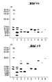
- the effective molecular weight of this polypeptide is 10.8 kD, but on sodium dodecyl sulfate polyacrylamide gel electrophoresis (SDS-PAGE), this peptide migrates as a peptide of 8 kD when compared with standard marker proteins.
- MRP-14 of formula II wherein Z 2 is Thr-Cys-Lys-Met- or hydrogen.
- the latter compound is also called MRP-14d.
- the effective molecular weight of these polypeptides is 13.1 kD and 12.6 kD, respectively, but on SDS-PAGE, these peptides migrate as peptides of 14 kD when compared with standard marker proteins.
- Preferred derivatives are dimers of MRP-8 of formula I, wherein Zi is Met, dimers of MRP-14 of formula ll, wherein Z 2 is Thr-Cys-Lys-Met- and the mixed dimers of MRP-8 of formula I, wherein Z i is Met, with MRP-14 of formula II, wherein Z 2 is Thr-Cys-Lys-Met-, all bound via the oxidized mercapto group of a cystein residue.
- the invention relates also to a process for the preparation of human macrophage migration inhibition factor related peptides, mutants, fragments and derivatives thereof, characterized in that a solution containing the desired compounds, for example a pre-purified extract, cell supernatant or culture filtrate of stimulated normal human leukocytes or of genetically engineered microorganims or permanent mammalian cell lines, is purified by chromatographic methods and the compounds isolated and, if desired, fragments or derivatives prepared therefrom.
- a solution containing the desired compounds for example a pre-purified extract, cell supernatant or culture filtrate of stimulated normal human leukocytes or of genetically engineered microorganims or permanent mammalian cell lines
- Pre-purified extracts, cell supernatants and culture filtrates of stimulated normal human leukocytes containing a compound of the formula ll or derivatives thereof are prepared as described in EP 162 812.
- normal human mononuclear cells are stimulated to produce macrophage migration inhibition factor (MIF) and other lymphokines by suitable adjuncts, for example concanavalin A or phytohaemagglutinin, and are cultured according to customary methods.
- Extracts, cell supernatants or culture filtrates are then pre-purified by immunoaffinity chromatography on a column loaded with antibodies specific for human MIF, e.g. with monoclonal antibodies 1C5.
- Extracts, cell supernatants and culture filtrates of genetically engineered microorganisms or permanent mammalian cell lines containing human macrophage migration inhibition factor related peptides are obtained and pre-purified as will be discussed hereinbelow.
- Chromatographic methods contemplated for the preparation of the desired compounds are ion exchange chromatography, reversed phase high performance liquid chromatography, gel filtration, affinity chromatography, chromatography on hydroxylapatite, hydrophobic interaction chromatography and the like.
- a suitable carrier material for ion exchange chromatography may be of organic or inorganic origin, e.g. cross-linked agarose, dextran, polyacrylamide, styrene/divinylbenzene copolymer, cellulose, or the like.
- This carrier material bears basic functional groups, e.g. tertiary amino functions, quaternary ammonium groups or acid functional groups, e.g. carboxylic or sulfonic acid residues.
- Examples for preferred ion exchangers are those bearing diethylaminoethyl (DEAE) or diethyl-2-hydroxypropyl-ammonioethyl functional groups and those bearing sulfopropyl (SP) or carboxymethyl (CM) functional groups, either attached to carriers suitable for normal liquid chromatography, fast protein liquid chromatography (FPLC) or high performance liquid chromatography (HPLC).
- the separations and purifications with ion exchange chromatography are performed following established procedures, e.g. in aqueous buffer solutions of pH 5 to pH 9 containing increasing amounts of salt, for example sodium chloride.
- Carrier material suitable for gel filtration or size exclusion chromatography includes cross-linked dextran, agarose, suitably modified polyacrylamide or silica, and the like. Optionally these carriers are modified with substituents bearing hydroxy functions, e.g. with 1-hydroxy- or 1,2-dihydroxy-lower alkyl groups.
- the chromatographic material is chosen so as to display optimal separation of peptides in the range of 5'000 to 20'000 Dalton (5 kD to 20 kD) molecular weight.
- Such gel filtration or size exclusion chromatography may be performed in a column suitable for normal liquid chromatography, FPLC or HPLC as above using aqueous buffer solutions around neutrality containing variable amounts of salt, e.g. sodium chloride.
- Reversed phase chromatography is performed on silica-based carrier material bearing hydrophobic groups, e.g. alkyl groups of 1 to 20 carbon atoms, preferably 4, 8,12 or 18 carbon atoms or mixtures of alkyl groups of 1 and 8 or 2 and 18 carbon atoms, respectively, or phenyl groups.
- hydrophobic groups e.g. alkyl groups of 1 to 20 carbon atoms, preferably 4, 8,12 or 18 carbon atoms or mixtures of alkyl groups of 1 and 8 or 2 and 18 carbon atoms, respectively, or phenyl groups.
- hydrophobic interaction chromatography wherein agarose or a related material coated with alkyl groups of up to 12 carbon atoms and/or phenyl groups is used.
- Solvents for processing of the polypeptides of the invention on silica-based reversed phase material are aqueous acids, e.g.
- aqueous trifluoracetic acid containing increasing amounts of a polar, water-miscible organic solvent, e.g. acetonitrile, lower alcohols, e.g. methanol, ethanol or propanol, tetrahydrofuran, and the like, preferably acetonitrile.
- a polar, water-miscible organic solvent e.g. acetonitrile
- lower alcohols e.g. methanol, ethanol or propanol, tetrahydrofuran, and the like, preferably acetonitrile.
- Affinity chromatography is also contemplated for the purification of the peptides of the invention, using a suitable carrier material, e.g. cross-linked agarose, dextran or polyacrylamide bearing molecules with high affinity for a compound of formula I, mutants, fragments and derivatives thereof, for example antibodies, in particular polyclonal and monoclonal antibodies specific for the peptides of the invention as described hereinbelow.
- a suitable carrier material e.g. cross-linked agarose, dextran or polyacrylamide bearing molecules with high affinity for a compound of formula I, mutants, fragments and derivatives thereof, for example antibodies, in particular polyclonal and monoclonal antibodies specific for the peptides of the invention as described hereinbelow.
- the preferred chromatographic methods are ion exchange chromatography with carriers bearing sulfopropyl groups and reversed phase high performance liquid chromatography (HPLC).
- the compounds of the invention are isolated by the usual techniques, for example filtration or ultrafiltration, dialysis, dissolution and reprecipitation in suitable salt and/or buffer solutions and solvent mixtures, solvent evaporation, lyophilization and the like.
- Fragments of a MIF-related peptide are prepared e.g. by treatment with a protease.
- a protease For example, papain, trypsin, a-chymotrypsin, thermolysin, pepsin, subtilisin, endoproteinase Lys-C from Lysobacter enzy- mogenes, V8 protease from Staphylococcus aureus or related proteases may be added to a solution of a compound of formula I or II, and the resulting mixture of fragments separated by chromatographic methods, e.g. by gel filtration and/or reversed phase HPLC.
- Dimers of compounds of formula I or 11 containing a Cys residue are obtained by mild oxidation, e.g. with air, oxygen, iodine, dimethylsulfoxide and HCI or HBr, or other chemical oxidants.
- Conjugates of compounds of formula I with compounds of formula II are prepared likewise by oxidation of a suitable mixture.
- compounds of the formula I or II, mutants, fragments and derivatives thereof can be prepared by recombinant DNA technique comprising, for example, culturing a transformed host expressing a peptide of the formula l or ll, a mutant or derivative thereof under conditions which allow expression of the heterologous polypeptide and isolating the desired compound. More specifically, the desired compounds are prepared by
- the invention relates also to DNAs coding for a compound of formula I or ll, to mutants thereof, e.g. DNAs wherein one or more, especially one, two, three or four, nucleotides are mutated, and to fragments of such DNA comprising at least 15 nucleotides. It is understood that such DNAs are single-stranded or double-stranded.
- the invention concerns a DNA coding for MRP-8 of the formula and a DNA coding for MRP-14 or of the formula wherein
- the invention relates to a DNA coding for MRP-8 of the formula and a DNA coding for MRP-14 of the formula wherein Yi is a flanking DNA residue of 12 nucleotides or more containing a promoter sequence, Y 2 is ATGACTTGCAAA or absent and Y 3 is a flanking DNA residue of one or more nucleotides or absent, a double-stranded DNA consisting of a DNA of formula V or Vl and of a complementary DNA thereto, that complementary DNA itself, genomic DNA, wherein one or more, especially one or two, introns interrupt the DNA of formula V or VI, a mutant of such DNAs, wherein one or more, especially one, two, three or four nucleotides are mutated, and fragments of such DNAs comprising at least 15 nucleotides.
- the invention relates also to a DNA which hybridizes with a DNA of formula V or VI or with a DNA complementary to the DNA of formula V or VI.
- An example of a DNA of the invention of the formula V coding for MRP-8 is e.g. the cDNA which is derived from mRNA of a human mononuclear leukocyte, of the formula
- Y i is e.g. AAGTCTGTGGGCATC-, ATGTCTCTTGTCAGCTGTCTTTCA-GAAGACCTGGTGGGGCAAGTTCCGTGGGCATC-, TTGTCTCTTGTCAGCTGTCTTTCAGAAGACCT-GAAGGTTCTGTTTTTCAGGTGGGGCAAGTTCCGTGGGCATC- or a sequence comprising nucleotides 108 to 123 or 86 to 123 of formula Vll with an insert of a T between nucleotides 112 and 113.
- a further example of a DNA of the formula V coding for MRP-8 is e.g. the genomic DNA which is isolated from human placenta and which contains an intron of 150 nucleotides between the amino acids 47 and 48 (nucleotides 141 and 142 of formula V), of the formula
- DNA of the invention of the formula VI coding for MRP-14 is e.g. the cDNA which is derived from mRNA of a human mononuclear leykocyte, of the formula
- a further example of a DNA of the formula VI coding for MRP-14 is e.g. the genomic DNA which is isolated from human placenta or fetal liver cells and which contains an intron between the amino acids 50 and 51 (nucleotides 138 and 139 of formula VI) of the formula
- RNAs coding for a compound of formula l or ll to mutants thereof, e.g. RNA wherein one or more nucleotides are mutated, and to fragments of such RNA, in particular to a RNA of the formula V or VI, wherein the various Y have the meanings given hereinbefore except that RNA residues replace DNA residues and hence uridine (U) replaces deoxy-thymidine (T), in particular to a RNA of formula VII or IX, wherein U replaces T.
- uridine U
- T deoxy-thymidine
- the DNAs coding for a compound of formula l or ll or for a mutant thereof and fragments of such DNAs or mutants can be prepared for example by culturing a transformed host and isolating the desired DNA therefrom, or by chemical synthesis through nucleotide condensation.
- such DNAs can be prepared by
- Polyadenylated messenger RNA is isolated from human mononuclear leukocytes by known methods.
- the leukocytes may be derived from fresh human blood, e.g., from buffy coats consisting of white blood corpuscules, or from leukocytes of an established continuous cell line which can be expanded in culture. Isolation methods involve, for example, homogenizing stimulated leukocytes in the presence of a detergent and a ribonuclease inhibitor, e.g.
- heparin, guanidinium isothiocyanate and mercaptoethanol extracting the mRNA with suitable chloroform-phenol mixtures, optionally in the presence of salt and buffer solutions, detergents and/or cation chelating agents, and precipitating mRNA from the remaining aqueous, salt-containing phase with ethanol, isopropanol or the like.
- the isolated mRNA may be further purified by centrifuging in a cesium chloride gradient followed by ethanol precipitation and/or by chromatographic methods, e.g. affinity chromatography, for example chromatography on oligo(dT) cellulose or on oligo(U) sepharose.
- chromatographic methods e.g. affinity chromatography, for example chromatography on oligo(dT) cellulose or on oligo(U) sepharose.
- such purified total mRNA is fractionated according to size by gradient centrifugation, e.g., in a linear sucrose gradient, or chromatography
- the desired mRNA is selected by screening with a DNA probe or by translation in suitable cells or cell-free systems and screening the obtained polypeptides.
- Fractionated mRNA may be translated in cells, e.g. in frog oocytes, or in cell-free systems, e.g. in reticulocyte lysates or wheat germ extracts.
- the obtained polypeptides are screened for macrophage migration inhibitory activity or for reaction with antibodies raised against native macrophage migration inhibition factor (MIF), e.g. in an immunoassay, for example radioimmunoassay, enzyme immunoassay or immunoassay with fluorescent markers.
- MIF macrophage migration inhibition factor
- an immunoassay for example radioimmunoassay, enzyme immunoassay or immunoassay with fluorescent markers.
- Such immunoassays and the preparation of polyclonal and monoclonal antibodies are well known in the art and are applied accordingly. Monoclonal antibodies to MIF and immunoassays using them are described, e.g. in European Patent Application EP 162 812.
- hybridization probe may be a fully synthetic DNA consisting of at least 17 nucleotides or a DNA or DNA fragment isolated from a natural source or from a genetically engineered microorganism.
- a synthetic DNA probe can be constructed on the basis of a partial amino acid sequence of a human MIF protein isolated from a natural source, e.g. the human MIF 8 kD described in EP 162 812 or a human MIF-related protein with molecular weight of approximatively 14 kD.
- a human MIF protein isolated from a natural source e.g. the human MIF 8 kD described in EP 162 812 or a human MIF-related protein with molecular weight of approximatively 14 kD.
- Preferably mixtures of oligonucleotide comprising 17 or more nucleotides are prepared, wherein each member of the mixture is complementary to one fragment defined by six or more consecutive triplet codons Y of formula III or IV.
- Such DNA probes are also comprised by the present invention.
- DNA probes of the invention are the 17-mer oligonucleotides complementary to the DNA fragments of formula YD-Yv-Yy-YH-YK-TA corresponding to amino acids 14-19 of MRP-8 of formula I and of formula YD-YV-YW-YF-YK-GA corresponding to amino acids 52-57 of MRP-8 of formula I, and the 26-mer oligonucleotide mixture complementary to the DNA fragment of formula YT-YI-YI-YN-YT-YF-YH-YO-TA corresponding to amino acids 14-22 of MRP-14 of formula II, in which formulas the meaning of YD, YF, YH, Y1, YK, YN, Yo, YT, Yv, y w and YY is as defined under formula IV.
- the 26-mer oligonucleotides contain three inosine residues in place of a nucleotide complementary to a nucleotide of a triplet Y, thus reducing the number of complementary nucleotide-nucleotide interactions to 23.
- oligonucleotides are synthesized according to known methods as detailed hereinbelow, preferably by stepwise condensation using the solid phase phosphotriester, phosphite triester or phosphoramidite method, e.g. the condensation of dinucleotide coupling units by the phosphotriester method.
- stepwise condensation using the solid phase phosphotriester, phosphite triester or phosphoramidite method, e.g. the condensation of dinucleotide coupling units by the phosphotriester method.
- These methods are adapted to the synthesis of mixtures of the desired oligonucleotides by using mixtures of two, three or four nucleotides dA, dC, dG and/or dT in protected form or the corresponding dinucleotide coupling units in the appropriate condensation step as described by Y. Ike et al. (Nucleic Acid Research, 1983, 11, 477).
- the DNA probes have to contain a marker so that hybridization with the desired mRNA can be detected and the mRNA identified and separated from other mRNA not coding for a polypeptide of the present invention.
- a marker so that hybridization with the desired mRNA can be detected and the mRNA identified and separated from other mRNA not coding for a polypeptide of the present invention.
- Suitable are e.g. radioactive labels, such as 32p in the 5'-end phosphate of the oligonucleotide, fluorescent markers or a label containing biotin which can be detected with suitably labelled avidin, e.g. avidin bearing a fluorescent marker or conjugated with an enzyme such as horseradish peroxidase.
- Hybridization of size-fractionated mRNA with the DNA probes containing a marker is performed according to known procedures, i.e. in buffer and salt solutions containing adjuncts, e.g. calcium chelators, viscosity regulating compounds, proteins, irrelevant DNA and the like, at temperatures favoring selective hybridization, e.g. between 0° and 70°C, for example between 25° and 40°C for the 17-mer oligonucleotides and between 30° and 50° C for the 26-mer oligonucleotides, preferably at around 20° lower than the hybrid dsDNA melting temperature.
- adjuncts e.g. calcium chelators, viscosity regulating compounds, proteins, irrelevant DNA and the like
- the preparation of a single-stranded complementary DNA from the selected mRNA template is well known in the art, as is the preparation of a double-stranded DNA from a single-stranded DNA.
- the mRNA template is incubated with a mix of deoxynucleoside triphosphates, optionally a radioactively labelled deoxynucleoside triphosphate (in order to be able to screen the result of the reaction), a primer sequence such as an oligo-dT residue hybridizing with the poly(A) tail of the messenger RNA and a suitable enzyme, e.g. a reverse transcriptase.
- the complementary DNA is incubated with a mix of deoxynucleoside triphosphates and a suitable enzyme as above to give a double-stranded DNA.
- suitable enzymes are a reverse transcriptase, the Klenow fragment of E. coli DNA polymerase I or T 4 DNA polymerase.
- the single-stranded DNA is first extended with a tail of like deoxynucleotides to allow the use of a primer sequence of complementary like deoxynucleotides, but the formation of dsDNA usually starts on spontaneous hairpin formation.
- Such dsDNA obtained as a result of hairpin formation is further processed with Si nuclease which cuts the hairpin.
- genomic DNA may be isolated and screened for DNA coding for the desired polypeptide.
- Genomic DNA is isolated from suitable human tissue, preferably from human placenta or human fetal liver cells, according to methods known in the art.
- a genomic DNA library is prepared therefrom by digestion with suitable restriction endonucleases, e.g. Alul and Haelll, and incorporation into ⁇ charon phage, e.g. ⁇ charon 4A, following established procedures.
- the genomic DNA library replicated on nitrocellulose membranes is screened with a DNA probe, e.g. a synthetic DNA probe of at least 17 nucleotides or a cDNA derived from mRNA coding for the desired polypeptide, as described hereinbefore.
- this cDNA is labelled e.g. by the well-known nick translation technique, then hybridized with the genomic DNA library in solutions containing salts and buffers and other adjuncts as described hereinbefore, preferably at temperatures between 40 and 80°C, e.g. around 65°C.
- dsDNA prepared from mRNA or of genomic origin into an appropriate vector is well known in the art.
- a suitable vector is cut and provided with tails of like deoxynucleotides.
- the dsDNA to be annealed then has to bear tails of complementary like deoxynucleotides, which is accomplished by incubation in the presence of the corresponding deoxynucleoside triphosphate and an enzyme such as terminal deoxynucleotidyl transferase.
- the dsDNA may be incorporated into the vector with the aid of linker oligodeoxynucleotides or else by blunt end ligation.
- E. coli are conditioned for transformation by incubation in media containing calcium chloride, then treated with the hybrid vector.
- Transformed hosts are selected by a suitable marker, for example antibiotics resistance marker, e.g. tetracycline or ampicillin resistance.
- the preparation of a DNA of the invention may also be performed by means of chemical synthesis. Suitable methods for the synthesis of DNA have been presented in summary form by S.A. Narang, Tetrahedron 1983, 39, 3. The known synthesis techniques allow the preparation of polynucleotides towards 40 bases in length, in good yield, high purity and in a relatively short time. Suitably protected nucleotides are linked with one another by the phosphodiester method [K.L. Agarwal et al., Angew. Chem. 1972, 84, 489], the more efficient phosphotriester method [C.B. Reese, Tetrahedron 1972, 34, 3143], phosphite triester method [R.L.
- a polynucleotide with up to 67 bases can thus be prepared in a short time and with good yields.
- the actual double-stranded DNA is built up enzymatically from chemically prepared overlapping oligonucleotides from both DNA strands, which are held together in the correct arrangement by base-pairing and are then chemically linked by the enzyme DNA ligase.
- Another possibility comprises incubating over lapping single oligonucleotides from the two DNA strands in the presence of the four required deoxynucleoside triphosphates with a DNA polymerase, for example DNA polymerase I, the Kienow fragment of polymerase I or T 4 DNA polymerase, or with AMV (avian myeloblastosis virus) reverse transcriptase.
- a DNA polymerase for example DNA polymerase I, the Kienow fragment of polymerase I or T 4 DNA polymerase, or with AMV (avian myeloblastosis virus) reverse transcriptase.
- the two oligonucleotides are thereby held together in the correct arrangement by base-pairing and are supplemented with the required nucleotides by the enzyme to give a complete double-stranded DNA [S.A. Narang et al., Anal. Biochem. 1982, 121, 356].
- the invention further relates to hybrid vectors comprising a DNA coding for a compound of formula I or II, mutants thereof and fragments of such DNA operatively linked to an expression control sequence, and to processes for the preparation thereof.
- the vector is selected depending on the host cells envisaged for transformation.
- suitable hosts are microorganisms, which are devoid of or poor in restriction enzymes or modification enzymes, such as yeasts, for example Saccharomyces cerevisiae, for example S. cerevisiae GRF 18, and strains of bacteria, in particular strains of Escherichia coli, for example E. coli X1776, E. coli HB 101, E. coli W3110, E. coli HB101/LM1035, E. coli JA221 or E.
- E. coli K12 strain 294 Bacillus subtilis, Bacillus stearothermophilus, Pseudomonas, Haemophilus, Streptococcus and others, and furthermore cells of higher organisms, in particular established human or animal cell lines, e.g. human embryonic lung fibroblasts L-132, human malignant melanoma Bowes cells, Hela cells, SV-40 virus transformed kidney cells of African green monkey COS-7 or chinese hamster ovary (CHO) cells.
- the above strains of E. coli for example E. coli HB101, E. coli K12 and E. coli W3110, and of Saccharomyces cerevisiae, for example S. cerevisiae GRF 18, are preferred as hosts, furthermore the human embryonic lung fibroblast cell line L-132.
- vectors which replicate and express the desired polypeptide gene according to the invention in the chosen host are suitable.
- vectors which are suitable for the expression of the compounds of formula I or 11 in an E. coli strain are bacteriophages, for example derivatives of ⁇ bacteriophages, or plasmids, such as, in particular, the plasmid ColE1 and its derivatives, for example pMB9, pSF2124, pBR317 or pBR322.
- the preferred vectors of the present invention are derived from plasmid pBR322.
- Suitable vectors contain a complete replicon and a marker gene, which allows to select and identify the hosts transformed with the expression plasmids on the basis of a phenotypical trait, and optionally signal sequences and enhancers. Suitable marker genes impart to the host, for example, resistance towards heavy metals, antibiotics and the like. Furthermore, preferred vectors of the present invention contain, outside the replicon and marker gene regions, recognition sequences for restriction endonucleases, so that the foreign DNA and, if appropriate, the expression control sequence can be inserted at these sites.
- the preferred vector, the plasmid pBR322 and derived plasmids e.g.
- pUC9, pUC-KO, pHRi148 and pPLc24 contain an intact replicon, marker genes, which confer resistance e.g. towards tetracycline and ampicillin (tetR and ampR), and a number of unique recognition sites for restriction endonucleases.
- expression control sequences can be used for regulation of the gene expression.
- expression control sequences of highly expressed genes of the host to be transformed are used.
- the expression control sequences (which contain, inter alia, the promoter and the ribosomal binding site) of the lactose operon, tryptophan operon, arabinose operon and the like, the ⁇ -lactamase gene, the corresponding sequences of the phage ⁇ N gene, especially those containing the P L promoter, or the phage fd-coat protein gene and others are suitable.
- the plasmid pBR322 already contains the promoter of the ⁇ -lactamase gene (P-lac gene), the other expression control sequences must be introduced into the plasmid.
- Vectors which are suitable for replication and expression in yeast contain a yeast replication start and a selective genetic marker for yeast.
- Hybrid vectors which contain a yeast replication start for example chromosomal autonomously replicating segment (ars), are retained extrachromosomally within the yeast cell after the transformation and are replicated autonomously.
- hybrid vectors which contain sequences homologous to the yeast 2p. plasmid DNA can be used. Such hybrid vectors will get integrated by recombination into 2p. plasmids already existing within the cell, or replicate autonomously. 2p. sequences are particularly suitable for plasmids with a high transformation frequency and permit high copy numbers.
- the preferred yeast vector of the present invention is the plasmid pJDB207.
- Suitable marker genes for yeasts are, in particular, those which impart antibiotic resistance to the host or, in the case of auxotrophic yeast mutants, genes which complement host lesions. Corresponding genes impart, for example, resistance towards the antibiotic cycloheximide or provide for protrophy in an auxotrophic yeast mutant, for example the URA3, LEU2, HIS3 or, in particular, TRP1 gene.
- Yeast hybrid vectors furthermore preferably contain a replication start and a marker gene for a bacterial host, in particular E. coli, so that the construction and cloning of the hybrid vectors and their intermediates can take place in a bacterial host.
- Expression control sequences which are suitable for expression in yeast are, for example, those of highly expressed yeast genes.
- the promoters of the TRP1 gene, the ADHI or ADHII gene, acid phosphatase (PH03 or PH05) gene, isocytochrome gene or a promoter involved with the glycolytic pathway such as the promoter of the enolase, glyceraldehyde-3-phosphate dehydrogenase (GAPDH), 3-phosphoglycerate kinase (PGK), hexokinase, pyruvate decarboxylase, phosphofructokinase, glucose-6-phosphate isomerase, 3-phosphoglycerate mutase, pyruvate kinase, triosephosphate isomerase, phosphoglucose isomerase and glucokinase genes, can be used.
- GPDH glyceraldehyde-3-phosphate dehydrogenase
- PGK 3-phospho
- Preferred vectors of the present invention contain promoters with transcriptional control, e.g. the promoters of the PH05, ADH 11 and GAPDH genes, which can be turned on or off by variation of the growth conditions.
- the PH05 promoter can be repressed or derepressed solely by increasing or decreasing the concentration of inorganic phosphate in the medium.
- Vectors suitable for replication and expression in mammalian cells are preferably provided with DNA from viral origin, e.g. from simian virus 40 (SV40), Rous sarcoma virus (RSV), adenovirus 2, bovine papilloma virus (BPV), papovavirus BK mutant (BKV), or mouse or human cytomegalovirus (CMV).
- viral origin e.g. from simian virus 40 (SV40), Rous sarcoma virus (RSV), adenovirus 2, bovine papilloma virus (BPV), papovavirus BK mutant (BKV), or mouse or human cytomegalovirus (CMV).
- SV40 simian virus 40
- RSV Rous sarcoma virus
- BPV bovine papilloma virus
- BKV papovavirus BK mutant
- CMV mouse or human cytomegalovirus
- shuttle vectors may be constructed from a pBR322 E.
- the plasmid may contain the enhancer unit of the mouse or human cytomegalovirus major immediate-early gene, the SV40 enhancer combined with the human a-globin promoter, and/or in addition inducible promoters, such as the ones derived from the heat shock or metallothionein genes. Further it is also possible to utilize promoter or control sequences which are normally associated with the desired gene sequence.
- An origin of replication may be provided either by construction of the vector to include an exogeneous origin, such as derived from SV40, other viral source or provided by the host cell chromosomal replication mechanism. If the vector is integrated into the host cell chromosome, the latter method is often more efficient.
- the present invention relates to hybrid vectors capable of replication and phenotypical selection in a host strain comprising a promoter and a DNA encoding a compound of formula I or II, a mutant or a fragment thereof, said DNA being positioned together with transcription start and termination signals as well as translation start and stop signals in said hybrid vector under the control of said promoter such that in a transformed host it is expressed to produce the polypeptide.
- the invention also relates to a process for the preparation of a transformed host, which comprises transforming or transfecting a host with an expression vector containing a DNA of the invention regulated by an expression control sequence, and to the transformed or transfected hosts themselves.
- suitable hosts are the above-mentioned microorganisms, such as strains of Saccharomyces cerevisiae, Bacillus subtilis and Escherichia coli.
- the transformation with the expression plasmids according to the invention is carried out, for example, as described in the literature, thus for S. cerevisiae [A. Hinnen, J.B. Hicks and G.R. Fink, Proc. Natl. Acad. Sci. USA 1978, 75, 1929], B. subtilis [Anagnostopoulos et al., J. Bacteriol. 1961, 81, 741] and E. coli [M. Mandel et al., J. Mol. Biol. 1970, 53, 159].
- the transformation procedure of E. coli cells includes Ca2+ pretreatment of the cells so as to allow DNA uptake, and incubation with the hybrid vector.
- the cells are transferred to a selective growth medium which allows separation of the transformed cells from the parent cells. Cells which do not contain the vector will not survive in such a medium.
- the transformation of yeast comprises, for example, the steps of (1) enzymatic removal of the yeast cell wall by means of glucosidases, (2) treatment of the obtained spheroplasts with the vector in the presence of polyethylene glycol and Ca2+ ions and (3) regeneration of the cell wall by embedding the spheroplasts into agar.
- the regeneration agar is prepared in a way to allow regeneration and selection of the transformed cells at the same time.
- suitable hosts are the above-mentioned mammalian cells, e.g. COS-7 cells, Bowes melanoma cells, chinese hamster ovary (CHO) cells or preferably embryonic lung cells L-132.
- the vectors are introduced into mammalian cells by transfection in the prescence of helper compounds, e.g. diethylami- noethyldextran, dimethyl sulfoxide, glycerol, polyethylene glycol or the like, or as co-precipitates of vector DNA and calcium phosphate.
- helper compounds e.g. diethylami- noethyldextran, dimethyl sulfoxide, glycerol, polyethylene glycol or the like, or as co-precipitates of vector DNA and calcium phosphate.
- Further suitable methods inlcude direct microinjection of vector DNA into the cell nucleus and electroporation, i.e.
- Selection markers include genes which confer resistance to antibiotics, e.g. G-418 (neomycin) or hygromycin, or genes which complement a genetic lesion of the host cell such as the absence of thymidine kinase or hypoxanthine phosphoribosyl transferase.
- a preferred selection system makes use of cells lacking dihydrofolate reductase (DHFR-), e.g. CHO cells, which absolutely require thymidine, glycine and purines for growth unless an exogenous DHFR gene is supplied.
- DHFR- dihydrofolate reductase
- suitable DHFR- cells e.g. CHO cells
- transformed cells are selected by increasing the concentration of the anti-folate drug methotrexate in the medium.
- Such treatment furthermore amplificates the production of the desired polypeptide through amplification of the DHFR gene together with substantial flanking chromosomal regions containing the gene coding for the desired polypeptide.
- a selection method wherein suitable mammalian cells, e.g. human embryonic lung cells L-132, are treated with co-precipitates of vector DNA containing the gene coding for a compound of formula I or II, a plasmid DNA containing a gene coding for antibiotics resistance, e.g. resistance to G-418, and calcium phosphate.
- the transformed cells are selected by culturing in the presence of the corresponding antibiotic, e.g. G-418, and by screening for expression of the desired polypeptide.
- the transformed host cells are cultured by methods known in the art in a liquid medium containing assimilable sources of carbon, nitrogen and inorganic salts.
- sources of carbon can be used for culture of the transformed hosts according to the invention.
- preferred sources of carbon are assimilable carbohydrates, such as glucose, maltose, mannitol or lactose, or an acetate, which an be used either by itself or in suitable mixtures.
- suitable sources of nitrogen are amino acids, such as casaminoacids, peptides and proteins and their degradation products, such as tryptone, peptone or meat extracts, yeast extracts, malt extract and also ammonium salts, for example ammonium chloride, sulfate or nitrate, which can be used either by themselves or in suitable mixtures.
- Inorganic salts which can also be used are, for example, sulfates, chlorides, phosphates and carbonates of sodium, potassium, magnesium and calcium.
- the medium furthermore contains, for example, growth-promoting substances, such as trace elements, for example iron, zinc, manganese and the like, and preferably substances which exert a selection pressure and prevent the growth of cells which have lost the expression plasmid.
- growth-promoting substances such as trace elements, for example iron, zinc, manganese and the like
- substances which exert a selection pressure and prevent the growth of cells which have lost the expression plasmid For example, ampicillin is added to the medium if the expression plasmid contains an ampR gene.
- antibiotic substances also has the effect that contaminating antibiotic-sensitive microorganisms are destroyed.
- the plasmid preferably contains a gene coding for an enzyme which complements the host defect. Cultivation of the yeast strain is performed in a minimal medium deficient in said amino acid.
- Vertebrate cells are grown under tissue culture conditions using commercially available media optionally supplemented with growth-promoting substances and/or mammal sera.
- the cells are grown either attached to a solid support, e.g. a microcarrier or porous glass fibres, or free-floating in appropriate culture vessels. Culturing is effected by processes which are known in the art.
- the culture conditions such as temperature, pH value of the medium and fermentation time, are chosen so that a maximum titre of the polypeptide of the invention is obtained.
- coli or yeast strain is preferably cultured under aerobic conditions by submerged culture with shaking or stirring at a temperature of about 20 to 40° C, preferably about 30° C, and a pH value of 4 to 8, preferably at about pH 7, for about 4 to 30 hours, preferably until maximum yields of the polypeptide of the invention are reached.
- the culture is interrupted and the polypeptide is isolated. If the polypeptide is fused with a suitable signal peptide sequence, it is excreted by the cell directly into the supernatant. Otherwise, the cells have to be destroyed, for example by treatment with a detergent, such as SDS, NP-40, Triton® or deoxycholic acid, or lysed with lysozyme, a similarly acting enzyme or with ultra-sound. If yeast is used as a host microorganism, the cell wall may be removed by enzymatic digestion with a glucosidase.
- a detergent such as SDS, NP-40, Triton® or deoxycholic acid
- mechanical forces such as shearing forces (for example X-press, French press, Dyno mill) or shaking with glass beads or aluminium oxide, or alternating freezing, for example in liquid nitrogen, and thawing, for example to 30° to 40°C, as well as ultra-sound can be used to break the cells.
- the cell supernatant or the solution obtained after centrifugation of the mixture obtained on breaking the cells, which contains proteins, nucleic acids and other cell constituents, is enriched in proteins, including the polypeptides of the invention, in a manner which is known oerse.
- proteins including the polypeptides of the invention
- oerse for example, most of the non-protein constituents are removed by polyethyleneimine treatment and the proteins including the polypeptides of the invention are precipitated, for example, by saturation of the solution with ammonium sulfate or with other salts. Otherwise, the cell supernatant or lysate is directly pre-purified using chromatographic methods.
- polypeptides prepared by genetically engineered microorganisms are purified by a combination of chromatographic separations, preferably by a combination of ion exchange chromatography with basic and acid functional groups and reversed phase high performance liquid chromatography as discussed hereinbefore.
- Other separation methods may be included in the purification protocol, e.g. filtration or ultrafiltration with molecular weight cut-off membranes, gel filtration, affinity chromatography, hydrophobic interaction chromatography, chromatography on hydroxylapatite, chromatofocusing, and methods of dialyzing, dissolving and reprecipitating in suitable salt and/or buffer solutions and solvent mixtures.
- Other preferred schemes are those wherein ion exchange chromatography is performed only on one carrier containing sulfonic acid residues, and/or wherein gel filtration, i.e. size exclusion chromatography, is included in the purification protocol.
- the invention concerns furthermore compounds of formula I or II, mutants, fragments and derivatives thereof, whenever prepared according to the methods of the present invention.
- the invention concerns especially the DNA, the hybrid vectors, the transformed host cells, compounds of formula I and 11 and the processes for the preparation thereof as described in the Examples.
- the immune regulating properties of a human macrophage migration inhibition factor related peptide, a mutant, fragment or a derivative thereof may be useful for the therapy of immune regulation diseases and chronic inflammatory diseases and for the protection against infections, preferably in the form of pharmaceutical preparations that contain a therapeutically effective amount of the active ingredient optionally together or in admixture with inorganic or organic, solid or liquid, pharmaceutically acceptable carriers.
- the pharmaceutical preparations according to the invention are those for enteral, e.g. rectal or oral, administration and preferably for parenteral, e.g. intranasal, intramuscular, subcutaneous or intravenous, administration to warm-blooded animals, for example humans.
- parenteral e.g. intranasal, intramuscular, subcutaneous or intravenous, administration to warm-blooded animals, for example humans.
- the pharmaceutical preparations may be in unit dose form, for example in ampoules, vials, suppositories, dragees, tablets, capsules or nasal sprays in liquid or solid form.
- the amount of the therapeutically effective compounds to be administered depends on the condition of the warm-blooded animal, for example the human, such as the body weight, the nature and severity of the disease and the general condition and also on the mode of administration, and is carried out in accordance with the assessment of the physician giving the treatment.
- the effective dose of a compound of formula I or 11, of a mutant or fragment thereof is in the order of magnitude of from 0.001 to 1 ⁇ g per kg of body weight per day.
- the pharmaceutical preparations according to the invention contain the customary inorganic or organic, solid or liquid pharmaceutically acceptable carriers, optionally together with other therapeutically effective compounds and/or adjuncts.
- solutions or suspensions of the active ingredient especially isotonic aqueous solutions or suspensions, or also lyophilized preparations which are dissolved in water shortly before use.
- the pharmaceutical preparations may be sterilized and/or contain preservatives, stabilizers, wetting agents, emulsifiers, solubilizers, viscosity-increasing substances, salts for regulating the osmotic pressure and/or buffers, and also other proteins, for example human serum albumin or human blood plasma preparations.
- liposomes in the form of liposomes in aqueous dispersion containing a therapeutically effective amount of a MIF-related peptide, a mutant, fragment or derivative thereof.
- liposomes having a population of as homogeneous a size as possible and a diameter of approximately from 2.0 x 10- 8 to 5.0 x 10- 6 m consisting of one or more double layers of lipid components, for example amphipatic lipids, for example phospholipids, such as lecithin, cephalin or phosphatidic acid, and optionally neutral lipids, for example cholesterol, enclosing an aqueous interior containing a MIF-related peptide according to the invention.
- liposomes consisting of a mixture of synthetic phosphatidylserine and phosphatidylcholine.
- the invention further concerns polyclonal and monoclonal antibodies specific for a human macrophage migration inhibition factor related peptide, in particular for MRP-8 of formula I and for MRP-14 of formula II, and derivatives thereof.
- Polyclonal antibodies against MIF-related peptides are of mammal origin, e.g. mouse, rat, rabbit, goat, sheep, equine, pig, chimpanzee or human origin. Preferred are mouse, rat, rabbit, goat or sheep antibodies, in particular rabbit antibodies. Such polyclonal antibodies may contain other antibodies than antibodies against compounds of formula I and compounds of formula II, respectively. In particular, polyclonal antibodies are a collection of antibodies with different affinity and selectivity towards MIF-related peptides. Preferred polyclonal antibodies are specific for MRP-8 and specific for MRP-14, respectively.
- Preferred antibodies of the invention are monoclonal antibodies against MIF-related peptides.
- Monoclonal antibodies are also of mammal origin, e.g. mouse, rat or human origin, preferably mouse antibodies.
- Particularly preferred are the monoclonal antibodies against MRP-8 with the designation 8-5C2 and 8-10D7 and the monoclonal antibodies against MRP-14 with the designation 14-6B2 and 14-19C9, and derivatives thereof.
- These monoclonal antibodies are secreted by the corresponding hybridoma cell lines with the designation 8-5C2, 8-10D7, 14-6B2 and 14-19C9,-respectively.
- Derivatives of antibodies of this invention are e.g. antibody fragments, radioactively labelled antibodies, and conjugates of the antibodies with e.g. enzymes, compounds with exceptional binding properties, e.g. avidin or biotin, fluorescent markers, chemiluminescent markers or paramagnetic particles.
- Fragments of antibodies of this invention are e.g. Fab, Fab' or F(ab') 2 fragments, which retain their specificity for the antigenic determinants, i.e. which retain the characteristic binding pattern of the parent antibody to MIF-related proteins.
- Radioactively labelled antibodies contain e.g. radioactive iodine ( 123 1, 125 1, 131 1), carbon ( 14 C), sulfur ( 3 5S), tritium ( 3 H) or the like. Preferred are antibodies labelled with radioactive iodine, e.g. monoclonal antibodies labelled with 125 1 .
- Antibody conjugates of the invention are e.g. conjugates with enzymes such as horseradish peroxidase, alkaline phosphatase, p-D-gaiactosidase, glucose-oxidase, glucoamylase, carboanhydrase, acetylcholinesterase, lysozyme, malate dehydrogenase or glucose-6-phosphate dehydrogenase, conjugates with biotin or avidin or conjugates with fluorescent markers, e.g. with fluorescein or rhodamine B, conjugates with chemiluminescent markers, e.g. acridinium esters, or conjugates with solid paramagnetic particles.
- enzymes such as horseradish peroxidase, alkaline phosphatase, p-D-gaiactosidase, glucose-oxidase, glucoamylase, carboanhydrase, acetylcholinesterase,
- conjugates the antibody is bound to the conjugation partner directly or by the way of a spacer or linker group.
- conjugates of monoclonal antibodies with the enzymes horseradish peroxidase or alkaline phosphatase and conjugates of polyclonal and monoclonal antibodies with biotin.
- the selectivity of an antibody towards a MIF-related protein can be detected qualitatively in an enzyme-immunoassay wherein the wells of a microtiter plate are coated with the protein, then treated with the antibody to be tested, and bound antibody is detected with labelled antiserum directed against the antibody.
- the selectivity of a mouse monoclonal antibody of the invention is determined in a sandwich type enzyme-immunoassay wherein the wells of a microtiter plate are coated with a rabbit polyclonal antibody to a MIF-related protein followed by the protein itself, then treated with the antibody to be tested, and bound monoclonal antibody is detected with labelled antiserum directed against the constant part of mouse antibodies.
- the monoclonal antibodies can be further analyzed with respect to their immunoglobulin class and subclass, e.g. by the immuno-diffusion Ouchterlony method using class-specific second antibodies.
- the antibodies of the invention and derivatives thereof are obtained by processes known per se.
- Polyclonal antibodies of the invention and derivatives thereof are obtained by a process, wherein a suitable mammal is immunized by two or more injections of a compound of formula I or 11 in the presence of an immune response enhancer, the blood serum of the immunized mammal is collected and the antibodies isolated and purified, and, if desired, the obtained antibodies are transformed into derivatives thereof.
- mice or rabbits are used. They are immunized by two, three, four or more injections of the compound of formula I or 11 intradermally, subcutaneously, intravenously or intraperitoneally in regular or irregular intervals of a few days, e.g. three to seven days, up to several months, for example four weeks.
- the immune response enhancer is an adjuvans which stimulates the lymphocyte production, e.g. complete or incomplete Freund's adjuvans.
- the immune response of the mammal is preferably monitored by a suitable antibody assay, e.g. an enzyme-immunoassay as described hereinbefore.
- the blood of the mammal is collected a few, e.g. two to five, days after the last booster immunization.
- the antibodies are isolated by known methods, e.g. by precipitation, centrifugation and/or chromatographic procedures.
- a crude immunoglobulin fraction may be obtained from the serum by precipitation with ammonium sulfate or the like.
- Such an immunoglobulin fraction can be further purified by gel filtration or molecular sieve chromatography, ion exchange chromatography, chromatography on DEAE cellulose or immunoaffinity chromatography, e.g. on carrier material bearing Staphylococcus protein A or, preferably, the corresponding MIF-related protein.
- Antibody fragments for example Fab, Fab' or F(ab') 2 fragments, which retain their specificity towards the antigenic determinants, can be obtained by methods known per se, e.g. by digestion of the antibodies with enzymes such as pepsin or papain and/or cleavage of disulfide bonds by chemical reduction.
- Antibodies labelled with radioactive iodine are prepared by iodination methods known in the art, e.g. by labelling antibodies with radioactive sodium or potassium iodide and a chemical oxidant such as sodium hypochlorite, chloramine T or the like, or an enzymatic oxidant such as lactoperoxidase or glucose oxidase and glucose.
- Conjugates of antibodies of the invention are prepared by methods known in the art, e.g. by reacting an antibody or a fragment thereof prepared as described hereinbefore with the enzyme in the presence of a coupling agent, e.g. glutaraldehyde, periodate, N,N'-o-pheny lenedimaleimide, N-(m-maleimidobenzoy- loxy)-succinimide, N-(3-[2'-pyridyidithio]-propionoxy)-succinimide, N-ethyl-N'-(3-dimethylaminopropyl)-carbodiimide or the like.
- Conjugates with avidin are prepared likewise.
- Conjugates with biotin are prepared e.g. by reacting antibodies with an activated ester of biotin such as the biotin N-hydroxysuccinimide ester.
- Conjugates with fluorescent or chemiluminescent markers are prepared in the presence of a coupling agent, e.g. those listed above, or by reaction with an isothiocyanate, preferably fluorescein-isothiocyanate.
- Conjugates with paramagnetic particles are obtained with preactivated particles or by coupling in the presence of e.g. glutaraldehyde or periodate.
- the monoclonal antibodies of the invention and derivatives thereof are obtained by processes known per se, characterized in that hybridoma cells secreting said monoclonal antibodies a) are cultivated in vitro and the monoclonal antibodies isolated from the culture supernatant, or b) are propagated in vivo in a suitable mammal and the monoclonal antibodies recovered from body fluids of said mammal, and,
- the obtained monoclonal antibodies are converted into a derivative thereof.
- Suitable culture media for the in vitro cultivation according to process a) are standard culture media such as Dulbecco's modified Eagle medium or RPMI 1640 medium, optionally replenished by a mammal serum, e.g. fetal calf serum, or other growth-sustaining supplements, e.g. 2-aminoethanol, insulin, transferrin, low density lipoprotein, oleic acid and the like, and trace elements.
- a mammal serum e.g. fetal calf serum
- growth-sustaining supplements e.g. 2-aminoethanol, insulin, transferrin, low density lipoprotein, oleic acid and the like, and trace elements.
- the isolation of the monoclonal antibodies is accomplished by precipitating the protein contained in the culture supernatants by ammonium sulfate or the like, followed by purifying the immunoglobulins by standard chromatographic methods, such as gel filtration, ion exchange chromatography, chromatography on DEAE cellulose or immunoaffinity chromatography.
- the desired monoclonal antibodies can also be obtained by the propagation of hybridoma cells according to process b).
- Cell clones are injected into syngeneic mammals, which causes antibody-producing tumors to grow. After one to three weeks the desired monoclonal antibodies are recovered from body fluids of said mammal.
- hybridoma cells derived from Balb/c mice are intraperitoneally injected into Balb/c mice optionally pretreated with a hydrocarbon such as pristane, and after one to two weeks, ascites fluid of these mice is collected.
- the desired monoclonal antibodies are isolated from the body fluids by methods known per e, e.g.
- Radioactively labelled monoclonal antibodies may also be prepared by adding radioactively labelled nutrients to the culture media of the in vitro cultivation of step a). Such labelled nutrients contain e.g. radioactive carbon ( 14 C).
- the invention further concerns hybridoma cell lines, characterized in that they secrete monoclonal antibodies directed against MIF-related peptides, in particular against MRP-8 of formula I and against MRP-14 of formula II.
- the invention concerns cell lines which are hybrids of myeloma cells and B lymphocytes of a mammal immunized with compounds of formula I or II.
- these cell lines are hybrids of mouse myeloma cells and B lymphocytes of a syngeneic mouse immunized with purified compounds of formula I or II.
- Preferred are the hybridoma cell lines with the designation 8-5C2, 8-10D7, 14-6B2 and 14-19C9.
- These hybridoma cell lines are hybrids of the mouse myeloma cell line P3x63-Ag8.653 and of B lymphocytes of the spleen of Balb/c mice immunized with MRP-8 and MRP-14, respectively. They are stable cell lines, secrete the monoclonal antibodies with the corresponding designation and may be kept in deep-frozen cultures and reactivated by thawing and optional re-cloning.
- the invention concerns also a process for the production of hybridoma cell lines secreting monoclonal antibodies which bind to compounds of formula I or II, characterized in that a suitable mammal is immunized with a compound of formula I or II, antibody-producing cells of this mammal are fused with myeloma cells, the hybrid cells obtained in the fusion are cloned, and cell clones secreting the desired antibodies are selected.
- Suitable antigens for the immunization of mammals in the process of the invention are the preferred compounds of formula I and II, MRP-8 and MRP-14, respectively, described hereinbefore.
- Preferred mammals for the immunization are mice, in particular Balb/c mice. The immunizations are performed e.g. as described hereinbefore for the preparation of polyclonal antibodies.
- Antibody-producing cells of the immunized mammals preferably spleen cells, taken two to five days after the final booster injection, are fused with myeloma cells of a suitable cell line in the presence of a fusion promoter.
- myeloma cell lines are known in the art. Preferred are myeloma cell lines lacking the enzyme hypoxanthine guanine phosphoribosyl transferase (HGPRT) or the enzyme thymidine kinase (TK), which therefore do not survive in a selective culture medium containing hypoxanthine, aminopterin and thymidine (HAT medium).
- HGPRT hypoxanthine guanine phosphoribosyl transferase
- TK thymidine kinase
- myeloma cells and derived cell lines that do not survive in HAT medium and do not secrete immunoglobulins or fragments thereof, such as the cell lines P3x63-Ag8.653 or Sp2/0-Ag14.
- Fusion promoters considered are e.g. Sendai virus or other paramyxo viruses, optionally in UV-inactivated form, calcium ions, surface-active lipids such as lysolecithin, or polyethylene glycol.
- the myeloma cells are fused with a three- to twentyfold excess of spleen cells from immunized mammals in a solution containing about 30 % to about 60 % polyethylene glycol or a molecular weight between 1000 and 4000 and about 10 % to about 20 % dimethyl sulfoxide.
- the cells are resuspended and cultivated in selective HAT medium. Thereby, only hybridoma cells will survive, because they combine the ability to grow and replicate in vitro inherited from myeloma cells and the missing HGPRT or TK genes essential for the survival in the HAT medium inherited from the antibody-producing spleen cells of the immunized mammals.
- Suitable culture media for the expansion of hybridoma cells are the standard culture media, such as Dulbecco's modified Eagle medium, minimum essential medium, RPMI 1640 medium and the like, optionally replenished by serum, e.g. 10 to 15 % fetal calf serum.
- Optionally feeder cells are added at the beginning of the cell growth, e.g. normal mouse peritoneal exudate cells, spleen cells, marrow bone macrophages, or the like.
- the culture media are supplemented with selective HAT medium, at later stages with hypoxanthine/thymi dine (HT) medium, in order to prevent normal myeloma cells overgrowing the hybridoma cells.
- HAT medium at later stages with hypoxanthine/thymi dine (HT) medium
- hybridoma cell culture supernatants are screened for the desired monoclonal antibodies, preferentially with an enzyme immunoassay or a radioimmunoassay.
- Positive hybridoma cells are cloned, e.g. by limiting dilution.
- the cloned cell lines may be frozen in a conventional manner.
- the polyclonal and monoclonal antibodies of the invention and/or their derivatives are useful for the qualitative and quantitative determination of MIF-related proteins, in particular of compounds of formula I or II.
- the antibodies or derivatives thereof such as enzyme conjugates or radioactive derivatives, can be used in any of the known immunoassays, which rely on the binding interaction between the antigenic determinant of the MIF-related proteins and the antibodies.
- assays are radioimmunoassays (RIA), enzyme-immunoassays, e.g. enzyme-linked immunosorbent assay (ELISA), immunofluorescence, immunoprecipitation, latex agglutination, and hemagglutination.
- Such immunoassays are useful e.g. in the qualitative and quantitative determination of the MIF-related proteins in biological fluids or tissues, e.g. of patients with inflammatory conditions and of patients or healthy human subjects with genetic predisposition of cystic fibrosis.
- the antibodies according to the invention can be used as such or in the form of radioactively labelled derivatives in a radioimmunoassay (RIA).
- a radioimmunoassay Any of the known modifications of an RIA can be used, for example RIA in homogeneous phase, solid phase RIA or heterogeneous RIA, single RIA or double (sandwich) RIA with direct or indirect (competitive) determination of the protein of the invention.
- a sandwich RIA in which a suitable carrier, for example the plastics surface of a microtitre plate or of a test tube, for example of polystyrene, polypropylene or polyvinyl chloride, glass or plastics beads, filter paper, or dextran, cellulose acetate or nitrocellulose sheets or the like, is coated with a polyclonal or monoclonal antibody of the invention by simple adsorption or optionally after activation of the carrier, for example with glutaraldehyde or cyanogen bromide, and incubated with the test solution and a solution of a monoclonal antibody radioactively labelled with 125 1, the dissolved monoclonal antibody recognizing another epitope of the proteins of the invention than the carrier-bound monoclonal antibody, if such is used, and the amount of the proteins of the invention is determined by measuring the radioactivity bound to the carrier.
- a suitable carrier for example the plastics surface of a microtitre plate or of a test tube, for example of polys
- a sandwich radioimmunoassay as described hereinbefore, wherein a monoclonal antibody of the invention is bound to a bead, for example a polystyrene bead, this coated bead is incubated in a test or standard solution containing MIF-related proteins and is finally developed with a radiolabelled monoclonal antibody recognizing a different epitope.
- the antibodies according to the invention can be used as such or in the form of enzyme-conjugated or biotin-conjugated derivatives in an enzyme-immunoassay (EIA).
- EIA enzyme-immunoassay
- Such immunoassays include test procedures in which enzyme-labelled monoclonal antibody derivatives according to the invention, enzyme-labelled antibodies known per se that recognize and bind an epitope of the antibodies according to the invention, or enzyme-avidin conjugates are used.
- Any of the known modifications of an EIA can be used, for example EIA in homogeneous phase, solid phase EIA or heterogeneous EIA, single EIA or double (sandwich) EIA with direct or indirect (competitive) determination of the MIF-related protein.
- an ELISA enzyme-linked immunoadsorbent assay
- a carrier as described above for an RIA is coated with a polyclonal or monoclonal antibody according to the invention, incubated with a test solution containing a MIF-related protein and then with a polyclonal or different monoclonal antibody conjugated to biotin, and, finally, the bound antibody-biotin conjugate is developed by an enzyme-avidin conjugate, and the amount of the protein bound is determined by an enzyme substrate reaction.
- an ELISA in which a carrier coated with a polyclonal or monoclonal antibody according to the invention is incubated with a test solution and with a solution of a monoclonal antibody that is conjugated with an enzyme, the dissolved monoclonal antibody recognizing a different epitope of the MIF-related protein than does the carrier-bound monoclonal antibody, if such is used.
- an enzyme substrate reaction that results, for example, in a colour change and can be observed by eye or with optical measuring devices, the amount of bound enzyme, which is proportional to the amount of the protein in the test solution, is measured.
- an ELISA in which a carrier coated with a polyclonal or monclonal antibody according to the invention is incubated with a test solution, with a solution of a monoclonal or polyclonal antibody of a different species than the carrier-bound antibody and then with an enzyme-labelled second antibody that recognizes and binds the species-specific part of the dissolved antibody.
- the amount of bound enzyme is proportional to the amount of the protein in the test solution and can be determined by an enzyme substrate reaction.
- the monoclonal antibodies according to the invention can be used as such or in the form of derivatives conjugated with fluorescent markers in immunofluorescence tests.
- immunofluorescence tests include procedures wherein monoclonal antibody derivatives according to the invention, e.g. derivatives conjugated with fluorescein, or fluorescent marker-labelled antibodies known per se that recognize and bind an epitope of the monoclonal antibodies according to the invention are used.
- an immunofluroescence test in which a carrier as described above for an RIA is coated according to standard methods with cells to be tested for the presence of a protein of the invention, the cells are fixed and permeabilized to allow interaction of proteinaceous material inside the cell with solutions applied, then incubated with a solution of a polyclonal or monoclonal antibody derivative according to the invention conjugated with a fluorescent marker, or incubated with a solution of a polyclonal or monoclonal antibody of the invention followed by a solution of a fluorescent marker-labelled second antibody that recognizes and binds the antibody of the invention, e.g. a fluorescein-labelled rabbit anti-mouse immunoglobulin. The presence of a protein of the invention is then detected and the protein localized by standard fluorescence microscopy or flow cytometry.
- the corresponding enzyme immunohistological test in which the fixed cells are incubated with a polyclonal or monoclonal antibody of the invention followed by a solution of a enzyme-labelled second antibody that recognizes and binds the antibody of the invention, e.g. a peroxidase-labelled anti-rabbit or anti-mouse antiserum. The presence of a protein of the invention is then detected and the protein localized by an enzyme substrate reaction.
- the use according to the invention of monoclonal antibodies and derivatives thereof as described hereinbefore for the qualitative and quantitative determination of the MIF-related proteins also includes other immunoassays known per se, for example immunodot analysis, immunoprecipitation tests with radiolabelled antibodies or radiolabelledMIF-related proteins, latex agglutination with antibody-coated or antigen-coated latex particles or hemagglutination with antibody-coated or antigen-coated red blood corpuscles or the like.
- the invention relates also to test kits for the qualitative and quantitative determination of MIF-related proteins, in particular of compounds of formula I or II, containing polyclonal and/or monoclonal antibodies of the invention and/or derivatives thereof and, optionally, other monoclonal or polyclonal antibodies and/or adjuncts.
- Test kits according to the invention for a radioimmunoassay contain, for example, a suitable carrier, uncoated or coated with a polyclonal or monoclonal antibody of the invention, optionally freeze-dried or concentrated solutions of a monoclonal or polyclonal antibody to a compound of formula I or 11 and/or a radiolabelled derivative thereof, standard solutions of the corresponding compound of formula I or II, buffer solutions and, optionally, polypeptides and detergents for preventing non-specific adsorption and aggregate formation, pipettes, reaction vessels, calibration curves, instruction manuals and the like.
- Test kits according to the invention for an enzyme-immunoassay contain, for example, a suitable carrier, e.g. microtiter plates or nitrocellulose sheets, optionally freeze-dried or concentrated solutions of a polyclonal or monoclonal antibody to a compound of the formula I or 11 and of an enzyme-labelled or biotin-labelled monoclonal or polyclonal antibody to this protein, solutions of an enzyme-avidin conjugate, if a biotin-labelled antibody is used, enzyme substrates in solid or dissolved form, standard solutions of a protein of the invention, buffer solutions and, optionally, polypeptides and detergents, pipettes, reaction vessels, calibration curves, colour scale tables, instruction manuals and the like.
- a suitable carrier e.g. microtiter plates or nitrocellulose sheets
- solutions of an enzyme-avidin conjugate if a biotin-labelled antibody is used
- enzyme substrates in solid or dissolved form standard solutions of a protein of the invention, buffer solutions and, optionally, polypeptid
- the monoclonal antibodies and antibody derivatives of the invention are used for the qualitative and quantitative determination of MIF-related proteins, in particular of compounds of formula I or II, e.g. MRP-8 and MRP-14, respectively, preferably in enzyme-immunoassays.
- MRP-8 and MRP-14 are used for the qualitative and quantitative determination of MIF-related proteins, in particular of compounds of formula I or II, e.g. MRP-8 and MRP-14, respectively, preferably in enzyme-immunoassays.
- the reliable determination of the amount of MRP-8 and MRP-14 in biological fluids, tissue sections and cells allows a simple detection of inflammatory conditions and/or genetic predisposition for cystic fibrosis.
- the monoclonal antibodies and antibody derivatives can be used in the isolation and purification of the MIF-related proteins of the invention from natural sources or from recombinant host cells by immunoaffinity chromatography.
- MRP-8 and MRP-14 of this invention occur in varying amounts of normal granulocytes and monocytes depending on the donors. Upon cultivation of granulocytes and monocytes, the number of MRP-8 and MRP-14 positive cells increase up to day 3 of culture and then decline approaching zero levels from day 10 and on. MRP-8 and MRP-14 seem not to be present in normal human tissue except in intravascular monocytes e.g. in liver, MRP-14 also in monocytes in the lung and in placenta.
- MRP-8-positive macrophages are detected.
- endothelial cells contain MRP-8.
- MRP-14-positive marcophages are seen in substantial number in rheumatoid arthritis tissue.
- MRP-8 and MRP-14 are expressed by subsets of tissue marcophages in primary chronic polyarthritis and other chronic inflammation in a pattern different from acute inflammation such as gingivitis.
- the invention therefore concerns a method of diagnosis of chronic inflammatory conditions, characterized in that the antibodies to MRP-8 and to MRP-14 described hereinbefore are used to determine the amount and pattern of expression of MRP-8 and MRP-14 in tissue.
- Cystic fibrosis is an autosomal recessive disease the aetiology of which is not yet known. There is a need for a simple method which allows rapid and reliable determination whether a subject is normal, CF heterozygous or CF homozygous. CF heterozygotes are clinically unaffected but may transmit the disease to the next generation.
- the average amount of MRP-14 found in healthy donors is 0.043 ⁇ g/ml with a maximum value of 0.132 ⁇ g/ml.
- Patients suffering from CF show a plasma concentration between 0.204 and 5.78 I lg/ml with a mean value of 1.076 ⁇ g/ml.
- CF heterozygotes i.e. plasma donors which are parents of CF patients but not clinically affected themselves, show average MRP-14 concentrations of 0.611 ⁇ g/ml with a minimum value of 0.161 ⁇ g/ml.
- the amount of MRP-8 found in plasma samples of healthy donors, CF homozygotes and CF heterozygotes is usually less than 0.01 ⁇ g/ml and not indicative of cystic fibrosis.
- the invention therefore concerns a method of reliable diagnosis of cystic fibrosis, characterized in that antibodies to MRP-14 described hereinbefore are used to determine the amount of MRP-14 in plasma samples of otherwise healthy subjects supposed to be homozygous or heterozygous in cystic fibrosis.
- the column is equilibrated in a mixture of 65 % TFA 0.1 % in water and 35 % TFA 0.07 % in acetonitrile, and the product eluted by a linear gradient over 30 min ending with a mixture of 45 % TFA 0.1 % in water and 55 % TFA 0.07 % in acetonitrile at a flow rate of 1 ml/min.
- the eluate is monitored for absorbance at 220 nm with a Kratos Spectroflow 773 HPLC UV detector.
- the protein of fraction I is identical with the known human MIF protein of apparent molecular weight 8 kD described in EP 162 812.
- Fraction ll contains dimeric MIF protein 8 kD appearing as a double band at 16.5 kD under non-reducing conditions and as strong band at 8 kD with a faint, slightly faster moving band under reducing conditions.
- the protein of fraction III appears at 14 kD under reducing and non-reducing conditions and is named MRP-14.
- the protein of fraction IV exhibits a double band at 13 kD and is named MRP-14'.
- the protein of fraction V with apparent molecular weight of 20 kD consists of the disulfide linked heterodimer of the 8 kD MIF protein and MRP-14 as shown under reducing conditions.
- the protein of fraction VI of 17.5 kD is a further disulfide linked heterodimer of a 8 kD protein, and the protein of fraction Vll appearing at 23.5 kD is the disulfide linked dimer of MRP-14
- MRP-8, MRP-14 and MRP-14' are eluted together in the same fractions between 250 ml and 280 ml of the total gradient volume corresponding to approximately 0.8 M - 0.9 M NaCi.
- the pool of MRP containing fractions is concentrated by ultrafiltration on a YM - 10® membrane (Amicon) and made 1 % in TFA. Isolation of pure MRP-8, MRP-14 and MRP-14' respectively is achieved on a Vydac @ 214 TP 510 reverse phase HPLC column (The Separations Group, USA).
- the column is equilibrated in a mixture of 75 % TFA 0.1 % in water and 25 % TFA 0.07 % in acetonitrile, and the proteins are eluted by a linear gradient over 45 min ending with a mixture of 45 % TFA 0.1 % in water and 55 % TFA 0.07 % in acetonitrile at a flow rate of 3 ml/min.
- the eluate is monitored for absorbance at 235 nm. Individual peaks are collected manually according to the absorption reading.
- MRP-8, MRP-14 and MRP-14' are identified by comparison with the peptides isolated according to
- Preparative separations of the peptide fragments are achieved on the same column with a linear gradient of 60 min from 100 % TFA 0.1 % in water to 100 % TFA 0.07 % in acetonitrile at a flow rate of 1 ml/min. Three fractions are collected manually according to the UV absorbance at 220 nm at retention times of 20.7 min (A), 22.7 min (B) and 24.8 min (C).
- Example 3 Amino acid sequence analysis of Staphylococcus aureus V8 protease fragments of MRP-14
- Fractions A, B and C are evaporated in vacuo, dissolved each in 25 ⁇ l 0.1 % aqueous TFA and subjected to an amino acid sequence determination on a gas phase protein sequencer model 470 from Applied Biosystems.
- N-terminal amino acid sequences found are: wherein X n means an undetermined amino acid at position n from the N-terminal.
- Example 4 Isolation of messenger RNA from human mononuclear blood leukocytes
- 1.6 x 10 10 mononuclear human blood leukocytes are isolated from buffy coats and treated with concanavalin A for 2 h as described in European Patent Application 162 812. After 16 h of incubation at 37°C in RPMI 1640 medium (5 % CO 2 ) in spinner cultures, the cells are collected by centrifugation for 15 min at 350 x g. The fluffy cell pellet of approximately 10 ml is dissolved in 50 ml GuSCN buffer and homogenized in a Sorvall omnimixer (100 ml) at maximum speed for 90 sec.
- the solution is thoroughly mixed with 60 ml of phenol previously equilibrated with a solution containing 10 mM Tris-HCI pH 7.5, 100 NaCl and 1 mM EDTA.
- Phase separation is achieved by addition of 60 ml of chloroform, mixing and centrifugation at 3000 rpm in a tabletop centrifuge.
- the aqueous phase including some of the nonviscous interphase is collected and 60 ml of equilibrated phenol and chloroform are added sequentially.
- the mixture is centrifuged, and the aqueous phase and the nonviscous interphase are recovered. These steps are repeated 3-4 times until all interphase has virtually disappeared.
- Nucleic acids are precipitated at -20°C by addition of 2 volumes of ethanol (100 ml).
- the RNA is further purified from contaminating DNA as follows: the nucleic acids are dissolved in 6 ml H 2 0 1.5 ml of 0.5 M EDTA pH 7.5, then a mixture of 7.5 g of baked CsCI and 215 ⁇ l 1 N HCI are added.
- the solution is layered over two 2 ml cushions of 5.7 M CsCl in 0.1 M EDTA pH 7.5 (final) in two TST41 tubes (Kontron).
- the tubes are filled up with H 2 0 and centrifuged in a TST41 rotor for 16 h at 20° C.
- RNA pellet is dissolved in 4 ml elution buffer by vortexing and occasional warming (2 min) at 37°C.
- the RNA is precipitated by addition of 10 mi of ethanol and centrifugation in a HB 4 rotor (Sorvall) for 10 min.
- the RNA (9.5 mg) is briefly dried and dissolved in 0.4 ml of elution buffer.
- RNA is eluted with 4 ml of elution buffer.
- the RNA is precipitated overnight at -20° C by addition of 0.25 ml of 3 M NaOAc pH 5.5 and 10 ml of ethanol.
- the precipitate (110 ⁇ g) is collected by centrifugation (15 min at 16000 x g), dissolved in 0.4 ml H 2 0 and re-precipitated by addition of 25 ⁇ l of 3 M NaOAc and 1 ml of ethanol.
- the RNA is collected by centrifugation for 5 min in an Eppendorf centrifuge. The pellet is air dried and dissolved in 110 ⁇ l of H 2 0.
- polyadenylated RNA (Example 4) are denatured at 80°C for 2 min in 200 ⁇ l of 50 % DMSO, 10 mM Tris-HCI pH 7.5, 1 mM EDTA and 0.5 % SDS. After cooling and addition of 300 ⁇ l H 2 0, the solution is layered onto 11.5 ml of a linear sucrose gradient 5-15 % w/v in 100 mM NaCI, 10 mM Tris-HCI pH 7.5, 1 mM EDTA and 0.5 % SDS, in a TST41 rotor (Kontron).
- RNA from individual fractions is precipitated by additon of 15 ⁇ l 0.3 M NaOAc pH 5.5 and 1 ml of ethanol. After incubation overnight at -20°C, the RNA is collected by centrifugation, dissolved and reprecipitated as above. Finally the RNA is dissolved in 20 ⁇ l H 2 0.
- Oligodeoxynucleotide mixture 1 has the composition 5'-TAYTTRTGRTANACRTC-3', wherein A, T, G, and C stand for adenosine, thymidine, guanosine, and cytosine, respectively, Y and R for pyrimidines (T,C) and purines (A,G), respectively, and N for any of the four deoxynucleotides.
- Oligodeoxynucleotide mixture 2 is composed of 5'-TCYTTRAACCANACRTC-3', wherein the codes have the same meaning as above.
- oligodeoxynucleotides are synthesized following the procedure of Y. Ike et al., Nucleic Acid Research 1983, 11, 477. The 5' ends of the oligodeoxynucleotides are rendered radioactive (1-2x10 9 dpm/ ⁇ g) using y-32P-dATP (5000 Ci/mmol) and polynucleotide kinase (Pharmacia) using standard procedures (T. Maniatis, E.F. Fritsch and J. Sambrook, "Molecular cloning, a laboratory manual", Cold Spring Harbor Laboratory, 1982).
- 2x2 ⁇ l of the size-fractionated RNA of Example 5 are spotted on replica filters (Pall-Biodyne@) and baked at 80°C for 2 h in a vacuum oven.
- the filters are prehybridized for 3 h at 32° C in 50 ml of Denhart's solution containing 0.9 M NaCI, 180 mM Tns-HCI pH 8.0, 6 mM EDTA, 0.5 % SDS and 50 ⁇ g/ml sheared single stranded calf thymus DNA.
- one filter is hybridized for 36 h at 29° C in 1.5 ml of prehybridization solution containing in additon 5x10 7 dpm of oligodeoxynucleotide mixture 1.
- the second filter is hybridized at 32° C in the same solution contianing 5x10 7 dpm of oligodeoxynucleotide mixture 2.
- the filters are washed at 25° C (filter 1) and 29° C (filter 2) 15 min with 250 ml of 0.6 M NaCl, 0.12 M Tris-HCI pH 8.0, 4 mM EDTA and 0.2 % SDS and four times 15 min with 250 ml 0.3 M NaCl, 0.06 M Tris-HCI pH 8.0, 2 mM EDTA and 0.2 % SDS.
- After drying the filters are exposed for 3 days at -80°C on X-ray film using an Ilford Fast Tungstate Screens. By comparison of the replicas, fraction 23 (9S) is estimatd to contain most MRP-8 mRNA.
- Example 7 Localization of MRP-14 mRNA among the size-fractionated RNA
- Size fractionated RNA is prepared as described in Example 5 except that 40 fractions of 0.3 mi are collected and the precipitated RNA is dissolved in 40 ⁇ l H 2 0.
- 3 ⁇ g of 9S mRNA coding for MRP-8 from fraction 23 are incubated for 90 min at 42° C in a 50 ⁇ l reaction mixture containing 100 mM Tris-HCI (pH 8.3 measured at 42°C), 10 mM MgCl 2 , 140 mM KCI, 10 mM DTT, 1 mM of each dNTP, 100 ⁇ g/ml oligo-dT 12-18 (Pharmacia), 90 units RNasin@ (Genofit), 40 units AMV reverse transcriptase (Genofit) and 30 ⁇ Ci of a- 32 P-dCTP (3000 Ci/mmol).
- the reaction is stopped by addition of 2 ⁇ l 0.5 M EDTA pH 7.5.
- the RNA is degraded by incubation with 25 ⁇ l of 0.15 N NaOH for 1 h at 65°C.
- the solution is neutralized by addition of 25 ⁇ l Tris-HCI pH 8.0 and 6 ⁇ l 1 N HCI.
- SDS SDS to 0.5 %
- the solution is extracted with 100 ⁇ l phenolchloroform (1:1) equilibrated with TNE buffer.
- the aqueous phase is passed over a 2 ml column containing Sephadex @ G-50 (Pharmacia) in TNE buffer.
- the reaction is diluted with 30 ⁇ l H 2 0, 20 ⁇ l of a solution containing 1 M NaCl, 250 mM NaOAc pH 4.5, 5 mM ZnS0 4 and 2.5 % glycerol, and 1 ul N HCI.
- 2 units Sl nuclease (Pharmacia) the mixture is incubated for 30 min at 30° C.
- the reaction is stopped by the addition of 5 ⁇ l 0.5 M EDTA pH 7.5, 5 ⁇ l 1 M Tris-HCI pH 8.3 and 5 ⁇ l 20 % SDS, extracted with phenol-chloroform and chromatgraphed on Sephadex® G-50 as above. 2 ⁇ g of double stranded cDNA are recovered and precipitated with ethanol.
- the ds cDNA of Example 8.1 is extended with homopolymeric dC-tails in a 200 ul reaction mixture containing 200 mM potassium cacodylate pH 6.9, 1 mM CoCI2, 1 mM DTT and 0.75 ⁇ M dCTP. After prewarming for 10 min at 30°C, 120 units terminal deoxynucleotidyl transferase (Pharmacia) are added and the mixture incubated for 15 min at 30°C. 4 ⁇ l of 05 M EDTA pH 7.5 and 2 ⁇ l 20% SDS are added, and the DNA is extracted, chromatographed and precipitated as above.
- Pharmacia 120 units terminal deoxynucleotidyl transferase
- dC-tailed cDNA 40 ng (20 ⁇ l) of this dC-tailed cDNA are mixed with 8 ⁇ l (100 ng) of oligo-dGi 0 - 20 tailed pUC9 DNA (Pharmacia) and 172 ⁇ l TNE buffer and sequentially incubated at 65° C for 10 min, at 46° C for 1 h, at 37° C for 1 h and at room temperature for 1 h.
- the annealed cDNA plasmid DNA is used to transform competent E.coli HB 101 cells (strain LM 1035), which have been prepared for transformation as described by D. Hanahan, J. Mol. Biol. 1983, 166, 557.
- annealed DNA 1 ⁇ l of annealed DNA is added to 200 ⁇ l of competent cells and left on ice for 30 min. This procedure is performed 60 times. After a heat shock of 90 sec and chilling in ice for 2 min, 0.8 ml of SOC medium are added per tube which is then incubated for 60 min at 37° C. After the incubation all tubes are combined and plated out on 3 McConkey agar plates (12 cm) containing 25 ⁇ g/ml of ampicillin. The plates are incubated overnight at 37°C. The resulting 2500 recombinants per plate are lifted onto nylon membranes (Pall-BiodyneTM) and two replicas made. The master filter is stored at 4° C on an agar plate and the replicas are processed for colony hybridization as described in the Maniatis handbook.
- the replica filters of Example 8.2 are prehybridized for 2 h at 32°C in 100 ml of 2x Denhart's solution containing 0.9 M NaCI, 0.18 M Tris-HCI pH 8.0, 6 mM EDTA, 0.2 % SDS and 50 ⁇ g/ml of denatured calf thymus DNA.
- Hybridization is performed for 36 h in 1 ml of the same solution containing 2x10 7 dpm oligonucleotide mixture 2 (Example 6) in a sealed plastic bag. After hybridization the filters are washed and exposed on a X-ray film overnight. Positives appear on both replica filters. The six positive colonies are grown up and their plasmid DNAs are isolated for restriction analysis.
- cDNA insert out of these 6 clones (approx. 500 base pairs, clone 3) is chosen to rescreen the same cDNA library and a second cDNA library, which is generated to obtain full length cDNA clones as follows:
- 10 ⁇ l (1 mg/ml) of 9S mRNA coding for MRP-8 (fraction 23, Example 6) are incubated in a solution containing 25 ⁇ l RVT buffer, 2.5 ⁇ l of 20 mM dNTP mix, 5 ⁇ l of 1 mg/ml oligo-dT 12-18 (Pharmacia),1 ⁇ l ⁇ - 32 P-dCTP (10 ⁇ Ci, 3000 Ci/mmol), 3 ⁇ l RNasinTM (60 units, Boitec), 3 ⁇ l AMV reverse transcriptase (66 units, Genofit), and 2 ⁇ l of H 2 0. The mixture is incubated for 1.5 h at 42° C, then the reaction stopped by addition of 2 ⁇ l 0.5 M EDTA pH 7.5.
- RNA is degraded and the cDNA collected as in Example 8.1.
- the cDNA is extended with oligo-dC tails in a reaction mixture containing 32 ⁇ l cDNA (2.8 ⁇ g), 10 ⁇ l 1 M potassium cacodylate pH 7.0, 5 ⁇ l 10 mM CoC) 2 , 5 ⁇ l 1 mM DTT and 10 ⁇ Ci of 3 H-dCTP (20 Ci/mmol, 10 ⁇ l lyophylized). After preincubation for 5 min at 37°C, 3 ⁇ l of terminal deoxynucleotidyl transferase (81 units, Pharmacia) are added and incubation is allowed to proceed for 10 min.
- 3 ⁇ l of terminal deoxynucleotidyl transferase 81 units, Pharmacia
- the aqueous phase is applied onto a Sephadex® G-50 column (2.5 ml in TNE buffer) and the breakthrough fraction (0.4 ml) containing 1.3 ⁇ g of ds cDNA is collected.
- the DNA is precipitated by addition of 1 ml ethanol and chilling in dry-ice.
- the resulting pellet is taken up in 32 ⁇ l H 2 0 and the DNA extended with oligo-dC tails as described above for single stranded cDNA.
- the reaction is stopped by addition of 1 ⁇ l 0.5 M EDTA pH 7.5, and the sample loaded onto a horizontal 1 % agarose gel in TBE buffer using slots with a width of 0.5 cm.
- the region containing cDNA with an approximate size between 0.35 and 0.7 kb is excised and placed in two micro-collodion bags (Sartorius) and presoaked in H 2 0. 0.3 ml H 2 0 are added and the bags are placed in an electrophoresis apparatus containing half-concentrated TBE buffer.
- the DNA is electroeluted at 5 V/cm electrode distance for 20 min and recovered from the bag by vigorous pipetting. After extraction with 0.6 ml phenol-chloroform mix, 40 ⁇ l of 3 M NaOAc pH 5.5 and 1.2 ml ethanol are added and the solution chilled in dry-ice for 10 min. After centrifugation (5 min, Eppendorf centrifuge) 164 ng of ds cDNA are recovered and dissolved in 18 ⁇ l of 10 mM Tris-HCI pH 8.0 and 1 mM EDTA.
- the cDNA insert from 1 ⁇ g plasmid of clone 3 (Example 8.3) is removed by digestion with Hindlll amd EcoRl restriction endonuclease and isolated by agarose gel electrophoresis and gel elution.
- the pure cDNA insert (100 ng) is rendered radioactive using a nick translation kit from Amersham (N.5000) following the instructions given by the supplier.
- the radioactive cDNA probe has a specific activity of 5 x 10 8 dpm/ ⁇ g.
- the replica filters of Example 8.4 are prehybridized for 2 h in 100 mi of 2x Denhart's solution containing 0.9 M NaCI, 0.18 M Tris-HCI pH 8.0, 6 mM EDTA, 0.2 % SDS and 50 ⁇ g/ml of denatured calf thymus DNA. Hybridization is performed overnight in 1 ml of the same solution containing the heat-denatured nick-translated cDNA probe (60 x 10 6 dpm) in a sealed plastic bag.
- the filters are washed in 200 ml of 0.9 M NaCI, 0.18 M Tris-HCI pH 8.0, 6 mM EDTA and 0.2 % SDS, twice at 65°C, followed by two washes at 65°C with 200 ml of 0.45 M NaCI, 0.09 M Tris-HCI pH 8.0, 3 mM EDTA and 0.2% SDS and two washes with 200 ml of 0.15 M NcCI, 0.03 M Tris-HCI pH 8.0, 1 mM EDTA and 0.2 % SDS.
- the filters are exposed on an X-ray film over night and positives appear on both replica filters.
- each of mRNA coding for MRP-14 from fractions 12 and 13 (Example 7) are incubated for 60 min at 42°C in a solution containing 70 ⁇ l RVT buffer, 7 ⁇ l of 20 mM dNTP mix, 10 ⁇ l of 1 mg/ml oligo-dT 12-18 (Pharmacia), 3 ⁇ l RNasin@ (60 units, Biotec), 2 ⁇ l AMV reverse transcriptase (66 units, Genofit) and 7 ⁇ l a- 32 P-dCTP (1 ⁇ Ci, 3000 Ci/mmol). The reaction is stopped by addition of 6 ⁇ l 0.5 M EDTA pH 7.5.
- RNA is degraded by incubation with 3.75 ⁇ l of 1 N NaOH for 1 h at 65° C.
- the solution is neutralized by addition of 25 ⁇ l Tris-HCI pH 8.0 and 6 ⁇ l 1 N HCI.
- SDS SDS to 0.5 %
- the solution is extracted with 100 ⁇ l phenol-chloroform (1:1) equilibrated with TNE buffer.
- the aqueous phase is passed over a 2 ml column containing Sephadex® G-50 (Pharmacia) in TNE buffer. 2x10 5 dpm 32 P (1.0 ⁇ g) single stranded cDNA are recovered from the breakthrough fraction (0.4 ml) and precipitated by additon of 1 ml ethanol. The precipitate is collected by centrifugaion.
- the cDNA is extended with oligo-dC tails as described for MRP-8 cDNA in Example 8.4., then treated with dNTP mix, oligo-dG a- 32 P-dCTP and 3 ⁇ l of reverse transcriptase in RVT buffer at 37° C for 60 min. After phenol-chloroform extraction, the aqueous phase is applied onto a Sephadex® G-50 column (2.5 ml in TND buffer) and the breakthrough fraction (0.4 ml) containing 0.53 ⁇ g of ds cDNA is collected. The DNA is extended with oligo-dC tails, then loaded onto a horizontal 1 % agarose gel in TBE buffer using slots with a width of 0.5 cm.
- the region containing cDNA with an approximate size between 0.5 and 0.75 kb is excised and electroeluted as described in Example 8.4. 100 ng of ds cDNA are recovered and dissolved in 200 ⁇ l of 10 mM Tris-HCI pH 8.0 and 1 mM EDTA.
- pUC-KO plasmid is a derivative of pUC9 (available from Pharmacia) in which the promoter/operator region of the lac Z gene is deleted between the Haell restriction site just outside the promoter sequence and the Hindlll restriction site within the polylinker, leaving the other sequences of the pUC9 plasmid unchanged.
- the annealed cDNA plasmid DNA is used to transform competent E.coli HB 101 cells (strain LM 1035) as described in Example 8.2 for MRP-8 cDNA. From 6 McConkey agar plates each containing 2500 recombinants two replicas are made. The master filters on nylon membranes are stored at 4° C on an agar plate and the replicas are processed for colony hybridization.
- the replica filters of Example 9.2 are hybridized at 37°C in a solution containing 2x10 7 dpm of the oligodeoxynucleotide mixture 3 of Example 7 in a sealed plastic bag as described under prescreening in Example 8.3.
- the positive colonies are grown up and thier plasmid DNAs are isolated for restriction analysis.
- a recombinant plasmid named pMRP-14-10 containing an insert of approx. 500 nucleotides is chosen to rescreen the same cDNA library.
- the cDNA insert from 1 ⁇ g plasmid of this clone pMRP-14-10 is removed by digestion with Hindlll and EcoRl restriction endonuclease and rendered radioactive (specific activity of 5 x 10 8 dpm/ ⁇ g) using a nick translation kit.
- This probe is used to screen once more the replica filters of Example 9.2 as described in Example 8.5. Positives appear on both replica filters.
- the cDNA insert of clone 3 (Example 8.3) and a number of cDNA inserts from positive clones of Example 8.5 are chosen for the determination of the full nucleotide sequence using the well-known methods of Sanger and of Maxam and Gilbert.
- the sequence of the coding region is determined on both strands and restriction sites used in the sequencing strategy determined on overlapping fragments as well.
- the strategy is summarized in Figure 2 (Part C), and restriction sites given (Part B).
- the coding cDNA sequence of clone 3 (and the amino acid sequence for which it codes) is given in formula VII.
- Part A of Figure 2 schematically represents the full sequence of this cDNA. Special features and discrepancies between this cDNA of clone 3 and other clones are indicated by the nunbers and are listed below:
- Example 11 DNA sequence coding for MRP-14 -
- the cDNA insert of two clones (Example 9.3, pMRP-14-10 and pMRP-14-16) are chosen for the determination of the full nucleotide sequence.
- the strategy is summarized in Figure 3.
- the coding cDNA sequence of these clones (and the amino acid sequence for which it codes) is identical and is given in formula IX.
- Example 12 Expression of MRP-8 in E.coli under control of the trp promoter
- An expression vector for E.coli containing a trp promoter and a DNA insert coding for MPR-8 is prepared essentially as described for the correspondng Eglin C expression vector in European Patent Application EP 146 785.
- plasmid pHRi 148 20 ⁇ g of plasmid pHRi 148 (EP 146 785) are digested to completion using BamHl. The ends are dephosphorylated using 3 units of calf intestinal alkaline phosphatase. The DNA is extracted with phenol-chloroform mix, then digested to completion using EcoRl. The vector DNA is isolated by agarose gel electrophoresis and electroelution as described above for cDNA in Example 8.4. The vector DNA is dissolved in H 2 0 at a concentration of 0.5 ⁇ g/ml.
- oligodeoxynucleogides 5'-AATTCATGCTGACTGAGC-3' and 5'-TCAGTCAGCATG-3' are synthesized using standard procedures (H. Rink et al., Nucleic Acid Research 1984, 12, 6369). 2 ⁇ g of the shorter oligodeoxynucleotide are phosphorylated using ATP and polynucleotide kinase and ligated to 2 ⁇ g of the unphosphorylated larger oligodeoxynucleotide with DNA ligase. Following ligation the dsDNA is digested to completion with Alul. The DNA is extracted with phenol-chloroform mix, precipitated with ethanol and dissolved in 10 ⁇ l H 2 0.
- clone 3 cDNA (Example 8.3) is digested to completion with BamHl and Pvull, and the cDNA insert of approx. 480 bp isolated by agarose gel electrophoresis and gel elution. 3 ⁇ g of this DNA fragment are digested partially with Alul, and a fragment of approx. 430 bp isolated as above. 70 ng of this DNA are ligated to 8 ⁇ l of the linker DNA solution of Example 12.2 in 8 ⁇ l H 2 0. The resulting DNA is digested to completion with EcoRl and BamHl, and the resulting fragment of approx. 440 bp isolated by PAGE and electroelution.
- Example 8.3 0.5 ug of the vector of Example 12.1 and 50 ng of the insert-linker DNA of Example 12.3 are ligated. The resulting DNA is used to transform competent E.coli HB 101 cells (strain LM 1035) as described above (Example 8.2). A recombinant pMRP-8-trp displaying the expected restriction enzyme pattern and DNA sequence of the insert is chosen for the expression of MRP-8.
- E.coli containing pMRP-8-trp (Example 12.4) is grown overnight at 37°C in 200 ml of medium containing 7 g Na 2 HP0 4 , 3 g KH 2 P0 4 , 0.5 g NaCl, 1 g NH 4 CI, 5 g glucose, 0.02 g CaCl 2 , 0.25 g MgS0 4 , 25 g casamino acids, 0.1 g thiamine and 50 mg ampicillin per liter.
- the culture is diluted with 800 mi of the same, prewarmed medium supplemented with 12.5 ⁇ g/ml ⁇ -indole-acrylic acid. After 4.5 h, the optical density OD 650 reaches 1.3.
- the cells are collected by centrifugation. Control cultures using E.coli containing pHRi148 are processed in the same way.
- the bacterial pellet is resuspended in 50 ml of 50 mM Tris-HCI pH 8.0, 30 Mm NaCl and 1 mg/ml chicken egg-white lysozyme.
- the suspension is kept 1 h on ice.
- 0.1 mM PMSF are added and the cells are broken by three cycles of freezing in liquid nitrogen and thawing at 37° C.
- the lysate is cleared by centrifugation for 30 min at 17000 rpm in a SS34 rotor (Sorvall) at 4°C.
- the supernatant is made 0.1 mM in CaCl 2 , MgCl 2 , CoCl 2 , ZnCl 2 , MnCl 2 , CuSO 4 and FeS0 4 and dialysed against three changes of 10 mM Hepes pH 7.5, 130 mM NaCI and 0.1 mM of the same salts as above.
- the dialysate is cleared by centrifugation as in Example 12.6.
- Example 13 Expression of MRP-14 in E.coli under control of the trp promoter
- An expression vector for E.coli containing a trp promoter and a DNA insert coding for MPR-14 is prepared essentially as described in Example 12.
- oligodeoxynucleotides are synthesized using standard procedures (H. Rink et al., Nucleic Acid Research 1984, 12, 6369): (1) 5'-AATTCATGACTTGCAAAATGTCGCAG-3', (2) 5'-CTGCGACATTTTG-CAAGTCATG-3', (3) 5'-AATTCATGTCGCAG-3' and (4) 5'CTGCGACATG-3'.
- the oligodeoxynucleotides 1 and 3 are phosphorylated using ATP and polynucleotide kinase according to standard procedures.
- the resulting DNAs are used to transform competent E.coli HB 101 cells (strain LM 1035) as described above.
- Recombinants pMRP-14-trp and pMRP-14d-trp displaying the expected restriction enxyme pattern and DNA sequence of the insert are chosen for the expression of MRP-14 and MRP-14d, respectively.
- E. coli containing pMRP-14-trp (Example 13.2) are grown and processed as described in Example 12.3.
- the cells are treated with lysozyme or broken by sonification in urea solution and the lysate worked up as described in Examples 12.6 and 12.7.
- Example 14 Expression of MRP-8 in E.coli under control of the promoter P L of phage ⁇
- Plasmid pPLc24 (E. Remaut, P. Stanssens and W. Fiers, Gene 1981, 15, 81) is digested with EcoRl, then treated with Klenow polymerase and dNTP mix in order to render the ends blunt-ended. The DNA is digested with BamHl, and the resulting vector DNA isolated by agarose gel electrophoresis and electroelution. pMRP-8-trp DNA (Example 12.4) is digested to completion with BamHl and digested partially with Hpal. The fragment of approx. 440 bp is isolated by agarose gel electrophoresis and electroelution.
- the fragment is ligated to the vector DNA derived from pPLc24 and the resulting DNA used to transform wild type E.coli K12.
- the transformants are plated out on L-plates containing 40 ⁇ g of ampicillin. Standard sequence analysis of recombinants is performed to ensure the correctness of the constructions. DNA of a correct construction is used to transform E.coli strains W3110 and HB101, both harbouring ⁇ cl857 (Remaut et al., loc. cit.).
- the transformants are placed out on L-plates containing 40 ⁇ g/ml of kanamycin and ampicillin. Resulting recombinants pMRP-8-P L are used for fermentation.
- Recombinants pMRP-8-P L are grown at 30° C overnight in LB medium containing 40 ⁇ g/ml of ampicillin and kanamycin. The cultures are diluted 1:5 with the same medium and incubated at 42° C for 2.5 h. The cells arelysed as described in Examples 12.6 or 12.7.
- Example 15 Expression of MRP-14 and MRP-14d in E.coli under control of the promoter P L of phage ⁇
- Vectors are constructed from pMRP-14-trp and pMRP-14d-trp DNA (Example 13.2), respectively, and plasmid pPLc24 and used to transform E.coli K12, W3110 and HB101 as described in Example 14.1. Resulting recombinants pMRP-14-P L and pMRP-14d-P L are grown in LB medium and lysed as described in Example 14.2.
- Example 16 Construction of a plasmid for MRP-8 expression in S.cerevisiae
- the DNA solution is applied to a 100 ⁇ l bed of DE52 (Whatman) anion exchanger equilibrated with 10 mM Tris-HCI pH 7.5 containing 150 mM NcCI and 1 mM EDTA. After washing with the same buffer, the DNA is eluted with 400 ⁇ l of 1.5 M NaCl, 10 mM Tris-HCI pH 7.5 and 1 mM EDTA and precipitated by ethanol. The large 6.85 kb BamHl fragment is separated from the small fragment on a 1 % agarose gel in TBE buffer. The DNA fragment is electroeluted from the gel, purified by DE52 anion exchange chromatography (see above), precipitated with ethanol and resuspended in H 2 0 at a concentration of 0.1 pmol/ ⁇ l.
- 10 ⁇ g plasmid p31/R (European Patent Application EP 100 561) are digested with EcoRI and BamHl. The resulting 3 fragments are separated on a 1.2 % agarose gel in TBE buffer. The 534 bp BamHI-EcoRl fragment comprises the PH05 promoter. The DNA fragment is electroeluted from the gel, purified by DE52 chromatography, ethanol precipitation and resuspended at a concentration of 0.1 pmol/ ⁇ l.
- plasmid pMRP-8-trp (Example 12.4) are digested with BamHl and EcoRl. The resulting two DNA fragments are separated on a 1.2 % agarose gel. The 0.4 kb fragment is isolated as above and resuspended in H 2 0 at a concentration of 0.1 pmol/ ⁇ l.
- 0.1 pmol (0.45 ⁇ g) of the 6.8 kb BamHl vector fragment, 0.2 pmol (70 ⁇ g) of the 534 bp BamHl-EcoRl PH05 promoter fragment and 0.2 pmol (52 ng) of the 0.4 kb EcoRI-BamHI fragment of pMRP-8-trp are ligated in 15 ⁇ l of 60 mM Tris-HCI pH 7.5,10 mM MgCl 2 , 10 mM DTT, 1 mM ATP with 600 units of T 4 DNA ligase (Biolabs) at 15°C for 6 h. A 1 ⁇ l aliquot of the ligation mixture is added to 100 ⁇ l of calcium treated, transformation competent E.coli HB101 cells.
- Plasmid DNA is prepared according to the method of Holmes et al., (Anal. Biochem. 1981, 114, 193) and analyzed by BamHl and Hindlll/EcoRl digests. The appearance of a 780 bp EcoRl-Hindlll fragment indicates the correct orientation of the PH05 promoter-MRP-8 DNA insert. Such a clone is isolated and referred to as pJDB207R.PH05-MRP-8.
- Example 17 Construction of plasmids for MRP-14 and MRP-14d expression in S.cerevisiae
- Plasmids for MRP-14 and MRP-14d expression in Saccharomyces cerevisiae are prepared from plasmid pJDB207R/PH05-TPA12-2 and 0.4 kb EcoRl-BamHl fragments of pMRP-14-trp and pMRP-14d-trp (Example 13.2), respectively, as described in Example 16.
- Plasmid DNA is prepared and analysed by BamHI and Hindlll/EcoRl digestion. The appearance of a 780 bp EcoRI-HindIII fragment indicates the correct orientation of the PH05 promoter-MRP-14 or -MRP-14d DNA insert. Such clones are isolated and referred to as pJDB207R/PH05-MRP-14 and pJDB207R/PH05-MRP-14d, respectively.
- Plasmids pJDB207R/PH05-MRP-8, pJDB207R/PH05-MRP-14 and pJDB207R/PH05-MRP-14d are introduced into Saccharomyces cerevisiae strain GRF18 (a, his 3-11, his 3-15, leu 2-3, leu 2-112, kanR) using the transformation protocol described by Hinnen et al. (Proc. Natl. Acad. Sci. USA 1978, 75, 1929). Transformed yeast cells are selected on yeast minimal media plates deficient in leucine.
- Single transformed yeast colonies are isolated and referred to as Saccharomyces cerevisiae GRF18/pJDB207R/PH05-MRP-8, GRF18/pJDB207R/PH05-MRP-14 and GRF18/pJDB207R/PH05-MRP-14d.
- yeast minimal medium Difco Yeast Nitrogen Base without amino acids to which 2 % glucose and 20 mg/I L-histidine are added
- the cells are washed in 0.9 % NaCl and used to inoculate 100 ml of low Pi minimal medium prepared according to the recipe of the Difco Yeast Nitrogen Base medium (without amino acids) with 0.03 g/I KH 2 P0 4 , 1 g/I KCI, 10 g/I L-asparagine instead of (NH 4 ) 2 S0 4 , 2 % glucose and 1 g/I L-histidine.
- the medium is inocualted to a starting OD 600 of 0.25.
- the cells are grown at 30°C for 24 h and harvested at an OD 600 of 2.
- Cells of 35 ml of low Pi medium are collected by centrifugation and resuspended in a total volume of 4 ml of cold 66 mM sodium phosphate buffer pH 7.4 and 0.1 % (v/v) Triton® X-100.
- the cell suspension is transferred to a 30 ml Corex tube, 8 g of glass beads (0.4 mm in diameter) are added and the suspension is shaken on a Vortex Mixer (Scientific Instruments Inc., USA) at full speed for 4 min and then cooled in an ice bath. More than 90 % of the cells are broken by this procedure.
- Example 12.6 72 ml of crude lysate of Example 12.6 is dialysed (Spectrapor® membrane No. 3, 3.5 kD cutoff, Spectrum Medical Industries) against 20 mM Tris-HCI, 0.01 % DTT pH 8.5.
- the dialysed solution is pumped onto a DEAE Trisacryl® M (LKB) ion exchange column (2.6 x 10 cm) equilibrated with the dialysis buffer. After loading of the sample the column is washed with dialysis buffer (80 ml) until the UV 254 nm absorption reaches baseline level (Uvicord® S, LKB).
- LLB DEAE Trisacryl® M
- Proteins bound to the column are eluted using a linear gradient of NaCl in dialysis buffer ranging from 0.0 M to 0.2 M NaCl, then dialysis buffer/0.2 M NaCI (80 ml) and dialysis buffer/1.0 M NaCl (200 ml) at a flow rate of 1.8 ml/min (peristaltic pump P-I, Pharmacia).
- Individual 9 ml fractions are analyzed by SDS-PAGE [U.K. Laemmli, Nature 1970, 227, 680] on 15 % polyacrylamide slab gels and pooled according to their MRP-8 content.
- the pool of fractions eluting between 180-380 ⁇ S Bio Rad conductivity monitor
- the concentrated pool from Example 20.1 is separated with a TSK G 3000 SWG high performance gel filtration column (LKB) equilibrated in 30 mM NaOAc and 150 mM NaCl pH 6.5 on a HPLC system consisting of a HPLC pump 2150, a 2 channel recorder 2210 from LKB and a HPLC UV detector Spectroflow 0 757 from Kratos.
- the maximal sample volume applied is 2 ml, the flow rate 3 ml/min, and fractions are collected automatically every minute (fraction collector Superrac® 2211, LKB). Based on the analysis by SDS-PAGE, the fractions eluting between an elution volume of 163 ml and 182 ml are pooled and concentrated to a volume of 6 ml by ultrafiltration as above.
- Example 20.2 0.8 ml of the concentrated pool of Example 20.2 is diluted with 3 ml water and adjusted to pH 8.5 with dilute ammonia. This solution is applied to a Mono Q® HR 5/5 column (FPLC, Fast Protein, Polypeptide, Polynucleotide Liquid Chromatography system, Pharmacia) equilibrated in 20 mM diethanolamine-HCI pH 8.5. The column is washed with 12 ml diethanolamine buffer and the proteins eluted with a linear NaCl gradient in the same buffer ranging from 0.0 M NaCl to 0.125 M NaCl over 21 min at a flow rate of 1.5 ml/min. Fractions of individual peaks are collected manually according to the UV 280 nm elution pattern.
- FPLC Fast Protein, Polypeptide, Polynucleotide Liquid Chromatography system, Pharmacia
- Example 20.1 As an alternative to the purification with size exclusion chromatography and Mono Q® ion exchange chromatography (Examples 20.2 and 20.3) the concentrated pool from Example 20.1 (52 ml containing approx. 250 mg protein) is dialysed against 50 mM NaOAc/0.01 % DTT, pH 5.5 (starting buffer) and pumped onto a SP Trisacryl® M (LKB) ion exchange column (2.5 x 10 cm) equilibrated in the same buffer. The column is washed with more buffer until the UV 254 nm absorption reaches baseline level.
- LLB SP Trisacryl® M
- Proteins bound to the column are eluted using a linear gradient of NaCl in starting buffer ranging from 0.0 M to 0.5 M NaCl (300 ml), then starting buffer/0.5 M NaCi (100 ml) and starting buffer/1.0 M NaCl (100 ml) at a flow rate of 2.0 ml/min.
- MRP-8 is eluted between 0.25 M - 0.35 M NaCI. Purity as judged by SDS-PAGE is greater than 90 % at this stage.
- Example 20.3 or 20.4 The protein fraction of Example 20.3 or 20.4 is further purified on a Vydac @ 218 TP 5415 reversed phase HPLC column (The Separations Group, Hesparia, CA, USA) using a Varian 500 liquid chromatograph.
- the colum is equilibrated in a mixture of 65 % TFA 0.1 % in water and 35 % TFA 0.07 % in acetonitrile. 5 min after sample injection, a linear gradient of 12 mins is started ending at 45 % TFA 0.1 % in water and 55 % TFA 0.07 % in acetonitrile at a flow rate of 1 ml/min.
- the eluate is monitored for UV absorbance at 215 nm.
- MRP-8 is eluted in 2 separate peaks with retention times of 14.5 min and 15.8 min, respectively. As shown by SDS-PAGE under reducing and non-reducing conditions, the faster eluting material represents the monomeric form of MRP-8 (apparent molecular weight 8 kD), whereas the second peak consists of the dimeric disulfide-linked derivative of MRP-8 (apparent molecular weight 16 kD).
- the purified MRP-8 of Example 20 is subjected to N-terminal amino acid sequence analysis using a gas-phase protein sequencer model 470 (Applied Biosystems) according to the method of M.W. Hunkapillar and L.E. Hood, Methods in Enzymology 1983, 91, 399.
- the anilino-thiazolinone derivatives are rearranged to phenylthiohydantoin (PTH) amino acids by treatment with 25 % aqueous TFA at 50°.
- PTH amino acids are analyzed on a Zorbax CN® HPLC column (DuPont, 200 x 4.6 mm) [R. Knecht et al., Anal. Biochem. 1983,130 65].
- the following N-terminal amino acid sequence is found:
- X 17 , X 27 , X 31 , X 42 , and X 47 represent non-determined amino acids. This sequence is in accord with the sequence of formulas I or VII determined by cDNA analysis (Example 10).
- Analytical reversed phase HPLC is performed on a Vydac® 218 TP-B5-5 ⁇ (4.0 x 120 mm) column with human MIF 8 kD of EP 162 812, MRP-8 of Example 20 and a mixture thereof using a linear 30 min gradient from 65 % TFA 0.1 % in water and 35 % TFA 0.08 % in acetonitrile to 45 % TFA 0.1 % in water and 55 % TFA 0.08 % in acetonitrile at a flow rate of 1 ml/min. Human MIF 8 kD and MRP-8 are indistinguishable under these conditions and elute with a retention time of 11.2 min (UV absorbance at 215 nm).
- MRP-8 and the dimeric disulfide-linked MRP-8 derivative of Example 20 are chromatographed on a Shimpack @ Diol 150 high performance size exclusion chromatography column (Shimadzu, 7.9 x 500 mm) in 30 mM Tris-HCI and 150 mM NaCi pH 7.0 at a flow rate of 1 ml/min. The UV absorption is measured at 215 nm.
- Monomeric MRP-8 is eluted after 16.4 min with an apparent molecular weight of 30 kD and the dimeric MRP-8 derivative after 15.1 min with an apparent molecular weight of 42 kD when compared to standard molecular weight markers cytochrome c (12 kD), myoglobin (17 kD), carbdnic anhydrase (30 kD), ovalbumin (45 kD) and BSA (66.2 kD).
- MRP-8 The molecular weight of MRP-8 is determined with the Fast Atom Bombardment (FAB-MS) method according to M. Barber et al. (Nature 1981, 293, 270) on a ZAB-SE spectrometer (VG Analytical Ltd., Manchester, GB) at 8 kV. lonisation is achieved with a cesium gun at 35 kV. Thioglycerol containing 0.1 % TFA is used as a matrix.
- FAB-MS Fast Atom Bombardment
- MRP-8 Aliquots of MRP-8 are digested with 1/10 the amount of either trypisn, chymotrypsin or V8 protein from Staphylococcus aureus in 50 mM NH 4 HCO 3 . V8 fragments are converted to methyl esters with 1.25 N HCI in methanol 1 h at room temperature. Trypsin fragments (50 ⁇ g) are oxidized with 50 ⁇ l performic acid (1 part 30 % H 2 0 2 and 19 parts HCOOH) for 2 h at room temperature. FAB-MS of underivatized, esterified and oxidized fragments allows the unequivocal identification of the MRP-8 fragments comprising amino acids 1 to 16, 19 to 41, 57 to 70 and 72 to 77, respectively.
- the pool of recombinant MRP-14 containing fractions from Example 22.1 (68 ml) is acidified to pH 5.5 with 10 % acetic acid and pumped onto a SP Trisacryl® M (LKB) ion exchange column (2.5 x 10 cm) equilibrated with 50 mM NaOAc/0.01 % DTT, pH 5.5 (starting buffer). The column is washed with starting buffer until the UV 254 nm absorption reaches baseline level.
- LLB SP Trisacryl® M
- Proteins bound to the column are eluted using a linear gradient of NaCl in starting buffer ranging from 0.0 M to 0.5 M NaCl, then starting buffer/0.5 M NaCI (80 mi) and starting buffer/1.0 M NaCI (200 ml) at a flow rate of 1.7 ml/min. Individual 17 ml fractions are collected and analyzed by SDS-PAGE. Recombinant MRP-14 from pooled fractions is judged to be mroe than 90 % pure at this stage according to SDS-PAGE.
- Recombinant MRP-14 of Example 22.2 is further purified on a HPLC column (0.4 x 12 cm) packed with 218 TP-B5-5 ⁇ (The Separations Group). The equipment and conditions of Example 1.1 are used. Thus, at a linear 30 min gradient of 65 to 45 % TFA 0.1 % in water and 35 to 55 % TFA 0.07 % in acetonitrile and a flow rate of 1 ml/min, MRP-14 is eluted after 13.8 min. Dimeric, disulfide-linked MRP-14 elutes after 16.2 min. This dimer can be converted to monomeric MRP-14 by treatment with 0.2 % DTT for 15 min at room temperature.
- the purified MRP-14 of Example 22.3 is subjected to N-terminal amino acid sequence analysis as described in Example 21.1.
- the following N-terminal amino acid sequence is found: X 2 , X 9 , Xio, X 20 , X 22 , X 27 and X 30 represents non-determined amino acids.
- the amino acid methionine expected at the N-terminal of MRP-14 has obviously been cleaved by the E. coli host harbouring pMRP-14-trp. 22.5.
- Example 13 320 ml of crude lysate of Example 13.3 from E. coli containing pMRP-14d-trp is dialysed and loaded on a DEAE Trisacryl® M ion exchange column (2.6 x 10 cm) as described in Example 20.1. The column is washed with 80 ml dialysis buffer, then eluted using a linear gradient of NaCl in dialysis buffer ranging from 0.0 M to 0.2 M NaCl. MRP-14d is eluted between 0.08 and 0.19 M NaCI.
- MRP-14d The combined fractions containing recombinant MRP-14d from Example 23.1 (70 ml) are acidified to pH 5.5 and loaded onto a SP Trisacryl® M ion exchange column as described in Example 22.2. MRP-14d is eluted using a linear gradient of 0.0 M to 0.5 M, then 0.5 M and 1.0 M NaCl in starting buffer as above and judged to be more than 95 % pure according to SDS-PAGE.
- Example 23.2 The purified MRP-14d of Example 23.2 is subjected to N-terminal amino acid sequence analysis as described in Example 21.1. The following N-terminal amino acid sequence if found:
- the amino acid methionine expected at the N-terminal of MRP-14d is missing.
- Example 24 Human genomic DNA coding for MRP-8
- a human placenta is minced to powder by freezing tissue slices in liquid nitrogen and crushing in a mortar.
- the DNA is isolated by gently lysing several aliquots of 2 ml of this fine tissue powder in 30 ml of 0.1 M EDTA, 0.1 M Tris-HCI pH 7.5, 1 % sarkosyl, 0.3 M ⁇ -mercaptoethanol and 100 ⁇ g/ml proteinase K at 50° C for 2 h on a rotary disk.
- the powder is passed through a fine sifter prior to addition to the lysis buffer.
- Example 24.2 The stained gel of Example 24.2 is exposed to UV irradiation at 260 nm for 5 min to allow efficient transfer of high molecular weight DNA molecules. The gel is then incubated for 30 min in 0.4 N NaOH and 0.6 M NaCl for denaturation and for 30 min in 0.5 M Tris-HCI pH 7.5 and 1.5 M NaCl for neutralization. The gel is placed on a glass plate previously packed in 3 MM Whatman paper and immersed in a 20xSSC buffer (3 M NaCI and 0.3 M sodium citrate).
- a "GeneScreen plus”@ membrane prewetted with 2xSSC and a 5 cm pile of 3 MM Whatman paper are layered on the gel to allow the fluid transfer of nucleic acids in 20xSSC during 18 h at room temperature.
- the BamHl, EcoRl, Hindlll and PstI restriction digests of total human placenta DNA are examined for fragments containing sequences homologous and partially related to the human MRP-8 cDNA (clone 3 of Example 8.3).
- the placenta DNA fragments on "GeneScreen plus"@ are hybridized at low and high stringency with a 32p labelled probe of MRP-8 cDNA (1.2x10 7 cpm/ ⁇ g). This probe is prepared form the plasmid of clone 3 (Example 8.3) by digestion with Pvull/Pstl, which cleaves a fragment of 369 bp, and nick translation as described in Example 8.5.
- the membrane with the DNA fragments is hybridized at 65°C in 6xSSC buffer, 5x Denhart's solution, 0.1 % SDS and 100 ⁇ g/ml calf thymus sonificated carrier DNA for 12 h with 5-10x10 6 cpm/ml of the MRP-8 cDNA probe.
- the membrane is washed at 65°C in 6xSSC, 4xSSC, 2xSSC, 1xSSC and 0.1xSSC buffer containing 0.1 % SDS successively, dried and exposed for autoradiography. Low stringency hybridization is performed likewise except for washing at 65°C in 6xSSC containing 0.1 % SDS only.
- a human ⁇ charon 4A genomic library is constructed from human fetal liver DNA by limited digestion with restriction endonucleases Haelll and Alul (R.M. Lawn, E.F. Fritsch, R.C. Parker, G. Blake and T. Maniatis, Cell 178,15,1157-1174).
- This library is screened with the MRP-8 cDNA probe as described in Example 24.4 600'000 independent phage lambda plaques are transferred to nylon membranes (Pall-BiodyneTM) and hybridized in duplicate with 8 x 10 6 cpm 32 DNA probe per membrane according to standard protocols (Maniatis handbook). The master plates are kept at 4°C for several months.
- the MRP-8 gene contains two introns (intron 1 and 2) and three exons (exon 1-3).
- Intron 1 interrupts the 5' untranslated region 23 nucleotides upstread from the ATG initiation codon while intron 2, which is 150 bp in length, interrupts the coding region at amino acid position 47.
- the non-coding exon 1 is 33 bp long.
- Exon 2 is 164 bp long and codes for the MRP-8 protein from amino acids 1 to 47 while exon 3 is 211 bp long and codes from amino acid 48 to 93 (C-terminus).
- the 56 bp long mRNA leader sequence is interrupted at 33 bp downstream the CAP site by the 484 bp long intron 1.
- the mRNA trailer up to the poly(A) addition site is 70 nucleotides long.
- the poly(A) addition site is deduced from the 3' end DNA sequence of the full length MRP-8 cDNA (Example 10).
- the length of the MRP-8 mRNA is 408 nucleotides.
- Example 26 Human genomic DNA coding for MRP-14
- Example 9 The BamHl, EcoRl, Hindlll and Pstl restriction digests of total human placenta DNA of Example 24.2 are examined for fragments containing sequences homologous and partially related to the human MRP-14 cDNA (clone pMRP-14-10 of Example 9.3).
- the placenta DNA fragments on "GeneScreen plus”® are hybridized at low and high stringency with a 32p labelled probe of MRP-14 cDNA (1.2x10 7 cpm/ ⁇ g).
- This probe is prepared from the plasmid of clone pMRP-14-10 (Example 9.3) by digestion with Dralll/Aval, which cleaves a fragment of 364 bp, and nick translation as described in Example 9.3.
- the membrane with the DNA fragments is hybridized at 65°C in 6xSSC buffer, 5x Denhart's solution, 0.1 % SDS and 100 ⁇ g/ml calf thymus sonificated carrier DNA for 12 h with 5-10x10 6 cpm/ml of the MRP-14 cDNA probe.
- the membrane is washed at 65°C in 6xSSC, 4xSSC, 2xSSC, 1xSSC and 0.2xSSC buffer containing 0.1 % SDS successively, dried and exposed for autoradiography.
- Low stringency hybridization is performed likewise except for washing at 65°C in 4xSSC containing 0.1 % SDS only.
- the BamHI digest yields a hybridizing 11.6 kb fragment, and Pstl a 6 kb fragment.
- the Hindlll and EcoRI digests both yield two fragments, i.e. for Hindlll 5.7 and 3.6 kb, for EcoRl 5.4 and 2.7 kb. This result strongly suggests that the MRP-14 gene is present as a single copy in the human genome.
- a human ⁇ charon 4A genomic library is constructed from human fetal liver DNA and screened with the MRP-14 cDNA probe as described in Example 24.5. 600'000 independent phage lambda plaques are transferred to 20 nylon membranes and hybridized according to standard protocols. Four positive plaques are picked up, purified and the lambda DNA isolated for further analysis. Southern blot analysis of the DNA showed that all four recombinant phages contain the complete gene on the phage human DNA insert.
- a Pstl 6 kb long DNA fragment containing the complete MRP-14 coding region is sub-cloned from phage ⁇ clone 2 into a wild type pUC9 vector linearized with Pstl giving rise to the plasmid pUCMRP-14/Pst6.
- the sequencing strategy is depicted in Figure 5 and the sequenced areas shown by lines and black circles pointing into the DNA sequencing direction.
- the DNA sequencing is performed with the dideoxy chain termination method on bacteriophage M13 single stranded templates and on double stranded supercoiled DNA templates according to the Maniatis handbook.
- the coding region of the MRP-14 gene is interrupted by two introns, intron 1 and intron 2.
- Intron 1 is 387 bp long and interrupts the 5' untranslated region 15 bp upstream from the ATG initiation codon.
- Intron 2 is about 2227 bp long and interrupts the coding region at amino acid position 50.
- the non-coding exon 1 is 28 bp long.
- Exon 2 is 165 bp long and codes for the MRP-14 protein from amino acid 1 to 50 while exon 3 is 389 bp long and codes for amino acid 51 to 114 (COOH terminus).
- Example 28 Construction of a vector for MRP-8 expression in mammalian cells
- An expression vector pCMVe/MRP-8 is constructed by standard DNA manipulation and is shown in Figure 6.
- the plasmid has the pBR322 origin of replication and the ampicillin resistance gene for propagation in E.coli. it contains the BamHI/EcoRl 4320 bp long DNA fragment derived from plasmid pBRMRP-8/3A-Hpall of Example 24.5, which carries the MRP-8 coding region, the 5' as well as the 3' DNA regulatory elements, further an eukaryotic transcription regulatory sequence in the form of a very stron constitutive enhancer on a 300 bp long DNA fragment located between the Accl site (position 2246 in pBR322) and the BamHl site from the BamHl/EcoRl 4320 bp long DNA fragment.
- HCMV human cytomegalovirus
- M. Boshart, F. Weber, G. Jahn, K. Dorsch-Hasler, B. Fleckenstein and W. Schaffner, Cell 1985, 41, 521-530 is derived from plasmid pBR322Accl by standard DNA manipulation (Maniatis handbook). Plasmid pBR322Accl is made by ligating the 2144 bp long AccI/HindIII DNA fragment from pBR322 (J.G. Sutcliffe, Proc. Nat. Acad. Sci.
- Example 29 Construction of a vector for MRP-14 expression in mammalian cells
- An expression vector pCMVe/MRP-14 is constructed by standard DNA manipulation and is shown in Figure 7.
- the plasmid is identical to the plasmid pCMVe/MRP-8 of Example 28 except that it contains the Pstl 6000 bp long DNA fragment derived from plasmid pUCMRP-14/Pst6 of Example 26.2, which carries the MRP-14 coding region, the 5' as well as the 3' DNA regulatory elements.
- Example 30 Gene transfer and transient expression of the plasmids pCMVe/MRP-8 and pCMVe/MRP-14 in mammalian cells
- the MPR-8 gene and the MRP-14 gene are transfected into mammalian cells with a modified DEAE-dextran technique (J.H. Cutchan and J. Pagano, J. Natl. Cancer Inst. 1968, 41, 351-357; J. Banerji, L. Olson and W. Schaffner, Cell 1983, 33, 729-740).
- Plasmid DNA of Example 28 is purified two times successively on CsCI/ethidium bromide density gradients and resuspended in 10 mM Tris-HCI and 1 mM EDTA pH 7.5, then mixed with DEAE-dextran (0.5 mg/ml, Pharmacia, molecular weight 5x10 5 ) in DMEM (Gibco) containing 10 mM Hepes (Gibco) to a final concentration of 1 ⁇ g/ml plasmid DNA. The solution is incubated at room temperature for 10 min. A mock control containing the plasmid without gene is treated identically.
- COS-7 cells (ATCC CRL 1651, SV-40 virus transformed kidey cells of African green monkey, Y. Gluzman, Cell 1981, 23, 175-182), Bowes cells (RPMI 7272, human malignant melanoma, D.C. Rijken and D. Collen, J.Biol.Chem. 1981, 256, 7035-7041) and L-132 cells (ATCC CCL 5, human embryonic lung fibroblasts, C. Davis et al., Fed. Proc. 1960,19,386) are plated in MEM containing 5 %-10 % FCS (Gibco) in 96 well microtiter plates or 10 cm Petri dishes (Falcon).
- MEM containing 5 %-10 % FCS (Gibco) in 96 well microtiter plates or 10 cm Petri dishes (Falcon).
- the cell density reaches 60 %-80 % confluency after 24 h.
- the cell monolayer is rinsed twice with DMEM.
- the plasmid DNA DEAE-dextran mixture is added (50 ul per microtiter plate well and 1.2 ml per 10 cm Petri dish).
- the cells are incubated for 30 min at 37° C/5 % C0 2 , then for 90 min to 120 min at 37° C/7.5 % C0 2 depending on the cell type.
- the cells are further incubated with 15 % (v/v) DMSO (Merck) in DMEM for 90 sec, rinsed twice with DMEM, then incubated in 100 ⁇ l (per microtiter plate well) or 10.0 ml (per Petri dish) MEM containing 5.0 % (v/v) FCS and 5 mM butyrate (Sigma) for 12 h at 37°C/5 % C0 2 .
- the medium is changed to MEM containing 5 % (v/v) FCS.
- the transfected cells are checked for expression of MRP-8 after 48 to 72 h.
- Plasmid DNA of Example 29 is purified, mixed with DEAE-dextran and added to L-132 cells as above (Example 30.1). The L-132 cells are incubated for 30 min at 37° C/7.5 % C0 2 , and further processed as above. The transfected cells are checked for expression of MRP-14 after 48 to 72 h.
- a pellet containing 7x10 6 L-132 cells is resuspended in 500 ⁇ l GuSCN buffer 48 h after transfection (Example 30.1). They are homogenized by passing the GuSCN solution several times through a sterile disposable 1 ml pipette tip. The lysed cells are treated with phenol, and the nucleic acids precipitated according to a standard procedure (Maniatis handbook). The nucleic acids are centrifuged, redissolved in 7.5 ml of 10 mM Tris-HCI pH 7.0 and 1 mM EDTA and added to 7.5 g of CsCl (Merck).
- the CsCl solution is loaded on a 2 ml 5.7 M CsCI cushion in a TST41 (15 ml) ultracentrifuge polyallomer tube.
- the RNA molecules are pelletted after 16 h at 2900 rpm at 20°C in a Kontron TST41 rotor. The DNA remaining on top of the CsCI cushion is removed.
- the RNA pellet is redissolved in 2 ml elution buffer, precipitated with ethanol, resuspended in 100 ⁇ l 0.1 % SDS and used for the primer extension experiment. The concentration is determined spectrophotometrically and amounts to 30-50 ⁇ g RNA per 10 6 cells.
- a radioactive primer is prepared by annealing at 60°C 500 ng of a synthetic DNA oligomer 5'-GGCTCGACCTCTTTCGGAAC-3' complementary to position 131 to 150 of MRP-8 cDNA of formula VII to 1 ⁇ g ot a M13 single stranded template containing the complete MRP-8 cDNA sequence (Example 10) for 60 min.
- the oligomer is elongated by incubating in 50 ⁇ l of a solution containing 5 units of Klenow DNA polymerase (Boehringer) and 60 ⁇ Ci of the four radiolabelled a- 32 P-dNTP at room temperature for 30 min.
- the reaction is chased with 5 ⁇ l chase mix (0.2 mM dNTP) for 15 min and stopped at 65° C during 5 min.
- the newly synthesized DNA is restricted with Pvull and the reaction mixture separated by denaturing 8 M urea 8 0/ 0 PAGE.
- the elongation product, a 68 neucleotides long DNA fragment, is cut out and eluted from the polyacrylamide gel.
- the specific activity of the synthesized primer is 0.5x10 7 cpm/ ⁇ g.
- RNA from transfected human L-132 cells (Example 31.1) are coprecipitated with 1 x 10 6 cpm of the synthetic primer of Example 31.2 with ethanol.
- the nucleic acids are resuspended in 27 ⁇ l sterile H 2 0 and 3 ⁇ l 2.5 M KCI by shaking for 30 min.
- RNA and primer are denatured at 99° C for 3 min and annealed at 60° C for 1 h.
- 30 ⁇ l of triply concentrated reverse transcriptase buffer (60 mM Tris-HCI pH 8.8, 30 mM MgC1 2 , 30 mM DTT) are added and the reaction mixture adjusted to 3 mM dNTP mix in 90 ⁇ l.
- RNA reverse transcriptase
- the RNA is hydrolysed by alkali treatment with 50 mM NaOH for 1 h at 65° C.
- the nucleic acids are neutralized, then precipitated with ethanol in the presence of carrier tRNA.
- the nucleic acids are resuspended in formamide sample buffer (80 % formamide, 10 mM NaOH, 1 mM EDTA, 0.1 % xylene cyanol, 0.1 % bromophenol blue) and the extended primer visualized on a DNA sequencing 8 M urea 8 % PAGE in TBE buffer.
- RNA molecules comigrate with a deoxyadenosine located 30 bp downstream the 5'-TATAAAA-3' regulatory element.
- poly(A) RNA isolated from human blood mononuclear cells are assayed with the same primer as described above. No such elongated product is detected out of the mock-transfected cell RNA molecules when the latter are assayed under identical conditions as for the pCMVe/MRP-8 transfected cells.
- Cells transfected with pCMVe/MRP-8 of Example 30.1 are fixed with glutaraldehyde using established protocols (J. Bruggen et al., Cancer lmmunol. lmmunother. 1983, 15, 200-205): The cell monolayer is rinsed twice with PBS and incubated with 0.05 % (v/v) glutaraldehyde (Fluka) in PBS for 5 min at room temperature followed by rinsing twice with PBS.
- the fixed cells are tested for the expression of MRP-8 using a modified version of an immunoperoxidase technique (J. Bruggen et al., 1983, loc. cit.; Suter et al., Cancer Immunol. Immunother. 1983, 16, 53-58) in the following way: After blocking of nonspecific binding with 10 % (v/v) normal swine serum (Gibco), the cells are incubated with a monospecific rabbit anti-MRP-8 serum (Example 34.1) for 30 min at 37°C. The specific antibodies bound to the cells are incubated with swine anti-rabbit IgG conjugated to horseradish peroxidase (DAKO) for 30 min at 37° C.
- DAKO horseradish peroxidase
- the peroxidase bound is reacted with 0.10 % (v/v) H 2 0 2 (Merck) and 0.26 % (w/v) 3-amino-9-ethylcarbazole (AEC, Sigma) in 0.1 M acetate buffer pH 5.2 for 7 min at room temperature.
- the evaluation is done on 500 cells microscopically; MRP-8 positive cells display a red coloured precipitate.
- Table 1 shows the percent expression in the tested cells.
- MRP-8 is expressed to a high percentage in human embryonic lung cells L-132. Expression is much lower in SV-40 transformed monkey kidney cells COS-7 and in human malignant melanoma cells (Bowes). Mock-treated controls and negative control sera are negative.
- Embryonic lung cells L-132 transfected with the MRP-8 gene as described in Example 30.1 are detached with 0.05 % (w/v) trypsin and 0.02 % (w/v) EDTA (Gibco), pelleted at 80 x g and lysed with 0.5 % (v/v) NP-40 or 0.1 % (w/v) SDS in buffer containing 20 mM Tris-HCI, 120 mM NaCI, 5 mM KCI, 1.4 mM Mg(OAc) 2 , 3.6 mM CaC1 2 , 6.0 mM ⁇ -mercaptoethanol and 1 mM PMSF (all reagents from Biorad) on ice for 15 min.
- Tris-HCI 120 mM NaCI, 5 mM KCI, 1.4 mM Mg(OAc) 2 , 3.6 mM CaC1 2 , 6.0 mM ⁇ -mercaptoethanol and 1 mM PMSF (all
- the cellular lysate is centrifuged at 48'000 x g for 60 min and the supernatant processed by a SDS-PAGE (15 % (w/v); U.K. Lammii, Nature 1970,227, 680-685).
- the amount of protein introduced per slot corresponds to a cellular equivalent of 5x10 5 cells.
- the separated protein is electrotransferred onto nitrocellulose (Millipore; Towbin et al., Proc. Nat. Acad. Sci. USA 1979, 76, 4350).
- the proteins transferred onto the nitrocellulose sheet are stained with 0.1 % (w/v) amidoblack (Merck) in 45 % (v/v) methanol and 10 % (v/v) acetic acid.
- An untreated sheet of nitrocellulose is developed with rabbit ant4-MRP-8 serum (Example 34.1) for 14 h at 4° C followed by swine anti rabbit IgG conjugated to horseraddish peroxidase (DAKO).
- the bound peroxidase is made visible by 0.1 % H 2 0 2 and 0.3 % (w/v) chloronaphthtol (Merck) diluted in PBS for 7 min.
- the rabbit anti-MRP-8 serum recognizes a protein band within the cell lysates which displays a molecular weight of 8 kg/mol and shows the same characteristics as the recombinant MRP-8 protein expressed in E. coli or yeast (Example 21).
- Transfected L-132 cells of Example 30.2 are fixed with glutaraldehyde and tested for the expression of MRP-14 using the method of Example 31.4.
- the cells are incubated with the monospecific rabbit anti-MRP-14 serum of Example 35.1.
- the evaluation is done on 500 cells microscopically; MRP-14 positive cells display a red coloured precipitate.
- MRP-14 is expressed in 75 % of the transfected human embryonic lung cells L-132. Mock-treated controls and negative control sera are negative.
- Embryonic lung cells L-132 transfected with pCMVe/MRP-14 as described in Example 30.2 are lysed and the lysate processed by a SDS-PAGE as described in Example 31.5.
- the separated proteins are electrotransferred onto nitrocellulose and developed with rabbit anti-MRP-14 serum (Example 35.1) for 14 h at 4° C followed by swine anti-rabbit IgG conjugated to horseraddish peroxidase (DAKO).
- the rabbit anti-MRP-14 serum recognizes a protein band within the cell lysates which displays a molecular weight of 14 mg/mol and shows the same characteristics as the recombinant MRP-14 protein expressed in E. coli or yeast (Example 22).
- Example 33 Isolation of stable human cell lines expressing MRP-8 or MRP-14
- plasmid containing the dominant selection marker Tn 5 neomycin conferring resistance to G-418 is coprecipitated with the plasmid containing the MRP-8 gene or the plasmid containing the MRP-14 gene on human embyronic lung cells L-132 using the calcium phosphate technique (F.L. Graham and A.J. van der Eb, Virology 1973, 52, 456-467; D. Picard and W. Schaffner, Proc.Nati.Acad.Sci. USA 1983, 80, 417-421).
- the plasmids pSV 2 neo P. Southern and P. Berg, J. Mol.
- the cells are incubated for 16 h at 37°C/5 % C0 2 , treated with 15 % (v/v) DMSO for 90 sec, refed with MEM containing 5 % (v/v) FCS and 5 mM sodium butyrate, incubated for 6 h at 37°C/5 % C0 2 , refed with MEM and incubated for 24 h at 37°C/5 % C0 2 .
- the selection agent G-418 (Gibco) is added to a final concentration of 1 mg/ml.
- the cells are cultured for 5 days, trypsinized, split in MEM/5 % FCS at a ratio of 1:5 into 96 well microtiter plates and incubated at 37°C/5 % C0 2 with G-418. After 10 days single cell clones are isolated and propagated to mass cell cultures according to standard procedures. The cells are examined for the expression of MRP-8 or MRP-14 as described in Examples 31 and 32, and positive clones selected.
- the rabbit anti-MRP-8 serum is generated by immunization of a rabbit with recombinant MRP-8 from E.coli (Example 12) purified by size exclusion chromatography (Example 20.2). 0.5 mg protein in complete Freund's adjuvans (Gibco) are injected followed by a booster injection of 0.5 mg protein in incomplete Freund's adjuvans after 20 days.
- the titer of the rabbit serum is monitored by an enzyme linked immunosorbent assay (ELISA) in microtiter plates coated with recombinant MRP-8 following established protocols. Examination of Western blots reveals that, after exhaustive absorption with lysates of untransfected E.coli, the only reactivity left in the anti-serum is directed against MRP-8.
- ELISA enzyme linked immunosorbent assay
- a MRP-8-Affi-Gel 10 immunoadsorbent column is prepared by coupling 4-5 mg of purified recombinant MRP-8 (Example 20) to 1 ml of Affi-Gel O 10 using the manufacturers procedure (Bio-Rad, Richmond, California).
- Immunoglobulin G (IgG) from the monospecific rabbit anti-MRP-8 serum (Example 34.1) is precipitated by ammonium sulfate at 50 % saturation. The precipitate is dissolved in PBS and dialysed against PBS. 15 ml of the dialysed solution containing approximately 100 mg of IgG is pumped through the immunoaffinity column at a flow rate of 10-12 ml/h.
- Unspecifically bound material is removed by washing the column which PBS/0.4 M sodium chloride. Specifically bound IgG is eluted with 0.1 M glycine hydrochloride, pH 2.5. Fractions containing the antibodies are pooled, neutralized by adding 1 M Tris and dialysed against PBS. Approximately 4 mg of IgG specific for MRP-8 are obtained.
- the rabbit anti-MRP-14 serum is generated by immunization of a rabbit with purified recombinant MRP-14 from E.coli (Example 22). 0.5 mg protein in complete Freund's adjuvans (Gibco) are injected followed by a booster injection of 0.5 mg protein in incomplete Freund's adjuvans after 20 days. The titer of the rabbit serum is monitored by an enzyme linked immunosorbent assay (ELISA) in microtiter plates coated with recombinant MRP-14 following established protocols. Examination of Western blots reveals that, after exhaustive absorption with lysates of untransfected E.coli, the only reactivity left in the anti-serum is directed against MRP-14.
- ELISA enzyme linked immunosorbent assay
- Example 36 Hybridoma cells producing monoclonal antibodies against MRP-8
- mice 3 female Balb/c mice are injected each intraperitoneally with 0.1 mg of recombinant MRP-8 (Example 12, purified as in Example 20.2) in complete Freund's adjuvans followed by two booster injections of 0.05 mg MRP-8 in incomplete Freund's adjuvans at 14 days interval. After 6 weeks 0.05 mg MRP-8 in physiological saline is injected and the mice sacrificed 4 days later.
- hybridoma After 10-14 days the supernatants of growing hybridomas are tested for binding to MRP-8 with a sandwich-type ELISA (Example 40.2). Of 633 hybridomas supernatants tested 48 scored strongly posititive in this assay. Suitable hybridoma are recloned by limiting dilution at least two times.
- Balb/c mice 8-10 weeks of age are pretreated intraperitoneally (i.p.) with 0.3 ml pristane (Aldrich). 2-3 weeks later, 5-10 x 10 6 cloned hybridoma cells and 0.2 ml pristane are injected i.p.. After 8-10 days ascites fluid is collected, centrifuged at 800xg and stored at -80°C.
- hybridomas are propagated in vitro at a large scale using hybridoma medium (Gibco).
- the supernatant is centrifuged at 800xg, filtered with a 0.45 11 m Nalgene® filter and stored at -80° C.
- Crude immunoglobulin is precipitated by dropwise addition of 0.9 volume equivalents of saturated ammonium sulphate at 0° C, then dissolved in 20 mM Tris-HCI, 50 mM NaCI, pH 7.9.
- An IgG fraction is obtained by using the Affigel O Protein A MAPS Kit procedure of Bio-Rad. The eluted IgG fraction is precipitated again with ammonium sulphate and dissolved in PBS at a concentration of 10 mg/ml and dialysed against the same buffer.
- the antibodies produced by the selected hybridomas 8-5C2 and 8-10D7 are tested for their specificity in the sandwich type ELISA using rabbit anti-MRP-8 and anti-MRP-14 (Examples 40 and 42), in the Western blot for MRP-8 and MRP-14 (Examples 31.5 and 32.2) and in the single cell assay for MRP-8 and MRP-14 (Examples 31.4.and 32.1).
- the monoclonal antibodies 8-5C2 and 8-10D7 selectively recognize the MIF 8 kD of EP 162 812 and the recombinant MRP-8 as expressed in E.
- Example 12 coli (Example 12), yeast (Example 19) and transfected embryonic lung cells L-132 (Example 30), but do not crossreact with either natural or recombinant MRP-14 or with other proteins.
- the recognized epitope is SDS-stable.
- the subclass of the monoclonal antibodies is determined following standard protocols (J. Bruggen et al., Cancer Immunol. Immunother. 1983, 15, 200) and is found to be IgG 1 for both 8-5C2 and 8-10D7.
- Example 38 Hybridoma cells producing monoclonal antibodies against MRP-14
- mice are immunized with 0.1 mg recombinant MRP-14 from E.coli (Example 13, purified according to Example 22) in complete Freund's adjuvans followed by two booster injections of 0.05 mg MRP-14 in incomplete Freund's adjuvans at 14 days intervals. After 6 weeks 0.05 mg MRP-14 in physiological saline are injected. 4 days later the spleen cells of the immunized mice are collected and fused with mouse myeloma cells P3x63-Ag8.653 (ATCC No. CRL 1580), and the resulting hybridoma cells screened as described in Example 36. Of 420 hybridoma supernatants tested 27 were positive in the sandwich-type ELISA of Example 42. Suitable hybridomas are recloned by limiting dilution at least two times.
- Selected hybridoma cells of Example 38 are propagated in vivo or cultured in vitro as described in Example 37.1.
- the precipitated IgG fraction is purified using the Affigel® Protein A MAPS kit, precipitated again with ammonium sulfate, dissolved in PBS at a concentration of 10 mg/ml, dialysed against PBS and stored at -80° C.
- the antibodies produced by the selected hybridomas 14-6B2 and 14-19C9 are tested for their specificity in the sandwich type ELISA, Western blot and single cell assay as described in Example 37.2.
- the monoclonal antibodies 14-6B2 and 14-19C9 selectively recognize natural MRP-14, MRP-14', recombinant MRP-14 and MRP-14d, but do not crossreact with MRP-8 or other proteins.
- the subclass of the monoclonal antibodies 14-6B2 and 14-19C9 is determined to be IgGi.
- Example 40 Detection of MRP-8 with an enzyme-immunoassay (ELISA)
- Microtiter plates (NUNC FI) are coated with the affinity purified rabbit anti-MRP-8 antibodies (5 ⁇ g/ml) of Example 34.2 in 0.05 M carbonate buffer, pH 9.6 at 50 gl/well and incubated overnight at 4°C. The coated plates can be stored for 2 weeks.
- the bound peroxidase is developed using 0.064 % (v/v) H 2 0 2 (Merck) and 2,2'-azino-bis-(3-ethylbenzthiazoline sulfonic acid) (ABTS, 0.54 mg/ml (w/v), Bohringer Mannheim) dissolved in 0.05 M citrate buffer, pH 4.0. After 30 min at 37°C the optical density is measured at 415 nm using an eight channel photometer (FLOW).
- ABTS 2,2'-azino-bis-(3-ethylbenzthiazoline sulfonic acid)
- the assay detects MRP-8 down to 50 pg/ml in cellular lysates of human monocytes, in MRP-8 transfected embryonic lung cells L-132 (Example 31) and in plasma samples of human patients and normal subjects.
- the biotinylated rabbit anti-MRP-8 can be replaced by the biotinylated mouse monoclonal antibody 8-5C2 (1 ⁇ g/ml).
- the assay procedure is as in Example 40.2, except that the microtiter plates are coated first with the monoclonal antibody 8-10D7 (10 ⁇ g/ml). After blocking nonspecific sites the test samples and standard solutions of MRP-8 are added and then reacted with the biotinylated antibody 8-5C2 (1 ⁇ g/ml). The remaining procedure is as described.
- biotinylated monoclonal antibody 8-5C2 can be replaced by biotinylated rabbit anti-MRP-8.
- Example 40.2 The assay procedure is as described in Example 40.2. As capture antibody 8-10D7 (or 8-5C2) is coated (10 ug/ml) on the microtitre plate. After blocking the nonspecific site, the test samples and standard solutions containing MRP-8 are added and then reacted with the rabbit anti-MRP-8. The bound rabbit antibodies are detected with species specific goat anti-rabbit lg peroxidase conjugate (DIANOVA) and further processed as in Example 40.2.
- DIANOVA species specific goat anti-rabbit lg peroxidase conjugate
- An equivalent assay uses polyclonal rabbit anti-MRP-8 for coating followed by the test samples and then the mouse monoclonal antibody 8-5C2 or 8-10D7.
- the mouse antibodies bound are reacted with species specific goat anti-mouse Ig peroxidase conjugate (DIANOVA).
- Example 41 Test kit for sandwich ELISA for MRP-8
- a test kit for the sandwich ELISA of Example 40.2. contains
- test kit for the sandwich type ELISA of Example 40.3. contains the same components as above except for the following replacements:
- test kit for the sandwich type ELISA of Example 40.4 contains the same components as above except for the following replacements: -
- Example 42 Detection of MRP-14 with an enzyme-immunoassay (ELISA)
- Polyclonal rabbit anti-MRP-14 antibody (Example 35) or monoclonal antibody 14-6B2 or 14-19C9 (Example 39) are biotinylated as described in Example 40.1.
- a sandwich type ELISA is set up for detection of MRP-14 as described in Examples 40.2 or 40.4 substituting polyclonal antibody anti-MRP-14 for anti-MRP-8 and monoclonal antibody 14-6B2 or 14-19C9 for 8-10D7 or 8-5C2.
- Reference standard solutions are made from MRP-14 of Example 22 (0.1 - 100 ng/ml).
- the plasma samples taken with heparin are made up to 1 mM phenylmethanesulfonyl fluoride (PMSF, Fluka) immediately and stored at -80°C.
- PMSF phenylmethanesulfonyl fluoride
- the samples are diluted from 1:5 to 1:10'000 and tested in the described sandwich assay.
- Example 43 Test kit for sandwich ELISA for MRP-14
- a test kit for the sandwich ELISA of Example 42 using polyclonal anti-MRP-14 antibody and biotinylated anti-MRP-14 or biotinylated monoclonal antibody 14-6B2 contains
- biotinylated polyclonal or monoclonal antibody and the streptavidin-peroxidase conjugate (4 and 5) may be replaced by the polyclonal or monoclonal antibodies themselves and a species specific anti-Ig serum peroxidase conjugate as in Example 41.
Landscapes
- Health & Medical Sciences (AREA)
- Chemical & Material Sciences (AREA)
- Organic Chemistry (AREA)
- Life Sciences & Earth Sciences (AREA)
- Genetics & Genomics (AREA)
- General Health & Medical Sciences (AREA)
- Engineering & Computer Science (AREA)
- Zoology (AREA)
- Molecular Biology (AREA)
- Biophysics (AREA)
- Biochemistry (AREA)
- Medicinal Chemistry (AREA)
- Proteomics, Peptides & Aminoacids (AREA)
- Wood Science & Technology (AREA)
- Bioinformatics & Cheminformatics (AREA)
- General Engineering & Computer Science (AREA)
- Biotechnology (AREA)
- Biomedical Technology (AREA)
- Physics & Mathematics (AREA)
- Immunology (AREA)
- Microbiology (AREA)
- Plant Pathology (AREA)
- Toxicology (AREA)
- Gastroenterology & Hepatology (AREA)
- General Chemical & Material Sciences (AREA)
- Animal Behavior & Ethology (AREA)
- Rheumatology (AREA)
- Chemical Kinetics & Catalysis (AREA)
- Public Health (AREA)
- Nuclear Medicine, Radiotherapy & Molecular Imaging (AREA)
- Pharmacology & Pharmacy (AREA)
- Pain & Pain Management (AREA)
- Veterinary Medicine (AREA)
- Preparation Of Compounds By Using Micro-Organisms (AREA)
- Peptides Or Proteins (AREA)
- Micro-Organisms Or Cultivation Processes Thereof (AREA)
- Medicines Containing Antibodies Or Antigens For Use As Internal Diagnostic Agents (AREA)
- Investigating Or Analysing Biological Materials (AREA)
- Saccharide Compounds (AREA)
- Medicines That Contain Protein Lipid Enzymes And Other Medicines (AREA)
Priority Applications (1)
| Application Number | Priority Date | Filing Date | Title |
|---|---|---|---|
| AT87810561T ATE103339T1 (de) | 1986-10-03 | 1987-09-28 | Lymphokin-aehnliche peptide. |
Applications Claiming Priority (4)
| Application Number | Priority Date | Filing Date | Title |
|---|---|---|---|
| GB868623850A GB8623850D0 (en) | 1986-10-03 | 1986-10-03 | Lymphokine related peptide |
| GB868628358A GB8628358D0 (en) | 1986-11-27 | 1986-11-27 | Lymphokine related peptides |
| GB8623850 | 1986-11-27 | ||
| GB8628358 | 1986-11-27 |
Publications (3)
| Publication Number | Publication Date |
|---|---|
| EP0263072A2 true EP0263072A2 (de) | 1988-04-06 |
| EP0263072A3 EP0263072A3 (en) | 1990-01-10 |
| EP0263072B1 EP0263072B1 (de) | 1994-03-23 |
Family
ID=26291377
Family Applications (1)
| Application Number | Title | Priority Date | Filing Date |
|---|---|---|---|
| EP87810561A Expired - Lifetime EP0263072B1 (de) | 1986-10-03 | 1987-09-28 | Lymphokin-ähnliche Peptide |
Country Status (11)
| Country | Link |
|---|---|
| US (2) | US5350687A (de) |
| EP (1) | EP0263072B1 (de) |
| JP (1) | JPH09188698A (de) |
| AU (1) | AU7930987A (de) |
| DE (1) | DE3789413T2 (de) |
| DK (1) | DK517787A (de) |
| ES (1) | ES2052602T3 (de) |
| FI (1) | FI874287L (de) |
| IE (1) | IE61773B1 (de) |
| NO (1) | NO874159L (de) |
| PT (1) | PT85849B (de) |
Cited By (17)
| Publication number | Priority date | Publication date | Assignee | Title |
|---|---|---|---|---|
| WO1990011301A1 (en) * | 1989-03-17 | 1990-10-04 | Genetics Institute, Inc. | Human macrophage migration inhibitory factor |
| EP0412050A1 (de) * | 1989-07-05 | 1991-02-06 | Ciba-Geigy Ag | Cytokine |
| US5149544A (en) * | 1989-11-13 | 1992-09-22 | Research Corporation Technologies, Inc. | Method of inhibiting progenitor cell proliferation |
| EP0585201A1 (de) * | 1992-08-24 | 1994-03-02 | Bma Biomedicals Ag | Diagnostische Testpackung zur Bestimmung von Proteinen |
| EP0550506A4 (en) * | 1990-09-05 | 1994-06-08 | Heart Res Inst | Novel chemotactic factor |
| EP0413477B1 (de) * | 1989-08-14 | 1995-11-22 | Kao Corporation | Monoklonaler Antikörper |
| WO1998031800A3 (en) * | 1997-01-21 | 1998-11-26 | Human Genome Sciences Inc | Human proteins |
| WO2004004770A1 (en) * | 2002-07-05 | 2004-01-15 | Universite Laval | Chemotactic factor inhibitor for modulating inflammatory reactions |
| WO2003069349A3 (en) * | 2002-02-15 | 2004-02-05 | Clemens Sorg | Method for diagnosis of inflammatory diseases using mrp8/mrp14 |
| US6706683B1 (en) | 1998-09-29 | 2004-03-16 | Asahi Kasei Pharma Corporation | Method for controlling the release of granules |
| WO2006005186A1 (en) * | 2004-07-13 | 2006-01-19 | UNIVERSITé LAVAL | S100 protein inhibitors for treating leukemia |
| US7084141B2 (en) | 2001-05-24 | 2006-08-01 | Avanir Pharmaceuticals | Inhibitors of macrophase migration inhibitory factor and methods for identifying the same |
| US7173036B2 (en) | 2003-02-14 | 2007-02-06 | Avanir Pharmaceuticals | Inhibitors of macrophage migration inhibitory factor and methods for identifying the same |
| US7361760B2 (en) | 2003-08-22 | 2008-04-22 | Avanir Pharmaceuticals | Substituted naphthyridine derivatives as inhibitors of macrophage migration inhibitory factor and their use in the treatment of human diseases |
| US7365200B2 (en) | 2005-03-24 | 2008-04-29 | Avanir Pharmaceuticals | Thienopyridinone derivatives as macrophage migration inhibitory factor inhibitors |
| US7659087B2 (en) | 2004-07-23 | 2010-02-09 | Aspenbio Pharma, Inc. | Methods and devices for diagnosis of appendicitis |
| US7670789B2 (en) | 2004-07-23 | 2010-03-02 | Aspenbio Pharma, Inc. | Methods and devices for diagnosis of appendicitis |
Families Citing this family (13)
| Publication number | Priority date | Publication date | Assignee | Title |
|---|---|---|---|---|
| FR2750604B1 (fr) * | 1996-07-02 | 2002-09-20 | Oreal | Heteromere liant l'acide arachidonique et son utilisation en cosmetique ou pharmacie |
| US6645493B1 (en) * | 1993-05-17 | 2003-11-11 | The Picower Institute For Medical Research | Composition containing anti-MIF antibody |
| US5776348A (en) * | 1995-02-07 | 1998-07-07 | Massachusetts Institute Of Technology | Mineral precipitation system and method for inhibiting mineral precipitate formation |
| WO2000026668A2 (en) * | 1998-11-05 | 2000-05-11 | The Regents Of The University Of Michigan | S100 proteins and autoantibodies as serum markers for cancer |
| US7361474B2 (en) * | 2003-02-24 | 2008-04-22 | United States Of America As Represented By The Department Of Veterans Affairs | Serum macrophage migration inhibitory factor (MIF) as marker for prostate cancer |
| EP1608397B1 (de) | 2003-03-28 | 2008-10-15 | Universite Laval | S100 protein als aktivator neutrophiler zellen zur verminderung der neutropenie bei der krebsbehandlung |
| AU2005216236A1 (en) * | 2004-02-25 | 2005-09-09 | The United States Of America As Represented By The Department Of Veterans Affairs | Methods for diagnosing and treating bladder cancer |
| SE0400886D0 (sv) * | 2004-04-02 | 2004-04-02 | Amersham Biosciences Ab | Process of purification |
| GB0605689D0 (en) * | 2006-03-21 | 2006-05-03 | Novartis Ag | Organic compounds |
| EP3075867B1 (de) * | 2008-09-03 | 2019-03-13 | Quantumdx Group Limited | Wahrnehmungsstrategien und verfahren zum nukleinsäurenachweis mit biosensoren |
| EP2711682B1 (de) | 2011-05-20 | 2019-07-10 | Riken | Klärungsreagens für biologische materialien und verwendung davon |
| US9958456B2 (en) | 2011-10-07 | 2018-05-01 | Baxalta Incorporated | OxMIF as a diagnostic marker |
| US10626166B2 (en) * | 2014-08-22 | 2020-04-21 | Baxalta Incorporated | Detection of CHO-MIF contaminations |
Citations (4)
| Publication number | Priority date | Publication date | Assignee | Title |
|---|---|---|---|---|
| EP0100561A1 (de) | 1982-08-09 | 1984-02-15 | Ciba-Geigy Ag | Hefe-Hybridvektoren und ihre Verwendung für die Herstellung von Polypeptiden |
| EP0143081A2 (de) | 1983-11-21 | 1985-05-29 | Ciba-Geigy Ag | Synthese von Zellgewebsaktivator(TPA) durch Hefe |
| EP0146785A1 (de) | 1983-11-21 | 1985-07-03 | Ciba-Geigy Ag | Verfahren zur Herstellung von Protease-Inhibitoren |
| EP0162812A2 (de) | 1984-05-24 | 1985-11-27 | Ciba-Geigy Ag | Lymphokin in reiner Form, neue monoklonale Antikörper, Hydridoma-Zellinien, Verfahren und Anwendungen |
Family Cites Families (5)
| Publication number | Priority date | Publication date | Assignee | Title |
|---|---|---|---|---|
| DE230876C (de) * | ||||
| US4322274A (en) * | 1980-08-28 | 1982-03-30 | Wilson Gregory B | Method of diagnosing cystic fibrosis patients and asymptomatic carrier of the cystic fibrosis gene |
| GB8318754D0 (en) * | 1983-07-11 | 1983-08-10 | Fagerhol M K F | Human proteins anti-sera test kits |
| JPS6041697A (ja) * | 1983-08-15 | 1985-03-05 | Asahi Chem Ind Co Ltd | 活性蛋白質誘導体の新規合成法 |
| GB8915414D0 (en) * | 1989-07-05 | 1989-08-23 | Ciba Geigy | Novel cytokines |
-
1987
- 1987-09-28 ES ES87810561T patent/ES2052602T3/es not_active Expired - Lifetime
- 1987-09-28 DE DE3789413T patent/DE3789413T2/de not_active Expired - Fee Related
- 1987-09-28 EP EP87810561A patent/EP0263072B1/de not_active Expired - Lifetime
- 1987-09-30 FI FI874287A patent/FI874287L/fi not_active IP Right Cessation
- 1987-10-01 PT PT85849A patent/PT85849B/pt not_active IP Right Cessation
- 1987-10-02 NO NO874159A patent/NO874159L/no unknown
- 1987-10-02 IE IE264587A patent/IE61773B1/en not_active IP Right Cessation
- 1987-10-02 AU AU79309/87A patent/AU7930987A/en not_active Abandoned
- 1987-10-02 DK DK517787A patent/DK517787A/da not_active Application Discontinuation
-
1990
- 1990-11-21 US US07/617,485 patent/US5350687A/en not_active Expired - Fee Related
-
1995
- 1995-07-27 US US08/508,142 patent/US5702920A/en not_active Expired - Fee Related
-
1996
- 1996-12-18 JP JP8338746A patent/JPH09188698A/ja active Pending
Patent Citations (4)
| Publication number | Priority date | Publication date | Assignee | Title |
|---|---|---|---|---|
| EP0100561A1 (de) | 1982-08-09 | 1984-02-15 | Ciba-Geigy Ag | Hefe-Hybridvektoren und ihre Verwendung für die Herstellung von Polypeptiden |
| EP0143081A2 (de) | 1983-11-21 | 1985-05-29 | Ciba-Geigy Ag | Synthese von Zellgewebsaktivator(TPA) durch Hefe |
| EP0146785A1 (de) | 1983-11-21 | 1985-07-03 | Ciba-Geigy Ag | Verfahren zur Herstellung von Protease-Inhibitoren |
| EP0162812A2 (de) | 1984-05-24 | 1985-11-27 | Ciba-Geigy Ag | Lymphokin in reiner Form, neue monoklonale Antikörper, Hydridoma-Zellinien, Verfahren und Anwendungen |
Non-Patent Citations (43)
| Title |
|---|
| A. HINNEN; J.B. HICKS; G.R. FINK, PROC. NATL. ACAD. SCI. USA, vol. 75, 1978, pages 1929 |
| ANAGNOSTOPOULOS ET AL., J. BACTERIOL., vol. 81, 1961, pages 741 |
| C. DAVIS ET AL., FED. PROC., vol. 19, 1960, pages 386 |
| C.B. REESE, TETRAHEDRON, vol. 34, 1972, pages 3143J |
| D. HANAHAN, J. MOL. BIOL., vol. 166, 1983, pages 557 |
| D. PICARD; W. SCHAFFNER, PROC.NATL.ACAD.SCI. USA, vol. 80, 1983, pages 417 - 421 |
| D.C. RIJKEN; D. COLLEN, J.BIOL.CHEM., vol. 256, 1981, pages 7035 - 7041 |
| E. REMAUT; P. STANSSENS; W. FIERS, GENE, vol. 15, 1981, pages 81 |
| F.L. GRAHAM; A.J. VAN DER EB, VIROLOGY, vol. 52, 1973, pages 456 - 467 |
| G. KOHLER; C. MILSTEIN, NATURE, vol. 256, 1976, pages 495 |
| H. RINK ET AL., LNUCLEIC ACIDS RESEARCH, vol. 12, 1984, pages 6369J |
| H. RINK ET AL., NUCLEIC ACID RESEARCH, vol. 12, 1984, pages 6369 |
| HINNEN ET AL., PROC. NATL. ACAD. SCI. USA, vol. 75, 1978, pages 1929 |
| HOLMES ET AL., ANAL. BIOCHEM., vol. 114, 1981, pages 193 |
| J. BANERJI; L. OLSON; W. SCHAFFNER, CELL, vol. 33, 1983, pages 729 - 740 |
| J. BRUGGEN ET AL., CANCER IMMUNOL. IMMUNOTHER., vol. 15, 1983, pages 200 |
| J. BRUGGEN ET AL., CANCER IMMUNOL. IMMUNOTHER., vol. 15, 1983, pages 200 - 205 |
| J.G. SUTCLIFFE, PROC. NAT. ACAD. SCI. USA, vol. 75, 1978, pages 3737 - 3741 |
| J.H. CUTCHAN; J. PAGANO, J. NATL. CANCER INST., vol. 41, 1968, pages 351 - 357 |
| J.R. DORIN ET AL., NATURE, vol. 326, 1987, pages 614 |
| K.L. AGARWAL ET AL., ANGEW. CHEM., vol. 84, 1972, pages 489J |
| LÄMMLI, NATURE, vol. 227, 1970, pages 680 - 685 |
| LERNER ET AL., J. EXP. MED., vol. 152, 1980, pages 1085 |
| M. BARBER ET AL., NATURE, vol. 293, 1981, pages 270 |
| M. BODANSZKY: "Principles of Peptide Synthesis", 1984, SPRINGER-VERLAG |
| M. BOSHART ET AL., CELL, vol. 41, 1985, pages 521 - 530 |
| M. MANDEL ET AL., J. MOL. BIOL., vol. 53, 1970, pages 159 |
| M.W. HUNKAPILLAR; L.E. HOOD, METHODS IN ENZYMOLOGY, vol. 91, 1983, pages 399 |
| P. SOUTHERN; P. BERG, J. MOL. APPL. GENET., vol. 1, 1982, pages 327 - 341 |
| R. BREATHNACH ET AL., PROC. NAT. ACAD. SCI. USA, vol. 75, 1978, pages 4853 - 4857 |
| R. KNECHT ET AL., ANAL. BIOCHEM., vol. 130, 1983, pages 65 |
| R.L. LETSINGER ET AL., J. AM. CHEM. SOC., vol. 98, 1976, pages 3655 J |
| R.M. LAWN ET AL., CELL, vol. 15, 1978, pages L157 - L174 |
| S. FRIEZNER-DEGEN; B. RAJPUT; E. REICH, J. BIOL. CHEM., vol. 261, 1986, pages 6972 |
| S.A. NARANG ET AL., ANAL. BIOCHEM., vol. 121, 1982, pages 356 |
| S.A. NARANG, TETRAHEDRON, vol. 39, 1983, pages 3 |
| S.L. BEAUCAGE; M.H. CARUTHERS, TETRAHEDRON LETTERS, vol. 22, 1981, pages 1859J |
| SUTER ET AL., CANCER IMMUNOL. IMMUNOTHER., vol. 16, 1983, pages 53 - 58 |
| T. MANIATIS; E.F. FRITSCH; J. SAMBROOK: "Molecular cloning, a laboratory manual", 1982, COLD SPRING HARBOR LABORATORY |
| TOWBIN ET AL., PROC. NAT. ACAD. SCI. USA, vol. 76, 1979, pages 4350 |
| U.K. LAEMMLI, NATURE, vol. 227, 1970, pages 680 |
| Y. GLUZMAN, CELL, vol. 23, 1981, pages 175 - 182 |
| Y. IKE ET AL., NUCLEIC ACID RESEARCH, vol. 11, 1983, pages 477 |
Cited By (37)
| Publication number | Priority date | Publication date | Assignee | Title |
|---|---|---|---|---|
| WO1990011301A1 (en) * | 1989-03-17 | 1990-10-04 | Genetics Institute, Inc. | Human macrophage migration inhibitory factor |
| EP0412050A1 (de) * | 1989-07-05 | 1991-02-06 | Ciba-Geigy Ag | Cytokine |
| EP0413477B1 (de) * | 1989-08-14 | 1995-11-22 | Kao Corporation | Monoklonaler Antikörper |
| US5149544A (en) * | 1989-11-13 | 1992-09-22 | Research Corporation Technologies, Inc. | Method of inhibiting progenitor cell proliferation |
| US5294544A (en) * | 1989-11-13 | 1994-03-15 | Research Corporation Technologies, Inc. | Biologically active factor |
| EP0550506A4 (en) * | 1990-09-05 | 1994-06-08 | Heart Res Inst | Novel chemotactic factor |
| EP0585201A1 (de) * | 1992-08-24 | 1994-03-02 | Bma Biomedicals Ag | Diagnostische Testpackung zur Bestimmung von Proteinen |
| WO1998031800A3 (en) * | 1997-01-21 | 1998-11-26 | Human Genome Sciences Inc | Human proteins |
| US6653445B1 (en) | 1997-01-21 | 2003-11-25 | Human Genome Sciences, Inc. | Human CCV polypeptides |
| US6706683B1 (en) | 1998-09-29 | 2004-03-16 | Asahi Kasei Pharma Corporation | Method for controlling the release of granules |
| US7732146B2 (en) | 2001-05-24 | 2010-06-08 | Avanir Pharmaceuticals | Method for screening an agent that modulates activity of macrophage migration inhibitory factor |
| US7514225B2 (en) | 2001-05-24 | 2009-04-07 | Avanir Pharmaceuticals | Method for screening an agent that modulates activity of macrophage migration inhibitory factor |
| US7432374B2 (en) | 2001-05-24 | 2008-10-07 | Avanir Pharmaceuticals | Inhibitors of macrophage migration inhibitory factor and methods for identifying the same |
| US7084141B2 (en) | 2001-05-24 | 2006-08-01 | Avanir Pharmaceuticals | Inhibitors of macrophase migration inhibitory factor and methods for identifying the same |
| US7105519B2 (en) | 2001-05-24 | 2006-09-12 | Avanir Pharmaceuticals | Inhibitors of macrophage migration inhibitory factor and methods for identifying the same |
| US7129236B2 (en) | 2001-05-24 | 2006-10-31 | Avanir Pharmaceuticals | Inhibitors of macrophage migration inhibitory factor and methods for identifying the same |
| US7157469B2 (en) | 2001-05-24 | 2007-01-02 | Avanir Pharmaceuticals | Inhibitors of macrophage migration inhibitory factor and methods for identifying the same |
| US7238809B2 (en) | 2001-05-24 | 2007-07-03 | Avanir Pharmaceuticals | Process for the preparation of inhibitors of macrophage migration inhibitory factor |
| US7192961B2 (en) | 2001-05-24 | 2007-03-20 | Avanir Pharmaceuticals | Inhibitors of macrophage migration inhibitory factor and methods for identifying the same |
| US7192955B2 (en) | 2001-05-24 | 2007-03-20 | Avanir Pharmaceuticals | Inhibitors of macrophage migration inhibitory factor and methods for identifying the same |
| US7202248B2 (en) | 2001-05-24 | 2007-04-10 | Avanir Pharmaceuticals | Inhibitors of macrophage migration inhibitory factor and methods for identifying the same |
| US7230106B2 (en) | 2001-05-24 | 2007-06-12 | Avanir Pharmaceuticals | Process for the preparation of inhibitors of macrophage migration inhibitory factor |
| US7435737B2 (en) | 2001-05-24 | 2008-10-14 | Avanir Pharmaceutials | Inhibitors of macrophage migration inhibitory factor and methods for identifying the same |
| US7235565B2 (en) | 2001-05-24 | 2007-06-26 | Avanir Pharmaceuticals | Inhibitors of macrophage migration inhibitory factor and methods for identifying the same |
| WO2003069349A3 (en) * | 2002-02-15 | 2004-02-05 | Clemens Sorg | Method for diagnosis of inflammatory diseases using mrp8/mrp14 |
| WO2004004770A1 (en) * | 2002-07-05 | 2004-01-15 | Universite Laval | Chemotactic factor inhibitor for modulating inflammatory reactions |
| US7553488B2 (en) | 2002-07-05 | 2009-06-30 | UNIVERSITé LAVAL | Antibodies against S100A8 and S100A9 proteins for modulating inflammatory reactions |
| US8282925B2 (en) | 2002-07-05 | 2012-10-09 | Universite Laval | Method for treating rheumatoid arthritis using anti-S100A9 antibody |
| US7312220B2 (en) | 2003-02-14 | 2007-12-25 | Avanir Pharmaceuticals | Inhibitors of macrophage migration inhibitory factor and methods for identifying the same |
| US7312221B2 (en) | 2003-02-14 | 2007-12-25 | Avanir Pharmaceuticals | Inhibitors of macrophage migration inhibitory factor and methods for identifying the same |
| US7235546B2 (en) | 2003-02-14 | 2007-06-26 | Avanir Pharmaceuticals | Inhibitors of macrophage migration inhibitory factor and methods for identifying the same |
| US7173036B2 (en) | 2003-02-14 | 2007-02-06 | Avanir Pharmaceuticals | Inhibitors of macrophage migration inhibitory factor and methods for identifying the same |
| US7361760B2 (en) | 2003-08-22 | 2008-04-22 | Avanir Pharmaceuticals | Substituted naphthyridine derivatives as inhibitors of macrophage migration inhibitory factor and their use in the treatment of human diseases |
| WO2006005186A1 (en) * | 2004-07-13 | 2006-01-19 | UNIVERSITé LAVAL | S100 protein inhibitors for treating leukemia |
| US7659087B2 (en) | 2004-07-23 | 2010-02-09 | Aspenbio Pharma, Inc. | Methods and devices for diagnosis of appendicitis |
| US7670789B2 (en) | 2004-07-23 | 2010-03-02 | Aspenbio Pharma, Inc. | Methods and devices for diagnosis of appendicitis |
| US7365200B2 (en) | 2005-03-24 | 2008-04-29 | Avanir Pharmaceuticals | Thienopyridinone derivatives as macrophage migration inhibitory factor inhibitors |
Also Published As
| Publication number | Publication date |
|---|---|
| DK517787D0 (da) | 1987-10-02 |
| IE872645L (en) | 1988-04-03 |
| DE3789413D1 (de) | 1994-04-28 |
| US5350687A (en) | 1994-09-27 |
| US5702920A (en) | 1997-12-30 |
| IE61773B1 (en) | 1994-11-30 |
| EP0263072B1 (de) | 1994-03-23 |
| PT85849A (en) | 1987-11-01 |
| FI874287A0 (fi) | 1987-09-30 |
| FI874287A7 (fi) | 1988-04-04 |
| DE3789413T2 (de) | 1994-07-28 |
| EP0263072A3 (en) | 1990-01-10 |
| NO874159L (no) | 1988-04-05 |
| AU7930987A (en) | 1988-05-19 |
| JPH09188698A (ja) | 1997-07-22 |
| PT85849B (pt) | 1990-07-31 |
| ES2052602T3 (es) | 1994-07-16 |
| NO874159D0 (no) | 1987-10-02 |
| DK517787A (da) | 1988-04-04 |
| FI874287L (fi) | 1988-04-04 |
Similar Documents
| Publication | Publication Date | Title |
|---|---|---|
| EP0263072B1 (de) | Lymphokin-ähnliche Peptide | |
| EP0413908B1 (de) | Löslisches extrazellulares Fragment des menschlischen IFN-beta 2/IL-6-Rezeptors, seine Herstellung und diesen Fragment enthaltende pharmazeutische Mischung | |
| US5049659A (en) | Proteins which induce immunological effector cell activation and chemattraction | |
| KR920002267B1 (ko) | 하이브리드 인터페론의 제조방법 | |
| EP0491878B1 (de) | Zusammensetzungen zur hemmung der bildung von proteinhormon und deren verwendungen | |
| EP0254249B1 (de) | Herstellung von verwandten Polypeptiden des Bindungsfaktors | |
| US5869264A (en) | Immunoassays for and immunopurification of interferon-induced human protein | |
| US5238839A (en) | Nucleic Acids Encoding proteins which induce immunological effector cell activation and chemattraction, vectors, and recombinant cells | |
| IE70519B1 (en) | Novel cytokines | |
| US5763210A (en) | Recombinant production of interferon-gamma binding proteins | |
| EP0242329B1 (de) | Monoklonale Antikörper gegen Interferon-induziertes humanes Protein in gereignigter Form, und diese Antikörper enthaltende Testsätze | |
| EP0174143A1 (de) | Verschiedenartige Familie von menschlichen Leukozyten-Interferonen, Zusammensetzungen, die diese enthalten, Verfahren zu ihrer Herstellung, sowie DNA und transfektierte Wirte hierfür | |
| US5198350A (en) | Interferon-induced human protein in pure form, monoclonal antibodies thereto and test kits containing these antibodies | |
| US6407209B1 (en) | Interferon-induced human protein in pure form, monoclonal antibodies thereto and test kits containing these antibodies | |
| JPS63157997A (ja) | 新規リンホカイン関連ペプチド | |
| IL90488A (en) | Ifn-beta2/il-6 receptor its preparation and pharmaceutical compositions containing it | |
| NZ227372A (en) | Murine monoclonal antibodies to human ifnalpha |
Legal Events
| Date | Code | Title | Description |
|---|---|---|---|
| PUAI | Public reference made under article 153(3) epc to a published international application that has entered the european phase |
Free format text: ORIGINAL CODE: 0009012 |
|
| 17P | Request for examination filed |
Effective date: 19870930 |
|
| AK | Designated contracting states |
Kind code of ref document: A2 Designated state(s): AT BE CH DE ES FR GB GR IT LI LU NL SE |
|
| PUAL | Search report despatched |
Free format text: ORIGINAL CODE: 0009013 |
|
| AK | Designated contracting states |
Kind code of ref document: A3 Designated state(s): AT BE CH DE ES FR GB GR IT LI LU NL SE |
|
| 17Q | First examination report despatched |
Effective date: 19920311 |
|
| GRAA | (expected) grant |
Free format text: ORIGINAL CODE: 0009210 |
|
| AK | Designated contracting states |
Kind code of ref document: B1 Designated state(s): AT BE CH DE ES FR GB GR IT LI LU NL SE |
|
| REF | Corresponds to: |
Ref document number: 103339 Country of ref document: AT Date of ref document: 19940415 Kind code of ref document: T |
|
| REF | Corresponds to: |
Ref document number: 3789413 Country of ref document: DE Date of ref document: 19940428 |
|
| ITF | It: translation for a ep patent filed | ||
| ET | Fr: translation filed | ||
| REG | Reference to a national code |
Ref country code: ES Ref legal event code: FG2A Ref document number: 2052602 Country of ref document: ES Kind code of ref document: T3 |
|
| REG | Reference to a national code |
Ref country code: GR Ref legal event code: FG4A Free format text: 3012459 |
|
| ITTA | It: last paid annual fee | ||
| PLBE | No opposition filed within time limit |
Free format text: ORIGINAL CODE: 0009261 |
|
| STAA | Information on the status of an ep patent application or granted ep patent |
Free format text: STATUS: NO OPPOSITION FILED WITHIN TIME LIMIT |
|
| EAL | Se: european patent in force in sweden |
Ref document number: 87810561.8 |
|
| 26N | No opposition filed | ||
| REG | Reference to a national code |
Ref country code: CH Ref legal event code: PFA Free format text: CIBA-GEIGY AG TRANSFER- NOVARTIS AG |
|
| PGFP | Annual fee paid to national office [announced via postgrant information from national office to epo] |
Ref country code: FR Payment date: 19970421 Year of fee payment: 11 |
|
| PGFP | Annual fee paid to national office [announced via postgrant information from national office to epo] |
Ref country code: NL Payment date: 19970424 Year of fee payment: 11 |
|
| PGFP | Annual fee paid to national office [announced via postgrant information from national office to epo] |
Ref country code: DE Payment date: 19970430 Year of fee payment: 11 |
|
| REG | Reference to a national code |
Ref country code: GB Ref legal event code: 732E |
|
| PGFP | Annual fee paid to national office [announced via postgrant information from national office to epo] |
Ref country code: SE Payment date: 19970502 Year of fee payment: 11 |
|
| PGFP | Annual fee paid to national office [announced via postgrant information from national office to epo] |
Ref country code: GR Payment date: 19970507 Year of fee payment: 11 |
|
| NLS | Nl: assignments of ep-patents |
Owner name: NOVARTIS AG |
|
| PGFP | Annual fee paid to national office [announced via postgrant information from national office to epo] |
Ref country code: GB Payment date: 19970715 Year of fee payment: 11 |
|
| REG | Reference to a national code |
Ref country code: ES Ref legal event code: PC2A |
|
| PGFP | Annual fee paid to national office [announced via postgrant information from national office to epo] |
Ref country code: CH Payment date: 19970822 Year of fee payment: 11 Ref country code: AT Payment date: 19970822 Year of fee payment: 11 |
|
| PGFP | Annual fee paid to national office [announced via postgrant information from national office to epo] |
Ref country code: ES Payment date: 19970908 Year of fee payment: 11 |
|
| PGFP | Annual fee paid to national office [announced via postgrant information from national office to epo] |
Ref country code: BE Payment date: 19971104 Year of fee payment: 11 |
|
| REG | Reference to a national code |
Ref country code: FR Ref legal event code: TP |
|
| PGFP | Annual fee paid to national office [announced via postgrant information from national office to epo] |
Ref country code: LU Payment date: 19971211 Year of fee payment: 11 |
|
| PG25 | Lapsed in a contracting state [announced via postgrant information from national office to epo] |
Ref country code: LU Free format text: LAPSE BECAUSE OF NON-PAYMENT OF DUE FEES Effective date: 19980928 Ref country code: GB Free format text: LAPSE BECAUSE OF NON-PAYMENT OF DUE FEES Effective date: 19980928 Ref country code: AT Free format text: LAPSE BECAUSE OF NON-PAYMENT OF DUE FEES Effective date: 19980928 |
|
| PG25 | Lapsed in a contracting state [announced via postgrant information from national office to epo] |
Ref country code: SE Free format text: LAPSE BECAUSE OF NON-PAYMENT OF DUE FEES Effective date: 19980929 Ref country code: ES Free format text: LAPSE BECAUSE OF EXPIRATION OF PROTECTION Effective date: 19980929 |
|
| PG25 | Lapsed in a contracting state [announced via postgrant information from national office to epo] |
Ref country code: LI Free format text: LAPSE BECAUSE OF NON-PAYMENT OF DUE FEES Effective date: 19980930 Ref country code: GR Free format text: LAPSE BECAUSE OF NON-PAYMENT OF DUE FEES Effective date: 19980930 Ref country code: CH Free format text: LAPSE BECAUSE OF NON-PAYMENT OF DUE FEES Effective date: 19980930 Ref country code: BE Free format text: LAPSE BECAUSE OF NON-PAYMENT OF DUE FEES Effective date: 19980930 |
|
| BERE | Be: lapsed |
Owner name: NOVARTIS A.G. Effective date: 19980930 |
|
| PG25 | Lapsed in a contracting state [announced via postgrant information from national office to epo] |
Ref country code: NL Free format text: LAPSE BECAUSE OF NON-PAYMENT OF DUE FEES Effective date: 19990401 |
|
| REG | Reference to a national code |
Ref country code: CH Ref legal event code: PL |
|
| GBPC | Gb: european patent ceased through non-payment of renewal fee |
Effective date: 19980928 |
|
| EUG | Se: european patent has lapsed |
Ref document number: 87810561.8 |
|
| PG25 | Lapsed in a contracting state [announced via postgrant information from national office to epo] |
Ref country code: FR Free format text: LAPSE BECAUSE OF NON-PAYMENT OF DUE FEES Effective date: 19990531 |
|
| NLV4 | Nl: lapsed or anulled due to non-payment of the annual fee |
Effective date: 19990401 |
|
| PG25 | Lapsed in a contracting state [announced via postgrant information from national office to epo] |
Ref country code: DE Free format text: LAPSE BECAUSE OF NON-PAYMENT OF DUE FEES Effective date: 19990701 |
|
| REG | Reference to a national code |
Ref country code: FR Ref legal event code: ST |
|
| REG | Reference to a national code |
Ref country code: ES Ref legal event code: FD2A Effective date: 20010201 |
|
| PG25 | Lapsed in a contracting state [announced via postgrant information from national office to epo] |
Ref country code: IT Free format text: LAPSE BECAUSE OF NON-PAYMENT OF DUE FEES;WARNING: LAPSES OF ITALIAN PATENTS WITH EFFECTIVE DATE BEFORE 2007 MAY HAVE OCCURRED AT ANY TIME BEFORE 2007. THE CORRECT EFFECTIVE DATE MAY BE DIFFERENT FROM THE ONE RECORDED. Effective date: 20050928 |